regulation of membrane flexibility in human erythrocytes?gilmer/pdfs/regulation_of_membrane.pdf ·...
TRANSCRIPT

K U R Y A N D M C C O N N E L L
Humphries, G . K., and McConnell, H. M . (1974), Proc.
Hunter, M. M. (1971), J . Physiol. 218, 49P. Juliano, R. L., and Rothstein, A. (1971), Biochim. Biophys.
Kaplan, J., Canonico, P. G., and Caspary, W . J . (1973),
Kornberg, R. D., McNamee, M. G., and McConnell, H . M.
(1972), Proc. Natl. Acad. Sci. U.S.A. 69, 1508.
50, 172. Natl. Acad. Sci. U.S .A . 7 1 , 1691. LaCelle, P. L., and Rothstein, A. (1966), J . Gen. Physiol.
Passow, H. (1968), Prog. Biophys. Mol. Biol. 19, 423. Schrier, S. L. ( 1 970), J . Lab. Clin. Med. 75, 422. Shin, B. C., and Carraway, K . L. (1974), Biochim. Bio-
Weiner, H. (1969), Biochemistry 8 , 526.
Acta 249, 227.
Proc. Natl. Acad. Sci. U.S.A. 70, 66. phys. Acta 345, 14 1,
Regulation of Membrane Flexibility in Human Erythrocytes?
Penny G . Kuryl and Harden M . McConnell”
ABSTRACT: We have used spin-labels to detect prostaglan- din E induced changes in erythrocyte membranes. The ob- served changes in spin-label resonance spectra can be mim- icked in erythrocyte ghosts by loading them with C A M P or cGMP. These changes can also be observed by adding ei- ther of these cyclic nucleotides to intact cells. This entry of cyclic nucleotides into intact cells is blocked by an inhibitor
T h e morphology, chemical composition, and dynamical properties of the plasma membranes of most biological cells are doubtlessly subject to continuous regulation. The pres- ent paper is concerned with the membrane of the mature human erythrocyte. Recent studies by Allen and Rasmus- sen (1971) and by Kury et al. (1974) have shown that the rheological properties of erythrocytes a t high hematocrit a re affected by physiological concentrations of prostaglan- dins and epinephrine. These studies suggest but do not prove that the flexibility of an isolated erythrocyte can be controlled by these substances. Studies by Allen and Valeri (1974) indicate further that erythrocyte morphology is reg- ulated by low concentrations of the prostaglandins PGE1,’ or PGE2, which can decrease, or increase, respectively, the internal volume of the cell by a small amount (-3%). The paramagnetic resonance spectra of fatty acid spin-labels bound nonspecifically to these membranes show small but reproducible changes on the addition of low concentrations ( 10-’o-10-’2 M; of the order of few molecules per cell) of the prostaglandins PGEl and PGE2 (Kury et al., 1974), and also show reproducible changes on the addition of physio- logical concentrations of adrenaline and carbamyl choline (Huestis and McConnell, 1974). The spin-label concentra- tion and resulting spectra suggest that the associated changes in membrane structure arise from small changes
’ From the Stauffer Laboratories for Physical Chemistry, Stanford University. Stanford, California 94305. Received January 27, 1975. This investigation was sponsored by the National Science Foundation Grant No. BMS 75-02381 and has benefited from facilities made available to Stanford University by the Advanced Research Projects Agency through the Center for Materials Research.
Postdoctoral Fellow of the Bank of America-Giannini Foundation, 1973- 1975.
I Abbreviations used are: CAMP, adenosine 3’,5’-cyclic phosphate; cGMP, guanosine 3’,5’-cyclic phosphate; PGEI, prostaglandin El; PGE2, prostaglandin E2; SITS, 4-acetamido-4’-isothiocyanatostilbene- 2.Z’-disulfonic acid.
of the anion channel. We suggest that the observed changes in paramagnetic resonance spectra are due to changes in lipid “fluidity” that are brought about by changes in the biochemical state of membrane-associated proteins (such as spectrin) and in the direct or indirect biophysical interac- tions of these proteins with membrane lipids.
throughout much of the membrane, rather than large changes localized to small regions of the membrane. The resonance spectra are a measure of the flexibility of the spin-label fatty acid chains in the bilayer region of the membrane. It is interesting that substances that increase the apparent single cell flexibility (e.g., P G E , ) also increase this fatty acid chain flexibility, and vice versa (PGE2 and epinephrine) (Kury et al., 1974). Recent studies of erythro- cyte ghosts using circular dichroism also show changes i n the presence of prostaglandins (Meyers and Swislocki, 1974).
The purpose of the present paper is to describe our para- magnetic resonance studies and to attempt to relate these biophysical changes to biochemical changes in the erythro- cyte membrane that have been studied by other investiga- tors.
Materials and Methods Reagents. The 10,3-fatty acid spin-label is the N-oxyl-
4’,4’-dimethyloxazoline derivative of 5-ketopalmitic acid. Ghost Preparation. Human erythrocyte ghosts were pre-
pared by a procedure adapted from Humphries and McConnell (1974). The cells were washed as described pre- viously (Kury et al., 1974) and brought to 50% hematocrit in Allen’s buffer (Allen and Rasmussen, 1971) (145 m M NaCI-5 m M KC1-1 m M MgS04-3.5 m M NazHP04-1.5 m M NaHzP04-10 m M glucose-1 m M CaC12, final pH 7.0). A 1.4 dilution of Allen’s buffer was prepared for the lysing solution. To 0.4 ml of 50% cells was added 2.0 ml of lysing solution. The cells were mixed and left on ice 10 min until translucent. They were centrifuged a t g max 4300 for I O min a t 0’. Then I .O ml of the supernatant was carefully removed without disturbing the pellet. The membranes were resuspended in the remaining supernatant. Then 1 .O ml of resealing solution (252 m M KCI-IO m M NaCI-IO m M MgC12-3.9 m M Na2HP04-1 m M Na2ATP-2.4 m M
2798 B I O C H E M I S T R Y , V O L . 1 4 , N O . 1 3 , 1 9 7 5

E R Y T H R O C Y T E M E M B R A N E F L E X I B I L I T Y
10-8 10-7 10-6 10-5
[c -AM?]
F I G U R E I : The effect of c A M P on 90 A S on ghosts loaded with ATP. These experiments were performed on erythrocqte ghosts loaded with 0.4 m M P T P and varying concentrations of CAMP. (0) Internal Mg2+ concentration 0.76 mM; (0) internal Mg2+ concentration 4.5 mM. The control value of S is 0.67 at 37'.
theophylline, final osmolarity 586 mosm, p H 7.0) (we used 1 m M MgClz instead of 10 m M MgClz for the low Mg2+ concentration experiments) was added and the suspension was thoroughly mixed. The cells were left on ice 5 rnin and then incubated 1 hr a t 37'. They were chilled on ice 5 rnin and then the ghosts were washed three times with Allen's buffer. This ghost preparation is only partially hemoglobin depleted because the lysis is performed under very mild conditions in order to minimize disruption of the mem- brane-associated proteins which solubilize a t low ionic strength (proteins 1, 2, and 5 ) (Fairbanks et al., 1971). Greater than 95% of the membranes are resealed as deter- mined by density gradient centrifugation (Bodemann and Passow, 1972). In the experiments with ghosts loaded with cyclic nucleotides, the appropriate amount of cyclic nucleo- tide is added just prior to addition of the resealing solution.
Spin-Label Studies. Washed erythrocytes were suspend- ed in Allen's buffer a t 70% hematocrit and incubated for I O min at 37'. In experiments containing an inhibitor of the anion channel, 0.1 m M SITS was added first to the cells followed by the cyclic nucleotide at the appropriate concen- tration. The treated cells were incubated I O rnin a t 37' and then an aliquot was added to a test tube containing a film of the 10,3-fatty acid (final concentration of the spin-label is 0.1 m M ) and incubated 10 min more a t 37'. The film was obtained by blowing argon over the ethanolic solution of the spin-label. In experiments containing ghosts loaded with cy- clic nucleotides, the loaded ghosts were incubated 10 min a t 37' and then added to the spin-label as abqve. The labeled cells (or loaded ghosts) were kept a t 12' after preparing the samples until the spin-label spectra could be determined. The spectra were determined a t 37' on either a Varian E-4 or E- 12 spectrometer. Because of the small changes in the electron spin resonance (ESR) spectra, the experiments were repeated several times.
In experiments where effectors were added to the extra- cellular solution, the concentration is expressed in terms of the entire volume even though 70% of the volume is cells.
The Mg2+ concentrations cited in the ghost experiments a re calculated concentrations assuming (a) that the internal Mg2+ concentration in intact cells is 3.5 m M and (b) that the Mg2+ concentration fully equilibrates after addition of the lysing solution and the resealing solution.
Recently Bieri et al. (1974) have reported changes in erythrocyte morphology, including classic echinocyte mor- phology, due to the presence of various concentrations of the (1 2,3) fatty acid spin-label. Under our experimental conditions ( 1 O7 labels/cell, and the buffer described above) we have not been able to observe any significant change in
3 - ' I - ' 3 4 5mM [ M g * + l 2.0 -
0.76mM [Mg"l 4 I
I II C 0 N TROL
L,, ~~LI_n,~_--,~__~-~,L,- -.--- A&v__
10-9 10-8 10-7 10-6 10-5
0
[c- GM PI
F l G L R E 2: The effect of c G M P on % A S on ghosts loaded with ATP. These expeiiments-were performed on erythrocyte ghosts loaded with 0.4 m M ATP and varying concentrations of cGMP. (0 ) Internal Mg2+ Concentration 0.76 mM; (0) internal Mg'+ concentration 4.5 mM. The control value of S is 0.67 at 37' .
erythrocyte morphology due to the (10,3) fatty acid label, using a light microscope.
Results Order Parameter. The present paper reports changes in
order parameters of the fatty acid spin-label (1'0,3) in erythrocytes on addition of prostaglandins and cyclic nu- cleotides. The derivation of order parameters from spin- label resonance spectra has been discussed extensively in the literature (McConnell and McFarland, 1972). The order parameters a re of course only measures of the hydro- carbon chain flexibility (lack of order) of the fatty acid spin-label itself. W e shall later consider the possible relation between the spin-label order parameters and the "fluidity" of the erythrocytes membrane. Changes in order parameter a t e reported as % A S , which is equal to 100 ASIS where A S is the change in the order parameter, S, derived from changes in the paramagnetic resonance spectra. For a quali- tative idea of what a certain % A S represents, it may be noted that a phospholipid spin-label with the nitroxide on the fatty acid chain exhibits approximately a 20% change in AS a t the phase transition of dipalmitoyllecithin (Hubbell and McConnell, 1971). For much of the work described in the present paper, the order parameter is only a convenient spectral parameter, and a number of other spectral features could have been used equally well.
Changes in Lipid Fluiditji in Erythrocyte Ghosts Loaded with Cyclic Nucleotides. In Figure I , the percentage change in order parameter, % AS, of the spin-label incorpo- rated in the membrane of erythrocyte ghosts loaded with cyclic nucleotides is a function of the concentration of c A M P as long as the internal MgZf concentration is high (4.5 m M ) but is independent of c A M P concentration a t a lower internal Mg2+ concentration (0.76 m M ) . Therefore, a t a high internal Mg2+ concentration c A M P increases the order parameter. The half-maximal change i n % A S occurs a t 2 X M CAMP.
In addition, c G M P causes a similar change in % AS, but this effect is not dependent on the internal Mg*+ concentra- tion over the same concentration range (Figure 2). The half-maximal change in % A S occurs a t 6 X M cGMP.
The erythrocyte ghosts used for Figures 1 and 2 were prepared by loading the lysed cells with a final calculated concentration of A T P equal to 0.4 mM. If an equivalent concentration of G T P is added in place of the A T P in the resealing step (under the conditions of low internal Mg2+ concentration), then the c G M P effect remains essentially unchanged, but now c A M P decreases the order parameter
B I O C H E M I S T R Y , V O L . 1 4 , N O . 1 3 , 1 9 7 5 2799
K U R Y A N D M C C O N N E L L
I
1 +1 5
t 1
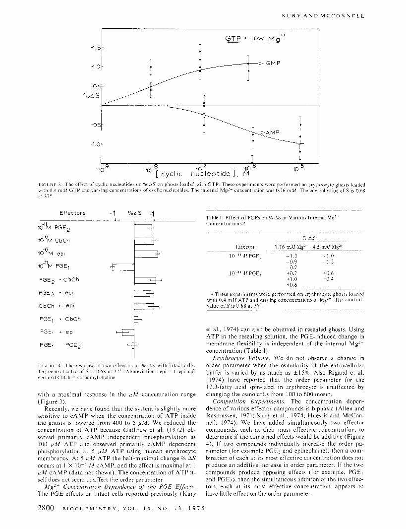
I I(;( KL: 3: The effect of cyclic nucleotides on "0 1.S on ghosts loaded w i t h GTP. These experiments were performed on erqthrocqte ghost5 loaded w i t h 0.4 m M GTP and varqing concentrations of c!clic nucleotides. The internal Mg" concentration U B E 0.76 mM. The control value o f .T is O.hX at 310.
Effectors -1 " / . A s +I
16% PGE2
10% CbCh
I O - ~ M e p i
10-"M PGEl
PGE2 + C b C h
PGE2 + epi
D C b C h + ePl
PGEl + CbCh 3. -l PGEl + e p t
PGEl PGE2
t I( ;( K I 1: The response of ~ U O effector\ on 90 1.S \ \ i t h intact cell\. The control value of S is 0.6X at 37'. Abbrebiations: epi = I -epineph- rine and CbCh = cdrbarnql choline.
with a maximal response in the p M concentration range (Figure 3 ) .
Recently, we have found that the system is slightly more sensitive to c A M P when the concentration of A T P inside the ghosts is lowered from 400 to 5 p M . We reduced the concentration of A T P because Guthrow et al. ( 1 972) ob- served primarily C A M P independent phosphorylation at 100 &I A T P and observed primarily c A M P dependent phosphorylation at 5 p M A T P using human erythrocyte membranes. At 5 p M A T P the half-maximal change o/o A S occurs a t I X IO-' M CAMP, and the effect is maximal a t 1 p M c A M P (data not shown). The concentration of A T P it- self does not seem to affect the order parameter.
Mg'+ Concentration Dependence of the PGE EHects. The PGE effects on intact cells reported previously (Kury
Table I . Effect of PGEs on c/c A S at Various Internal M g Z t Concentrations.0
Effector 0.76 m11 hlgZt 4.5 mU hlgZt
10 - - I 1 .If PGE, --1.3 ~ 1.0 -0.9 - 1 .z -0.7
10-".1IPGE2 +0.7 +0.6 +1.0 +0.4 +0.6
a These e ~ p e r i m e n t s were performed on erythrocyte ghosts loaded \vith 0.4 mZf ATP and varying concentrations of 51gz+. The control value of S is 0 .68 a t 37".
_ _ _ . ~ ~ ~ ~ _ ~ . ~... .. ...
et al.. 1974) can also be observed in resealed ghosts. Using A T P in the resealing solution, the PGE-induced change i n membrane flexibility is independent of the internal Mg'+ concentration (Table I ) .
Erj.throc.j'te Volume. We do not observe a change in order parameter when the osmolarity of the extracellular buffer is varied by as much as f15%. Also Rigand et al. (1974) have reported that the order parameter for the 12.3-fatty acid spin-label in erythrocyte is unaffected by changing the osmolarity from IO0 to 600 mosm.
Competition Experiments. The concentration depen- dence of various effector compounds is biphasic (Allen and Rasmussen, 197 1: Kury et al.. 1974; Huestis and McCon- nell, 1974). W e have added simultaneously two effector compounds, each a t their most effective concentration. to determine if the combined effects would be additive (Figure 4). If two compounds individually increase the order pa- rameter (for example PGE, and epinephrine), then a com- bination of each at its most effective concentration does not produce an additive increase in order parameter. I f the two compounds produce opposing effects (for example, PGEl and PGE,). then the simultaneous addition of the two effec- tors. each at its most effective concentration. appears to have little effect on the order parameter.
2800 B I O C H E M I S T R Y , V O L . . 1 4 , N O . 1 3 , 1 9 7 5

E R Y T H R O C Y T E M E M B R A N E F L E X I B I L I T Y
m
C-GMP 0 C-AMP
c -GMP+ .ImM S I T S c - A M P + JmM SITS +2 0-
*1 5-
+ I 0-
+ 5 -
1
% 0 s 0
[cyclic nucleotide 1) FIGURE 5: The effect of cyclic nucleotides on 96 A S in intact erythrocytes. The control value of S is 0.68 at 37'
Cyclic Nucleotides Added Intra- and Extracellularly to Erythrocytes in the Absence or Presence of an Inhibitor of the Anion Channel. W e added cyclic nucleotides extracellu- larly to intact erythrocytes and observed reproducible in- creases in order parameter (Figure 5). There is a greater re- sponse a t p M cyclic nucleotide concentrations than a t m M concentrations. W e find that it is possible to block this ap- parent entry of extracellular cyclic nucleotides by pretreat- ing the cells with an inhibitor of the erythrocyte anion chan- nel (Figure 5). The inhibitor is 4-acetamido-4'-isothiocya- natostilbene-2,2'-disulfonic acid (SITS) (Cabantchik and
I so,- SITS
Rothstein, 1972, 1974). T o ensure that a cyclic nucleotide- induced change in resonance spectra can still be observed when the anion channel is blocked, we loaded ghosts with a cyclic nucleotide, resealed, and then added or omitted SITS. W e found that the changes in spin-label resonance spectra were not altered by the presence of S I T S as long as the cyclic nucleotides were preloaded into the resealed ghosts.
Discussion At an average concentration of a single prostaglandin
molecule per intact erythrocyte, we have observed changes in the paramagnetic resonance spectrum of a membrane bound spin-labeled fatty acid (Kury et al., 1974). The fatty acid spin-labels bound to the membranes are present in rapid reversible equilibrium with labels present in solution. Labels are probably distributed more or less uniformly throughout the membrane; they are certainly not present in concentrated patches, otherwise they would show exchange broadened resonance spectra. W e shall assume that the res- onance spectra reflect the bulk flexibility of the membrane phospholipid fatty acid chains. We have, however, not ruled out the possibility that the change in the resonance spectra may arise from a change in the surface charge, pH, or ionic composition in the immediate region of the membrane sur-
face. It is likely that one or several prostaglandin effector molecules regulate the membrane flexibility of an entire erythrocyte. Thus, there must be an efficient amplification mechanism. It is likely that this mechanism involves c A M P and/or cGMP.
Although a n erythrocyte guanyl cyclase has not yet been reported (Goldberg et al., 1973), there is increasing evi- dence that there is an adenyl cyclase in the mature human erythrocyte (Kaiser et al., 1974; Rubin and Rosen, 1973) although it has not yet been demonstrated that it is hor- mone sensitive. W e observe changes in the spin-label spec- trum when erythrocyte ghosts are loaded with c A M P or c G M P (Figures 1 and 2). The c A M P induced change de- pends critically on the magnesium ion concentration inside the ghost. The dependence on Mg2+ concentration could be due to a Mg*+-sensitive protein kinase associated with the erythrocyte membrane which is activated by c A M P (Gu- throw et al., 1972). This protein kinase is known to phos- phorylate an erythrocyte protein called spectrin (Guthrow et al., 1972). We suggest that the changes in the paramag- netic resonance spectra arise from changes in the state of phosphorylation of membrane-associated proteins such as spectrin. There are a t least two types of enzymes that can regulate the degree of phosphorylation of proteins. They are the ATP-requiring, Mg2+-dependent protein kinases that phosphorylate protein substrates and the protein phospha- tases that hydrolyze phosphoproteins (Taborsky, 1974). In avian erythrocytes the P-adrenergic agonist, I-isoproterenol, stimulates incorporation of 32P into a protein that is analo- gous to spectrin in human erythrocytes (Rudolph and Greengard, 1974). Extracellular c A M P (1 m M ) mimics this effect. Huestis and McConnell (1974) find that the 8- adrenergic agent, L-epinephrine, increases spin-label order parameters in human erythrocytes.
From the data of Guthrow et al. (1972) we calculate that under their experimental conditions (5 p M ATP) only 2.5% of the spectrin is phosphorylated in the absence of c A M P and 3.6% phosphorylated in the presence of 1 p M CAMP. At 5 p M A T P we observe c A M P dependent changes in lipid order parameters. At higher concentrations of A T P (66- 100 p M ) an appreciable fraction of the spectrin molecules are phosphorylated (Guthrow et al., 1972; Rubin and Rosen, 1973), but the fraction of c A M P stimulation of 32P incorporation into protein decreases a t higher A T P concen-
B I O C H E M I S T R Y , V O L . 1 4 , N O . 1 3 , 1 9 7 5 2801

K U R Y A N D M C C O h N E L L
trations (Guthrow et al., 1973). The physiological concen- tration of A T P is approximately 0.7-1 .O m M (Bishop and Surgenor, 1964) so presumably in vivo most spectrin mole- cules are phosphorylated, and only a small percentage of those result from specific c A M P stimulation of phosphoryl- ation. Fairbanks et al. (1971) have estimated from sodium dodecyl sulfate gels that there are approximately 3.4 x 10' molecules of spectrin/cell. Kant and Steck (1973) estimate from binding studies that there are 6 X 10' internal binding sites for c A M P per cell (see, however, Swillens et al., 1974). These c A M P molecules are thought to bind to the regulato- ry subunit of the protein kinase. Using these data we con- clude that there are approximately 50 spectrin molecules/ kinase regulatory subunit.
There are two lines of evidence that these changes in lipid order parameter are directly related to the phosphorylation of proteins. One is that we observe approximately the same concentration of c A M P that half-maximally increases the order parameter as Guthrow et al. (1972) observes for half- maximal stimulation of incorporation of phosphate into spectrin. The other is that the erythrocyte protein kinase (Guthrow et al. (1972) is sensitive to the same concentra- tion of ME2+ as the CAMP-induced change in order param- eter (Figure 2). The change in the paramagnetic resonance spectrum induced in ghosts by c G M P is not sensitive either to the internal Mg2+ concentration or to whether A T P or G T P is used in the resealing solution (Figures 2 and 3). c G M P may regulate the activity of a protein phosphatase. The fact that prostaglandin-induced changes in erythrocyte ghost lipid fluidity are not sensitive to intracellular Mg'+ concentration (Table I ) indicates that PGE, and P G E l may alter the guanyl cyclase activity rather than the adenyl cy- clase activity.
Ryan and Hendrick (1974) and Guthrow et al. (1972) have reported a biphasic response for c A M P stimulation of "P-incorporation into membrane proteins. The biphasic character of the phosphorylation might be due to dual ef- fects of the two cyclic nucleotides, depending on their con- centrations and on the intracellular ionic constituents.
Although it is commonly thought that cyclic nucleotides cannot enter intact cells, there are various systems reported in the literature where effects are observed upon addition of extracellular cyclic nucleotides (Rudolph and Greengard, 1974: Ryan and Hendrick, 1974; Ford and Omachi, 1972; Siggins et al., 197 1 ; Bloom et al., 1975). We have presented evidence that p M concentrations of cyclic nucleotides readi- ly enter the intact erythrocyte (Figure 5) a t 37'. Our data are consistent with the possibility that cyclic nucleotides enter the cell via the anion channel since blockage of the channel prevents the response.
There are a number of protein-lipid interactions that can affect lipid fluidity. For example, Sefton and Gaffney ( 1 974) have shown that the order parameter of fatty acid as well as phospholipid spin-labels bound to the membrane of Sindbis virus is decreased by 9% when the membrane pro- teins are removed. Also the insertion of intrinsic proteins into membranes (e.g., rhodopsin) increases the order pa- rameters of phospholipid spin-labels (Hong and Hubbell, 1972). Thus one can imagine a variety of relatively simple, direct mechanisms whereby a change i n the biochemical state of a membrane protein affects the label order parame- ter. For example, the degree of phosphorylation of spectrin could affect specific as well as nonspecific binding to the cy- toplasmic surface of the erythrocyte. From the experiments of Sefton and Gaffney (1 974), yielding a 9% reduction in S
on removing membrane proteins, we can readily see that even a relatively small change in the degree of spectrin binding (--10-20%) could produce a 1-2% change in S. AI- though this is only an order-of-magnitude argument, i t does show clearly that a mere change in the degree of binding, or strength of binding, of spectrin to the cytoplasmic surface might well produce the observed changes in lipid flexibility. A second quite general and quite plausible mechanism is that the phosphorylation of cytoplasmic proteins results in a "muscle-like" change in the state of these proteins that al- ters the lateral compression or extention of the membrane lipids. Both effects may be simultaneously operative, so it may be difficult to design experiments that distinguish them.
Acknowledgments The prostaglandins were a gift of Dr. Margaret Merritt
of the Upjohn Company. The 10,3-fatty acid spin-label was a gift of Dr. Betty Jean Gaffney; we are particularly indebt- ed to Dr. Gaffney for pointing out the possibility that fatty acid labels may exhibit spectral changes due to membrane surface charges. We thank Professor H. Rasmussen for suggesting the experiment of measuring % A S on changing cell volume. W e also thank the Hematology Department a t Stanford University Medical School for supplying us with the human erythrocytes.
References Allen, J. E., and Rasmussen, H . (1971),Science 174, 512. Allen, J . E., and Valeri, C . R. (1974), Arch. In?. Med. 133,
86. Bieri, V . G., Wallach, D. F. H. , and Liu, P. S. (1 974). Pror.
niat l . Acad. Sci. U.S.A. 71, 4797. Bishop. C. , and Surgenor, D. M. (1964), The Red Blood
Cell, New York, N.Y., Academic Press, p 54. Bloom, F. E., Siggins, G. R. , Hoffer, B. J. , Segal, M., and
Oliver, A. P. (1975), Adv. Cyclic Nucleotide Res. ( in press).
Bodemann, H. , and Passow, H . (1972), J . Membr. B i d . 8 , 1 .
Cabantchik, Z . I . , and Rothstein, A. (1972), J . Membr. B i d . I O , 31 1 .
Cabantchik, Z. I. , and Rothstein, A. (1974), J . Membr. Biol. 15, 207.
Fairbanks, G., Steck, T. H., and Wallach, D. F. H . (1971). Biochemistry IO. 2606.
Ford, D. L., and Omachi, A. (1972), Biochim. Biophys. Acta 279, 587.
Goldberg, N. D., O'Dea, R. F., and Haddor, M. K. ( l973) , Adv. Cyclic Nucleotide Res. 3, 155.
Guthrow, C . E., Allen, J . E., and Rasmussen, H . (1972). J . B i d . Chem. 247, 8145.
Hong, K.. and Hubbell, W . 1. (1972), Proc. Natl. Acad. Sci. U.S.A. 69, 2617.
Hubbell, W. L., and McConnell, H. M. (1971), J . A m . Chem. Soc. 93, 314.
Huestis, W . H. , and McConnell, H. M . (1974). Biochem. Biophys. Res. Commun. 57, 726.
Humphries, G. K., and McConnell, H. M. (1974), Proc. .'Vat/. Acad. Sci. C.S .A . 71, 1691.
Kaiser, G. Quiring, K., Gauger, D., Palm, D., Becker, H . , and Schoepp, W. ( 1 974), Blur 29. 1 15.
Kant, J. A,, and Steck, T. L. (1973), Biochem. Biophj*s. Res. Commun. 54, 116.
Kury. P. G.. Ramwell, P. W., and McConnell, H . M .
2802 B I O C H E M I S T R Y , V O L 1 4 , h O 1 3 , 1 9 7 5

M E M B R A N E P H O S P H O L I P I D M E T A B O L I S M
( 1 974) , Biochem. Biophys. Res. Commun. 56, 178.
N.Y. Acad. Sci. 195, 207.
249, 5684.
cleotide Res. 4, 8 l . McConnell, H. M., and McFarland, B. G . (1972) , Ann.
Meyers, M . B., and Swislocki, N. I. (1974) , Arch Biochem.
Rigand, J. L., Gary-Bobo, C. M., and Taupin, C. (1974) ,
Rubin, C. S., and Rosen, 0. M . (1973) , Biochem. Biophys.
Rudolph, S. A., and Greengard, P. (1974) , J . Biol. Chem.
Ryan, W. L., and Hendrick, M. L. ( l 9 7 4 ) , Adv. Cyclic N u -
Sefton, B. M., and Gaffney, B. J. (1974) , J . Mol. Biol. 90,
Siggins, G . R., Oliver, A. P., Hoffer, B. J., and Bloom, F. E.
Swillens, S., Van Canter, E., and Dumont, J. E. ( 1 974) ,
Taborsky, G. (1974) , Adv. Protein Chem. 28, 1 .
Biophys. 164, 544. 343.
Biochim. Biophys. Acta 373, 21 1 .
Res. Commun. 50, 42 1 .
(1971),Science 171, 192.
Biochim. Biophys. Acta 364, 250.
Aldosterone-Induced Membrane Phospholipid Fatty Acid Metabolism in the Toad Urinary Bladder?
David B. P. Goodman,* Mitzi Wong, and Howard Rasmussen
ABSTRACT: Aldosterone action in the isolated toad urinary bladder has been studied by incubation of the tissue with several specifically labeled lipogenic precursors. Within 30 min after hormone addition phospholipid synthesis is stimu- lated; the metabolism of oleic acid is particularly enhanced. Additionally, during this time interval a phospholipid dea- cylation-reacylation cycle is stimulated by aldosterone.
T h e addition of aldosterone to the isolated toad urinary bladder stimulates transcellular sodium transport (Sharp and Leaf, 1966; Edelman and Fimognari, 1968). Aldoste- rone treatment alters a t least three other membrane-related processes. It increases ( 1 ) the permeability of the tissue to water and low-molecular solutes in the presence of antidi- uretic hormone (Goodman et al., 1969; Handler et al., 1969); ( 2 ) the sensitivity of sodium transport to inhibition by the cardiac glycoside, ouabain, a known inhibitor of Na+- K+-activated ATPases (Goodman et al., 1969); and ( 3 ) the sensitivity of sodium transport to inhibition by increased partial pressures of oxygen (Allen et al., 1973). Because an alteration in membrane lipid composition could account for these diverse hormone-induced changes in tissue function, we initiated an investigation of the effects of aldosterone on membrane lipid metabolism. In our earlier studies we found that aldosterone treatment increased both lipid synthesis and the turnover of membrane phospholipid fatty acids within 30 min (Goodman et al., 1971). Additionally, pre- treatment of the tissue with phospholipase A resulted in a significant reduction in the latent period between hormone addition and the inception of the physiological response, an increase in transcellular sodium transport. Aldosterone also induced an increase in the weight percentage of phospholip- id long-chain polyunsaturated fatty acids. These studies thus provided the first biochemical evidence that a modifi-
From the Department of Pediatrics and Biochemistry, University of Pennsylvania School of Medicine, Philadelphia, Pennsylvania 19174. Received November 25, 1974. Supported by U S . Public Health Service Grants AM 09650 and CA 14345 and Office of Naval Re- search Contract NR 202-005.
After 4 hr aldosterone increases the oxidation of all fatty acids utilized, but enhances, specifically, the elongation and desaturation of oleic acid, as well as the recycling of [I4C]a- cetyl-CoA derived from [ l-’4C]oleic acid into membrane phospholipid fatty acid. These data provide further evidence for a rapid and specific action of aldosterone on toad blad- der membrane phospholipid fatty acid metabolism.
cation of lipid metabolism might be a significant effect of the action of aldosterone. The purpose of the present inves- tigation was to characterize more fully and specifically the effects of aldosterone on lipid metabolism in the toad uri- nary bladder.
Utilizing specifically labeled [I4C]- and [3H]fatty acids as precursors and analyzing tissue phospholipid fatty acids by radio-gas chromatography, we have shown that aldoste- rone stimulates specifically the elongation and desaturation of oleic acid (18: lw9) . By prelabeling tissue lipids with [ 14C]acetate, we have also demonstrated that aldosterone stimulates the release of I4C from phospholipid into [ I4C]fatty acid.
Materials and Methods Experimental Animals and Preincubation. Urinary blad-
ders were removed from female toads (Bufo marinus, Na- tional Reagents Co., Bridgeport, Conn.) treated as pre- viously described (Goodman et al., 1971). Tissue was rou- tinely preincubated overnight a t room temperature in sub- strate-free aerated Ling-Ringer phosphate buffer (Ling, 1962), p H 7.4, containing 50 mg/l. of both penicillin G and streptomycin sulfate. In experiments where tissue lipids were prelabeled, 1 m M sodium acetate, containing 1 .O kCi/ ml of sodium [1,2-I4C]acetate, was included in the over- night incubation solution. The following morning experi- ments were performed.
Tissue Incubations; [I4C] Lipid Analysis. Individual matched hemibladders from six toads were incubated for 1 hr in Ling-Ringer phosphate (pH 7.4) containing 4 m M glucose. The tissue was then transferred into a fresh Ling- Ringer glucose buffer containing either 0.1 m M sodium ac-
B I O C H E M I S T R Y , V O L . 1 4 , N O . 1 3 , 1 9 7 5 2803