regulation of junction protein · and expression (ben-ze’ev. 1984, 1986, 1991; ungar et al.,...
TRANSCRIPT
REGULATION OF ADHERENS JUNCTION PROTEIN LEVELS: ITS ROLE IN CELL MOTILITY AND
TUMORIGEN IClTY
Avri Ben-Ze’ev, Jose Luis Rodriguez Fernandez,
Ursula Gluck, Daniela Salomon, Margot Zoller, and
Benjamin Geiger
I. INTRODUCTION . . . . . . . . . . . . . . . . . . . . . . . . . . . . . . . . 144 11. GROWTHSTIMULATIONANDAJPROTEINEXPRESSION . . . . . . . 145
OVEREXPRESSION AND ANTISENSE TRANSFECTION OF VINCULIN AFFECT CELL MOTILITY . . . . . . . . . . . . . . . . . . . . 148 SUPPRESSION OF TUMORIGENICITY IN CELLS TRANSFECTED WITH VINCULIN cDNA . . . . . . . . . . . . . . . . . . 152 SUPPRESSION OF TUMORIGENICITY IN MALIGNANT CELLS
111.
IV.
V. TRANSFECTED WITH ALPHA-ACTININ cDNA . . . . . . . . . . . . . . 157
Advances in Molecular and Cell Biology Volume 12, pages 143-163. Copyright 0 1995 by JAI Press Inc. All rights of reproduction in any form reserved. ISBN: 1-55938-845-5
143
144 AVRl BEN-ZE’EV ET AL.
VI. CONCLUSIONS AND PERSPECTIVES . . . . . . . . . . . . . . . . . . . . 159 ACKNOWLEDGMENTS . . . . . . . . . . . . . . . . . . . . . . . . . . . . 160
1. INTRODUCTION
Cell-cell and cell-extracellular matrix (ECM) contacts are central determinants of cell and tissue morphology (Hay, 1981; Takeichi, 1991). These interactions of the cell with its environment are mediated by numerous transmembrane receptors of the integrin and cadherin families of receptors (Takeichi, 199 1; Hynes, 1992). In the cytoplasmic domain, many of these adhesive interactions are linked to the microfilament system by plaque proteins which are especially prominent in adher- ens type junctions (AJ) (Burridge et al., 1988; Geiger and Ginsberg, 1991; Tsukita et al., 1992), thus forming a structural link between the extracellular and the intracellular domain of the plasma membrane. Earlier observations have demon- strated effects on gene expression, cell growth, motility, and differentiation in response to altered cell-cell and cell-ECM interactions (Ben-Ze’ev, 1991; 1992). In the last decade, the number of components that have been shown to localize in AJ has steadily increased (Burridge et al., 1988; Geiger and Ginsberg, 1991; Tsukita et al., 1992; Sadler et al., 1992; Ben-Ze’ev, 1992). In addition to proteins which are involved in establishing the structural and thus more mechanical interaction be- tween the outside and inside of the cell, AJ contain a considerable number of regulatory molecules including tyrosine kinases, protein kinase C, and various oncogene products and proteases, which reside in the cytoplasmic side of the plaque area(Burridgeet a]., 1988; GeigerandGinsberg, 1991;LunaandHitt, 1992).These findings suggested that the junctional areas of cell-cell and cell-ECM interaction are directly involved in signal transduction and thereby elicit long range effects on cells.
Tyrosine phosphorylation of AJ proteins has been proposed to be a major mechanism in this signal transduction, based’on studies showing abundant tyrosine phosphorylation-dephosphorylation activity in AJ of both normal and transformed cells (Rohrschneider, 1980; Comoglio et al., 1984; Maher et al., 1985; Volberg et al., 1992). The recently characterized adhesion plaque tyrosine kinase pl 25FAK (Schalleret al., 1992) has raised wide interest since this protein is rapidly phospho- rylated on tyrosine by both cytokine stimulation and by adhesion. It proceeds through activation of integrins in fibroblasts adhering to the ECM (Guan et al., 1991; Bumdge et al., 1992) and in platelet activation by soluble factors (Lipfert et al., 1992), but it.is also apparent in the transformation of fibroblasts by RSV (Guan and Shalloway, 1992), and after stimulation of neuropeptide receptors by various soluble components (Zachary and Rozengurt, 1992). This tyrosine phosphoryla- tion, in turn, enhances the kinase activity of pl 25FAK. These studies therefore suggest that signal transduction by cell-ECM interaction and by different cytokines may converge at an early stage, in adhesion plaques. They also imply that there is
Adherens Junction Protein Regulation: Role in Motilityand Malignancy 145
a common mechanism for the effects on cell shape, motility, growth and differen- tiation resulting from either cell adhesion and/or soluble factor action on cells.
The assembly of AJ apparently proceeds through an initial binding of the transmembrane contact receptor to its extracellular ligand (an ECM protein se- quence, or a homologous cell adhesion (CAM) receptor (Geiger et al., 1990, 1992a)). However, recent studies have clearly demonstrated that following this initial step, the recruitment and binding of the plaque and cytoskeletal components to the clustered receptor constitutes an important maturation step in the formation of stable AJ (La Flamme et al., 1992; Geiger et al., 1992a,b). The binding of cytoskeletal components to the transmembrane contact receptor may be required for the long range effects on cells (Geiger et al., 1992a). This maturation andor stabilization step in AJ formation can be controlled both by post-transcriptional mechanisms (i.e., phosphorylation-dephosphorylation), or by regulating the amount of a particular junctional protein.
In this essay we summarize studies demonstrating AJ modulation through changes in the level of expression of junctional components, and show that this modulation has a dramatic effect not only on cell structure, but also on cell motility and the tumorigenic ability of cells.
I I . GROWTH STIMULATION AND AJ PROTEIN EXPRESSION
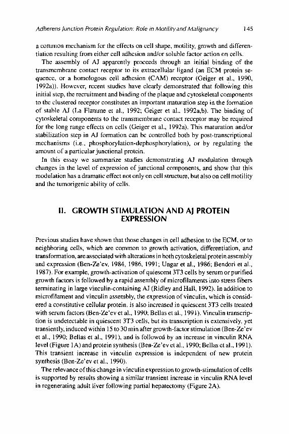
Previous studies have shown that those changes in cell adhesion to the ECM, or to neighboring cells, which are common to growth activation, differentiation, and transformation, are associated with alterations in both cytoskeletal protein assembly and expression (Ben-Ze’ev. 1984, 1986, 1991; Ungar et al., 1986; Bendori et al., 1987). For example, growth-activation of quiescent 3T3 cells by serum or purified growth factors is followed by a rapid assembly of microfilaments into stress fibers terminating in large vinculin-containing AJ (Ridley and Hall, 1992). In addition to microfilament and vinculin assembly, the expression of vinculin, which is consid- ered a constitutive cellular protein, is also increased in quiescent 3T3 cells treated with serum factors (Ben-Ze’ev et al., 1990 Bellas et a]., 1991). Vinculin transcrip- tion is undetectable in quiescent 3T3 cells, but its transcription is extensively, yet transiently, induced within 15 to 30 min after growth-factor stimulation (Ben-Ze’ev et al., 1990; Bellas et a]., 1991), and is followed by an increase in vinculin RNA level (Figure 1A)andprotein synthesis (Ben-Ze’evet al., 1990; Bellas et al., 1991). This transient increase in vinculin expression is independent of new protein synthesis (Ben-Ze’ev et al., 1990).
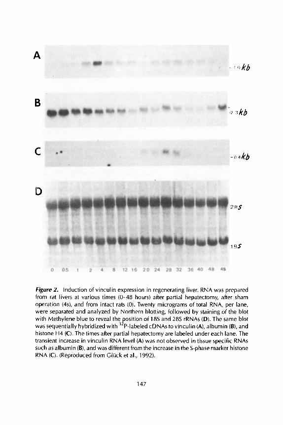
The relevance of this change in vinculin expression to growth-stimulation of cells is supported by results showing a similar transient increase in vinculin RNA level in regenerating adult liver following partial hepatectomy (Figure 2A).
146 AVRl BEN-ZE’EV ET AL.
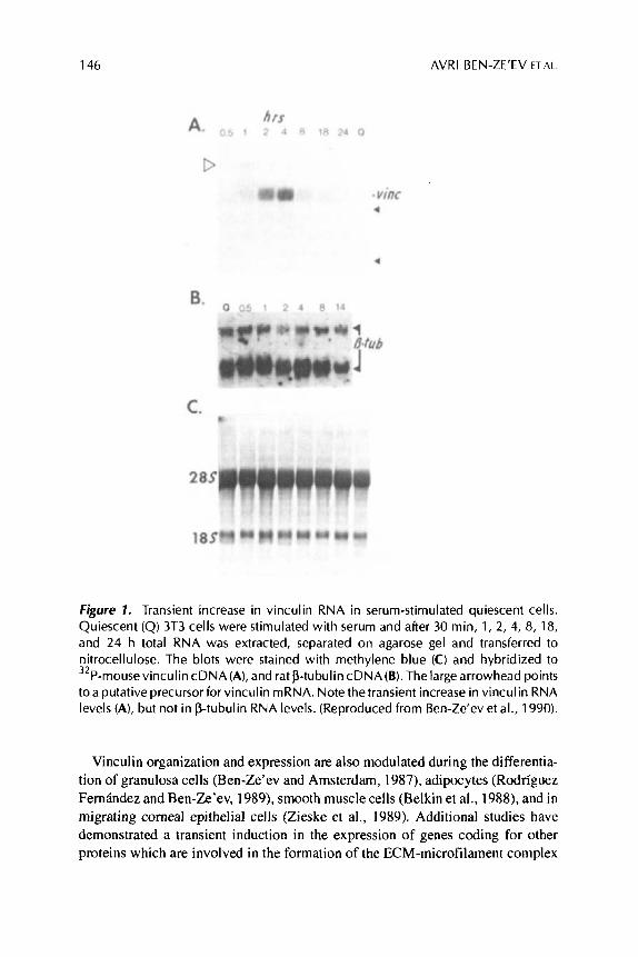
Figure 1. Transient increase in vinculin RNA in serum-stimulated quiescent cells. Quiescent (Q) 3T3 cells were stimulated with serum and after 30 min, 1, 2, 4, 8, 18, and 24 h total RNA was extracted, separated on agarose gel and transferred to nitrocellulose. The blots were stained with methylene blue (C) and hybridized to 32P-mousevinculincDNA(A), and rat P-tubulin cDNA(B).The largearrowhead points to a putative precursor for vinculin mRNA. Note the transient increase in vinculin RNA levels (A), but not in P-tubulin RNA levels. (Reproduced from Ben-Ze‘ev et al., 1990).
Vinculin organization and expression are also modulated during the differentia- tion of granulosa cells (Ben-Ze’ev and Amsterdam, 1987), adipocytes (Rodriguez Fernhdez and Ben-Ze’ev, 1989), smooth muscle cells (Belkin et al., 1988), and in migrating corneal epithelial cells (Zieske et a]., 1989). Additional studies have demonstrated a transient induction in the expression of genes coding for other proteins which are involved in the formation of the ECM-microfilament complex
A
B
C
Figure 2. Induction of vinculin expression in regenerating liver. RNA was prepared from rat livers at various times (0-48 hours) after partial hepatectomy, after sham operation (4s), and from intact rats (0). Twenty micrograms of total RNA, per lane, were separated and analyzed by Northern blotting, followed by staining of the blot with Methylene blue to reveal the osition of 18s and 28s rRNAs (D). The same blot was sequentially hybridized with P-labeled cDNAs to vinculin (A), albumin (B), and histone H4 (C). The times after partial hepatectomy are labeled under each lane. The transient increase in vinculin RNA level (A) was not observed in tissue specific RNAs such as albumin (B), and was different from the increase in the S-phase marker histone RNA (0. (Reproduced from Cluck et al., 1992).
P
147
148 AVRl BEN-ZE’EV ET AL.
i n both growth-factor stimulated fibroblasts (Rysek et al., 1989; Bellas et al., 199 I ; Cluck et al., 1992), in growth-stimulated hepatocytes in culture (Ben-Ze’ev et al., 1988; Mooney et al., 1992), and in regenerating liver (Gliick et al., 1992). These include ECM components such as fibronectin, the transmembrane receptor PI-integrin, and components of adhesion plaques and actin-binding proteins such as a-actinin, talin, and tropomyosin. The correlation between the regulation of AJ protein expression, i n both cultured cells and in vivo, suggests that this modulation of cytoskeletal gene expression may have a physiologically relevant function.
111. OVEREXPRESSION AND ANTISENSE TRANSFECTION OF VlNCULlN AFFECT CELL MOTILITY
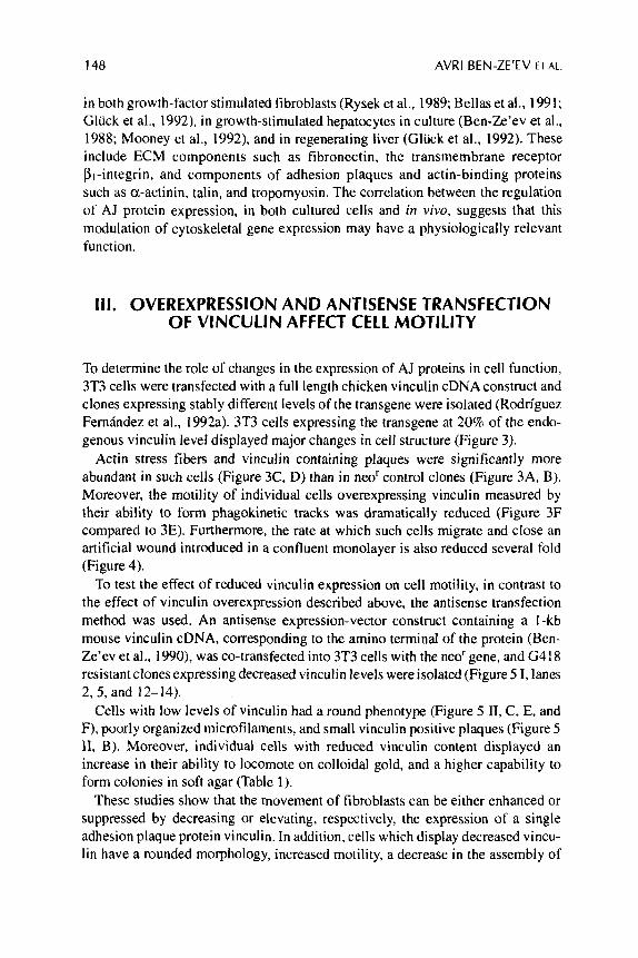
To determine the role of changes in the expression of AJ proteins in cell function, 3T3 cells were transfected with a full length chicken vinculin cDNA construct and clones expressing stably different levels of the transgene were isolated (Rodriguez Fernindez et al., 1992a). 3T3 cells expressing the transgene at 20% of the endo- genous vinculin level displayed major changes in cell structure (Figure 3).
Actin stress fibers and vinculin containing plaques were significantly more abundant in such cells (Figure 3C, D) than in neOr control clones (Figure 3A, B). Moreover, the motility of individual cells overexpressing vinculin measured by their ability to form phagokinetic tracks was dramatically reduced (Figure 3F compared to 3E). Furthermore, the rate at which such cells migrate and close an artificial wound introduced in a confluent monolayer is also reduced several fold (Figure 4).
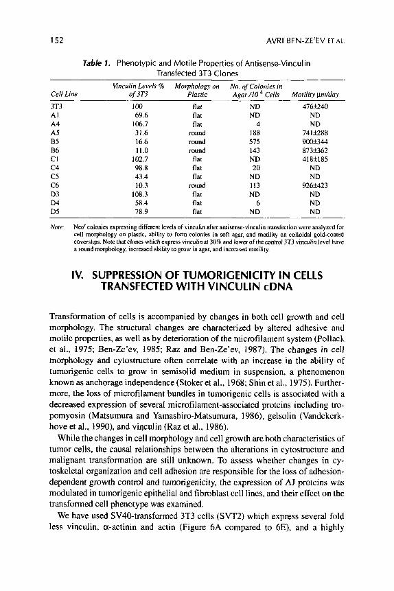
To test the effect of reduced vinculin expression on cell motility, in contrast to the effect of vinculin overexpression described above, the antisense transfection method was used. An antisense expression-vector construct containing a I -kb mouse vinculin cDNA, corresponding to the amino terminal of the protein (Ben- Ze’ev et a]., 1990), was co-transfected into 3T3 cells with the neo‘gene, and G418 resistant clones expressing decreased vinculin levels were isolated (Figure 5 I , lanes 2.5, and 12-14).
Cells with low levels of vinculin had a round phenotype (Figure 5 11, C, E, and F), poorly organized microfilaments, and small vinculin positive plaques (Figure 5 11, B). Moreover, individual cells with reduced vinculin content displayed an increase in their ability to locomote on colloidal gold, and a higher capability to form colonies in soft agar (Table 1).
These studies show that the movement of fibroblasts can be either enhanced or suppressed by decreasing or elevating, respectively, the expression of a single adhesion plaque protein vinculin. In addition, cells which display decreased vincu- lin have a rounded morphology, increased motility, a decrease in the assembly of
Adherens junction Protein Regulation: Role in Motility and Malignancy 149
Figure 3. Increased stress fiber and vinculin assembly and decreased motility in 3T3 cells overexpressing vinculin. Double fluorescent labeling for vinculin (A, C) and actin (B, D) and dark field visualization of phagokinetic tracks (E, F ) of 3T3 cells transfected with chicken vinculin (C, D, F), or with the neor selective marker (A, B, E). The level of chicken vinculin in the transfected cells is about 20% relative to the endogenous protein. The bar in D indicates 10 pm. (Reproduced from Geiger et al., 1992a).
actin filaments, and an increase in their ability togrow in suspension culture in agar. These properties are characteristic of transformed fibroblasts (Shin et al., 1975).
In other studies, another microfilament-associated protein gelsolin, which severs actin filaments, was overexpressed in 3T3 cells. This overexpression resulted in increased cell motility (Cunningham et al., 1991). In contrast, increasing the expression of actin binding protein (ABP-280) which enhances actin polymeriza- tion, was shown to decrease cell locomotion (Cunningham et al., 1992). These results are consistent with the view that modulations in the expression of individual proteins which are involved in the stabilization of microfilament-membrane com- plexes can regulate cell morphology and motility.
VlOl 3T3 N87
h) P J-
hoiirs
150
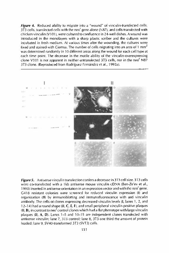
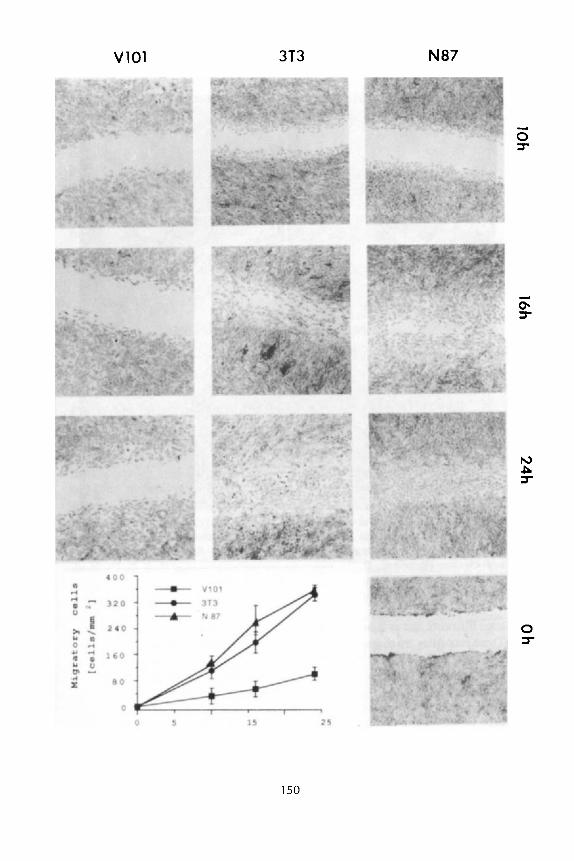
Figure 4. Reduced ability to migrate into a "wound" of vinculin-transfected cells. 3T3 cells, transfected cells with the neo' gene alone (N87), and cells transfected with chicken vinculin (V1 O l ) , werecultured to confluence in 24-well dishes. Awound was introduced in the monolayers with a sharp plastic scriber and the cultures were incubated in fresh medium. At various times after the wounding, the cultures were fixed and stained with Giemsa. The number of cells migrating into an area of 1 mm2 was determined randomly in 10 different areas along the wound for each cell type at each time point. The decrease in the motile ability of the vinculin-overexpressing clone V101 i s not apparent in neither untransfected 3T3 cells, nor in the neor N87 3T3 clone. (Reproduced from Rodriguez Fernandez et al., 1992a).
Figure5. Antisense vinculin transfection confers a decrease in 3T3 cell size. 3T3 cells were co-transfected with a 1 kb antisense mouse vinculin cDNA (Ben-Ze'ev et al., 1990) inserted in antisense orientation in an expression vector and with the neor gene. G418 resistant colonies were screened for reduced vinculin expression (I) and organization (11) by immunoblotting and immunofluorescence with anti vinculin antibody. The cells of clones expressing decreased vinculin levels ( I , lanes 1, 2, and 12-14) had a round shape (11, C, E, F), and small peripheral vinculin-positive plaques (11, B), in contrast to neo'control clones which had a flat phenotype with largevinculin plaques (11, A, D). Lanes 1-5 and 10-15 are independent clones transfected with antisense vinculin; lane 7, 3T3 control; lane 8, 3T3 one third the amount of protein loaded; lane 9, SV40-transformed 3T3 (SVT2) cells.
151
152 AVRl BEN-ZE’EV ET AL.
Table 7. Phenotypic and Motile Properties of Antisense-Vinculin Transfected 3T3 Clones
Wnculin Levels 9i Morphology on No. of Colonies iri Cell Line of 3T3 Plastic Agar/lO Cells Motility Hm/day
3T3 A1 A4 A5 B5 B6 CI c 4 c5 C6 D3 D4 D5
100 69.6
106.7 31.6 16.6 11.0
102.7 98.8 43.4 10.3
108.3 58.4 78.9
flat flat flat
round round round
flat flat flat
round flat flat flat
ND ND
4 188 575 143 ND 20
ND 113 ND
6 ND
476f240 ND ND
741k2.288 9 w 3 4 4 873t362 418f185
ND ND
926k423 ND ND ND
Nore: Neo‘ colonies expressing different levels of vinculin after antisense-vinculin transfection were analyzed for cell morphology on plastic, ability to form colonies in soft agar, and motility on colloidal gold-coated coverslips. Note that clones which express vinculin at 30% and lower of thecontrol 3T3 vinculin level have a round morphology, increased ability to grow in agar, and increased motility.
IV. SUPPRESSION OF TUMORlGENlClTY IN CELLS TRANSFECTED WITH VlNCULlN cDNA
Transformation of cells is accompanied by changes in both cell growth and cell morphology. The structural changes are characterized by altered adhesive and motile properties, as well as by deterioration of the microfilament system (Pollack et al., 1975; Ben-Ze’ev, 1985; Raz and Ben-Ze’ev, 1987). The changes in cell morphology and cytostructure often correlate with an increase in the ability of tumorigenic cells to grow in semisolid medium in suspension, a phenomenon known as anchorage independence (Stoker et al., 1968; Shin et al., 1975). Further- more, the loss of microfilament bundles in tumorigenic cells is associated with a decreased expression of several microfilament-associated proteins including tro- pomyosin (Matsumura and Yamashiro-Matsumura, 1986), gelsolin (Vandekerk- hove et al., 1990), and vinculin (Raz et al., 1986).
While the changes in cell morphology and cell growth are both characteristics of tumor cells, the causal relationships between the alterations in cytostructure and malignant transformation are still unknown. To assess whether changes in cy- toskeletal organization and cell adhesion are responsible for the loss of adhesion- dependent growth control and tumorigenicity, the expression of AJ proteins was modulated in tumorigenic epithelial and fibroblast cell lines, and their effect on the transformed cell phenotype was examined.
We have used SV40-transformed 3T3 cells (SVT2) which express several fold less vinculin, a-actinin and actin (Figure 6A compared to 6E), and a highly
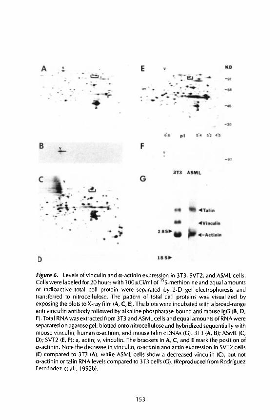
Figure 6. Levels of vinculin and a-actinin expression in 3T3, SVT2, and ASML cells. Cells were labeled for 20 hours with 100 pCi/ml of 35S-methionine and equal amounts of radioactive total cell protein were separated by 2-D gel electrophoresis and transferred to nitrocellulose. The pattern of total cell proteins was visualized by exposing the blots to X-ray film (A, C, E). The blots were incubated with a broad-range anti vinculin antibody followed by alkaline phosphatase-bound anti mouse IgG (B, D, F). Total RNAwas extracted from 3T3 and ASML cells and equal amounts of RNA were separated on agarose gel, blotted onto nitrocellulose and hybridized sequentially with mouse vinculin, human a-actinin, and mouse talin cDNAs (G) . 3T3 (A, B); ASML (C, D); SVT2 (E, F); a, actin; v, vinculin. The brackets in A, C, and E mark the position of a-actinin. Note the decrease in vinculin, a-actinin and actin expression in SVT2 cells (E) compared to 3T3 (A), while ASML cells show a decreased vinculin (C), but not a-actinin or talin RNA levels compared to 3T3 cells (GI. (Reproduced from Rodriguez Fernhndez et al., 1992b).
153
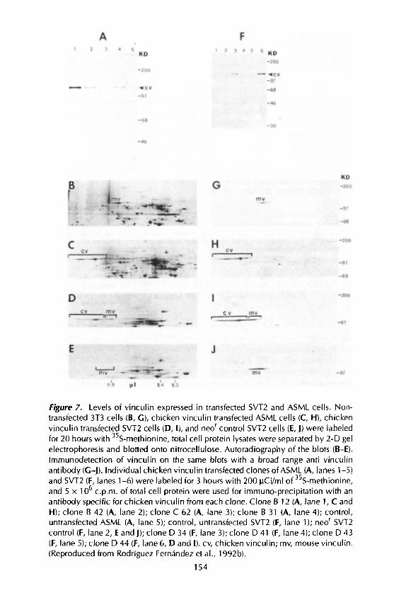
Figure 7. Levels of vinculin expressed in transfected SVTZ and ASML cells. Non- transfected 3T3 cells (B, G ) , chicken vinculin transfected ASML cells (C, H), chicken vinculin transfected SVT2 cells (D, I), and neor control SVT2 cells (E, J) were labeled for 20 hours with 35S-methionine, total cell protein lysates were separated by 2-D gel electrophoresis and blotted onto nitrocellulose. Autoradiography of the blots (B-El. lmrnunodetection of vinculin on the same blots with a broad range anti vinculin antibody (C-J). Individual chicken vinculin transfected clones of ASML (A, lanes 1-5) and SVT2 (F, lanes 1-6) were labeled for 3 hours with 200 pCi/ml of 35S-methionine, and 5 x 1 O6 c.p.m. of total cell protein were used for immuno-precipitation with an antibody specific for chicken vinculin from each clone. Clone B 12 (A, lane 1, C and H); clone B 42 (A, lane 2); clone C 62 (A, lane 3); clone B 31 (A, lane 4); control, untransfected ASML (A, lane 5); control, untransfected SVTZ (F, lane 1); neor SVTZ control (F, lane 2, E and J); clone D 34 (F, lane 3); clone D 41 (F, lane 4); clone D 43 (F, lane 5); clone D 44 (F, lane 6, D and I). cv, chicken vinculin; rnv, mouse vinculin. (Reproduced from Rodriguez Fernandez et al., 1992b).
154
Adherens junction Protein Regulation: Role in Motility and Malignancy 155
malignant ASML adenocarcinoma (Matzku et al., 1983) which expresses no detectable vinculin at either the RNA (Figure 6G) or protein level (Figure 6C and D).
An expression vector containing the full length chicken vinculin cDNA was introduced into these cells together with the neo' gene, and (3418 resistant colonies expressing different levels o f the transgene were isolated (Figure 7).
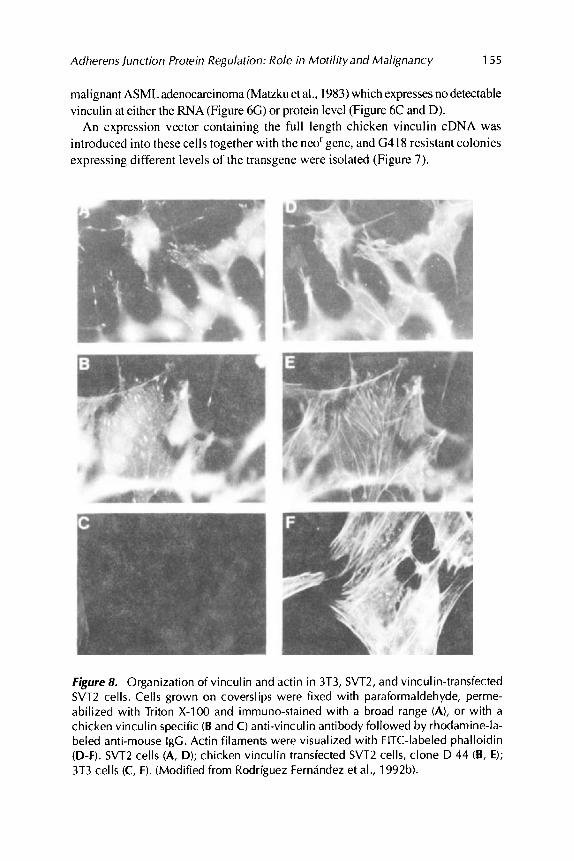
Figure 8. Organization of vinculin and actin in 3T3, SVT2, and vinculin-transfected SVT2 cells. Cells grown on coverslips were fixed with paraformaldehyde, perme- abilized with Triton X-100 and immuno-stained with a broad range (A), or with a chicken vinculin specific (B and C) anti-vinculin antibody followed by rhodamine-la- beled anti-mouse IgG. Actin filaments were visualized with FITC-labeled phalloidin (D-F). SVT2 cells (A, D); chicken vinculin transfected SVT2 cells, clone D 44 (B, E); 3T3 cells (C, F). (Modified from Rodriguez Fernandez et al., 1992b).
156 AVRl BEN-ZE'EV ET AL.
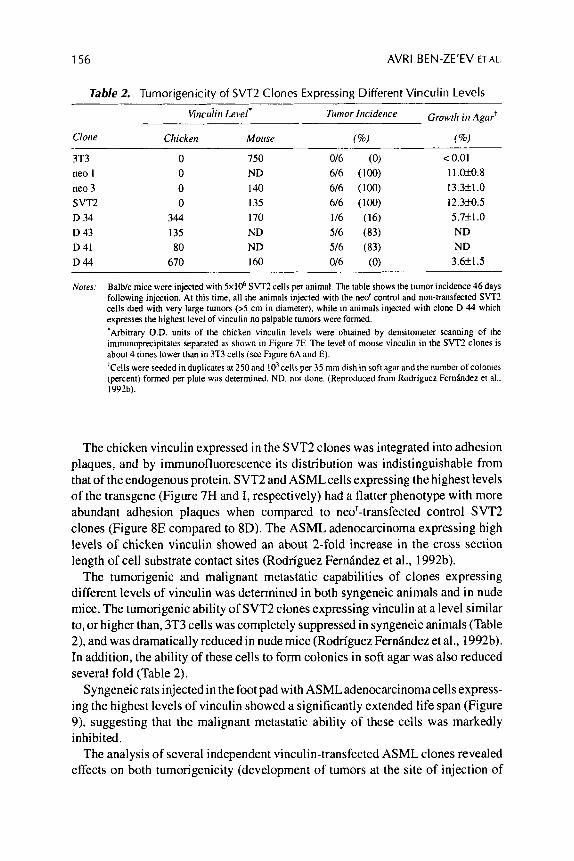
Table 2. Tumorinenicitv of SVT2 Clones Expressing Different Vinculin Levels
3T3 0 750 016 (0) < 0.01 neo 1 0 ND 6/6 (100) 11.039.8 neo 3 0 I40 6/6 (100) 13.3+1.0 SVT2 0 135 6/6 (100) 12.3fo.5 D 34 344 170 1/6 (16) 5.7f1.0 D 43 135 ND 5/6 (83) ND D 41 80 ND 5/6 (83) ND D44 670 160 0/6 (0) 3.6k1.5
Notes: Balb/c mice were injected with 5x10⁶ SVTZ cells per animal. The table shows the tumor incidence 46 days following injection. At this time. all the animals injected with the neo' control and non-transfected SVT2 cells died with very large tumors (>S cm in diameter), while i n animals injected with clone D 44 which expresses the highest level of vinculin no palpable tumors were formed. 'Arbitrary O.D. units of the chicken vinculin levels were obtained by deiisitomeler scanning of the immunoprecipitates separated as shown i n Figure 7F. The level of mouse vinculin in the SVTZ clones is about 4 times lower than in 3T3 cells (see Figure 6A and E). 'Crlls were seeded in duplicates at 250and 10' cells per 35 mm dish in soft agar and the number of colonies (percent) formed per plate was determined. ND. nor done. (Reproduced from Rodriguez Fernhdez et al.. 1992b).
The chicken vinculin expressed in the SVT2 clones was integrated into adhesion plaques, and by immunofluorescence its distribution was indistinguishable from that of the endogenous protein. SVT2 and ASMLcells expressing the highest levels of the transgene (Figure 7H and I, respectively) had a flatter phenotype with more abundant adhesion plaques when compared to nee'-transfected control SVT2 clones (Figure 8E compared to 8D). The ASML adenocarcinoma expressing high levels of chicken vinculin showed an about 2-fold increase in the cross section length of cell substrate contact sites (Rodriguez Fernindez et al., 1992b).
The tumorigenic and malignant metastatic capabilities of clones expressing different levels of vinculin was determined in both syngeneic animals and in nude mice. The tumorigenic ability of SVT2 clones expressing vinculin at a level similar to, or higher than, 3T3 cells was completely suppressed in syngeneic animals (Table 2), and was dramatically reduced in nude mice (Rodriguez Fernindez et al., 1992b). In addition, the ability of these cells to form colonies in soft agar was also reduced several fold (Table 2).
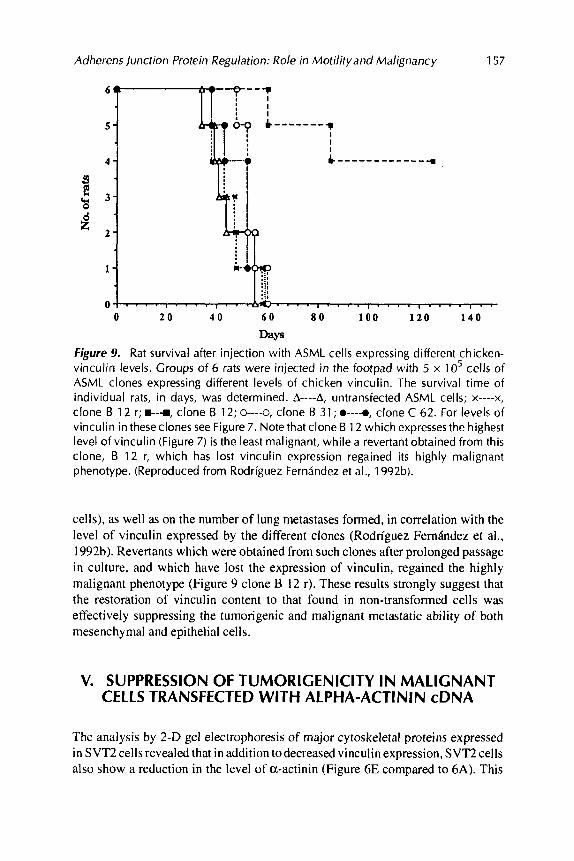
Syngeneic rats injected in the foot pad with ASMLadenocarcinomacells express- ing the highest levels of vinculin showed a significantly extended life span (Figure 9). suggesting that the malignant metastatic ability of these cells was markedly inhibited.
The analysis of several independent vinculin-transfected ASML clones revealed effects on both tumorigenicity (development of tumors at the site of injection of
Adherens junction Protein Regulation: Role in Motilityand Malignancy 157
3 E
0 2 0 4 0 6 0 8 0 1 0 0 1 2 0 1 4 0
Days Figure 9. Rat survival after injection with ASML cells expressing different chicken- vinculin levels. Groups of 6 rats were injected in the footpad with 5 x lo5 cells of ASML clones expressing different levels of chicken vinculin. The survival time of individual rats, in days, was determined. A----A, untransfected ASML cells; x----x, clone B 12 r; e-r, clone B 12; 0---a, clone B 31 ; 0----0, clone C 62. For levels of vinculin in theseclones see Figure 7. Note that clone B 1 2 which expresses the highest level of vinculin (Figure 7) is the least malignant, while a revertant obtained from this clone, B 12 r, which has lost vinculin expression regained its highly malignant phenotype. (Reproduced from Rodriguez Ferndndez et al., 1992b).
cells), as well as on the number of lung metastases formed, in correlation with the level of vinculin expressed by the different clones (Rodriguez Fernandez et a]., 1992b). Revertants which were obtained from such clones after prolonged passage in culture, and which have lost the expression of vinculin, regained the highly malignant phenotype (Figure 9 clone B 12 r). These results strongly suggest that the restoration of vinculin content to that found in non-transformed cells was effectively suppressing the tumorigenic and malignant metastatic ability of both mesenchymal and epithelial cells.
V. SUPPRESSION OF TUMORlGENlClTY IN MALIGNANT CELLS TRANSFECTED WITH ALPHA-ACTININ cDNA
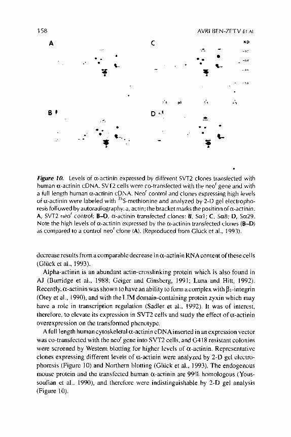
The analysis by 2-D gel electrophoresis of major cytoskeletal proteins expressed in SVT2 cells revealed that in addition to decreased vinculin expression, SVT2 cells also show a reduction in the level of a-actinin (Figure 6E compared to 6A). This
158 AVRl BEN-ZE'EV ET AL
KD
-I, . ,I - 0
*- * * -hR
-a , ,
. * f
- -To
Figure 10. Levels of a-actinin expressed by different SVT2 clones transfected with human a-actinin cDNA. SVT2 cells were co-transfected with the neo' gene and with a full length human a-actinin cDNA. Neor control and clones expressing high levels of a-actinin were labeled with 35S-methionine and analyzed by 2-D gel electropho- resis followed by autoradiography. a, actin; the bracket marks the position ofa-actinin. A, SVT2 neor control; B-D, a-actinin transfected clones: I?, Sal; C, Sa8; D, Sa29. Note the high levels of a-actinin expressed by the a-actinin transfected clones (B-D) as compared to a control neor clone (A). (Reproduced from Cluck et al., 1993).
decrease results from a comparable decrease in a-actinin RNAcontent of these cells (Gliick et al., 1993).
Alpha-actinin is an abundant actin-crosslinking protein which is also found in AJ (Burridge et al., 1988; Geiger and Ginsberg, 1991; Luna and Hitt, 1992). Recently, a-actinin was shown to have an ability to form acomplex with PI-integrin (Otey et al., 1990), and with the LIM domain-containing protein zyxin which may have a role in transcription regulation (Sadler et al., 1992). It was of interest, therefore, to elevate its expression in SVT2 cells and study the effect of a-actinin overexpression on the transformed phenotype.
A ful l length human cytoskeletal a-actinin cDNAinserted in an expression vector was co-transfected with the neo'gene into SVT2 cells, and (3418 resistant colonies were screened by Western blotting for higher levels of a-actinin. Representative clones expressing different levels of a-actinin were analyzed by 2-D gel electro- phoresis (Figure 10) and Northern blotting (Gliick et al., 1993). The endogenous mouse protein and the transfected human a-actinin are 99% homologous (Yous- soufian et al., 1990), and therefore were indistinguishable by 2-D gel analysis (Figure 10).
Adherens Junction Protein Regulation: Role in Motil i tyand Malignancy 159
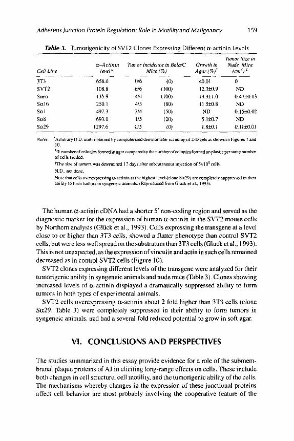
Table 3. Tumorigenicity of SVTZ Clones Expressing Different a-actinin Levels
Tumor Size in a-Acrinin Tumor Incidence in Balb/C Growrk in Nude Mice
Cell Line level* Mice (8) Agar(%)' (cm')$
3T3 SVTZ Snro Sa16 Sat Sa8 Sa29
658.0 016 (0) <0.01 0 108.8 616 (100) 12.3fo.9 ND 135.9 414 (100) 13.3k1.0 0.4739.13 250.1 415 (80) 1 1 Sfo .8 ND 491.3 214 (50) ND 0.15M.02 693.0 115 (20) 5.lM.7 ND
1297.6 or5 (0) 1.8M. I 0. I 1M.01
Norm 'Arbitrary O.D. units obtained by computerized densitometer scanning of 2-Dgels as shown in Figures 7 and 10. ' 7c numkrofcoloniesformed inagarcomparedtothenumkrofcoloniesformedonplasticpersamenum~ of cells seeded. 'The size of tumors was determined 12 days after subcutaneous injection of 5x10' cells. N.D.. not done. Note that cells overexpressing a-actinin at the highest level (clone Sa29) are completely suppressed in their ability to form tumors in syngeneic animals. (Reproduced from Gluck et al.. 1993).
The human a-actinin cDNA had a shorter 5' non-coding region and served as the diagnostic marker for the expression of human a-actinin in the SVT2 mouse cells by Northern analysis (Gluck et al., 1993). Cells expressing the transgene at a level close to or higher than 3T3 cells, showed a flatter phenotype than control SVT2 cells, but were less well spread on the substratum than 3T3 cells (Gluck et al., 1993). This is not unexpected, as the expression of vinculin and actin in such cells remained decreased as in control SVT2 cells (Figure 10).
SVT2 clones expressing different levels of the transgene were analyzed for their tumongenic ability in syngeneic animals and nude mice (Table 3). Clones showing increased levels of a-actinin displayed a dramatically suppressed ability to form tumors in both types of experimental animals.
SVT2 cells overexpressing a-actinin about 2 fold higher than 3T3 cells (clone Sa29, Table 3) were completely suppressed in their ability to form tumors in syngeneic animals, and had a several fold reduced potential to grow in soft agar.
VI. CONCLUSIONS AND PERSPECTIVES
The studies summarized in this essay provide evidence for a role of the submem- branal plaque proteins of AJ in eliciting long-range effects on cells. These include both changes in cell structure, cell motility, and the tumorigenic ability of the cells. The mechanisms whereby changes in the expression of these junctional proteins affect cell behavior are most probably involving the cooperative feature of the
160 AVRl BEN-ZE’EV ETAL.
interactions between these proteins in the process of AJ assembly, and the binding of microfilament bundles to these structures (Kreis et al., 1984; Geiger et al., 1990, 1992). According to this model, an increase in vinculin or a-actinin levels will lead to an increase in their level in the AJ of the transfected cells, which will then lead to the formation of more stable microfilament-membrane interactions, increased adhesion, and decreased motility. In contrast, a decrease in the level of an AJ component such as vinculin, obtained by antisense transfection, will lead to less actin-membrane assembly, a round cell shape, and will impose less restriction on motility. This could result in increased motility, and may influence the ability of cells to grow in soft agar.
The recruitment and immobilization of transmembrane adhesion receptors by AJ plaque components most probably constitute an important mechanism for regulat- ing cell adhesion and motility by an “inside out” mechanism, as suggested by recent studies with chimeric cell adhesion receptors of both the integrin and cadherin family of receptors (La Flamme et al., 1992; Geiger et al., 1992b).
The observations showing that growth-activation, differentiation, and cell trans- formation are associated with modulations in both the organization and the expres- sion of AJ proteins, may therefore point to important changes in AJ protein function during these vital cellular processes. Further studies involving gene targeting in somatic cells and in transgenic animals are needed for addressing the role of such changes in AJ levels during animal development and physiology.
A C K N O W L E D G M E N T S
The studies from the authors’laboratory were supported by grants from the National Council for Research and Development-German Cancer Research Center Collaboration Fund, and from the Leo and Julia Forchheimer Center for Molecular Genetics at the Weizmann Institute of Science. A. Ben-Ze’ev is the Lunenfeld-Kunin Professor of Cell Biology and Genetics, and B. Geiger is the E. Neter Professor of Cell and Tumor Biology.
REFERENCES
Belkin. A.M., Ornatsky, 0.1.. Kabanov, A.E., Glukhova, M.A.. & Koteliansky, V.E. (1988). Diversity of vinculidmeta-vinculin in human tissues and cultivated cells. J. Biol. Chem. 263,14631-14635.
Bellas. R.E., Bendori, R., & Fanner, S.R. (1991). E p i d e m l growth factor activation of vinculin and pi-integrin gene transcription in quiescent Swiss 3T3 cells. J. Biol. Chem. 266, 12008-12014.
Bendori, R., Sdomon, D., &Geiger. B. (1987). Contact dependent regulation of vinculin expression in cultured fibroblasts: A study with vinculin specific cDNAprobes. EMBO J. 6.2897-2905.
Ben-Ze’ev, A. (1984). Differential control of cytokeratins and vimentin synthesis by cell-cell contact and cell spreading in cultured epithelial cells. J. Cell. Biol. 99, 1424-1433.
Ben-Ze’ev, A. (1985). The cytoskeleton of cancer cells. Biochim. Biophys. Acta. 780,197-212. Ben-Ze’ev, A. (1986). The relationship between cytoplasmic organization, gene expression and mor-
Ben-Ze’ev, A. (1991). Animal cell shape changes and gene expression. BioEssays. 13,207-212. phogenesis. Trends Biochem. Sci.. 11,478481.
Adherens Junction Protein Regulation: Role in Motilityand Malignancy 161
Ben-Ze’ev, A. ( 1992). Cytoarchitecture and signal transduction. Critical Rev. Eukaryotic Gene Expres- sion 2, 265-281.
Ben-Ze’ev. A. & Amsterdam. A. (1987). Invitroregulationofgranulosacell differentiation: Involvement of cytoskeletal protein expression. J. Biol. Chem. 262,53665376.
Ben-Ze’ev, A., Reiss. R.. Bendori, R., & Gorodecki, B. (1990). Transient induction of vinculin gene expression in 3T3 fibroblasts stimulated by serum-growth factors. Cell Regul. I . 621-636.
Ben-Ze’ev. A., Robinson, G.. Bucher, N.L.R., & Farmer, S.R. (1988). Cell-cell and cell-matrix interactions differentially regulate the expression of hepatic and cytoskeletal genes in primary cultures of rat hepatocytes. Proc. Natl. Acad. Sci. USA 85. 2161-2165.
Burridge, K., Fath, K.. Kelly, T.. Nuckolls, G., &Turner, C. (1988). Focal adhesions: Transmembrane junctions between the extracellular matrix and the cytoskeleton. Annu. Rev. Cell Biol. 4.487-525.
Burridge, K., Turner, E.E., & Romer, L.H. (1992). Tyrosine phosphorylation of paxillin and pp 125FAK accompanies cell adhesion to extracellular matrix: A role in cytoskeletal assembly. J. Cell Biol.
Comoglio, P.M., DiRenzo. M.F., Tarone, G., Giancotti, F.G., Naldini, L., & Marchisio, P.C. (1984). Detection of phosphotyrosine-containing proteins in the detergent-insoluble fraction of RSV- transformed fibroblasts by azobenzene phosphate antibodies. EMBO J. 3 ,483489.
Cunningham,C.C.. Gorlin.J.B.,Kwiatkowski.D.J.,Hartwig,J.H.,Janmey, P.A..Byers,R.H..&Stossel, T.P. (1992). Actin-binding protein requirement for cortical stability and efficient locomotion. Science 255,325-327.
Cunningham,C.C., Stossel,T.P.,& Kwiatkowski,DJ.( 1991). EnhancedmotilityinNIH3T3fibroblasts that overexpress gelsolin. Science 251, 1233-1236.
Geiger, B. & Ginsberg, D. (1991). The cytoplasmic domain of adherens-type junctions. Cell Motil. Cytoskel. 20, 1-6.
Geiger, B.. Ginsberg, D., Ayalon. 0.. Volberg, T.. Rodriguez Fernhdez. J.L.. Yarden. Y.. & Ben-Ze’ev, A. (1992). Cytoplasmic control of cell-adhesion. Cold Spring Harbor Symp. Quant. Biol. (in press).
Geiger, B.. Ginsberg, D.. Salomon, D., & Volberg, T. (1990). The molecular basis for the assembly and modulation of adherens-type junctions. Cell Diff. Dev. 32.343-354.
Geiger. B., Salomon. D.,Takeichi. M..& Hynes. R. (1992b). Achimeric N-cadheridpi-integrinreceptor which localizes to both cell-cell and cell-matrix adhesions. J. Cell Sci. 103,943-952.
Cluck, U., Kwiatkowski, D.J.. & Ben-Ze’ev, A. (1993). Suppression of tumorigenicity in simian virus 40-transformed 3T3 cells transfected with a-actinin cDNA. Proc. Natl. Acad. Sci. USA 90.
Cluck, U., Rodriguez Fernhdez, J.L., Pankov, R., & Ben-Ze’ev, A. (1992). Regulation of adherens junction protein expression in growth-activated 3T3 cells and in regenerating liver. Exp. Cell Res. 202,477436.
Guan, J.L. & Shalloway. D. (1992). Regulation of focal adhesion-associated protein tyrosine kinase by both cellular adhesion and oncogenic transformation. Nature 358.690-692.
Guan. J.L., Trevithick, J.E.. & Hynes. R.O. (1991). Fibronectidintegrin interaction induces tyrosine phosphorylation of a 120 kDa protein. Cell Regul. 2, 951-964.
Hay, E.D. (ed.) (1981). In: Cell Biology of Extracellular Matrix, 417 pp., Plenum Publishing Corp., New York.
Hynes, R.O. (1992). Integrins: Versatility, modulation, and signaling in cell adhesion. Cell 69, 11-25. Kreis, T.E., Avnur, Z., Schlessinger, J., & Geiger, B. ( 1984). Dynamic properties of cytoskeletal proteins
in focal contacts. In: Molecular Biology of the Cytoskeleton (Borisy. G., Cleveland, D., and Murphy, D.. eds.) pp. 45-57, Cold Spring Harbor Laboratory, Cold Spring Harbor.
La F l a m e , S.E., Akiyama. S.K.. & Yamada. K.M. (1992). Regulation of fibronectin receptor distribu- tion. J. Cell Biol. 117 .43747.
119, 893-903.
383-387.
162 AVRl BEN-ZE’EV ET AL.
Lipfert. L.. Haimovich, B.. Schaller, M.D.. Cobb, B.S., Parsons, J.T., & Brugge, J.S. (1992). Integnn- in platelets. J. dependent phosphorylation and activation of the protein tyrosine kinase
Cell Biol. 119, 905-912. Luna, E.J. & Hitt, A.L. ( 1992). Cytoskeleton-plasma membrane interactions. Science 258,955-964. Maher, P.A.. Pasquale, E.B.. Wang, J.Y.J.. & Singer, S.J. (1985). Phosphotyrosine-containing proteins
are concentrated in focal adhesions and intercellular junctions in normal cells. Proc. Natl. Acad. Sci. USA 82,6576-6580.
Matsumura. F. & Yamashiro-Matsumura, S . (1986). Tropomyosin in cell transformation. Cancer Rev. 6.21-39.
Matzku, S., Komitowski, D., Mildenberger, M. & Zoller, M. (1983). Characterization of BSp73, a spontaneous rat tumor and its in vivo selected variants showing different metastasizing capacities. Invasion Metastasis. 3, 109-123.
Mooney, D., Hansen, L.. Vacanti, J., Langer. R.. Farmer, S.. & Ingber, D. (1992). Switching between growth and differentiation in hepatocytes: Control by extracellular matrix. J. Cell. Physiol. 151, 497-505.
Otey, C.A., Pavalko, EM.. & Bumdge, K. (1990). An interaction between a-actinin and the pi integrin subunit in vitro. J. Cell Biol. 11 1, 721-729.
Pollack, R., Osborn, M.. & Weber, K. (1975). Patterns of organization of actin and myosin in normal and transformed cells. Proc. Natl. Acad. Sci. USA 72,994-998.
Raz, A. & Ben-Ze’ev. A. (1987). Cell contact and architecture of malignant cells and their relationship to metastasis. Cancer Met. Rev. 6, 3-21.
R a r A.,Zoller, M.. & Ben-Ze’ev, A. (1986). Cell configuration and adhesive properties of metastasizing and nonmetastasizing Bsp73 rat adenocarcinoma cells. Exp. Cell Res. 162, 127-141.
Ridley, A.J. & Hall, A. (1992). The small GTP-binding protein rho regulates the assembly of focal adhesions and actin stress fibers in response to growth factors. Cell 70,389-399.
Rodriguez Femdndez, J.L. & Ben-Ze’ev, A. (1989). Regulation of fibronectin. integnn and cytoskeleton expression in differentiating adipocytes: Inhibition by extracellular matrix and polylysine. Differ- entiation 42, 65-74.
Rodriguez Fernhdez. J.L., Geiger, B.. Salomon, D., and Ben-Ze’ev, A. (l992a). Overexpression of vinculin suppresses cell motility in Balb/c 3T3 cells. Cell Motil. Cytoskel. 22, 127-134.
Rodriguez Femdndez. J.L.. Geiger. B.. Salomon, D.. Sabanay, H.. Ziiller. M., & Ben-Ze’ev, A. (l992b). Suppression of tumongenicity in transformed cells after transfection with vinculin cDNA. J. Cell Biol. 119,427438.
Rohrschneider, L.R. (1980). Adhesion plaques of Rous sarcoma virus-transformed cells contain the src gene product. Proc. Natl. Acad. Sci. USA77.3514-3518.
Rysek, R.-P., MacDonald-Bravo, H., Zerial. M., & Bravo, R. (1989). Coordinate induction of fi- bronectin. fibronectin receptor, tropomyosin, and actin genes in serum-stimulated fibroblasts. Exp. Cell Res. 180,537-545.
Sadler. I., Crawford, A.W., Michelsen. J.W., & Beckerle, M.C. (1992). Zyxin and cCRP: ’bointeractive LIM domain proteins associated with the cytoskeleton. J. Cell Biol. 119. 1573-1587.
Schaller, M.D., Borgman, C.A., Cobb. B.S., Vines, R.R., Reynolds, A.B., & Parsons, J.T. (1992). ~ ~ 1 2 5 ~ ~ ~ a structurally unique protein tyrosine kinase associated with focal adhesions. Proc. Natl. Acad. Sci. USA 89,5192-5196.
Shin, S.-L Freedman, V.H., Risser, R., & Pollack, R. (1975). Tumorigenicity of virus-transformed nude mice is correlated specifically with anchorage independent growth in vitro. Proc. Natl. Acad. Sci. USA 72,4435-4439.
Stoker. M., O’Neill, C.H., Benyman, S., & Waxman, V. (1968). Anchorage and growth regulation in normal and virus-transformed cells. Int. J. Cancer 3, 683-688.
Takeichi, M. (1991). Cadherin cell adhesion receptors as a morphogenetic regulator. Science 251, 145 1-1455.
Adherens ]unction Protein Regulation: Role in Motility and Malignancy 163
Tsukita, S., Tsukita. S.. Nagafuchi, A., & Yonemura, S. (1992). Molecular linkage between cadherins and acti\e filaments in cell-cell adherens junctions. Curr. Opin. Cell Biol. 4, 834-839.
Ungar. E, Geiger, B., & Ben-Ze’ev, A. (1986). Cell contact and shape dependent regulation of vinculin synthesis in cultured fibroblasts. Nature 319,787-791.
Vandekerckhove, J., Bauw, G.K.. Vancompemolle, G., Honore. B., & Celis, J. (1990). Comparative two dimensional gel analysis and microsequencing identifies gelsolin as one of the most prominent downregulated markers of transformed human fibroblast and epithelial cells. J. Cell Biol. 1 1 1 , 95-102.
Volberg, T., Zick, Y.. Dror, R., Sabanay, I., Gilon, C., Levitzki. A,, & Geiger, B. (1992). The effect of tyrosine-specific protein phosphorylation on the assembly of adherens-type junctions. EMBO J . I I , 1733-1742.
Youssoufian, H., McAfee, M., & Kwiatkowski. D.J. (1990). Cloning and chromosomal localization of the human cytoskeletal a-actinin gene reveals linkage to the P-spectrin gene. Am J. Hum. Genet. 47.62-72.
Zachary, I. & Rozengurt, E. (1992). Focal adhesion kinase ( ~ 1 2 5 ~ ~ ~ ) : A point of convergence in the action of neuropeptides, integrins, and oncogenes. Cell 71, 891-894.
Zieske, J.D., Bukusoglu. G., & Gipson, I.K. (1989). Enhancement of vinculin synthesis by migrating stratified squamous epithelium. J . Cell Biol. 109, 571-576.