regulation of developmental and environmental signaling by … · 2017-08-27 · m ini- review...
TRANSCRIPT

MINI-REVIEW
Regulation of developmentaland environmental signaling by interactionbetween microtubules and membranes in plantcells
Qun Zhang&, Wenhua Zhang&
College of Life Sciences, State Key Laboratory of Crop Genetics and Germplasm Enhancement, Nanjing AgriculturalUniversity, Nanjing 210095, China& Correspondence: [email protected] (Q. Zhang), [email protected] (W. Zhang)
Received August 31, 2015 Accepted October 31, 2015
ABSTRACT
Cell division and expansion require the orderedarrangement of microtubules, which are subject tospatial and temporal modifications by developmentaland environmental factors. Understanding how signalstranslate to changes in cortical microtubule organizationis of fundamental importance. A defining feature of thecortical microtubule array is its association with theplasma membrane; modules of the plasma membraneare thought to play important roles in the mediation ofmicrotubule organization. In this review, we highlightadvances in research on the regulation of corticalmicrotubule organization by membrane-associated andmembrane-tethered proteins and lipids in response tophytohormones and stress. The transmembrane kinasereceptor Rho-like guanosine triphosphatase, phospho-lipase D, phosphatidic acid, and phosphoinositides arediscussed with a focus on their roles in microtubuleorganization.
KEYWORDS abiotic stresses, cortical microtubule,lipids, plasma membrane
INTRODUCTION
In plants, the cytoskeleton consists of two main components:microtubules and actin filaments. Specific cytoskeletonconfigurations are required for diverse essential processessuch as chromosome segregation, intracellular transport,
cell motility, and cell shape determination (Hashimoto, 2015).Organization of the interphase cortical microtubule array,which is anchored tightly to the plasma membrane, guidesplant growth and morphogenesis by acting in cell divisionand polarity, and in responses to abiotic stresses (Linde-boom et al., 2013; Pleskot et al., 2013, 2014). The dynamicnature of microtubules provides the flexibility to rearrangethem into different arrays in response to developmental andenvironmental stimuli (Wang et al., 2007, 2012; Zhang et al.,2012). To support these diverse functions, the corticalmicrotubule arrays are accurately organized by microtubule-associated proteins and lipids in the plasma membrane(Zhang et al., 2012; Pleskot et al., 2013).
Understanding how cortical microtubules are organizedinto specific array patterns and the underlying molecularmechanisms remains a challenge (Lucas and Shaw, 2008;Hamada, 2014). Real-time observations of microtubuledynamics in axially growing cells, in combination withanalysis of phospholipid regulation of cytoskeletal organi-zation, have provided a deep appreciation of the regulatorynetworks involved in cytoskeletal organization (Lin et al.,2014; Pleskot et al., 2014). Cytoskeletal dynamics and itsregulation have been the subject of multiple reviews (Dixitand Cyr, 2004; Lloyd and Chan, 2004; Ehrhardt and Shaw,2006; Pleskot et al., 2013, 2014). In this review, wedescribe recent advances in elucidating the functions ofcortical microtubules in response to phytohormones andabiotic stresses, and their functional regulation by mem-brane-associated and membrane-tethered proteins andlipids.
© The Author(s) 2015. This article is published with open access at Springerlink.com and journal.hep.com.cn
DOI 10.1007/s13238-015-0233-6 Protein&Cell
Protein
&Cell
Protein Cell 2016, 7(2): –81 88
MICROTUBULE FUNCTIONS IN HORMONE-MEDIATED DEVELOPMENTAL PROCESSES
Microtubule reorganization and auxin response
Auxin participates in various developmental processes. Onemajor effect of auxin is cell expansion, which relies on thecoordinated activities of cellular processes involving micro-tubules (Ruan and Wasteneys, 2014; Adamowski and Friml,2015). When cells elongate, cortical microtubules arearranged perpendicularly to the axis of cell elongation(transverse microtubules), while a longitudinal alignmentinduces growth inhibition. In response to auxin, root micro-tubules change from transverse to longitudinal, inhibiting cellexpansion (Chen et al., 2014). Using the TILLING mutant,which is defective in AUXIN BINDING PROTEIN1 (ABP1)(abp1-5), it was further demonstrated that the effect of auxinrequires ABP1 and involves the contribution of downstreamsignaling components, including Rho-like GTPase fromplants 6 (ROP6), and the ROP-interacting protein RIC1 (Linet al., 2013; Chen et al., 2014). In leaf pavement cells ofArabidopsis, the plasma membrane-localized transmem-brane kinases (TMKs) belonging to the receptor-like kinasefamily has been found to interact with ABP1. The TMK-ABP1interaction is required to activate ROPs, which play a role inregulating cytoskeleton organization and the endocytosis ofPIN-FORMEDs (PINs), which are auxin efflux carrier pro-teins (Xu et al., 2014; Fig. 1).
Overall, the stated functions of ABP1 are inconsistent. Aviable abp1-5 TILLING allele was used to identify the func-tions of ABP1, including the auxin-responsive rearrange-ment of microtubules, PIN protein internalization, and othermolecular and cellular processes (Robert et al., 2010; Basteret al., 2013; Effendi et al., 2013; Chen et al., 2014; Paqueet al., 2014; Xu et al., 2014). More recently, however, Gaoet al. (2015) used ribozyme-based CRISPR technology togenerate an abp1 mutant with a 5-bp deletion in the firstexon of ABP1, and they isolated a T-DNA insertion abp1allele. None of the mutants showed either auxin signaling ordevelopmental phenotypes. Furthermore, genomesequencing of the abp1-5 mutant revealed that backgroundmutations may lead to auxin and other phenotypes (Enderset al., 2015). Complementation tests and a re-valuation ofthe functions of ABP1 have been proposed for the futurework; additional information about ABP1 can be found inother reports (Enders et al., 2015; Liu, 2015).
Cortical microtubules in turn influence polar auxin trans-port (Heisler et al., 2010; Ambrose et al., 2013; Zhang et al.,2013; Ruan and Wasteneys, 2014). Short-term treatmentwith the microtubule-disrupting drug oryzalin had no effect onthe polarity of PIN proteins (Boutte et al., 2006; Geldneret al., 2001); however, prolonged oryzalin treatment inter-fered with basal PIN2 targeting in young cortical cells andwith PIN1 targeting in the stele, resulting in reduced polardistribution (Kleine-Vehn et al., 2008). The Arabidopsismicrotubule-associated protein CLASP interacts with the
retromer component sorting nexin 1 (SNX1) protein tomediate the association between endosomes and micro-tubules. Plants carrying the clasp-1 mutation displayenhanced PIN2 degradation and aberrant auxin distribution,which is promoted by microtubule depolymerization (Am-brose et al., 2013; Brandizzi and Wasteneys, 2013). Thesefindings indicate that intact microtubules are required for thepolar distribution of PIN proteins and auxin function.
Microtubules, stomatal development, and abscisic acidsignaling
Stomatal morphogenesis takes place after the symmetricdivision of a guard mother cell, followed by the developmentof wall thickening in each daughter cell and their separationto form the stomatal pore in a microtubule-dependent pro-cess (Galatis and Apostolakos, 2004; Lucas et al., 2006).The highly organized microtubules in Arabidopsis stomatalcells play key roles in the morphogenesis of stomatal com-plexes (Galatis and Apostolakos, 2004; Lucas et al., 2006).The preprophase bands (PPBs) of microtubules in maturemother cells are located away from stomata, and radiallyoriented microtubules converge near the central rim of thestomatal pore, suggesting an essential function of micro-tubules in asymmetric division (Lucas et al., 2006). Muta-tions in Arabidopsis MUSTACHES (MUS), a leucine-richrepeat receptor-like kinase, disrupt stomatal symmetryresulting in stomatal defects and depolarized radial micro-tubule arrays (Keerthisinghe et al., 2015).
Reorganization of the cortical microtubule cytoskeleton iscritical for guard cell function, particularly in the abscisic acid(ABA) signaling pathway (Marcus et al., 2001; Eisinger et al.,2012a, b; Jiang et al., 2014). An apparent loss of microtubuleswas observed in guard cells upon stomatal closure, probablydue to microtubule instability or rearrangement. The depoly-merization of guard cell microtubules by oryzalin preventedArabidopsis stomatal opening, while the stabilization ofmicrotubules delayed stomatal closure (Eisinger et al.,2012a). Microtubules were further observed using green flu-orescent protein fused to α-tubulin 6 (GFP-TUA6). The totalamount of polymerized tubulin was higher in open than inclosed guard cells; this was correlated with an increase in thetotal fluorescence (Eisinger et al., 2012b). These results are inagreement with genetic evidence showing that themutation ofCONSTITUTIVELY PHOTOMORPHOGENIC 1 (COP1),which encodes an Arabidopsis RING finger-type ubiquitin E3ligase, results in tubulin degradation and stomatal closure(Khanna et al., 2014). COP1 has been studied extensively asa critical destabilizer of photomorphogenesis-promoting fac-tors. Because light is an important factor in the regulation ofstomatal movement, the finding of a COP1-mediated micro-tubule array opens a new avenue for understanding the reg-ulatory mechanisms underlying microtubule organization(Mao et al., 2005). Taken together, these results suggest thatthe microtubule array organization is correlated with and
MINI-REVIEW Qun Zhang, Wenhua Zhang
© The Author(s) 2015. This article is published with open access at Springerlink.com and journal.hep.com.cn
Protein
&Cell
82

required for stomatal opening and closure. Microtubules maycontrol the activity of plasmamembrane ion channels such asthose that transport calcium, and lipid signaling may beinvolved in this process. Phospholipase D (PLD) catalyzesphospholipid hydrolysis to produce phosphatidic acid (PA)and a free head group. It was demonstrated that PLD and PAare involved in the ABA-induced stomatal closure (Zhanget al., 2009). Treatment with calcium induces depolymeriza-tion of microtubules and stomatal closure in wild-type Ara-bidopsis, but not in the pldα1 mutant (Jiang et al., 2014). Inaddition, both ABA-induced microtubule depolymerizationand stomatal closure were impaired in pldα1, and cotreatmentwith ABA and microtubule-disrupting drugs rescued the pldα1phenotype (Jiang et al., 2014).
The cop1mutation not only induces tubulin degradation, italso impairs the calcium ion-dependent activation of S-typeanion channel currents in guard cells, which are activated todrive stomatal closure. However, the cop1 mutation did notchange the activation of inward K+ channel currents requiredfor stomatal opening (Khanna et al., 2014). It is still an openquestion whether S-type anion channels and microtubulesmay function independently, or whether they act together toregulate stomatal movement.
Roles of the hormones GA, ethylene,and brassinosteroid in microtubule organization
DELLA nuclear proteins restrain cell proliferation and expan-sion, leading to inhibited plant growth (Peng et al., 1999), andthey integrate salt-activated ethylene (ETH) and ABA signalingin response to environmental changes (Achard et al., 2006). Arecent study established DELLA proteins as a mechanistic linkbetween GA and cortical microtubule organization (Locascioet al., 2013). DELLA proteins interact with the prefoldin (PFD)complex, a cochaperone required for tubulin folding (Locascioet al., 2013). In the presence of GA, DELLAs are degradedand the FPD complex is shuttled into the cytoplasm where itproduces active tubulin subunits. In the absence of GA, PFD islocalized to the nucleus, where it compromises α/β-tubulinheterodimer availability, affecting microtubule organization(Locascio et al., 2013). A loss of function of PFD impairsmicrotubule organization, rendering the pfd mutant hypersen-sitive to salt stress (Rodriguez-Milla and Salinas, 2009). Theseresults demonstrate that GA-mediated microtubule organiza-tion plays an essential role in salt tolerance.
As a gaseous plant hormone, ETH is essential for plantgrowth and development, including seed germination, leafsenescence, fruit ripening, and responses to environmentalstresses (Kendrick and Chang, 2008; Muller and Munne-Bosch,2015). ETH affects the organization of cortical microtubules inplant cells (Takahashi et al., 2003; Polko et al., 2012). Themicrotubule-associated protein WAVE-DAMPENED2-LIKE5(WDL5) is a microtubule-stabilizing protein in Arabidopsis (Sunet al., 2015). Treatment with 1-aminocyclopropane-1-carboxylicacid (ACC) significantly enhanced the WDL5 expression and
cortical microtubule stability, resulting in decreased etiolatedhypocotyl cell elongation, and the reorganization of corticalmicrotubules in the wdl5-1 mutant showed reduced sensitivityto ACC treatment (Sun et al., 2015). The above results suggestthat cell elongation depends on the microtubule reorganization,and that stabilized microtubules are required for EHT-inhibitedetiolated hypocotyl cell elongation, which involves WDL5 as apositive participant. In addition, WDL3 overexpression resultedin overall shortening of hypocotyl cells and stabilization of cor-tical microtubules in the light, and WDL3 protein was abundantin the light, but was degraded through the 26S proteasomepathway in the dark (Liu et al., 2013).
Brassinosteroid (BR) mediates hypocotyl cell elongationby a mechanism that may control the orientation and stabilityof cortical microtubules. The key transcription factor BRAS-SINAZOLE-RESISTANT1 (BZR1) targets and upregulatesmicrotubule destabilizing protein 40 (MDP40) directly,thereby serving as a positive regulator of hypocotyl cellelongation (Li, 2010; Gudesblat and Russinova, 2011; Wanget al., 2012). Genetic evidence shows that the light/GA-sig-naling pathway affects the properties of microtubulesrequired to reorient growth (Sambade et al., 2012). Ara-bidopsis AUGMIN subunit 8 (AUG8) is a novel microtubuleplus end-binding protein that contributes to light-inducedmicrotubule reorientation and modulates cell elongation(Cao et al., 2013). The studies above suggest the existenceof a molecular mechanism of putative crosstalk betweenphytohormones, microtubule dynamics, and cell elongationin response to light or dark environments.
MICROTUBULE REORGANIZATION IN RESPONSETO STRESS
Cortical microtubules are not only targets of signaling, butalso actively participate in signal transduction itself. Saltstress induces the rapid depolymerization of microtubulesand the formation of a new microtubule network via repoly-merization (Wang et al., 2007, 2011; Zhang et al., 2012).SPR1, a microtubule-stabilizing protein, is degraded by the26S proteasome in response to salt stress, and this degra-dation is essential for salt stress tolerance (Nakajima et al.,2004; Sedbrook et al., 2004; Wang et al., 2011). Moreover,the spr1 mutant displays a right-handed helical growthphenotype, and interestingly, mutations of the plasmamembrane Na+/H+ antiporter, SOS1, suppress the helicalgrowth phenotype (Shoji et al., 2006). The root microtubulesarrays of sos1 plants are oriented much more randomly thanthose of wild-type cells under mild salt treatment (Shoji et al.,2006). A recent study showed that the Arabidopsis saltoverly sensitive 3 (SOS3) protein plays an important role insalt tolerance through regulation of actin filaments (Ye et al.,2013). These findings indicate that the cytoskeleton interactswith the SOS pathway to signal salt stress in plant cells.
PLDα1-derived PA binds to microtubule-associated pro-tein 65-1 (MAP65-1) and regulates cortical microtubule
Signaling via microtubule and membrane interaction MINI-REVIEW
© The Author(s) 2015. This article is published with open access at Springerlink.com and journal.hep.com.cn
Protein
&Cell
83
organization in Arabidopsis in response to salt stress (Zhanget al., 2012). Under salt stress, knockout of the PLDa1 genecauses increased NaCl-induced disorganization of micro-tubules, which cannot be recovered during or after removalof the stress but can be alleviated by exogenous PA. Furtherevidence reveals that PA binds to residues 53KRK55,61KSR63, and 428SK429 of MAP65-1, and that this bindingis involved in MAP65-1 binding to microtubules (Zhang et al.,2012). Interestingly, PA also binds to mitogen-activatedprotein kinase 6 (MPK6) and increases its phosphataseactivity, which phosphorylates SOS1 and enhances plantsalt tolerance (Yu et al., 2010). In addition, MAP65-1 isphosphorylated by mitogen-activated protein kinase 4(MAPK4 or MPK4) and MPK6 (Smertenko et al., 2006; Becket al., 2011), their putative orthologs MAPK NRK1/NTF6(Sasabe et al., 2006), and cyclin dependent protein kinase(CDPK) (Mollinari et al., 2002). These findings have estab-lished the existence of crosstalk among phospholipids,microtubules, and phosphatase in response to a stressfulenvironment (Fig. 1).
MEMBRANE-MICROTUBULE INTERACTION
Interplay between cortical microtubules and plasmamembrane domains
To ensure the proper spatial and temporal regulation ofmicrotubule dynamics, the activity and binding properties ofMAPs are further modulated by upstream signaling mole-cules. Physical linkages between microtubules and themembrane were recently observed using high-resolutionscanning electron microscopy (Barton et al., 2008). Only afew candidate MAPs have been proposed as potential link-ers between the plasma membrane and microtubules (e.g.,PLD [Gardiner et al., 2001]) and Arabidopsis membrane-in-tegrated formin (AtFH4 [Deeks et al., 2010]). A 90 kDatubulin-binding protein from tobacco was identified as aputative PLDδ based on an activity assay and sequencealignment (Gardiner et al., 2001), and the activation of PLDinduced cortical microtubules to release from the plasmamembrane and partially depolymerize (Dhonukshe et al.,2003). However, the detailed mechanical functions of PLDon microtubule organization remain to be elucidated. AtFH4coaligns the endoplasmic reticulum with microtubules andalso nucleates filamentous (F)-actin. Although an AtFH4-GFP fusion protein was shown to accumulate at the endo-plasmic reticulum (ER), it may be trafficked to the plasmamembrane to act as a scaffold for cytoskeletal organization(Deeks et al., 2010).
ROP11 is distributed broadly at the plasma membrane.ROP11, after being activated by Rho of the plant guaninenucleotide exchange factor 4 (ROPGEF4), recruits themicrotubule depletion domain 1 (MIDD1) protein to inducethe local disassembly of cortical microtubules. Conversely,cortical microtubules eliminate active ROP11 from theplasma membrane through MIDD1 (Oda and Fukuda, 2012).
The mutually inhibitory interaction between active ROPdomains and cortical microtubules is essential to establishthe secondary wall pattern in xylem cells (Oda and Fukuda,2012, 2013).
Lipid signaling in plant cells
Phospholipids play a key role in maintaining the bilayerstructure of membranes and in separating the cytosol fromorganelles and the extracellular space. The proportions ofphospholipids such as PA, inositol 1,4,5-trisphosphate(InsP3), and diacylglycerol (DAG) change rapidly, and toge-ther with phospholipid-metabolizing proteins, are involved inplant growth and development (Wang et al., 2014). As arough approximation, PA, an abundant negatively chargedphospholipid, constitutes 1%–4% of total cellular lipids (Staceand Ktistakis, 2006). Although PA does not bind to tubulinsin vitro (Zhang et al., 2012), it may mediate cytoskeletalorganization and dynamics by binding to and modulatingcytoskeleton-associated proteins (Pleskot et al., 2013).
We demonstrated that PA acts as a linker between theplasma membrane and microtubules via MAP65-1, which isessential for salt-stress signaling in Arabidopsis (Zhanget al., 2012). Under salt stress, Arabidopsis PLDα1 is acti-vated to produce PA, which binds to MAP65-1, leading toenhanced microtubule polymerization and bundling activity(Zhang et al., 2012). Exogenous application of PA rescuesthe salt-sensitive phenotype of microtubules in pldα1, but notin map65-1, clearly indicating that the PA-MAP65-1 interac-tion is essential for cortical microtubule organization inresponse to salt stress (Zhang et al., 2012). The twomicrotubule-destabilizing proteins, MAP18 (AtPCaP2) andMDP25 (AtPCaP1), bind PtdIns(3,4,5)P3 and PtdIns(3,5)P2
in vitro indicating that both proteins are involved in intracel-lular signaling by regulating microtubule organization andinteracting with PtdInsPs (Nagasaki et al., 2008; Kato et al.,2010), although no direct evidence for the involvement ofPtdInsPs in the regulation of MAP18 and MDP25 has beenreported to date.
Like microtubules, the organization and dynamics of actinfilaments are mediated by membrane phospholipids. Ara-bidopsis heterodimeric capping protein (AtCP) binds to thebarbed ends of actin filaments (Huang et al., 2003), and thisactivity is regulated by PA (Huang et al., 2006). The inter-action between PA and AtCP renders filament ends moredynamic, which significantly enhances filament-filamentannealing and filament elongation from free ends (Li et al.,2012). In a separate report, actin and β-tubulin were pulleddown with GFP-PLDδ from Arabidopsis suspension cells,suggesting that PLDδ connects microtubules with actin fila-ments in plant cells (Ho et al., 2009). In tobacco (Nicotianatabacum) pollen, actin interacts with NtPLDβ1, and F-actinenhances, while G-actin inhibits, PLDβ1 activity (Pleskotet al., 2010). Thus, PA regulates microtubules and actinthrough PA-binding proteins, and PLD directly links micro-tubules and actin. These results suggest that individual PLD
MINI-REVIEW Qun Zhang, Wenhua Zhang
© The Author(s) 2015. This article is published with open access at Springerlink.com and journal.hep.com.cn
Protein
&Cell
84
isoforms and their product, PA, anchor the cytoskeleton tospecific sites on membranes to reorganize them in responseto diverse signals. On the other hand, microtubule depoly-merization induced by oryzalin activates PLDα1, anddepolymerized G-actin inhibits PLDβ1 activity, indicatingfeedback regulation of PLD activity (Pleskot et al., 2010;Zhang et al., 2012).
CONCLUSIONS AND OUTLOOK
Microtubules in plant cells are regulated by multiple MAPs toenrich the scope of microtubule behavior, and some MAPsare bound tightly to the plasma membrane (Gardiner et al.,
2001; Ambrose and Wasteneys, 2008; Gu et al., 2008; Liet al., 2011). Cortical microtubules and the plasma mem-brane reorganize themselves and transduce external stimulito internal systems. Most of these interactions are mediatedby membrane-based molecules and microtubule linker pro-teins. The acidic phospholipid PA, a minor, dynamic com-ponent of the bilayer, does not bind to tubulins in vitro;instead, it may mediate microtubule organization by inter-acting with MAPs (Zhang et al., 2012). Moreover, otheracidic phospholipids such as phosphatidylserine (PS),phosphatidylglycerol (PG), and phosphatidylinositol (PI) mayalso mediate cytoskeletal organization directly or regulateMAPs activity involved in microtubule arrays, but supporting
MAP65-1
NaCl
PA
PA
PA MPK6
SOS1
GTP-ROP11
MIDD1 Kin13A
PLD
δ
CLASP
SNX1
PIN2
AtFH4
ER
RIC1
KTN1
Auxin
GAs
DELLA PFD
WDL5
MDP40
BR
Plasma membrane
PtdInsPs
ETH
MAP18
PLDα1
Nucleus
TMK1 ABP1
PLDβ1 MTs
AFsAtC
P
MDP25
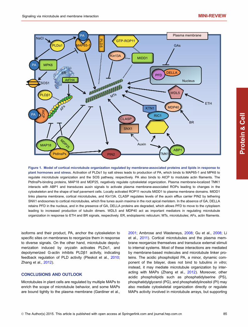
Figure 1. Model of cortical microtubule organization regulated by membrane-associated proteins and lipids in response to
plant hormones and stress. Activation of PLDα1 by salt stress leads to production of PA, which binds to MAP65-1 and MPK6 to
regulate microtubule organization and the SOS pathway, respectively. PA also binds to AtCP to modulate actin filaments. The
PtdInsPs-binding proteins, MAP18 and MDP25, negatively regulate cytoskeletal organization. Plasma membrane-localized TMK1
interacts with ABP1 and transduces auxin signals to activate plasma membrane-associated ROPs leading to changes in the
cytoskeleton and the shape of leaf pavement cells. Locally activated ROP11 recruits MIDD1 to plasma membrane domains. MIDD1
links plasma membrane, cortical microtubules, and Kin13A. CLASP regulates levels of the auxin efflux carrier PIN2 by tethering
SNX1 endosomes to cortical microtubules, which fine tunes auxin maxima in the root apical meristem. In the absence of GA, DELLA
retains PFD in the nucleus, and in the presence of GA, DELLA proteins are degraded, which allows PFD to move to the cytoplasm
leading to increased production of tubulin dimers. WDL5 and MDP40 act as important mediators in regulating microtubule
organization in response to ETH and BR signals, respectively. ER, endoplasmic reticulum; MTs, microtubules; AFs, actin filaments.
Signaling via microtubule and membrane interaction MINI-REVIEW
© The Author(s) 2015. This article is published with open access at Springerlink.com and journal.hep.com.cn
Protein
&Cell
85

evidence remains limited (Pleskot et al., 2013). Therefore,further study is needed to identify additional MAPs that bindto the plasma membrane and phospholipids and interact withcytoskeleton-associated proteins in plant cells.
ACKNOWLEDGMENTS
This work was supported by grants from the National Basic
Research Program (973 Program) (No. 2012CB114200), the
National Natural Science Foundation of China (Grant No.
91117003), the Fundamental Research Funds for the Central
Universities (KYTZ201402), and RAPD project to W Zhang, and
grants from the National Natural Science Foundation of China (Grant
No. 31470364) and the Fundamental Research Funds for the Cen-
tral Universities (KYZ201423) to Q Zhang.
ABBREVIATION
ABA, abscisic acid; CDPK, cyclin dependent protein kinase; ER,
endoplasmic reticulum; ETH, ethylene; MIDD1, microtubule deple-
tion domain 1; MPK6, mitogen-activated protein kinase 6; PFD,
prefoldin; PINs, PIN-FORMEDs; SNX1, sorting nexin 1; TMKs,
transmembrane kinases
COMPLIANCE WITH ETHICS GUIDELINES
Qun Zhang and Wenhua Zhang declare that they have no conflict of
interest. This article does not contain any studies with human or
animal subjects performed by the any of the authors.
OPEN ACCESS
This article is distributed under the terms of the Creative Commons
Attribution 4.0 International License (http://creativecommons.org/
licenses/by/4.0/), which permits unrestricted use, distribution, and
reproduction in any medium, provided you give appropriate credit to
the original author(s) and the source, provide a link to the Creative
Commons license, and indicate if changes were made.
REFERENCES
Achard P, Cheng H, De Grauwe L, Decat J, Schoutteten H, Moritz T,
Van Der Straeten D, Peng J, Harberd NP (2006) Integration of
plant responses to environmentally activated phytohormonal
signals. Science 311:91–94
Adamowski M, Friml J (2015) PIN-dependent auxin transport: action,
regulation, and evolution. Plant Cell 27:20–32
Ambrose JC, Wasteneys GO (2008) CLASP modulates microtubule-
cortex interaction during self-organization of acentrosomal micro-
tubules. Mol Biol Cell 19:4730–4737
Ambrose C, Ruan Y, Gardiner J, Tamblyn LM, Catching A, Kirik V,
Marc J, Overall R, Wasteneys GO (2013) CLASP interacts with
sorting nexin 1 to link microtubules and auxin transport via PIN2
recycling in Arabidopsis thaliana. Dev Cell 24:649–659
Barton DA, Vantard M, Overall RL (2008) Analysis of cortical arrays
from Tradescantia virginiana at high resolution reveals discrete
microtubule subpopulations and demonstrates that confocal
images of arrays can be misleading. Plant Cell 20:982–994
Baster P, Robert S, Kleine-Vehn J, Vanneste S, Kania U, Grunewald
W, De Rybel B, Beeckman T, Friml J (2013) SCF(TIR1/AFB)-
auxin signalling regulates PIN vacuolar trafficking and auxin
fluxes during root gravitropism. EMBO J 32:260–274
Beck M, Komis G, Ziemann A, Menzel D, Samaj J (2011) Mitogen-
activated protein kinase 4 is involved in the regulation of mitotic
and cytokinetic microtubule transitions in Arabidopsis thaliana.
New Phytol 189:1069–1083
Boutte Y, Crosnier MT, Carraro N, Traas J, Satiat-Jeunemaitre B
(2006) The plasma membrane recycling pathway and cell polarity
in plants: studies on PIN proteins. J Cell Sci 119:1255–1265
Brandizzi F, Wasteneys GO (2013) Cytoskeleton-dependent
endomembrane organization in plant cells: an emerging role for
microtubules. Plant J 75:339–349
Cao L, Wang L, Zheng M, Cao H, Ding L, Zhang X, Fu Y (2013)
Arabidopsis AUGMIN subunit8 is a microtubule plus-end binding
protein that promotes microtubule reorientation in hypocotyls.
Plant Cell 25:2187–2201
Chen X, Grandont L, Li H, Hauschild R, Paque S, Abuzeineh A,
Rakusova H, Benkova E, Perrot-Rechenmann C, Friml J (2014)
Inhibition of cell expansion by rapid ABP1-mediated auxin effect
on microtubules. Nature 516:90–93
Deeks MJ, Fendrych M, Smertenko A, Bell KS, Oparka K, Cvrckova
F, Zarsky V, Hussey PJ (2010) The plant formin AtFH4 interacts
with both actin and microtubules, and contains a newly identified
microtubule-binding domain. J Cell Sci 123:1209–1215
Dhonukshe P, Laxalt AM, Goedhart J, Gadella TW, Munnik T (2003)
Phospholipase D activation correlates with microtubule reorga-
nization in living plant cells. Plant Cell 15:2666–2679
Dixit R, Cyr R (2004) The cortical microtubule array: from dynamics
to organization. Plant Cell 16:2546–2552
Effendi Y, Jones AM, Scherer GF (2013) AUXIN-BINDING-PRO-
TEIN1 (ABP1) in phytochrome-B-controlled responses. J Exp Bot
64:5065–5074
Ehrhardt DW, Shaw SL (2006) Microtubule dynamics and organiza-
tion in the plant cortical array. Annu Rev Plant Biol 57:859–875
Eisinger W, Ehrhardt D, Briggs W (2012a) Microtubules are essential
for guard-cell function in Vicia and Arabidopsis. Mol Plant 5:601–610
Eisinger WR, Kirik V, Lewis C, Ehrhardt DW, Briggs WR (2012b)
Quantitative changes in microtubule distribution correlate with
guard cell function in Arabidopsis. Mol Plant 5:716–725
Enders TA, Oh S, Yang Z, Montgomery BL, Strader LC (2015)
Genome sequencing of Arabidopsis abp1-5 reveals second-site
mutations that may affect phenotypes. Plant Cell 27:1820–1826
Galatis B, Apostolakos P (2004) The role of the cytoskeleton in the
morphogenesis and function of stomatal complexes. New Phytol
161:613–639
Gao Y, Zhang Y, Zhang D, Dai X, Estelle M, Zhao Y (2015) Auxin
binding protein 1 (ABP1) is not required for either auxin signaling
or Arabidopsis development. Proc Natl Acad Sci U S A 112:2275–
2280
Gardiner JC, Harper JD, Weerakoon ND, Collings DA, Ritchie S,
Gilroy S, Cyr RJ, Marc J (2001) A 90-kD phospholipase D from
tobacco binds to microtubules and the plasma membrane. Plant
Cell 13:2143–2158
MINI-REVIEW Qun Zhang, Wenhua Zhang
© The Author(s) 2015. This article is published with open access at Springerlink.com and journal.hep.com.cn
Protein
&Cell
86

Geldner N, Friml J, Stierhof YD, Jurgens G, Palme K (2001) Auxin
transport inhibitors block PIN1 cycling and vesicle trafficking.
Nature 413:425–428
Gu Y, Deng Z, Paredez AR, DeBolt S, Wang ZY, Somerville C (2008)
Prefoldin 6 is required for normal microtubule dynamics and
organization in Arabidopsis. Proc Natl Acad Sci USA 105:18064–
18069
Gudesblat GE, Russinova E (2011) Plants grow on brassinosteroids.
Curr Opin Plant Biol 14:530–537
Hamada T (2014) Microtubule organization and microtubule-asso-
ciated proteins in plant cells. Int Rev Cell Mol Biol 312:1–52
Hashimoto T (2015) Microtubules in plants. Arabidopsis Book 13:
e0179
Heisler MG, Hamant O, Krupinski P, Uyttewaal M, Ohno C, Jönsson
H, Traas J, Meyerowitz EM (2010) Alignment between PIN1
polarity and microtubule orientation in the shoot apical meristem
reveals a tight coupling between morphogenesis and auxin
transport. PLoS Biol 8:e1000516
Ho AY, Day DA, Brown MH, Marc J (2009) Arabidopsis phospho-
lipase D δ as an initiator of cytoskeleton-mediated signalling to
fundamental cellular processes. Funct Plant Biol 36:190–198
Huang S, Blanchoin L, Kovar DR, Staiger CJ (2003) Arabidopsis
capping protein (AtCP) is a heterodimer that regulates assembly at
the barbed ends of actin filaments. J Biol Chem 278:44832–44842
Huang S, Gao L, Blanchoin L, Staiger CJ (2006) Heterodimeric
capping protein from Arabidopsis is regulated by phosphatidic
acid. Mol Biol Cell 17:1946–1958
Jiang Y, Wu K, Lin F, Qu Y, Liu X, Zhang Q (2014) Phosphatidic acid
integrates calcium signaling and microtubule dynamics into
regulating ABA-induced stomatal closure in Arabidopsis. Planta
239:565–575
Kato M, Nagasaki-Takeuchi N, Ide Y, Maeshima M (2010) An
Arabidopsis hydrophilic Ca2+ -binding protein with a PEVK-rich
domain, PCaP2, is associated with the plasma membrane and
interacts with calmodulin and phosphatidylinositol phosphates.
Plant Cell Physiol 51:366–379
Keerthisinghe S, Nadeau JA, Lucas JR, Nakagawa T, Sack FD
(2015) The Arabidopsis leucine-rich repeat receptor-like kinase
MUSTACHES enforces stomatal bilateral symmetry in Arabidop-
sis. Plant J 81:684–694
Kendrick MD, Chang C (2008) Ethylene signaling: new levels of
complexity and regulation. Curr Opin Plant Biol 11:479–485
Khanna R, Li J, Tseng TS, Schroeder JI, Ehrhardt DW, Briggs WR
(2014) COP1 jointly modulates cytoskeletal processes and
electrophysiological responses required for stomatal closure.
Mol Plant 7:1441–1454
Kleine-Vehn J, Langowski L, Wisniewska J, Dhonukshe P, Brewer
PB, Friml J (2008) Cellular and molecular requirements for polar
PIN targeting and transcytosis in plants. Mol Plant 1:1056–1066
Li J (2010) Regulation of the nuclear activities of brassinosteroid
signaling. Curr Opin Plant Biol 13:540–547
Li J, Wang X, Qin T, Zhang Y, Liu X, Sun J, Zhou Y, Zhu L, Zhang Z,
Yuan M, Mao T (2011) MDP25, a novel calcium regulatory
protein, mediates hypocotyl cell elongation by destabilizing
cortical microtubules in Arabidopsis. Plant Cell 23:4411–4427
Li J, Henty-Ridilla JL, Huang S, Wang X, Blanchoin L, Staiger CJ
(2012) Capping protein modulates the dynamic behavior of actin
filaments in response to phosphatidic acid in Arabidopsis. Plant
Cell 24:3742–3754
Lin D, Cao L, Zhou Z, Zhu L, Ehrhardt D, Yang Z, Fu Y (2013) Rho
GTPase signaling activates microtubule severing to promote
microtubule ordering in Arabidopsis. Curr Biol 23:290–297
Lin F, Qu Y, Zhang Q (2014) Phospholipids: molecules regulating
cytoskeletal organization in plant abiotic stress tolerance. Plant
Signal Behav 9:e28337
Lindeboom JJ, Nakamura M, Hibbel A, Shundyak K, Gutierrez R,
Ketelaar T, Emons AM, Mulder BM, Kirik V, Ehrhardt DW (2013) A
mechanism for reorientation of cortical microtubule arrays driven
by microtubule severing. Science 342:1245533
Liu CM (2015) AUXIN BINDING PROTEIN 1 (ABP1): a matter of
fact. J Integr Plant Biol 57:234–235
Liu X, Qin T, Ma Q, Sun J, Liu Z, Yuan M, Mao T (2013) Light-
regulated hypocotyl elongation involves proteasome-dependent
degradation of the microtubule regulatory protein WDL3 in
Arabidopsis. Plant Cell 25:1740–1755
Lloyd C, Chan J (2004) Microtubules and the shape of plants to
come. Nat Rev Mol Cell Biol 5:13–22
Locascio A, Blazquez MA, Alabadi D (2013) Dynamic regulation of
cortical microtubule organization through prefoldin-DELLA inter-
action. Curr Biol 23:804–809
Lucas J, Shaw SL (2008) Cortical microtubule arrays in the
Arabidopsis seedling. Curr Opin Plant Biol 11:94–98
Lucas JR, Nadeau JA, Sack FD (2006) Microtubule arrays and
Arabidopsis stomatal development. J Exp Bot 57:71–79
Mao J, Zhang YC, Sang Y, Li QH, Yang HQ (2005) From The Cover:
A role for Arabidopsis cryptochromes and COP1 in the regulation
of stomatal opening. Proc Natl Acad Sci U S A 102:12270–12275
Marcus AI, Moore RC, Cyr RJ (2001) The role of microtubules in
guard cell function. Plant Physiol 125:387–395
Mollinari C, Kleman JP, Jiang W, Schoehn G, Hunter T, Margolis RL
(2002) PRC1 is a microtubule binding and bundling protein
essential to maintain the mitotic spindle midzone. J Cell Biol
157:1175–1186
Muller M, Munne-Bosch S (2015) Ethylene response fekerehub in
hormone and stress signaling. Plant Physiol 169:32–41
Nagasaki N, Tomioka R, Maeshima M (2008) A hydrophilic cation-
binding protein of Arabidopsis thaliana, AtPCaP1, is localized to
plasma membrane via N-myristoylation and interacts with
calmodulin and the phosphatidylinositol phosphates PtdIns
(3,4,5)P3 and PtdIns(3,5)P2. FEBS J 275:2267–2282
Nakajima K, Furutani I, Tachimoto H, Matsubara H, Hashimoto T
(2004) SPIRAL1 encodes a plant-specific microtubule-localized
protein required for directional control of rapidly expanding Ara-
bidopsis cells. Plant Cell 16:1178–1190
Oda Y, Fukuda H (2012) Initiation of cell wall pattern by a Rho- and
microtubule-driven symmetry breaking. Science 337:1333–1336
Oda Y, Fukuda H (2013) The dynamic interplay of plasma
membrane domains and cortical microtubules in secondary cell
wall patterning. Front Plant Sci 4:1–6
Paque S, Mouille G, Grandont L, Alabadi D, Gaertner C, Goyallon A,
Muller P, Primard-Brisset C, Sormani R, Blazquez MA, Perrot-
Rechenmann C (2014) AUXIN BINDING PROTEIN1 links cell
wall remodeling, auxin signaling, and cell expansion in Arabidop-
sis. Plant Cell 26:280–295
Signaling via microtubule and membrane interaction MINI-REVIEW
© The Author(s) 2015. This article is published with open access at Springerlink.com and journal.hep.com.cn
Protein
&Cell
87

Peng J, Richards DE, Hartley NM, Murphy GP, Devos KM, Flintham
JE, Beales J, Fish LJ, Worland AJ, Pelica F et al (1999) ‘Green
revolution’ genes encode mutant gibberellin response modula-
tors. Nature 400:256–261
Pleskot R, Potocky M, Pejchar P, Linek J, Bezvoda R, Martinec J,
Valentova O, Novotna Z, Zarsky V (2010) Mutual regulation of
plant phospholipase D and the actin cytoskeleton. Plant J
62:494–507
Pleskot R, Li J, Zarsky V, Potocky M, Staiger CJ (2013) Regulation
of cytoskeletal dynamics by phospholipase D and phosphatidic
acid. Trends Plant Sci 18:496–504
Pleskot R, Pejchar P, Staiger CJ, Potocky M (2014) When fat is not
bad: the regulation of actin dynamics by phospholipid signaling
molecules. Front Plant Sci 5:1–6
Polko JK, van Zanten M, van Rooij JA, Maree AF, Voesenek LA,
Peeters AJ, Pierik R (2012) Ethylene-induced differential petiole
growth in Arabidopsis thaliana involves local microtubule reori-
entation and cell expansion. New Phytol 193:339–348
Robert S, Kleine-Vehn J, Barbez E, Sauer M, Paciorek T, Baster P,
Vanneste S, Zhang J, Simon S, Covanova M, Hayashi K,
Dhonukshe P, Yang Z, Bednarek SY, Jones AM, Luschnig C,
Aniento F, Zazimalova E, Friml J (2010) ABP1 mediates auxin
inhibition of clathrin-dependent endocytosis in Arabidopsis. Cell
143:111–121
Rodriguez-Milla MA, Salinas J (2009) Prefoldins 3 and 5 play an
essential role in Arabidopsis tolerance to salt stress. Mol Plant
2:526–534
Ruan Y, Wasteneys GO (2014) CLASP: a microtubule-based
integrator of the hormone-mediated transitions from cell division
to elongation. Curr Opin Plant Biol 22:149–158
Sambade A, Pratap A, Buschmann H, Morris RJ, Lloyd C (2012) The
influence of light on microtubule dynamics and alignment in the
Arabidopsis hypocotyl. Plant Cell 24:192–201
Sasabe M, Soyano T, Takahashi Y, Sonobe S, Igarashi H, Itoh TJ,
Hidaka M, Machida Y (2006) Phosphorylation of NtMAP65-1 by a
MAP kinase down-regulates its activity of microtubule bundling
and stimulates progression of cytokinesis of tobacco cells. Genes
Dev 20:1004–1014
Sedbrook JC, Ehrhardt DW, Fisher SE, Scheible WR, Somerville CR
(2004) The Arabidopsis sku6/spiral1 gene encodes a plus end-
localized microtubule-interacting protein involved in directional
cell expansion. Plant Cell 16:1506–1520
Shoji T, Suzuki K, Abe T, Kaneko Y, Shi H, Zhu JK, Rus A,
Hasegawa PM, Hashimoto T (2006) Salt stress affects cortical
microtubule organization and helical growth in Arabidopsis. Plant
Cell Physiol 47:1158–1168
Smertenko AP, Chang HY, Sonobe S, Fenyk SI, Weingartner M,
Bogre L, Hussey PJ (2006) Control of the AtMAP65-1 interaction
with microtubules through the cell cycle. J Cell Sci 119:3227–
3237
Stace CL, Ktistakis NT (2006) Phosphatidic acid- and phos-
phatidylserine-binding proteins. Biochim Biophys Acta 1761:913–
926
Sun J, Ma Q, Mao T (2015) Ethylene regulates Arabidopsis
microtubule-associated protein WDL5 in etiolated hypocotyl
elongation. Plant Physiol 169:325–337
Takahashi H, Kawahara A, Inoue Y (2003) Ethylene promotes the
induction by auxin of the cortical microtubule randomization
required for low-pH-induced root hair initiation in lettuce (Lactuca
sativa L.) seedlings. Plant Cell Physiol 44:932–940
Wang C, Li J, Yuan M (2007) Salt tolerance requires cortical
microtubule reorganization in Arabidopsis. Plant Cell Physiol
48:1534–1547
Wang S, Kurepa J, Hashimoto T, Smalle JA (2011) Salt stress-
induced disassembly of Arabidopsis cortical microtubule arrays
involves 26S proteasome-dependent degradation of SPIRAL1.
Plant Cell 23:3412–3427
Wang X, Zhang J, Yuan M, Ehrhardt DW, Wang Z, Mao T (2012)
Arabidopsis microtubule destabilizing protein 40 is involved in
brassinosteroid regulation of hypocotyl elongation. Plant Cell
24:4012–4025
Wang X, Guo L, Wang G, Li M (2014) PLD: phospholipase Ds in
plant signaling. Springer, Berlin, pp 3–26
Xu T, Dai N, Chen J, Nagawa S, Cao M, Li H, Zhou Z, Chen X, De
Rycke R, Rakusová H (2014) Cell surface ABP1-TMK auxin-
sensing complex activates ROP GTPase signaling. Science
343:1025–1028
Ye J, Zhang W, Guo Y (2013) Arabidopsis SOS3 plays an important
role in salt tolerance by mediating calcium-dependent microfila-
ment reorganization. Plant Cell Rep 32:139–148
Yu L, Nie J, Cao C, Jin Y, Yan M, Wang F, Liu J, Xiao Y, Liang Y,
Zhang W (2010) Phosphatidic acid mediates salt stress response
by regulation of MPK6 in Arabidopsis thaliana. New Phytol
188:762–773
Zhang Y, Zhu H, Zhang Q, Li M, Yan M, Wang R, Wang L, Welti R,
Zhang W, Wang X (2009) Phospholipase Dalpha1 and phospha-
tidic acid regulate NADPH oxidase activity and production of
reactive oxygen species in ABA-mediated stomatal closure in
Arabidopsis. Plant Cell 21:2357–2377
Zhang Q, Lin F, Mao T, Nie J, Yan M, Yuan M, Zhang W (2012)
Phosphatidic acid regulates microtubule organization by interact-
ing with MAP65-1 in response to salt stress in Arabidopsis. Plant
Cell 24:4555–4576
Zhang C, Raikhel NV, Hicks GR (2013) CLASPing microtubules and
auxin transport. Dev Cell 24:569–571
MINI-REVIEW Qun Zhang, Wenhua Zhang
© The Author(s) 2015. This article is published with open access at Springerlink.com and journal.hep.com.cn
Protein
&Cell
88