regulated virulence controls the ability of a pathogen to compete
TRANSCRIPT

DOI: 10.1126/science.1222195, 1325 (2012);336 Science
et al.Nobuhiko Kamadawith the Gut MicrobiotaRegulated Virulence Controls the Ability of a Pathogen to Compete
This copy is for your personal, non-commercial use only.
clicking here.colleagues, clients, or customers by , you can order high-quality copies for yourIf you wish to distribute this article to others
here.following the guidelines
can be obtained byPermission to republish or repurpose articles or portions of articles
): September 13, 2012 www.sciencemag.org (this information is current as of
The following resources related to this article are available online at
http://www.sciencemag.org/content/336/6086/1325.full.htmlversion of this article at:
including high-resolution figures, can be found in the onlineUpdated information and services,
http://www.sciencemag.org/content/suppl/2012/05/09/science.1222195.DC1.html can be found at: Supporting Online Material
http://www.sciencemag.org/content/336/6086/1325.full.html#relatedfound at:
can berelated to this article A list of selected additional articles on the Science Web sites
http://www.sciencemag.org/content/336/6086/1325.full.html#ref-list-1, 10 of which can be accessed free:cites 22 articlesThis article
http://www.sciencemag.org/content/336/6086/1325.full.html#related-urls3 articles hosted by HighWire Press; see:cited by This article has been
http://www.sciencemag.org/cgi/collection/microbioMicrobiology
subject collections:This article appears in the following
registered trademark of AAAS. is aScience2012 by the American Association for the Advancement of Science; all rights reserved. The title
CopyrightAmerican Association for the Advancement of Science, 1200 New York Avenue NW, Washington, DC 20005. (print ISSN 0036-8075; online ISSN 1095-9203) is published weekly, except the last week in December, by theScience
on
Sept
embe
r 13,
201
2w
ww
.sci
ence
mag
.org
Dow
nloa
ded
from
inflammation, the adaptive immune system canlimit the presence of live bacteria in the periphery.Collectively, these data suggest that ILCs areessential to promote anatomical containment ofAlcaligenes to lymphoid tissues and limit the in-duction of systemic inflammation in lymphocyte-replete hosts.
Loss of containment of commensal bacteriaand chronic systemic inflammation is associatedwith several chronic human diseases (6–8). Todetermine whether these diseases were also as-sociated with a loss of containment of Alcaligenesspp., we analyzed serum samples from cohorts ofpediatric Crohn’s disease patients or chronicallyhepatitis C virus (HCV)–infected adults for thepresence of Alcaligenes-specific IgG. In compar-ison to age-matched controls, serum from pedia-tric Crohn’s disease patients and plasma fromcirrhotic HCV-infected individuals awaiting livertransplantation exhibited significantly elevated lev-els of relative IgG specific for Alcaligenes spp.(Fig. 4, I and J). Although further analysis ofHCV-infected individuals with and without cir-rhosis demonstrated no correlations betweenAlcaligenes-specific IgG levels and patient ageor serum alanine transaminase (fig. S11, A andB), there were significant correlations betweenplasma levels of Alcaligenes-specific IgG and lab-oratory measures of liver disease, includingincreased serum bilirubin and international nor-malized ratio (INR) of prothrombin time as wellas decreased serum albumin and platelets (Fig. 4,K to N).
Mammals have evolved multiple immuno-logic and physiologic mechanisms to promotethe anatomical containment of commensal bacte-ria to intestinal sites, including promoting phys-ical barriers (via epithelial cell tight junctions),biochemical barriers (via production of mucuslayers and antimicrobial peptides), and immuno-logic barriers (via IgA-mediated immune exclusion;intraepithelial lymphocytes; and innate pathwaysinvolving phagocytosis, Toll-like receptor–mediatedsensing, and oxidative bursts) (1, 2, 18, 19, 25).The demonstration that depletion of ILCs resultsin the selective dissemination and survival ofAlcaligenes spp. in peripheral tissues of mice in-dicates that, in addition to established pathwaysthat nonselectivelymaintain intestinal barrier func-tion, more discriminatory processes may haveevolved to promote the selective anatomical con-tainment of phylogenetically defined communi-ties of lymphoid-resident commensal bacteria(fig. S12). It is notable that Alcaligenes spp. hasrecently been identified as a dominant lymphoid-resident commensal species colonizing the PPsandmLNs ofmammals (4).Moreover, peripheraldissemination of Alcaligenes spp. has been re-ported in patients with HIV infection, cancer, andcystic fibrosis (26–29). The identification of a path-way throughwhich IL-22–producing ILCs can pre-vent dissemination of lymphoid-resident Alcaligenesspp. and limit systemic inflammation highlightsthe selectivity of immune-mediated containmentof defined commensal bacterial species and could
offer therapeutic strategies to limit inflammationassociated with multiple debilitating chronic hu-man diseases.
References and Notes1. D. A. Hill, D. Artis, Annu. Rev. Immunol. 28, 623 (2010).2. L. V. Hooper, A. J. Macpherson, Nat. Rev. Immunol. 10,
159 (2010).3. R. E. Ley, D. A. Peterson, J. I. Gordon, Cell 124, 837
(2006).4. T. Obata et al., Proc. Natl. Acad. Sci. U.S.A. 107, 7419
(2010).5. A. J. Macpherson, T. Uhr, Science 303, 1662 (2004).6. J. M. Brenchley, D. C. Douek, Annu. Rev. Immunol. 30,
149 (2012).7. N. G. Sandler et al., Gastroenterology 141, 1220, 1230,
e1 (2011).8. M. A. McGuckin, R. Eri, L. A. Simms, T. H. Florin,
G. Radford-Smith, Inflamm. Bowel Dis. 15, 100 (2009).9. D. Lescut et al., Gastroenterol. Clin. Biol. 14, 811 (1990).10. A. Parlesak, C. Schäfer, T. Schütz, J. C. Bode, C. Bode,
J. Hepatol. 32, 742 (2000).11. G. F. Sonnenberg, L. A. Fouser, D. Artis, Nat. Immunol.
12, 383 (2011).12. W. Ouyang, J. K. Kolls, Y. Zheng, Immunity 28, 454
(2008).13. G. F. Sonnenberg, L. A. Monticelli, M. M. Elloso,
L. A. Fouser, D. Artis, Immunity 34, 122 (2011).14. H. Spits, T. Cupedo, Annu. Rev. Immunol. 30, 647 (2012).15. D. A. Hill et al., Nat. Med. 18, 538 (2012).16. Y. Zheng et al., Nat. Med. 14, 282 (2008).17. S. J. Aujla et al., Nat. Med. 14, 275 (2008).18. S. Vaishnava et al., Science 334, 255 (2011).19. E. Slack et al., Science 325, 617 (2009).20. A. J. Macpherson, N. L. Harris, Nat. Rev. Immunol. 4,
478 (2004).21. K. S. Bergstrom et al., PLoS Pathog. 6, e1000902 (2010).22. G. Funke, D. Monnet, C. deBernardis, A. von Graevenitz,
J. Freney, J. Clin. Microbiol. 36, 1948 (1998).23. H. J. Busse, A. Stolz, in Prokaryotes, M. E. A. Dworkin, Ed.
(Springer, New York, 2006), pp. 675–700.24. C. Ryckman et al., J. Immunol. 169, 3307 (2002).25. R. E. Ley, C. A. Lozupone, M. Hamady, R. Knight,
J. I. Gordon, Nat. Rev. Microbiol. 6, 776 (2008).26. G. Aisenberg, K. V. Rolston, A. Safdar, Cancer 101, 2134
(2004).
27. F. Espinoza-Gómez, O. A. Newton-Sánchez, V. Melnikov,O. Virgen-González, J. Unrau, Braz. J. Infect. Dis. 11,603 (2007).
28. J. M. Duggan, S. J. Goldstein, C. E. Chenoweth,C. A. Kauffman, S. F. Bradley, Clin. Infect. Dis. 23, 569(1996).
29. L. Liu et al., J. Clin. Microbiol. 40, 1210 (2002).
Acknowledgments: We thank members of the Artis laboratoryfor discussions and critical reading of the manuscript.We also thank S. Olland, R. Zollner, K. Lam, and A. Root atPfizer for the preparation of IL-22 cytokine and antibodies.The research is supported by the NIH (grants AI061570,AI087990, AI074878, AI083480, AI095466, and AI095608to D.A.; T32-AI007532 to G.F.S and L.A.M.; T32-AI055428 toG.F.S.; T32-RR007063 and K08-DK093784 to T.A.; andAI47619 to K.-M.C.); the NIH-funded Penn Center for AIDSResearch (grant P30 AI 045008 to G.F.S. and D.A.); theBurroughs Wellcome Fund Investigator in Pathogenesis ofInfectious Disease Award (to D.A.); the Philadelphia VA MedicalResearch and Merit Review and American GastroenterologicalAssociation (to K.-M.C.); the Ministry of Education, Culture,Sports, Science and Technology of Japan (to J.K., N.S., andH.K); and the Program for Promotion of Basic and AppliedResearches for Innovations in Bio-Oriented Industry (to J.K.).We also thank the Matthew J. Ryan Veterinary HospitalPathology Lab, the National Institute of Diabetes and Digestiveand Kidney Disease Center for the Molecular Studies inDigestive and Liver Disease Molecular Pathology andImaging Core (grant P30DK50306), the Penn MicroarrayFacility, and the Abramson Cancer Center Flow Cytometry andCell Sorting Resource Laboratory [partially supported byNational Cancer Institute (NCI) Comprehensive Cancer CenterSupport grant #2-P30 CA016520] for technical advice andsupport. Several human tissue samples were provided bythe Cooperative Human Tissue Network, which is funded by theNCI. The data presented in the paper are tabulated in the mainpaper and in the supplementary materials.
Supplementary Materialswww.sciencemag.org/cgi/content/full/336/6086/1321/DC1Materials and MethodsFigs. S1 to S12Tables S1 and S2Reference (30)
28 November 2011; accepted 24 April 201210.1126/science.1222551
Regulated Virulence Controls theAbility of a Pathogen to Compete withthe Gut MicrobiotaNobuhiko Kamada,1 Yun-Gi Kim,1 Ho Pan Sham,2 Bruce A. Vallance,2 José L. Puente,3
Eric C. Martens,4 Gabriel Núñez1*
The virulence mechanisms that allow pathogens to colonize the intestine remain unclear. Here, weshow that germ-free animals are unable to eradicate Citrobacter rodentium, a model for human infectionswith attaching and effacing bacteria. Early in infection, virulence genes were expressed and requiredfor pathogen growth in conventionally raised mice but not germ-free mice. Virulence gene expressionwas down-regulated during the late phase of infection, which led to relocation of the pathogen to theintestinal lumen where it was outcompeted by commensals. The ability of commensals to outcompeteC. rodentium was determined, at least in part, by the capacity of the pathogen and commensals togrow on structurally similar carbohydrates. Thus, pathogen colonization is controlled by bacterialvirulence and through competition with metabolically related commensals.
Enterohemorragic Escherichia coli (EHEC)and enteropathogenic E. coli (EPEC) areimportant causes of diarrhea and mor-
tality worldwide (1, 2). These Gram-negative
bacteria attach to and colonize the intestinal tractby inducing attaching and effacing (AE) lesionson the intestinal epithelium (1, 2). The genomesof AE pathogens harbor the locus of enterocyte
www.sciencemag.org SCIENCE VOL 336 8 JUNE 2012 1325
REPORTS
on
Sept
embe
r 13,
201
2w
ww
.sci
ence
mag
.org
Dow
nloa
ded
from
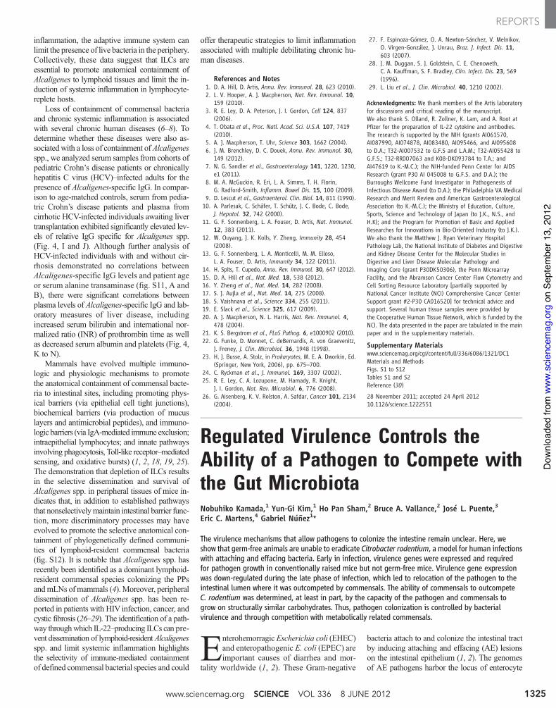
effacement (LEE) that is critical for these bacteriato colonize their hosts and cause pathology (3, 4).Infection with Citrobacter rodentium—a naturalpathogen of mice that is used to model humaninfections with EPEC and EHEC (5, 6)—is asso-ciated with a significant but reversible decreasein the number of total commensals in the colon(7, 8). To assess the role of the microbiota in thisenteric infection, germ-free (GF) and specificpathogen–free (SPF) C57BL/6 mice were orallyinoculated with C. rodentium. The pathogen col-onized the intestines of SPFmice, reachingmaxi-mal concentrations in the feces on days 7 to 10post infection, followed by a decline by day 12,and becoming undetectable by day 22 (Fig. 1A).In contrast, GF mice harbored 10 times as muchC. rodentium on days 7 to 10 post infection, andunlike SPF mice, they were unable to clearC. rodentium even by day 42 when the experi-ments were terminated (Fig. 1A). It was remark-able that all GF infected mice remained alivedespite high and persistent pathogen burdens(Fig. 1B). Notably, the recruitment of neutrophils,inflammatory macrophages, and CD3+ T cells inresponse to infection was similar in SPF and GFmice (fig. S1). Consistently, histological analysisrevealed comparable pathology scores on day 12post infection, which declined on day 22 in bothGF and SPF mice (fig. S1). Furthermore, expres-sion of antimicrobial peptides including RegIIIg,b-defensin-1, b-defensin-3, and b-defensin-4 wascomparable in the colons of infected SPF and GFmice (fig. S2).
The expression of most LEE genes in C.rodentium is controlled by Ler, a member of theH-NS protein family (3, 4, 9, 10). Ler was ex-pressed in the feces on day 7 post infection in bothGF and SPF mice (Fig. 2A). It was noteworthythat expression of ler and tir, a Ler-regulated gene,were both down-regulated on day 12 after in-fection in both GF and SPF mice and were notexpressed at day 42 in GF mice, despite robustpathogen colonization (Fig. 2A and fig. S3). Tomonitor the expression of ler in the intestine, weengineered a bioluminescent reporterC. rodentiumstrain in which the ler promoter was fused to theluxCDABE operon of Photorhabdus luminescens(fig. S4) (11). Ler expression was detected in thefeces on day 5, but down-regulated by days 7 to12 post infection (Fig. 2B). Furthermore, ler ex-pression was visualized on day 5 post infection inthe ileum, cecum, and distal colon but was down-regulated on day 14 in both SPF and GF mice(Fig. 2C). The ler luminescent signal in GF mice
was ~10 times that in SPF mice, consistent withhigher pathogen load inGFmice. If theC. rodentiumwere harvested from infected GF mice at day 21and reinoculated into SPF mice, the pathogenrobustly grew in the intestines and elicited co-lonic inflammation (fig. S5).
Because expression of virulence LEE genes isessential for pathogen colonization in the intes-tines of SPF mice (4), we asked whether LEEvirulencewas required for colonization inGFmice.To assess this, we orally infected GF and SPFmice with wild-type (WT) C. rodentium and iso-genic strains deficient in Ler (Dler) or in EscN(DescN), a Ler-regulated adenosine triphospha-tase required for injection of virulence factors viathe type III secretion system (T3SS) into hostcells (4). As expected, WT, but not the Dler orDescN strains, grew in the intestine of SPF mice(Fig. 2D). Notably, the Dler and DescN mutantsgrew robustly in GF mice, reaching numberssimilar to those of theWT pathogen (Fig. 2D andfig. S6). Thus, Ler-dependent LEE virulence isnot required for intestinalC. rodentium growth inthe absence of the microbiota. To determinewhether Ler and the T3SS regulate the ability ofthe microbiota to outcompete the pathogen, weorally infected GF mice with WT or the Dler orthe DescN mutant C. rodentium, and the infectedGF mice were colonized with commensals bycohousing them at either day 3 or day 21 postinfection with SPF mice. Notably, the burdenof WT C. rodentium in GF mice harboring thepathogen for 21 days declined significantly after3 days of cohousing and was further reduced by5 to 6 logs by day 7, similarly to the decline ob-served in mice infected with the Dler or theDescN mutant for 3 and 21 days (Fig. 2E andfig. S6). In contrast, the microbiota could notoutcompete the WT bacterium on day 3 postinfection when GFmice were cohoused with SPFmice (Fig. 2E), consistent with the observationthat ler expression is active in WT C. rodentiumat day 3 post infection (fig. S7).
C. rodentium induces marked inflammationin the distal colon, which requires LEE virulencefactors including the T3SS (4). If the T3SSpromotes outgrowth ofC. rodentium by enablingthe pathogen to colonize a niche at or near theepithelium, it would be expected that impairedcolonization of the DescN mutant could not berescued when inflammation is provided by othermeans. Oral administration of dextran sulfatesodium (DSS), a chemical that directly damagesthe colonic epithelium (12), enhanced the colo-nization of the WT bacterium by 3 logs butnot that of the DescN mutant (fig. S8). In addi-tion, coinfection of the DescN mutant with WTC. rodentium, which induces inflammation, didnot rescue colonization by the T3SSmutant strain(fig. S8). These results indicate that inflamma-tion promotes colonization of WT C. rodentiumnot in the absence of a functional T3SS. Theadaptive immune system—including immuno-globulin G (IgG) production and T helper 17responses against the pathogen—are important
for eradication of C. rodentium (13–16). It wasnoteworthy that the production of IgG, IgM, andIgA against C. rodentium was similar in GF andSPF mice (fig. S9). Furthermore, the generationof colonic interleukin-17 (IL-17)– and interferon-g–producing T cells in response to C. rodentiumwas comparable in SPF and GF mice (fig. S10).
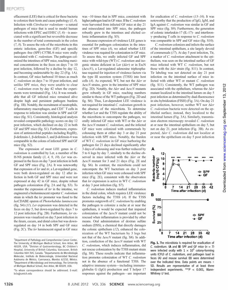
C. rodentium colonizes and infects the surfaceof the intestinal epithelium, a site largely devoidof commensals (17). At day 5 post infection, Tir,a marker of C. rodentium attachment to the epi-thelium, was seen on the intestinal surface of GFmice infected with WT C. rodentium, but notthose with the Dler strain (fig. S11). In contrast,Tir labeling was not detected on day 21 postinfection on the intestinal surface of mice in-fected with either WT or mutant C. rodentium(fig. S11). Consistently, the WT pathogen wasassociated with the epithelium, whereas the Dlermutant localized to the intestinal lumen on day 5post infection as determined by dual-fluorescencein situ hybridization (FISH) (Fig. 3A). On day 21post infection, however, neither WT nor DlerC. rodentium bacteria were detected on the ep-ithelial surface; instead, they localized to theintestinal lumen (Fig. 3A). Similarly, transmis-sion electron microscopy revealed C. rodentiumat or near the intestinal epithelium on day 5, butnot on day 21, post infection (Fig. 3B). As ex-pected, Dler C. rodentium did not localize ator near the epithelium on day 5 post infection
Fig. 1. The microbiota is required for eradication ofC. rodentium. (A and B) SPF and GF mice (n = 7)were infected orally with 1 ! 109 colony-formingunits (CFU) of C. rodentium, and pathogen load infeces (A) and mouse survival (B) were determinedover the indicated time. Data points are means TSD. Results are representative of at least threeindependent experiments. ***P < 0.001, Mann-Whitney U test.
1Department of Pathology and Comprehensive Cancer Center,The University of Michigan Medical School, Ann Arbor, MI48109, USA. 2Division of Gastroenterology, BC Children'sHospital, University of British Columbia, Vancouver, BritishColumbia V6H 3V4, Canada. 3Departamento de MicrobiologíaMolecular, Instituto de Biotecnología, Universidad NacionalAutónoma de México, Cuernavaca, Morelos 62210, México.4Department of Microbiology and Immunology, The Universityof Michigan Medical School, Ann Arbor, MI 48109, USA.
*To whom correspondence should be addressed. E-mail:[email protected]
8 JUNE 2012 VOL 336 SCIENCE www.sciencemag.org1326
REPORTS
on
Sept
embe
r 13,
201
2w
ww
.sci
ence
mag
.org
Dow
nloa
ded
from

(Fig. 3B). Consistent with these results, ler-lux–expressing WT C. rodentium found attached tothe cecal and colonic epithelium were abundantin the early phase of infection but dramaticallydecreased later during infection, even thoughpathogen burdens in the feces were comparable(Fig. 3C and fig. S12). These results indicate thatthe localization of the pathogen differs in the
early and late phase of infection, and this is con-trolled by LEE virulence gene expression.
The intestine harbors a large number of bac-terial species (18). To determine whether commen-sal bacteria exhibit different abilities to outcompeteC. rodentium, we orally infected GF mice withC. rodentium and on day 21 post infection themice were colonized with either E. coli or one of
two different anaerobic Bacteroides species, allisolated from the intestines of SPFmice (19). Theburden of C. rodentium in GF mice declined to~1/200th by day 3 and to less than 1/500th byday 14 upon colonization with E. coli, but not atall withB. thetaiotaomicron orB. vulgatus (Fig. 4A).Secondary administration of E. coli to GF micealready colonized with B. thetaiotaomicron or
Fig. 2. Expression and role of ler during C. rodentium infection in SPF and GFmice. (A) ler mRNA levels were determined by quantitative polymerase chainreaction in fecal pellets of SPF and GF mice infected with C. rodentium atthe indicated days post infection. Expression was normalized to that of thekanamycin-resistance gene carried by the C. rodentium strain. Control exper-iments were performed by determining lermRNA levels in C. rodentium grownunder inducing Dulbecco’s modified Eagle’s medium (DMEM) and repressingLuria broth (LB) in vitro culture conditions (4, 10). Data represent mRNAexpression relative to that in C. rodentium cultured in LB medium. Results aremeans T SD of individual mice (n = 3). Results are representative of at leasttwo experiments. *P < 0.05, ***P < 0.001. (B) Expression of ler in fecal pelletsof SPF and GF mice infected with the reporter ler-lux C. rodentium strain atthe indicated day post infection. Results show luminescence (relative lightunits) and CFU of ler-lux C. rodentium in the same samples. Data are
expressed as means T SD of individual mice (n = 4). Results are repre-sentative of at least two experiments. (C) Bioluminescent imaging of lerexpression in the intestines of SPF and GF mice infected with the ler-luxC. rodentium strain. Imaging was performed on day 5 and 14 post infection,and the signal was quantified on the basis of the color scale shown below.Results are representative of three individual mice. (D) SPF and GF mice (n =5) were infected orally with 109 CFU of WT and Dler mutant C. rodentium,and pathogen load in feces was determined over the indicated time. Datapoints are means T SD. Results are representative of at least two ex-periments. (E) GF mice were infected with WT and Dlermutant C. rodentium.At day 3 or day 21 post infection, mice were cohoused with SPF mice (1:1).Pathogen load was determined in feces on the indicated days after co-housing. Dots represent individual mice. Results are representative of at leastthree experiments.
www.sciencemag.org SCIENCE VOL 336 8 JUNE 2012 1327
REPORTS
on
Sept
embe
r 13,
201
2w
ww
.sci
ence
mag
.org
Dow
nloa
ded
from
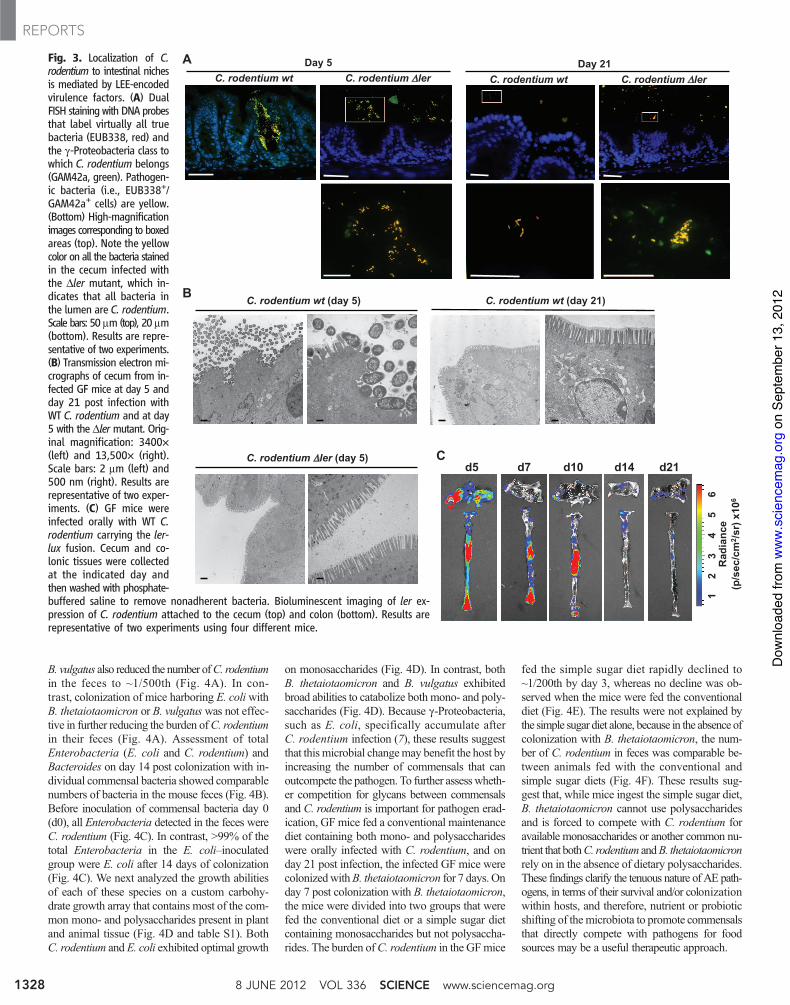
B. vulgatus also reduced the number ofC. rodentiumin the feces to ~1/500th (Fig. 4A). In con-trast, colonization of mice harboring E. coliwithB. thetaiotaomicron or B. vulgatuswas not effec-tive in further reducing the burden ofC. rodentiumin their feces (Fig. 4A). Assessment of totalEnterobacteria (E. coli and C. rodentium) andBacteroides on day 14 post colonization with in-dividual commensal bacteria showed comparablenumbers of bacteria in the mouse feces (Fig. 4B).Before inoculation of commensal bacteria day 0(d0), all Enterobacteria detected in the feces wereC. rodentium (Fig. 4C). In contrast, >99% of thetotal Enterobacteria in the E. coli–inoculatedgroup were E. coli after 14 days of colonization(Fig. 4C). We next analyzed the growth abilitiesof each of these species on a custom carbohy-drate growth array that contains most of the com-mon mono- and polysaccharides present in plantand animal tissue (Fig. 4D and table S1). BothC. rodentium and E. coli exhibited optimal growth
on monosaccharides (Fig. 4D). In contrast, bothB. thetaiotaomicron and B. vulgatus exhibitedbroad abilities to catabolize both mono- and poly-saccharides (Fig. 4D). Because g-Proteobacteria,such as E. coli, specifically accumulate afterC. rodentium infection (7), these results suggestthat this microbial changemay benefit the host byincreasing the number of commensals that canoutcompete the pathogen. To further assesswheth-er competition for glycans between commensalsand C. rodentium is important for pathogen erad-ication, GF mice fed a conventional maintenancediet containing both mono- and polysaccharideswere orally infected with C. rodentium, and onday 21 post infection, the infected GF mice werecolonizedwithB. thetaiotaomicron for 7 days. Onday 7 post colonization with B. thetaiotaomicron,the mice were divided into two groups that werefed the conventional diet or a simple sugar dietcontaining monosaccharides but not polysaccha-rides. The burden ofC. rodentium in the GFmice
fed the simple sugar diet rapidly declined to~1/200th by day 3, whereas no decline was ob-served when the mice were fed the conventionaldiet (Fig. 4E). The results were not explained bythe simple sugar diet alone, because in the absence ofcolonization with B. thetaiotaomicron, the num-ber of C. rodentium in feces was comparable be-tween animals fed with the conventional andsimple sugar diets (Fig. 4F). These results sug-gest that, while mice ingest the simple sugar diet,B. thetaiotaomicron cannot use polysaccharidesand is forced to compete with C. rodentium foravailable monosaccharides or another common nu-trient that bothC. rodentium andB. thetaiotaomicronrely on in the absence of dietary polysaccharides.These findings clarify the tenuous nature of AE path-ogens, in terms of their survival and/or colonizationwithin hosts, and therefore, nutrient or probioticshifting of themicrobiota to promote commensalsthat directly compete with pathogens for foodsources may be a useful therapeutic approach.
Fig. 3. Localization of C.rodentium to intestinal nichesis mediated by LEE-encodedvirulence factors. (A) DualFISH staining with DNA probesthat label virtually all truebacteria (EUB338, red) andthe g-Proteobacteria class towhich C. rodentium belongs(GAM42a, green). Pathogen-ic bacteria (i.e., EUB338+/GAM42a+ cells) are yellow.(Bottom) High-magnificationimages corresponding to boxedareas (top). Note the yellowcolor on all the bacteria stainedin the cecum infected withthe Dler mutant, which in-dicates that all bacteria inthe lumen are C. rodentium.Scale bars: 50 mm (top), 20 mm(bottom). Results are repre-sentative of two experiments.(B) Transmission electron mi-crographs of cecum from in-fected GF mice at day 5 andday 21 post infection withWT C. rodentium and at day5 with the Dlermutant. Orig-inal magnification: 3400!(left) and 13,500! (right).Scale bars: 2 mm (left) and500 nm (right). Results arerepresentative of two exper-iments. (C) GF mice wereinfected orally with WT C.rodentium carrying the ler-lux fusion. Cecum and co-lonic tissues were collectedat the indicated day andthen washed with phosphate-buffered saline to remove nonadherent bacteria. Bioluminescent imaging of ler ex-pression of C. rodentium attached to the cecum (top) and colon (bottom). Results arerepresentative of two experiments using four different mice.
8 JUNE 2012 VOL 336 SCIENCE www.sciencemag.org1328
REPORTS
on
Sept
embe
r 13,
201
2w
ww
.sci
ence
mag
.org
Dow
nloa
ded
from

References and Notes1. J. B. Kaper, J. P. Nataro, H. L. Mobley, Nat. Rev.
Microbiol. 2, 123 (2004).2. R. Mundy, T. T. MacDonald, G. Dougan, G. Frankel,
S. Wiles, Cell. Microbiol. 7, 1697 (2005).3. W. Deng, Y. Li, B. A. Vallance, B. B. Finlay, Infect. Immun.
69, 6323 (2001).4. W. Deng et al., Proc. Natl. Acad. Sci. U.S.A. 101, 3597
(2004).5. S. A. Luperchio et al., J. Clin. Microbiol. 38, 4343 (2000).6. D. Borenshtein, M. E. McBee, D. B. Schauer, Curr. Opin.
Gastroenterol. 24, 32 (2008).7. C. Lupp et al., Cell Host Microbe 2, 204 (2007).8. C. Hoffmann et al., Infect. Immun. 77, 4668 (2009).9. J. L. Mellies, S. J. Elliott, V. Sperandio, M. S. Donnenberg,
J. B. Kaper, Mol. Microbiol. 33, 296 (1999).10. J. Barba et al., J. Bacteriol. 187, 7918 (2005).11. J. Bjarnason, C. M. Southward, M. G. Surette, J. Bacteriol.
185, 4973 (2003).12. H. S. Cooper, S. N. Murthy, R. S. Shah, D. J. Sedergran,
Lab. Invest. 69, 238 (1993).
13. C. P. Simmons et al., Infect. Immun. 71, 5077(2003).
14. C. Maaser et al., Infect. Immun. 72, 3315 (2004).15. L. Bry, M. B. Brenner, J. Immunol. 172, 433 (2004).16. I. I. Ivanov et al., Cell 139, 485 (2009).17. K. S. Bergstrom et al., PLoS Pathog. 6, e1000902
(2010).18. J. Qin et al., Nature 464, 59 (2010).19. S. M. Bloom et al., Cell Host Microbe 9, 390 (2011).
Acknowledgments: The authors thank the University ofMichigan Germ-Free Animal Core, Microscopy and ImageAnalysis Laboratory, and the Center for Molecular Imaging forsupport; S. Koonse for animal husbandry; A. Huerta-Saquerofor constructing the pler-lux plasmid; N. Pudlo for anaerobicbacteria culture; T. Stappenbeck for mouse commensal strains;J. Rousseau for technical assistance; and M. H. Shaw andG. Chen for expert review of the manuscript. B.A.V. is theCanada Research Chair (Tier 2) in Pediatric Gastroenterologyand the CH.I.L.D. Foundation Chair in Pediatric InflammatoryBowel Disease Research. This work was supported by grants
from the NIH, grants DK61707 and DK091191 (G.N.);Consejo Nacional de Ciencia y Tecnología, CONACyT (J.L.P.);Canadian Institutes of Health Research (B.A.V.); and theUehara Memorial Foundation and Crohn’s and ColitisFoundation of America Fellowship Awards (N.K). N.K. andG.N. hold U.S. provisional patent application no. 61/616,707regarding inhibition of LEE virulence as a potential therapeuticfor infection with AE pathogens. The data reported in thispaper are tabulated in the main paper and in thesupplementary materials.
Supplementary Materialswww.sciencemag.org/cgi/content/full/science.1222195/DC1Materials and MethodsFigs. S1 to S12Table S1References (20–22)
18 November 2011; accepted 30 March 2012Published online 10 May 2012;10.1126/science.1222195
D A
B
2.0
C. roden
tium
E. coli
B. thet
aiota
omicr
on
B. vulg
atus
MSs
Animal PSs
Other PSs
Plant PSs
0
1.0
Commensal Commensal
C
E F Fig. 4. Similar catabolic preferences for saccharides may determine the competingability of commensal bacteria with the enteric pathogen. (A) GF mice were infectedwith WT C. rodentium (Cr). At day 21 post infection, E. coli (Ec) or B. thetaiotaomicron(Bt) or B. vulgatus (Bv) were inoculated. As a second inoculation, a mixture of Btand Bv was inoculated into the Ec-harboring group, and Ec was inoculated into theBacteroides-harboring group, respectively. Pathogen load was determined in feceson the indicated days after inoculation of commensal bacteria. Dots representindividual mice from two independent experiments. (B) Total Enterobacteria (Crand Ec) and Bacteroides culture in feces at day 0 and day 14 [first inoculation in(A)]. Data are means T SD (n = 4). Results are representative of at least twoexperiments. (C) Number of Cr (kanamycin resistant) in the total Enterobacteria(without antibiotics) are indicated as a percentage. Data are means T SD (n = 4).Results are representative of at least two experiments. (D) Carbohydrate catabolicprofiles of Cr and commensal bacteria strains. Robust growth under supplemen-
tation of monosaccharides (MSs) or polysaccharides (PSs) indicated as red, and no growth indicated as yellow. Raw data are provided in table S1. (E)GF mice were infected with WT Cr. At day 21 post infection, Bt was inoculated. On day 7 post colonization with Bt, the mice were divided into twogroups that were fed a conventional maintenance diet (C) or a simple sugar diet (ss). Pathogen load was determined in feces on day 3 after dietswitching. Results are means T SD of individual mice (initially n = 10, and divided into two groups n = 5 each). Results are representative of twoexperiments. (F) Cr monoassociated GF mice (day 21) were fed a simple sugar diet (ssDiet) for 7 days. Pathogen load was compared before and afterswitching to ssDiet from conventional diet (cDiet). Dots represent individual mice and are representative of three independent experiments **P < 0.01.N.S. denotes not significant.
www.sciencemag.org SCIENCE VOL 336 8 JUNE 2012 1329
REPORTS
on
Sept
embe
r 13,
201
2w
ww
.sci
ence
mag
.org
Dow
nloa
ded
from