regional ems guidelines - northcentralctems.orgnorthcentralctems.org/images/2014 nccems guidelines...
TRANSCRIPT
July 9, 2014
Regional
EMS
Guidelines

EMR EMT AEMT
Paramedic Medical Control June 25, 2012
This Guideline format is based on that of New Britain EMS. Many thanks to them
for allowing us to use it.
These Guidelines replace all previous versions, including those
dated July 1, 2013

EMR EMT AEMT
Paramedic Medical Control June 11, 2013 - -
1
Table of Contents
General Guidelines Communication
Universal Patient Care Algorithm
Adult Airway Guideline
Cardiac Guidelines Acute Coronary Syndrome
STEMI Destination Guideline
STEMI Alert Procedure
Routine Adult Cardiac Arrest Care
AED Guidelines
Guidelines for NonTrauma Cardiac Arrests
Bradycardia
Tachycardia
Induced Hypothermia for ROSC Post Cardiac Arrest
Return of Spontaneous Circulation/Post Resuscitation Care
Respiratory Guidelines Acute Pulmonary Edema
Continous Positive Airway Pressure (CPAP)
Complete Airway Obstruction
Respiratory Distress
Sedation to Manage Airway Post-Intubation
Medical Guidelines Routine Medical Care
Allergic Reaction
Anaphylaxis
Altered Level of Consciousness
Heat Related Emergencies
Near Drowning
Hypothermia
Hypothermic Arrest
Nausea/Vomiting
Overdose / Poisonings
Pain Management Adult
Seizures
Shock
Anxiety/Behavioral Emergencies
Dystonic Reaction
Stroke
Alcohol Withdrawal

EMR EMT AEMT
Paramedic Medical Control June 11, 2013 - -
2
Adult Trauma Guidelines – 13 Years Old Patient Triage Guideline
Management of the Trauma Patient
Burns
Spinal Precautions for Ambulatory Patients
Omitting Spinal Immobilization
OB/Gyn Guidelines Antepartum Hemorrhage
Pregnancy Induced Hypertension and Seizures
Emergency Childbirth
Delivery Complications
Nuchal Cord
Prolapsed Cord
Breech Birth
Extremity Presentation
Post Partum Care of Mother
Post Partum Care of the Infant
Neonatal Resuscitation
Trauma in Pregnancy
Pediatric Medical Guidelines Pediatric Patient Assessment
Pediatric Airway
General Guidelines for Pediatric Respiratory Distress
Pediatric Asthma
Suspected Croup or Epiglottitis
Pediatric Obstructed Airway
Pediatric Pain Management
Pediatric Allergic Reaction
Pediatric Anaphylaxis
Pediatric Fever
Pediatric Altered Mental Status / Hypoglycemia / Coma
Pediatric Seizures / Status Epilepticus
Pediatric Overdose/Poisoning
Pediatric Bradycardia
Pediatric Tachycardia (Adequate Perfusion)
Pediatric Tachycardia (Poor Perfusion)
Pediatric Pulseless Arrest
Pediatric Trauma Guidelines < 13 Years Pediatric Trauma Triage
Pediatric Burn Patient

EMR EMT AEMT
Paramedic Medical Control June 11, 2013 - -
3
Appendix
Procedures 12-Lead ECG
Cincinnati Prehospital Stroke Scale
Endotracheal Tube Inducer (Bougie)
Esophageal-Tracheal Combitube
Intranasal Nalaxone
Intraosseous Infusion
Morgan Lens
Needle Cricothyrotomy
Needle Thoracostomy
Pediatric Glasgow Coma Scale
Rule of Nines Adult
Rule of Nines Pediatric
Surgical Cricothyrotomy
Tube Confirmation Adjuncts
Capnography
Tourniquet
STEMI Alert Procedure
Medications Acetaminophen (Tylenol)
Activated Charcoal
Adenosine (Adenocard)
Albuterol (Ventolin, Proventil)
Amiodarone (Cordorone)
Aspirin
Atropine
Benzocaine Spray
Calcium Chloride
Dextrose
Diazepam (Valium)
Diltiazem (Cardizem)
Diphenhydramine (Benadryl)
Dopamine (Intropin)
Epinephrine 1:10,000
Epinephrine 1:1000
Fentanyl
Glucagon
Haloperidol (Haldol)
Ipratropium (Atrovent)
Lactated Ringers
Lidocaine

EMR EMT AEMT
Paramedic Medical Control June 11, 2013 - -
4
Lorazepam (Ativan)
Magnesium Sulfate
Methylprednisone (Solu-Medrol)
Metoclopramide Hydrochloride (Reglan)
Metoprolol (Lopressor)
Midazolam (Versed)
Morphine Sulfate
Naloxone (Narcan)
Nitroglycerin
Norepinephrine (Levophed)
Normal Saline
Olanzipine (Zyprexa)
Odansetron (Zofran)
Oxygen
Phenylephrine (Neo-Synephrine)
Procainamide (Pronestyl)
Racemic Epinephrine (Vaponephrine)
Sodium Bicarbonate
Tetracaine Ophthalmic Solution
Vasopressin (Pitressin)
Policies Documentation of Prehospital Patient Care
Transfer of Care from Paramedic to Basic Life Support
Discontinuation of Prehospital Resuscitation Interfacility Transport of Intubated Patients
EMS Response to Detention Facilities / Jails AHA 2010 AED Guidelines
Capitol Region Council of Governments RESP Plan
The Role of EMS in Hospital Diversions
Connecticut Diversion Guidelines
SMART Triage
Pain Control Policy
Mass Casualty Policy
Lights and Sirens
Guidelines for Non Trauma Cardiac Arrests

EMR EMT AEMT
Paramedic Medical Control June 11, 2013 - -
5
Communication Failure
IMPORTANT CAUTION
The information contained in these Guidelines is compiled from sources believed to be reliable and
significant efforts have been expended to make sure there are no inaccuracies. However, this cannot
be guaranteed. Despite our best efforts, there may be typographical errors or omissions. The North
Central CT EMS Council is not liable for any loss or damage that may result from these errors.
ON-LINE MEDICAL DIRECTION
It is agreed upon in the North Central Connecticut Regional Policy Manual that prehospital providers
will contact the receiving hospital regarding obtaining patient care orders. This agreement includes
all EMS providers.
COMMUNICATION FAILURE
In the event of complete communication failure, these Guidelines will act as the parameters for pre-
hospital patient care. If communication failure occurs, the paramedic may follow the guidelines to
render appropriate and timely emergency care to the patient.
Upon arrival at the receiving hospital, the EMS provider will immediately complete an incident
report relating to the communication failure describing the events including the patient’s condition
and treatment given. This incident report must be filed with the paramedic’s sponsor hospital EMS
Medical Director and/or EMS Coordinator within 24 hours of the event. A copy of the patient’s run
form will also accompany the incident report.
CMED Telephone Number for Telephone Patches
EMS Providers who wish to contact hospitals by phone may do so by contacting CMED at
(860) 769-6051 and request a phone patch.
Communications, Page 1 of 1 Last Updated: June 13, 2008

EMR EMT AEMT
Paramedic Medical Control June 11, 2013 - -
6
UNIVERSAL PATIENT CARE GUIDELINE
Scene Safety
BSI
Initial Assessment
Adult or Pediatric
C-Spine stabilization if indicated Cardiac Arrest?
Vital Signs
Including Temperature* and
Pain Severity **
Cardiac Arrest Guideline
Adult or Pediatric
Airway Guideline
Adult or Pediatric
Consider Pulse Oximetry
Consider Cardiac Monitor and
12 Lead EKG
Appropriate Guideline
If patient doesn’t fit a Guideline
Contact Medical Control
* Temperature and pain may be either quantitive (a specific reading) or qualitative (a description, hot,
cool, etc.)
**Pain severity should be recorded using a pain scale as outlined in the pain Guideline
PEARLS: - Any patient contact which does not result in an EMS transport must have a completed PCR.
- Exam: Minimal exam if not noted on the specific Guideline is vital signs, mental status, and location of injury or
complaint.
- Required vital signs on every patient include blood pressure, pulse, respirations, and pain/severity.
- Pulse oximetry and temperature documentation is dependent on the specific complaint.
- Timing of transport should be based on patient’s clinical condition.
Universal Patient Care Guideline, Page 1 of 1 Last Updated: June 13, 2008

EMR EMT AEMT
Paramedic Medical Control June 11, 2013 - -
7
Adult Airway Guideline
Adult Airway Guideline , Page 1 of 2 Last Updated: December 28, 2011

EMR EMT AEMT
Paramedic Medical Control June 11, 2013 - -
8
Pearls:
For this Guideline, adult is defined as 13 years old or greater.
Quantitative waveform capnography to measure CO2 is mandatory with all methods of
intubation. Document results.
Maintain c-spine immobilization for ETT placement for all intubated patients.
Do not assume hyperventilation is psychogenic –use oxygen, not a paper bag.
External Laryngeal Manipulation and/or the gum bougie or other devices as approved by
medical control, should be used to assist with difficult intubations.
Paramedics should consider using an alternative advanced airway when they are unable to
intubate a patient or in lieu of endotracheal intubation when it is appropriate.
Continuous pulse oximetry should be utilized in all patients with an inadequate respiratory
function.
No more than two attempts/visualizations at intubation should be performed. One
additional attempt may be made by one other paramedic if they are available on scene.
Paramedics should utilize auto-ventilators whenever possible.
Definition of a Failed Airway – An airway in which you can’t intubate and can’t ventilate.
Neosynephrine and Benzocaine Spray may be utilized to assist in nasotracheal intubation.
Adult Airway Guideline, Page 2 of 2 Last Updated: December 28, 2011

EMR EMT AEMT
Paramedic Medical Control June 11, 2013 - -
9
North Central Connecticut
Regional
Paramedic Guidelines
Cardiac Guidelines

EMR EMT AEMT
Paramedic Medical Control June 11, 2013 - -
10
Acute Coronary Syndromes / Chest Pain
Universal Patient Care Guideline
(Assessment of ABC’s)
Oxygen: Oxygen Therapy if patient in respiratory distress (or Sa02 is less
than 94%)
ASPIRIN: Aspirin 324 mg
(Baby ASA PO 325mg (81mgx4))
MONITOR: Cardiac Monitor Perforom12-Lead ECG (if 12-lead shows
STEMI, contact hospital as soon as possible for STEMI alert and
transport to appropriate primary PCI facility)
IV: Establish IV NS @ KVO
NITROGLYCERIN: Nitroglycerin (NTG) 0.4mg (1/150 gr.) sublingual or
NTG spray (1) metered dose if SB/P > 100 systolic NTG may be repeated q
5 minutes to a total of 3 doses, until symptom free or SB/P ≤100
EMT-B/I assists pt. with
prescribed Nitroglycerin (as
per dosing and limits to left)
If pain persists after 3rd
administered NTG
and SB/P remains >100 administer
Morphine Sulfate (MS) 2mg to 6mg SIVP in 2mg increments q 5
minutes titrated to discomfort/pain relief provided SB/P >100.
or
Fentanyl 1mcg/kg
to maximum single dose of 50mcg repeated up to three times q 5 min
ANTI-NAUSEA: Consider
Odansetron (Zofran) 4 mg Slow IV Over 2 - 5 Minutes or Deep IM or
Metoclopramide (Reglan) 10mg
2nd
IV: Consider establishing 2nd
IV in high-risk patients.
Acute Coronary Syndromes, Page 1 of 2 Last Updated: October 10, 2011

EMR EMT AEMT
Paramedic Medical Control June 11, 2013 - -
11
Acute Coronary Syndromes p. 2
Establish Medical Control
Possible Physician Orders:
Additional MS 2mg SIVP every five minutes (up to maximum dose of 0.1mg/kg) or Fentanyl
1mcg/kg to maximum single dose of 50mcg
titrated to discomfort/pain relief provided SB/P >100.
Additional sublingual Nitroglycerin (Additional of the patient’s own Nitroglycerin for EMT’s and
EMT-I’s)
Pearls:
Supplemental oxygen is not needed for patients without evidence of respiratory distress,
heart failure or shock if the oxyhemoglobin saturation is > 94%.
Avoid ASA administration in patients with hypersensitivity to ASA
Confirm that patient has not used erectile dysfunction meds in the past 48 hours due
to the potential for severe hypotension if Nitroglycerin administered.
If patient has taken Nitroglycerin without relief, consider potency of the medication.
If positive EKG changes, establish a second IV while en route to the hospital.
Monitor for hypotension after administration of Nitroglycerin and Morphine.
Diabetics and geriatric patients often have atypical pain, or only generalized complaints.
Paramedics should perform 12-lead prior to administration of NTG. If 12-lead shows
inferior STEMI, do not administer NTG prior to performing a right sided ECG. If
right side leads reveal possible right ventricular infarct, establish a large bore IV.
Giving NTG to patients with right ventricular infarction is contraindicated.
The use of nitrates in patients with hypotension (SBP <100 mm Hg or 30 mm Hg below
baseline), extreme bradycardia (<50 bpm), or tachycardia in the absence of heart failure
(>100 bpm) is also contraindicated.
If patient SB/P drops below 100, place patient supine, elevate legs and administer 250 cc
bolus of Normal Saline, and remove any NTG paste/patch.
Early transport and notification of the hospital are essential for patients suspected of ACS.
If patient is wearing a nitroglycerin patch remove it prior to administering sublingual
nitroglycerin.
Absence of an IV shall not preclude use of first NTG dose provided SB/P remains >100
If patient has taken Nitroglycerin without relief, consider potency of the medication
Morphine should be used with caution in patients with unstable angina and NSTEMI.
Monitor for hypotension after administration of Nitroglycerin and Morphine.
Diabetics and geriatric patients often have atypical pain, or only generalized complaints.
Early transport and notification of the hospital are essential for patients suspected of ACS.
Acute Coronary Syndromes, Page 2 of 2 Last Updated: October 10, 2011

EMR EMT AEMT
Paramedic Medical Control June 11, 2013 - -
12
STEMI Destination Guideline

EMR EMT AEMT
Paramedic Medical Control June 11, 2013 - -
13
STEMI ALERT PROCEDURE 1. Acquire a 12-lead on all patients suspected of Acute Coronary Syndrome (active chest pain or equivalent symptoms (SOB, nausea, etc.) on first contact. 2. If 12-lead is diagnostic for STEMI and paramedic believes patient is having STEMI, contact CMED for STEMI Alert with Medical Control patch, and transmit ECG if possible. If possible and less than 30 minutes from PCI center, do not wait until transporting to call hospital. Failure to notify hospital until 5 minutes out will delay reperfusion. 3. When hospital answers phone, confirm MD Control, and state “I have a STEMI Alert and am requesting STEMI activation.” If you are uncertain the patient is having a STEMI, say “I have a Possible STEMI Alert.” 4. Describe 12-lead and patient condition. Based on the conversation between paramedic and ED MD and if applicable, the transmitted 12-Lead, the cath lab will either be activated in advance of arrival, placed on standby or not activated until the physician can make a more detailed assessment at the hospital. 5. Provide Appropriate Care during transport per guideline. Have defib pads ready in case patient goes into unstable ventricular tachycardia or ventricular fibrillation. Consider disrobing patient if time permits. Have latest 12-lead ready to show ED MD on arrival. Be prepared to transport patient to cardiac cath lab on EMS stretcher if given the go- ahead from ED staff. 6. Please leave copy of PCR and all 12-lead strips at the hospital prior to departing. PCRs should include Time of 911 Dispatch, Time at Patient side, Time of 1st 12-lead, Arrival at the Hospital, as well as all care rendered. 7. If applicable to hospital, fill out QA/Patient Follow-up form in ED. NOTES: STEMI Definition for Field Activation STEMI is defined by ECG of good quality with all of the following: a. ST elevation in 2 or more contiguous leads of >2 mm (V1-V4 or > 1 mm (limb or lateral) b. QRS duration < 0.12 second c. ***Acute MI*** or equivalent prints on 12-lead and paramedic agrees. If the machine does not read ***Acute MI*** but the paramedic still strongly believes the ECG shows a STEMI, the paramedic may proceed with the activation request. STEMI Alert, Page 1 of 2 Last Updated: June 12, 2013

EMR EMT AEMT
Paramedic Medical Control June 11, 2013 - -
14
Computer Interpretation Paramedics should not diagnose STEMI based solely on 12-lead computer interpretation. While the interpretation can by used to support your diagnosis, the computer is not infallible. The computer will not read all STEMIs as ***Acute MI Suspected***.” And the computer may read ***Acute MI*** when the ECG is clearly not a STEMI. The computer is less accurate with wide complex and tachycardic rhythms. STEMI Imposters Paramedics should be familiar with and take into consideration all of the known STEMI Imposters, including Bundle Branch Block Paced Rhythms Early Repolarization Pericarditis Left Ventricular Hypertrophy (LVH) Other PEARLS Do serial 12-leads (ideally all patients with ACS should have 12-lead on patient contact, on beginning transportation and on arrival at ED). STEMIs are often evolving. The STEMI may not appear until 3rd 12-lead or the STEMI captured on 1st 2-lead may disappear by arrival at the ED. A prehospital 12-lead documenting the transient elevation is critical in these patients. Regional PCI Hospitals Hartford Hospital John Dempsey Hospital New Britain Saint Francis Baystate Memorial Hospital (Springfield) Early Notification Saves Lives!
STEMI Alert, Page 2 of 2 Last Updated: June 12, 2013

EMR EMT AEMT
Paramedic Medical Control June 11, 2013 - -
15
Routine Adult Cardiac Arrest Universal Patient Care Guideline (Primary Assessment, think C-A-B)
Request Paramedic Intercept
Cardiac in Origin? Initiate CCR (200 chest compressions, at least 100/min, 2 inches in depth with full chest recoil, place
patient on Non-rebreather mask at 15lpm of oxygen)
Analyze rhythm, give one shock (maximum energy), no pulse check
Continue CCR
(200 chest compressions, at least 100/min, 2 inches in depth with full chest recoil)
Analyze rhythm, give one shock (maximum energy), no pulse check
Continue CCR
(200 chest compressions, at least 100/min, 2 inches in depth with full chest recoil)
Analyze rhythm, give one shock (maximum energy), no pulse check
Continue CCR
(200 chest compressions, at least 100/min, 2 inches in depth with full chest recoil)
Start Here for arrests that are NOT cardiac in origin: Resume Standard
ACLS, 30 Compressions/ 2 breaths
Consider Intubation (Do not interrupt compressions to do so)
Utilize Semi-Automatic (or
Automatic) External
Defibrillator with Adult size
defibrillation patches. If
“Shock” advised administer
shock x 1 in accordance to
specific equipment energy
waveform and manufacturer
recommendations
Attach Monitor & quantitative waveform capnography
If Arrest Rhythm, continue down guideline
EMS Providers should provide aggressive resuscitation on scene for at least 20 minutes prior
to consideration of transport or termination. Consider if transportation will have a benefit for
patient care.
CPR x 2 minute if indicated
IV/IO Access – 200mL Bolus
Routine Adult Cardiac Arrest, Page 1 of 5 Last Updated: April 6, 2014

EMR EMT AEMT
Paramedic Medical Control June 11, 2013 - -
16
Reanalyze patient rhythm “Shock” x 1 if indicated
Epinephrine 1:10,000 1mg, IVP repeated q 3-5 minutes
or Vasopressin (Pitressin), 40 units IVP as the first or second dose of Epinephrine
CPR x 2 minute if indicated
Reanalyze patient rhythm “Shock” x 1 if indicated
Amiodarone (Cordorone) for VF/VT 300 mg IVP, repeat once in 10 min at 150 mg IVP
Continuing defibrillation as directed by AED
Establish Medical Control Potential Orders for EMT and EMT-I
Paramedic Intercept, Additional IV Line Additional Fluid Bolus, Additional Shocks
If ROSC at anytime,
see ROSC Guideline. If no ROSC, consider
termination of efforts
according to guideline
Routine Adult Cardiac Arrest, Page 2 of 5 Last Updated: April 6, 2014

EMR EMT AEMT
Paramedic Medical Control June 11, 2013 - -
17
CARDIOCEREBRAL RESUSCITATION
Utilize for all arrests of suspected cardiac origin. Do not use for non-cardiac related arrests such as
traumatic arrest, drowning or respiratory arrest.
Start Immediate compressions, unless effective bystander CPR already in place.
Charge defibrillator during CPR (15 seconds before rhythm analysis).
Insert oral airway, apply 02 NRB at 15 lpm.
Switch compressors every 200 compressions. If one compressor is providing significantly stronger
CPR than another, stick with best compressor until he or she shows signs of fatigue.
Consider using CPR feedback device such as metronome or monitor.
Delay application of mechanical device until 5th
cycle of CPR unless it can be reliably applied with
less in less than 10 seconds, without delay in compressions.
Routine Adult Cardiac Arrest, Page 3 of 5 Last Updated: April 6, 2014

EMR EMT AEMT
Paramedic Medical Control June 11, 2013 - -
18
Give 1 mg Epi 1:10,000 every 5 minutes once IV in place.
Give 300 mg Amiodarone after first epi if shockable rhythm is present. Amiodarone may be repeated
at 150 mg after 3-5 minutes.
Delay advanced airway during first 8-10 minutes of CCR. Never interrupt compressions to place
ETI. Consider alternative airways such as King LT, LMA, Combi-tube .
Provide resuscitation on scene for at least 20 minutes provided conditions are safe.
If BLS is on scene first, paramedics should resume CCR on their arrival, taking into account amount
of time BLS has spent on CCR.
Once advanced airway is in place, ventilate at 8 breaths a minute. Be careful not to hyperventilate.
Do not provide over 600 ml per ventilation. Consider using pediatric BVM.
Cardiac arrests with ROSC should be brought to closest hospital unless post ROSC 12-lead shows
STEMI, in which case patient should be brought to PCI center.
Pearls:
Compressions - Start chest compressions for any unresponsive adult victim with no breathing or no normal breathing (ie,
only gasps). Initiate chest compressions before giving rescue breaths (C-A-B rather than A-B-C). Push hard (>2 inches)
and fast (> 100/min) and allow complete chest recoil. Minimize Interruptions. Consider rotating compressors every two
minutes. If no advanced airway in place, 30:2 compression-ventilation ratio
For biphasic defibrillators, follow manufacturer recommendations regarding defibrillation energy settings. If
recommendations are unknown for a biphasic unit, utilize 200 joules for all defibrillations. For monophasic units, deliver
all defibrillations at 360 joules.
Advanced Airways - Advanced airways can be delayed if the patient can be effectively ventilated by bag- valve mask.
The gold standard is not an ET, but an airway that can be effectively maintained. Minimize interruptions in CPR to
secure an airway.
Ventilation - Deliver each rescue breath over 1 second. Give a sufficient tidal volume to produce visible chest rise.
Once advanced airway is in place, (ET, Combi-tube, LMA, or King-LT) maintain continuous compressions. Ventilate
with 600 ml of an adult ambu bag. 8-10 a minute. Utilize waveform capnography.
IV/IO access is the preferred route over ET for medications, with IV being preferred, If it is not readily available then use
IO as back-up vascular access device.
Meds which can be given through the ET tube:
o Epinephrine dose should be 2-2.5mg 1:1000 diluted in 10cc NS.
o Atropine dose should be 2 mg diluted in 10cc NS.
o Vasopressin (Pitressin) dose should be 80 units
Continue Quality CPR

EMR EMT AEMT
Paramedic Medical Control June 11, 2013 - -
19
Treat Reversible Causes
Sodium bicarbonate 1meq/kg IV may be given for tricyclic overdoses, known pre-existing hyperkalemia, and
acidosis.
If hypovolemia is suspected, administer 500cc bolus NS.
If pneumothorax, perform needle decompression.
If hypoglycemia, administer Dextrose IV.
If hypothermic, follow hypothermia Guideline.
In the setting of cardiac arrest and history of renal failure or dialysis, give calcium chloride
1 g IV over 1 minute.
Capnography - Utilize waveform quantitative waveform capnography to confirm and monitor
ET placement. Quantitative waveform capnography should be used for all advanced airways.
Observe ETCO2 readings during CPR to monitor quality of CPR. Sudden rise in ETCO2 may
indicate return of spontaneous circulation (ROSC).
CPR Devices - CPR Devices should only be used with the approval of the service’s medical
control. At no time should the deployment of the device delay or disrupt quality CPR. Services
who utilize such devices are expected to train often in use of the device to ensure smooth and
rapid deployment. Delay application of mechanical device until 5th
cycle of CPR unless it can be
reliably applied with less in less than 10 seconds, without delay in compressions
Epinephrine Infusion for Cardiac Arrest
o Paramedics may consider giving patients in cardiac arrest an epinephrine infusion at 1
mg/5 min in place of additional epi boluses, following first epi 1 mg bolus.
o Drip should be shut off if ROSC is achieved.
o Sample way to mix drip
Mix 5 mg Epi 1:1000 in 250 ml bag
Give 1 mg every 5 minutes (50 cc every 5 minutes) which is 0.2 mg Epi every
minute (10 cc/min)
Norepinephrine is added as an alternative vasopressor to dopamine effective July 1, 2014. If a
service carries both vasopressors, norepinephrine will be the preferred vasopressor for
cardiogenic shock and hypotension post ROSC. (see attached drug sheet)
o Dose: 1-30 mcg/min titrated to blood pressure of 90 mmHg.
o Pediatric Dose: infusion 0.1 – 2micrograms/kg/min titrated to effect
Routine Adult Cardiac Arrest, Page 5 of 5 Last Updated: April 6, 2014

EMR EMT AEMT
Paramedic Medical Control June 11, 2013 - -
20

EMR EMT AEMT
Paramedic Medical Control June 11, 2013 - -
21
North Central EMS Regional Medical Advisory Committee
Position on Nontraumatic Cardiac Arrest Scene Care and Transportation
“Cardiac compressions are less effective during ambulance transport than they are on scene.
The 2010 American Heart Association (AHA) guidelines emphasize the importance of high
quality, minimally interrupted compressions. Thus, TOR (Termination of Resuscitation)
protocols that emphasize on-scene resuscitation may not only mitigate the risk of an ambulance
crash, but also improve the probability of successful resuscitation by avoiding interruptions in
compressions.” - NATIONAL ASSOCIATION OF EMS PHYSICIANS (2011)
While recognizing that each cardiac arrest scene has its own unique circumstances, as a general
guideline, patients in nontraumatic cardiac arrest should receive full resuscitative efforts on scene.
Moving a patient to the ambulance to start ALS resuscitation, to get to the hospital quicker or to meet a
paramedic intercept may be counterproductive by lowering the quality of compressions in the critical
early period of resuscitation. Any interruption in quality cardiac compressions decreases a patient’s
chance of survival.
In general, patients should receive at least 20 minutes of resuscitative efforts on scene prior to
considering movement. If a patient remains in asystole after twenty minutes of paramedic effort,
termination of resuscitation guidelines should be considered. If the decision is made (at any time) to
move the patient, care must be maintained to ensure that quality compressions are maintained
throughout extrication and transportation. Failure to do so mitigates the patient’s chances for survival.
Unless there are special circumstances, it is unlikely that a patient who cannot be resuscitated on scene
with quality CPR and defibrillation will be resuscitated either at a paramedic intercept point or at the
hospital.
Please refer to State Termination of Resuscitation Guidelines.
Routine Adult Cardiac Arrest, Page 4 of 4 Last Updated: June 11, 2013

EMR EMT AEMT
Paramedic Medical Control June 11, 2013 - -
22
North Central Connecticut EMS Council
AED Policy regarding 2010 AHA Guidelines
In the current CPR guidelines by the American Heart Association (AHA), there is a recommendation
to change the shock sequence in existing AEDs. The AHA also states that local medical directors
may consider allowing for two minutes of CPR prior to defibrillation of patients who present in either
un-witnessed cardiac arrest or an arrest with a down time of greater than 3 – 4 minutes.
Effective January 1, 2007 the North Central Connecticut EMS Council will require the following:
1. Existing AEDs may continue to be used, including those that administer 3 successive
shocks.
2. Services were expected to upgrade their AEDs to administer single shocks within the next
year, before January 1, 2008.
3. If you are given programming options for your AED, it should be to analyze and shock
once it is turned on. Please note that this is consistent with current (Guidelines 2010)
AHA teachings. Shock energy levels should be in accordance with manufacturer
recommendations.
4. When more than one rescuer is present: Upon arrival at a cardiac arrest CPR should be
started immediately and continued until the AED pads are in place and the machine is
ready to analyze. The AED should be placed on the patient as soon as it is available,
regardless of downtime or if the arrest was witnessed or un-witnessed. In cases in which a
defibrillator is not immediately available, CPR should be done until such time as a
defibrillator is available.
5. When there is only a single rescuer present: Unwitnessed arrests should have the AED
placed on the patient if no other help has arrived. (If additional help arrives they should
place the AED on the patient as soon as they arrive). Witnessed arrests should have the
AED placed immediately.
AED Policy, Page 1 of 1 Last Updated: December 28, 2011

EMR EMT AEMT
Paramedic Medical Control June 11, 2013 - -
23
Bradycardia
Universal Patient Care
Protocol
IV Access
Bradycardia, determine
either absolute (<50 bpm)
or relative
NO Serious Signs or
Symptoms? YES
Observe and
Transport Establish Medical
Control
Intervention sequence
Atropine 0.5 mg IV, may repeat in 0.5 mg
increments, up to 3 mg total
Transcutaneous Pacing (use first in 3rd
degree HB)
Dopamine (Intropin) 2-10 μg/kg/min IV drip
Epinephrine 2-10 μg/min IV drip
If successfully employing Transcutaneous
Pacing, follow Pain Management guideline to
maintain patient comfort.
PEARLS:
If patient has chronic renal failure contact Medical Control for a possible order of:
Calcium Chloride 1g IV and/or Sodium Bicarbonate 1 mEq/kg IV.
Atropine do not rely on atropine for 3rd
degree heart block, wide complex ventricular escape
beats, or Mobitz type II heart block or in patients with third-degree av block with a new wide
QRS complex.
Atropine should be used with caution in cases of suspected acute myocardial infarction.
Heart transplant patients will not respond to atropine.
Bradycardia, Page 1 of 1 Last Updated: October 25, 2011
EMR EMT AEMT
Paramedic Medical Control June 11, 2013 - -
24
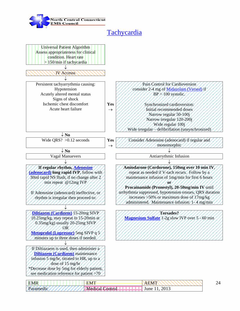
Tachycardia
Universal Patient Algorithm
Assess appropriateness for clinical
condition. Heart rate > 150/min if tachycardia
IV Accesss
Persistent tachyarrythmia causing:
Hypotension Acutely altered mental status
Signs of shock Ischemic chest discomfort
Acute heart failure Yes
Pain Control for Cardioversion consider 2-4 mg of Midazolam (Versed) if
BP > 100 systolic.
Synchronized cardioversion: Initial recommended doses
Narrow regular 50-100j Narrow irregular 120-200j
Wide regular 100j Wide irregular – defibrillation (unsynchronized)
No Wide QRS? >0.12 seconds Yes
Consider Adenosine (adenocard) if regular and
monomorphic
No Vagal Manuevers Antiarrythmic Infusion
If regular rhythm, Adenosine
(adenocard) 6mg rapid IVP, follow with
30ml rapid NS flush, if no change after 2
min repeat @12mg IVP
If Adenosine (adenocard) ineffective, or
rhythm is irregular then proceed to:
Amiodarone (Cordorone), 150mg over 10 min IV,
repeat as needed if V-tach recurs. Follow by a
maintenance infusion of 1mg/min for first 6 hours or
Procainamide (Pronestyl), 20-50mg/min IV until
arrhythmia suppressed, hypotension ensues, QRS duration
increases >50% or maximum dose of 17mg/kg
administered. Maintenance infusion: 1- 4 mg/min
Diltiazem (Cardizem) 15-20mg SIVP
(0.25mg/kg, may repeat in 15-20min at
0.35mg/kg) usually 20-25mg SIVP OR
Metoprolol (Lopressor) 5mg SIVP q 5
minutes up to three doses if needed.
Torsades? Magnesium Sulfate 1-2g slow IVP over 5 - 60 min
If Diltiazaem is used, then administer a
Diltiazem (Cardizem) maintenance
infusion 5 mg/hr, titrated to HR, up to a
dose of 15 mg/hr *Decrease dose by 5mg for elderly patient,
see medication reference for patient >70

EMR EMT AEMT
Paramedic Medical Control June 11, 2013 - -
25
Tachycardia, Page 1 of 2 Last Updated: October 25, 2011
EMR EMT AEMT
Paramedic Medical Control June 11, 2013 - -
26
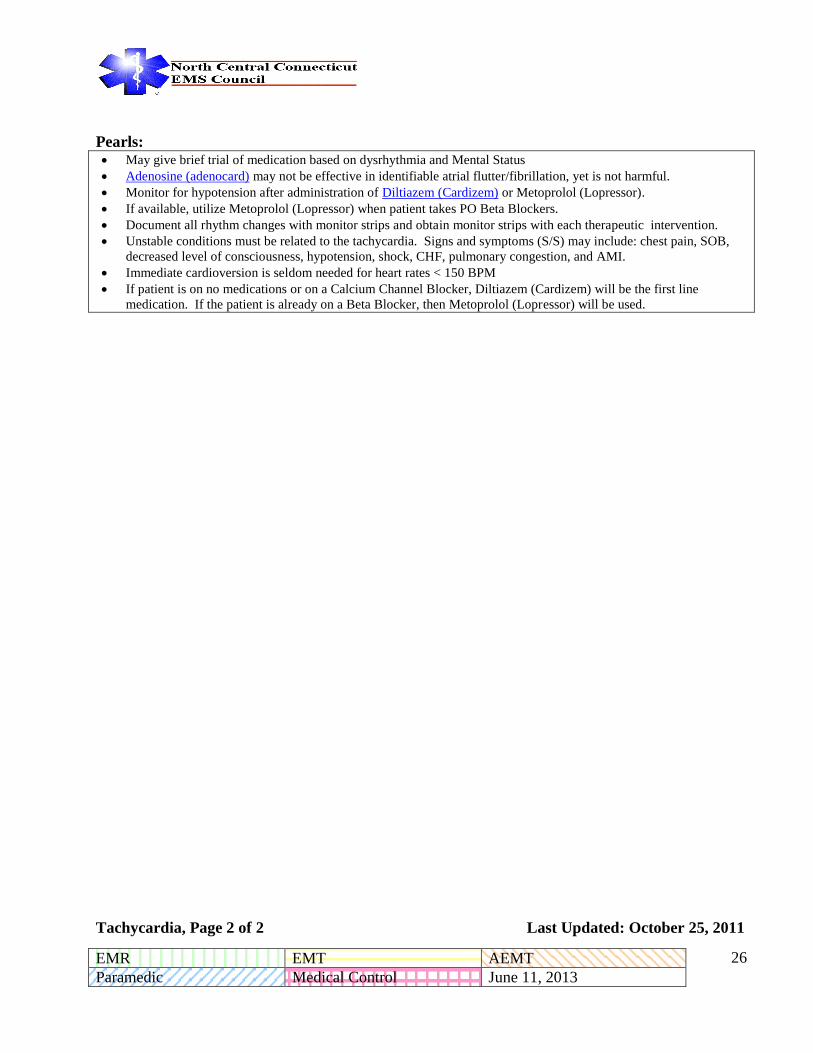
Pearls: May give brief trial of medication based on dysrhythmia and Mental Status
Adenosine (adenocard) may not be effective in identifiable atrial flutter/fibrillation, yet is not harmful.
Monitor for hypotension after administration of Diltiazem (Cardizem) or Metoprolol (Lopressor).
If available, utilize Metoprolol (Lopressor) when patient takes PO Beta Blockers.
Document all rhythm changes with monitor strips and obtain monitor strips with each therapeutic intervention.
Unstable conditions must be related to the tachycardia. Signs and symptoms (S/S) may include: chest pain, SOB,
decreased level of consciousness, hypotension, shock, CHF, pulmonary congestion, and AMI.
Immediate cardioversion is seldom needed for heart rates < 150 BPM
If patient is on no medications or on a Calcium Channel Blocker, Diltiazem (Cardizem) will be the first line
medication. If the patient is already on a Beta Blocker, then Metoprolol (Lopressor) will be used.
Tachycardia, Page 2 of 2 Last Updated: October 25, 2011

EMR EMT AEMT
Paramedic Medical Control June 11, 2013 - -
27
Induced Hypothermia for ROSC, Post Cardiac Arrest
Universal Patient Care Guideline
Patient is Post-Cardiac Arrest? Currently has a pulse and ROSC? No
Yes Go to Appropriate
Cardiac Arrest
Guideline
ROSC after cardiac arrest not related to trauma or hemorrhage.
Age greater than 18
Without obvious gravid uterus
No signs of hypothermia, (Initial temperature > 34 C
(93.2 F))
Patient is intubated and remains comatose (no purposeful response to pain)
If no, go to Routine
ALS Care
Advanced Airway Placed (ET preferred) and ETCO2 reading
> 20 mmHg NO
Yes Go to Adult Airway
Guideline, if successful
Perform and Record Neuro Exam
Induce Hypothermia: Expose patient; Apply Ice Packs to Axilla &
Groin, Midazolam (Versed) 2-5 mg (0.1 mg/kg) slow IV push if
systolic pressure is over 90 mmHG,
If available cold saline bolus 30mL/kg to max of 2 liters,
◄─ ───────┘
If SBP below 90mmHg, administer Dopamine (Intropin) 5-
20mcg/kg/min IV titrate to SBP of 90mmHg
Patient MUST be transported to a receiving facility capable of continuing induced therapeutic hypothermia
Induced Hypothermia for ROSC, Page 1 of 2 Last Updated: October 10, 2011
EMR EMT AEMT
Paramedic Medical Control June 11, 2013 - -
28
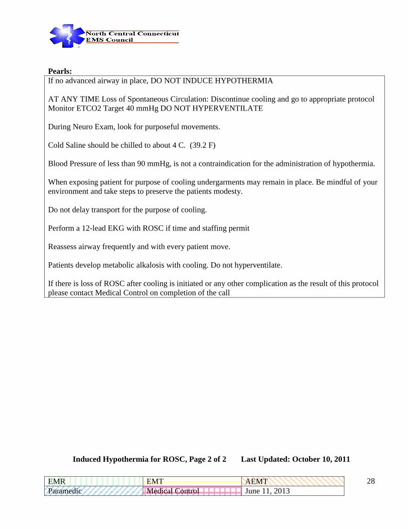
Pearls:
If no advanced airway in place, DO NOT INDUCE HYPOTHERMIA
AT ANY TIME Loss of Spontaneous Circulation: Discontinue cooling and go to appropriate protocol
Monitor ETCO2 Target 40 mmHg DO NOT HYPERVENTILATE
During Neuro Exam, look for purposeful movements.
Cold Saline should be chilled to about 4 C. (39.2 F)
Blood Pressure of less than 90 mmHg, is not a contraindication for the administration of hypothermia.
When exposing patient for purpose of cooling undergarments may remain in place. Be mindful of your
environment and take steps to preserve the patients modesty.
Do not delay transport for the purpose of cooling.
Perform a 12-lead EKG with ROSC if time and staffing permit
Reassess airway frequently and with every patient move.
Patients develop metabolic alkalosis with cooling. Do not hyperventilate.
If there is loss of ROSC after cooling is initiated or any other complication as the result of this protocol
please contact Medical Control on completion of the call
Induced Hypothermia for ROSC, Page 2 of 2 Last Updated: October 10, 2011

EMR EMT AEMT
Paramedic Medical Control June 11, 2013 - -
29
Return of Spontaneous Circulation (ROSC) / Post
Resuscitation Care
Return of Spontaneus Circulation
(ROSC)
Optimized ventilation and oxygenation Maintain oxygen saturation to >94%
If not already in place, consider advanced
airway and quantitative waveform
capnography Do not hyperventilate
Treat hypotension (systolic BP <90
mmHg) IV/IO fluid bolus (1 – 2 L of LR or NS) Consider treatable causes of the arrest
12-lead EKG
Consider vaspressor infusion (after fluid bolus)
Follow Induced Hypothermia Guideline
if appropriate Dopamine (Intropin) 5 – 10 mcg/kg/min or
Norepinephrine (Levophed) 1-30 mcg/min titrated to
blood pressure of 90 mmHg
Return of Spontaneous Circulation, p. 1 of 1 Last Updated: April 4, 2014

EMR EMT AEMT
Paramedic Medical Control June 11, 2013 - -
30
North Central Connecticut
Regional
Paramedic Guidelines
Respiratory Guidelines

EMR EMT AEMT
Paramedic Medical Control June 11, 2013 - -
31
ACUTE PULMONARY EDEMA
Universal Patient Care Algorithm
Oxygen Therapy (90% - 100%)
Cardiac Monitor
12 Lead EKG
Consider CPAP if available (See CPAP guideline) →
Lorazepam (Ativan) 0.5mg up to a max of 1mg may be
given on standing order for patient with extreme anxiety if
the medic judges that lessening their anxiety will enable
them to better tolerate CPAP.
IV Normal Saline KVO
If SBP >100 mmHg, Nitroglycerin 0.4 – 0.8 mg SL*
Or
if Systolic BP is >150mmHG then Nitroglycerin
Paste 1.5 inches, if >200 then 2 inches
Repeat q 3 – 5 minutes prn
*absence of an IV shall not preclude the use of first
NTG dose provided that the SBP is >100 mmHg. If
patients with Nitroglycerin paste become hypotensive,
remove paste.
→
If SBP < 100 mmHg, or if erectile dysfunction
Medication use within the last 48 hours
→ → Consult Medical Control
PEARLS
Sublingual dose of NTG can be either metered spray dose or tablet that dissolves.
Judgment can be used with patients who are marginally hypotensive and CPAP may be used before
Nitroglycerin
CHF can at times be confused with sepsis and pneumonia. Use great care with NTG if diagnosis is unclear and
the patient does not seem to be improving with the NTG.
Acute Pulmonary Edema, Page 1 of 1 Last Updated: June 11, 2013

EMR EMT AEMT
Paramedic Medical Control June 11, 2013 - -
32
CONTINUOUS POSITIVE AIRWAY PRESSURE (CPAP)
CPAP, Page 1 of 1 Last Updated April 6, 2014
Patient has a condition contraindicating CPAP use?
Respiratory Rate ≤10 breaths/minute
Confusion: Inability to understand and cooperate with application of CPAP
History of pneumothorax or recent tracheo-bronchial surgery
Active nausea or vomiting despite anti-emetic therapy
Severe Respiratory distress?
Accessory muscle use?
Hypoxemia despite oxygen therapy?
Marked work of breathing?
Inability to speak in full sentences?
Believed to be primarily
Pulmonary Edema?
Apply CPAP at 7.5-10 cm H2O.
If possible, adjust FiO2 to maintain
Oxygen Saturation >94%.
Believed to be Other
Respiratory Distress?
Apply CPAP at 2.5-5 cm H2O. If
possible, adjust FiO2 to maintain
Oxygen Saturation >94%.
Continue Reassessment. Titrate pressure setting based on patient
response. Do not exceed 15 cm H2O pressure. If patient shows evidence
of deterioration, discontinue CPAP. Consider BVM assist and possible
intubation. If patient vomits, immediately discontinue CPAP.
If equipment allows, continue indicated nebulized bronchodilators in-line with CPAP. It is allowable to briefly
interrupt CPAP to continue administration of ‘spray’ formulations of nitrates (if indicated)
Reduce the risk of increasing their retention of CO2 or causing a pneumothorax.
Notify receiving hospital early to allow preparations for continued CPAP/BiPAP.

EMR EMT AEMT
Paramedic Medical Control June 11, 2013 - -
33
COMPLETE AIRWAY OBSTRUCTION
Conscious Unconscious
Assess to determine airway obstruction Assess to determine unresponsiveness
Perform Heimlich Maneuver for conscious
patient Attempt to establish airway to determine airway
obstruction
Continue Heimlich Maneuver until airway
is cleared or patient becomes unconscious Perform Heimlich Maneuver for unconscious patient
If airway is still obstructed perform direct laryngoscopy
and remove any foreign body using Magill Forceps
If airway is still obstructed, attempt endotracheal
intubation
If airway is still obstructed, consider Transtracheal
Ventilation or Surgical Airway
Establish Medical Control
Complete Airway Obstruction, Page 1 of 1 Last Updated: June 13, 2008
EMR EMT AEMT
Paramedic Medical Control June 11, 2013 - -
34
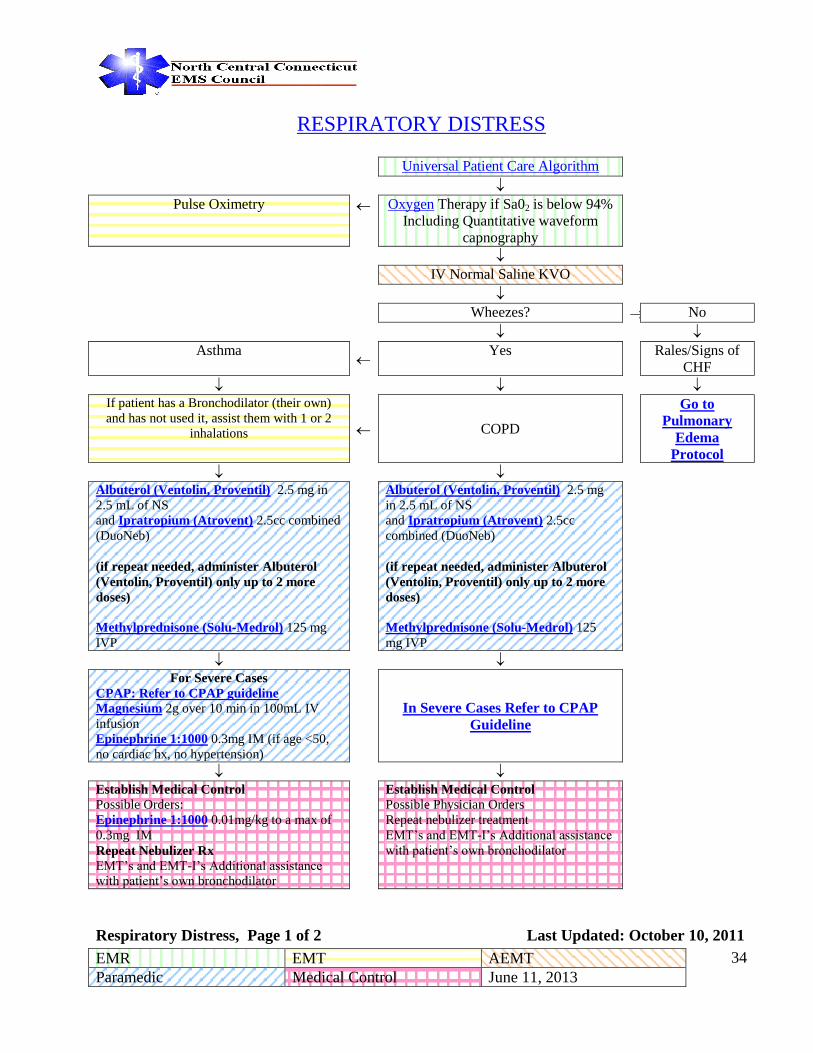
RESPIRATORY DISTRESS
Universal Patient Care Algorithm
Pulse Oximetry Oxygen Therapy if Sa02 is below 94%
Including Quantitative waveform
capnography
IV Normal Saline KVO
Wheezes? No
Asthma
Yes Rales/Signs of
CHF
If patient has a Bronchodilator (their own)
and has not used it, assist them with 1 or 2
inhalations COPD
Go to
Pulmonary
Edema
Protocol
Albuterol (Ventolin, Proventil) 2.5 mg in
2.5 mL of NS
and Ipratropium (Atrovent) 2.5cc combined
(DuoNeb)
(if repeat needed, administer Albuterol
(Ventolin, Proventil) only up to 2 more
doses)
Methylprednisone (Solu-Medrol) 125 mg
IVP
Albuterol (Ventolin, Proventil) 2.5 mg
in 2.5 mL of NS
and Ipratropium (Atrovent) 2.5cc
combined (DuoNeb)
(if repeat needed, administer Albuterol
(Ventolin, Proventil) only up to 2 more
doses)
Methylprednisone (Solu-Medrol) 125
mg IVP
For Severe Cases
CPAP: Refer to CPAP guideline
Magnesium 2g over 10 min in 100mL IV
infusion
Epinephrine 1:1000 0.3mg IM (if age <50,
no cardiac hx, no hypertension)
In Severe Cases Refer to CPAP
Guideline
Establish Medical Control
Possible Orders:
Epinephrine 1:1000 0.01mg/kg to a max of
0.3mg IM
Repeat Nebulizer Rx
EMT’s and EMT-I’s Additional assistance
with patient’s own bronchodilator
Establish Medical Control Possible Physician Orders
Repeat nebulizer treatment
EMT’s and EMT-I’s Additional assistance
with patient’s own bronchodilator
Respiratory Distress, Page 1 of 2 Last Updated: October 10, 2011

EMR EMT AEMT
Paramedic Medical Control June 11, 2013 - -
35
PEARLS If respirations begin to decrease in rate or depth with a change in mental status, begin to assist ventilations
immediately.
A patient who is experiencing moderate to severe respiratory distress with a respiratory rate > 24 with wheezing
presumed to be reactive airway disease.
All that wheezes is not asthma – It could be a sign of ACS, use care when administering β agonist medications
Use Epinephrine with caution with preexisting dysrhythmias, hypertension, cardiac history, or history of
ischemic cardiac chest pain, and patients over the age of 50.
Respiratory Distress, Page 2 of 2 Last Updated: June 2, 2009

EMR EMT AEMT
Paramedic Medical Control June 11, 2013 - -
36
Sedation to Manage Patient Airway Post-Intubation
The patient is intubated and being managed according to the proper Guideline and begins to “fight
the tube.” In order to protect the patient’s airway and to manage the patient in a safe and effective
manner the following Guideline should be utilized.
Patient is intubated and has positive confirmation of tube placement.
Patient begins to “buck” or “fight the tube.”
Lorazepam (Ativan) 2-4 mg SIVP
or
Midazolam (Versed) 2 -4 mg IVP if BP is >100 systolic
Reassess in 5 -10 min and repeat once if needed
Reconfirm tube placement in the usual manner, including quantitative waveform capnography as per
Intubated Confirmation Procedure
Establish Medical Control Possible Physician Orders:
Additional medication to sedate patient
Sedation Post Intubation, Page 1 of 1 Last Updated: June 13, 2008

EMR EMT AEMT
Paramedic Medical Control June 11, 2013 - -
37
North Central Connecticut
Regional
Paramedic Guidelines
Medical Guidelines

EMR EMT AEMT
Paramedic Medical Control June 11, 2013 - -
38
ROUTINE MEDICAL CARE
PURPOSE: All patients, after receiving their initial assessment and priority assignment, are to receive routine
medical care followed by the initiation of the appropriate Guideline.
ABCs, Address life threats immediately per appropriate Guideline
Maintain and protect airway, using adjuncts as necessary
Protect C-spine at all times if any possibility of injury
Oxygen per Guideline if Sa02 is less than 94%
PATIENT ASSESSMENT
Develop a DIFFERENTIAL DIAGNOSIS. Avoid “tunnel vision” in your diagnostic impression!!
Place patient in position of comfort unless otherwise contraindicated
Obtain and record vital signs every:
15 minutes for stable patient
5 minutes for the unstable patient After administration of medication
Initiate pulse oximetry monitoring
Request a paramedic intercept as early as possible
IV Access Cardiac monitoring as appropriate for patient’s presentation
Treat the patient based upon appropriate patient care Guideline based upon diagnostic impression
Destination hospital based upon patient condition, trauma regulation, request, or medical condition
Contact Medical Control as early as possible
Routine Medical Care, Page 1 of 1 Last Updated: June 25, 2012

EMR EMT AEMT
Paramedic Medical Control June 11, 2013 - -
39
ALLERGIC REACTION
Commonly a single system response to an allergen, involving the skin.
Universal Patient Care Guideline
Oxygen as per Guideline
Establish IV with Normal Saline
Cardiac monitor
Diphenhydramine (Benadryl) 1mg/kg IV or IM or PO (max 50mg)
Methylprednisone (Solu-Medrol) 125mg slow IVP
Establish Medical Control Possible Physician orders:
Epinephrine 1:1,000 0.3mg IM (Epi-Pen for EMT’s and EMT-I’s)
PEARLS:
An allergic reaction is a hypersensitivity to a given antigen. It is usually not life threatening,
merely uncomfortable for the patient.
The patient is hemodynamically stable and complains of minor to moderate skin manifestation
(erythema, pruritus or urticaria).
If angioedema is present refer to anaphylaxis Guideline
If wheezes or respiratory distress is present, refer to the anaphylaxis Guideline
Allergic Reaction, Page 1 of 1 Last Updated: June 13, 2008

EMR EMT AEMT
Paramedic Medical Control June 11, 2013 - -
40
ANAPHYLAXIS
A multisystem response to an allergen including one or more of the following signs or symptoms:
Severe respiratory distress.
Airway compromise/impending airway compromise (wheezing, swelling of the lips/tongue, throat
tightness).
Widespread hives, itching, swelling
Severe abdominal pain, vomiting and/or diarrhea
Signs of Shock
Universal Patient Care Guideline
Airway Management Guideline
Oxygen
Epinephrine 1:1,000 0.3mg IM (Epi-Pen autoinjector)
The anaphylaxis patients who has at least two of the following criteria:
1. Unstable Hemodynamics with hypotension (SBP < 100
mmHg)
2. Difficulty in breathing or wheezing
3. Hives, redness and/or itching
4. Difficulty in swallowing
Epinephrine 1:1,000 0.3mg IM
IV Ringers Lactate or Normal Saline titrated to a BP > 100 systolic
Cardiac monitoring
If patient remains unstable hemodynamically, administer Epinephrine 1:10,000 0.1 mg slow IV
over 3 minutes, can be repeated X 2 to a maximum dose of 0.3mg IV or IO, titrated to effect. Repeat
in 2 min prn.
Diphenhydramine (Benadryl) 1mg/kg Slow IVP (max. 50mg)
Methylprednisone (Solu-Medrol) 125mg slow IVP
Albuterol (Ventolin, Proventil) 2.5mg via nebulizer for respiratory distress
Establish Medical Control Possible Physician orders:
Dopamine (Intropin) Drip
Repeat doses of Epinephrine or Epi-Pen
Anaphylaxis, Page 1 of 2 Last Updated: April 6, 2014

EMR EMT AEMT
Paramedic Medical Control June 11, 2013 - -
41
Pearls:
Epinephrine 1:10,000 0.1mg needs to be given slowly over 3 mins. It can be mixed in a 50 to 100 cc
bag NS to give better control of administration.
Anaphylaxis, Page 2 of 2 Last Updated: April 6, 2014

EMR EMT AEMT
Paramedic Medical Control June 11, 2013 - -
42
ALTERED LEVEL OF CONSCIOUSNESS
Universal Patient Care Guideline
Oxygen Therapy if Sa02 is less
than 94%
Including quantitative waveform
capnography
Spinal Immobilization
IV Access
Blood Glucose Analysis
Blood Glucose Low
<70mg/dl
Blood Glucose
Between 70 – 300 mg/dl
Blood Glucose
>300 mg/dl
Oral Glucose if Alert with intact
gag reflex
Opiate overdose suspected? Normal Saline @
1000mL/hour
Dextrose 10% up to 25 Gm
SIVP if blood glucose level is
low, may repeat one dose if
clinically indicated.
When IV access is unavailable,
administer Glucagon 1.0 mg
IM
If the patient demonstrates a low
respiratory rate (<10) or
hypoventilation, administer:
Naloxone (Narcan) 0.4-2.0 mg
IV/IO or IM or 2.0mg IN or 2.0
ET (if intubated) and no IV
access
Monitor patient
for fluid overload
No Return to Baseline? Consider other causes: Head
injury, Overdose, Stroke,
Hypoxia
Yes Establish Medical Control Possible Physician orders:
Additional Oral Glucose,
Dextrose and/or Naloxone
(Narcan)
Altered Level of Consciousness, Page 1 of 2 Last Updated: June 11, 2013
EMR EMT AEMT
Paramedic Medical Control June 11, 2013 - -
43
PEARLS
Altered Level of Consciousness, Page 2 of 2 Last Updated: June 11, 2013
Be aware of AMS as presenting sign of an environmental toxin or Haz-Mat exposure and protect personal safety.
It is safer to assume hypoglycemia than hyperglycemia if doubt exists.
Do not let alcohol confuse the clinical picture. Alcoholics frequently develop hypoglycemia.
Consider restraints if necessary for patient’s and/or personnel’s protection per the restraint policy.
Treatment options are not mutually exclusive, consider other or combined causes. Can be given in any concentration 50%, 25% or 10% solutions as needed to bring patient back to baseline.
D10 is the preferred solution for hypoglycemic emergency. D10 can be given either as an open drip or drawn up in a large
syringe and given as IV pushes. If D10 unavailable, D25 or D50 may be used.

EMR EMT AEMT
Paramedic Medical Control June 11, 2013 - -
44
HEAT RELATED EMERGENCIES
Universal Patient Care
Guideline
Move patient to a cool environment
Oxygen
Heat Cramps Heat Exhaustion Heat Stroke
Establish IV
Normal Saline Consider Fluid
Bolus
Remove Clothing as practical
and fan moistened skin
Remove as much clothing as practical,
cool patient with a cool wet sheet
Establish IV Normal Saline
Consider Fluid Bolus
Apply cold packs under the arms, around
the neck and at the groin
Establish Medical
Control Cardiac Monitor & 12 Lead
ECG
Establish IV Normal Saline
Consider Fluid Bolus
Establish Medical Control Cardiac Monitor & 12 Lead ECG
Establish Medical Control
Heat Related Emergencies, Page 1 of 2 Last Updated: June 2, 2009

EMR EMT AEMT
Paramedic Medical Control June 11, 2013 - -
45
PEARLS Heat Cramps: Pain in muscles due to loss of fluid and salt. Frequently affects lower
extremities and abdomen. Cool, moist skin, normal to slightly elevated temperature; nausea.
Heat Exhaustion: The state of more severe fluid and salt loss leading to syncope,
headache, nausea, vomiting, diaphoresis, tachycardia, pallor and/or weak pulse.
Heat Stroke: A very serious condition. The patient may present with hot and flushed
skin, strong bounding pulse and altered mental status. The situation may progress to coma and/or seizures. CAUTION: Sweating may still be present in 50% of heat stroke patients.
Do not massage cramping muscles
Do not give patient oral fluids if patient is nauseated or confused.
Place patient in cool environment and determine need for advanced life support.
Determine patient’s past medical history and history related to present event.
Heat Related Emergencies, Page 2 of 2 Last Updated: June 2, 2009

EMR EMT AEMT
Paramedic Medical Control June 11, 2013 - -
46
NEAR DROWNING
Universal Patient Care Guideline
While protecting the cervical spine, establish a patent airway appropriate to the clinical situation
If hypothermic, follow Hypothermic Guideline
Bronchodilator via nebulizer as required for bronchospasm
(follow Acute Respiratory Distress Guideline)
Treat according to appropriate Guideline
Contact Medical Control
Near Drowning, Page 1 of 1 Last Updated: June 13, 2008

EMR EMT AEMT
Paramedic Medical Control June 11, 2013 - -
47
HYPOTHERMIA
Universal Patient Care Guideline
Avoid rough handling or excessive movement If CPR is required refer to Hypothermic
Arrest Guideline
Maintain the Airway Assist ventilations if respiratory rate is less than
5/minute, but do not hyperventilate; Administer
humidified oxygen at 100%
Protect C-spine as necessary
Remove patient from cold environment Remove all wet clothing
Protect from further heat loss
Establish IV Normal Saline (warmed) Check
blood glucose level with IV start If BP <100 administer 1L NS as a fluid
challenge
Monitor cardiac rhythm
Mild Hypothermia (greater than 93.2 F
or 34 C)
Moderate Hypothermia (86-93.2 F or 30 – 34 C)
Severe Hypothermia (Less than 86 F or 34 C)
Passive Rewarming
(Warm blankets,
Warm Environment)
Active External Rewarming Hot packs wrapped in a towel may be applied
to axillae, groin, abdomen
Active External Rewarming Hot packs wrapped in a towel
may be applied to axillae,
groin, abdomen
Dextrose 25 GMs IVP Naloxone (Narcan) 0.4-2.0mg IVP
If indicated
DO NOT DELAY
TRANSPORT Transport the patient supine in
a 10° head-down tilt
Contact Medical Control Possible Physician orders:
Contact Medical Control

EMR EMT AEMT
Paramedic Medical Control June 11, 2013 - -
48
Hypothermia 1 of 2 Last Updated: April 5, 2011

EMR EMT AEMT
Paramedic Medical Control June 11, 2013 - -
49
PEARLS:
When the body’s core temperature decreases, the body will first respond by shivering. This is an attempt by the body
to generate heat from muscle activity. Vasoconstriction will shunt blood from the skin and an increase in the patient’s
metabolic rate will increase heat. If these mechanisms cannot compensate for severe temperature drops and the body’s systems begin to fail, i.e.
respiratory function will deteriorate and lead to hypoxemia. The patient may also develop dysrhythmias and
cardiopulmonary arrest may occur. Patients are particularly at risk for cardiac dysrhythmias during the warming phase of treatment. D10 is the preferred solution for hypoglycemic emergency. D10 can be given either as an open drip or drawn up in a
large syringe and given as IV pushes. If D10 unavailable, D25 or D50 may be used.
HANDLE GENTLY: The cold heart is more susceptible to fibrillation
Clinical Presentation for moderate hypothermia may include: Conscious, but often lethargic often shivering, with skin
that is pale and cold to touch Clinical presentation for severe hypothermia may include: Unconsciousness or decreased LOC, ice cold skin,
inaudible heart sounds; unobtainable BP, or severe hypotension; unreactive Pupils, Very slow or absent respirations Avoid:
Hyperventilation because an extreme drop in CO2 may cause ventricular fibrillation. Rubbing the skin. Rewarming frostbitten extremities until after the core is rewarmed to prevent vascular complications to
the limb and the transportation of cold blood and detrimental by-products to the core. All unnecessary rough movements as they may precipitate arrhythmias

EMR EMT AEMT
Paramedic Medical Control June 11, 2013 - -
50
Hypothermia 2 of 2 Last Updated: April 5, 2011

EMR EMT AEMT
Paramedic Medical Control June 11, 2013 - -
51
HYPOTHERMIC ARREST
Yes ←
Vfib/Vtach
on monitor? →
No
Defibrillate at 360 joules or
appropriate biphasic setting.
Initiate CPR
Start rewarming
If no conversion, initiate CPR
Establish Medical Control for
consideration of any further
orders.
Contact Medical Control
Potential orders include increased
time between medications for
moderate hypothermia; however Do
not administer medications unless
directed to do so by Medical
Control Physician.
Potential orders include increased
time between medications for
moderate hypothermia; however Do
not administer medications unless
directed to do so by Medical
Control Physician.
PEARLS:
Once you have started CPR - DO NOT GIVE UP!
THE HYPOTHERMIC PATIENT IS NOT DEAD UNTIL THEY ARE WARM AND DEAD!
NOTE: Severely hypothermic patients may be without detectable pulse, blood pressure, or
respirations. This may be physiologic for a hypothermic patient. Successful resuscitation without
CNS complications has been accomplished in patients with a core temperature less than 70°F.
Patients who are severely hypothermic are generally not given medications until they are
warmed to >86 F or 30 C
Those that are moderately hypothermic are given medications but at increased intervals
between doses.
Hypothermic Arrest 1 of 1 Last Updated: June 13, 2008

EMR EMT AEMT
Paramedic Medical Control June 11, 2013 - -
52
NAUSEA / VOMITING GUIDELINE
Pearls Odansetron (Zofran) should routinely be used as the first line anti-emetic agent. Metoclopramide may be
preferred in patients that are more calm and relaxed but are allergic to Odansetron (Zofran) or where gastric
emptying is desired.
Do not exceed two doses of any one anti-emetic agent.
Nausea and Vomitting 1 of 1 Last Updated: June 25, 2012
Universal Patient Care
Guideline
Nausea and/or Vomiting Persists 5 Minutes after Administration?
Repeat (once) same anti-emetic as previously administered at the same dose (as listed above).
Nausea and/or Vomiting Persists at Least 10 Additional Minutes after Last Anti-emetic?
Consider 12 Lead ECG
Odansetron (Zofran) (Zofran®) 4 mg Slow IV
OR
Metoclopramide (Metoclopramide (Reglan)®) 10 mg Slow IV
If Patient is Hypotensive or Displays Orthostatic Hypotension: Administer 0.9% NS IV Bolus
IV Access
No
No
Yes
Yes
Administer single dose of alternate (not yet administered) anti-emetic. Either:
Odansetron (Zofran) 4 mg Slow IV or Deep IM
OR
Metoclopramide (Reglan) 10 mg Slow IV
Continued Assessment
EMR EMT AEMT
Paramedic Medical Control June 11, 2013 - -
53
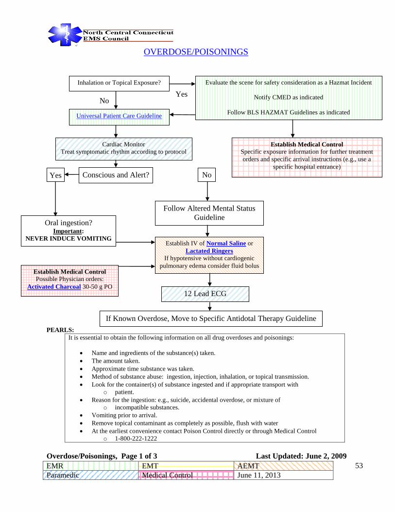
OVERDOSE/POISONINGS
PEARLS:
It is essential to obtain the following information on all drug overdoses and poisonings:
Name and ingredients of the substance(s) taken.
The amount taken.
Approximate time substance was taken.
Method of substance abuse: ingestion, injection, inhalation, or topical transmission.
Look for the container(s) of substance ingested and if appropriate transport with
o patient.
Reason for the ingestion: e.g., suicide, accidental overdose, or mixture of
o incompatible substances.
Vomiting prior to arrival.
Remove topical contaminant as completely as possible, flush with water
At the earliest convenience contact Poison Control directly or through Medical Control
o 1-800-222-1222
Overdose/Poisonings, Page 1 of 3 Last Updated: June 2, 2009
Universal Patient Care Guideline
Establish IV of Normal Saline or
Lactated Ringers If hypotensive without cardiogenic
pulmonary edema consider fluid bolus
12 Lead ECG
Cardiac Monitor
Treat symptomatic rhythm according to protocol
Inhalation or Topical Exposure?
Oral ingestion?
Important:
NEVER INDUCE VOMITING
Follow Altered Mental Status
Guideline
Conscious and Alert?
No
No Yes
Yes
Establish Medical Control Specific exposure information for further treatment
orders and specific arrival instructions (e.g., use a
specific hospital entrance)
Establish Medical Control
Possible Physician orders:
Activated Charcoal 30-50 g PO
Evaluate the scene for safety consideration as a Hazmat Incident
Notify CMED as indicated
Follow BLS HAZMAT Guidelines as indicated
If Known Overdose, Move to Specific Antidotal Therapy Guideline

EMR EMT AEMT
Paramedic Medical Control June 11, 2013 - -
54
Overdose/Poisonings
Continued
Overdose/Poisonings, Page 2 of 3 Last Updated: June 2, 2009
For Persistent Hypotension or Symptomatic
Bradycardia Refractory to Atropine and Fluids
Consider:
Glucagon 0.1 mg/kg (max 5mg) IV
Repeat in 5 minutes PRN
For hypotension or bradycardia with poor
perfusion despite previous measures consider:
Epinephrine 1mcg/kg/min IV Infusion
Titrate to effect
Beta Blocker Calcium Channel Blocker
For persistent hypotension with poor perfusion
despite previous measures consider:
Dopamine (Intropin) 10-20+ mcg/kg/minute IV
For Refractory Bradycardia <40 BPM with poor
perfusion consider:
Transcutaneous Pacing
For Persistent Hypotension or Symptomatic
Bradycardia Refractory to Atropine and Fluids:
Calcium Chloride 20 mg/kg (max 1g) IV
Repeat in 5 minutes if hypotension persists.
Consider Glucagon 0.1 mg/kg (max 5mg) IV
Repeat in 5 minutes PRN
For Refractory Bradycardia (<40 BPM) with
poor perfusion:
Consider Transcutaneous Pacing. Set rate
between 50-60 PPM.
Providers are encouraged to consult Medical Control early in the management of toxicological
emergencies when possible.
Ensure large, patent vein when administering calcium chloride to avoid extravasation and tissue
injury.

EMR EMT AEMT
Paramedic Medical Control June 11, 2013 - -
55
Overdose/Poisonings
Continued
Overdose/Poisonings, Page 3 of 3 Last Updated: June 2, 2009
Administer: Atropine 2mg IV or IM
Repeat, doubling dose every 5 minutes until bronchorrhea ceases. i.e. Initial
dose of 2mg; Second dose of 4mg; Third dose of 8 mg, etc.
Consider: Sodium Bicarbonate
1 mEq/kg IV
Repeat once in 5 minutes if
QRS still >0.10 sec
Stimulant or Anticholinergic
QRS > 0.10 sec?
Sodium Channel Blockers
(Tricyclic Antidepressants,
Diphenhydramine
(Diphenhydramine
(Benadryl)), Cocaine)
Monitor body temperature.
Consider active external cooling
Manage agitation, chest pain and
seizures following appropriate
guidelines.
Consider Repeat Fluid Bolus if
Hypotensive
Organophosphate Poisoning
(SLUDGE symptoms)
Cocaine or Diphenhydramine
(Diphenhydramine (Benadryl))?
For Persistent Ventricular
Dysrhythmias: Consider Magnesium Sulfate 1-2g slow
IVP over 5 - 60 min or Lidocaine 1 to 1.5mg/kg
Yes
Providers are encouraged to consult Medical Control early in the management of toxicological
emergencies when possible.
Ensure large, patent vein when administering calcium chloride to avoid extravasation and tissue
injury.
In TCA overdose, Sodium Bicarbonate is the preferred treatment, with Magnesium or Lidocaine
for refractory arrytmias
Administer Lorazepam (Lorazepam (Ativan)) 0.1
mg/kg (max 2mg) IM or IV
If seizures are observed, follow seizure guideline

EMR EMT AEMT
Paramedic Medical Control June 11, 2013 - -
56
PAIN MANAGEMENT (ADULT)
Universal Patient Care Guideline
(Include Pain Scales in assessment)
Use non-pharmacological pain
management. (positioning, splinting,
padding, reassurance, guided imagery,
hot and cold therapy) when possible
IV Access
Yes Acute Coronary Syndrome /
Chest Pain?
Follow Acute Coronary
Syndrome Guideline No
Patient Reports Moderate to
Severe Pain (4 or greater on
1-10 scale) after BLS
interventions?
Yes
Significant Head Trauma?
or
GCS < 13?
or
SBP < 100?
Yes
→
Establish Medical Control
Possible Physician Orders:
Morphine
Fentanyl
No
If patient is immobilized to a backboard or has a history of nausea / vomiting from Narcotics and no contraindications
exist, Then Administer:
Odansetron (Zofran®) 4mg slow IV
or
Metoclopramide (Reglan) 10 mg Slow IV
Ask Patient “Would you like some pain medicine?”
If answer “YES”
Fentanyl
Fentanyl up to 1mcg/kg IV
over 1-2 minutes, IM or IN
(to maximum single dose of
100mcg, for patients over 65
divide in two equal halves
administered five minutes
apart. Withhold the second
half of the dose if it is no
longer indicated.
Morphine
0.1 mg/kg Morphine Sulfate (2mg/min increments) up to 10 mg slow IV via
syringe or IV infusion (over at least 5 minutes) For patients over 65 administer 0.05
mg/kg.
or
If IV access is unavailable, Administer 0.1 mg/kg Morphine Sulfate up to 10 mg
IM
Pain Control, Adult, Page 1 of 3 Last Updated: March 5, 2012
EMR EMT AEMT
Paramedic Medical Control June 11, 2013 - -
57
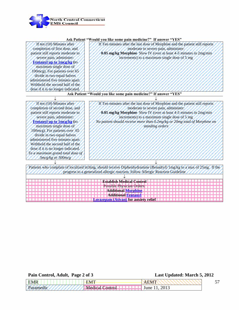
Ask Patient “Would you like some pain medicine?” If answer “YES” If ten (10) Minutes after
completion of first dose, and
patient still reports moderate to
severe pain, administer:
Fentanyl up to 1mcg/kg (to
maximum single dose of
100mcg). For patients over 65
divide in two equal halves
administered five minutes apart.
Withhold the second half of the
dose if it is no longer indicated.
If Ten minutes after the last dose of Morphine and the patient still reports
moderate to severe pain, administer:
0.05 mg/kg Morphine Slow IV (over at least 4-5 minutes in 2mg/min
increments) to a maximum single dose of 5 mg
Ask Patient “Would you like some pain medicine?” If answer “YES”
If ten (10) Minutes after
completion of second dose, and
patient still reports moderate to
severe pain, administer:
Fentanyl up to 1mcg/kg (to
maximum single dose of
100mcg). For patients over 65
divide in two equal halves
administered five minutes apart.
Withhold the second half of the
dose if it is no longer indicated.
To a maximum grand total dose of
3mcg/kg or 300mcg
If Ten minutes after the last dose of Morphine and the patient still reports
moderate to severe pain, administer:
0.05 mg/kg Morphine Slow IV (over at least 4-5 minutes in 2mg/min
increments) to a maximum single dose of 5 mg
No patient should receive more than 0.2mg/kg or 20mg total of Morphine on
standing orders
Patients who complain of localized itching, should receive Diphenhydramine (Benadryl) 1mg/kg to a max of 25mg. If the
progress to a generalized allergic reaction, follow Allergic Reaction Guideline
Establish Medical Control
Possible Physician Orders:
Additional Morphine
Additional Fentanyl
Lorazepam (Ativan) for anxiety relief
Pain Control, Adult, Page 2 of 3 Last Updated: March 5, 2012

EMR EMT AEMT
Paramedic Medical Control June 11, 2013 - -
58
PAIN MANAGEMENT (ADULT) Continued
Pearls:
All patients receiving prehospital narcotic analgesics or benzodiazepines should have
continuous pulse oximetry monitoring, ECG and non-invasive quantitative waveform
capnography (if available).
Consider administering morphine as an infusion over 5 -10 minutes in 50 – 100 mL of D5W or
0.9% NS to minimize side effects.
Stop medication administration if significant adverse effects (severe nausea, vomiting,
hypotension, respiratory depression) or sedation (decreased mental status) develop.
Respiratory depression should be treated with oxygen and ventilatory support if necessary.
Attempt verbal and tactile stimulation to reverse respiratory depression prior to considering
Naloxone (Narcan).
Administer the smallest possible reversal dose of Naloxone (Narcan) to maintain adequate
respirations. Dilute 0.4 mg Naloxone (Narcan) in 10cc 0.9% NS syringe and slowly titrate to
effect.
Morphine and Fentanyl should not be mixed without permission of medical control.
Patients who complain of localized itching, should receive Diphenhydramine (Benadryl) 1
mg/kg to a max of 25mg. If generalized allergic reaction, follow guidelines under Allergic
Reaction.
Fentanyl maybe given intranasally under the following dosing regime:
Administer Fentanyl IN, initial dose 1.5 mcg/kg (100 mcg max single dose), may
administer a second dose 1.5mcg/kg (100 mcg max single dose) if needed after 10
minutes, for a total maximum dose of 200 mcg. **Administer half a single dose in
each nare**
Pain Control, Adult, Page 3 of 3 Last Updated: March 5, 2012

EMR EMT AEMT
Paramedic Medical Control June 11, 2013 - -
59
PAIN ASSESSMENT (ADULT)
Purpose:
To identify and facilitate appropriate management of painful conditions in the prehospital setting.
Guiding Principles:
Pain is a medical condition and patients possess a right to have their pain treated.
All patients should be assessed for the presence of pain which should then be managed appropriately.
Procedure:
The EMS provider will evaluate all conscious patients (regardless of presenting complaint) for the
presence and severity of pain once immediate life threats have been addressed.
This assessment will be repeated after any pain management intervention, change in apparent pain level or at
least every 15 minutes. This evaluation will consist of, at a minimum, either a verbal numeric score or a visual
analog score. If possible, also use the verbal score. Pain scores must be documented on the patient care
report.
Visual Analog Scale
Ask the patient to mark or point to the severity of their pain on a scale of zero to ten with zero being no pain
and ten being unbearable pain, the worst pain they have ever felt.
Verbal Numeric Pain Score
Ask the patient to rate the severity of their pain on a scale of zero to ten with zero being no pain and ten being
unbearable, the worst pain ever.
Verbal Pain Score
Ask the patient to assign one of the following adjectives to rate their pain:
NONE MILD MODERATE SEVERE UNBEARABLE
Pain Assessment (Adult), Page 1 of 2 Last Updated: March 5, 2012

EMR EMT AEMT
Paramedic Medical Control June 11, 2013 - -
60
Documentation
Run form documentation will include an assessment of the patient’s pain, the nature of the pain, treatment of
the pain, a reassessment of the pain, and patient satisfaction with pain relief efforts.
If a paramedic chooses not to medicate a patient in moderate to severe pain, the reasons for withholding
analgesia must be documented.
Pain Assessment (Adult), Page 2 of 2 Last Updated: March 5, 2012

EMR EMT AEMT
Paramedic Medical Control June 11, 2013 - -
61
Seizures
Universal Patient Care Guideline
Consider Trauma, Hypoglycemia, Overdose
Go to appropriate Guideline if indicated
Protect the patient from personal injury
High flow oxygen if Sa02 is < 94%
Establish an IV of Normal Saline @ KVO
and obtain blood glucose level and record
If patient actively seizing, do not delay medicine administration to
obtain IV
If Blood Glucose is <70
Administer the following
If patient is actively seizing
Dextrose up to 25 Gm IVP (using
D10, D25, or D50W)
Glucagon 1mg IM if IV access
unavailable
Midazolam (Versed) 10 mg IM or 0.2 mg/kg to a max of 10mg IN if
patient >39 kg
Midazolam (Versed) 5 mg IM if patient <39 kg
Midazolam (Versed) can be repeated q 5 minutes to a max total standing
order dose of 20 mg if the patient is still seizing
Or if IV already established
Lorazepam (Ativan) 4 mg IV >39 kg
Lorazepam (Ativan) 2 mg IV if patient <39 kg>13 kg
Can be repeated q 5 minutes to a max total standing order dose of 8 mg if
patient still seizing
Establish Medical Control Possible Physician orders:
Additional anti-seizure medication Additional Dextrose
1. In absence of an established IV, Midazolam (Versed) IM is the preferred option in status epilepticus as studies have shown it stops the seizure quicker and results in fewer hospital admissions than IV Lorazepam (Ativan). 2. In absence of the other drug, Midazolam (Versed) can be given IV and Lorazepam (Ativan) can be given IM. 3. Midazolam (Versed) can be given IN, but this route is considered less reliable then IM. 4. The dosing above is intended for convulsing patients in status epilepticus. More moderate doses can be considered in partial seizures. 5. The sooner seizures can be stopped, the easier they are to stop and the less damage the patient may suffer from the seizure. 6. After giving first dose of Midazolam (Versed) IM, attempt to get an IV, and if obtained and patient still seizing after 5 minutes, give next dose Lorazepam (Ativan) IV.
Seizures, Page 1 of 1 Last Updated: June 11, 2013

EMR EMT AEMT
Paramedic Medical Control June 11, 2013 - -
62
SHOCK
Universal Patient
Care Guideline
Control obvious
bleeding
High flow oxygen
if Sa02 is < 94%
Early transport of
patient
Yes ←
Cardiogenic
Shock? →
No
Establish IV Normal Saline KVO
Fluid Challenge of 300-500 ml
En route to hospital
Establish large bore IV of Normal Saline
or Ringers Lactate and titrate to a
systolic BP > 100 mmHg
Cardiac Monitor
12 Lead EKG
Treat any underlying arrhythmias as per
Guideline
Dopamine (Intropin) 5 µg/kg/min up to 20
µg/kg/min titrated to a systolic
BP ≈ 100 mmHg
or
Norepinephrine (Levophed) 1-30
mcg/min titrated to blood pressure
of 90 mmHg
Consider second large bore IV line
Continuously monitor and record vital
signs
In trauma cases, monitor Glasgow Coma
Scale
Establish Medical Control Cardiac Monitor
Establish Medical Control
Note: Lung sounds and respiratory status must be continuously monitored to avoid pulmonary edema.
Shock, Page 1 of 1 Last Updated: April 6, 2014

EMR EMT AEMT
Paramedic Medical Control June 11, 2013 - -
63
Behavioral Emergencies
The following medical control options may be utilized for the patient with a psycho-social condition exhibiting extreme
anxiety, and who is hemodynamically stable.
Universal Patient
Care Guideline
Cardiac Monitor
IV Access
No ← Patient Violent? → Yes
Midazolam (Versed) 0.1mg/kg IM or IV
up to 10mg.
(5mg average dose)
May be repeated to a total max of 10 mg or
0.2mg/kg to a max 10mg IN.
or
Lorazepam (Ativan) up to 0.5 -2 mg slow
IVP or IM
(Refer to Behavioral Emergencies
Guidelines)
Establish Medical Control
Possible Physician Orders:
Repeat any of the above options as ordered
For Adult Patients
Midazolam (Versed) 0.1mg/kg IM up to
10mg (5mg average dose)
May be repeated to a total max of 10 mg or
0.2mg/kg to a max 10mg IN.
and Haloperidol (Haldol) 5 mg IM or
Olanizpine (Zyprexa) 5-10 mg IM
For Pediatric Patients see table below
Pearls
or
Lorazepam (Ativan) 0.05mg/kg up to 4mg
IM (2 mg average dose)
and Haloperidol (Haldol) 5 mg IM or
Olanizpine (Zyprexa) 5-10 mg IM
For Pediatric Patients see table below
Pearls
Establish Medical Control
Possible Physician Orders:
Additional Medication
Behavioral Emergencies, Page 1 of 3 Last Updated: June 11, 2013

EMR EMT AEMT
Paramedic Medical Control June 11, 2013 - -
64
PEARLS:
Administration of Haloperidol (Haldol) decreases a person’s seizure threshold. Use with caution on patients
with a history of seizure disorders or cocaine overdose.
Use caution with patients with a known prolonged QT segment
Midazolam (Versed) and Haloperidol (Haldol) or Lorazepam (Ativan) and Haloperidol (Haldol) may be
mixed in the same syringe. Midazolam (Versed) and Haloperidol (Haldol) should be the first option for
violent patients without an established IV because Midazolam (Versed)’s mechanism of action is quicker.
Olanizpine (Zyprexa) may not be combined with either Midazolam (Versed) or
Haloperidol (Haldol) in the same syringe.
EKG, Pulse Oximetry and quantitative waveform capnography should all be monitored continuously
Drug Age Dose Route
Haloperidol (Haldol)
0-6 YOA Not Recommended
N/A
6-12 YOA 1-3mg I.M.
12-adult dose 5mg I.M.
Lorazepam (Ativan)
3-12 YOA 0.05-0.2mg/kg
up to 2 mg
I.M.
12-adult dose 0.05mg/kg up to 4mg
IM
(2 MG average)
I.M.
Behavioral Emergencies, Page 2 of 3 Last Updated: June 11, 2013

EMR EMT AEMT
Paramedic Medical Control June 11, 2013 - -
65
North Central EMS Behavioral Emergency Guidelines
EMS providers may use physical and/or chemical restraints on patients who pose a danger to
themselves or others.
Providers should make every effort to ensure that law enforcement and adequate assistance are
present when attempting to restrain a violent or combative patient.
Only the minimum amount of restraint necessary to protect providers and the patient should be used.
Providers should first attempt to verbally calm the patient down. If the patient does not comply,
physical restraint may be attempted.
Providers should assess the patient for medical conditions that could be contributing to the patient’s
behavior. If an assessment cannot be performed prior to physical restraint, it should occur as soon as
possible after restraint is applied when it is safe and feasible.
Physical restraints must be soft in nature and pose no threat to the patient’s safety. Only the
extremities shall be restrained and these restraints must be assessed every five minutes.
Patients must never be hog-tied, restrained in a prone position with hands tied behind their backs or
placed between backboards or mattresses. No restraint shall ever be tied around the head, neck or
chest. A surgical mask, spit shield, or an oxygen mask may be placed loosely on the patient to
prevent spitting.
Handcuffs may only be used by law enforcement or correction officials on patients in their custody.
If the law enforcement officer insists that the patient remain handcuffed during transport, they must
either accompany the patient or provide a key to EMS personnel.
Chemical restraint may be used per guideline following unsuccessful attempts at verbal and/or
physical restraint or when a patient continues to forcibly struggle against physical restraints.
All restrained patients must have continual reassessment of vital signs and neurovascular status of
distal extremities. In chemically restrained patients (safety permitting) this should include ECG,
pulse oximetry, and quantitative waveform capnography if the patient is no longer alert.
Documentation must include justification for restraint, type of restraint used, restraint procedure,
results of continual reassessment, medications administered, the indications for the administration,
and any other care rendered.
Do not hesitate to involve medical direction in any call involving restraint.
Behavioral Emergency Guidelines, Page 3 of 3 Last Updated: June 11, 2013

EMR EMT AEMT
Paramedic Medical Control June 11, 2013 - -
66
DYSTONIC REACTION
Universal Patient Care Guideline
IV Access
Administer 25 – 50 mg of Diphenhydramine (Benadryl), IV or IM
Establish Medical Control
Dystonic Reaction, Page 1 of 1 Last Updated: June 13, 2008

EMR EMT AEMT
Paramedic Medical Control June 11, 2013 - -
67
Stroke Guideline
Universal Patient Care Guideline
IV Access 18g or larger
Check Blood Glucose Level
If blood glucose is below 70 mg/dl then
Administer up to 25G of Dextrose (D10Wpreferred)
Cincinnati Prehospital Stroke Scale
Transport Patient to appropriate receiving facility
Contact Medical Control
Pearls:
Brief history focusing on time of onset of symptoms, when was the last time the patient was symptom
free?
Transport patient and witness if possible
Cincinnati Prehospital Stroke Scale:
Assess for new unilateral arm drift
Assess speech – slurred or inappropriate words?
Assess for new facial droop
Contact receiving hospital for ‘Acute Stroke Alert’ and include following information:
Time of symptom onset
Description of neurologic deficits (Cincinnati stroke scale)
Blood glucose level
Early notification to the receiving hospital is essential to ensure the immediate availability of an
appropriate in-hospital response.
D10W is the preferred solution for hypoglycemic emergency. D10W can be given either as an open
drip or drawn up in a large syringe and given as IV pushes. If D10W unavailable, D25W or D50W may
be used.
Stroke Guideline, Page 1 of 1 Last Updated: June 11, 2013
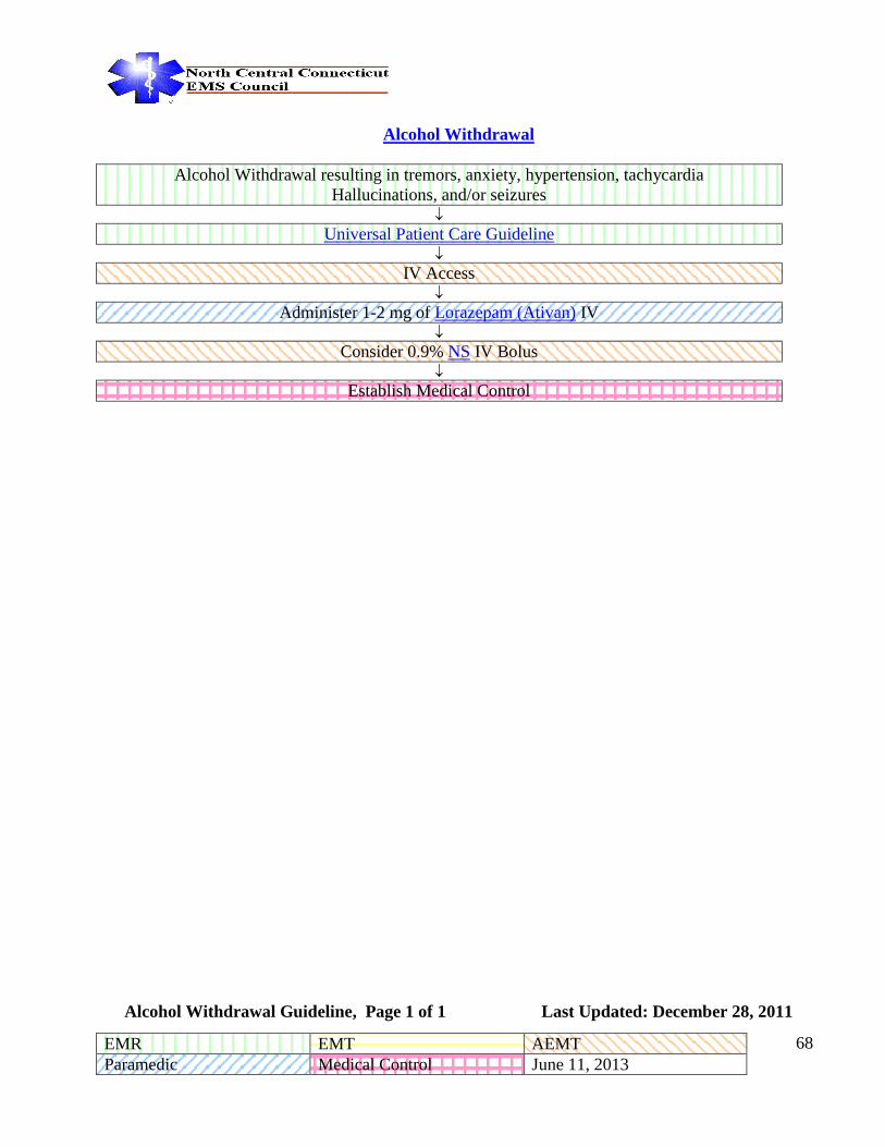
EMR EMT AEMT
Paramedic Medical Control June 11, 2013 - -
68
Alcohol Withdrawal
Alcohol Withdrawal resulting in tremors, anxiety, hypertension, tachycardia
Hallucinations, and/or seizures
Universal Patient Care Guideline
IV Access
Administer 1-2 mg of Lorazepam (Ativan) IV
Consider 0.9% NS IV Bolus
Establish Medical Control
Alcohol Withdrawal Guideline, Page 1 of 1 Last Updated: December 28, 2011

EMR EMT AEMT
Paramedic Medical Control June 11, 2013 - -
69
North Central Connecticut
Regional
Paramedic Guidelines
Adult
Trauma Guidelines
≥13 years Old

EMR EMT AEMT
Paramedic Medical Control June 11, 2013 - -
70
INJURED PATIENT TRIAGE GUIDELINE*
When transport to a Level I or II Trauma facility is indicated, but the ground transport time to that hospital is judged to be
greater than twenty (20) minutes, determination of destination hospital shall be in accordance with medical control.
Severely injured patients <13 years should be taken to a Level I or II facility with pediatric resources including pediatric ICU.
All EMS providers transporting trauma patients to hospitals shall provide receiving hospital with a complete OEMS approved
patient care form prior to departing from the hospital.
WHEN IN DOUBT, CONSULT WITH MEDICAL CONTROL *State of Connecticut Regulation of Department of Public Health, Concerning Statewide Trauma System: Section 19a-177-5.
Injured Patient Triage, Page 1 of 1 Last Updated: June 13, 2008
No
Yes
Yes
Yes
Measure Vital Signs and level of Consciousness
Glasgow Coma Scale 12 or less
Call Medical Control
for direction
Assess Anatomy of Injury
1. Gunshot wound to head, neck, chest, abdomen or groin?
2. Third degree burns > 15% or of the face or airway?
3. Evidence of spinal cord injury?
4. Amputation other than digits?
5. Two or more proximal long bone fractures?
Assess mechanism of injury and other factors
1. Falls >20 feet
2. Apparent high speed impact
3. Ejection of patient from vehicle
4. Death of same vehicle occupant
5. Pedestrian hit by car at > 20 mph
6. Rollover accident
7. Significant vehicle deformity – especially of steering wheel
8. Age <5 or >55
9. Known cardiac disease or respiratory distress
10. Penetrating injury to neck, thorax, or abdomen other than
gunshot wounds
Take to Level I or Level
II Trauma Facility
Evaluate as per usual
Guidelines
No
No

EMR EMT AEMT
Paramedic Medical Control June 11, 2013 - -
71
Management of the Trauma Patient
Scene Assessment
Initial Patient Assessment
Stabilize C-Spine during assessment
Open airway using Modified Jaw thrust, if indicated
Consider Rapid Extrication
↓
Manage Patient Airway /Administer Oxygen /Ventilate
Determine Patient’s Hemodymanic Status ( Vital signs)
Control External Bleeding with Direct Pressure
↓
Maintain SpO2 >94% (Utilize Pulse Oximetry, Quantitative waveform capnography & ETCO2)
Complete Spinal Immobilization
↓
The following treatments should not delay
transport
* Initiate Vascular Access
2 large bore peripheral IV
or 1 single IO
If patient is hypotensive - infuse
Normal Saline or Lactated
Ringers at W/O then titrate
to maintain SBP >100. Do
not exceed 2000cc infusion
Injury Specific Treatments
Head Injury –ventilate 350-500ml at rate
10 / min. If herniation signs develop,
increase rate to 20/min & maintain CO2
Level of 34-38.
Chest Wound –apply an occlusive
dressing. Release dressing if S&S of
Tension Pneumothorax occurs.
Flail Chest – Airway management with
manual splinting
Abdominal Evisceration –apply
moistened NS sterile occlusive dsg.
Impaled Objects –do not remove &
stabilize in place
Extremity Fractures – check
neurovascular status & immobilize
Isolated Femur Fracture –Traction
Splint
Pelvic Fx with hypotension –
consider pelvic binder
Pregnant Pt – check externally for
contractions, vaginal bleeding, amniotic
fluid. If hypotensive elevate right side of
backboard
Bleeding Control – Direct Pressure,
Tourniquet
* Cardiac Monitor - treat
dysrhythmias according to
ACLS
Spinal Cord Injury –consider
Dopamine (Intropin) drip to titrate to
Maintain SBP>100 or Norepinephrine
(Levophed) 1-30 mcg/min titrated to blood
pressure of 90 mmHg
If Tension Pneumothorax (dyspnea,
hypotension & diminished breath sounds)
present - needle decompression
↓
Begin Transport to Trauma Center according to the guidelines outlined in the State of Connecticut Injured
Patient Triage Guideline

EMR EMT AEMT
Paramedic Medical Control June 11, 2013 - -
72
Management of Trauma Patients, Page 1 of 2 Last Updated: June 11, 2013
PEARLS
Vital signs include Blood Pressure, Pulse ( rate, strength and location), Respiratory Rate,
Skin ( color, moisture and temperature), Pain Level & Glasgow Coma Scale
To control hemorrhage direct pressure is the 1st choice. Do not elevate extremity or use
pressure points. The use of tourniquets must be considered in severe hemorrhage if direct
pressure fails or is not able to be done effectively. If a tourniquet is applied it should be
just proximal to site of hemorrhage & tightened until bleeding stops. Mark time of
application on tourniquet leave site exposed for visual monitoring of hemorrhage
Paramedics may straighten severely angulated fractures if the distal extremity has signs of
decreased perfusion. Premedication with sedation or analgesia should be strongly
considered. EMRs, EMTs and AEMTs should splint angulated fractures in position
found. In unusual circumstances or extremely prolonged transport times, EMTs and
AEMTs may contact medical control for authorization to straighten severely angulated
fractures if the distal extremity has signs of decreased perfusion.
Dopamine (Intropin) infusion at 5-20 mcg/kg/ min. Generally start at 5 mcg/kg/min and
increase every 10 minutes by an additional 5 mcg/kg/min until SBP>100. Do not exceed
20 mcg/kg/min

EMR EMT AEMT
Paramedic Medical Control June 11, 2013 - -
73
Management of Trauma Patients, Page 2 of 2 Last Updated: June 11, 2013

EMR EMT AEMT
Paramedic Medical Control June 11, 2013 - -
74
BURNS
Universal Patient Care Guideline
↓
Oxygen
↓
Establish an IV Lactated Ringers or Normal
Saline (in area not affected by burn if
possible).
↓
Type of Burn
Assess percentage of Total Body Surface Area
Burned
↓
Thermal Burns Chemical Burns Ophthalmic Burns Electrical Burns
↓ ↓ ↓ ↓
Cover burns with clean, dry
dressing. If the burns are
less than 10% and are
superficial or partial
thickness you can moisten
towels or sheets with sterile
normal saline for comfort.
Otherwise use dry sterile
dressings
Consider any chemical burn situation as a
Hazmat situation. If potential Hazmat situation
exists, notify receiving hospital ASAP
Identify the chemical if possible
Immediate and
continuous flushing
of the affected eye is
performed using
Lactated Ringers or
Normal Saline. If
contact lenses are
known to be in the
patient’s eyes, an
attempt should be
made to remove them
and continue
flushing.
Suspect spinal injury
secondary to tetanic
muscle contraction
Immobilize Patient.
Assess for entrance
and exit wounds.
↓ ↓ ↓ ↓
If burn injury greater than
20% BSA, begin fluid
resuscitation at 500 ml/hr.
If hypotensive, administer
250-500ml fluid bolus and
titrate to patient’s BP >100
SB/P
Remove affected clothing & jewelry (if not
already done)
Unless
contraindicated instill
1 or 2 drops of
ophthalmic
anesthesia. Place
medication onto
lower lid.
IV Normal Saline
↓ ↓ ↓ ↓
Control Pain According to
Pain Control Guideline
Brush off dry powder. Flush with copious
amounts of water or saline unless
contraindicated. Irrigate burns to the eyes with
a minimum of 1 liter of lactated ringers or
Normal Saline. Alkaline burns should receive
continuous irrigation throughout transport.
Consider the Morgan Lens for eye irrigation,
(see right).
Place the Morgan
Lens in the affected
eye(s) continuously
flush with Lactated
Ringers or Normal
Saline while enroute
to the hospital. Run
2 liters of fluid wide
per eye, then
administer KVO rate.
Cardiac Monitor
Treat any cardiac
rhythm disturbances
per Guideline
Control Pain
According to Pain
Control Guideline
↓ ↓ ↓ ↓
Establish Medical Control Possible Physician Orders:
Additional MS, Intubation.
Control Pain According to Pain Control
Guideline
Control Pain
According to Pain
Control Guideline
Establish Medical
Control
↓
Establish Medical Control Possible Physician Orders:
Morphine Sulfate IVP
Burns, Page 1 of 2 Last Updated: June 2, 2009
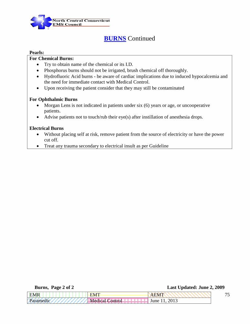
EMR EMT AEMT
Paramedic Medical Control June 11, 2013 - -
75
BURNS Continued
Pearls:
For Chemical Burns:
Try to obtain name of the chemical or its I.D.
Phosphorus burns should not be irrigated, brush chemical off thoroughly.
Hydrofluoric Acid burns - be aware of cardiac implications due to induced hypocalcemia and
the need for immediate contact with Medical Control.
Upon receiving the patient consider that they may still be contaminated
For Ophthalmic Burns
Morgan Lens is not indicated in patients under six (6) years or age, or uncooperative
patients.
Advise patients not to touch/rub their eye(s) after instillation of anesthesia drops.
Electrical Burns
Without placing self at risk, remove patient from the source of electricity or have the power
cut off.
Treat any trauma secondary to electrical insult as per Guideline
Burns, Page 2 of 2 Last Updated: June 2, 2009

EMR EMT AEMT
Paramedic Medical Control June 11, 2013 - -
76
EMS Spine Precautions & Use of the Long Back Board for Ambulatory Patients
EMS Spine Precautions & Use of the Long Back Board for All Providers Last Updated: June 11, 2013

EMR EMT AEMT
Paramedic Medical Control June 11, 2013 - -
77
OMITTING SPINAL IMMOBILIZATION
Paramedics shall make spinal immobilization decisions based on mechanism of injury and clinical
criteria. The decision to not immobilize a patient is the responsibility of the paramedic.
Indications for Spinal Immobilization:
1. Any patient who has sustained a significant mechanism of injury (includes windshield spider,
dash deformity, ejection, rollover, fall from > 10 feet, and vehicle space invasion > 1 foot).
2. Any patient with positive or questionable mechanism of injury and who has one of the following
clinical findings:
a. Altered mental status
b. Hemodynamic instability
c. Evidence of intoxication or unreliability
d. A significantly distracting painful injury
e. Neurological Deficit
f. Spinal Pain or tenderness
Procedure:
1. Determine Mechanism of Injury
Significant mechanism (including windshield spider, dash deformity, ejection, rollover, fall from >
10 feet, and vehicle space invasion > 1 foot) immobilize patient.
Positive Mechanism or questionable mechanism (including patients with trauma above the clavicle,
falls, MVAs, trauma to the spine head or neck, abrupt accelerating, decelerating or rotational forces)
maintain stabilization and proceed with spinal assessment.
2. Assess Patients
Assess mental status. If patient is not alert and oriented, immobilize.
Assess hemodynamic stability. If patient is hemodynamically unstable, immobilize.
Assess for intoxication and reliability. If patient has evidence of intoxication, mental impairment, or
gives unreliable answers, immobilize.
Omitting Spinal Immobilization, Page 1 of 2 Last Updated: June 13, 2008

EMR EMT AEMT
Paramedic Medical Control June 11, 2013 - -
78
Assess for distracting injury. If patient has an injury, which may distract from patient’s awareness to
pain, immobilize.
Assess neurological function. If patient has neurological deficit, immobilize.
Assess spine. If patient has pain on palpation of spinous process of cervical, thoracic or lumbosacral
spine, immobilize.
If the above are negative then:
Assess range of motion. Direct patient to touch their chin to their chest, look up extending their neck,
and then turn head from side to side. If patient has any neck pain during their normal active range of
motion, immobilize.
Decision:
Patients, who pass the above assessment, may have immobilization omitted at the discretion of the
paramedic.
Extra caution must be used in pediatric and geriatric patients. When in doubt, immobilize.
All pertinent exam and history findings must be included in run form.
In cases where the paramedic does not accompany the patient to the hospital, the paramedic needs to
provide their name to the transporting BLS provider for documentation purposes.
Considerations
Spinal Immobilization does not always require securing patient to a long back board.
While a long backboard is often essential to extrication and patient movement, it’s further use can
cause harm to the patient. When available vacuum backboards are preferred.
Immobilization to a long back board should be withheld in cases of ambulatory patients,
penetrating trauma (absent focal neurological deficit), interfacility transports and transports
exceeding 30 minutes.
If in the judgment of a paramedic, immobilization to a long back board will delay correction of life-
threatening treatment, impair a patient’s airway or ability to breathe, or cause unsafe force to be
applied to the affected area due to patient movement or anxiety, the paramedic may omit the long
back board and consider alternatives such as a KED with cervical collar, a cervical collar only, or
other means of safe immobilization. All reasons for deviation from the standard guideline should be
documented.
Remember: The decision to not immobilize a patient is the responsibility of the paramedic.
Omitting Spinal Immobilization, Page 2 of 2 Last Updated: June 11, 2013

EMR EMT AEMT
Paramedic Medical Control June 11, 2013 - -
79
North Central Connecticut
Regional
Paramedic Guidelines
OB/Gyn Guidelines
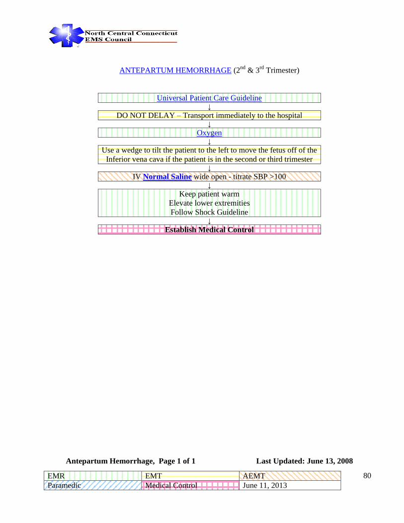
EMR EMT AEMT
Paramedic Medical Control June 11, 2013 - -
80
ANTEPARTUM HEMORRHAGE (2nd
& 3rd
Trimester)
Universal Patient Care Guideline ↓
DO NOT DELAY – Transport immediately to the hospital ↓
Oxygen ↓
Use a wedge to tilt the patient to the left to move the fetus off of the
Inferior vena cava if the patient is in the second or third trimester ↓
IV Normal Saline wide open - titrate SBP >100 ↓
Keep patient warm
Elevate lower extremities
Follow Shock Guideline ↓
Establish Medical Control
Antepartum Hemorrhage, Page 1 of 1 Last Updated: June 13, 2008

EMR EMT AEMT
Paramedic Medical Control June 11, 2013 - -
81
PREGNANCY INDUCED HYPERTENSION AND SEIZURES
(ECLAMPSIA / TOXEMIA)
Universal Patient Care Guideline ↓
If hypoglycemia or drug overdose induced Status Epilepticus is suspected, treat according to
appropriate Guideline. ↓
Lorazepam (Ativan) 0.1mg/kg IVP with a max single dose of
2mg, repeated q 5 minutes to a max total dose of 8 mg
If IV access unavailable:
Midazolam (Versed) 0.1mg/kg to a max single dose of 5 mg IM
Magnesium Sulfate 4 Gms in 20 ml normal saline Slow IVP (over 5 minutes)
Follow with infusion of Magnesium Sulfate @ 1 - 2 Gms/Hour ↓
If seizures recur or do not subside, contact medical control for repeat of above.
These seizures can occur up to four weeks post partum.
PEARLS:
Eclampsia is preceded by preclampsia which consists of hypertension, potential altered mental status
and diffuse edema. Gentle ALS transport should be considered for these patients.
Pregnancy Induced Hypertension and Seizures, Page 1 of 1 Last Updated: June 13, 2008

EMR EMT AEMT
Paramedic Medical Control June 11, 2013 - -
82
EMERGENCY CHILDBIRTH
Universal Patient Care Guideline
↓
Oxygen
↓
Establish IV Normal Saline at KVO rate
↓
No Crowning or urge to push Crowning or urge to push
Imminent Delivery
↓ ↓
Transport to OB facility Prepare for childbirth
↓
Control delivery with the palm of the hand so the infant does not
“explode” out of the vagina. Support the infants head as it emerges and
support perineum with gentle hand pressure.
↓
Support and encourage the mother to control the urge to push.
↓
Tear the amniotic membrane, if it is still intact and visible outside the
vagina. Check for cord around the neck.
↓
Gently suction mouth and nose (with bulb syringe) of infant as soon
as head is delivered.
↓
IF Meconium is present & the infant has a
HR<100, poor respiratory effort or poor
muscle tone*
Note the presence or absence of meconium staining.
↓ ↓
Intubate and suction prior to stimulating
breathing. Ventilate with BVM after
suctioning
As shoulders emerge, guide head and neck slightly downward to deliver
anterior shoulder, then the posterior shoulder.
↓
The rest of the infant should deliver with passive participation. Get a firm
hold on the baby.
↓
Repeat gentle suctioning then proceed to postpartum care of infant and
mother.
↓
Dry and keep infant warm. If possible skin-to-skin contact with the
mother while covering the infant with a blanket provides a good warming
source.
↓
Establish date and time of birth and record, do APGAR at 1 and 5
minutes.
*If the infant has a HR >100, strong respiratory effort and good muscle tone, intubation and
suctioning is not indicated.
Emergency Childbirth, Page 1 of 1 Last Updated: June 13, 2008

EMR EMT AEMT
Paramedic Medical Control June 11, 2013 - -
83
DELIVERY COMPLICATIONS
Nuchal Cord (cord around baby’s neck)
Universal Patient Care Guideline ↓
Emergency Delivery Guideline ↓
Slip two fingers around the cord and lift over baby’s head. ↓
If unsuccessful: Double clamp cord, cut cord between clamps with sterile scissors (blunt side next to baby,
never use a scalpel) allow cord to release from baby’s neck. ↓
Continue with normal delivery guideline. ↓
Contact Medical Control
Prolapsed Cord (cord presenting before the baby)
Universal Patient Care Guideline ↓
Elevate mother’s hips in knee-chest position or left side down in Trendelenberg position. ↓
Protect cord from being compressed by placing a sterile gloved hand in vagina and pushing up firmly on the
presenting part of the fetus ↓
Palpate cord for pulsation ↓
Keep exposed cord moist and warm. ↓
Keep hand in position and transport immediately. ↓
Contact Medical Control ↓
Do not remove hand until relieved by OB personnel.
Delivery Complications, Page 1 of 2 Last Updated: June 13, 2008

EMR EMT AEMT
Paramedic Medical Control June 11, 2013 - -
84
Delivery Complications continued
Breech Birth (legs or buttocks presenting first)
Universal Patient Care Guideline ↓
Emergency Delivery Guideline ↓
Never attempt to pull baby from the vagina by the legs or trunk. ↓
After shoulders are delivered, gently elevate the trunk and legs to aid in delivery of head (if face down)* ↓
Head should deliver in 30 seconds, if not; reach 2 fingers into the vagina to locate the baby’s mouth. Fingers
in mouth will flex baby’s head and should assist in spontaneous delivery. If not: Press vaginal wall away
from the baby’s mouth to create an airway. If head does not deliver in 2 minutes, keep your hand in position
and transport ASAP. ↓
ESTABLISH MEDICAL CONTROL
Extremity Presentation
Universal Patient Care Guideline ↓
Proceed immediately to the hospital Place in Trendelenberg Position
↓
Establish Medical Control ↓
Do not attempt out of hospital delivery ↓
Encourage mother to perform slow deep breathing
Delivery Complications, Page 2 of 2 Last Updated: June 13, 2008

EMR EMT AEMT
Paramedic Medical Control June 11, 2013 - -
85
Postpartum Care of the Mother
Universal Patient Care Guideline ↓
Emergency Delivery Guideline ↓
Massage the fundus of the uterus ↓
Placenta should deliver within a few minutes to up to 30 minutes. DO NOT pull on cord to facilitate placental
delivery. If delivered bring the placenta to the hospital, do not delay on scene waiting for the placenta to
deliver. ↓
If the perineum is torn and bleeding, apply direct pressure with trauma dressing to outside of vagina only. DO
NOT PACK VAGINA. Observe for excessive bleeding ↓
Titrate IV to maintain SBP >100 mm Hg, up to 3L ↓
If appropriate, put the infant to breast ↓
Establish Medical Control
.
Post Partum Care of the Mother, Page 1 of 1 Last Updated: June 13, 2008

EMR EMT AEMT
Paramedic Medical Control June 11, 2013 - -
86
POST PARTUM CARE OF THE INFANT
Universal Patient Care Guideline
↓
Note time and date of delivery. Dry the infant immediately and keep
warm. Continue gentle bulb suctioning of
the mouth and nose. Stimulate baby by rubbing its back
or flicking the soles of its feet, this
should be enough to stimulate the
baby to begin crying and breathing. *Spontaneous respirations should
begin within 30 seconds after
stimulation
↓
Yes
Spontaneous Breathing?
No →
Begin artificial
ventilations at 30 - 40
breaths/minute with
infant B-V-M. Watch
for chest rise.
↓ ↓
Cyanotic? No →
Obtain 1 minute APGAR score No ←
Pulse <80?
Yes ↓ Yes ↓
Place a pediatric face mask
approximately 4 inches from
the baby’s face and run oxygen
at 15 L/min. until color
improves. Gentle suctioning as
needed.
Clamp cord 6” to 8” from infants
body. Cut cord with sterile scissors
(blunt side next to infant) between
clamps. Clamping of cord is not
critical, and does not need to be
done immediately
Begin CPR and follow
Neonatal Resuscitation
Guideline
↓ ↓ ↓
Establish Medical Control
Allow mother to hold baby next to
her if her condition does not
contradict this. Wrap both baby and
mother together in blanket to
diminish heat loss.
Establish Medical
Control
↓
Obtain 5 minute APGAR score
If there is any bleeding from the cord clamp, reclamp again in close proximity to the “leaking” clamp.
* IF Meconium is present AND the infant has a HR<100, poor respiratory effort or poor muscle tone Intubate and suction
prior to stimulating breathing
IF the umbilical cord is not clamped, keep the infant level with mom. This will prevent infant CHF (blood from mom to
baby) or infant anemia (blood from baby to mom).
Post Partum Care of the Infant, Page 1 of 1 Last Updated: June 13, 2008

EMR EMT AEMT
Paramedic Medical Control June 11, 2013 - -
87
NEONATAL RESUSCITATION
Universal Patient Care Guideline
↓
Post Partum Care for Infants Guideline
↓
Position infant on his/her back with head down. Check for meconium.
↓
Suction mouth and nose with bulb syringe.
Dry infant and keep warm.
↓
Stimulate infant by rubbing his/her back or flicking the soles of the feet. If the infant shows decreased LOC, mottling or
cyanosis, and/or presents with a heart rate below 100 beats per minute
↓
Breathing, HR
>100
Breathing HR >100 but cyanotic Apneic or HR <100
↓ ↓ ↓
Observe Administer Supplementary Oxygen Positive Pressure Ventilation
↓ ↓
If after 30 seconds, persistent cyanosis
administer positive pressure ventilation
If after 30 seconds, HR <60, administer positive
pressure ventilation and chest compressions
↓
If after 30 seconds, HR <60, administer Epinephrine
0.01 mg/kg (1:10,000) IV/IO; 0.1 mg/kg (1:1,000) ET
Consider maternal condition including medications -
Naloxone (Narcan) 0.1 mg/kg
↓
IM/IV/IO/ET IV/IO access, 10-20 ml/kg Normal
saline bolus
↓
Obtain blood glucose level
↓
Establish Medical Control
Possible Physician orders: Repeat Epinephrine,
Naloxone (Narcan)
Dextrose 5% 5-10 ml/kg over 20 minutes other
treatment options per consultation
Neonatal Resuscitation, Page 1 of 2 Last Updated: June 13, 2008
PEARLS:

EMR EMT AEMT
Paramedic Medical Control June 11, 2013 - -
88
Current AHA Guidelines no longer recommend routine suctioning of meconuim. If thick meconium with a HR< 100
weak respiratory effort or poor muscle tone. Ventilate with BVM after suctioning.
Neonatal Resuscitation, Page 2 of 2 Last Updated: June 13, 2008

EMR EMT AEMT
Paramedic Medical Control June 11, 2013 - -
89
TRAUMA IN PREGNANCY
Universal Patient Care Guideline
↓ Adult Trauma Management Guideline
↓ Rapidly assess fetal viability - is uterus (fundus) above (viable) or
below the umbilicus (non-viable fetus).
↓ Treat mother aggressively for injuries based on mechanism of injury.
Follow Trauma Guideline with the following considerations.
↓ Oxygen
↓ Check externally for uterine contractions.
↓ Check externally for vaginal bleeding and amniotic fluid leak (Broken water).
↓ If patient becomes hypotensive while supine on blackboard elevate right side of backboard (to relieve pressure
on the inferior Vena Cava by uterus).
Pearls:
The most common cause of fetal death is maternal death. Fetus may be in jeopardy while mother’s vital signs appear stable.
Trauma in Pregnancy, Page 1 of 1 Last Updated: June 13, 2008

EMR EMT AEMT
Paramedic Medical Control June 11, 2013 - -
90
North Central Connecticut
Regional
Paramedic Guidelines
Pediatric Medical Guidelines

EMR EMT AEMT
Paramedic Medical Control June 11, 2013 - -
91
PEDIATRIC PATIENT ASSESSMENT
Evaluate Airway
Patent? No
→ Follow Pediatric Airway Guideline
No Problem
← Evaluate Breathing and
Circulation
Continue
Transport Reassess as
necessary
Problem with Either Problem with Circulation
BVM with 100% oxygen
Establish IV/IO and administer 20ml/kg
Normal Saline Reassess
May repeat fluid infusion x1
Establish Medical Control
Pediatric Patient Assessment, Page 1 of 1 Last Updated: June 13, 2008

EMR EMT AEMT
Paramedic Medical Control June 11, 2013 - -
92
PEDIATRIC AIRWAY ALGORITHM
Pediatric Assessment
Guideline
Clear, no assistance needed ← Assess Airway → Maintainable
Supplemental Oxygen Un-maintainable Position Head, Suction, Supplemental
Oxygen, BVM, OPA or NPA
Transport and reassess
Pulse Oximetry
Spontaneous respirations with
gag reflex?
Short transport time?
←
Basic Airway Maneuvers
(BVM, OPA or NPA)
→
Apneic with no gag reflex?
Consider managing airway with
BLS techniques Airway Obstruction?
BVM or consider ET or LMA
Heimlich Maneuver
Direct Laryngoscopy
Removal with Magill
Forceps
Age <8 years: Needle
Cricothyrotomy
Age >8 years: Surgical
Cricothyrotomy
Establish Medical Control
Pediatric Airway Guideline, Page 1 of 1 Last Updated: June 13, 2008

EMR EMT AEMT
Paramedic Medical Control June 11, 2013 - -
93
PEDIATRIC RESPIRATORY DISTRESS
Pediatric Assessment Guideline
↓
Determine the appropriate weight of the patient
If more than 50 kg (110 pounds), treat as an adult.
(Use pediatric resuscitation tape if weight unavailable)
↓
Ensure patency of airway
Assess respiratory rate and effort
↓
If airway is obstructed follow Obstructed Airway Guideline
↓
Assess for sign of respiratory distress
Use of accessory muscles, stridor, retractions, nasal flaring or noisy respirations
↓
Administer oxygen in the least irritating manner possible
↓
Allow the children to assume the most comfortable position for themselves
as practical and safe during transport
↓
See Guidelines for Croup/Epiglottitis or Pediatric Asthma if indicated
If patient requires ventilatory assistance, remember:
DO NOT OVER EXTEND NECK
Ventilate with a B-V-M first
Follow Pediatric Airway Guideline
↓
Early transport of the pediatric patient is critical
↓
Establish Medical Control
Pediatric Respiratory Distress, Page 1 of 1 Last Updated: June 13, 2008

EMR EMT AEMT
Paramedic Medical Control June 11, 2013 - -
94
PEDIATRIC ASTHMA
Pedia
Pediatric Asthma, Page 1 of 2 Last Updated: June 2, 2009
Pediatric Patient
Assessment Guideline
Pediatric Respiratory
Distress Guideline
Monitor Patient Condition
Severe Distress? (Marked accessory muscle use, hypoxia, hypercapnia, etc.)
Establish IV Normal saline Normal Saline
Administer Bolus 20 mL/kg
If minimal tidal volume, consider: Epinephrine
Epinephrine 0.01 ml/kg/dose IM (1:1,000) Max dose of 0.3 mg
If exacerbation is greater than 2 hours in duration:
Methylprednisone (Solu-Medrol) 2 mg/kg IV over
15 minutes
Repeat nebulized Albuterol (Ventolin, Proventil)
Albuterol (Ventolin, Proventil) as needed
Consider BVM Assist
Albuterol (Ventolin, Proventil) Albuterol
(Ventolin, Proventil) 5.0 mg (0.5cc) in 1.5ml
Normal Saline nebulized at 6 L/min O2
Repeat nebulized Albuterol
(Ventolin, Proventil) as needed
Yes No
Monitor Patient Condition
Administer Ipratropium Bromide 0.5 mg
nebulized concurrently with Albuterol (Ventolin,
Proventil) (i.e. Combivent® / Duoneb®). Do not
repeat Ipratropium.

EMR EMT AEMT
Paramedic Medical Control June 11, 2013 - -
95
Pearls:
Pediatric Asthma, Page 2 of 2 Last Updated: June 2, 2009
Endotracheal Intubation should be avoided if possible.
Do not routinely initiate IV access for pediatric asthma.

EMR EMT AEMT
Paramedic Medical Control June 11, 2013 - -
96
SUSPECTED CROUP or EPIGLOTTITIS
Pediatric Patient
Assessment Guideline
↓
Pediatric Respiratory
Distress Guideline
↓
Allow child to achieve
position of comfort
↓
Yes ← Respiratory
Arrest
→ No
↓ ↓
Attempt ventilation with
Pediatric B-V-M
If respiratory status warrants,
attempt to administer humidified
100% oxygen via mask (held by
mother or significant other) 4
inches in front of child’s face, but
ONLY if well tolerated by child.
↓ ↓
If ineffective, may use adult B-V-M Transport
↓ ↓
Transport
5 mg of Epinephrine (1:1000)
Nebulized with 2.5-3ml of NS if
the patient has respiratory distress
or stridor at rest.
or
Racepinephrine 2.25%
(Vaponephrine) in 0.5 ml mixed
with 2-3 mL of Normal Saline,
Nebulized if the patient has
respiratory distress or stridor at
rest.
* If the patient has a complicated
or cardiac hx contact medical
control before administering
epinephrine
↓ ↓
If still ineffective, endotracheal intubation may be
indicated
NOTE: In an unconscious patient, if there is strong
suspicion for epiglottitis and if the patient is unable
to be ventilated with a B-V-M and if an enlarged
epiglottis is visualized, ONE attempt at intubation is
allowed if the airway is able to be visualized.
Consider using a smaller size tube than you normally
would.
Establish Medical Control
Possible Physician orders:
Nebulized Epinephrine (5mg of
1:1,000) in 2.5-3ml NS.
↓
If unsuccessful:
Needle Cricothyrotomy if under 8 years
Surgical Cricothyrotomy if over 8 years
Normal Saline nebulizer 5ml at 6L/min O2 to
provide humidified oxygen (no medications in
nebulizer)
Establish
Medical
Control
Croup/Epiglottitis, Page 1 of 2 Last Updated: June 25, 2012

EMR EMT AEMT
Paramedic Medical Control June 11, 2013 - -
97
SUSPECTED CROUP or EPIGLOTTITIS continued
Pearls:
Obtain history and assess respiratory status to include:
Presence of stridor
Respiratory rate and effort
Drooling or mouth breathing
Degree of cyanosis
Increased skin temperature
DO NOT LOOK IN THE MOUTH
IMPORTANT KEEP PATIENT CALM AND UPRIGHT
DO NOT ATTEMPT TO ESTABLISH AN IV
Croup/Epiglottitis, Page 2 of 2 Last Updated: June 25, 2012

EMR EMT AEMT
Paramedic Medical Control June 11, 2013 - -
98
PEDIATRIC OBSTRUCTED AIRWAY
Pediatric
Assessment
Guideline
↓
Pediatric
Airway
Guideline
Partial Obstruction
Patient can breathe,
cough, cry or speak
Complete Obstruction
Patient is unconscious, or unable to ventilate, or cyanotic with no air
exchange
↓ ↓
Oxygen 100% by
face mask held
adjacent to face
If patient is conscious, but totally obstructed perform BLS airway
clearing maneuvers appropriate to age
↓ ↓
Transport - Open airway, attempt direct visualization with laryngoscope, and
attempt removal of foreign body using Magill forceps as needed
↓
If unsuccessful continuing BLS airway clearing maneuvers, trying to
ventilate with high pressure.
↓
If unsuccessful with above airway maneuvers and child is over the age
of 8 years consider surgical cricothyrotomy. If child is under the age of
8years, consider needle cricothyrotomy. Needle size is dependent upon
the age/size of the child.
↓
Establish Medical Control
NOTE: In an unconscious patient, if there is a strong suspicion for epiglottitis and if the patient is
unable to be ventilated with a B-V-M and if an enlarged epiglottis is visualized, ONE attempt at
intubation is allowed if the airway is able to be visualized.
Consider using a smaller size tube than you normally would.
Pediatric Obstructed Airway , Page 1 of 1 Last Updated: June 13, 2008

EMR EMT AEMT
Paramedic Medical Control June 11, 2013 - -
99
PAIN MANAGEMENT (PEDIATRIC) Pediatric Patient Assessment Guideline
(Include Pain Scales in assessment, if pain greater than 4)
IV Access
YES
← Significant Head Trauma?
or
GCS < 13?
or
SBP < age appropriate according to reference guide?
NO
Contact Medical Control
Possible Physician Orders:
Morphine Sulfate
or
Fentanyl
Any other pediatric patient presenting with a persistent complaint of moderate to severe
pain (rating 4 or greater on pain scale) including but not limited to:
Significant Extremity Injuries
Burn Patients
Sickle Cell Crisis
Crush Injury Patients
Back and Spinal Pain
Ask Patient “Would you like some pain medicine?” If answer “YES”
Administer 0.1 mg/kg of Morphine Sulfate up to 10 mg Slow IV over at least 5 minutes.
If IV access is unavailable, administer 0.1mg/kg of Morphine Sulfate IM or SC.
or
Fentanyl 1mcg/kg IV over 1-2 minutes or IM, or IN (to maximum single dose of 100mcg) Divide the dose into two equal
halves administered five minutes apart. Withhold the second half of the dose if it is no longer indicated.
Ask Patient “Would you like some pain medicine?” If answer “YES”
If 10 Minutes after completion of the last morphine dose, the patient still reports moderate to severe pain (4 or greater on 1-
10 scale) and the Systolic BP is appropriate, administer:
0.05mg/kg of Morphine Sulfate up to 5 mg Slow IV over 4-5 minutes
to a Maximum Single Dose of 5 mg
or
If ten (10) Minutes after completion of last Fentanyl dose, and patient still reports moderate to severe pain, administer:
Fentanyl 1mcg/kg (to maximum single dose of 100mcg) Divide the dose into two equal halves administered five minutes
apart. Withhold the second half of the dose if it is no longer indicated.
Ask Patient “Would you like some pain medicine?” If answer “YES”
If 10 Minutes after completion of the last morphine dose, the patient still reports moderate to severe pain (4 or greater on 1-
10 scale) and the Systolic BP is appropriate, administer:
0.05mg/kg of Morphine Sulfate up to 5 mg Slow IV over 4-5 minutes
to a Maximum Single Dose of 5 mg
No patient should receive more than 0.2mg/kg or 20mg total of Morphine on standing orders
or
If ten (10) Minutes after completion of last Fentanyl dose, and patient still reports moderate to severe pain, administer:
Fentanyl 1mcg/kg (to maximum single dose of 100mcg) Divide the dose into two equal halves administered five minutes
apart. Withhold the second half of the dose if it is no longer indicated.
To a maximum grand total dose of 3mcg/kg or 300mcg

EMR EMT AEMT
Paramedic Medical Control June 11, 2013 - -
100
Contact Medical Control
Possible Physician Orders:
Additional Morphine Sulfate, or Fentanyl
Pearls:
Maximize the use of non-pharmaceutical pain management techniques (e.g. positioning, padding and
splinting, reassurance, guided imagery, heat/cold therapy, etc.) whenever possible
All patients receiving prehospital narcotic analgesics or benzodiazepines should have continuous
pulse oximetry monitoring, ECG and non-invasive quantitative waveform capnography (if
available).
If possible, dilute 10 mg morphine in at least 10 mL NS or D5W to facilitate slow administration.
Stop morphine administration if significant adverse effects (severe nausea, vomiting, hypotension,
respiratory depression) or sedation (decreased mental status) develop.
Respiratory depression should be treated with oxygen and ventilatory support if necessary.
Attempt verbal and tactile stimulation to reverse respiratory depression prior to considering
Naloxone (Narcan) administration.
If necessary, administer the smallest possible dose of Naloxone (Narcan) to maintain adequate
respirations. Dilute 0.4 mg in 10cc 0.9% NS syringe and slowly titrate to effect.
Document pain level before and after morphine or fentanyl administration:
<3 years old – Behavioral tool or FACES Scale 3-7 years old – FACES Scale or visual analog scale 8 – 13 years old – Visual Analog Scale
Pediatric Systolic BP should be approximately 2 x Age in Years +70
Fentanyl maybe given intranasally under the following dosing regime:
Administer Fentanyl IN, initial dose 1.5 mcg/kg (100 mcg max single dose), may administer
a second dose 1.5mcg/kg (100 mcg max single dose) if needed after 10 minutes, for a total
maximum dose of 3 mcg/kg or 200 mcg. **Administer half a single dose in each nare**
Pediatric Pain Management , Page 2 of 2 Last Updated: October 27, 2011
EMR EMT AEMT
Paramedic Medical Control June 11, 2013 - -
101
PEDIATRIC PAIN ASSESSMENT (PEDIATRIC)
Purpose:
To identify and facilitate appropriate management of painful conditions in the prehospital setting.
Guiding Principles:
Pain is a medical condition and patients possess a right to have their pain treated.
All patients should be assessed for the presence of pain which should then be managed appropriately.
Procedure:
The EMS provider will evaluate all conscious pediatric patients (regardless of presenting
complaint) for the presence and severity of pain once immediate life threats have been
addressed.
This assessment will be repeated after any pain management intervention, change in apparent pain
level or at least every 15 minutes. This evaluation will consist of, at a minimum, either a verbal
numeric score or a visual analog score. If possible, also use the verbal score. Pain scores must be
documented on the patient care report.
Determine the ability of the child to verbally express themselves and choose an appropriate pain
scale. The following are general guidelines:
<3 years old – Behavioral tool or FACES Scale
3-7 years old – FACES Scale or visual analog scale
8 – 13 years old – Visual Analog Scale
Pediatric Pain Assessment , Page 1 of 3 Last Updated: June 13, 2008
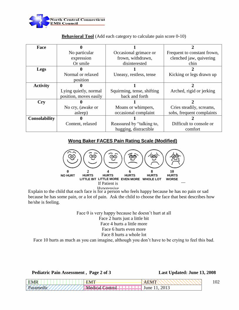
EMR EMT AEMT
Paramedic Medical Control June 11, 2013 - -
102
Behavioral Tool (Add each category to calculate pain score 0-10)
Face 0
No particular
expression
Or smile
1
Occasional grimace or
frown, withdrawn,
disinterested
2
Frequent to constant frown,
clenched jaw, quivering
chin
Legs 0
Normal or relaxed
position
1
Uneasy, restless, tense 2
Kicking or legs drawn up
Activity 0 Lying quietly, normal
position, moves easily
1
Squirming, tense, shifting
back and forth
2
Arched, rigid or jerking
Cry 0 No cry, (awake or
asleep)
1
Moans or whimpers,
occasional complaint
2
Cries steadily, screams,
sobs, frequent complaints
Consolability 0 Content, relaxed
1 Reassured by “talking to,
hugging, distractible
2 Difficult to console or
comfort
Wong Baker FACES Pain Rating Scale
Explain to the child that each face is for a person who feels happy because he has no pain or sad
because he has some pain, or a lot of pain. Ask the child to choose the face that best describes how
he/she is feeling.
Face 0 is very happy because he doesn’t hurt at all
Face 2 hurts just a little bit
Face 4 hurts a little more
Face 6 hurts even more
Face 8 hurts a whole lot
Face 10 hurts as much as you can imagine, although you don’t have to be crying to feel this bad.
Pediatric Pain Assessment , Page 2 of 3 Last Updated: June 13, 2008
0 NO HURT
2 HURTS
LITTLE BIT
4 HURTS
LITTLE MORE
If Patient is
Hypotensive
or Displays
Orthostatic
Hypotensio
n:
Administer
0.9% NS IV
Bolus
6 HURTS
EVEN MORE
8 HURTS
WHOLE LOT
10 HURTS
WORSE
Wong Baker FACES Pain Rating Scale (Modified)

EMR EMT AEMT
Paramedic Medical Control June 11, 2013 - -
103
Visual Analog Scale
Ask the patient to mark or point to the severity of their pain on a scale of zero to ten with zero being
no pain and ten being unbearable pain, the worst pain they have ever felt.
Verbal Numeric Pain Score
Ask the patient to rate the severity of their pain on a scale of zero to ten with zero being no pain and
ten being unbearable, the worst pain ever.
Verbal Pain Score
Ask the patient to assign one of the following adjectives to rate their pain:
NONE MILD MODERATE SEVERE UNBEARABLE
Pediatric Pain Assessment , Page 3 of 3 Last Updated: June 13, 2008
MODERATE
DISTRESSING PAIN
SEVERE
UNBEARABLE PAIN
NO
PAIN

EMR EMT AEMT
Paramedic Medical Control June 11, 2013 - -
104
PEDIATRIC ALLERGIC REACTION
Pediatric Allergic Reaction , Page 1 of 1 Last Updated: June 2, 2009
Oxygen as needed and
tolerated
Diphenhydramine (Benadryl) Elixir, tablet or dissolving strip 1 mg/kg orally.
Maximum dose 50 mg.
Universal Patient Care Guideline
Avoid IV access and parenteral medication if possible to reduce patient discomfort and stress.
Monitor Patient Condition.
If respiratory impairment or hemodynamic
instability develop or patient condition worsens,
move to anaphylaxis guideline.
Stable Hemodynamics Without Respiratory Impairment Minor or
moderate skin manifestations. Blood pressure appropriate for age by
formula or length based tape. Signs of adequate perfusion are present
(appropriate mental status, skin color, temp, capillary refill, etc.)
Pediatric Patient Assessment Guideline
Consider administration of oral Diphenhydramine (Diphenhydramine (Benadryl)) and careful
monitoring in the patient that receives an Epi-Pen prior to EMS arrival (for signs and
symptoms (s/sx) of an allergic reaction) but then presents to EMS with no s/sx of a reaction
and/or with an acuity level consistent with this guideline.

EMR EMT AEMT
Paramedic Medical Control June 11, 2013 - -
105
PEDIATRIC ANAPHYLAXIS
Pediatric Assessment Guideline ↓
Oxygen and
Pediatric Airway Guideline ↓
Paramedic
Epinephrine (1:1,000) 0.01 mg/kg
IM
Or
Epi-Pen Jr. (if under 30kg)
EMT-B, EMT-I
Administer Epi-Pen Jr. if under 30kg Standard Epi-Pen if
over 30kg For anaphylaxis patients who has at least two of the following criteria:
1.Hypotension (according to formula or length based tape)
2. Difficulty in breathing or wheezing
3. Hives, redness and/or itching
4. Difficulty in swallowing ↓
Establish IV access (IO if needed for Paramedic)
Fluid Bolus 20ml/kg of Normal Saline or Lactated Ringers ↓
If bronchospasm, administer Albuterol (Ventolin, Proventil) nebulized treatment ( 5 mg)
Diphenhydramine (Benadryl) 1mg/kg IV/IO push (over one minute) IM if no IV access. Maximum
dose 50 mg.
If above treatment does not improve patient status:
Epinephrine (1:10,000) 0.01 mg/kg (maximum dose of 0.3mg) slow IV/IO push
Methylprednisone (Solu-Medrol) 2mg/kg infusion over 15 minutes ↓
Establish medical Control Possible Physician orders:
Repeat Epinephrine IM or IV doses q 5 minutes
Epinephrine infusion 0.1 to 0.3 µg/kg/min increasing to 1.0 µg/kg/min as necessary
Fluid Bolus 20ml/kg of Normal Saline or Lactated Ringers
Unstable Hemodynamics - hypotensive patient according to normal values for age and weight;
pending upper airway obstruction with wheezing and/or stridor; or severe obstruction with wheezing
and/or stridor; or severe respiratory distress.
Reminder: Cardiac monitor is indicated for all patients receiving epinephrine.
Pediatric Anaphylaxis , Page 1 of 1 Last Updated: June 13, 2008

EMR EMT AEMT
Paramedic Medical Control June 11, 2013 - -
106
PEDIATRIC FEVER
Pediatric Patient Care Guideline
↓
Wear N95 mask if highly
infectious agent suspected or
bioterrorism related event
- Obtain temperature if available
(Temporal artery scan preferred)
- Passive cooling without
inducing shivering
↓
Temperature 101.5°F (38.5°C)
or greater
Or
Patient believed to be febrile
and temperature is unavailable
or unable to obtain.
No
↓ No
Patient >6 months old?
↓
Patient has received acetaminophen (Tylenol) within 4 hours? No
No ↓ Yes ↓
Administer:
Acetaminophen
(Tylenol) 15 mg/kg
PO
Patient has received less than 15
mg/kg acetaminophen (Tylenol)
within last 4 hours?
Yes ↓
Consider administering a “make-up” dose to bring the total dose of
acetaminophen (Tylenol) up to 15 mg/kg PO
↓
Continued Reassessment
Contact Medical Control
Pearls:
Pediatric Fever , Page 1 of 1 Last Updated: June 13, 2008
This Guideline is NOT to be used for patients suffering from environmental hyperthermia
If the patient is vomiting, suppositories are more appropriate and oral acetaminophen (Tylenol) should be
withheld.
Administer once the patient is in the ambulance to avoid patient/parent refusal after treatment.
Concentrated infant drops (80 mg per 0.8 mL) are recommended and may be dispensed using a needless
syringe.
Do not administer acetaminophen (Tylenol) if the patient has received greater than 15 mg/kg dose
in the last 6 hours.

EMR EMT AEMT
Paramedic Medical Control June 11, 2013 - -
107
PEDIATRIC ALTERED MENTAL
STATUS/HYPOGLYCEMIA/COMA
Pediatric Assessment Guideline
↓
Pediatric Airway Guideline
↓
Consider etiology (trauma,
hypoglycemia, overdose, seizure,
hypoxemia, etc.)
Treat according to appropriate Guideline
Determine GCS
↓
Check Blood Glucose Level
In the following circumstances:
Glucose <70
Blood Glucose not available and
patient is known diabetic or
History consistent with
hypoglycemia
Intact Gag Reflex
Administer Oral Glucose
↓
Establish IV access
Administer Dextrose
10% 0.5 Gm/kg IV
push
If IV access cannot be
readily established
Administer Glucagon
0.02 mg/kg up to 1mg
IM
If a narcotic overdose is suspected or
unknown and respiratory insufficiency is
present:
Administer Naloxone (Narcan) 0.1 mg/kg
IV or IM or IN. May repeat to a maximum
dose of 2.0 mg.
Establish Medical Control Possible Physician orders:
If no IV access, establish IO
Repeat D10W
Repeat Naloxone (Narcan)
Pearl: For children less than a week old, see drug guide for dose of dextrose and contact medical control
D10 is the preferred solution for hypoglycemic emergency. D10 can be given either as an open drip or drawn up in
a large syringe and given as IV pushes. If D10 unavailable, D25 or D50 may be used.
Pediatric Altered Mental Status , Page 1 of 1 Last Updated: June 13, 2008

EMR EMT AEMT
Paramedic Medical Control June 11, 2013 - -
108
PEDIATRIC SEIZURES/STATUS EPILEPTICUS
Pediatric Patient Assessment Guideline
↓
If post-trauma and age <9 years
cervical and full spinal immobilization
as appropriate while maintaining
airway
↓
Closely assess respiratory activity. Use
blow-by oxygen. Assist ventilations
with B-V-M and 100% O2 as necessary.
Suction as necessary.
↓
IV Access
↓
Check Blood Glucose Level
↓
If glucose level <70 administer:
Dextrose 0.5 Gm/kg
↓
Cardiac monitor
↓
HAS THE SEIZURE PERSISTED
BEYOND 10 MINUTES (from onset)?
↓
Yes No
↓ ↓
Administer: Lorazepam (Ativan)
0.1 mg/kg (up to 4 mg) SIVP (or)
Diazepam (Valium) 0.25 mg/kg
(up to 3 mg) SIVP (or)
If unable to establish IV access
administer: Midazolam (Versed)
0.1 mg/kg (up to 10 mg) IM or
0.2mg/kg (up to 10mg) IN
Lorazepam (Ativan), Diazepam
(Valium) or Midazolam (Versed)
may be repeated once as needed
Establish Medical Control
Possible Physician orders:
Administer: Lorazepam
(Ativan) 0.1 mg/kg (up to 4 mg)
SIVP (or) Diazepam (Valium)
0.25 mg/kg (up to 3 mg) SIVP
(or)
If unable to establish IV access
administer: Midazolam (Versed)
0.1 mg/kg (up to 10 mg) IM or
0.2mg/kg (up to 10mg) IN
Lorazepam (Ativan), Diazepam
(Valium) or Midazolam (Versed)
may be repeated once as needed
Repeat Dextrose 25%
↓
Establish Medical Control
Possible Physician orders:
Repeat administration of anti-
seizure medications
Repeat Dextrose 25%
Pediatric Seizures , Page 1 of 2 Last Updated: June 2, 2009

EMR EMT AEMT
Paramedic Medical Control June 11, 2013 - -
109
PEARLS:
Initiate treatment based on history and clinical presentation. It is essential to make the distinction between focal motor,
general motor seizures, and Status Epilepticus.
Most seizures do not require emergent intervention.
Perform an initial assessment. Attempt to determine the etiology i.e. whether the patient has a history of diabetes, seizure
disorder, narcotic use, head trauma, poisoning or fever.
NOTE: If the seizure is controlled by one of the benzodiazepines, continuous assessment of respiratory status is critical
as respiratory arrest can occur with use of these medications.
Pediatric Seizures , Page 2 of 2 Last Updated: June 2, 2009

EMR EMT AEMT
Paramedic Medical Control June 11, 2013 - -
110
PEDIATRIC OVERDOSE/POISONING
Pediatric Patient Assessment
Guideline
At the earliest convenience
contact Poison Control
directly or through Medical
Control
1-800-222-1222
Conscious Oral Ingestion? Establish and maintain airway,
Support ventilations as needed
Inhalation or Topical Exposure?
Yes Oxygen Yes
Routine Patient Care
Guideline
Pediatric Altered Level of
Consciousness
Guideline if indicated
Evaluate the scene for safety
consideration as a Hazmat Incident
Notify CMED as indicated
Follow BLS HAZMAT Guidelines as
indicated
Oxygen Establish IV of Normal
Saline or Lactated Ringers
Fluid bolus if hypotensive
Routine Patient Care Guideline
Establish Medical Control
Possible Physician orders:
Activated Charcoal*
Cardiac Monitor
Treat symptomatic rhythm
according to Guideline
Establish Medical Control Specific exposure information for
further treatment orders and specific
arrival instructions (e.g., use a
specific hospital entrance)
Important: NEVER
INDUCE VOMITING
If a Narcotic Overdose is
suspected see Pediatric altered
mental state-opiate Guideline
If patient remains
unresponsive:
Rapid glucose determination
Administer Dextrose 0.5 G/kg
IV for glucose <70
Establish Medical Control Possible Physician orders:
Management specific for
agent exposure, Additional
Dextrose
*An aqueous solution rather than a solution that includes Sorbital should be used for pediatric patients
.
Pediatric OD/Poisoning , Page 1 of 1 Last Updated: June 13, 2008
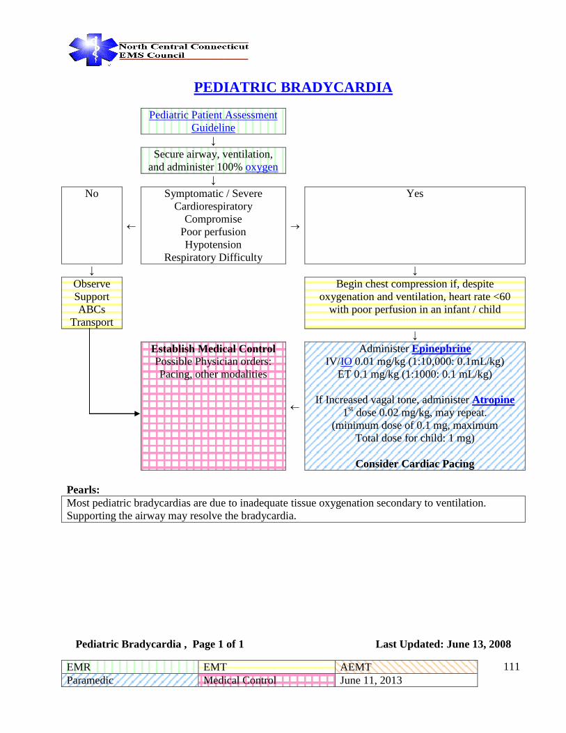
EMR EMT AEMT
Paramedic Medical Control June 11, 2013 - -
111
PEDIATRIC BRADYCARDIA
Pediatric Patient Assessment
Guideline
↓
Secure airway, ventilation,
and administer 100% oxygen
↓
No
Symptomatic / Severe
Cardiorespiratory
Compromise
Poor perfusion
Hypotension
Respiratory Difficulty
Yes
↓ ↓
Observe
Support
ABCs
Transport
Begin chest compression if, despite
oxygenation and ventilation, heart rate <60
with poor perfusion in an infant / child
↓
Establish Medical Control Possible Physician orders:
Pacing, other modalities
Administer Epinephrine
IV/IO 0.01 mg/kg (1:10,000: 0.1mL/kg)
ET 0.1 mg/kg (1:1000: 0.1 mL/kg)
If Increased vagal tone, administer Atropine
1st dose 0.02 mg/kg, may repeat.
(minimum dose of 0.1 mg, maximum
Total dose for child: 1 mg)
Consider Cardiac Pacing
Pearls:
Most pediatric bradycardias are due to inadequate tissue oxygenation secondary to ventilation.
Supporting the airway may resolve the bradycardia.
Pediatric Bradycardia , Page 1 of 1 Last Updated: June 13, 2008

EMR EMT AEMT
Paramedic Medical Control June 11, 2013 - -
112
Pediatric Tachycardia Rapid heart rate with adequate perfusion
Pediatric Patient
Assessment Guideline
Oxygen
↓
Obtain 12 lead EKG if
possible
↓
QRS duration
Normal for age ≤ 0.09sec Evaluate QRS Duration QRS duration wide for age >0.09 sec
↓ ↓
Evaluate rhythm
Probable SVT
Amiodarone (Cordorone) 5mg/kg IV over
20 – 60 min
or
Procainamide (Pronestyl) 15 mg/kg IV over
30 – 60 minutes
Or
Adenosine (adenocard) 0.1 mg/kg IV (max
first dose of 6 mg) if unclear of width of QRS
↓ ↓
Probable sinus tachycardia Establish IV access
↓ ↓
Identify and treat possible
causes:
Fever, shock, hypoxia,
hypovolemia, drug
ingestion, pneumothorax
Consider vagal maneuvers
Adenosine (adenocard) 0.1
mg/kg IV (max first dose of 6
mg)
May double (0.2 mg/kg) and
repeat dose once (max second
dose of 12 mg)
Yes Termination
↓ ↓
Establish Medical Control
No
↓
Establish Medical Control
May consider cardioversion after sedation
at 0.5 – 1 j/kg (may increase to 2 j/kg if
ineffective) Sedate before cardioversion
Pediatric Tachycardia , Page 1 of 2 Last Updated: October 27, 2011

EMR EMT AEMT
Paramedic Medical Control June 11, 2013 - -
113
PEDIATRIC TACHYCARDIA
Rapid heart rate with poor perfusion
Pediatric Patient
Assessment Guideline
Oxygen
↓
Obtain 12 lead EKG if possible
↓
QRS duration
Normal for age ≤ 0.09sec Evaluate QRS Duration QRS duration wide for age >0.09 sec
↓ ↓
Evaluate rhythm
Probable SVT
If cardiopulmonary compromise, attempt
synchronized cardioversion with 0.5 to 1 j/kg
(may increase to 2 j/kg if needed), if no
compromise then continue to
↓ ↓
Probable sinus
tachycardia
Establish IV access
↓ ↓
Identify and treat possible
causes:
Fever, shock, hypoxia,
hypovolemia, drug
ingestion, pneumothorax
Consider vagal maneuvers
Adenosine (adenocard) 0.1
mg/kg IV (max first dose of 6
mg)
May double and repeat dose
once (max second dose of 12
mg)
or
Attempt synchronized
cardioversion with 0.5 to 1
j/kg (may increase to 2 j/kg if
needed)
Adenosine (adenocard) 0.1 mg/kg IV (max
first dose of 6 mg)
Amiodarone (Cordorone) 5mg/kg IV over 20
– 60 min
or
Procainamide (Pronestyl) 15 mg/kg IV over
30 – 60 minutes
↓
Yes Termination
↓ ↓
If drug therapy successful start
infusion drip
Establish Medical Control
No
↓
Establish Medical Control
Pediatric Tachycardia, Page 2 of 2 Last Updated: October 27, 2011
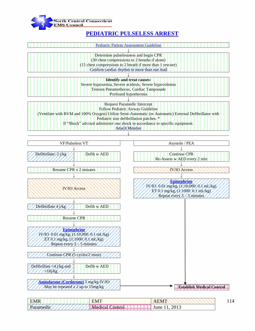
EMR EMT AEMT
Paramedic Medical Control June 11, 2013 - -
114
PEDIATRIC PULSELESS ARREST
Pediatric Patient Assessment Guideline
↓
Determine pulselessness and begin CPR
(30 chest compressions to 2 breaths if alone)
(15 chest compressions to 2 breath if more than 1 rescuer)
Confirm cardiac rhythm in more than one lead
↓
Identify and treat causes:
Severe hypoxemia, Severe acidosis, Severe hypovolemia
Tension Pneumothorax, Cardiac Tamponade
Profound hypothermia
↓
Request Paramedic Intercept
Follow Pediatric Airway Guideline
(Ventilate with BVM and 100% Oxygen) Utilize Semi-Automatic (or Automatic) External Defibrillator with
Pediatric size defibrillation patches. *
If “Shock” advised administer one shock in accordance to specific equipment.
Attach Monitor
↓
VF/Pulseless VT Asystole / PEA
↓ ↓
Defibrillate: 2 j/kg Defib w AED Continue CPR
Re-Assess w AED every 2 min
↓ ↓
Resume CPR x 2 minutes IV/IO Access
↓ ↓
IV/IO Access
Epinephrine
IV/IO 0.01 mg/kg, (1:10,000: 0.1 mL/kg)
ET 0.1 mg/kg, (1:1000: 0.1 mL/kg)
Repeat every 3 – 5 minutes
↓
Defibrillate 4 j/kg Defib w AED
↓
Resume CPR
↓
Epinephrine
IV/IO 0.01 mg/kg, (1:10,000: 0.1 mL/kg)
ET 0.1 mg/kg, (1:1000: 0.1 mL/kg)
Repeat every 3 – 5 minutes
↓
Continue CPR (5 cycles/2 mins)
↓
Defibrillate >4 j/kg and
<10j/kg
Defib w AED
↓
Amiodarone (Cordorone) 5 mg/kg IV/IO
May be repeated x 2 up to 15mg/kg Establish Medical Control

EMR EMT AEMT
Paramedic Medical Control June 11, 2013 - -
115
North Central Connecticut
Regional
Paramedic Guidelines
Pediatric
Trauma Guidelines
<13 Years

EMR EMT AEMT
Paramedic Medical Control June 11, 2013 - -
116
PEDIATRIC TRAUMA TRIAGE
Pediatric Patient
Assessment Guideline
↓
Does the patient meet Level I or II
trauma center criteria in accordance
with State of Connecticut trauma regulations (19a-177-5a)?
↓
Yes
GCS < 12 or SBP < 90 (hypotensive
for age) or RR < 10 or > 29
(< or > normal for age)
GSW to chest, head, neck, abdomen, or groin
Third degree burns > 15% TBSA, or
third degree burns of face or with airway involvement
Evidence of spinal cord injury or
Amputation, other than digits or two or more obvious proximal long bone
fractures
No
↓ ↓
Transport to the closest
appropriate Level I or II
Trauma center.
Does the patient meet any of the following Criteria requiring medical control in determining patient
destination?
↓ ↓
If the patient is less than 13
years of age
Transport to a Level I or Level
II trauma facility
With pediatric resources
including a pediatric ICU.
Yes Falls > 20 feet or 3 times the patient’s height, High Speed impact, Ejection of patient from vehicle, Death of same car
occupant, Ped struck by car at > 20 MPH, Rollover MVC,
Age<5 Penetrating injury to thorax, abdomen neck, or groin other than GSW
↓ ↓ ↓
If transport to an appropriate trauma center is Determined to be
>20 minutes, contact your
Medical control for direction
Utilize your department’s
preauthorized Guideline or request
medical direction In determining
most appropriate patient
destination
No
↓
Follow normal operating procedures for determining medical destination
↓
If the patient’s central pulses
cannot be palpated,
Airway cannot be managed, or
bleeding is
uncontrollable transport to
nearest hospital
Pediatric Trauma Triage , Page 1 of 1 Last Updated: June 13, 2008

EMR EMT AEMT
Paramedic Medical Control June 11, 2013 - -
117
PEDIATRIC BURN PATIENT
Pediatric Patient Assessment
Guideline
↓
Oxygen
↓
Establish an IV Lactated Ringers (in area not
affected by burn if possible).
→ IO Access if indicated
↓
Type of Burn Assess percentage of Total Body Surface Area
Burned
↓
Thermal Burns Chemical Burns Ophthalmic Burns Electrical Burns
↓ ↓ ↓ ↓
Cover burns with clean, dry
dressing. If the burns are
less than 10% and are
superficial or partial
thickness you can moisten
towels or sheets with sterile
normal saline for comfort.
Otherwise use dry sterile
dressings
Consider any chemical burn situation as a
Hazmat situation. If potential Hazmat situation
exists, notify receiving hospital ASAP
Identify the chemical if possible
Immediate and
continuous flushing of
the affected eye is performed using
Lactated Ringers or
Normal Saline. If contact lenses are
known to be in the
patient’s eyes, an attempt should be made
to remove them and
continue flushing.
Suspect spinal
injury
secondary to tetanic muscle
contraction
Immobilize Patient if age
less than 9
years. Assess for entrance and
exit wounds.
↓ ↓ ↓ ↓
If burn injury greater than 20%
BSA, administer 20ml/kg fluid
bolus. Repeat bolus if indicated by poor perfusion.
Remove affected clothing & jewelry (if not already
done)
Unless contraindicated
instill 1 or 2 drops of
ophthalmic anesthesia. Place medication onto
lower lid.
IV Normal
Saline
↓ ↓ ↓ ↓
Control Pain
Refer to Pediatric Pain
Control Guideline
Brush off dry powder. Flush with copious amounts of water or saline unless contraindicated. Irrigate
burns to the eyes with a minimum of 1 liter of
Lactated Ringers or Normal Saline. Alkaline burns should receive continuous irrigation throughout
transport. Consider the Morgan Lens for eye
irrigation if older than 6, (see right).
Place the Morgan Lens (if the patient is older
than 6 years old) in the
affected eye(s) continuously flush with
Lactated Ringers or
Normal Saline while enroute to the hospital.
Run 2 liters of fluid wide per eye, then
administer kvo rate.
Cardiac Monitor
Treat any
cardiac rhythm disturbances
per guideline
↓
Control Pain
Refer to
Pediatric Pain
Control
Guideline
↓ ↓ ↓
Establish Medical Control Possible Physician Orders:
Additional MS, Intubation.
Refer to Pediatric Pain Control Guideline Refer to Pediatric Pain
Control Guideline
Establish
Medical
Control
↓
Establish Medical Control Possible Physician Orders:
Morphine Sulfate IVP
Pediatric Burns , Page 1 of 2 Last Updated: June 13, 2008

EMR EMT AEMT
Paramedic Medical Control June 11, 2013 - -
118
Pearls:
The approach to the pediatric burn patient should be similar in your approach to any burn patient, assuring
your safety, the patient’s safety, stopping the burning process, and airway management all remain
paramount. These Guidelines will deal with specific fluid resuscitation measures and special considerations. Please refer to the appendix for the “Pediatric Rule of Nines.” Please refer to the Adult Trauma - Burn
section of these Guidelines for your “systems” approach to patient care. The anatomical map of the pediatric patient changes with age, if in doubt as to the Body Surface Area
involved in the burn see the “Rule of Nines.” Be suspicious for burn patterns that may indicate child abuse, i.e. “stocking” or “glove” pattern burns. Ophthalmic Chemical Burns The Morgan Lens may be utilized in children >6yrs who are cooperative. Care must be take to prevent any child who has had topical ophthalmic anesthesia from rubbing their eye - additional injury may occur since the pain receptors have been blocked
Pediatric Burns , Page 2 of 2 Last Updated: June 13, 2008

EMR EMT AEMT
Paramedic Medical Control June 11, 2013 - -
119
Appendix

EMR EMT AEMT
Paramedic Medical Control June 11, 2013 - -
120
North Central Connecticut
Regional Paramedic
Procedures

EMR EMT AEMT
Paramedic Medical Control June 11, 2013 - -
121
12-LEAD ECG
When to obtain: On patient contact (In conjunction with initial set of vitals).
Indications:
Any patient suspected of acute coronary syndrome, including any of the following (but not limited to):
1. Chest pain, pressure or discomfort
2. Radiating pain to neck or left arm. Also right arm, shoulder or back
3. Dyspnea
4. CHF
5. Cardiac Arrhythmias
6. Syncope/near syncope
7. Profound weakness
8. Epigastric discomfort
9. Hyperglycemia in diabetic patients
10. Sweating incongruent with environment
11. Nausea, vomiting
12. Previous cardiac history or other cardiac factors
13. Presence of anginal equivalents
14. Overdoses
15. Altered Mental Status
Before and after any rhythm conversion including PSVT and rapid afib.
In patients with suspected Acute Coronary Syndrome, a 12-Lead ECG should ideally be done on first
patient contact, during transport and on arrival at the ED. ECG results should be transmitted and
medical control notified.
Technique:
1. Remove patient clothing above waist. Use a gown or sheet to preserve patient modesty
2. Apply limb leads (to the shoulders)
3. Print rhythm strip
4. Apply precordial leads
5. Place patient in position of comfort (supine preferred).
6. Verify that all leads are securely attached
7. Acquire 12-lead
8. Prepare to repeat 12-lead at 10 minute intervals or on change in condition
If 12-lead reveals possible STEMI, inform Medical Control Immediately so the hospital will be prepared for
your patient. DO NOT WAIT UNTIL LATE IN YOUR TRANSPORT TO CALL. Early notification saves
lives!
Notes:
12-leads should only be performed by paramedics who have taken an approved course and been evaluated for competency
by medical control.
A normal 12-Lead ECG does not rule out the possibility of ischemic cardiac disease and must not be used to screen
patients.
12 Lead ECG , Page 1 of 1 Last Updated: June 11, 2013

EMR EMT AEMT
Paramedic Medical Control June 11, 2013 - -
122
CINCINNATI PREHOSPITAL STROKE SCALE
Sign/Symptom: Facial Droop
How Tested: Have patient show their teeth or smile.
Normal: Both sides of the face move equally.
Abnormal: One side of the face does not move as well as the other.
Sign/Symptom: Pronator Drift
How Tested: The patient closes their eyes and extends both arms straight out, palms up, for 10
seconds.
Normal: Both arms move the same, or both do not move at all.
Abnormal: One arm either does not move, or one-arm drifts downward (pronator drift) compared to
the other.
Sign/Symptom: Speech:
How Tested: The patient repeats “The sky is blue in Cincinnati.”
Normal: The patient says correct words with no slurring of words.
Abnormal: The patient slurs words, says the wrong words, or is unable to speak.
Patients with 1 of these findings – as a new event – have a 72% probability of an ischemic stroke.
If all 3 findings are present the probability of an acute stroke is more than 85%.
Early notification to the receiving hospital is essential to ensure the immediate availability of an
appropriate in-hospital response.
Cincinnati Stroke Scale, Page 1 of 1 Last Updated: June 2, 2009

EMR EMT AEMT
Paramedic Medical Control June 11, 2013 - -
123
Endotracheal Tube Introducer (BOUGIE)
Indication:
For use as a guide during adult endotracheal intubation, either routine or difficult, when the endotracheal tube can not be
passed despite a good view of the vocal cords, an incomplete view of the glottic opening, or when only the epiglottis is
visualized.
Precautions:
The ETI is not to be used in a completely blind fashion. At a minimum, the epiglottis or an incomplete view of the glottic
opening must be visualized. Excessive force, passage beyond the carina, or blind introduction may result in damage to
the soft tissue of the oropharynx or trachea/bronchi.
Procedure:
1. A 15F ETI should be used for ET tube sizes 6.0 to 10.0.
2. Lubricate ETI with a water soluble lubricant.
3. Perform laryngoscopy. If cords not visible after optimal manipulation of the thyroid cartilage, identify
landmarks (epiglottis at minimum) to aid intubation.
4. Place introducer (with curved tip) into the mouth and direct into glottis or underneath epiglottis with the firm end
of the ETI pointing anterior. The intubator and/or person holding cricoid pressure may feel clicking as the ETI
passes over the tracheal rings (this supports tracheal placement of the ETI).
5. Advance the ETI to approximately 20 cm at the teeth (look for black mark (s) on ETI)
6. The intubator may feel the ETI scathe to one side if the ETI passes the carina. If advanced to deeply, there may
also be increased resistance upon entrance into the bronchus. Failure to meet resistance after almost complete
insertion should suggest esophageal placement.
This concept can be used to confirm proper endotracheal tube placement if other techniques and/or signs of
proper tube placement are difficult to interpret. GENTLY pass the ETI via the endotracheal tube. The
carina/bronchus should impede ETI advancement at 30-35 cm depth. If no distal resistance is noted, the
endotracheal tube may be in the esophagus.
7. If possible, continue laryngoscopy to elevate the pharyngeal soft tissue while an assistant threads an endotracheal
tube over the introducer into the trachea. If the endotracheal tube meets resistance at the laryngeal inlet and will
not pass, stop advancement efforts, withdraw the endotracheal tube 1-2 cm with rotation of the tube 90° in either
or both directions while reattempting to advance the tube may help negotiate passage of the tube.
8. While the assistant holds the ETI, advance the endotracheal tube to the desired depth and hold the tube firmly in
place while the ETI is gently withdrawn.
Remove laryngoscope, secure the endotracheal tube, and confirm tube placement as usual.
Bougie , Page 1 of 1 Last Updated: June 13, 2008

EMR EMT AEMT
Paramedic Medical Control June 11, 2013 - -
124
ESOPHAGEAL-TRACHEAL COMBITUBE Indication: Apneic patient without a gag reflex
Contraindications: 1. Patient under the age of 16 years.
2. Patient under 5’0’’ or over 6’6” in height. (for patients under 5’0” there is a SA tube)
3. Ingestion of a caustic substance. “SA” for short adult
4. Severe oral facial trauma.
5. Esophageal disease.
6. Patient with a stoma.
Procedure: 1. Use basic precautions including gloves and goggles.
2. Hyperoxygenate patient before attempting placement.
3. Test equipment while patient is being oxygenated.
4. If basic airway is in place remove it; keep head in neutral or slightly flexed position.
5. With one hand, grab tongue/mandible and lift towards ceiling.
6. With the other hand place the Combitube so that it follows the natural curve of the
pharynx.
7. Insert to the tip of the mouth and advance gently until the printed ring is aligned with
the teeth.
8. Do Not Force. If the Combitube does not advance easily withdraw and reinsert.
9. Inflate the blue tube balloon with 100 cc of air. Inflate the white tube balloon with
15cc of air.
#1 Blue - will inflate the posterior pharyngeal balloon.
#2 White - will inflate the distal balloon.
10. Begin ventilation through the longer blue connecting tube. If auscultation of breath
sounds is positive and auscultation of gastric insufflation is negative, continue
ventilations.
11. IF NECESSARY, if auscultation of breath sounds is negative, and gastric insufflation
is positive, immediately begin ventilation through the shorter connecting clear tube.
Confirm tracheal ventilation by auscultation of breath sounds and absence of gastric
insufflation.
12. Removal of Combitube:
a. Reassure patient
b. Have suction ready and roll patient on their side.
c. Remove 100cc of air from #1 (Blue line).
d. Remove 15cc of air from #2 (White line).
e. Gently withdraw Combitube, suction patient as necessary.
Notes:
The paramedic should not hesitate to utilize the Combitube as a backup or as an alternative advanced airway in a patient
where the establishment of an ET tube may delay securing the patient’s airway.
Paramedics should generally leave the airway in if the airway is patent.
Combitube , Page 1 of 1 Last Updated: June 13, 2008

EMR EMT AEMT
Paramedic Medical Control June 11, 2013 - -
125
INTRANASAL NALOXONE (NARCAN)
Indication:
Suspected opiate overdose (See Altered Mental Status Guideline)
Procedure:
1. Load syringe with 2 mg (2ml) Naloxone (Narcan) and attach nasal atomizer
2. Place atomizer 1.5 cm within the nostril
3. Briskly compress syringe to administer 1 ml of atomized spray.
4. Remove and repeat in other nostril, spraying remaining 1ml.
5. If no results within 3 minutes, establish IV and administer IV Naloxone (Narcan).
Note:
It is recommended to use Naloxone (Narcan) 1mg/1ml concentration
Intranasal Naloxone (Narcan), Page 1 of 1 Last Updated: June 13, 2008

EMR EMT AEMT
Paramedic Medical Control June 11, 2013 - -
126
INTRAOSSEOUS INFUSION
The following guideline is to be utilized for FDA approved intraosseous access devices only. You should follow the
specific manufacturer’s guidelines for the insertion procedure.
INDICATIONS: May be inserted on standing order for the following:
Adult (> 40 kg) & Pediatric (3 – 39 kg)
1. Intravenous fluids or medications needed and a peripheral IV cannot be established in 2 attempts or 90
seconds AND the patient exhibits any of the following:
a. An altered mental status
b. Respiratory compromise
c. Hemodynamic instability
d. Status Epilepticus unresponsive to IM or rectal medication
2. If IV access is not readily available, then you may consider using IO as a backup PRIOR to peripheral IV
attempts in the following situations:
a. Cardiac arrest (medical or traumatic).
b. Profound hypovolemia (Shock) with altered mental status.
c. Patient in extremis with immediate need for delivery of medications and or fluids.
CONTRAINDICATIONS:
Fracture of the tibia or femur or humerus
Previous orthopedic procedure (knee replacement) or IO within 24 hours
Infection over insertion site
Inability to locate landmarks due to either significant edema or excessive tissue
Note: If contraindication exists, utilize an alternate insertion site.
PROCEDURE:
1. If the patient is conscious, explain the procedure and provide the rationale for it.
2. Use appropriate body substance isolation equipment.
3. If the patient is conscious, prime the extension set with 2%, preservative-free Lidocaine* and leave the syringe
attached (up to 50 mg Lidocaine total).
4. Identify the insertion site.
5. Prep the site with betadine** or alcohol.
6. Prepare the IO needle.
7. Stabilize, insert the IO needle and remove the stylet.
8. Confirm proper placement (aspiration of marrow; flushes freely without extravasation).
Introsseous, Page 1 of 2 Last Updated: June 13, 2008

EMR EMT AEMT
Paramedic Medical Control June 11, 2013 - -
127
9. Connect the extension set and flush with 2% preservative-free Lidocaine*:
a. Adults 20-50 mg
b. Pediatric 0.5 mg/kg
10. Rapidly flush with 0.9% NaCl:
a. Adults 10cc
b. Pediatric 5cc
11. Start infusion utilizing a pressure bag or BP cuff (if pressure bag unavailable).
12. Secure the catheter and tubing.
13. Attach identification wrist band, notify receiving facility staff and deliver removal instruction form to treating
physician or nurse.
14. Frequently monitor IO catheter site and patient condition.
* If the patient is unconscious or allergic to Lidocaine, prime the extension set with 5-10 mL 0.9% NaCl.
** Betadine is preferred (if patient is not allergic to iodine). If time permits, swab three separate times in an outward,
circular motion utilizing a fresh applicator each time.
Note: Paramedics must have attended a medical control approved, device-specific in-service and demonstrated
competency prior to utilizing an IO device in clinical practice.
COMPLICATIONS:
Infection
Compartment syndrome
Subcutaneous extravasation
Clotting of marrow in needle
Osteomyelitis/cellulitis
PEARLS
Sites:
Should be used on sites in accordance with manufacturer recommendations and guidance from local
medical control.
Introsseous, Page 2 of 2 Last Updated: June 13, 2008
Complications are infrequent (0.6%) and consist mostly of pain and extravasation.
IO flow rates are typically slower than with IV catheters. Use a pressure bag or pump.
Insertion of IO needles in conscious patients causes mild-moderate discomfort and is usually no more
painful than a large bore IV.
Infusion through an IO line may cause severe discomfort for conscious patients and preservative-free
lidocaine should be administered.
Onset of analgesia with lidocaine typically takes approximately 1 minute and lasts 40 minutes to 1 hr.
The most common side effect of lidocaine toxicity is seizures.

EMR EMT AEMT
Paramedic Medical Control June 11, 2013 - -
128
MORGAN LENS
Indication:
For use in patients age 6 years and older who have sustained an exposure injury to the eye(s), (i.e. dry
or liquid chemical).
Equipment:
1. Gloves
2. 1000ml IV bag Lactated Ringers or Normal Saline
3. IV tubing (macro drip)
4. Morgan Lens
5. Tetracaine or other ophthalmic anesthetic
6. Towels or chux
Procedure:
Explain procedure to patient and give rationale.
Use BSI (Body Substance Isolation)
Unless contraindicated*, instill one or two drops of Tetracaine.
Instruct patient not to touch/rub eye(s).
Spike IV bag and attach/flush tubing, connect Morgan Lens, maintain sterile
environment of Morgan Lens.
Have the patient look down, insert the Morgan Lens under the upper lid, then have
the patient look up, retract lower lid and allow lens to drop into place.
Begin flow rate at wide open and maintain this rate per patient tolerance. Have
plenty of towels or chux to absorb flow.
*Contraindication: allergic reaction to local anesthetics, i.e. Novocain, Lidocaine
Morgan Lens , Page 1 of 1 Last Updated: June 2, 2009

EMR EMT AEMT
Paramedic Medical Control June 11, 2013 - -
129
NEEDLE CRICOTHYROTOMY
Indication:
The inability to secure the patient’s airway by other invasive procedures, (endotracheal intubation).
Cautions:
1. Needle cricothyrotomy is an invasive procedure and requires proper training and certification
through one of the Regional Sponsor Hospitals.
2. Carbon dioxide (CO2) build-up occurs rapidly. The procedure can be used only for a
short period of time (30 minutes maximum) at which time a definitive airway must be
established.
3. The patient must have a patent airway or a means established to allow outflow of air
from the lungs.
Contraindications:
1. The ability to establish an easier and less invasive airway rapidly.
2. Acute laryngeal disorders such as laryngeal fractures that cause landmark distortion
or obliteration of landmarks.
3. Bleeding disorders.
4. Injury or obstruction below the level of the cricothyroid membrane.
Complications:
Pneumothorax
Subcutaneous emphysema
Catheter dislodgment
Hemorrhage
Esophageal or mediastinal injury
Hypercarbia
Equipment:
14 gauge or larger over-the-needle catheter, or commercial product
10 cc syringe
3 cc syringe
15 mm adapter from a 3.0 ET tube
Bag-valve-mask
Oxygen
Needle Crichothyrotomy , Page 1 of 2 Last Updated: June 13, 2008
EMR EMT AEMT
Paramedic Medical Control June 11, 2013 - -
130
NEEDLE CRICOTHYROTOMY (CONTINUED)
Equipment:
Povidone iodine swabs
Adhesive tape
Trauma shears
Suction equipment
Gloves
Goggles
Procedure:
Observe basic precautions
Prepare equipment: remove plunger from barrel of 3cc syringe. Attach 15mm
adapter from the 3.0 ET tube.
Palpate the thyroid cartilage, cricothyroid membrane, and suprasternal notch.
Prep the skin with two povidone iodine or alcohol swabs.
You may attach the 10 cc syringe to the over-the-needle catheter, or you may elect
to use the catheter-needle assembly by itself. Puncture the skin over the
cricothyroid membrane.
Advance the needle at a 45-degree angle caudally (toward the feet).
Carefully push the needle until it pops into the trachea (aspirating on the syringe as
you advance the needle if you are using a syringe).
Free movement of air confirms you are in the trachea.
Advance the plastic catheter over the needle, holding the needle stationary, until the
catheter hub comes to rest against the skin.
Holding the catheter securely, remove the needle.
Reconfirm the position of the catheter. Securely tape the catheter.
Attach the 3 cc syringe with the 3.0 ET adapter to the catheter hub. Attach the BVM
to the adapter and forcefully ventilate the patient. Forcefully squeeze the BVM
over one second to inflate and then remove the BVM to allow for exhalation (for 4
seconds).
Constantly monitor the patient’s breath sounds, ventilation status, and color.
Adequate exhalation never forcefully occurs with this technique. The patient may
develop hypercarbia (increased CO2) and increased air pressure in the lungs
possibly causing alveoli to rupture.
EMS Providers with Commercially made devices should follow the manufacturer’s recommendations
and directions for use.
Needle Crichothyrotomy , Page 2 of 2 Last Updated: June 13, 2008

EMR EMT AEMT
Paramedic Medical Control June 11, 2013 - -
131
NEEDLE THORACOSTOMY
Indication:
Tension pneumothorax associated with either traumatic or spontaneous lung collapse and manifested
by hypotension, severe respiratory distress, absent breath sounds with hyper resonance on the
affected side. There may be possible tracheal shift to the unaffected side.
Contraindications:
There are no contraindications for field use.
Procedure:
1. The patient is supine with head and chest to 30 degrees (semi-sitting position).
2. Explain procedure and rationale if patient is conscious. Bare the chest.
3. Select site for procedure—usually the anterior second or third intercostal space in
the midclavicular line.
4. Cleanse the skin.
5. Select needle or over-the-catheter needle size 14 gauge or larger.
6. Holding the needle/catheter perpendicular to the chest wall, insert it straight into the
thorax, going just above a rib when one is encountered. Insert until air is heard
escaping. Advance catheter and remove needle. This converts a tension
pneumothorax to a simple pneumothorax. A chest tube thoracostomy will need to
be placed in the ED.
7. Cover puncture site, stabilize catheter to transport. Although not mandatory, when
possible, attach tube to flutter valve or flap valve.
“Pearls”
Use the largest needle or catheter possible since “plugging” with tissue may occur.
Intercostal nerve or artery damage, be sure to go above not below the rib.
Injury to the diaphragm - site is too low or the patient is not positioned correctly.
Subcutaneous placement - insertion not perpendicular to chest wall (remember it
is curved, not flat).
Infections - late complication - prevent this by prepping the skin well.
* Note: Since recent studies have shown that the standard 2” catheter may not be long enough to
penetrate the chest wall of 10 to 35% of the population, in large patients with thick chest walls, if
clinical improvement does not occur after initial attempt at decompression with a standard 2”
catheter, decompression with a longer (up to 3.5“) catheter may be attempted. Insert needle only
until air is heard escaping. Advance catheter until hub is on chest wall or until resistance is met. In
extremely large patients, the paramedic has the option to use the larger catheter on first attempt.
Needle Thoracostomy , Page 1 of 1 Last Updated: June 2, 2009

EMR EMT AEMT
Paramedic Medical Control June 11, 2013 - -
132
Pediatric Glasgow Coma Scale
CHILD INFANT
Eye Opening 4 - opens spontaneously 4 - opens spontaneously
3 - opens to speech 3 - opens to speech
2 - opens to pain 2 - opens to pain
1 – none 1 – none
5 - oriented 5 - coos and babbles
Verbal Response 4 - confused 4 - irritable cry
3 - inappropriate words 3 - cries in pain
2 - incomprehensible words 2 – moans in pain
1 - none 1 - none
6 - obeys commands 6 - spontaneous movement
Motor Response 5 - localizes pain 5 - withdraws to touch
4 - withdrawal to pain 4 - withdraws to pain
3 - flexion (pain) 3 - flexion (pain)
2 - extension (pain) 2 - extension (pain)
1 – none 1 - none
Changes in neurologic status can be of significance to the trauma surgeon or to the neurosurgeon. Significant
alteration can change the outcome for the patient

EMR EMT AEMT
Paramedic Medical Control June 11, 2013 - -
133
Pediatric Glasgow Coma Scale , Page 1 of 1 Last Updated: June 13, 2008

EMR EMT AEMT
Paramedic Medical Control June 11, 2013 - -
134
RULE of NINES ADULT
Adult Rule of Nines , Page 1 of 1 Last Updated: June 13, 2008

EMR EMT AEMT
Paramedic Medical Control June 11, 2013 - -
135
RULE of NINES PEDIATRIC
Area Age – 0 Age – 1 Age – 5 Age – 10 Age – 15
A = ½ of head 9 ½ 8 ½ 6 ½ 5 ½ 4 ½
B = ½ of thigh 2 ¾ 3 ¼ 4 ½ 4 ¼ 4 ½
C = ½ of leg 2 ½ 2 ½ 3 3 3 ¼
Lund and Browder method of calculating pediatric BSA for burns.
Pediatric Rule of Nines , Page 1 of 1 Last Updated: June 13, 2008

EMR EMT AEMT
Paramedic Medical Control June 11, 2013 - -
136
SURGICAL CRICOTHYROTOMY
Indication:
This is a surgical procedure and may be performed in extreme situations. It should be considered for
field use only when all other techniques have failed.
Equipment:
1. Goggles
2. Gloves
3. Betadine prep swabs
4. #10 scalpel
5. 2x2 and 4x4 gauze sponges
6. Adhesive tape or umbilical tape
7. Endotracheal tube(s) 5.0 through 6.5
Procedure:
1. Basic precautions.
2. Stabilize larynx with thumb and index fingers.
3. Identify cricothyroid membrane and prep with Betadine swabs.
4. Using careful dissection cut through the skin and soft tissue in a vertical manner
approximately 1cm.
5. Gently retract the skin to reveal the cricothyroid membrane.
6. Make a stab wound horizontally through the membrane to a depth of ¼” to ½.” Care
should be taken not to enter to deeply as posterior structures may be injured.
7. Remove the scalpel and using the blunt end, insert it into the stab wound, rotate the
blunt end 90 degrees to create an opening. Alternatively, hemostats, tracheal hooks, a bougie or
a finger may all be used to dilate the opening. Maintain this opening and with gentle
yet controlled pressure, pass the appropriate size endotracheal tube 1cm past the
cuff.
8. Inflate the balloon. Attach a BVM and ventilate the patient.
9. Ascultate for breath and epigastric sounds, attach end-tidal CO2.
10. Secure ETT in place. Consider “shortening” the ETT by cutting it, care should be
taken not to cut it below the level of the pilot balloon.
Contraindication:
This procedure is not indicated for pediatric patients, less than 8 years of age.
Surgical Crichothyrotomy, Page 1 of 1 Last Updated: June 13, 2008

EMR EMT AEMT
Paramedic Medical Control June 11, 2013 - -
137
TUBE CONFIRMATION ADJUNCTS
Statement: There are a number of different types and brands of end-tidal CO2 detectors, it will not be
dictated by the Region which type or which brand to use. However, it is the policy in the Region that
each patient who is intubated will have quantitative wave-form end-tidal CO2 and/or Esophageal
Intubation Detector (EID) used to confirm placement and to monitor placement.
Indications: All intubated patients.
Procedure:
1. Following oral or nasal intubation, confirm positive and equal breath sounds and the absence of
epigastric sounds. Place end-tidal CO2 and EID.
2. Ventilate patient with 100% oxygen.
3. Depending upon the type/brand of end-tidal CO2 detector used the paramedic will
confirm tube placement by noting color change or CO2 numbers.
4. If the EID (Esophageal Intubation Detector) is used, a rapid reinflation will occur
with correct placement of the ETT.*
5. If confirmation that the tube is correctly placed is noted, ETT will be secured in place
in the usual manner and monitored en route to the hospital.
6. If the end-tidal CO2 detector or EID indicates incorrect ETT, immediate visualization
of tube placement should be done. If ETT is incorrectly placed, immediately remove
ETT, hyperoxygenate patient and reattempt intubation.
7. If visualization shows the ETT properly placed, secure tube in the usual manner and
continue to ventilate and monitor patient en route to the hospital. Report finding to
physician caring for patient.
Note: All ET Tube Attempts Must Be Documented On Regional ET Form Proper documentation on the patient’s PCR should indicate use of end-tidal CO2
and/or use of EID and findings.
*It is possible to have a positive placement finding with the EID. If the tube tip is
at the level right above the vocal cords, but not through the cords rapid inflation
of the bulb may occur.
Once a patient has stopped cellular respiration (death) color change is not always
possible even with a properly placed ETT.
It is possible for an end-tidal CO2 detector to have a positive color change with an
esophageal intubation. This may occur for a limited time (usually on 4 or 5
ventilations) correct color change can be assured after this.
Tube Confirmation Adjuncts , Page 1 of 1 Last Updated: June 13, 2008
EMR EMT AEMT
Paramedic Medical Control June 11, 2013 - -
138
Quantitative Waveform Capnography
Capnography is the vital sign of ventilation, providing a breath by breath trend of respirations and an
early warning system of impending respiratory crisis. Capnography will indicate an impending
problem much sooner than pulse oximetry or patient observation.
Advanced Airway Indications
ET Tube Confirmation and Monitoring Tube Position
Quantitative waveform capnography must be used on all intubated patients. Continuous end-tidal
CO2 monitoring can confirm both successful placement of a tracheal intubation and its continuing
proper position. A good wave form indicating the presence of CO2 ensures the ET tube is in the
trachea. You must still assess for equal lung sounds. Capnography cannot detect right main-stem
intubations.
Monitoring CPR Monitoring ETC02 indirectly measures cardiac output, thus monitoring ETCO2 is a good way to
measure the effectiveness of CPR. A decrease in cardiac output will lower the delivery of carbon
dioxide to the lungs decreasing the ETCO2. The better the CPR, the higher the ETC02.
Return of Spontaneous Circulation
ETCO2 can be the first sign of return of spontaneous circulation (ROSC). During a cardiac arrest, if
you see the ETCO2 number suddenly shoot up, stop CPR and check for pulses. End-tidal CO2 will
often overshoot baseline values when circulation is restored due to carbon dioxide washout from the
tissues. After a minute, the number should return to baseline.
Loss of Spontaneous Circulation In a resuscitated patient, if you see the stabilized ETCO2 number significantly drop in a person with
ROSC, immediately check pulses. You may have to restart CPR.
Predictor of Resuscitation Outcome
End tidal CO2 monitoring can confirm the futility of resuscitation as well as forecast the likelihood of
resuscitation. An ETCO2 level of 10 mmHg or less after 20 minutes of advanced cardiac life support
can help accurately predict death. In general, patients with a high initial ETCO2 reading are more
likely to be resuscitated than those with lower values. It is important to note, however, while a low
initial ETCO2 makes resuscitation less likely than a higher initial ETCO2, patients have been
successfully resuscitated with an initial ETCO2 >10 mmHg.
Ventilating Head Injured Patients Capnography can help paramedics avoid hyperventilation in intubated head injured patients. Try to
keep the patient as normocapnic as possible. A target value of 35 mmHg is recommended.
Quantitative Waveform Capnography, Page 1 of 3 Last Updated: December 10, 2011

EMR EMT AEMT
Paramedic Medical Control June 11, 2013 - -
139
Combitubes and LMAs
Quantitative waveform capnography should also be used on Combi-tubes and LMAs as well as on all
ET tubes. A capnography filter can also be used in between a bag valve mask and the face mask to
access ETCO2 during BVM use.
Nonintubated Indications
Monitoring for Hypoventilation
When a person hypoventilates, their ETCO2 goes up. Hypoventilation can be caused by altered
mental status such as overdose, sedation, intoxication, postictal states, head trauma, or stroke, or by a
tiring CHF patient. A high or steadily rising ETCO2 can help a paramedic anticipate when a patient
may require assisted ventilations or intubation. Monitoring ETCO2 provides a better gauge of
ventilatory status than respiratory rate. ETCO2 will show a heroin overdose with a respiratory rate of
24 (with many shallow ineffective breaths) and an ETCO2 of 60 is more in need of arousal than a
patient with a respiratory rate of 8 and an ETCO2 of 35.
ETCO2 in Asthma, COPD End-tidal CO2 monitoring with a nasal cannula can be used to assess the severity of Asthma/COPD,
and the effectiveness of treatment. Bronchospasm will produce a characteristic “shark fin” wave
form, as the patient has to struggle to exhale, creating a sloping “B-C” upstroke. The shape is caused
by uneven alveolar emptying. Successful treatment may lessen or eliminate the shark fin shape and
return the ETCO2 to normal range. ETCO2 values may also fluctuate with severity of the episode.
With a mild asthma, the CO2 will drop (below 35) as the patient hyperventilates to compensate. As
the asthma worsens, the C02 levels will rise to normal. When the asthma becomes severe, and the
patient is tiring and has little air movement, the C02 numbers will rise to dangerous levels. Pay as
much attention to the trend as to the number.
Monitoring Sedated Patients Quantitative waveform capnography should be used to monitor any patients receiving pain
management or sedation (enough to alter their mental status) for evidence of hypoventilation and/or
apnea. Beware of rising ETC02 values in these patients and be prepared to stimulate them to breathe
if necessary.
Documentation
Paramedics should document their use of continuous ETCO2 monitoring and attach wave form strips
to their PCRs.
Quantitative Waveform Capnography, Page 2 of 3 Last Updated: December 10, 2011

EMR EMT AEMT
Paramedic Medical Control June 11, 2013 - -
140
Notes
ETCO2 of 35-45 mm Hg is the normal value for capnography. Some experts consider 30-45 as
normal.
Hyperventilation will cause the number to go down. Hypoventilation will cause it to go up.
Some patients who are baseline hypercapnic will have much higher values as their norm.
Imperfect positioning of nasal cannula capnofilters may cause distorted low readings. Unique nasal
anatomy, obstructed nares and mouth breathers may skew results and/or require repositioning of
cannula. Also, oxygen by mask may lower the reading by 10% or more.
Pay as much attention to the ETCO2 trend as to the number!
Quantitative Waveform Capnography, Page 3 of 3 Last Updated: December 10, 2011

EMR EMT AEMT
Paramedic Medical Control June 11, 2013 - -
141
Tourniquet Usage Guidelines INDICATIONS A. A tourniquet should be used to control hemorrhagic wounds that have not responded adequately to direct pressure or in situations of significant extremity bleeding with the need for additional interventions (example: significant extremity bleeding with airway compromise. A tourniquet should be used to quickly control bleeding, freeing up Personnel to concentrate on airway issues.) PRECAUTIONS A. Use BSI
B. A tourniquet applied incorrectly can increase blood loss and lead to death.
C. Applying a tourniquet can cause nerve and tissue damage whether applied correctly or not. Proper patient selection is of the utmost importance.
D. Damage is unlikely if the tourniquet is removed within 2 hours. Low risk to tissue is acceptable over death secondary to hypovolemic shock.
E. Tourniquets should never be covered up by patient clothing or packaging. TECHNIQUE A. Attempt to control hemorrhage with direct pressure or pressure dressing.
B. If unable to control hemorrhage using the above means, apply a tourniquet, using the procedure below, and minding the above considerations a. Select commercially manufactured tourniquet, blood pressure cuff, or improvised “Spanish Windlass” is applied to the extremity proximal to the wound, preferably on single-bone structures (humerus and femur) above wound. Do not place over joints.
b. Tighten tourniquet until bleeding stops.
c. The time and date of application (“TK 20:30” indicates that the tourniquet was placed at 8:30 pm) should be written on a piece of tape and secured to the tourniquet or written directly on the patient’s skin next to the tourniquet with a permanent marker.
d. The tourniquet should be left uncovered so that the site can be monitored for recurrent hemorrhage.
e. Keep tourniquet on throughout transport – a correctly applied tourniquet should only be removed by the receiving hospital.
f. Continue monitor patient vitals and wound g. Ensure receiving personnel are aware of tourniquet placement
Tourniquet Use, Page 1 of 1 Last Updated: December 10, 2011

EMR EMT AEMT
Paramedic Medical Control June 11, 2013 - -
142
STEMI ALERT PROCEDURE 1. Acquire a 12-lead on all patients suspected of Acute Coronary Syndrome (active chest pain or equivalent symptoms (SOB, nausea, etc.) on first contact. 2. If 12-lead is diagnostic for STEMI and paramedic believes patient is having STEMI, contact CMED for STEMI Alert with Medical Control patch, and transmit ECG if possible. If possible and less than 30 minutes from PCI center, do not wait until transporting to call hospital. Failure to notify hospital until 5 minutes out will delay reperfusion. 3. When hospital answers phone, confirm MD Control, and state “I have a STEMI Alert and am requesting STEMI activation.” If you are uncertain the patient is having a STEMI, say “I have a Possible STEMI Alert.” 4. Describe 12-lead and patient condition. Based on the conversation between paramedic and ED MD and if applicable, the transmitted 12-Lead, the cath lab will either be activated in advance of arrival, placed on standby or not activated until the physician can make a more detailed assessment at the hospital. 5. Provide Appropriate Care during transport per guideline. Have defib pads ready in case patient goes into unstable ventricular tachycardia or ventricular fibrillation. Consider disrobing patient if time permits. Have latest 12-lead ready to show ED MD on arrival. Be prepared to transport patient to cardiac cath lab on EMS stretcher if given the go- ahead from ED staff. 6. Please leave copy of PCR and all 12-lead strips at the hospital prior to departing. PCRs should include Time of 911 Dispatch, Time at Patient side, Time of 1st 12-lead, Arrival at the Hospital, as well as all care rendered. 7. If applicable to hospital, fill out QA/Patient Follow-up form in ED. NOTES: STEMI Definition for Field Activation STEMI is defined by ECG of good quality with all of the following: a. ST elevation in 2 or more contiguous leads of >2 mm (V1-V4 or > 1 mm (limb or lateral) b. QRS duration < 0.12 second c. ***Acute MI*** or equivalent prints on 12-lead and paramedic agrees. If the machine does not read ***Acute MI*** but the paramedic still strongly believes the ECG shows a STEMI, the paramedic may proceed with the activation request. Computer Interpretation STEMI Alert, Page 1 of 2 Last Updated: June 12, 2013

EMR EMT AEMT
Paramedic Medical Control June 11, 2013 - -
143
Paramedics should not diagnose STEMI based solely on 12-lead computer interpretation. While the interpretation can by used to support your diagnosis, the computer is not infallible. The computer will not read all STEMIs as ***Acute MI Suspected***.” And the computer may read ***Acute MI*** when the ECG is clearly not a STEMI. The computer is less accurate with wide complex and tachycardic rhythms. STEMI Imposters Paramedics should be familiar with and take into consideration all of the known STEMI Imposters, including Bundle Branch Block Paced Rhythms Early Repolarization Pericarditis Left Ventricular Hypertrophy (LVH) Other PEARLS Do serial 12-leads (ideally all patients with ACS should have 12-lead on patient contact, on beginning transportation and on arrival at ED). STEMIs are often evolving. The STEMI may not appear until 3rd 12-lead or the STEMI captured on 1st 2-lead may disappear by arrival at the ED. A prehospital 12-lead documenting the transient elevation is critical in these patients. Regional PCI Hospitals Hartford Hospital John Dempsey Hospital New Britain Saint Francis Baystate Memorial Hospital (Springfield) Early Notification Saves Lives!
STEMI Alert, Page 2 of 2 Last Updated: June 12, 2013

EMR EMT AEMT
Paramedic Medical Control June 11, 2013 - -
144
North Central Connecticut
Regional Paramedic
Medications

EMR EMT AEMT
Paramedic Medical Control June 11, 2013 - -
145
Acetaminophen (Tylenol)
(Tylenol)
Class: Antipyretic; Analgesic
Action: Antipyretic effect via direct action on the hypothalamus
heat-regulation center; Unknown mechanism of analgesia
Indications: Pediatric fever; Minor pain
Contraindication: Hypersensitivity to acetaminophen (Tylenol)
Adverse effects: Hepatotoxicity in overdose
Nausea
Pedi Dose: 15 mg/kg every 4 – 6 hours as needed
Route: PO; PR
Note: Concentrated infant drops (80 mg per 0.8 mL) are
recommended and may be dispensed using a needleless
syringe.
Appears in Pediatric Fever guideline
Last Updated: June 13, 2008
EMR EMT AEMT
Paramedic Medical Control June 11, 2013 - -
146
Activated Charcoal
Class: Absorbent
Action: Absorbs many drugs and poisons in the GI tract
Indication: Toxic ingestions - not caustics or pure petroleums
Contraindication: None for emergency use
Dose: 30-50-100 grams
Route: PO - usually in liquid form to drink
Pedi dose: 1-2 grams/kg
Appears in Overdose & Poisoning guideline
Appears in Pediatric OD/Poisoning guideline
Last Updated: June 13, 2008

EMR EMT AEMT
Paramedic Medical Control June 11, 2013 - -
147
Adenosine
(Adenocard)
Class: Endogenous nucleoside
Action: Stimulates adenosine receptors; decreases conduction through the AV
node
Indication: PSVT, Monomorphic Wide Complex Tachycardia
Contraindication: Patients taking Persantine or Tegretol.
Precaution: Short half-life must administer rapid normal saline bolus immediately after
administration of drug. Use IV port closest to IV site.
Side effect: Arrhythmias, chest pain, dyspnea, bronchospasm (rare)
Dose: Adult - 6mg IV over 1-2 seconds; may repeat 12mg in 2 minutes. Pedi - 0.1mg/kg,
may repeat once at 0.2mg/kg
Route: Rapid IV push, followed by a flush
Appears in Tachycardia – Afib/Aflutter guideline
Appears in Pediatric Tachycardia with Adequate Perfusion guideline
Appears in Pediatric Tachycardia with Poor Perfusion guideline
Last Updated: June 25, 2012
EMR EMT AEMT
Paramedic Medical Control June 11, 2013 - -
148
Albuterol
(Ventolin, Proventil)
Class: ß2 Agonist
Synthetic sympathomimetic
Bronchodilator
Action: Stimulates ß2 receptors in the smooth muscle of the bronchial tree.
Indication: Relief of bronchospasm.
Contraindication: None for field use.
Precaution: Patient with tachycardia.
Side effect: Tachycardia
Dose: 2.5mg (0.5ml of the 0.5% solution diluted to 3ml NS) for nebulized updraft.
Route: Inhaled as a mist via nebulizer.
Pediatric Dose: 2.5mg nebulized updraft.
Appears in Respiratory Distress guideline
Appears in Anaphylaxis guideline
Appears in Pediatric Asthma guideline
Appears in Pediatric Allergic Reaction guideline
Appears in Pediatric Anaphylaxis guideline
Last Updated: June 13, 2008

EMR EMT AEMT
Paramedic Medical Control June 11, 2013 - -
149
Amiodarone
(Cordorone)
Class: Antiarrythmic
Action: Reduces myocardial cell membrane excitability by increasing the
effective refractory period
Inhibits alpha and beta adrenergic stimulation, causing peripheral
vasodilation and decreased heart rate
Indication: Cardiac arrest -- ventricular fibrillation
Wide Complex Tachycardia w/pulse>150 bpm
Contraindication: none for cardiac arrest, contraindicated for wide complex tachycardia
with hypotension (synchronized shock indicated). Bradycardia.
Dose: Cardiac arrest – 300 mg IV; May repeat at 150 mg
Wide Complex Tachycardia w/pulse>150 bpm – 150mg IV over 10
minutes
Drip – 1mg/min
Route: IV
Side Effects: Hypotension, bradycardia, headache, dizziness, nausea, vomiting
Appears in Tachycardia – Wide Complex guideline
Appears in V-fib/Pulseless V-tach guideline
Appears in Pediatric Tachycardia with Adequate Perfusion guideline
Appears in Pediatric Tachycardia with Poor Perfusion guideline
Appears in Pediatric Cardiac Arrest guideline
Last Updated: June 13, 2008

EMR EMT AEMT
Paramedic Medical Control June 11, 2013 - -
150
Aspirin
(Acetylsalicylic acid)
Class: Antiplatelet
Action: Inhibitor of platelet aggregation
Effects: Decrease clotting time
Indication: Chest pain of cardiac origin
Contraindication: Allergy to aspirin
Dose: 325mg tab or 4-baby aspirin (81mg per tab)
Route: PO
Side Effects: None with field use
Last Updated: June 13, 2008

EMR EMT AEMT
Paramedic Medical Control June 11, 2013 - -
151
Atropine
(Atropine Sulfate)
Class: Antimuscarinic
Parasympathetic blocker
Anticholinergic
Action: Blocks acetylcholine (ACh) at muscarinic sites
Indication: Symptomatic bradyarrhythmias
Cholinergic poisonings
Refractory bronchospasm
Contraindication: Relative contraindication wide complex bradycardia in the setting of acute
ischemic chest pain
Side effects: Tachyarrhythmias
Exacerbation of Glaucoma
Precipitation of myocardial ischemia
Dose: Bradyarrhythmias - 0.5mg , may repeat every 3-5 minutes
Organophosphate poisonings - 2mg IV or IM Repeat, doubling dose every 5
minutes until bronchorrhea ceases. i.e. Initial dose of 2mg; Second dose of
4mg; Third dose of 8 mg, etc.
Route: IV push
Pedi dose: 0.02mg/kg IV
Appears in the Bradycardia guideline
Appears in Pediatric Bradycardia guideline
Appears in the Poisoning and Overdose guideline
Last Updated: June 2013

EMR EMT AEMT
Paramedic Medical Control June 11, 2013 - -
152
Benzocaine spray
(Cetacaine)
Class: Topical anesthetic
Action: Blocks conduction of impulses at the sensory nerve endings.
Indication: Nasal intubations or oral intubations where patient may still have gag reflex.
To improve patient comfort and tolerance of intubation.
Contraindication: Known sensitivity to Benzocaine products.
Children under 1 year of age.
Precaution: Children under 6 years of age.
Side effects: Rash; May induce methemoglobinemia (especially in overdose or in infants
and young children). This is a condition in which altered hemoglobin no
longer effectively carries oxygen. It is characterized (in late stages) by a
chocolate-brown color to the blood and cyanosis. Signs of
methemoglobinemia are related to hypoxia and include: confusion, coma,
seizures, respiratory distress and tachydysrythmias. Standard pulse oximetry is
not accurate in methemoglobinemia.
Dose: ½ second spray to the posterior pharynx; allow approximately 20-30 seconds
after administration for effect to occur. Do not exceed one second total spray
duration (expulsion rate ~200 mg/second)
Route: PO
Last Updated: June 13, 2008

EMR EMT AEMT
Paramedic Medical Control June 11, 2013 - -
153
Calcium Chloride
Class: Electrolyte
Action: Facilitates the actin/myosin interaction in the heart muscle.
Indication: Hypocalcemia
Hyperkalemia with arrhythmia
Calcium channel blocker intoxication with hypotension or symptomatic
bradycardia
Contraindication: Not to be mixed with any other medication - precipitates easily.
Precaution: Patients receiving calcium need cardiac monitoring
Side effect: Cardiac arrhythmias
Precipitation of digitalis toxicity
Dose: Usual dose is 5-10ml of 10% Calcium Chloride in 10ml.
Route: IV
Pedi Dose: 0.2ml/kg of 10% concentration
Appears in adult cardiac arrest guideline
Appears in Bradycardia guideline
Appears in V-fib/Pulseless V-tach guideline
Last Updated: June 13, 2008

EMR EMT AEMT
Paramedic Medical Control June 11, 2013 - -
154
Dextrose
(D50W), (D25W) (D10W)
Class: Carbohydrate
Action: Raises the blood sugar
Indication: Diabetic patients with low blood sugar level (<70 mg/dL in adults)
Altered mental states
Seizure
Contraindication: acute strokes with normal finger stick blood glucose i.e. neurotoxic
Precaution: Tissue necrosis if infiltration occurs
Side effects: As above-infiltration
Intracerebral hemorrhages in neonates with undiluted D50
Dose: Up to 25 Gms Slow IV push, may repeat (Can be given to adults as eitherD50,
D25 or D10) A concentration of D10W is the preferred method of
administration of dextrose.
Route: IV slow, confirm IV placement prior to and during administration.
Pedi Dose: A concentration of D10W is the preferred method of administration of
dextrose. Otherwise1ml/kg of D50W slow IV push. Dilute 1 to 4 in those less
than 1 week old and dilute 1 to 2 (D25W) in those 1 week to 16 years.
Appears in adult cardiac arrest guideline
Appears in Altered Level of Consciousness guideline
Appears in Hypothermia guideline
Appears in Seizure guideline
Appears in Stroke guideline
Appears in Pediatric Altered Mental Status/Hypoglycemia guideline
Appears in Pediatric Seizure guideline
Appears in Pediatric OD/Poisoning guideline
Last Updated: June 11, 2013

EMR EMT AEMT
Paramedic Medical Control June 11, 2013 - -
155
Diazepam
(Valium)
Class: Benzodiazepine
Action: Decreases cerebral irritability
Calms CNS
Indication: Major motor seizures
Acute anxiety states
Pre-cardioversion
Contraindication: none for emergency field use
Dose: 2 to 20mg to control seizure activity
2 to 5mg for anxiety or pre-cardioversion
Route: IV push - slow
Pedi dose: 0.25 mg/kg (up to 3 mg) SIVP
Appears in Pediatric Seizure guideline

EMR EMT AEMT
Paramedic Medical Control June 11, 2013 - -
156
Last Updated: June 13, 2008
Diltiazem
(Cardizem)
Class: Calcium channel blocker
Action: Partial blockade of AV node conduction
Indication: Atrial fibrillation, Atrial flutter, narrow complex tachycardia
Contraindication: Hypotension
Hypersensivity to drug
Wide complex tachycardia
Known history of Wolf Parkinson White (WPW)
2° or 3° AV block
Relative contraindication: Already on Digoxin and Beta Blocker
Side effect: May induce VF if given to patient with wide complex tachycardia that is due to
WPW.
May cause hypotension
Dose: Initial dose: 0.25mg/kg slow IV (average dose 20mg in adult male)
May repeat with 0.35 mg/kg (25 mg average) in 10-15 minutes if no or
diminishing effect. Decrease by 5 mg per bolus for elderly (>70 yr/old).
Route: IV push (bolus) given over 2 minutes; reconstitute according to
manufacturer’s recommendation.
Pedi dose: 0.25mg/kg
Important points: If patient is hypotensive secondary to drug administration:
- If not in failure give IV fluids
- If bradycardic administer CaCl2
- If still bradycardia give Atropine
- Transcutaneous pacing may be necessary for markedly symptomatic
bradycardia.
- If CHF is present or worsens administer Dopamine (Intropin) infusion
- If all of above fail (persistent hypotension >2-5 minutes) administer glucagon
1 mg IV
Appears in Tachycardia – Afib/Aflutter guideline
Appears in Tachycardia – PSVT guideline
Last Updated: June 13, 2008

EMR EMT AEMT
Paramedic Medical Control June 11, 2013 - -
157
Diphenhydramine
(Benadryl)
Class: Antihistamine
H1 blocker
Action: Blocks histamine receptor sites
Indication: Systemic anaphylaxis
Drug induced extrapyramidal reactions
Contraindication: None with emergency use
Precaution: Asthma
Side effect: Sedation
Hypotension
Dose: 25 -50mg
Route: IV push, may also be given IM
Pedi Dose: 1mg/kg
Appears in Allergic Reaction guideline
Appears in Anaphylaxis guideline
Appears in Dystonic Reaction guideline
Appears in Pediatric Allergic Reaction guideline
Appears in Pediatric Anaphylaxis guideline
Last Updated: June 13, 2008

EMR EMT AEMT
Paramedic Medical Control June 11, 2013 - -
158
Dopamine (Intropin)
(Intropin)
Class: Naturally occurring catecholamine, adrenergic agents
Action: Stimulates α, β1 and Dopamine (Intropin)rgic receptors
Effects: 0.5 to 2 μg/kg/min - Renal and mesenteric vasodilation.
2 to 10 μg/kg/min - Renal and mesenteric vasodilation persists and
increased force of contraction (FOC).
10 to 20 μg/kg/min - Peripheral vasoconstriction and increased FOC (HR may
increase).
20 μg/kg/min or greater - marked peripheral vasoconstriction (HR may increase).
Indication: Shock - Cardiogenic
- Septic
- Anaphylactic
Bradycardia
Contraindication: Pre-existing tachydysrhythmias or ventricular dysrhythmias.
Precaution: Infuse in large vein only
Use lowest possible dose to achieve desired hemodynamic effects,
because of potential for side effects.
Do not D/C abruptly; effects of Dopamine (Intropin) may last up to 10 minutes after
drip is stopped.
Do not mix with NaHCO3 as alkaline solutions will inactivate Dopamine (Intropin).
Side effect: Tachydysrhythmias
Ventricular ectopic complexes
Undesirable degree of vasoconstriction
Hypertension relate to high doses
Nausea and vomiting
Anginal pain
Dose: 2.0 - 20. μg/kg/min titrated to desired effect
Route: IV drip
Pedi dose: same as adult dose - titrate to effect
Appears in Bradycardia guideline Appears in Shock guideline
Appears in V-fib/Pulseless V-tach guideline Appears in Management of the Trauma Patient
Appears in Anaphylaxis guideline
Last Updated: June 2011

EMR EMT AEMT
Paramedic Medical Control June 11, 2013 - -
159
Epinephrine 1:10,000
Class: Natural catecholamine, adrenergic
Action: Stimulates both alpha (a) and beta (ß1 and ß2) receptors.
Indication: Cardiac arrest – Adult
Cardiac arrest - Pediatric
Anaphylaxis with shock
Contraindication: Use in pregnant women should be conservative
Pre-existing tachydysrhythmias
Side effects: Tachydysrhythmias
Hypertension
May induce early labor in pregnancy
Headache, nervousness, decreased level of consciousness
Dose: 0.5 to 1.0 mg (usual) for cardiac arrest
0.1 mg slow IV over 3 minutes, can be repeated X 2 to a maximum dose of
0.3mg for anaphylaxis
2-10 mcgs/min IV drip for bradycardia
Route: IV, IO
ET if given this route the dose should be doubled
Pedi Dose: 0.01 mg/kg (0.1 ml/kg)
Appears in Asystole guideline
Appears in Bradycardia guidelines (mixed in 1000mL IV drip)
Appears in V-fib/Pulseless V-tach guideline
Appears in PEA guideline
Appears in Anaphylaxis guideline
Appears in Neonatal Resuscitation guideline
Appears in Pediatric Anaphylaxis guideline
Appears in Pediatric Bradycardia guideline
Appears in Pediatric Cardiac Arrest guideline
Last Updated: June 2011

EMR EMT AEMT
Paramedic Medical Control June 11, 2013 - -
160
Epinephrine 1:1,000
Class: Same as Epi 1:10,000
Action: Same as Epi 1:10,000
Indication: Severe allergic reaction, status asthmaticus,
laryngeal or lingual edema
Contraindication: Use with caution in the presence of:
Pre-existing tachydysrhythmias
Hypertension
Significant cardiac history
Pregnancy
Side effect: Same as Epi 1:10,000
Dose: 0.3 mg
Route: IM
Pedi dose: 0.01 mg/kg (0.01 ml/kg) to a max. 0.3 mg (0.3ml)
See PALS guidelines
For croup administer 5mg (5ml) nebulized with 2.5-3ml of NS
Appears in Bradycardia guidelines (mixed in 1000mL IV drip)
Appears in Respiratory Distress guideline
Appears in Allergic Reaction guideline
Appears in Anaphylaxis guideline
Appears in Pediatric Asthma guideline
Appears in Croup/Epiglottitis guideline
Appears in Pediatric Allergic Reaction guideline
Appears in Pediatric Anaphylaxis guideline
Last Updated: June 13, 2008

EMR EMT AEMT
Paramedic Medical Control June 11, 2013 - -
161
Fentanyl Citrate
Class: Narcotic analgesic
Action: Decreases pain perception and anxiety
Onset of action: Fentanyl citrate is a narcotic analgesic. A dose of 100 mcg (0.1 mg)
(2 mL) is approximately equivalent in analgesic activity to 10 mg of
morphine or 75 mg of meperidine.
Indications: Moderate to severe pain adults
Contraindications: Significant Head injury
GCS < 13
SBP < 100
Allergy to Fentanyl
Side effects: Respiratory depression or arrest
Decreased LOC
Hypotension
Increased vagal tone (slowed heart rate)
Nausea/vomiting
Pinpoint pupils
Increased cerebral blood flow
Urticaria
Chest Wall Rigidity
Precautions: Continuous patient monitoring after administration of Fentanyl is required
including (when physically possible) pulse oximetry, ECG, quantitative
waveform capnography (when available), vital signs and respiratory effort;
Rapid administration increases the likelihood of side effects. Use aliquots of
25 mcgs to 50 mcgs for patients over 65.
Adult Dose: Analgesia general: Initial dose 1 mcg/kg (upto 100 mcg) slow IV over 3 to 5
minutes. Repeat dose of 1 mcg/kg (up to an additional 100 mcg) slow IV after
10 minutes if needed. Repeat dose of 1 mcg/kg (up to an additional 100 mcg)
slow IV after an additional 10 minutes if needed for a total maximum dose of
300 mcg
Route: Slow IV; IO push – slow, IN, IM
Appears in Adult Pain Control Guideline
Last Updated: October 2011

EMR EMT AEMT
Paramedic Medical Control June 11, 2013 - -
162
Glucagon
Class: Pancreatic hormone
Action: Increases blood glucose by converting liver glycogen to glucose
Indication: Hypoglycemic patient who does not have IV access
Beta-blocker or calcium channel blocker overdose
Food bolus impaction in the esophagus
Contraindication: Known hypersensivity
Pheochromocytoma / insulinoma
Precaution: Mix with own diluent - do not mix with saline
Side effect: Nausea / vomiting
Hyperglycemia
Dose: 1mg (1unit)
Route: IM
Pedi dose: 0.5 - 1mg
Appears in Bradycardia guideline (at 5mg IV)
Appears in Altered Level of Consciousness guideline
Appears in Seizure guideline
Appears in Pediatric Altered Mental Status/Hypoglycemia guideline
Last Updated: June 13, 2008

EMR EMT AEMT
Paramedic Medical Control June 11, 2013 - -
163
Haloperidol
(Haldol)
Class: Tranquilizer, antipsychotic
Action: Inhibits CNS catecholamine receptors, strong anti-Dopamine (Intropin)rgic
and weak anticholinergic.
Acts on CNS to depress subcortical areas, mid-brain and ascending
Reticular Activating System
Indication: Chemical restraint for violent, agitated, and aggressive patients who
present a danger to themselves or to others and who cannot be safely managed
otherwise.
Contraindications: Agitation secondary to shock or hypoxia
Hypersensitivity
Parkinson’s Disease
CNS Depression
Relative contraindication if has seizure history
Side effects: Extrapyramidal symptoms (dystonic reaction), restlessness, spasms
Lowers seizure threshold
Hypotension
Tachycardia
Vomiting
Blurred vision
May prolong QT segment
Dose: 5 mg IM may be combined with 2 mg Lorazepam (Ativan) in same syringe.
Route: IM
Appears in Behavioral Emergency guideline
Appears in Chemical Restraint Policy
Last Updated: June 2011
EMR EMT AEMT
Paramedic Medical Control June 11, 2013 - -
164
Ipratropium
(Atrovent)
Class: Anticholinergic Bronchodilator
Action: Relaxes bronchial smooth muscle
Effect: Bronchodilation
Indication: For use in severe COPD and Asthma cases after Albuterol (Ventolin,
Proventil)
Contraindication: Hypersensitivity to ipratropium
Dose: 0.5 mg (2.5ml); Do not repeat
Route: Nebulized updraft
Side effects: Tachycardia, palpitations, headache (most common)
Appears in Respiratory Distress guideline
Last Updated: June 2011

EMR EMT AEMT
Paramedic Medical Control June 11, 2013 - -
165
Lactated Ringers
Class: Isotonic crystalloid
Action: Approximates the electrolyte concentration of blood
Indication: Hypovolemic shock
Contraindications: Congestive Heart Failure
Renal Failure
Precaution: Monitor patient for fluid overload
Side Effect: Rare
Dose: Patient condition dependent; TKO, rapid bolus, “wide open”
Route: IV infusion
Pediatric Dosage: TKO, 20ml/kg (bolus)
Appears in Anaphylaxis guideline
Appears in Overdose & Poisoning guideline
Appears in Shock guideline
Appears in Management of the Trauma Patient guideline
Appears in Burn guideline (adult)
Appears in Pediatric Anaphylaxis guideline
Appears in Pediatric OD/Poisoning guideline
Appears in Pediatric Burn guideline
Appears in Morgan Lens Procedure
Last Updated: June 13, 2008
EMR EMT AEMT
Paramedic Medical Control June 11, 2013 - -
166
Lidocaine
(Xylocaine)
Class: Antiarrhythmic
Action: Decreases ventricular irritability
Elevates fibrillation threshold
Indication: Refractory Ventricular Tachycardia or ventricular fibrillation
Recurrent runs of Ventricular tachycardia and after successful defibrillation to
prevent the reoccurrence of VF or VT
Contraindication: AV blocks
Sensitivity to medication
Idioventricular rhythms
Sinus bradycardias, SA arrest or block
Ventricular conduction defects
Not used to treat occasional PVCs
Precaution: Reduce dose in patients with CHF, renal or hepatic diseases
Side effect: Anxiety, apprehension,
Toxicity: Early: decreased LOC, tinnitus, visual
disturbances, euphoria, combativeness, nausea, twitching,
numbness, difficulty breathing or swallowing, decreased heart rate.
Late: Seizure, hypotension, coma, widening QRS complex, prolongation of the
P-R interval, hearing loss, and hallucinations.
Dose: 1.0 -1.5 mg/kg, may repeat 3-5 minutes
IV - Drip usual dosage rate 2-4 mg/min
Route: IV, IO
ET - double usual IV dose.
Pedi dose: 1.0mg/kg total pedi dose-3mg/kg
Appears in IO Procedure
EMR EMT AEMT
Paramedic Medical Control June 11, 2013 - -
167
Lorazepam
(Ativan)
Class: Benzodiazepine
Action: Decreases cerebral irritability; sedation
Effect: Stops generalized seizures; produces sedation
Indications: Status Epilepticus
Sedation for TCP or synchronized cardioversion
Anxiety relief
Delirium tremens (i.e. alcohol withdrawal resulting in tremors, anxiety, hypertension,
tachycardia, hallucinations and/or seizures)
Contraindications: Hypersensitivity to benzodiazepines or benzyl alcohol
Adult Dose: Status Epilepticus: 4 mg slow IV (<2 mg/min) if patient weighs >39kg; 2mg if patient
is <39kg Repeat if condition persists every 5 minutes to a maximum dose of 8mg if
patient still seizing
.
Agitation / Anxiety Relief: 0.5 – 2 mg slow IV (<2 mg/min) or IM
Behavioral Emergency: 0.05mg/kg up to 4mg IM (2 mg average dose)
Pediatric Dose: Status Epilepticus: 0.1 mg / kg (max 2 mg per dose) slow IV (<2 mg/min)
Route: Slow IV (< 2 mg/min) diluted in at least an equal volume of NS; IM (undiluted)
Side Effects: CNS and respiratory depressant
Precautions: Continuous patient monitoring after administration of Lorazepam (Ativan) is required
including (when physically possible) pulse oximetry, ECG, quantitative waveform
capnography (when available), vital signs and respiratory effort; Rapid administration
increases the likelihood of side effects. IV Lorazepam (Ativan) must be diluted in at
least an equal volume of saline and administered no faster than 2 mg/minute.
Appears in Sedation Post Intubation guideline
Appears in Adult Pain Control guideline
Appears in Seizure guideline
Appears in Anxiety guideline
Appears in Pre-Eclampsia guideline
Appears in Pediatric Seizure guideline
Appears in Chemical Restraint Policy
Last Updated: June 2013

EMR EMT AEMT
Paramedic Medical Control June 11, 2013 - -
168
Magnesium Sulfate
Class: Electrolyte
Action: Facilitates the proper function of many enzyme systems in the body
Facilitates the Na-K magnesium dependent ATPase pump
Blocks calcium non-selectively
Indication: Polymorphic ventricular tachycardia (Torsades de pointes)
Refractory or recurrent VF or pulseless VT if has documented
hypomagnesemia
Eclampsia
Severe Asthma
Contraindication: None for field emergency use
Precaution: Use with caution or not at all in the presence of renal insufficiency
or high degree AV block.
Side effect: Hypotension - mild but common
Heart block - uncommon
Muscular paralysis, CNS and respiratory depression - toxic effects
Dose: Torsades (pulseless)
- 2 grams over 1-2 minutes
Eclampsia
- 4 grams over IV drip over ½ hour if actively seizing
VF/VT (suspected hypomagnesemia)
- 2 grams IV bolus
Severe Asthma
- 2 grams in 100cc over 10 minutes IV infusion
VT/Torsades with a pulse
- 1- 2g slow IVP over 5 - 60 min
Route: IV drip or IV push
Appears in Tachycardia – Wide Complex guideline
Appears in V-fib/Pulseless V-tach guideline
Appears in Respiratory Distress guideline
Appears in Eclampsia guideline
Last Updated: June 13, 2008

EMR EMT AEMT
Paramedic Medical Control June 11, 2013 - -
169
Methylprednisolone
(Solu-Medrol)
Class: Steroid
Glucocorticoid
Anti-inflammatory
Action: Thought to stabilize cellular and intracellular membranes
Indication: Asthma attack of greater than 2 hour’s duration
COPD exacherbation
Anaphylactic reaction
Contraindication: none for emergency field use
Dose: Reactive airway disease - 40 to 125mg
Route: IV push – slow
IV drip (infusion)
Pedi dose: Reactive airway disease - 2 mg/kg (max 125 mg)
Appears in Respiratory Distress guideline
Appears in Allergic Reaction guideline
Appears in Anaphylaxis guideline
Appears in Pediatric Asthma guideline
Appears in Pediatric Allergic Reaction guideline
Appears in Pediatric Anaphylaxis guideline
Last Updated: June 2011

EMR EMT AEMT
Paramedic Medical Control June 11, 2013 - -
170
Metoclopramide Hydrochloride
(Reglan)
Class: Anti-nausea / antiemetic
Action: Increase tone at the esophageal sphincter and increased gastric emptying
Indication: Nausea and vomiting
Contraindication: Hypersensitivity to drug
Pheochromocytoma
Seizure disorder
GI bleeding
GI obstruction
Peritonitis
Side effect: Nervousness
Somnolence
Dystonic reactions
Dizziness, weakness
Bowel disturbances
Dose: 10mg (adults only >14yrs)
Route: IV push slow (over 1-2 minutes)
Appears in ACS Guideline
Appears in Nausea & Vomiting guideline
Appears in Adult Pain Control guideline
Last Updated: June 13, 2008

EMR EMT AEMT
Paramedic Medical Control June 11, 2013 - -
171
Metoprolol (Lopressor)
(Lopressor)
Class: Beta Blocker
Action: Partial blockade of Beta Receptors
Indication: Atrial fibrillation, Atrial flutter, narrow complex tachycardia
Contraindication: Hypotension (SBP < 110mmHg)
Bradycardia (HR < 70bpm)
Hypersensivity to drug
1st, 2
nd or 3
rd Degree Heart Block
Asthma
Acute Pulmonary Edema
Recent Cocaine Use
Side effect: Pulmonary Edema
Hypotension
Weakness
Dose: 5mg SIVP; Can be repeated q 5 minutes X 2 to a maximum total dose of 15
mg
Route: IV push (bolus) given over 5 minutes
Pedi dose: None
Important points: Utilize Metoprolol (Lopressor) for patients experiencing narrow complex
tachycardias that
are taking oral Beta Blockers.
Appears in Tachycardia – Afib/Aflutter guideline
Appears in Tachycardia – PSVT guideline
Last Updated: June 2011

EMR EMT AEMT
Paramedic Medical Control June 11, 2013 - -
172
Midazolam
(Versed)
Class: Benzodiazepine (short acting)
Action: CNS depressant
Effect: Sedation
Indications: Pre-cardioversion; TCP; seizure control; sedation post-intubation, ROSC
hypothermia protocol, Anxiety, Agitation, Behavioral Emergency
Dose: 0.1mg/kg IM or IV up to 10mg. (5mg average dose) May be repeated to
a total max of 10 mg; Administer 50% dose to patients >70 years old:
IN dose: Adults over 50kg, 10mg (2ml) of Midazolam (Versed); Pediatrics
0.2mg/kg
not to exceed 10mg.
Route: Slow IV push; IM; IN
Side Effects: Decreased level of consciousness
Hypotension
Respiratory depression
Special Information: Any patient receiving Midazolam (Versed) must be closely monitored. This
must include (if physically possible) pulse oximetry, ECG, quantitative
waveform capnography (if available), all vital signs and respiratory effort.
For IN doses, load syringe with appropriate milliliter volume of Midazolam
(Versed) (use only 5mg/ml concentration) and attach MAD nasal atomizer.
Administer half volume as atomized spray to each nostril.
Appears in Tachycardia – Afib/Aflutter guideline
Appears in Tachycardia – PSVT guideline
Appears in Tachycardia – Wide Complex guideline
Appears in Sedation Post Intubation guideline
Appears in Seizure guideline
Appears in Pre-Eclampsia guideline
Appears in Pediatric Seizure guideline
Last Updated: June 11, 2013
EMR EMT AEMT
Paramedic Medical Control June 11, 2013 - -
173
Morphine Sulfate
Class: Narcotic analgesic
Action: Decreases pain perception and anxiety
Onset of action: Intravenous – immediate with peak effect at 10 – 15 minutes.
Indications: Moderate to severe pain adults
Moderate to severe pain pediatrics
AMI
Pulmonary Edema
Burns adults
Burns pediatrics
Contraindications: Significant Head injury
GCS < 13
SBP < 100
Allergic to Morphine, Codeine, Percodan
Side effects: Respiratory depression or arrest
Decreased LOC
Hypotension
Increased vagal tone (slowed heart rate)
Nausea/vomiting
Pinpoint pupils
Increased cerebral blood flow
Urticaria
Precautions: Continuous patient monitoring after administration of morphine is required
including (when physically possible) pulse oximetry, ECG, quantitative
waveform capnography (when available), vital signs and respiratory effort;
Rapid administration increases the likelihood of side effects. Use initial dose
of 0.5 mg/kg for patients over 65.
Adult Dose: Analgesia general: Initial dose 0.1 mg/kg (5 – 10 mg) slow IV over 4 to 5
minutes; Repeat dose 0.05 mg/kg slow IV after 10 minutes if needed X 2
ACS: 2 mg slow IV q 5 as needed to a total of 6 mg
Pedi dose: 0.1mg/kg (usual dose) slow IV or IM
Route: Slow IV; IO push – slow; IM
Last Updated: October 2011
EMR EMT AEMT
Paramedic Medical Control June 11, 2013 - -
174
Naloxone
(Narcan)
Class: Narcotic antagonist
Action: Reverses the effects of narcotics by competing for opiate receptor
sites.
Will reverse respiratory depression cause by narcotics
Indications: Suspected overdose with depression of respiration and/or hypoxia
Contraindication: none for emergency field use
Side effect: Narcotic withdrawal
Dose: 0.4mg to 2.0mg - titrate to respiratory effort
Route: IV push; IM; IN; ET
Pedi dose: 0.1mg/kg (max 2 mg per dose)
Appears in adult cardiac arrest guideline
Appears in Altered Level of Consciousness guideline
Appears in Hypothermia guideline
Appears in Adult Pain Control guideline
Appears in Neonatal Resuscitation guideline
Appears in Pediatric Pain guideline
Appears in Pediatric Altered Mental Status/Hypoglycemia guideline
Last Updated: June 13, 2008

EMR EMT AEMT
Paramedic Medical Control June 11, 2013 - -
175
Nitroglycerin
Class: Vascular smooth muscle relaxant
Action: Systemic vasodilator which decreases myocardial workload and
oxygen consumption.
Indication: Angina Pectoris
Pulmonary edema
Contraindication: Hypotension
Children under 12 yrs
Use of Erectile Dysfunction drugs within 48 hours
Right Ventricle Infarction
In Acute Coronary Syndrome: Severe bradycardia <50 bpm or tachycardia
(>100 per minute)
Precaution: In Acute Coronary Syndrome avoid decreasing blood pressure > 30 mm Hg
below baseline
Side effect: Hypotension, rarely brief asystole,
Headache and facial flushing
Dizziness, decreased LOC
Dose: 0.4mg may repeat q 3-5 minutes, titrate to pain, effect and blood
pressure for angina. 0.4 – 0.8 mg for pulmonary edema.
Route: Sublingual - spray or tablet
Appears in Pulmonary Edema guideline
Last Updated: June 2011

EMR EMT AEMT
Paramedic Medical Control June 11, 2013 - -
176
Norepinephrine Bitartrate (Levophed)
Class: Vasopressor, Apha and Beta 1 receptor adronergic receptor agonist
Action:
Norepinephrine functions as a potent peripheral vasoconstrictor and as an inotropic stimulator of the
heart and dilator of coronary arteries by stimulating the alpha and beta-1 receptors.
Indication:
1. Primary indication is cardiogenic shock, including hypotension post ROSC
2. Septic or neurogenic shock unresponsive to fluid challenge.
Precautions:
1. Norepinephrine is a potent vasoconstrictor and may cause hypertension. The rate of flow should be
carefully monitored and blood pressures should be taken every 2-3 minutes.
2. Extravasation of Levophed into tissue may cause tissue necrosis. The IV should be checked prior
to administration for patency, and should be monitored frequently during administration. Should the
IV infiltrate notify hospital staff ASAP for possible antidote treatment.
3. Use of a larger vein is recommended whenever possible to reduce the risk of necrosis.
Administration:
1-30 mcg/minute titrated to blood pressure of 90 mm Hg
Pediatric Dose: infusion 0.1 – 2micrograms/kg/min titrated to effect
Appears in return of spontaneous circulation/post resuscitation care,
shock
trauma patient care guidelines
Last Updated: April 2014
EMR EMT AEMT
Paramedic Medical Control June 11, 2013 - -
177
Normal Saline (0.9% NaCl)
Class: Isotonic electrolyte
Action: Fluid and sodium replacement
Indications: IV access in emergency situations
Fluid replacement in hypovolemic states
Used as a diluent for IVPB medications
Contraindications: None for field use
Precaution: Fluid overload
Side Effect: Rare
Dose: Dependent upon patient condition and situation, TKO, fluid bolus,
“wide open”
Route: IV infusion
Pediatric Dose: TKO or 20ml/kg bolus
Appears in Allergic Reaction guideline Appears in Pediatric Anaphylaxis guideline
Appears in Anaphylaxis guideline Appears in Pediatric OD/Poisoning guideline
Appears in Altered Level of Consciousness guideline Appears in Pediatric Burn guideline
Appears in Heat Related Emergencies guideline Appears in IO Procedure
Appears in Hypothermia guideline Appears in ACS guideline
Appears in Nausea & Vomiting guideline Appears in Morgan Lens Procedure
Appears in Overdose & Poisoning guideline
Appears in Seizure guideline
Appears in Shock guideline
Appears in Management of the Trauma Patient guideline
Appears in Ante-partum Hemorrhage guideline
Appears in Emergency Childbirth guideline
Appears in Neonatal Resuscitation guideline
Appears in Pediatric Assessment guideline
Appears in Pediatric Asthma guideline
Appears in Croup/Epiglottitis guideline
Appears in Pediatric Pain guideline
Appears in Pediatric Allergic Reaction guideline

EMR EMT AEMT
Paramedic Medical Control June 11, 2013 - -
178
Last Updated: May 26, 2009

EMR EMT AEMT
Paramedic Medical Control June 11, 2013 - -
179
Olanzipine
(Zyprexa)
Class: Atypical Antipsychotic / Thienobenzodiazipine
Action: The exact mechanism is unknown. However, it is believed that its effect is
mediated through a combination of Dopamine (Intropin) and serotonin 5-HT 2
antagonism. It is a selective monoaminergic antagonist with a strong affinity
for serotonin 5-HT 2A and 5 HT 2C receptors, and Dopamine (Intropin) D1,
D2, D3, and D4 receptors. It also binds weakly and antagonizes GABA A,
benzodiazepine, cholinergic muscarinic, and beta adrenergic receptors. In
comparison to other antipsychotics Olanzipine (Zyprexa) also has a lower
affinity for histamine H1 and Alpha 1 receptors.
Onset of action: Rapid onset with Peak effect 15 – 45 minutes
Indications: Chemical restraint for violent, agitated, and aggressive patients who
present a danger to themselves or to others and who cannot be safely managed
otherwise. Acute psychosis resulting in agitation.
Contraindications: Agitation secondary to shock or hypoxia, CNS depression, known
hypersensitivity.
Side effects: Dry mouth, feeling hot, thirsty, dizziness, sedation, insomnia, orthostatic
hypotension.
Olanizpine (Zyprexa) has a significantly low incidence of extrapyramidal
symptoms
Precautions: Use caution in the elderly, and with patients who are suspected of being on
CNS depressants. Use caution with seizure history and head injured patients.
Do not combine Olanizpine (Zyprexa) in same syringe with Lorazepam
(Ativan) or Midazolam (Versed).
Adult Dose: 5-10 mg IM
13 or under consult with medical control
Route: Intramuscular
Appears in Behavioral Emergency Guideline
Last Updated: June 11, 2013

EMR EMT AEMT
Paramedic Medical Control June 11, 2013 - -
180
Odansetron
(Zofran)
Class: Antiemetic; Serotonin Receptor Antagonist, 5-HT3
Action: Selectively antagonizes serotonin 5-HT3 receptors
Indication: Nausea; Vomiting
Contraindication: Hypersensitivity to Odansetron (Zofran)
Precautions: Hypersensitivity to other selective 5-HT3 antagonists
Adverse effects: Headache (40% incidence)
QTc Prolongation
Tachycardia; Anginal chest pain (rare)
Constipation; diarrhea; dry mouth
Dizziness (5% incidence)
Transient Blindness (rare)
Pregnancy Class: B
Adult Dose: 4 mg or Slow IV over 2 – 5 minutes
Pediatric Dose: 0.1 mg/kg (max. single dose of 4 mg) IM or slow IV over 2 –
5 minutes
Routes: Slow IV over 2 – 5 minutes
Notes: Odansetron (Zofran) causes less sedation and incurs minimal risk of
dystonia as compared to other antiemetics such as
Promethazine (Phenergan ®), prochlorperazine
(Compazine®), or Metoclopramide (Reglan).
Appears in Nausea & Vomiting guideline
Appears in Adult Pain Control guideline
Last Updated: June 13, 2008

EMR EMT AEMT
Paramedic Medical Control June 11, 2013 - -
181
Oxygen
Class: Gas
Action: Odorless, tasteless, colorless gas that that is necessary for life. Brought into the
body via the respiratory system and delivered to each cell via the hemoglobin
found in RBCs.
Indications: Any hypoxic patient or patient who may have increased oxygen demands for
any reason.
Contraindications: None for field use
Precautions: If patient has COPD avoid rebreather or >50% oxygen. However O2 should
never be withheld from any severely hypoxic patient (O2 sat <90%) In which
case provide oxygen titrated to a SAT of >94%. Avoid hyperoxia.
Side effects: Hypercarbia and somnolence in COPD patients who retain CO2
Dose: Titrate to >94%. Patient dependent 1 liter/minute via Nasal Prongs to 100%
via rebreather face mask.
Route: Inhaled, or delivered via supplemental respiratory drive.
Appears in Respiratory Distress guideline Appears in Pediatric Assessment guideline
Appears in Routine Medical Care guideline Appears in Pediatric Airway guideline
Appears in Allergic Reaction guideline Appears in Pediatric Routine Cardiac Arrest
Appears in Anaphylaxis guideline Appears in Pediatric Respiratory Distress
Appears in Altered Level of Consciousness guideline Appears in Croup/Epiglottitis guideline
Appears in Heat Related Emergencies guideline Appears in Pediatric Obstructed Airway
Appears in Hypothermia guideline Appears in Pediatric Allergic Reaction guideline
Appears in Overdose & Poisoning guideline Appears in Pediatric Anaphylaxis guideline
Appears in Seizure guideline Appears in Pediatric Seizure guideline
Appears in Shock guideline Appears in Pediatric OD/Poisoning guideline
Appears in Management of the Trauma Patient guideline Appears in Pediatric Bradycardia guideline
Appears in Burn guideline (adult) Appears in Pediatric Burn guideline
Appears in Ante-partum Hemorrhage guideline Appears in Adult Airway guideline
Appears in Emergency Childbirth guideline Appears in ACS guideline
Appears in Post Partum Care of the Infant guideline
Appears in Neonatal Resuscitation guideline
Appears in Trauma in Pregnancy guideline
Last Updated: June 2011

EMR EMT AEMT
Paramedic Medical Control June 11, 2013 - -
182
Phenylephrine (Neo-Synephrine)
(Neo-Synephrine)
Class: Topical vasoconstrictor
Action: Stimulates alpha (a) receptors in blood vessels of the nasal mucosa
causing vasoconstriction. Decreases risk and amount of nasal
bleeding.
Indication: Facilitation of nasotracheal intubation
Contraindication: Severe hypertension.
Precaution: Administer prior to setting up equipment to allow medication a
chance to take effect.
Side effect: Hypertension
Palpitations
Dose: 2-4 sprays each nostril
Route: Nasal spray
Last Updated: June 13, 2008

EMR EMT AEMT
Paramedic Medical Control June 11, 2013 - -
183
Procainamide
(Pronestyl)
Class: Antiarrhythmic
Action: Suppress ventricular activity. May be effective when Lidocaine is
not.
Indication: Ventricular dysrhythmias
- recurrent VT
- refractory PSVT
- refractory VF/Pulseless VT
Contraindication: Complete heart block
PVCs in conjunction with bradycardia, prolonged QT
Precaution: Hypotension following rapid injection
Widening of the QRS complex and lengthening of the PR or the QT
interval may induce AV conduction disturbances.
Use with caution in patients with AMI
IVP should not exceed 20mg/min. Not to exceed 1 gram total dose.
Do not routinely use Procainamide (Pronestyl) and Amiodarone (Cordorone) together.
Side effect: Hypotension
Heart blocks, asystole, VF
Anxiety
Nausea/vomiting
Seizures
Dose: 20mg/min until one of the following is observed:
- arrhythmia is suppressed
- hypotension ensues
- QRS complex is widened by 50% of its original width
- a total of 1 gram has been given
Route: IV push
IV infusion (usual dose is 1-4 mg/min IV drip)
Pedi dose: 15mg/kg IV over 30 to 60 minutes for V-tach with a pulse with good and poor
perfusion
Appears in Pediatric Tachycardia with Adequate Perfusion guideline
Appears in Pediatric Tachycardia with Poor Perfusion guideline
Last Updated: October 11, 2011

EMR EMT AEMT
Paramedic Medical Control June 11, 2013 - -
184
RACEMIC EPINEPHRINE (VAPONEPHRINE)
Class: Sympathomimetic
Action: Vasoconstriction may reduce swelling in the upper airway, and ß effects on
bronchial smooth muscle may relieve bronchospasm.
Indication: Bronchospasm in bronchiolitis
Stridor at rest in croup
Suspected epiglottitis
Contraindication: Acute angle glaucoma
CAD
Side effect: Tachycardia
Palpitations
Dose: 0.5 ml Racemic Epinephrine (Vaponephrine) mixed in 3 ml saline via
nebulizer
Route: Nebulized
Notes: Onset 1-5 minutes, Duration 1-3 Hours
Appears in Suspected Croup or Epiglottis Guideline
Last Updated: June 25, 2012

EMR EMT AEMT
Paramedic Medical Control June 11, 2013 - -
185
Sodium Bicarbonate (NaHCO3)
Class: Alkalotic agent
Action: Increases protein binding of tricyclic antidepressant and shunts potassium
intracellularly as well as increasing renal elimination. Neutralizes acid in the
blood. May help pH return to normal limits.
Indication: Tricyclic antidepressant overdose, hyperkalemia (consider strongly if cardiac
arrest in renal dialysis patient).
Contraindication: Digitalis, Respiratory acidosis
Not to be used routinely in cardiac arrest (exceptions noted above).
Side effect: Metabolic alkalosis
Lowers K+ which may increase cardiac irritability
Worsens respiratory acidosis if ventilation is inadequate
Dose: 1.0 mEq/kg, may repeat if indicated at ½ initial dose
Route: IV push
Appears in adult cardiac arrest guideline
Appears in Bradycardia guideline
Last Updated: June 13, 2008

EMR EMT AEMT
Paramedic Medical Control June 11, 2013 - -
186
Tetracaine Ophthalmic Solution
Class: Topical anesthetic for the eye only
Action: Produces anesthesia in the eye approximately 30 seconds after
application
Indication: For pain control in burns to the eye
Contraindication: Known allergic reaction to Tetracaine or Novocain type
medications.
Dose: 1 or 2 drops to the affected eye
Route: Topically to the eye
Pedi dose: 1 or 2 drops to the affected eye
Appears in Morgan Lens Procedure
Last Updated: June 13, 2008

EMR EMT AEMT
Paramedic Medical Control June 11, 2013 - -
187
Vasopressin
(Pitressin)
Class: Vasopressor, antidiuretic
Action: Potent alpha agonist in cardiac arrest, causes vasoconstriction
Indication: Cardiac arrest to replace first or second dose of epinephrine
Contraindication: History of hypersensitivity to Vasopressin (Pitressin)
Dose: One-time dose of 40 units IV push
Route: IV
Appears in adult cardiac arrest guideline
Appears in Asystole guideline
Appears in V-fib/Pulseless V-tach guideline
Appears in PEA guideline
Last Updated: June 13, 2008

EMR EMT AEMT
Paramedic Medical Control June 11, 2013 - -
188
North Central Connecticut
Regional EMS
Policies

EMR EMT AEMT
Paramedic Medical Control June 11, 2013 - -
189
Documentation of Prehospital Care
Documentation of assessments and patient care shall be done on all patients evaluated including, but
not limited to: emergency, transfer, patient refusals, downgrades and stand by circumstances.
Documentation of patient care shall be done immediately upon completion of patient care, and/or
transfer of care. The only exceptions to this practice are personal safety issues.
The EMS Patient Care Report (PCR) is a medical record and the primary source of information for
continuous quality improvement review. Prehospital care personnel shall be responsible for providing
clear, concise, complete and accurate documentation. The prehospital provider who authors the report
must include his/her name and signature on the report.
When a patient is transported, the PCR will be delivered with the patient to the hospital. Vital
information should also be immediately communicated to the Emergency Department staff for
efficient and safe transfer of care.
The PCR shall be left at the receiving emergency department. Every effort shall be made to be certain
that the nurse/and or physician responsible for care receive the record. In the event the crew is called
out of the hospital to respond to an emergency call, the run form must either be faxed to the facility
immediately following the call, or hand-delivered. All PCRs must be left within eight hours.
Failure to leave a run form is considered to be just cause for disciplinary action.
Each emergency department shall prominently post in their EMS area their procedure for leaving
PCRs. Copy machines will be made available to EMS.
Hospitals may require a second copy of the PCR be left in a designated box for review by the
hospital’s EMS Clinical Coordinator.
* * *
Question: What is so important about leaving a run form if no one is going to read it?
Answer: While it may often seem like no one reads the run form a paramedic leaves, this is not
the case. The prehospital run form is essential reading to the emergency physician and later
physicians in the patient’s continuing care. It is also essential that that prehospital ECG strips be
affixed to the PCR.
Last Updated: June 13, 2008

EMR EMT AEMT
Paramedic Medical Control June 11, 2013 - -
190
Transfer of Adult Care from Paramedic to Basic Life Support
Tiered EMS response systems often rely upon the ability of Paramedics to triage patients to Basic Life Support
Care to maximize the efficiency of limited numbers of Paramedic response units.
The following are some examples of high-risk conditions that will, for the most part, merit Paramedic care
and/or monitoring.
1. Primary complaint of chest pain, chest discomfort, palpitations, or syncope in patients of any age.
2. Complaint of shortness of breath or difficulty breathing.
3. Patients with a new neurological deficit or presentation of stroke.
4. Patients with an initial diagnostic finding of blood glucose <80 or >400
5. Patients who meet the physiologic or anatomic triage criteria for transport to a level 1 or 2 trauma
center.
6. Patients for whom the transporting service requests the presence of the Paramedic.
7. Patients for whom Paramedic treatment (not assessment only) has been initiated.
On-line medical control may be utilized for downgrade consultation or for mitigating circumstances.
Documentation shall be consistent with the North Central Connecticut EMS Council Guidelines
“Documentation of Patient Care” and include the reasons for the downgrade.
Last Updated: June 13, 2008

EMR EMT AEMT
Paramedic Medical Control June 11, 2013 - -
191
DISCONTINUATION OF PREHOSPITAL RESCUSITATION
CONNECTICUT EMS MEDICAL ADVISORY COMMITTEE (CEMSMAC)
GUIDELINES FOR MRT, EMT – B, I, P
DETERMINATION OF DEATH / DISCONTINUATION OF
PRE-HOSPITAL RESUSCITATION FOR ADULTS OVER AGE 18
NON-MASS CASUALTY SITUATIONS
PROCEDURE FOR DETERMINATION OF DEATH
Local emergency responders and EMS personnel in Connecticut who are trained in any of the National
Standard curricula are instructed to follow the most recent national guidelines of the American Heart
Association.
All clinically dead patients will receive all available resuscitative measures including cardiopulmonary
resuscitation (CPR) unless contraindicated by one of the exceptions defined below. A clinically dead
patient is defined as any unresponsive patient found without respirations and without a palpable carotid
pulse.
The person who has the highest level of currently valid EMS certification (above MRT level), has
active medical control and who has direct voice communication for medical orders, who is affiliated
with an EMS organization present at the scene will be responsible for, and have the authority to direct,
resuscitative activities.
In the event there is a personal physician present at the scene who has an ongoing relationship with the
patient, that physician may decide if resuscitation is to be initiated. In the event there is a registered
nurse from a home health care or hospice agency present at the scene who has an ongoing relationship
with the patient, and who is operating under orders from the patient’s private physician, that nurse
(authorized nurse) may decide if resuscitation is to be initiated. If the physician or nurse decides
resuscitation is to be initiated, usual medical direction procedures will be followed.
Resuscitation must be started on all patients who are found apneic and pulseless UNLESS the
following conditions exist (SECTION I- (a-d) are applicable to a MRT level provider):
I. Traumatic injury or body condition clearly indicating biological death (irreversible
brain death), limited to:
EMR EMT AEMT
Paramedic Medical Control June 11, 2013 - -
192
a. Decapitation: the complete severing of the head from the remainder of the
patient’s body.
b. Decomposition or putrefaction: the skin is bloated or ruptured, with or without
soft tissue sloughed off. The presence of at least one of these signs indicate
death occurred at least 24 hours previously.
c. Transection of the torso: the body is completely cut across below the shoulders
and above the hips through all major organs and vessels. The spinal column
may or may not be severed.
d. Incineration: 90% of body surface area 3 burn as exhibited by ash rather than
clothing and complete absence of body hair with charred skin.
Section (e) and (f) require additional assessment confirmation found under “General
Procedures,” a-c.
e. Dependent lividity with rigor mortis (when clothing is removed there is
a clear demarcation of pooled blood within the body, and the body is generally
rigid). DOES NOT APPLY TO VICTIMS OF LIGHTNING STRIKES,
DROWNING OR HYPOTHERMIA –in which case follow your specific
protocols.
f. Injuries incompatible with life (such as massive crush injury, complete
exsangination, severe displacement of brain matter)
II. Pronouncement of death at the scene by a licensed Connecticut physician or
authorized registered nurse by a:
Physician or authorized registered nurse at the scene in person.
III. A valid DNR bracelet is present (per CGS 19a-580d), when it:
a. Conforms to the state specifications for color and construction.
b. Is intact: it has not been cut, broken or shows signs of being repaired.
c. Is on the wrist or ankle
d. Displays the patient’s name and the physician’s name.

EMR EMT AEMT
Paramedic Medical Control June 11, 2013 - -
193
GENERAL PROCEDURES:
In cases of dependent lividity with rigor mortis and in cases of injuries incompatible
with life, the condition of clinical death must be confirmed by observation of the
following:
a. Reposition the airway and look, listen, and feel for at least 30 seconds for
spontaneous respirations; respiration is absent.
b. Palpate the carotid pulse for at least 30 seconds; pulse is absent.
c. Examine the pupils of both eyes with a light; both pupils are non-reactive.
d. Absence of a shockable rhythm with an AED for 30 seconds or lack of cardiac
activity with a cardiac monitor [paramedic] (in at least 2 leads) for 30 seconds.
If all the components above are confirmed, no CPR is required.
If CPR has been initiated but all the components above have been subsequently confirmed,
CPR may be discontinued and medical direction contacted as needed.
Special Consideration: For scene safety and/or family wishes, provider may decide to
implement CPR even if all the criteria for death are met.
If any of the findings are different than those described above, clinical death is NOT
confirmed and resuscitative measures must be immediately initiated or continued and the
patient transported to a receiving hospital unless paramedic intercept is pending.
Termination of resuscitative efforts could then be implemented by the paramedic protocol
below.
DO NOT RESUSCITATE (DNR) WITH SIGNS OF LIFE
If there is a DNR bracelet or DNR transfer form and there are signs of life (pulse and respiration),
EMS providers should provide standard appropriate treatment under existing protocols matching the
patient’s condition. To request permission to withhold treatment under these conditions for any
reason, contact DMO (Direct medical oversight).
If there is documentation of a DO NOT INTUBATE (DNI) advanced directive, the patient should
receive full treatment per protocols with the exception of intubation. If for any reason intubation is
being considered in a patient with a documented DNI directive, DMO must be contacted.

EMR EMT AEMT
Paramedic Medical Control June 11, 2013 - -
194
TERMINATION OF RESUSCITATIVE EFFORTS (PARAMEDIC LEVEL ONLY):
NONTRAUMATIC CARDIAC ARREST
Discontinuation of CPR and ALS intervention may be implemented after contact with medical
direction if all of the following criteria have been met.
1. Patient must be least 18 years of age.
2. Patient is in cardiac arrest at the time of arrival of advanced life support, no pulse, no
respirations, and no heart sounds.
3. ACLS is administered for at least twenty (20) minutes, according to AHA/ACLS
Guidelines
4. There is no return of spontaneous pulse and no evidence of neurological function (non-
reactive pupils, no response to pain, no spontaneous movement).
5. Patient is asystolic in two (2) leads
6. No evidence or suspicion of any of the following: drug/toxin overdose, hypothermia,
active internal bleeding, preceding trauma.
7. All Paramedic personnel involved in the patient’s care agree that discontinuation of the
resuscitation is appropriate.
All seven items must be clearly documented in the ambulance patient care report
(PCR).
DMO should be established prior to termination of resuscitation in the field. The final decision to
terminate resuscitative efforts should be a consensus between the on-scene paramedic and the DMO
physician. CONTACT DMO for confirmation of terminating resuscitation efforts.
If any of the above criteria are not met and there are special circumstances whereby
discontinuation of pre-hospital resuscitation is desired, contact DMO.
Logistical factors should be considered, such as collapse in a public place, family wishes, and safety
of the crew and public.
Examples: Inability to extricate the patient, significant physical environmental barriers, unified
family wishes with presence of a living will.
All patients who are found in ventricular fibrillation or whose rhythm changes to ventricular
fibrillation should in general have full resuscitation continued and be transported.
Patients who arrest after arrival of EMS should be transported.

EMR EMT AEMT
Paramedic Medical Control June 11, 2013 - -
195
TRAUMATIC CARDIAC ARREST:
1. Patients must be at least 18 years of age.
2. Resuscitation efforts may be terminated with approval of medical direction in any blunt trauma
patient who, based on thorough primary assessment, is found apneic, pulseless, and asystolic on
ECG upon arrival of emergency medical services at the scene.
3. Victims of penetrating trauma found apneic and pulseless by EMS, should be rapidly assessed for
the presence of other signs of life, such as pupillary reflexes, spontaneous movement, response to
pain and electrical activity on ECG. Resuscitation may be terminated with permission of DMO if
these signs of life are absent. If resuscitation is not terminated, transport per protocol.
4. Do not delay initiating proper BLS resuscitation in order to contact DMO.
5. Cardiopulmonary arrest patients in whom mechanism of injury does not correlate with clinical
condition, suggesting a non-traumatic cause of arrest, should have standard ALS resuscitation
initiated.
DISPOSITION OF REMAINS:
I. Disposition of dead bodies is not the responsibility of EMS personnel but efforts must
be taken to insure that there is a proper transfer of the responsibility for scene security.
However, to be helpful to family, police, and others, EMS personnel may assist those
who are responsible.
II. When a decision has been made to withhold or withdraw resuscitation, the body may
be removed in one of the following ways:
a. The Office of the Chief Medical Examiner (860-679-3980 or 1-800-842-8820) must
be notified of any death which may be subject to investigation by the Chief Medical
Examiner (CGS19a-407), which includes all deaths that occur outside a health care
institution. Normally the police make this notification -otherwise EMS personnel
should make the notification and document on the patient care record.
b. If the body is in a secure environment (protected from view by the public or from
being disturbed or moved by unauthorized people), the police should be contacted if
not present already. The personal physician or coverage must be notified if at all
possible and EMS personnel may leave when the patient has been turned over to the
police. Example: a death at home

EMR EMT AEMT
Paramedic Medical Control June 11, 2013 - -
196
c. If the body is not in a secure environment and police have not yet arrived, transport the
body to the hospital if scene safety is a concern. Example: death in the street with an
unruly crowd of people.
d. If the body is not in a secure environment notify the police. The police may contact the
Office of the Chief Medical Examiner for authorization to move the body by hearse, or
the medical Examiner may elect to send a vehicle for the body. EMS personnel may
leave after turning the scene over to other appropriate authority. Example: death
occurring on the street.
DETERMINATION OF DEATH/DISCONTINUATION OF RESUSCITATION NOTES:
Consider the needs of survivors when considering the discontinuation of resuscitation, especially if
crisis management services may be needed. Transport from the scene may be the better option.
Scene management and safety of the crew and public may prevent withholding/discontinuation of
resuscitation. In general, do not cease resuscitation in public places/establishments.
Tubes and IV lines may be removed if patient is being picked up by a funeral home. If the patient is
deemed a medical examiner’s case, leave tubes and lines in place. In all cases of trauma, tubes and
IV lines must be left in place.
Documentation of all encounters with the patient’s family, personal physician, scene physician or
nurse, medical examiner, law enforcement, and DMO should be on the PCR.
DNR TRANSFER FORM
a. To transmit a DNR order during transport by an EMS provider between healthcare
institutions, the DNR order shall be documented on the DNR transfer form.
b. The DNR transfer form shall be signed by a licensed physician or a registered nurse
and shall be recognized as such and followed by EMS providers.
c. The DNR remains in place during transport as well as to the point of admission to the
receiving facility.
EMR EMT AEMT
Paramedic Medical Control June 11, 2013 - -
197
REVOCATION OF THE DNR
A valid DNR order can be revoked by the patient’s representative according to CT
statutes.
However, in the event that EMS providers cannot verify the DNR status, the patient
should be transported with normal care protocols followed.
A copy of all PCRs documenting pre-hospital deaths must be provided to medical direction within 24
hours of the event.
CEMSMAC RATIFIED 5/14/09
Last Updated: June 2, 2009

EMR EMT AEMT
Paramedic Medical Control June 11, 2013 - -
198
North Central Connecticut EMS Council
Scheduled Interfacility Transportation of Mechanical Ventilator Patients
In order to maintain a high quality of care during a interfacility transfer of
Mechanical Ventilator Dependent patients, the North Central EMS Council has enacted the following
policies:
Mechanical Ventilator Dependent patients who are hemodynamically stable with low risk of
deterioration
1. Pulse Oximetry, and End-tidal CO2 production will be measured continuously during the transfer
of Mechanical Ventilator Depended patients. Continuous ECG may be an option.
2. The Mechanical Ventilator Dependent patients should be on a portable ventilator during transport.
Reliance on BVM ventilation should only occur during equipment failure or transfer between
devices. In the rare event a ventilator is not available; a second attendant must be present in the back
of the ambulance to assist with ventilation.
Mechanical Ventilator Dependent patients who are hemodynamically stable/unstable with medium to
high risk of deterioration
1. Continuous ECG, Pulse Oximetry, and End-tidal CO2 production will be measured continuously
during the transfer of Mechanical Ventilator Dependent patients.
2. The Mechanical Ventilator Dependent patient should be on a portable ventilator during transport.
Reliance on BVM ventilation should only occur during equipment failure or transfer between
devices.
3. A second healthcare provider shall be available to assist with patient transport. In certain
circumstances where the medications and/or equipment being utilized are not within the scope of
practice for the out-of-hospital provider, the hospital shall supply an additional provider for transport.
4. A medium to high risk patient is defined as meeting at least, but not limited to one of the following
criteria:
a. is being transferred from one Hospital Critical Care area to another Hospital Critical Care area
b. is currently endotracheal / nasotracheal intubated
c. who has had any deterioration of vital signs
Last Updated: June 1, 2008 Page 1 of 1

EMR EMT AEMT
Paramedic Medical Control June 11, 2013 - -
199
EMS Response to Detention/Holding Facilities
CONNECTICUT EMS MEDICAL ADVISORY COMMITTEE
(CEMSMAC)
EMS RESPONSE TO DETENTION/HOLDING FACILITIES
EMS providers are often called to detention or holding facilities to assess, treat and transport
detainees. It is important to keep in mind that detainees have the same rights to medical treatment as
does the lay public.
Request for Evaluation Only
While it is beyond the practice for paramedics or EMTs to provide intentional treat and release
services, EMS responders often encounter situations where a patient (or law enforcement) desires
evaluation, but does not want transportation.
When in such a situation, EMS responders must treat the scenario the same as they would a patient in
a home or at an accident scene who requests evaluation only.
The EMS responder should follow good medical judgment in these situations, including doing a full
history and assessment. Vitals signs should be assessed, including checking blood sugar if relevant.
Patient/detainee Refusal of Transport
If in the judgment of the EMS provider the patient/detainee should be medically evaluated at the
hospital, every attempt should be made to convince the patient/detainee (and law enforcement) to
allow ambulance transportation to a local medical facility.
EMS responders should offer transportation several times; fully explain the potential medical
consequences of refusing care to the patient/detainee and make every effort to ensure all parties
understand the risks, and advise the patient/detainee to ask the law enforcement officer to recall 911
if necessary.
Should the patient/detainee refuse this offer of transport, a full refusal PCR should be completed.
The law officer should witness it. In the event the patient/detainee refuses care and refuses to sign
the PCR, document this fact and have the law officer attest to the patient’s refusal to sign.

EMR EMT AEMT
Paramedic Medical Control June 11, 2013 - -
200
Police Officer Ordered Transport
In the event the patient/detainee refuses treatment and transportation, but law enforcement orders it,
EMS should transport the patient/detainee and document all circumstances in the PCR. In all cases a
law enforcement officer should accompany a detainee in the ambulance.
Law Enforcement Refused Transport
In the event the patient/detainee requests transport, but the law enforcement officer refuses to allow
the patient/detainee to be transported, document this fact, including the name of the officer in your
report. The officer can legally sign a refusal for a patient/detainee who requests transportation
(however in practice this is not done – normally the patient/detainee will sign). Documentation
should also include the EMS responder’s cautions to the law enforcement officer on the
consequences of withholding necessary evaluation and or treatment. The EMS responder should
request that the law enforcement officer sign under this documentation. Medical Direction must be
contacted (see section below).
Medical Control
EMS responders are always encouraged to contact Medical Direction to allow the on-line physician
to speak directly with the patient/detainee or law enforcement officer in an effort to convince them of
the need for further medical evaluation. In all circumstances in which a patient/detainee is given an
approved EMS medication such as a breathing treatment or dextrose, and then refuses transport or
has transport denied by the law enforcement officer, the EMS responder must contact Medical
Direction
Scope of Practice
At no time should an EMS responder perform any treatments or evaluation methods beyond their
scope of practice such as administering insulin, dispensing or verifying medications .
Transport Destination
The law enforcement officer may determine the hospital of choice unless it conflicts with
patient/detainee need as determined by regional guideline or state regulation. Medical Direction
should be contacted with any questions.
CEMSMAC RATIFIED (WITH CORRECTIONS) 4/9/09
Last Updated: June 2, 2009

EMR EMT AEMT
Paramedic Medical Control June 11, 2013 - -
201
North Central Connecticut EMS Council
AED Policy regarding 2010 AHA Guidelines
The Current CPR guidelines by the American Heart Association (AHA recommend to change the
shock sequence in existing AEDs.
Effective January 1, 2007 the North Central Connecticut EMS Council will require the following:
1. Existing AEDs may continue to be used, including those that administer 3 successive
shocks.
2. Services are expected to upgrade their AEDs to administer single shocks within the next
year, before January 1, 2008.
3. If you are given programming options for your AED, it should be to analyze and shock
once it is turned on. Please note that this is consistent with current AHA teachings.
Shock energy levels should be in accordance with manufacturer recommendations.
4. When more than one rescuer is present: Upon arrival at a cardiac arrest CPR should be
started immediately and continued until the AED pads are in place and the machine is
ready to analyze. The AED should be placed on the patient as soon as it is available,
regardless of downtime or if the arrest was witnessed or un-witnessed. In cases in which a
defibrillator is not immediately available, CPR should be done until such time as a
defibrillator is available.
5. When there is only a single rescuer present: Unwitnessed arrests should have the AED
placed on the patient if no other help has arrived. (If additional help arrives they should
place the AED on the patient as soon as they arrive). Witnessed arrests should have the
AED placed immediately.
Last Updated: December 28, 2011

EMR EMT AEMT
Paramedic Medical Control June 11, 2013 - -
202
Capitol Region Council of Governments RESP Plan
The Capitol Region Council of Governments Regional Emergency Support (RESP) Plan.
http://www.crcog.org/homeland_sec/plan.html
Last Updated: June 13, 2013

EMR EMT AEMT
Paramedic Medical Control June 11, 2013 - -
203
The Role of EMS in Hospital Diversions
As hospital diversions become increasingly common in the North Central region it is important for EMS
personnel to understand their role and responsibilities when a local hospital is on diversion.
A hospital diversion occurs when a hospital’s resources or ability to treat certain patients has been temporarily
compromised. By diverting a patient to another hospital, the emergency medical system is providing for the
timeliest rendering of care for all patients entering the system.
It is understood however that any hospital must accept a patient who is so unstable that, in the opinion
of the ambulance crew, the patient must be taken to the closest hospital regardless of the diversion
status of that institution.
This may include, but not be limited to, patients with unmanageable airways, patients with CPR in progress,
patients with uncontrolled external hemorrhage, patients requiring immediate ALS, but having no paramedic
in attendance, and obstetrical patients in active labor.
In these cases, EMS personnel shall attempt to contact the receiving facility as soon as possible to report on the
patient and the reasons they must override diversion status.
EMS should consider the additional transport time between hospitals. The distance for Hartford EMS services
will obviously not be as big an obstacle as services requested to divert from outer lying areas.
We rely on EMS personnel to use their best judgment in each situation to do what is right for the individual
patient and the larger EMS system.
Whenever in doubt, do not hesitate to contact medical control at the diverting hospital to discuss the
transport destination for your patient.
Notes:
If there are compelling circumstances for a patient to go to a particular hospital, EMS should contact medical
control at that facility to discuss destination.
Last Updated: June 13, 2008

EMR EMT AEMT
Paramedic Medical Control June 11, 2013 - -
204
State of Connecticut Hospital Diversion Policy
Last Updated: January 17, 2008

EMR EMT AEMT
Paramedic Medical Control June 11, 2013 - -
205
Last Updated: June 13, 2008

EMR EMT AEMT
Paramedic Medical Control June 11, 2013 - -
206
Last Updated: January 17, 2008

EMR EMT AEMT
Paramedic Medical Control June 11, 2013 - -
207
SMART Triage
The SMART Triage algorithm is outlined below. EMS providers are encouraged to attend a SMART
Triage training program. A list of upcoming programs may be found at CTTrain.com. SMART
Triage is the official triage system of the State of Connecticut.
Last Updated: January 17, 2008
EMR EMT AEMT
Paramedic Medical Control June 11, 2013 - -
208
Pain Control Policy
Last Updated June 2, 2009

EMR EMT AEMT
Paramedic Medical Control June 11, 2013 - -
209
North Central C-MED
Region 3 Mass Casualty Incident Protocol
Pre-Hospital
North Central CT EMS Council
P.O. Box 1833
120 Holcomb Street
Hartford, CT 06144-1833
Business Phone: 860-769-6055
CMED Phone: 860-769-6051
Business Fax: 860-286-3034
Email: [email protected]
Last Updated: June 2, 2009 Page 1 of 18

MRT EMT EMT-I
Paramedic Medical Control June 11, 2013
210
The MCI section of this field manual was created with
generous support from the ESF-8 EMS Section MCI
Subcommittee
Special thanks to:
Dr. Steven C. Wolf, M.D., Chairman for the Department of Emergency Medicine, Saint Francis Care
Brenda Murphy, Chief Medical Officer, East Hartford Fire Department Dave Koscuk, EMT-P, EMSI, Captain of Clinical Services,
New Britain EMS Scott A. Woods, EMS Service Chief,
Newington Volunteer Ambulance Corps, Inc.
Last Updated: June 2, 2009 Page 2 of 18

MRT EMT EMT-I
Paramedic Medical Control June 11, 2013
211
Table of Contents
Purpose…………………………………………………………………………….. 2
Introduction to MCI……………………………………………………….............. 3
Definitions……………………………. …………………………………………. 3
MCI Thresholds …………………………………………………………………... 3
MCI Threshold- Region 3………………………………………………………… 4
MCI Levels………………………………………………………………………... 5
First Unit on Scene………………………………………………………………. 6
Notifying CMED of MCI………………………………………………………… 7
CMED Notification to Area Hospitals…………………………………………… . 7
Use of MED Channels…..………...………………………………………………. 8
MED Channel Assignments……………………………………………………….. 9
MEDNET Notification …………………………………………………………… 9
Mutual Aid Call Out ……………..……………………………………………….. 10
Ambulance Staging Areas………..……………………………………………….. 11
Patient Dispersal ………………………………………………………………….. 11
Medical Control Policy……………………………………………………………. 12
After Action Reporting…………………………………………………………… 12
MCI Trailer Deployment Procedure …………………………………………… 13-15
RED Plan Activation………………………………………………………………. 16
Medical Branch Incident Command Structure……………………………………. 17
Medical Branch Incident Command Structure Definitions………………………... 18-19
Checklists………………………………………………………………… 20
Medical Branch Director…………………………………………………. 21
Medical Group Supervisor……………………………………………….. 22
Triage Unit Leader……………………………………………………….. 23
Triage Personnel………………………………………………………….. 24
Morgue Manager…………………………………………………………. 25
Treatment Unit Leader…………………………………………………… 26
Treatment Dispatch Manager...................................................................... 27
Immediate Txt. Area Manager…………………………………………… 28
Urgent Txt. Area Manager……………………………………………….. 29
Delayed Txt. Area Manager……………………………………………… 30
Transportation Unit Leader…… …………………………………………. 31
Medical Comm. Coordinator….. …………………………………………. 32
Ambulance Coordinator………. …………………………………………. 33
Medical Supply Coordinator…………………………………………….. 34
CMED Radio Frequencies ………………………………………………………. 35
CT Trauma Centers………………………………………………………………. 37
CT Hospitals w/Directions……………………………………………………….. 37-51
Out of State Hospitals Bordering CT……………………………………………. 52-66
U.S. Burn Centers………………………………………………………………… 66-68
CT CMED Centers……………………….…………………………………….... . 68-69
Last Updated: June 2, 2009 Page 3 of 18

MRT EMT EMT-I
Paramedic Medical Control June 11, 2013
212
North Central CMED System
Purpose
North Central CMED’s utmost concern is to provide pre-hospital and hospital users with the most efficient
and reliable communications system possible.
This information is designed to familiarize you with North Central CMED’s procedures during a Multi-
Casualty Incident, and is intended to assist with your communication needs if such an incident occurs.
Any questions or concerns regarding these guidelines should be addressed directly to North Central
Connecticut EMS Council Management.
Last Updated: June 2 2009 Page 4 of 18
MRT EMT EMT-I
Paramedic Medical Control June 11, 2013
213
Introduction to the Mass Casualty Incident
“Multi-Casualty and “Mass Casualty” traditionally are interchangeable terms in Connecticut. Connecticut’s
Term references an incident that meets locally defined thresholds in accordance with the jurisdiction
emergency response plan.
Large numbers of injured persons
Large multi-agency response teams
Inherently hazardous environments
High stress environments
Local disaster plans identify the specific formula for each jurisdiction; knowing the local criteria is crucial to
early recognition and declaration of MCI
What is a Mass Casualty?
FEMA Mass Casualty Incident Definition
Mass casualty incidents are incidents resulting from man-made or natural causes resulting in illness or
injuries that exceed or overwhelm the EMS and hospital capabilities of a locality, jurisdiction, or region. A
mass casualty incident is likely to impose a sustained demand for health and medical services rather than the
short, intense peak demand for these services typical of multiple casualty Incidents.
What is a Multi-Casualty Incident?
FEMA Multi-Casualty Incident Definition
Multi-casualty incidents are incidents involving multiple victims that can be managed, with heightened
response (including mutual aid if necessary), by a single EMS agency or system. Multi-casualty incidents
typically do not overwhelm the hospital capabilities of a jurisdiction and/or region, but may exceed the
capabilities for one or more hospitals within a locality. There is usually a short, intense peak demand for
health and medical services, unlike the sustained demand for these services typical of mass casualty
incidents.
Last Updated: June 2, 2009 Page 5 of 18

MRT EMT EMT-I
Paramedic Medical Control June 11, 2013
214
MCI Threshold Definition
The point at which the number of patients at an MCI and the severity of their conditions are beyond the
ability of available resources to provide adequate care.
The day-to-day EMS response is designed to assure scene safety and to triage, treat and transport no more
than a few patients. If day-to-day procedures were followed at the scene of a large number of casualties,
several problems could occur with scene management, triage, treatment, and transport.
The threshold formula is….
# Ambulances within 15 minutes X 2 victims +1 would constitute an MCI declaration for that
community
Example: 6 ambulances X 2 victims = 12 victims
12 victims + 1 = 13 (MCI declaration)
MCI Threshold = 13 victims
If the numbers of victims exceeds the threshold, but few, if any, appear to be seriously injured, consideration
should be given to not declaring an MCI.
North Central Region Threshold
“For Area Hospital Notification Only”
Field units are required to notify North Central CMED of incidents involving:
Three ambulances to any incident
Three critical (red) victims and/or
Ten victims
Last Updated: June 2, 2009 Page 6 of 18

MRT EMT EMT-I
Paramedic Medical Control June 11, 2013
215
MCI LEVELS
The establishment of MCI levels is to automatically trigger operational movement of resources without the
CMED communicator needing special authority/direction. In theory the EMS officer would declare an MCI
(level 1-4) and CMED following established protocol would automatically deploy resources as outlined:
Level 1 MCI (11-20 victims)
10 Ambulances (no need to specify ALS v BLS)
2 EMS Supervisors
1 Local MCI equipment resource
Level 2 MCI (21-50 victims)
15 ambulances
3 EMS Supervisors
1 Regional MCI trailer
Consider 1 bus
RED Plan Notification
Level 3 MCI (51-100 victims)
20 Ambulances
Consider 2 buses
5 EMS Supervisors
1 Regional MCI Trailer
RED Plan Notification
Level 4 MCI (>100 victims)
20 Ambulances (per 100 victims)
Consider 2 Buses (per 100 victims)
5 EMS Supervisors (per 100 victims)
1 Regional MCI trailer (per 100 victims)
RED Plan Notification
Hazardous Materials (HAZMAT) Weapons of Mass Destruction (WMD)
HAZMAT, CBRNE/WMD incidents will often require the use of local or regional HAZMAT teams.
Last Updated: June 2, 2009 Page 7 of 18
MRT EMT EMT-I
Paramedic Medical Control June 11, 2013
216
FIRST UNIT ON THE SCENE
First unit on scene gives visual size-up, assumes and announces command, and confirms incident location,
then…the 5 S’s
SAFETY assessment: Assess the scene observing for:
Electrical hazards.
Flammable liquids.
Hazardous Materials.
Other life threatening situations.
Be aware of the potential for secondary explosive devices.
SIZE UP the scene: How big and how bad is it? Survey incident scene for:
Type and/or cause of incident.
Approximate number of patients.
Severity level of injuries (either Major or Minor).
Area involved, including problems with scene access.
SEND information:
Contact CMED with your size-up information.
Request additional resources.
SETUP the scene for management of the casualties:
Establish staging.
Identify access and egress routes.
Identify adequate work areas for Triage, Treatment, and Transportation.
SMART triage:
Begin where you are.
Ask anyone who can walk to move to a designated area.
Use SMART Triage tags to mark patients.
Move quickly from patient to patient.
Maintain patient count.
Provide only minimal treatment.
Keep moving!
Remember…Establish COMMAND, SAFETY, SURVEY, SEND, SET-UP AND SMART.
Last Updated: June 2, 2009 Page 8 of 18

MRT EMT EMT-I
Paramedic Medical Control June 11, 2013
217
Radio Procedures
It is essential that proper radio etiquette is used during transmissions to CMED. Unit to unit communications
should be left to a minimum. Use plain language, avoid jargon and codes. Transmissions shall be
professional, brief and concise.
When calling a unit or station, identify the unit or station being called then your I.D. Speak clearly into the
microphone and build in pauses when giving reports to confirm the other party receives the message.
Notification to CMED for Declared Mass Casualty Incident
Upon declaration of an MCI, per protocol, CMED will confirm receipt of notification, alert area agencies and
notify all hospitals in the North Central Region of the developing MCI with a simultaneous broadcast.
CMED will not forward information to other agencies, hospitals, etc… until the incident is declared an MCI.
The Medical Branch Director/Medical Group Supervisor will request a CMED channel assignment. Once
assigned, the Medical Branch Director/the Medical Group Supervisor will determine and communicate
to CMED the following information:
1. Name/Title of the Medical Group Supervisor on scene.
2. Name/Title of the Patient Transportation Unit Leader/Ambulance Coordinator
3. Nature of the Incident (cause of situation).
4. Exact Location (town & street).
5. MCI Level (1-4)
6. Estimated number of victims. (Number of known injuries and estimated possible casualties).
7. Number of ambulances requested to the scene (if CMED is requested to perform mutual aid
call out) and if an MCI trailer is needed.
8. Exact ambulance staging area and contact information
CMED Notification to Hospitals for Declared Mass Casualty Incident
Upon confirmation and receipt of declared MCI by the Medical Branch Director/ Medical Group
Supervisor on scene, CMED will notify all hospitals in the North Central region of the developing mass
casualty incident with a simultaneous broadcast, and telephone communications as necessary.
Patients will be sorted according to SMART criteria of red, yellow, green. Upon receiving direction from
the Medical Group Supervisor, CMED will contact all area hospitals to determine red, yellow, and green
capabilities.
During the incident, CMED will provide periodic updates to the hospitals in the affected area. These
hospitals should report any changes in their status during an incident that may affect scene management,
directly to CMED.
Last Updated: June 2, 2009 Page 9 of 18

MRT EMT EMT-I
Paramedic Medical Control June 11, 2013
218
CMED will notify hospitals when ambulances depart the scene of an MCI. The following information will
be reported for each transport:
Ambulance number and destination hospital
Patient SMART Tag
Triage color
Age and sex of patient(s)
Nature of injury
ETA
Incidents involving more than 10 patients CMED will notify Colchester Communications (MEDNET
Control) of the incident.
Use of MED Channels during Mass Casualty Incident
MED Channels are used to facilitate your direction requests to CMED. MED channels will not be used
as an “EMS ground frequency” or an uninterrupted direct link to any hospital. EMS units responding to an
MCI are to sign on with CMED on MED 10. Units will then be directed to the assigned MCI MED channel.
MCI Channel Assignment
To maintain a sound communication system, CMED will authorize up to three MED channels to be used
during an MCI.
MCI Command and Control Channel
This channel will be utilized for communications between the Medical Branch Director/Supervisor and
CMED. This channel will be used to:
Coordinate between scene and CMED
Update CMED with established casualties
Update CMED with escalation of the incident
Update scene as to hospital bed availability
MCI Transportation Channel
This channel will be used by the Patient Transportation Unit Leader/Medical Communications
Coordinator during MCI operations. The Patient Transportation Unit Leader should give concise patient
SMART Tag reports to CMED for hospital notification. This will prevent ambulances from lengthy
individual reports. This channel will be used to:
Request mutual aid
Coordination of arriving units (directions, new information, staging, etc…)
Update scene of mutual aid status

MRT EMT EMT-I
Paramedic Medical Control June 11, 2013
219
Provide patient reports to CMED
Provide transportation information to CMED
Note: Entry notifications to the hospitals will be made by CMED. The Patient Transportation Unit
Leader/Medical Communications Coordinator should give CMED patient reports which include:
Ambulance number and hospital destination, SMART Tag number and color, age and sex, nature of injury,
and ETA will be documented on the CMED patient tracking form.
MCI Channel Assignment
MCI Additonal Channel
Depending upon the nature and scope of the MCI, North Central CMED may assign a third MED channel.
Use of this channel will be determined after discussion between the CMED Center and
the Medical Branch Director. Examples for channel use are listed below and are not all inclusive:
Forward Movement of Patients
Ambulance Strike Team Request
Governmental agency requests
Supply requests
Stockpile request
Further scene coordination
Communication link between medical control hospital and medical control officer on scene. (This
will be a non-repeaterized channel).
MEDNET (CT EMS Communications Network) Notification
CMED will notify various communications centers and appropriate public safety agencies via MEDNET, as
necessary.
Last Updated: June 2, 2009 Page 11 of 18

MRT EMT EMT-I
Paramedic Medical Control June 11, 2013
220
Mutual Aid
Mutual Aid is the process by which resources from one town/service area are deployed to respond to request
for service in another town or service area. Mutual aid is used in the following circumstances:
1. There are more calls in a town service area than the primary responder can handle
2. There is need for additional resources above what the town/service provider can provide at a single
incident
3. A mass casualty situation has occurred
4. The primary service has failed to initiate a response within established response parameters
As North Central CMED is not the primary dispatch center for any EMS service, they will have no role in
mutual aid callout until such time as they are requested to assist in procuring mutual aid or when a MCI
declaration occurs. In either instance, at the time of the request, North Central CMED will become the sole
agency with the exception of pre-planned Special Operations to request additional units and responses.
During Special Operations, it is the responsibility of the EMS Commander to advise CMED of the number of
transport units on scene. At the time of the request North Central CMED should be provided with a turnover
of agencies requested and responding, their unit numbers, clinical levels and ETA.
North Central CMED as part of the Statewide MEDNET System is responsible for mobilizing EMS assets in
its service area for response to major incidents through out the State of Connecticut. Pending completion of
the Department of Public Health EMS Mobilization Plan, North Central CMED and its client EMS Provider
Services will be guided by the following principles when requested to provide mutual aid in other areas of
the State (outside of Region 3).
1. Only 25% of the on duty ambulance/paramedic units available in the North Central CMED Service
area at the time of the request will be allocated to an out of region incident.
2. Upon a state DPH request for North Central CMED service area EMS assets, all EMS provider
services will be requested to staff all of their available response units, to ensure coverage in Region 3.
3. At no time will on duty ambulance/paramedic units fall below 75% due to responses requested by the
State, other regions or other CMED’s.
4. EMS providers will refrain from deploying assets from their service areas to other areas of the State
except as may be directed by North Central CMED.
Hospital Distribution
As a general rule, in the case of an emergency, EMS transports patients to the closest geographic hospital.
Sometimes, EMS and hospital conditions makes it more appropriate to take the patient to a hospital that is
not the closest.
This point-of-entry plan addresses circumstances when, because of the health of the system, the system
would benefit from distributing patients to a more distant hospital(s) emergency department. North Central
CMED will monitor the overall status of the EMS and hospital systems. In the event of an MCI or other
high volume incident or incidents, North Central CMED will assign hospital destinations to transport units.
Last Updated: June 2, 2009 Page 12 of 18

MRT EMT EMT-I
Paramedic Medical Control June 11, 2013
221
Staging Areas
All responding EMS units should go directly to the assigned STAGING AREA and await further
instructions. Do not leave the staging area until you are instructed to do so by the Medical Branch
Director/Medical Group Supervisor or the direction of North Central CMED.
Patient Dispersal from the Scene
Patients will be sorted according to SMART Tag criteria of RED/YELLOW/GREEN/BLACK. Upon
receiving direction from the the Medical Branch Director/Medical Group Supervisor, CMED will contact all
area hospitals to determine RED/YELLOW/GREEN capabilities.
Red: Priority 1
Life-threatening but treatable injuries requiring immediate medical attention
Yellow: Priority 2
Potentially serious injuries, but are stable enough to wait a short while for urgent medical treatment
Green: Priority 3
Injuries that can wait for longer periods of time for delayed treatment
Black/Blue: Dead/Expectant
Dead or (expectant still with life signs but injuries are incompatible with survival in austere
conditions
To assure hospital capabilities have not reached capacity, transporting units will be assigned hospital
destination by North Central CMED.
Patient Dispersal to Receiving Hospitals
Purpose
The purpose of this protocol is to assure that the treatment of patients at the scene of a mass casualty incident
and transportation to receiving hospitals is done in accordance with accepted medical and communication
standards. Radio traffic should be kept at a minimum. In accordance with the statewide program of Mass
Casualty Care in Connecticut, patients requiring advanced life support will have effective medical control
communications providing guidance for, advanced life support care without the need for individual orders,
alternative transportation for patients receiving advance life support when insufficient MICU unit are
available, and assurance
that trauma patients are taken to appropriate trauma centers. Communications to hospitals and requests for
medical control will be processed through the individual that has assumed responsibility for the EMS
function at the scene of an incident.

MRT EMT EMT-I
Paramedic Medical Control June 11, 2013
222
Scene Management
Upon arrival at the scene of a mass casualty incident, the EMS provider sets up EMS scene control and
designates and the Medical Branch Director/Medical Group Supervisor per their Mass Casualty Incident
Plan. Whenever possible CMED should be advised of the incident’s scope. CMED will alert the hospitals
closet to the incident’s scene. During the incident, CMED will provide periodic updates to the hospitals in
the affected area. These hospitals should report any changes in their status during an incident that may affect
scene management, directly to CMED.
Medical Control / Communication at the Scene
All EMS personnel providing treatment at the scene of a declared Mass Casualty Incident will follow
standing orders protocols. It is not necessary to contact medical control of the individual service.
Standing ALS orders will apply during a declared Mass Casualty Incident. If communication to
medical control is necessary, CMED will provide a MED channel for the designated Treatment Officer. The
Sponsor Hospital nearest the incident will be designated as the Medical Control Hospital. This should not
be considered an “open patch.” The Treatment Officer should establish communication with CMED first to
assure that a physician is online.
Med control policy
MEDICAL DIRECTION DURING A MASS
CASUALTY INCIDENT
In order to reduce radio congestion and allow scene personnel to accomplish their tasks during a declared
mass casualty incident, all regional protocols will revert to standing orders during this time. However,
medical personnel cannot function beyond the scope of their training or above the authorized level of the
service with which the personnel are responding. All patients treated under standing orders must have this
documented on the PCR.
Nevertheless, scene personnel are encouraged to contact on line medical direction as needed to aid in the
treatment efforts
After Action Reporting
The EMS Section of Region 3 Emergency Support Function (ESF)-8 will make itself available to facilitate
an After Action Report of any MCI within Region 3. The After Action Report may be requested by the
incident agency/town, the Region 3 Medical Advisory Committee (MAC), the Region 3 EMS Council or
North Central CMED.
Last Updated: June 2, 2009 Page 14 of 18

MRT EMT EMT-I
Paramedic Medical Control June 11, 2013
223
Last Updated: June 2, 2009 Page 15 of 18

MRT EMT EMT-I
Paramedic Medical Control June 11, 2013
224
Last Updated: June 2, 2009 Page 16 of 18

MRT EMT EMT-I
Paramedic Medical Control June 11, 2013
225
Last Updated: June 2, 2009 Page 17 of 18

MRT EMT EMT-I
Paramedic Medical Control June 11, 2013
226
Resp Plan Activation
The purpose of the Regional Emergency Support Plan (RESP Plan) is to provide a framework for member
communities and agencies to collaborate in planning, communication information sharing and coordination
activities before, during, and after a regional emergency.
The Resp Plan does not supersede existing emergency operations plans or procedures that CMED currently
has in place but works in coordination with the RESP Plan.
RESP Plan Incident/Event Status Levels
Level One – Single agency/community
Level Two – Regular mutual aid event
Level Three – Region resources activated through the RED Plan (Standard Regional Incident-SRI)
Level Four – Regional and State resources activated (Disruptive Regional Incident- DRI)
Level Five – Regional, State and Federal Resources activated (Major Regional Incident – MRI)
Notification and RESP Plan Activation
Key decision makers, subject matter experts, and RESF chairpersons are notified of a potential or actual
incident through Regional Integrated Communication System (RICS). “RICS” is the regional radio
designation for the Regional Integrated Communication System headquartered at Central Connecticut State
University Campus Police Department.
RICS Activation Format:
A designated authority or incident commander (or agency’s dispatcher) calls RICS at 860-832-3477
anytime 24/7 and requests specific resources or the activation of the RED Plan where RCC will
anticipate resource needs without a specific request.
RICS will broadcast the message over the “intercity” radio frequency.
RICS will notify the chairperson of each RESF of the situation/event and provide the contact number
in the message for the chairperson of RESF5 Emergency Management or the designated back-up
official.
Last Updated: June 2, 2009 Page 18 of 18

MRT EMT EMT-I
Paramedic Medical Control June 11, 2013
227
Lights and Sirens Use Policy STATE OF CONNECTICUT DEPARTMENT OF PUBLIC HEALTH
OFFICE OF EMERGENCY MEDICAL SERVICES
RESPONSE AND TRANSPORTATION GUIDELINES FOR AUTHORIZED EMERGENCY MEDICAL
VEHICLES (INCLUDING LIGHTS AND SIREN USE)
Due to the inherent risk of operating with lights and siren. Department of Public Health Authorized Emergency Medical Vehicles (AEMV),
(specifically ambulances and EMS non-transport vehicles) should use emergency lights and siren only when responding to calls involving or
transporting patients believed to need immediate life or limb threatening medical intervention. The mode of transport is a patient care medical
decision.
Preparation
EMS personnel must use patient compartment vehicle occupant restraints whenever practical based upon patient critical needs. EMS personnel
must use occupant restraints when driving. Front seat and patient compartment passengers/patients must use occupant restraints. EMS employers
must ensure that EMS personnel who operate AEMVs are qualified and trained appropriately. Consideration should be given to the use of
electronic behavior modifying instant feedback systems as a skills improvement and coaching tool.
The Department of Public Health should strongly encourage and financially support;
1. Emergency Vehicle Operators Training for all EMS Providers and,
2. The use of vehicle monitoring systems that encourage coaching and provide operators with immediate driving
technique feedback and organizations with data for system improvement.
System Status
Connecticut Statute 14-283 must be adhered to.
Patient Response
Authorized Emergency Medical Vehicles should respond lights and siren only when directed by their dispatch center based on EMD criteria.
Should additional information be received from public safety personnel suggesting that a response no longer merits a lights and siren mode while
the AEMV is en route to the scene, the AEMV response should be downgraded to non-lights and siren mode. Similarly. should additional
information be received from public safety personnel suggesting that a non lights and siren response merits a lights and siren mode while the
AEMV is en route to the scene, the response should be upgraded to a lights and siren mode.
Patient Transport
The highest level certified/licensed EMS provider responsible for the patient's care will advise the driver of the appropriate mode of
transportation based upon the medical condition of the patient.
When transporting the patient utilizing lights and sirens, the need for immediate medical intervention should be beyond the capabilities of the
ambulance crew using available supplies and equipment and be documented on the patient care report.
The mode of transport for emergency interfacility transfers should be based upon the directions of the referring physician and on the condition of
the patient unless the patient's condition has deteriorated en route.
Exceptions to these policies can be made under extraordinary circumstances.
Last Updated: June 2, 2009 Page 1 of 2

MRT EMT EMT-I
Paramedic Medical Control June 11, 2013
228
Appendix A
Sec. 14-283. Rights of emergency vehicles. Obstruction of. (a) "Emergency vehicle", as used in this section. means any ambulance or vehicle
operated by a member of an emergency medical service organization responding to an emergency call, any vehicle used by a fire department or
by any officer of a fire department while on the way to a fire or while responding to an emergency call but not while returning from a fire or
emergency call any state or local police vehicle operated by a police officer or inspector of the Department of Motor Vehicles answering an
emergency call or in the pursuit of fleeing law violators or any Department of Correction vehicle operated by a Department of Correction officer
while in the course of such officers employment and while responding to an emergency call.
(b) The operator of any emergency vehicle may (1) park or stand such vehicle, irrespective of the provisions of this chapter, (2) proceed past
any red light or stop signal or stop sign, but only after slowing down or stopping to the extent necessary for the safe operation of such vehicle, (3)
exceed the posted speed limits or other speed limits imposed by or pursuant to section 14-218a or 14-219 as long as such operator does not
endanger life or property by so doing, and (4) disregard statutes, ordinances or regulations governing direction of movement or turning in specific
directions.
(c) The exemptions herein granted shall apply only when an emergency vehicle is making use of all audible warning signal device, including
but not limited to a siren, whistle or bell which meets the requirements of subsection (f) of section 14-80, and visible flashing or revolving lights
which meet the requirements of sections 14-96p and 14-96q, and to any state or local police vehicle properly and lawfully making use of an
audible warning signal device only.
(d) The provisions of this section shall not relieve the operator of an emergency vehicle from the duty to drive with due regard for the safety
of all persons and property.
(e) Upon the immediate approach of an emergency vehicle making use of such an audible warning signal device and such visible flashing or
revolving lights or of any state or local police vehicle properly and lawfully making use of all audible warning signal device only, the operator of
every other vehicle in the immediate vicinity shall immediately drive to a position parallel to, and as close as possible to, the right-hand edge or
curb of the roadway clear of any intersection and shall stop and remain in such position until the emergency vehicle has passed, except when
otherwise directed by a state or local police officer or a firefighter.
(f) Any officer of a fire department may remove, or cause to be removed, any vehicle upon any public or private way which obstructs or
retards any fire department, or any officer thereof, in controlling or extinguishing any fire.
(g) Any person who wilfully or negligently obstructs or retards any ambulance or vehicle operated by a member of an emergency medical
service organization while answering any emergency call or taking a patient to a hospital. or any vehicle used by a fire department or any officer
or member of a fire department while on the way to a fire, or while responding to an emergency call, or any vehicle used by the state police or
any local police department, or any officer of the Division of State Police within the Department of Public Safety or any local police department
while on the way to an emergency call or in the pursuit of fleeing law violators, shall be fined not more than two hundred dollars or imprisoned
not more than seven days, or both.
(h) Nothing in this section shall be construed as permitting the use of a siren upon any motor vehicle other than an emergency vehicle, as
defined in Subsection (a) of this section, or a rescue service vehicle which is registered with the Department of Motor Vehicles pursuant to
section 19a-181.
(1) A police officer may issue a written warning or a summons to the owner of a vehicle based upon an affidavit signed by the operator of an
emergency vehicle specifying (I) the license plate number, color and type of any vehicle observed violating any provision of subsection (e) or (g)
of this section and (2) the date, approximate time and location of such violation.
Approved CEMSMAC 5/07 Approved with modifications CT EMS Advisory Board 6/13/07 Approved
modifications CEMSMAC 9/20/07
Approved for submission to Commissioner by Steering Committee 10/07
Last Updated: June 2, 2009 Page 2 of 2.
MRT EMT EMT-I
Paramedic Medical Control June 11, 2013
229
North Central EMS Regional Medical Advisory Committee
Position on Nontraumatic Cardiac Arrest Scene Care and Transportation
“Cardiac compressions are less effective during ambulance transport than they are on scene.
The 2010 American Heart Association (AHA) guidelines emphasize the importance of high
quality, minimally interrupted compressions. Thus, TOR (Termination of Resuscitation)
protocols that emphasize on-scene resuscitation may not only mitigate the risk of an ambulance
crash, but also improve the probability of successful resuscitation by avoiding interruptions in
compressions.” - NATIONAL ASSOCIATION OF EMS PHYSICIANS (2011)
While recognizing that each cardiac arrest scene has its own unique circumstances, as a general
guideline, patients in nontraumatic cardiac arrest should receive full resuscitative efforts on scene.
Moving a patient to the ambulance to start ALS resuscitation, to get to the hospital quicker or to meet a
paramedic intercept may be counterproductive by lowering the quality of compressions in the critical
early period of resuscitation. Any interruption in quality cardiac compressions decreases a patient’s
chance of survival.
In general, patients should receive at least 20 minutes of resuscitative efforts on scene prior to
considering movement. If a patient remains in asystole after twenty minutes of paramedic effort,
termination of resuscitation guidelines should be considered. If the decision is made (at any time) to
move the patient, care must be maintained to ensure that quality compressions are maintained
throughout extrication and transportation. Failure to do so mitigates the patient’s chances for survival.
Unless there are special circumstances, it is unlikely that a patient who cannot be resuscitated on scene
with quality CPR and defibrillation will be resuscitated either at a paramedic intercept point or at the
hospital.
Please refer to State Termination of Resuscitation Guidelines.
Last Updated: June 12, 2013 Page 1 of 1.