referral and clinical guidelines
TRANSCRIPT

REFERRAL AND CLINICAL GUIDELINES
FOR
LUNG CANCER WITHIN NORTH TRENT
Produced by the North Trent Lung Cancer Group – July 2011 Review date – July 2014

North Trent Lung Cancer Group: Lung Cancer Guidelines Page 2 of 68
Table of Contents
Page 1. INTRODUCTION
5
2. PRESENTATION OF LUNG CANCER
5
3. SERVICE ORGANISATION
5
4. AUDIT
6
5. ACCESS TO SERVICES
6
6. INVESTIGATION OF A POSSIBLE LUNG CANCER 7 6.1 Introduction 7 6.2 Initial assessment and investigation following referral 7 7. STAGING 8 7.1 Non Small Cell Lung Cancer (NSCLC) 8 7.2 Small Cell Lung Cancer (SCLC) 9 8. RADIOLOGY GUIDELINE 9 8.1 Imaging in Known, or Suspected, Lung Cancer 9 8.2 CT Protocol 10 8.3 Reporting 10 8.4 Image Guided Lung Biopsy 11 8.5 Transfer of Images to STHFT PACS 11 8.6 Follow-up Images 11 8.7 Feedback 11 8.8 STHFT Nodule Follow-up Protocol 12 9. PATHOLOGY GUIDELINES 12 9.1 Service Provision 12 9.2 Service Specification 13 9.3 Review and Referral Policy 14 10. THORACIC SURGERY 14 10.1 Service Provision 15 10.2 Access to Service 15 10.3 Patients to Refer 15 10.4 Pre-referral Work-up 15 10.5 Timing of Surgery 18 11. RADIOTHERAPY 18 11.1 Service Provision 18 11.2 Access to Service 18 11.3 Non Small Cell Lung Cancer –Pancoasts 19 11.4 Small Cell Lung Cancer – Concomitant Chemoradiotherapy 21 11.5 Mesothelioma 22 11.6 Thymoma 23 11.7 Follow-up 23 12. CHEMOTHERAPY 23 12.1 Service Provision 23 12.2 Access to the Service 23

North Trent Lung Cancer Group: Lung Cancer Guidelines Page 3 of 68
Page 12.3 Patients to Refer 24 12.4 Pre-Referral Work-up : SCLC 24 12.5 Chemotherapy Indications in NSCLC 24 12.6 Chemotherapy Treatment Regimens 25 12.7 Trials 13. UNUSUAL REFERRAL PATHWAYS
27
14. NORTH TRENT MESOTHELIOMA PROJECT 28 14.1 Diagnosis 28 14.2 Treatment 29 14.3 Compensation Issues 32 14.4 Peritoneal Mesothelioma 33 14.5 Specialist MDT 34 14.6 Mesothelioma Proforma 35 15. COMMUNICATION AND PALLIATIVE CARE 37 15.1 Background 37 15.2 Timing of Referrals 37 15.3 Screening for Palliative Care Needs 38 15.4 How will the Checklist help to Trigger Referrals to Palliative Care? 38 15.5 Storage of Checklist Forms 39 15.6 Referral to Community-based Palliative Care Services 39 15.7 Information providing 39 15.8 Co-ordinating Clinician 40 16. BREAKING OF BAD NEWS 40 16.1 Introduction 40 16.2 Staff Groups Communicating Significant News 41 16.3 The Staff Groups, which should refer the Giving of Bad News to
those in 16.2 41
16.4 Those who should be Present when the News is Given 41 16.5 The Setting 41 16.6 Giving Significant News 42 16.7 Unplanned requests for Significant News 42 17. LUNG CANCER NURSE SPECIALISTS
43
18. APPENDIX 1: TNM STAGING OF LUNG CANCER
47
19. APPENDIX 2: MANAGMENT OF SUPERIOR VENA CAVA OBSTRUCTION (SVCO) COMPLICATING LUNG CANCER
49
19.1 Introduction 49 19.2 Recognition 49 19.3 Investigation 49 19.4 Treatment 49 19.5 Radiotherapy and Chemotherapy 49 19.6 Endovascular Management of SVCO 50 20. APPENDIX 3: MANAGEMENT OF PANCOAST TUMOUR 50 20.1 Background 51 20.2 Treatment Protocol 53 20.3 References 55

North Trent Lung Cancer Group: Lung Cancer Guidelines Page 4 of 68
Page 21. APPENDIX 4: REVIEW OF CHEMORADIOTHERAPY AND
INDUCTION CHEMOTHERAPY IN NSCLC 57
21.1 Introduction 57 21.2 Chemoradiotherapy 57 21.3 Primary (Induction) Chemotherapy 57 21.4 Conclusion 58 21.5 Recommendations 58 22. APPENDIX 5: ACKNOWLEDGEMENTS 23. APPENDIX 6: TEENAGERS AND YOUNG ADULTS

North Trent Lung Cancer Group: Lung Cancer Guidelines Page 5 of 68
1. INTRODUCTION
The initial version of this guideline, produced in 2002, has been previously updated in 2005, in line with that years NICE Guidance, and also in July 2008.
The most recent review took place in May 2011 and incorporated the 2011 Lung Cancer NICE
Guidance. The BTS documents on Giving Information to Lung Cancer patients and the Mesothelioma
Statement have been amended for local use and are also available. Members of the Network Mesothelioma MDT have produced a local investigation algorithm and
referral proforma that are also available on the network website.
2. PRESENTATION OF LUNG CANCER Patients suspected of having lung cancer should be seen by a Respiratory Physician within 2
weeks of receipt of the referral (Government ‘2 week wait target’, when referred from the GP). The
majority of lung cancer patients are smokers 40 years. Most patients will have an abnormal CXR, but a normal CXR does not exclude lung cancer.
Common symptoms: Haemoptysis Unresolved infection/pneumonia Persistent cough Persistent chest/ shoulder pain Persistent breathlessness Weight loss Hoarseness Common Signs: focal chest signs hoarseness finger clubbing features suggestive of metastasis from a lung cancer (for example in brain, bone, liver or skin) cervical / supraclavicular lymphadenopathy
3. SERVICE ORGANISATION
All patients with a likely diagnosis of lung cancer should be referred to a member of a lung cancer MDT (usually the chest physician).
Where a CXR has been requested in primary or secondary care and is incidentally suggestive of
lung cancer, a second copy of the report should be sent to a designated member of the lung cancer MDT. The MDT should have a mechanism to follow up these reports to ensure referral to the lung cancer clinic where appropriate.
The care of all patients with a working diagnosis of lung cancer should be discussed at a lung
cancer MDT meeting. Early diagnosis clinics should be provided where possible for the investigation of patients with
suspected lung cancer, because they are associated with faster diagnosis and less patient anxiety.
North Trent Lung Cancer Group: Lung Cancer Guidelines Page 6 of 68
All cancer units and the cancer centre should have at least one trained lung cancer nurse
specialist to see patients before and after diagnosis, to provide continuing support, and to facilitate communication between the secondary care team (including the MDT), the patient’s GP, the community team and the patient. Their role includes helping patients to access advice and support whenever they need it.
Patients who have lung cancer suitable for radical treatment or chemotherapy, or need
radiotherapy or ablative treatment for relief of symptoms, should be treated without undue delay, according to the Department of Health recommendations (within 31 days of the decision to treat and within 62 days of their urgent referral).
All patients should be offered regular appointments for specialist follow up rather than relying on
patients requesting appointments when they experience symptoms. Offer all patients an initial follow up appointment within 6 weeks of completing treatment to discuss ongoing care.
Consider offering protocol driven follow up led by a lung cancer clinical nurse specialist as an
option for patients with a life expectancy of more than 3 months. Ensure that patients know how to contact the lung cancer clinical nurse specialist involved in their
care between their scheduled hospital visits. Patients with lung cancer – in particular those with a better prognosis – should be encouraged to
stop smoking. The opinions and experiences of lung cancer patients and carers should be collected and used to
improve the delivery of lung cancer services. Patients should receive feedback on any action taken as a result of such surveys.
4. AUDIT
All lung cancer MDTs in the North Trent Cancer Network contribute to the National Lung Cancer Data Project (LUCADA), a national ongoing audit programme for lung cancer. The National Lung Cancer Audit reports are reviewed and discussed to generate Network Audit projects to explore particular areas of practice in more detail. Further details of the LUCADA project can be found at: http://www.ic.nhs.uk/services/national-clinical-audit-support-programme-ncasp/cancer/lung
5. ACCESS TO SERVICES If a chest X-ray or chest computed tomography (CT) scan suggests lung cancer (including pleural effusion and slowly resolving consolidation), patients should be offered an urgent referral to a member of the lung cancer multidisciplinary team (MDT), usually the chest physician If the chest X-ray is normal but there is a high suspicion of lung cancer, patients should still be offered urgent referral to a member of the lung cancer MDT, usually the chest physician. Patients should be offered an urgent referral to a member of the lung cancer MDT, usually the chest physician, while awaiting the result of a chest X-ray, if any of the following are present: persistent haemoptysis in smokers/ex-smokers older than 40 years signs of superior vena caval obstruction stridor.

North Trent Lung Cancer Group: Lung Cancer Guidelines Page 7 of 68
Emergency referral should be considered for patients with superior vena caval obstruction or stridor. Where a chest X-ray has been requested in primary or secondary care and is incidentally suggestive of lung cancer, a second copy of the radiologist’s report should be sent to a designated member of the lung cancer MDT, usually the chest physician. The MDT should have a mechanism in place to follow up these reports to enable the patient’s GP to have a management plan in place.
6. INVESTIGATION OF POSSIBLE LUNG CANCER
6.1 Introduction
All reasonable attempts should be made to obtain a histological or cytological diagnosis of lung cancer where this would change the patients management. This diagnosis should be made and transmitted to the patient within 31 days of the initial referral. Patients unsuitable for, or declining investigations, should have the reason(s) clearly documented in their notes by a member of the MDT. Where a chest X-ray has been requested in primary or secondary care and is incidentally suggestive of lung cancer, a second copy of the radiologist’s report should be sent to a designated member of the lung cancer MDT, usually the chest physician. The MDT should have a mechanism in place to follow up these reports to enable the patient’s GP to have a management plan in place. The care of all patients with a working diagnosis of lung cancer should be discussed at a lung cancer MDT meeting. 6.2 Initial assessment and investigation following referral
All patients should be assessed with: -
History and physical examination (looking for signs of SVCO, LN, pleural effusion, signs in the chest, hepatomegaly, bony tenderness, neurological signs and mental state).
Assessment of performance status and weight loss
Patients with known or suspected lung cancer should be offered a contrast-enhanced chest CT scan early in the diagnostic pathway to further the diagnosis and stage the disease. The scan should also include the liver, adrenals and lower neck. Chest CT should be performed before: - an intended fibreoptic bronchoscopy any other biopsy procedure
Bronchoscopy should be performed on patients with central lesions who are able and willing to
undergo the procedure. Sputum cytology is rarely indicated and should be reserved for the investigation of patients who
have centrally placed nodules or masses and are unable to tolerate, or unwilling to undergo, bronchoscopy or other invasive tests.
Percutaneous transthoracic needle biopsy is recommended for diagnosis of lung cancer in patients
with peripheral lesions where there are no nodes / suspected metastatic sites. For some patients it may be appropriate to consider surgery without a tissue diagnosis.
With the advent of targeted therapies, consideration should be given to the adequacy of
specimens obtained to facilitate the distinction of squamous from adenocarcinoma and for assessment of EGFR-mutation status in non-squamous cases, balancing the benefits of obtaining an adequate number and size of biopsies, against safety and distress to the patient.

North Trent Lung Cancer Group: Lung Cancer Guidelines Page 8 of 68
Surgical biopsy should be performed for diagnosis where other less invasive methods of biopsy
have not been successful or are not possible. Where there is evidence of distant metastases, biopsies should be taken from the metastatic site if
this can be achieved safely. An 18F-deoxyglucose positron emission tomography (FDG-PET) scan should be performed to
investigate solitary pulmonary nodules in cases where a biopsy is not possible or has failed, depending on nodule size, position and CT characterisation.
7. STAGING
7.1 Non Small Cell Lung Cancer
In the assessment of mediastinal and chest wall invasion: - CT alone may not be reliable other techniques such as ultrasound should be considered where there is doubt surgical assessment may be necessary if there are no contraindications to resection. Magnetic resonance imaging (MRI) should not routinely be performed to assess the stage of the primary tumour (T-stage) in NSCLC, but should be performed when the extent of superior sulcus tumours needs to be established. Patients who are staged as candidates for surgery on CT should have an FDG-PET CT scan to look for involved intrathoracic lymph nodes and distant metastases. Enlarged mediastinal nodes (2-3cm) on staging CT scan should be sampled prior to PET CT to confirm stage – this may obviate the need for PET CT. Patients who are otherwise surgical candidates and have, on CT, limited (1–2 stations) N2/3 disease of uncertain pathological significance, should have an FDG-PET CT scan. Patients who are candidates for radical radiotherapy on CT should have an FDG-PET CT scan. Patients who are staged as N0 or N1 and M0 (stages I and II) by CT and FDG-PET CT and are suitable for surgery should not have cytological/histological confirmation of lymph nodes before surgical resection. Histological/cytological investigation should be performed to confirm N2/3 disease where FDG-PET CT is positive. Transbronchial needle aspiration (TBNA) and endobronchial ultrasound/endoscopic ultrasound (EBUS/EUS)-guided TBNA are recommended as an initial diagnostic and staging procedure according to findings on CT or PET-CT scans. EBUS/EUS-guided TBNA is more appropriate when nodes are 10-20 mm in diameter (intermediate probability of malignancy). Bronchoscopy and non-US guided TBNA, or neck ultrasound and biopsy may be successful if nodes of >20 mm in diameter (high probability of malignancy) are present, or the lesion is sub mucosal. Negative results obtained by TBNA and EBUS/EUS-guided TBNA should be confirmed by mediastinoscopy and lymph node biopsy where clinically appropriate. Histological/cytological confirmation is not required: - where there is definite distant metastatic disease where there is a high probability that the N2/N3 disease is metastatic (for example, if
there is a chain of high FDG uptake in lymph nodes) when nodal status would not influence management.
North Trent Lung Cancer Group: Lung Cancer Guidelines Page 9 of 68
When an FDG-PET CT scan for N2/N3 disease is negative for N2/3 disease, biopsy is not required even if the patient’s nodes are enlarged on CT.
The presence of isolated distant metastases/synchronous tumours should be confirmed by biopsy or further imaging in patients being considered for radical treatment, including biopsy of the adrenal lesions that show abnormal FDG uptake.
An MRI or CT scan should be performed in preference to PET CT for patients with clinical signs or symptoms of brain metastasis. An X-ray should be performed in the first instance for patients with localised signs or symptoms of bone metastasis. If the results are negative or inconclusive, either a bone scan or an MRI scan should be offered.
7.2 Small Cell Lung Cancer
SCLC should be staged by a contrast-enhanced CT scan of the patient’s chest, liver and adrenals and by selected imaging of any symptomatic area. The value of PET CT in SCLC is presently unclear.
8. RADIOLOGY GUIDELINES
8.1 Imaging in Known, or Suspected Lung Cancer
(a) The chest radiograph is generally the initial investigation. The following features should be “alerted” and a report copied to the nominated member of the lung cancer MDT: - mass lesion lobar collapse in the absence of an acute history of infection. “pneumonia” (consolidation) that fails to resolve following a course of antibiotics on
the six week follow up radiograph (b) A staging CT scan of the thorax and abdomen forms the basic investigation. This
should be performed: - or both NSCLC and SCLC. Prior to intended bronchoscopy where possible. Prior to any other biopsy.
(c) PET-CT scanning is recommended for the following:-
Patients with resectable disease on CT who are candidates for radical surgery. Patients with limited N2/3 disease on CT of uncertain significance that are
otherwise surgical candidates. Patients being considered for radical radiotherapy on the basis of CT. Patient with a solitary nodule where biopsy is not possible or has failed Patients in whom post chemotherapy radical radiotherapy or surgery is being
considered. The PET-CT scan should be performed PRIOR to chemotherapy. Good PS patients with locally advanced disease where the presence of metastatic
disease would prevent potentially toxic radical treatments PET-CT scanning in this group of patients can be problematic. Therefore please note: -
if there are queries related to the PET CT request, discussion with an ARSAC certified radiologist at the PET CT centre is advised
in patients in whom the differential lies between infection and tumour, initial treatment with antibiotics and a 6 week follow up CT scan is advised prior to PET-CT referral

North Trent Lung Cancer Group: Lung Cancer Guidelines Page 10 of 68
PET-CT scans cost >£1000. If the patient appears to have an adrenal metastasis on CT scan (i.e. lesion >3cm, HU>approx 12-15), an MRI adrenal is advised prior to PET CT. Indeterminate adrenal nodules are exceedingly common and do not preclude PET-CT scanning.
All recent relevant imaging should be transferred to STH PACS electronically prior to the PET-CT appointment
(d) Ultrasound of the neck is advised for tissue sampling (FNA or core biopsy) prior to
bronchoscopy in patients with enlarged N2/N3 nodes. (e) MRI may be required for imaging of superior sulcus tumours (see Appendix 3:
Management of Pancoasts tumour) and in cases of suspected spinal cord compression.
(f) CT of the head (or MRI) is advised for patients with symptoms and signs of brain
metastases. (g) Plain radiography is recommended initially in the investigation of focal bone pain
thought to be due to metastatic disease. If the result is negative or inconclusive, either bone scan or MRI should be offered.
(h) Routine CT scan of the head in confirmed lung cancer is not currently recommended.
This may be considered for selected patients that are being assessed for radical treatment (radiotherapy or surgery.)
8.2 CT Protocol
Contrast enhanced multidetector CT imaging with thin slice reconstruction is recommended as follows: - (a) Arterial phase thorax (approx 25-30s): base of neck to diaphragm (b) Portal venous phase liver (approx 65-70s): whole liver and adrenal glands.
All images should be reviewed on PACS workstation or dedicated CT workstation to enable reformats and review at different window settings.
8.3 Reporting
Structured Reporting
(Imaging for Oncology: Collaboration between Clinical Radiologists and Clinical Oncologists in diagnosis, staging and radiotherapy planning. Faculty of Clinical Oncology, RCR 2004. BFCO(04)2) ‘In addition to standardising techniques and protocols there is an increasing need to standardise formal radiological reporting so that all of the information required for management decisions is always included’. The report should include the following; -
(a) General Description
A description of primary tumour and sites of spread, with appropriate
measurements including:- Size and location (lobe/ segment) primary tumour. Extent of tumour in relation to adjacent anatomy

North Trent Lung Cancer Group: Lung Cancer Guidelines Page 11 of 68
Presence/number/size or absence of mediastinal, hilar, supraclavicular nodes Presence of distant metastases or statement re absence of metastases at the
common sites of metastases e.g. liver, adrenals Description of disease associated with malignancy but unlikely to be cancer e.g.
pulmonary consolidation. Description of all other abnormalities, even if unrelated to malignancy
(b) Conclusion
summary of the major features of the primary tumour and radiological T,N,M
staging. It would be helpful to identify the most suitable site and method of obtaining
histological diagnosis other important non cancer diagnoses an “alert” should be added to the report and a copy sent to the nominated lung
cancer MDT representative if the referral is not from a chest physician or if the lung cancer diagnosis was not suspected.
8.4 Image Guided Lung Biopsy (a) MDT discussion of biopsy advised (b) Biopsy may not be required if the patient is otherwise suitable for surgery (c) Baseline respiratory function tests, full blood count and clotting advised
(d) Beware of the risks of co-morbidities especially emphysema (e) Alternative methods of biopsy may be more appropriate e.g. ultrasound FNA of neck
nodes, EBUS, mediastinoscopy, liver biopsy etc (f) The site of biopsy chosen should ideally enable confirmation of cancer stage e.g.
biopsy probable metastatic site rather that primary tumour 8.5 Transfer of Images to STHFT PACS
Relevant scans for all patients undergoing thoracic surgery and all patients referred for PET CT scans should be electronically transferred to STH PACS. (It is preferable that images are NOT sent via CD.)
8.6 Follow-up Imaging
(a) 3 month baseline CT scan is advised post radical radiotherapy (b) A baseline CT scan is not advised post surgical resection unless there is extensive
chest radiographic change on the post operative film which is not “typical” (c) Indeterminate lesions. A local policy for the follow up of indeterminate nodules is
advised to limit excessive CT imaging. The STH policy for nodule follow up is included in the Appendix and based on the Fleischner Society Guidelines (MacMahon et al. Radiology 2005;237:395-400) and adjusted according to local STH policy.
8.7 Feedback
Any problems encountered at the Cancer Centre with the quality of the staging examinations and/or reports from the Cancer Units should be documented and fed back to the relevant MDT via the visiting surgeon or oncologist. It would be of educational value for any discrepancies between imaging findings and surgical findings to be fed back to the relevant radiologist via the MDT.

North Trent Lung Cancer Group: Lung Cancer Guidelines Page 12 of 68
8.8 STHFT Nodule Follow-up Protocol Investigation of Pulmonary Nodules
Size of nodule
Low risk (interval in months from 1st scan)
High risk (interval in months from 1st scan)
< 4 mm No follow up 4, 12, 24 *
4 – 8 mm 8, 24 4, 12, 24 *
> 8 mm Treat as SPN
Treat as SPN (See below)
* or as per appropriate MDT protocol SPN = solitary pulmonary nodule
1. Nodule to be measured in greatest dimension. 2. The management of the nodule/s is dependant on the size of the largest nodule
found. 3. Low risk defined as: -
less than 40 years non smoker/minimal smoking history no known cancer
It is not necessary to meet every criterion to be considered a low risk patient/lesion
4. High risk defined as: -
spiculated nodule or mixed solid/ground glass nodule significant smoking history known primary malignancy.
5. SPN: solitary pulmonary nodule. This is defined as a lesion between 8 mm and 3
cm in size. SPNs should be treated as malignant lesions until proved otherwise.
Management of SPN: 1. For lesions that look highly suspicious for malignancy, refer to lung cancer MDT for further management. 2. For patients at low risk with a lesion that appears benign on CT, repeat CT in 4, 12 and 24 months from initial scan. If CT demonstrates lesion growth, treat as for lung cancer. Follow up CT Protocol: Low dose unenhanced CT (unless part of another screening program e.g. CRC)

North Trent Lung Cancer Group: Lung Cancer Guidelines Page 13 of 68
9. PATHOLOGY GUIDELINES
9.1 Service Provision Pathological investigation and diagnosis of lung cancer / other thoracic
neoplastic disease should be managed in cancer units by consultants with a special/dedicated interest in these subjects and appropriate attendance at MDTs. The cancer units are staffed by general pathologists, but these cases need to be managed by pathologists with a designated interest in this area.
Pathologists operating within the cancer centres for thoracic/lung neoplasia should have the same attributes of specialist interest, although specialisation in this arena is the norm. There should be clear evidence of the management of local cases and assessment of regional referrals. Evidence for case referral and review should exist. Attendance at MDTs is required and appropriate EQA performance for the subject is also required. A named lead pathologist (core MDT member) should be in place for servicing the local MDT meetings, and to interface with other staff as deemed appropriate.
9.2 Service Specification
The laboratories in question (unit/centre) should all provide a timely, accurate and safe diagnostic service. The pathological investigation of lung/thoracic neoplasia requires cytology, histology and other tests (see below) in order to both diagnose, assess and guide clinical decision-making, in order to optimise patient outcomes. All laboratories should be able to offer basic cytology, histology, standard histochemistry stains and standard immunohistochemistry with appropriate turnarounds and quality assurance. Special immunohistochemistry and other specialist tests (asbestos fibre assay, electron microscopy, gene testing, metal analysis) should also be available via the cancer centre. Cytological investigations include sputum cytology, broncho-alveolar lavage cytology, bronchial brush sampling, transbronchial needle aspiration and endoscopic bronchial ultrasound samples. Standard staining of the cytology preparations is required with Papanicolaou, Giemsa and other staining protocols as indicated. The possibility to convert samples to cell block format for standard histology tests is required. Histological examination ranges from small biopsies through to large resections. Samples can include bronchial biopsies, targeted lymph node sampling, pleural biopsies, lung biopsies/wedge resections, lobectomy/pneumonectomy samples, thymic resections, pericardial resections, pleurectomy and pleuro-pneumonectomy samples. These should be assessed, blocked and analysed in accordance with the Royal College of Pathologists Guidelines and minimum dataset reporting schedules. Standard Haematoxylin & Eosin staining must be available. The possibility for special histochemistry and advanced panels of immunohistochemistry must be available locally, or at the cancer centre.

North Trent Lung Cancer Group: Lung Cancer Guidelines Page 14 of 68
Special investigations such as asbestos/mineral fibres assay via light/electron microscopy should be available on site on by external request. The availability of genetic testing (e.g. EGFR) should ideally be available locally although it is acceptable for distal referrals to be managed if of a timely quality. Safeguarding of paraffin embedded/patient record samples is required. Many cases of thoracic neoplasia require correlation at autopsy, as part of the quality assurance for the treatment and outcomes of patient pathways. Review of the original diagnosis and the effects of surgery/oncology should be available with clinical outcomes being monitored by the local MDT. Autopsy work should conform to NCEPOD Guidelines and Royal College of Pathologist standards. Most cases are performed under the aegis of HM Coroner and histological analysis and case review should be managed within that framework.
9.3 Review and Referral Policy
Primary lung cancer of non-small cell/small cell quality can be diagnosed and managed by cancer units providing there is an appropriate immunohistochemistry, MDT and clinical management of the case. Complex or difficult should be reviewed centrally by the cancer centre pathology team and assessed at the cancer centre MDT accordingly. The cancer centre MDT will clearly manage its own primary cases as well as the referral / review pathology workload. Cases of mesothelioma may be diagnosed by cancer units but all cases should now be reviewed centrally at the MDT specifically for mesothelioma. Correlation of the diagnosis and progress of the case should be considered by appropriate autopsy investigations. The cancer centre MDT must accept any case for review. Rare primary lung tumours (sarcoma/lymphoma/other) can be managed by the primary MDT (whether it be cancer unit or centre) and can be passed directly to the appropriate non-thoracic MDT without transit through the lung cancer MDT. Likewise, metastatic/neoplasia can follow a direct referral policy to the relevant MDT. All diagnostic data should be made available to local physicians, surgeons, oncologists and properly interested parties. Regular/appropriate attendance of pathologists at MDT meetings is required. Access to data and pathological material will be made available to local groups, national research organisations and specialist groups providing that appropriate formal documentary application/ guardianship is made. The cancer centre and unit pathologists will show appropriate interest in managing the surgical/cytology/autopsy patient record tissue material.
10. THORACIC SURGERY
10.1 Service Provision The individual MDTs are supported by thoracic or cardiothoracic surgeons as follows:

North Trent Lung Cancer Group: Lung Cancer Guidelines Page 15 of 68
Mr John Edwards / Mr Jagan Rao Northern General Hospital Mr John Edwards / Mr Jagan Rao Barnsley Mr John Edwards / Mr Jagan Rao Doncaster Mr Tim Locke Rotherham Mr David Hopkinson Chesterfield
10.2 Access to Service Patients should be referred for surgery at the local MDT meeting, and only outside this forum in exceptional circumstances. 10.3 Patients to Refer Special considerations:
T3 disease Patients with T3N0-1M0 disease should be offered radical treatment
T4 disease Selected patients with T4N0-1M0 disease should be considered for radical multimodality treatment. N2 disease Radical radiotherapy or chemoradiotherapy should be considered in patients with T1-4N2 (bulky or fixed) M0 disease. Surgery should be considered as part of multimodality management in patients with T1-3N2 (non-fixed, non-bulky, single zone) M0disease. N3 disease Consider clinical trials of radical treatment for patients with T1-4N3M0 disease. M1 disease Consider clinical trials of radical treatment for patients with M1a and M1b disease.
Bronchioloalveolar carcinoma Suitable patients with single-site bronchioloalveolar carcinoma should be offered anatomical lung resection. Multiple wedge resections should be considered in suitable patients with a limited number of sites of bronchioloalveolar carcinoma.
Patients with a probable peripheral lung cancer where histology has not been
obtained. Patients where surgical biopsy is needed to establish the diagnosis. Patients who require a mediastinoscopy to assess the stage of their tumour. Patients downstaged by neoadjuvant treatment in the context of a clinical
trial, after discussion at an MDT meeting. 10.4 Pre-Referral Work Up
Reference should be made to the relevant section of the Guidelines on the Radical Management of Patients with Lung Cancer published by the British Thoracic Society and the Society for Cardiothoracic Surgery in Great Britain and Ireland, Thorax 2010:65 (Supplement III) (a) Operability

North Trent Lung Cancer Group: Lung Cancer Guidelines Page 16 of 68
DETERMINING WHETHER THE SURVIVAL ADVANTAGE AFTER RESECTION OUTWEIGHTS OPERATIVE MORTALITY AND MORBIDITY
1. Use of the American College of Cardiology guidelines 2007 is
recommended as a basis for assessing perioperative cardiovascular risk.
2. Lung resection should not be done within 30 days of myocardial infarction.
3. A cardiology review should be sought in patients with an active cardiac condition or >3 risk factors or poor cardiac functional capacity.
4. Surgery should be offered without further investigations to patients with no or one risk factor(s) and good cardiac functional capacity.
5. Optimisation of medical therapy and secondary prophylaxis should be commenced for coronary disease as early in the patient pathway as possible.
6. Anti-ischaemic treatment should be continued in the perioperative period including aspirin, statins and beta-blockade.
7. Management of patients with a coronary stent should be discussed with a cardiologist to determine perioperative antiplatelet management.
8. Patients with chronic stable angina and conventional ACC/AHA indications for treatment (coronary artery bypass grafting and percutaneous coronary intervention) should be considered for revascularisation prior to thoracic surgery.
ESSENTIAL PARAMETERS FOR ASSESSMENT OF CARDIAC RISK: 1. History (including assessment of functional status) 2. Physical examination 3. Resting ECG 4. All patients with an audible murmur or unexplained dyspnoea should
also have an echocardiogram. 5. The first step in cardiac risk assessment is to identify patients with an
active cardiac condition, as they all require evaluation by a cardiologist and correction before surgery (table 3).
Table 3 Active cardiac conditions
Condition Examples
Unstable coronary syndromes
Unstable or severe angina -CCS class III or IV* Recent M**I
Decompensated heart failure
NYHA functional class IV Worsening heart failure New onset heart failure
Significant arrhythmias
High-grade atrioventricular block Mobitz II atrioventricular block Third degree atrioventricular heart block Symptomatic ventricular arrhythmias Supraventricular arrhythmias including atrial fibrillation with uncontrolled ventricular rate: HR >100 beats/min at rest Symptomatic bradycardia Newly recognised ventricular tachycardia
Severe heart valve disease Severe aortic stenosis: mean pressure gradient >40 mm Hg, aortic valve area <1.0 cm2 or symptomatic Symptomatic mitral stenosis: progressive dyspnoea

North Trent Lung Cancer Group: Lung Cancer Guidelines Page 17 of 68
on exertion, exertional presyncope or heart failure
*May include ‘stable’ angina in patients who are unusually sedentary. **The American College of Cardiology National Database Library defines recent MI as >7 days but <1 month. CCS, Canadian Cardiovascular Society grading for angina pectoris; HR, heart rate; MI, myocardial infarction; NYHA, New York Heart Association.
In patients who do not have an active cardiac condition, risk assessment is performed using the revised cardiac index (table 4), a validated model with receiver operator characteristic (ROC) area under the curve (AUC) of 0.81. Patients with <2 risk factors and good cardiac functional capacity (able to climb a flight of stairs without cardiac symptoms) can proceed to surgery without further investigations.
Table 4 Revised cardiac risk index
Number of factors Risk of major cardiac complication*
0 0.4%
1 1%
2 7%
>3 11%
Risk factors: high-risk type of surgery (includes all thoracic surgery) ischaemic heart disease, history of congestive cardiac failure history of cerebrovascular disease insulin therapy for diabetes preoperative serum creatinine >177 mmol/l. *Cardiac complications defined as: myocardial infarction pulmonary oedema ventricular fibrillation or primary cardiac arrest complete heart block. Patients with poor cardiac functional capacity or with >3 risk factors should have further investigations to screen for reversible cardiac ischemia (e.g. exercise stress testing, exercise thallium scanning) and, if necessary, cardiology review prior to surgery.
ASSESSMENT OF LUNG FUNCTION
Lung carbon monoxide transfer factor should be measured in all
patients regardless of spirometric values, because each is an important and independent predictor of postoperative morbidity and death. The DLCO remains important when the spirometry is normal.
Surgical resection should be offered to patients with low risk of postoperative dyspnoea and patients at moderate to high risk of postoperative dyspnoea if they are aware of and accept the risks of dyspnoea and associated complications.
Ventilation scintigraphy or perfusion scintigraphy should be considered to predict postoperative lung function if a ventilation or perfusion mismatch is suspected.
Quantitative CT or MRI should be considered to predict postoperative lung function if available.

North Trent Lung Cancer Group: Lung Cancer Guidelines Page 18 of 68
Shuttle walk testing should be used as functional assessment in patients with moderate to high risk of postoperative dyspnoea using a distance walked of >400 m as a cut-off for good function.
Cardiopulmonary exercise testing should be considered to measure peak oxygen consumption as functional assessment in patients with moderate to high risk of postoperative dyspnoea using >15 ml/ kg/min as a cut-off for good function.
Age
Age is not a contra-indication (b) Resectability
DETERMINING WHETHER REMOVAL OF MACROSCOPICALLY VISIBLE TUMOUR CAN BE ACHIEVED IN PART (PALLIATION) OR TOTALLY (RESECTION FOR CURE) Site of Tumour At Bronchoscopy, tumour appears resectable according to procedure required
CT staging (cTNM) Stage I, II and selected stage IIIa patients should be discussed with a surgeon
(Formal TNM and Lung Cancer staging are described in Appendix 1)
10.5 Timing of Surgery
Whilst individual MDTs receive named consultant input, in the interests of expedient care, surgery may be carried out by a different surgeon. Local arrangements are in place to take account of the 31 day rule for definitive treatment.
11. RADIOTHERAPY 11.1 Service Provision
The radiotherapy service is provided by Clinical Oncologists working at Weston Park Hospital as follows: RHH Dr Fisher NGH Dr Fisher DRI (+Bassetlaw) Dr Hatton RDGH Dr Mohanamurali CNDRH Dr Mohanamurali BDGH Dr Lee
11.2 Access to Service Patients should be referred for radiotherapy at the local MDT meeting, and only
outside this forum in exceptional circumstances. 11.3 Non Small Cell Lung Cancer
Radical Radiotherapy/CHART (See also appendix 4 (review of Chemoradiotherapy)

North Trent Lung Cancer Group: Lung Cancer Guidelines Page 19 of 68
Patient characteristics:
Performance status 0 – 2. CHART should only be offered if PS 0 or 1,
No excessive weight loss (< 10%),
Reasonable respiratory function (FEV1 or transfer factor > 40% predicted), full PFTs including transfer factor should be available at MDT discussion,
Able to tolerate treatment position.
Tumour characteristics:
Bulky tumours (>5cm) are unlikely to be controlled by radiotherapy alone,
N1 and N2 disease may be encompassible in CHART volumes,
No evidence of metastatic disease.
Lack of pathological confirmation would not preclude patients with a clear radiological diagnosis from treatment. However, where ever possible this must be obtained.
Radical Radiotherapy Lung Trials Consider trials eligibility in all patients receiving potentially curative radiotherapy e.g.
CHART-ED dose escalation study.
Hyperpolarised Helium MRI study and other radiotherapy planning studies. SBRT – Stereotactic Body Radiotherapy for Lung Cancer This is a new technique of radical radiotherapy for lung cancer, but is not new to oncology, having been used to treat brain tumours for many years. Uses a higher dose per fraction of radiotherapy, in fewer fractions, to a small tumour area to eliminate disease. Suitable for T1/2 N0 M0 small, peripheral tumours; especially those that are medically inoperable (i.e. due to comorbidities). This service is currently not available in WPH but can be offered by colleagues in Leeds.
Palliative Radiotherapy Indications: Emergency Treatment N.B. All can be treated with alternative modalities (see parentheses). Spinal cord compression (surgery) Superior Vena Cava Obstruction (stent, chemotherapy – if tissue diagnosis) Stridor (stent) Massive Haemoptysis (endobronchial therapy) Definite Indications Haemoptysis Chest Pain Pain from bone metastases Possible Indications Dyspnoea where this is due to bronchial obstruction by tumour Cough Brain metastases

North Trent Lung Cancer Group: Lung Cancer Guidelines Page 20 of 68
Nodal masses and skin deposits No indication Dyspnoea due to lymphangitis, parenchymal metastases or pleural effusion Post-thoracotomy pain Recommended Regimens 17 Gy in 2# one week apart should be considered the standard regimen. 10 Gy in a single # should be given to patients of poor performance status (> 2), patients with metastatic disease and those who find it difficult to tolerate the treatment position. High dose palliation, 36 Gy in 12#, should be reserved for patients with good performance status (0 - 2), without excessive weight loss, with no evidence of metastatic disease. Retreatment Will not be offered routinely. May be appropriate to consider if: - the first course of irradiation was well tolerated and resulted in good symptomatic improvement, there has been a long interval since initial treatment, good performance status, local symptoms likely to respond to radiotherapy.
All patients must be warned about the risk of radiation myelopathy and care will be taken to minimise the dose to the cord. The prescribed dose will depend on the dose / fractionation used for the initial treatment but 20Gy in 10# will be considered standard. Referral for endoluminal brachytherapy may also be considered if local symptoms due to endobronchial disease.
Brain Metastases Those patients with brain metastases from NSCLC have a poor prognosis and the role of radiotherapy is currently the subject of a national trial – QUARTZ (randomisation between best supportive care (BSC) with dexamethasone (DXM) versus BSC+DXM+ whole brain radiotherapy (WBRT) - 20Gy/5#. Consider patients for the QUARTZ trial where possible. Off trial WBRT, the dose/fractionation schedule from the brain metastasis section of the handbook will be used (12Gy/2#).
Post-operative Radiotherapy (PORT)

North Trent Lung Cancer Group: Lung Cancer Guidelines Page 21 of 68
Patients with residual disease (macroscopic or microscopic) should usually be offered post-operative radiotherapy to reduce the incidence of locoregional recurrence. Patients with N2 disease require careful MDT discussions particularly concerning extent of lymph node sampling / dissection. Where it is considered that there is likely to be residual disease in remaining mediastinal nodes, PORT should be considered in the context of the LUNG-ART study (when open) which is examining the role of post operative radiotherapy in N2 disease. If all disease is considered to be fully resected then patients should be offered adjuvant chemotherapy only. Patients undergoing surgery for Pancoast tumours who did not receive neoadjuvant (chemo)radiotherapy should normally be referred for PORT, as local control is important in this group of patients. Other en bloc chest wall resections should have individual decisions depending on extent of disease, performance status, lung function and comorbid medical conditions. Any patient being considered for PORT should have spirometry performed prior to discharge. If FEV1 <1 litre then formal lung function tests should be repeated prior to discussion at MDT. Radiotherapy should start within 8 to 12 weeks of surgery.
Pancoast Tumours Please see appendix 3.
11.4 Small Cell Lung Cancer Thoracic Consolidation
Thoracic radiotherapy should be offered to patients with: limited disease, good performance status (0 - 2), adequate lung function (FEV1 > 1 litre or > 40% predicted),
complete remission or good partial remission following chemotherapy.
50Gy in 20 fractions over 28 days
Patients who have responded but still have a significant volume of disease, or who are otherwise felt not to be appropriate for CT planning, will be treated with anterior / posterior fields to a dose of 36Gy in 12#.
Concomitant chemo-radiotherapy
Patient selection
1. Limited stage SCLC.
2. Manchester Score 0 or 1.
3. Performance Status 0 or 1.
4. FEV1 or Transfer factor > 40% predicted.
5. Radical radiotherapy volume can encompass the primary tumour and involved nodes (see radiotherapy planning).

North Trent Lung Cancer Group: Lung Cancer Guidelines Page 22 of 68
6. Adequate haematological, hepatic and renal reserves for cisplatin based chemotherapy.
Chemotherapy in concomitant treatment
6 courses, 21 day cycle. Cisplatin 75 mgs/m2 iv day 1 Etoposide 120 mgs/m2 iv day 1 Etoposide 240 mgs/m2 oral day 2 and 3. Standard anti-emetics, and prophylactic antibiotics. Radiotherapy starts on day 1 of the 2nd cycle.
Radiotherapy in concomitant treatment
50Gy in 24 - 25 fractions over 35 days 45Gy in 30 fractions delivered twice daily over 21 days Prophylactic Cranial Irradiation Prophylactic cranial irradiation should be offered to patients with: Patients completing concurrent chemo-radiotherapy as their primary treatment Limited or extensive* stage disease at diagnosis, a complete or partial response to primary chemotherapy and good performance status Aged < 70 years (consider selected patients between the ages of 70 and 75 years) PCI should be offered with caution in those patients with a history of epilepsy. Must be discussed with patient and risk/benefit in such patients weighed up. *Slotman et al NEJM 2007 357; 664-73. Prophylactic cranial irradiation in extensive small cell lung cancer. This paper reports the results of a randomised trial, PCI reduced the risk of cerebral metastasis (hazard ratio 0.27, 95% CI 0.16 – 0.44) with an associated significant improvement in 1 year survival. Palliative/Miscellaneous SCLC
Consider the REST trial (when open) of thoracic radiotherapy in patients with extensive disease receiving PCI. Otherwise palliative radiotherapy to the chest for SCLC will normally consist of a single 10Gy fraction though 17Gy/2# and 36Gy/12# can also be used. Occasional patients of good performance status may benefit from high dose palliation.
11.5 Mesothelioma
Patients with biopsy proven mesothelioma will be offered prophylactic radiotherapy to chest wall scars to prevent tumour seeding. This must be started within 2 weeks of the surgical procedure where possible.

North Trent Lung Cancer Group: Lung Cancer Guidelines Page 23 of 68
A DXR direct field to cover the scar plus a margin is used. The prescribed dose is 21Gy in 3 daily fractions Patients with diffuse pleural involvement and pain, brachial plexus symptoms or established chest wall masses may benefit from palliative radiotherapy via glancing parallel fields. Prescribed dose 30Gy in 10#. Post extra-pleuropneumonectomy radiotherapy is experimental and will only be done in the context of the MARs trial (see protocol). This is a complex technique and patients referred for radiotherapy will be discussed at the Monday Lung Cancer ward round.
11.6 Thymoma
The indications for post operative radiotherapy are incomplete resection of stage I disease or locally invasive disease (stages II and III). Radiotherapy may also be considered for recurrent or inoperable thymoma.
A two phase technique using and anterior/posterior parallel pair of 27Gy in 15 fractions (phase I) followed by 3 field conformal plan for the remaining 27Gy in 15 fractions (phase II) is generally used for post operative patients. A single phase (3 field plan) is usually more appropriate for patients with recurrent disease. Dose/fractionation.
Post operative 54Gy in 28 - 30 fractions (as above) Recurrence: 55Gy in 19 - 20 fractions If necessary, consider giving the final treatment as a bd dose rather than reducing the overall number of fractions to 18 resulting in a large dose per fraction.
11.7 Follow Up All patients will be reviewed weekly during treatment and followed up in their
respective consultant’s outpatient clinics 6 weeks post treatment. They will be given written information and instructions on whom to contact in the interim if they become unwell.
12. CHEMOTHERAPY
12.1 Service Provision STH Professor Woll, Dr Danson & Dr Fisher
DRI (+Bassetlaw) Dr Hatton RDGH Dr Mohanamurali CNDRH Dr Mohanamurali
BDGH BDGH Dr Lee 12.2 Access to the Service Patients should be referred for chemotherapy at the local MDT meeting, and only outside this forum in exceptional circumstances.

North Trent Lung Cancer Group: Lung Cancer Guidelines Page 24 of 68
12.3 Patients to Refer
1. All patients with small cell lung cancer should be considered for chemotherapy via the MDT. 2. Patients with non-small cell lung cancer (NSCLC) stage II or III who are not
suitable for surgery should be considered for combination chemoradiotherapy (NICE guidance, 2011).
3. Palliative chemotherapy should be considered in patients with stage III or IV NSCLC and good performance status (WHO 0-1) who are unsuitable for curative-intent treatment, to improve survival, disease control and QoL (NICE guidance 2005).
4. Adjuvant chemotherapy should be offered to NSCLC patients who have had a complete resection, with discussion of the risks and benefits.
12.4 Pre-Referral Work Up: Small Cell Lung Cancer (SCLC) Staging Small Cell Lung Cancer (See Stahel RA et al. Staging and prognostic
factors in small cell lung cancer: a consensus report. Lung Cancer 1989;5:119-126)
Patients are staged as:-
LIMITED DISEASE - tumour confined to one hemithorax, including evidence of pleural effusion (negative histology) and/or involvement of the supraclavicular lymph nodes (ipsilateral and/or contralateral) and mediastinal lymph nodes. EXTENSIVE DISEASE - evidence of tumour beyond the above.
Treatment Options in SCLC
Manchester score (reference: Cerny T et al. Pre-treatment prognostic factors and scoring system in 407 small cell lung cancer patients. International Journal of Cancer 1987;39:146-149). Serum Sodium < lower limit of normal (<132)
Performance status WHO PS 2 (Karnofsky <60) Extensive stage disease LDH > upper limit of normal
Serum Alkaline Phosphatase 1.5 x upper limit of normal Any positive in this series scores 1 point Score 0 – 1 Good prognosis Score 2 – 3 Intermediate prognosis Score 4 – 5 Poor prognosis Treatment regimes are tailored to the prognostic score.
12.5 Chemotherapy Indications in NSCLC
1. Patients with stage II or III NSCLC and good performance status (PS 0 & 1) should be offered chemoradiotherapy. In most cases, sequential chemo-radiotherapy is appropriate, but where the tumour is encompassable by a radiation field, concurrent chemo-radiotherapy can be considered. Patients
North Trent Lung Cancer Group: Lung Cancer Guidelines Page 25 of 68
declining chemotherapy or considered fit enough for radiotherapy but not chemotherapy, may be offered radiotherapy alone.
2. Chemotherapy for non-small cell lung cancer (NSCLC) should be offered to patients with stage IV disease and good performance status (WHO 0, 1) to improve survival, disease control and quality of life.
3. Adjuvant chemotherapy should be offered to NSCLC patients who have had a complete resection, with discussion of the risks and benefits. Following discussion in the WPH Lung Cancer Research Group and Lung Cancer Executive for North Trent (LuCENT), the following policy is now in operation: Patients with completely resected NSCLC stages IB-IIIA should have the
opportunity to discuss adjuvant chemotherapy with an oncologist. (IB ≥4cm)
Patients should be fully informed of the modest benefits and substantial risks of adjuvant chemotherapy, including the increased risks of toxicity in older patients.
Patients will be eligible for chemotherapy if: Fit for chemotherapy within 8 weeks of surgery, with complete resolution
of all toxicity. Performance status 0-1
12.6 Chemotherapy Treatment Regimens
NSCLC (a) Adjuvant
Consenting patients should receive 4 cycles of cisplatin 80mg/m2 D1 + vinorelbine 30mg/m2 D1+D8, repeated every 3 weeks. G-CSF prophylaxis should be given with each cycle. The numbers treated, the patient characteristics and their outcomes should be audited annually.
(b) Stage III and IV – non squamous histology
Test for EGFR sensitizing mutation. If present, for first line gefitinib (Mok T et al. N Engl J Med 2009; 361:947-995). Post operative specimens should also be tested for future reference/guidance on treatment (see below)
Gefitinib (PS 0-2) Gefitinib (Iressa) 250mg od continuous review q28 days Otherwise for chemotherapy, as suggested below:-
(c) Stage III - for downstaging (3-4 cycles)
Cisplatin/ Pemetrexed (adeno or large cell histology) Cisplatin 75mg/m2 IV infusion Day 1 Pemetrexed 500mg/m2 IV infusion Day 1 Folic Acid 500mg od Continuously Vit B12 1000 mcg im Before 1st cycle and each 3rd cycle thereafter Repeat every 21 days

North Trent Lung Cancer Group: Lung Cancer Guidelines Page 26 of 68
Gemcitabine / Cisplatin (non adeno, non large-cell) Gemcitabine 1250 mg/m2 IV Infusion Days 1 and 8 Cisplatin 80mg/m2 IV Infusion Day 1 Repeat every 21 days Gemcitabine/Carboplatin (where GFR insufficient for Cisplatin)
Gemcitabine 1200mgs/m2 IV Infusion Days1 and 8 Carboplatin AUC 5 IV Infusion Day1
Repeat every 21 days (d) Stage IV - palliative
Gemcitabine/Carboplatin (PS 0-1) Gemcitabine 1200mgs/m2 IV Infusion Days 1 and 8
Carboplatin AUC 5 IV Infusion Day1 Repeat every 21 days Carboplatin/Taxol (PS 0-1) Paclitaxel 175mg/m2 IV infusion Day 1 Carboplatin AUC 6 IV infusion Day 1 Repeat every 21 days Vinorelbine (PS2) Vinorelbine 30mg/m2 IV bolus Days 1 and 8 Repeat every 3 weeks
(e) Second Line Palliative Chemotherapy Second line therapy should only be considered in those with good performance status (0-1). Response Rates to chemotherapy are about 5-10%. Regime options depend on the availability of EGFR status. EGFR Positive Erlotinib (Tarceva) (if not already had Gefitinib 1st line) EGFR Negative / Unknown Docetaxel or (Erlotinib) Docetaxel Docetaxel 75mg/m2 IV infusion Day 1 Repeat every 21 days up to a maximum of 4 cycles Erlotinib 150mg daily continuous, review monthly
SCLC Cisplatin/etoposide for Manchester score 0/1, encompassable by a radiation field and adequate renal function, either sequentially or preferably concurrently as part of CONVERT study. Cisplatin – 75mg/m2 d1 and etoposide 120mg/m2 d1 and 240mg/m2 d 2 and 3 (all iv), 3wkly. CT to assess response after 3 cycles. Proceed to 6 cycles if responding

North Trent Lung Cancer Group: Lung Cancer Guidelines Page 27 of 68
Carboplatin/etoposide for those patients otherwise unsuitable for cis/etoposide – Carboplatin AUC 6 and etoposide as above, 3wkly and assess response and proceed, as above.
12.7 Trials
All patients diagnosed with lung cancer of any stage should be considered for FRAGMATIC study, provided the histological diagnosis is within 6wks and must be randomised prior to starting chemotherapy/radiotherapy LUN 066- ReSoLuCENT (Epidemiological study: lung cancer patients with either i) 1st degree relative with lung cancer aged <60 or ii) with 2 or more 1st or 2nd degree relatives with cancer at any age. Involves questionnaire and one blood test.) Quality of Life Study Questionnnaire and bloods for biomarkers for patients having treatment in Sheffield. All patients suitable MAGRIT Study. Double-blind randomised placebo controlled phase III trial. Those patients with completely resected tumours, can be referred for testing for the presence of the MAGE antigen (only present in ~1 in 4 patients). If positive may be suitable for study using MAGE vaccine. If patient requires adjuvant chemotherapy can be referred towards the end of chemo. Not eligible if wedge resection. Note recently closed to IB patients having chemo and IIIA patients not having chemotherapy
13. UNUSUAL REFERRAL PATHWAYS
Referral for oncological, surgical and palliative treatment is discussed in the individual sections. Occasionally patients may need other treatments, such as endobronchial therapy, photodynamic therapy (PDT) or brachytherapy. Endobronchial therapy is performed at NGH, Rotherham DGH and Doncaster Royal Infirmary: Sheffield Rotherham Doncaster LASER YES NO NO DIATHERMY YES YES YES CRYOTHERAPY NO NO NO STENTING YES YES YES BALLOON DILATATION YES YES NO Sheffield Treated on bronchoscopy lists on Thursday afternoon or Friday morning. Dr O Pirzada and Dr J Hill – please contact via MDT co-ordinator 2266318. We are able to accept patients from other units within the Network. Rotherham Daily bronchoscopy lists, also out of hours provision in exceptional circumstances. Dr N Qureshi 01709 427168 (direct line) 01709 820000 (switchboard).

North Trent Lung Cancer Group: Lung Cancer Guidelines Page 28 of 68
I am able to accept patients from other units within the Network. Doncaster Treated on bronchoscopy lists on Monday afternoon or Thursday morning with adequate notice. Dr T Rogers and Dr TJ Noble. Secretary: Paula Emery, 01302 647021 (or ext 3511). Due to capacity issues we are unable to accept patients from other units within the Network. Referral to the Specialist Mesothelioma MDT hosted by the Sheffield Lung Cancer MDT is covered in the Mesothelioma Protocol. PDT is available outside the network, at Goole Hospital and brachytherapy is available in Nottingham. Like all treatments, such referrals are to be made after discussion of cases at local MDT meetings, unless treatment is urgent, in which case discussion may be retrospective at a subsequent MDT meeting. Several non lung cancer MDTs regularly refer patients into, or provide opinions to, the lung cancer MDTs. Notable clinical scenarios include: Lung nodules in patients with Colorectal cancer - inclusion in the PulMICC trial should
be encouraged where possible Brain metastases - positive CT head scans are routinely copied to the Neuro
Oncology MDT where careful liaison between the 2 MDTs is required to ensure a clinically appropriate decision is made in a timely fashion – patients with NSCLC and brain metastases should be considered for the QUARTZ trial
However this list is not exhaustive and where referral is required to other MDTs both within and outside the Network the importance of careful liaison with all appropriate information and timely decision making is paramount. Primary sarcomas of the thoracic cavity. Primary sarcomas of the lung are very rare and may present with large masses which may involve the pleura, mediastinum, lung, great vessels or heart. Tumours may also involve the chest wall/ribs, thoracic inlet, sternum or diaphragm. In any case where sarcoma may be part of the differential diagnosis, the case should be referred to the sarcoma MDT for pre-operative discussion to confirm the diagnosis and agree a management plan. For complex cases with a high potential surgical morbidity, a PET scan may be an additional requirement prior to surgery. Sarcomatous Lung Metastases. The most common metastatic site for sarcomas is pulmonary. In some cases these may be of rapid onset after the primary was diagnosed, multiple and affecting both lobes. In some cases however they may be suitable for metastasectomy which may prolong survival. In all instances the case should be discussed at the sarcoma MDT before surgery. It is important to exclude local recurrence preoperatively and a scan of the primary site and in some cases a PET scan may be required. Patients deemed to have inoperable disease will be managed by Professor Woll who specialises in both Lung and sarcoma oncology

North Trent Lung Cancer Group: Lung Cancer Guidelines Page 29 of 68
14. NORTH TRENT MESOTHELIOMA PROTOCOL
14.1 Diagnosis It is essential to utilise the combination of history, examination, radiology and
pathology to reach a diagnosis of MM.
The history of asbestos exposure is very important but is often not easily recalled. An occupation may strongly suggest that exposure has occurred, although it is important to recognise less obvious occupations such as teacher, decorator etc. The possibility of neighbourhood or para-occupational exposure needs to be considered. Many patients will be attempting to recall working conditions up to 50 years earlier. Plain chest radiographic abnormalities may strongly suggest a malignancy. Key investigations are a pleural tap (if effusion present) CT scan, and biopsy. A pleural tap can be performed in the clinic and fluid should be sent for cytology and immunocytochemistry. The risk of seeding from a pleural tap site is low but the site of a puncture should be recorded. If clinical, radiological and cytological results support a diagnosis of mesothelioma then this can be accepted. However, immunocytochemistry can demonstrate that cells are mesothelial in origin, but it may be difficult to distinguish malignant from highly reactive cells. A biopsy is recommended if there is doubt about the diagnosis on radiological or clinical grounds. It is not uncommon for a patient to have an undiagnosed pleural effusion despite a pleural tap and a CT scan. A potential diagnosis of mesothelioma may not have been considered by the managing team. A policy of prompt referral of such cases for a respiratory opinion is recommended. The choices of technique for biopsies are an ultrasound or CT guided percutaneous pleural biopsy, or a thoracoscopic biopsy. Thoracoscopy is appropriate where there is pleural fluid and allows complete drainage of the fluid and biopsy, and also immediate talc pleurodesis where appearances are clearly malignant. Where there is doubt about the macroscopic appearance, pleurodesis should be deferred. Biopsies are essential even if the pleura appears normal. Sometimes, even after biopsy and MDT discussion, there is uncertainty about the diagnosis. Occasionally, a diagnosis of malignant mesothelioma only emerges after time and repeated CT scans and/or biopsies. This strategy needs to be carefully explained to patients and their families. Most equivocal cases eventually turn out to have mesothelioma when indolent disease is accompanied by negative biopsies. Persistent effusions are unusual in the presence of benign pleural thickening. MM should be considered in any patient with either pleural fluid or pleural thickening, especially if chest pain is present. A detailed occupational history is essential.

North Trent Lung Cancer Group: Lung Cancer Guidelines Page 30 of 68
Any patient in whom mesothelioma is suspected should be promptly referred to a respiratory physician for further assessment. Pathological confirmation of the diagnosis is recommended, unless the patient is frail or has extremely advanced disease. Negative pleural biopsy and cytological results do not exclude mesothelioma and should lead to further investigation or follow-up. CT, US and thoracoscopic biopsy are the preferred techniques for obtaining tissue. CT scanning cannot reliably differentiate MM from other causes of malignant pleural disease. Ultrasound guided pleural aspiration should be used as a safe and accurate method of obtaining fluid if the effusion is small or loculated. A patient with mesothelioma should be discussed at an MDT, and be under the care of a specialist. An appropriately trained specialist nurse should be involved from the outset to support the care of the patient and liaise between hospital services, primary care, and specialist palliative care services.
The diagnosis should be communicated skilfully and sympathetically with a clear picture of the disease and the management plan. This information should be communicated immediately to the general practitioner.
Written information about the disease, its medico-legal aspects and relevant organisations, should be available to the patient and family. The general practitioner should be reminded that all deaths have to be reported to the Coroner; an inquest is usually required. The British Thoracic Society guidelines for the management of malignant pleural effusions can be found at: http://www.brit-thoracic.org.uk/Portals/0/Clinical%20Information/Pleural%20Disease/Guidelines/PleuralDiseaseMalignantPE.pdf
Figure 1: Suspected Malignant Mesothelioma – Diagnostic Algorithm.
Aspiration cytology Contrast enhanced CT scan
Venous phase CT chest and abdomen
Pleural Effusion ± thickening Pleural thickening alone
History, examination, chest X-ray, blood tests and lung function
Local lung cancer MDT

North Trent Lung Cancer Group: Lung Cancer Guidelines Page 31 of 68
14.2 Treatment Essential management points to be considered on diagnosis are: -
1. Immediate supportive care requirements. Most patients need symptom palliation from the time of diagnosis onwards.
The lung cancer CNS acts as the key worker facilitating the pathway of care for the patient and the family throughout the illness. Clinical nurse specialists are pivotal to meeting patients’ specific supportive care needs. The CNS should maintain complex communication pathways. Physical, psychological, social and spiritual assessment may need to be repeated at several key times during the illness. Patient preference is particularly relevant when making treatment decisions about MM. Early involvement of a pain relief specialist is indicated if pain is not controlled after initial measures. Dyspnoea, cough and other symptoms should be managed according to palliative care guidelines.
2. Management of the Pleural Effusion
Early pleurodesis is a key aim for symptom control and prevention of the development of a trapped lung. Thoracoscopy is an extremely useful diagnostic and therapeutic tool. Calibrated talc is the pleurodesis agent of choice.
Clear Diagnosis (MM)
Review cytology and radiology Obtain pleural tissue by
a) US or CT-guided biopsy or b) Thoracoscopic or surgical biopsy if
a. not suitable for closed biopsy b. high suspicion for neoplasia c. need for pleurodesis
Clear Diagnosis (not MM) Uncertain Diagnosis
Consider: a) interval CT b) repeat biopsy –
another technique c) PET-CT d) Refer Specialist
Mesothelioma MDT
MANAGE Specialist Mesothelioma
MDT

North Trent Lung Cancer Group: Lung Cancer Guidelines Page 32 of 68
Indwelling pleural catheters are useful for symptom control in cases of trapped lung or where chemical pleurodesis has failed.
3. Indications for Radiotherapy
Prophylactic radiotherapy may reduce chest wall implantation following invasive procedures, but may be most applicable for patients with a better prognosis and after more invasive procedures.
Palliative radiotherapy provides pain relief in about half of all patients. Palpable masses respond to radiotherapy in about half of all patients. Breathlessness and superior vena caval obstruction rarely respond to radiotherapy.
4. Indications for Chemotherapy Several chemotherapeutic agents can reduce tumour bulk and help
symptoms.
Patients with mesothelioma and performance status 0-2 should have the opportunity to discuss the merits of chemotherapy with an oncologist. The combination of pemetrexed and cisplatin significantly prolongs survival compared with cisplatin alone. This regimen would be suitable for patients of good performance status (0 or 1) only. Single agent vinorelbine was investigated in MSO1 where an exploratory analysis suggested that it may have activity similar to pemetrexed and raltitrexed. Vinorelbine would currently be offered to patients of PS2 who would be unlikely to tolerate platinum based chemotherapy. Clinical trials of chemotherapy should be encouraged.
5. Suitability for Surgery There are no randomised control trials to establish the role of radical surgery, although such trials are on-going. Patients with resectable mesothelioma (clinical stage T1-3, N0-1) and sufficient physiological reserve should have the opportunity to discuss the merits of surgical resection with a thoracic surgeon. Clinical trials of surgery should be encouraged.
Patients should be aware of the potential for trimodality treatment and be given realistic information about outcomes.
6. Suitability for Clinical Trial Entry
There are no randomised control trials to establish the role of radical surgery, although such trials are on-going.

North Trent Lung Cancer Group: Lung Cancer Guidelines Page 33 of 68
Patients with resectable mesothelioma (clinical stage T1-3, N0-1) and sufficient physiological reserve should have the opportunity to discuss the merits of surgical resection with a thoracic surgeon. Clinical trials of surgery should be encouraged.
Patients should be aware of the potential for trimodality treatment and be given realistic information about outcomes.
14.3 Compensation Issues The CNS should provide help and guidance to patients and their carers
concerning entitlement to benefits and allowances.

North Trent Lung Cancer Group: Lung Cancer Guidelines Page 34 of 68
14.4 Peritoneal Mesothelioma
Peritoneal mesothelioma is related to asbestos exposure but is less common than pleural mesothelioma. The outlook is poor and no treatment has been shown to alter prognosis. As peritoneal mesothelioma is very rare and has been excluded from previous chemotherapy trials there is no data on the role of chemotherapy in this disease. However, patients of performance status 0-2 should have the opportunity to discuss the pros and cons of palliative chemotherapy with an oncologist. Some patients may be candidates for debulking surgery, although again due to the rarity of this condition, there is no trial evidence to support this approach.
ACKNOWLEDGEMENT
This protocol is based on the 2007 BTS statement, with modifications for local practice and procedures. British Thoracic Society Standards of Care Committee Statement on malignant mesothelioma in the United Kingdom Thorax, Nov 2007; 62: ii1 – ii19.

North Trent Lung Cancer Group: Lung Cancer Guidelines Page 35 of 68
14.5 Specialist Mesothelioma MDT
The North Trent Network Specialist Mesothelioma MDT is hosted by the Northern General Hospital Lung Cancer MDT and is held weekly at 8.15am on a Tuesday morning. All patients with a diagnosis of mesothelioma and those with undiagnosed malignant pleural effusions should be referred. Referral is via proforma (attached). It is vital that all relevant information is provided, including the local MDTs management plan, to avoid delay in acquiring images and pathological specimens and therefore delay in discussion at the specialist MDT. The proforma should be sent to the MDT office at the NGH, ideally electronically. It is the responsibility of the local MDT to complete the proforma and send to the Specialist MDT in a timely fashion. The specialist MDT will discuss the patient at the next MDT meeting providing that all relevant information is available. The opinion section of the proforma will then be completed and the proforma will be e mailed back to the local MDT co ordinator and clinical team. It is the responsibility of the specialist MDT to complete the proforma and return to the referring MDT within 24 hours of the MDT discussion to ensure no undue delay in the patients pathway. However, the local team should not delay in actioning their treatment plan once agreed at the local MDT as to date the specialist MDT has ratified the decision of the local team in 95% of cases. Contact details: Please remember to always contact one of the MDT co-ordinators as well as Dr Fisher if you are contacting her directly. [email protected] Nicola Burden 01142 266318 fax / 01142 266289
[email protected] Julie Cusick 01142 266135 / fax 01142 610350 [email protected] or [email protected] or via secretary on telephone 0114 2265063 / fax 0114 2265512.

North Trent Lung Cancer Group: Lung Cancer Guidelines Page 36 of 68
14.6 MDT to MDT Referral for Mesothelioma/undiagnosed Malignant Pleural Effusions
Patient name: Sex: Male Female Date of birth: Patient ID: Referring MDT (including name of clinician) and date of referral: Opinion / management plan of local MDT: Relevant history Smoking history: Asbestos Exposure? No Yes What? Any history of previous malignancy? Is there anything to suggest infection / TB? Has this patient had previous RT or surgery to the relevant area? Has this patient had previous effusions aspirated / pleurodesis? Symptoms Pain SOB Cough Weight Loss Other Fitness Performance status: 0 1 2 3 4 Co morbidity: The following are also required if may be candidate for radical therapy: Lung function: FEV1 FVC TLCO Cardiac function: Renal function: Liver function: Imaging CT date and location: Are there any old films for comparison? Yes No Additional imaging e.g. MRI / PET? NB Please send copy of radiology report(s) Pathology Biopsy date and location (please include laboratory number): Biopsy procedure Blind Image Guided VATS If VATS comment on pleural appearance: Any previous biopsies / cytology? NB Please send copies of pathology report(s)

North Trent Lung Cancer Group: Lung Cancer Guidelines Page 37 of 68
Dear Dr Re:
This patient was discussed at the NGH Lung Cancer / Mesothelioma
MDT today (date: ). The outcome of that discussion was as
follows. Pathology opinion: Radiological opinion including IMIG stage: The treatment recommended is: (Delete those not appropriate). Potentially resectable, consider surgical opinion Potentially suitable for palliative/debulking surgery Potentially fit for chemotherapy, consider appropriate trials Symptomatic treatment only (including port site radiotherapy) NOTE – WE’D BE VERY GRATEFUL FOR FEEDBACK AS TO THE EVENTUAL TREATMENT AND OUTCOME FOR THIS PATIENT. Additional comments: Yours sincerely, Dr P M Fisher, Consultant Clinical Oncologist On behalf of NGH Lung Cancer / Mesothelioma MDT Copies to: Referring clinician MDT Co Ordinator Patients GP

North Trent Lung Cancer Group: Lung Cancer Guidelines Page 38 of 68
15. COMMUNICATION AND PALLIATIVE CARE
15.1 Background
Palliative medicine and nursing specialists are now incorporated in most of North Trent cancer MDTs, but the way that they receive patients from other clinicians may vary. This section of the guideline is a start to identify sensible and feasible criteria, or ‘triggers’, to enable more prompt and appropriate referral for patients to palliative care services via cancer MDTs. These referral guidelines are concerned primarily with the interface between ‘basic’ and ‘specialised’ palliative care, i.e. the transfer of patients from physicians, surgeons and oncologists to members of specialist palliative care teams who are available to each North Trent cancer MDT.
The Cancer Standards indicate that all MDTs should include a representative from local specialist palliative care services, which could be from medicine or nursing. Ideally this should be strengthened with attendance of both a palliative medicine physician (consultant level or specialist registrar) and a specialist nurse at each MDT. Unfortunately for many units this is not available, and as a result much of the recommendations below are aspirational. It is hoped however that this document may define a model of care that may help units to make the case for increased palliative medicine input. When no members of the palliative care team are present at an MDT then there should be a clear means of contacting them regarding potential new patients or current patients who need review. The decision to make a referral via the MDT should be recorded in the meeting minutes, as for any other referral.
As well as a palliative care physician and nurse, for many patients it may be appropriate to see other members of the broader palliative care multiprofessional team, e.g. physiotherapy, OT, psychology, social work, dietetics, chaplaincy, speech and language therapy etc. We recommend that access to the broader palliative care should be the responsibility of the palliative care members of the MDT, and that a list is maintained of named individuals who can be accessed by each MDT.
15.2 Timing of Referral
Many cancer patients present symptomatically and even when asymptomatic have a high risk of anxiety and other psychosocial distress. It is important therefore that patients are considered for referral to specialist palliative care at the earliest stage, which may be even before a definite diagnosis is made, This is especially relevant if patients present with metastatic disease; are elderly; or very young. however it is acknowledged that in many instances the site specific nurse will manage and monitor the patient’s symptoms and refer to specialist palliative care when they feel it is appropriate. It is emphasized that palliative care should no longer be seen as the pathway for metastatic or terminally ill patients only. For all patients, the need for palliative care referral should be considered at all MDT meetings when their progress is reviewed, and in between meetings if a clinician becomes aware of a new problem, e.g. emergency admission for symptom control.

North Trent Lung Cancer Group: Lung Cancer Guidelines Page 39 of 68
15.3 Screening for Palliative Care Needs To screen for referral to the palliative care team, it is recommended that a standardized quality of life questionnaire (e.g. EORTC QLQ-C30) be presented to patients as they arrive at the clinic, for completion before seeing the clinician. The form can then be used as a checklist for the clinician during the consultation, in which it provides patient-generated scores ‘in real time’ for many of the questions that would normally be asked anyway, and also for others which are often overlooked in busy clinics. Recent studies have indicated that this approach can increase the chance of significant problems being picked up and discussed during consultations, and leads to higher patient satisfaction. They do not add to clinic waiting or consultation time. Using a standardised palliative care needs checklist in MDT meetings would not only regularise the means of referral to palliative care but also enhance the quality of consultations for all patients and clinicians. Appendix A gives a proposed sample screening checklist for cancer patients attending North Trent MDTs. It is based on the EORTC QLQC30, an internationally recognised quality of life instrument. Research at Leeds has shown that it is well received by patients and provides useful practical information for clinicians to use during the consultation. In Leeds various completion techniques have been evaluated, including direct computer entry by patients. We propose to use the EORTC QLQ-C30 initially as the screening checklist for symptoms, psychological, social and financial problems for patients attending North Trent MDTs. This proposal will be reviewed after a test period, as other potentially suitable checklists are also available. Patients who are unable to complete the checklist should be helped to do this by a clinic staff member. This could be the Lung Cancer nurse, the palliative care nurse, or the clinician seeing the patient during the consultation. Translations are available of the EORTC QLQ-C30 in all major languages: the scoring system is the same for all so that the scores can be read off even if the questions are not printed in English. If it is inappropriate for the patient to complete a questionnaire, even with help, then we recommend that the clinician seeing the patient should use the checklist as a prompt for asking – at least - the specific questions relating to the topics listed below.
15.4 How will the Checklist Help to Trigger Referrals to Palliative Care?
Patients with uncontrolled symptoms, or symptoms which can only be controlled with unacceptable toxicity to the patient, should be referred. The following lists the usual physical symptoms which arise in cancer patients, at any stage of the disease: - Pain Dyspnoea Nausea/vomiting Constipation/diarrhoea Anorexia/cachexia Fatigue In addition, psychological distress (in particular anxiety and depression) is prevalent from the time of diagnosis, and may reach the point where intervention is required. Social and financial difficulties may also need specialist interventions. These could be activated via the community-based services (see above) or via the Lung Cancer CNS.

North Trent Lung Cancer Group: Lung Cancer Guidelines Page 40 of 68
Using the EORTC QLQ-C30 as a checklist, we propose that the scores given in ‘real time’ should be used to activate referral, unless the MDT member seeing the patient can readily improve his/her symptoms using the 4-point verbal scale (not at all; a little; quite a bit; a lot), patients who report pain or other symptoms as ‘quite a bit’, should be discussed with a palliative care specialist. As a result of the discussion, the following could happen: - Patient is given advice and GP advised to initiate therapy Patient is seen in same clinic by palliative care specialist, or brought back
within two weeks to another clinic. Patients who score ‘a lot’, should be seen promptly by a palliative care
physician or nurse unless symptom improvement can be rapidly effected. Ideally this should be in the same clinic visit, or if that is not possible, then the patient should be initiated on appropriate therapy and brought back within one week to see the palliative care specialist. Appropriate therapy should be identified from the current palliative care formulary.
Patients who have persisting symptoms at levels of ‘a lot’ even after being seen by the MDT palliative care specialist, should be discussed at the local palliative care MDT meeting. If appropriate, the patient could then be seen by relevant members of the broader palliative care team (see above).
15.5 Storage of Checklist forms
The checklists completed by patients (or by clinicians either on behalf of patients or as an interview prompt, including uncompleted ones, with the reason for non-completion) could be valuable as audit material for the MDT’s handling of palliative care issues and should be inserted into the patient’s notes.
15.6 Referral to Community-Based Palliative Care Services
In many cases the need will arise during a clinic visit, for the patient to be referred to the local community-based palliative care services. This could be at a ‘basic’ level, e.g. GP or district nurse; or at ‘specialised’ level, local community team. Ideally this need should be discussed during the MDT meeting, but it may only surface once the patient has been seen in the clinic. It is reasonable for any MDT member to make a referral to community-based palliative care. However, it is recommended that if such a referral is made by a MDT physician, surgeon or oncologist, then the specialist palliative care team members of that MDT and the Lung Cancer CNS should be informed (either by direct contact or copy letter). This would ensure continuity of care for palliative care needs between hospital and community – especially if hospital-based palliative care staff are already providing an outreach service themselves - and will facilitate appropriate information flow. Furthermore, any referrals to community based palliative care should be recorded in the next MDT minutes and entered into the patient’s database.
15.7 Information Providing
All cancer patients (and those being investigated for suspected cancer) are now required to have access to appropriate information. This usually takes the form of written material (locally produced leaflets, nationally produced booklets such as from CancerBACUP/Macmillan Cancer Support). Increasingly, information is being made available via the internet, from national and overseas sources (DIPEX, NCI); and in the North Trent Cancer Network website. For example, patient information leaflets and other details for current North Trent clinical trials are being posted on the internet. It is recommended that each MDT should produce its own leaflet with names and contact details for each of members; this

North Trent Lung Cancer Group: Lung Cancer Guidelines Page 41 of 68
should be made available to all new patients, ideally via the letter of first appointment. A clinic nursing staff member (lung cancer nurse specialist or palliative care nurse) should have responsibility for ensuring the constant availability of patient and carer information. This should be minimally a notice-board with flyers and posters about information services; and preferably a wall-rack of local and national leaflets and booklets until information prescriptions are fully implemented. Ideally each cancer clinic in North Trent should have an internet access point conveniently and privately situated. This internet point should be available to enable an information prescription to be generated, a copy of which should be filed in the patient’s casenotes. The prescriptions can be accessed via www.nhs.uk/ips. These have been developed by the National Cancer Action Team, the roll-out of which commenced in September 2010 and is expected to be fully implemented nationally within 2 years.
15.8 Co-ordinating Clinician
As patients in cancer MDTs are discussed by several professionals, and may be under the are of more than one at any time, there is scope for confusion and crossed wires of communication, by both patients and staff (and especially with respect to the primary secondary care interface). The MDT discussion should make it clear who will see the patient at each clinic visit. One solution is that the MDT should declare who at any moment is the current ‘coordinating clinician’ for the patient, and this should be added to the patient’s database. If the coordinating clinician hands the care over to another member of the MDT in between visits, then this should be documented at the next visit and entered in the database. The current coordinating clinician should also be identified in each clinic letter. If it is decided during the MDT discussion, or later during the clinic consultation, to make a referral to palliative care, then there should be a handover from the physician, surgeon or oncologist to the relevant colleague. This often happens when the patient returns to the clinic after investigations, to hear the results. Ideally this handover should be by personal introduction during the clinic. If the reason for handover to palliative care is that investigations show advanced or progressive disease, then it is the duty of the investigating clinician to impart that news to the patient first. If the handover is done after the clinic visit, or in between visits, then it is the responsibility of the clinician handing over to inform the patient and the GP of the change.
16. BREAKING OF BAD NEWS
16.1 Introduction
Professionals involved in caring for patients with cancer realise that the communication of significant news is often difficult for professionals, patients and their carers. The purpose of these guidelines is to assist professionals to communicate significant news sensitively and compassionately in line with best practice. The guidelines have been written recognising that the giving of significant news is a process not an event and that the information offered is intended to support

North Trent Lung Cancer Group: Lung Cancer Guidelines Page 42 of 68
the patient to make objective decisions regarding their treatment options – throughout their illness/cancer journey. Although primarily focused on cancer patients these guidelines are, in the main, applicable to giving significant news in any setting. The guidelines will focus on: - 1. The staff groups which should give the news 2. The staff groups which should refer giving the news to (1) 3. Those who should be present 4. The setting 5. Giving Significant News 6. Strategies for dealing with unplanned requests for significant news. Any significant news should be given in a manner appropriate to the patient’s level of understanding regardless of their age, disability, gender, class, status, race or sexual orientation.
16.2 Small Groups Communicating Significant News
Wherever possible the staff who communicates significant news will be senior core members of the tumour specific MDT, i.e. Consultants, Staff Grades, Specialist Registrars and Specialist Nurses These members of the team should have undertaken some recognised training that includes interactive role-play with experiential learning. That is: - National Advanced Communications Skills Training Advanced Communication Skills Training Academically Accredited Courses (as detailed in the Manual for Cancer
Services. Quality measure 2A-125) 16.3 The Staff Groups which should refer the Giving of News to Those in 16.2 Any staff group not identified in 16.2. 16.4 Those Who Should be Present When the News is Given
The people present when the news is given should include: - The Patient A person or persons chosen by the patient The senior medical personnel who will be conveying the news The most appropriate nurse (e.g. specialist, clinical or out-patient nurse) NB: Extraneous personnel should not be present. Although it is acknowledged that a limited number of people in training may be present. However, numbers of professionals present should not exceed 3.
16.5 The Setting
Wherever possible patients should be moved away from an open ward/bay or general outpatient environment into a setting that is as private as possible and free from interruptions. There should be no physical barriers (e.g. desk) and the

North Trent Lung Cancer Group: Lung Cancer Guidelines Page 43 of 68
patient should be suitably dressed according to the environment, for example a patient not gowned for investigations.
16.6 Giving Significant News
When it is known that results are to be given, the patient is advised to ask a relative/significant other to be present at this meeting. Ensure environment is acceptable Check all results are available Arrange that this meeting will be interruption free, including the switching off
any bleeps. Sit down on the same level as the patient Introduce self and colleagues Find out what the patient knows and how much information is wanted Listen carefully to the concerns of the patient and then prioritise them Use a lead-in sentence warning the patient that the news is not good Avoid euphemisms Encourage the expression of feelings Give time for reflection Summarise and forward plan Offer on-going support; follow-up telephone call, clinic visit – Identification of
point of contact (key-worker) Ensure that there is a document put in to the patient’s notes that records the
information/facts given to the patient at the meeting. In line with local guidelines, verbal information should be supported by other information e.g. written information leaflets, taped consultations, videotapes, which would be appropriate to the needs of the patient
16.7 Unplanned Request for Significant News
All personnel should be aware of both network and local guidelines. However, there will always be unplanned requests for significant news and each request should be considered in isolation. If significant news needs to be conveyed by non-core MDT, or other, personnel those staff members should: -
1. Ensure that the patient/carer cannot be offered an appointment to see the staff outlined in 1
2. Answer questions honestly and appropriately to their knowledge and skills
3. Convey the news as outlined in (3, 4 & 5)
4. Work within their professional code of conduct
5. Refer to a specialist team as soon as possible. These guidelines have been developed from the examples of best practice from across the network including: - Breaking Bad News. Guidelines for Best Practice. Doncaster & Bassetlaw Cancer Unit Breaking Bad News. Staff Information. Chesterfield and North Derbyshire Royal Hospital. NHS Foundation Trust Breaking Bad News. Staff Information. Barnsley Hospital NHS Foundation Trust
North Trent Lung Cancer Group: Lung Cancer Guidelines Page 44 of 68
17. LUNG CANCER NURSE SPECIALIST introduction In 1995 A Policy Framework for Commissioning Cancer Services was published, the aim of which was to ensure uniformly high standard of care to patients wherever they live within England and Wales. One of the recommendations was that the nursing service must be structured to ensure access to specialist nurses with site specific expertise. (D.o.H. & the Welsh Office). This was endorsed in Guidance on Commissioning Cancer Services: Improving Outcomes in Lung Cancer (N.H.S.E. 1998) which states that there should be a clear route by which patients can access a multiprofessional treatment team that should include; - ‘Nurse Specialist. A nominated individual with specialist knowledge of lung disease and cancer should be able to provide patient support and advocacy, to facilitate communication and flow of information, and to liaise with other services.’ (N.H.S.E. 1998 P28). This concept was adopted by NCAT (2008) developing the measure of Key Worker “a person who, with the patient’s consent and agreement, takes a key role in co-ordinating the patient’s care and promoting continuity”. NICE (2010) further recommends that a Lung cancer Nurse Specialist should be available to support patients and carers at all stages of the care pathway and that the service should “Offer protocol-driven follow-up led by a lung cancer nurse specialist as an option for patients with a life expectancy of more than 3 months” (p166) NICE. (2005) recommended; ’Non drug interventions for breathlessness should be delivered by a multidisciplinary group, co-ordinated by a professional with expertise in the techniques (such as nurse, physiotherapist or occupational therapist’. (p11) The Operational Policy of Lung Cancer Nurse Specialists within North Trent should reflect the ability to meet the recommendations depending on local service needs and workforce. Lung Cancer Nurses Service may include Nurse-led follow up which may be delivered in clinic, by home visits and / or by telephone. Mission Statement To facilitate the provision of holistic care to patients and carers with lung cancer which is evidence based and quality driven. Identification of Lung Cancer Nurse Specialist as Key Worker for the patient. Philosophy of Care To establish ways to raise public awareness concerning issues related to lung
cancer, the signs and symptoms to be aware of and the triggers for presentation to the GP (NICE 2010)
Provide health education to patients and the wider public To provide physical, psychological, social and spiritual support for patients
undergoing investigations and treatments for lung cancer in collaboration with the multidisciplinary team both in the hospital and primary care setting.
To provide patients and family/carers with quality information about treatment options and service provision and where appropriate act as an advocate in order to enable them to make informed decisions.
To provide information and advice on symptom control To assess, plan and facilitate referral to appropriate support services including
smoking cessation. To empower patients, family/carers and the multidisciplinary team through support
and education.

North Trent Lung Cancer Group: Lung Cancer Guidelines Page 45 of 68
There are established close links with; Social service department Discharge planning co-ordinators. Chaplaincy and other religious leaders and link workers. Physiotherapists Occupational therapists Dieticians. District nurses Community and hospital Specialist Palliative Care Nurses. Complementary therapy services Service Provision Referrals Who can refer to the service; Patients Relatives/Carers Medical staff Nurses Professions allied to medicine The service is open to patients and carers however they do not have to use the service unless they feel it is necessary. The service is patient centred and the patient must be aware of the referral. If the patient does not wish to speak to the Lung Cancer nurse then advice to staff can be given. Triggers for referral. Patients undergoing investigation for lung cancer. Newly diagnosed patients with primary lung cancer. Patients/carers requiring information and education regarding lung cancer and
treatment/ support available. Lung cancer patients with problematic symptom management needs e.g. Pain
breathlessness, fatigue. Patients with lung cancer requiring psychological support, including assessment and
management of complex needs. Staff requiring support in order to care effectively for patients with lung cancer e.g.
liaising with family members, symptom control, discharge planning. Staff requiring education to assist in providing research based care. To provide contact for patient/carer when they are at home. To co-ordinate care and information between primary, secondary and tertiary care. Levels of Intervention Access to the Lung Cancer Support Service may be on several different levels, depending on the specific needs of each patient, carer or health care professional. 1. Advice / Information regarding lung cancer support service / lung cancer care
pathway may be accessed by professionals, directly with the Lung Cancer nurse. No contact with the patient will be made. Statistical information required will include name, address and diagnosis.
2. An assessment of the patients /carers needs will be made. If no further specialist
intervention is required at this time /stage withdrawal in liaison with the key worker, i.e. Primary Health Care Team, Hospital, Community Palliative Care Team

North Trent Lung Cancer Group: Lung Cancer Guidelines Page 46 of 68
3. Short term intervention to deal with specific problems of symptom management. Withdrawal in liaison with Primary Health Care Team, Hospital, Community Palliative
Care Team. 4. Identified patients/ carers who have complex needs, i.e. anxiety, breathlessness,
pain, requiring specialist nurse intervention, necessitating regular re-assessment and evaluation of their needs.
These levels of interventions are designed to meet the needs of the patients based on Weber. J, (1994) The Evolving Role Of the Macmillan Nurse. Nursing Times Vol. 90 No. 25. Service Standards Care Group; Patients / carers affected by the diagnosis of lung cancer Standard Statement To provide a structure which ensures the care group are assessed and supported appropriately during the lung cancer care pathway. Facilitate referral to appropriate support services. Structure. Patient documentation (based on O’Berle & Davies Nursing Model- Support and Caring/ Exploring the Concepts 1992 Oncology Nursing Forum. Vol. 19, No. 5
p.763- 767) Levels of intervention. Service information leaflets. Process. The lung cancer nurse obtains information from the referrer and completes patient
documentation. Referrals are prioritised using levels of intervention based on information received at
time of referral. Based on the information received at time of referral, if level 1 intervention is
appropriate no contact with the patient will be made. Contact will be made for all patients / carers meeting level 2-4 criteria. The lung cancer nurse will liaise and refer to Primary Health Care / Hospital &
Community Palliative Care Team and other agencies as appropriate. Patients / carers requiring level 3-4 intervention will, following diagnosis/ treatment
planning, if not for radical treatment be referred to the most appropriate support agency.
Patients / carers requiring level 2-4 intervention will be offered a contact telephone number to the lung cancer service help line.
Cover arrangements – whenever possible the lung cancer nurses will not take annual leave simultaneously
Activities of the Service –Family and Carers Attendance at new referral clinic to support patients undergoing investigations. The
lung cancer CNS should be available to support patients and carers at all stages of the care pathway (NICE 2010 p56)
Attendance at the lung M.D.T. meeting Attendance at the Lung Cancer Clinic(s) Attendance at any other outpatient clinic to support patients receiving the diagnosis
of lung cancer.

North Trent Lung Cancer Group: Lung Cancer Guidelines Page 47 of 68
Initial assessment. Emotional support. Symptom control advice. Nursing Breathlessness Intervention. Monitoring of symptoms and efficacy of treatment. Participation in discharge planning. North Trent Lung Cancer Group: Lung Cancer Guidelines, page 34 Facilitate referral to appropriate agencies. Benefits advice. Provision of information. Follow up in outpatient clinics . Activities of the Service- Health Care Professionals. • Resource to health care professionals regarding the management of patients with lung cancer. • Staff support. • Education in the clinical area and via formal teaching sessions. • Undertaking audit and research. • Dissemination of audit findings. • Awareness of national and local research. References Department of Health & the Welsh Office (1995) A Policy Framework for Commisssioning Cancer Services. A Report by the Expert Advisory Group on Cancer to the Chief Medical Officers of England and Wales. D.o.H. London & the Welsh Office Cardiff. National Cancer Action Team (2008). National Cancer Peer Review programme Manual for Cancer Services 2008: Lung Measures. NCAT London National Health Service Executive (1998) Guidance on Commissioning Cancer Services Improving Outcomes in Lung Cancer The Manual D.o.H. Wetherby National Institute for Clinical Excellence (2005) Quick reference guide. Lung Cancer – The diagnosis and treatment of lung cancer. NICE London National Institute for Clinical Excellence (2010) The diagnosis and treatment of lung cancer (update). NICE London The following are the lung cancer nurse specialists in North Trent: Bassetlaw Linda Pollard (team leader for Bassetlaw & D.R.I.) 01909 500990 ext2982 Doncaster Royal Infirmary Carmel Causer Nicky Godfrey Pamela Crookes 01302 553285 Barnsley District General Alison Bennett 01226 434926 Rotherham District General

North Trent Lung Cancer Group: Lung Cancer Guidelines Page 48 of 68
Suzanne Roberts Josie Roberts 01709 304727 Weston Park Hospital Sheffield Andrea Sorsby Vicky Hill 0114 2265409 Northern General Hospital Sheffield Paula Munro Anne Clegg Helena Stanley 0114 2266956
18. APPENDIX 1: TNM STAGING OF LUNG CANCER 7th Edition UICC TNM Classification (Journal of Thoracic Oncology 2007; 2: 706-714, JTO 2007; 2: 603-612) The amended TNM Staging was introduced in January 2010.
N0 N1 N2 N3 M1
T1 (a/b)
IA IIA IIIA IIIB IV
T2 (a/b)
IB/ IIA
IIA/ IIB
IIIA IIIB IV
T3 IIB IIIA IIIA IIIB IV
T4 IIIA IIIA IIIB IIIB IV
M1 IV IV IV IV
Occult cancer Tx N0 M0 Stage 0 (carcinoma in situ) Tis N0 M0 Stage IA T1a N0 M0, T1b N0 M0 Stage IB T2a N0 M0 Stage IIA T1a N1 M0, T1b N1 M0, T2a N1 M0, T2b N0 M0 Stage IIB T2b N1 M0, T3 N0 M0 Stage IIIA T1 N2 M0, T2 N2 M0, T3 N1 M0, T3 N2 M0,
T4 N0 M0, T4 N1 M0 Stage IIIB T4 N2 M0, any T N3 M0 Stage IV any T, any N, M1a/M1b

North Trent Lung Cancer Group: Lung Cancer Guidelines Page 49 of 68
Tumour Stage (T) T0 No evidence of primary tumour T1 Tumour measuring 3 cm or less in its largest dimension surrounded by lung or visceral pleura, without bronchoscopic evidence of invasion more proximal than the lobar bronchus. T1a: Tumour less than or equal to 2cm in greatest dimension T1b: Tumour > 2cm and < 3cm T2 Tumour measuring > 3 cm but </equal to 7cm OR with any of the following features:
- involves main bronchus > or equal to 2cm distal to carina - invades visceral pleura - atelectasis or obstructive pneumonitis that extends to the hilar region but does
not involve the entire lung T2a: Tumour >3cm but < or equal to 5cm T2b: Tumour >5cm but < or equal to 7cm T3 Tumour > 7cm in greatest dimension OR one that directly invades any of the following:
- chest wall (including superior sulcus tumours), diaphragm, phrenic nerve, mediastinal pleura, parietal pericardium
OR tumour in the main bronchus < 2cm distal to the carina but without involvement of the carina OR associated atelectasis or obstructive pneumonitis of entire lung OR separate tumour nodules in the same lobe as primary T4 Tumour of any size that invades any of the following:
- mediastinum, heart, great vessels, trachea, recurrent laryngeal nerve, oesophagus, vertebral body, carina.
OR separate tumour nodules within a different ipsilateral lobe Nodal Stage (N) - no changes from previous classification Nx Regional lymph nodes cannot be assessed N0 No regional lymph node metastases N1 Metastases in ipsilateral peribronchial and /or ipsilateral hilar lymph nodes and intrapulmonary nodes, excluding involvement by direct extension N2 Metastases in ipsilateral mediastinal and/or subcarinal nodes N3 Metastases to the contralateral mediastinal and/or hilar lymph nodes OR ipsilateral or contralateral scalene or supraclavicular lymph nodes

North Trent Lung Cancer Group: Lung Cancer Guidelines Page 50 of 68
Metastatic Stage (M) MX Metastasis cannot be assessed M0 No metastases M1 Metastases present: M1a: Separate tumour nodule(s) in a contralateral lobe, pleural/pericardial tumour nodules or malignant pleural or pericardial effusion M1b: Distant metastasis
19. APPENDIX 2: MANAGEMENT OF SUPERIOR VENA CAVA OBSTRUCTION (SVCO) COMPLICATING LUNG CANCER
19.1 Introduction This was previously regarded as an acute medical emergency, but it is now
appreciated that there is time for adequate diagnostic evaluation prior to treatment. SVCO is not an independent prognostic factor in lung cancer
19.2 Recognition
The clinical features include:
swelling of the face (including conjunctiva), neck and arms breathlessness in 50-80% chest pain, cough, hoarseness dizziness, syncope, lethargy, headache, dysphagia usually acute in onset
19.3 Investigation
CT scanning should be performed with IV contrast enhancement. Venography is usually only undertaken as a part of SVC stenting. Investigations should be undertaken to establish a diagnosis with sufficient precision for the correct treatment decision to be made, which usually requires a tissue diagnosis.
19.4 Treatment
Symptoms are improved by sitting the patient up, and giving supplemental oxygen. The role of steroids is unproven but clinical experience supports their efficacy: suggested dose is dexamethasone 4 mg bd. with rapid tapering as control is obtained. Full-dose, unfractionated heparin is also recommended prior to endovascular treatment (see below) and may have a more general role, although this is unproven. Whilst many patients can be stabilised in this way to allow specific anti-cancer treatment, thrombolytic therapy and stenting have benefits in selected patients. Such endovascular treatment is the treatment of choice in recurrence after radiotherapy/chemotherapy and may also be required in patients with distressing symptoms, especially when delay is anticipated prior to histological diagnosis, for example when mediastinoscopy is required.

North Trent Lung Cancer Group: Lung Cancer Guidelines Page 51 of 68
19.5 Radiotherapy and Chemotherapy
Radiotherapy (CHART, radical or palliative) remains the preferred treatment for NSCLC. 17 Gy in 2 Fractions is considered standard. Supraclavicular fossa treatment may reduce local relapse rate.
In SCLC patients symptomatic response rates to chemotherapy occur in 70 - 90 %, and objective response rates in 30 - 70 %. It should be given according to the Manchester score in the usual way. Radiotherapy is also highly effective in SCLC, associated with symptomatic response rates of 70 - 90%.
19.6 Endovascular Management of SVCO
If SVCO stenting is to be considered, please contact the on-call Consultant Vascular Radiologist (in hours this can be done via the angiography suite at the NGH – 15346, out of hours via the NGH switchboard). This individual will indicate the likely way that this will be approached. Below is a brief summary to assist in management and to help inform patients. Stenosis is technically easier, quicker and safer to relieve than thrombotic occlusion, which may be suggested by a more rapid deterioration. Infusion of unfractionated heparin should be started as soon as possible to prevent thrombosis of the stenosis (preferably via the arm which is most swollen). Stents are usually placed from the groin, but bilateral arm venography may be necessary to guide the procedure, and occasionally jugular access is needed. The objective of stenting is to allow drainage of at least 1 internal jugular vein; bilateral stenting may be considered if there is a good prognosis (although any added benefit of this is unproven and may be detrimental in some circumstances). Low dose thrombolysis with or without mechanical thrombectomy or thrombus aspiration is required to clear clot prior to stenting (e.g. rtPA 0.5 mg in 10 ml N saline per hour infused via catheters from the arms, with the tips in the thrombus, 500 IU heparin per hour is usually infused in combination to prevent pericatheter thrombosis). Thrombolysis is contraindicated if there is a recent history of bleeding or stroke, and carries significant risk of haemorrhagic complications. Repeat venography should be performed, catheters repositioned as necessary, and lysis stopped once there is rapid forward flow. A stent should then be placed to open the underlying stenosis.
20. APPENDIX 3: MANAGEMENT OF PANCOAST TUMOUR
20.1 Background
Pancoast tumours, or superior sulcus tumours, arise in the apex of the lung and invade local structures including apical ribs, the brachial plexus, subclavian vessels, the stellate ganglion and adjacent vertebral bodies. Classically this tumour results in pain radiating down the arm, wasting of the small muscles of the hand and Horner’s syndrome1. These tumours were originally described by Hare in 1928 and became more recognised in 1932 when Henry Pancoast described the clinical and radiological characteristics1. Pancoast tumours are usually Non-small cell lung cancers (NSCLC), particularly squamous cell carcinomas. The attitude towards these tumours was largely one of nihilism until 1953 when Chardack and MacCallum reported prolonged survival (beyond five years) after

North Trent Lung Cancer Group: Lung Cancer Guidelines Page 52 of 68
en-bloc resection followed by radiotherapy (65Gy) in a patient with a superior sulcus tumour2. The role of pre-operative radiotherapy in the management of superior sulcus tumours, which became the traditional means of managing these tumours until relatively recently, was discovered by serendipity. In 1956 Shaw treated a patient with a Pancoast tumour with 30Gy of radiation to try to palliate pain. Following radiotherapy the tumour reduced in size and was rendered operable. Following surgery the patient was noted to have prolonged survival. In fact, he was still alive without recurrence 27 years later3. In 1961, Shaw and Paulson published a series consisting of 18 patients treated with a palliative dose of radiation (30-35Gy over 2 weeks) followed by en-bloc surgical resection. At the time of writing, 14 of the 18 patients remained alive, and 12 had no evidence of recurrent disease, although the duration of follow-up was less than 2 years in 89% of patients4. Several reports have confirmed the relative success of pre-operative radiation initially demonstrated by Shaw and Paulson5-17. Five year overall survival using this approach is reported to range from 27% to 46%5-17. Several retrospective analyses have demonstrated that complete resection with negative margins is associated with improved outcome while N2 or N3 lymph node involvement is a major adverse prognostic factor and thus should be considered a contra-indication to surgery5,7,16-20. Vertebral body involvement has also been shown to be a poor prognostic factor5,7,16-20. Despite advances in surgical techniques allowing increasing numbers of tumours being considered operable, particularly T4 tumours, this did not translate into marked improvements in survival and so the outcome for patients with Pancoast tumours remained unsatisfactory. Over the past twenty years, however, encouraging results from combined modality therapy in patients with Stage III NSCLC led to interest in using this approach in Pancoast tumours.
The largest study using induction chemoradiation followed by surgery in superior sulcus tumours was performed by the Southwest Oncology Group (SWOG) Trial 9416 (Intergroup Trial 0160)21. Patients with T3-4 superior sulcus NSCLC received 2 cycles of cisplatin and etoposide concurrently with 45Gy radiation. Patients who responded to treatment or who had stable disease proceeded to thoracotomy. 110 patients entered this trial and 95% (n=104)) completed induction therapy21. Induction therapy was well tolerated by the majority with leukopaenia, neutropaenia and anaemia being the most common Grade 3 or 4 toxicities (in 38%, 39% and 16% of patients respectively). There were 3 (2.7%) treatment related deaths (one neutropaenic sepsis and two myocardial infarctions). Oesophagitis of Grade 3 or 4 occurred on five patients21. 86% of patients (n=95) were eligible for surgery and 80% (n=88)) underwent thoracotomy. 76% of patients (n=83) had a complete resection and 2 patients (1.8%) died post-operatively21. A pathological complete response was seen in 33.7% of resection specimens and minimal microscopic residual disease was found in 31.3% with poor concordance between pathological and radiological complete responses: 72% of patients considered to have a radiological partial response to induction treatment were found to have a pathological complete response or minimal microscopic residual disease and 65% of patients with radiologically stable disease had either a pathological complete response or minimal residual disease21. Thus patients with radiologically stable disease

North Trent Lung Cancer Group: Lung Cancer Guidelines Page 53 of 68
should not be considered to have had an insufficient response to induction treatment and should not be denied surgery on these grounds alone. Five year survival was 44% overall and 54% for those patients achieving complete resection. There was no difference in survival in patients with T3 or T4 disease22. Pathological complete response predicted improved survival over any residual disease (p=0.02)21. Most relapses were at distant sites, the most common single site being the brain (19 of 57 patients; 41%)21. Following surgery, it was planned that all patients should receive a further 2 cycles of cisplatin and etoposide. Equally, if patients had stable disease or a response to induction chemoradiation, but did not proceed to surgery (patient declined or unfit), then a further 2 cycles of chemotherapy was planned. In practice, however, only 63% (n=60 out of 95 eligible patients) actually began this phase of treatment (the remainder not receiving this treatment due to frailty, death, patient refusal, progressive disease or other reasons) and only 52% of all eligible patients (n=49) completed both cycles21. Encouraging results supporting the use of induction chemoradiotherapy followed by surgery have also been reported by Wright et al22. There was an increased rate of pathological complete response and improved survival in a series of 15 patients treated with radiotherapy and concurrent cisplatin based chemotherapy compared to 20 patients treated with induction radiotherapy alone (pathological complete response: 87% in the chemoradiation group and 35% in the radiation alone group; p= 0.001, four year survival 84% in the chemoradiation group and 49% in the radiation alone group; p=0.01) 22. Similarly, Kwong et al in 2005 reported a complete resection rate of 97.3% and overall median survival of 2.6 years in a series of 36 patients who received pre-operative chemoradiation (with platinum based combination chemotherapy) followed by surgery (and one patient who received radiotherapy alone) 23. Pathological complete response was seen in 40.5% and median survival was 7.8 years in this group23. More recently, in January 2009, De Leyn et al published a cohort of 32 patients with superior sulcus and central T4 non-small cell lung cancers who had received induction chemoradiotherapy (with cisplatin and etoposide chemotherapy) then surgery24. Of the 32 patients, 28 had a response to or stable disease on imaging following induction therapy. The rate of complete resection was 78% with pathological complete response or minimal residual microscopic disease being seen in 74%. Radiological and pathological response rates were not reported separately for patients with superior sulcus tumours. Five year survival was 74% overall and 77% in completely resected patients. There was no significant difference in survival between patients with superior sulcus tumours and central T4 lung tumours24. The Netherlands Cancer Institute recently highlighted the problem that only a limited number of patients with Pancoast tumours are actually suitable for optimal treatment with induction chemoradiation and surgery25. They reported a series of 52 patients with Pancoast tumours, stage IIB to IV. Only 23% of patients (n=12) received induction (chemo)radiotherapy (3 patients had radiotherapy alone) and then proceed to surgery. In 8 cases a pathological complete response was found. Two and 5 year survival for patients completing induction treatment and surgery was 75 and 39% respectively. The remaining patients were not suitable for induction therapy followed by surgery as a result of stage IV disease, co-morbidity, unresectable disease or insufficient response to induction therapy. Not unsurprisingly, the survival in patients not receiving induction treatment followed by surgery was less good. There were no incidences of local recurrence in the group treated with induction chemoradiotherapy followed by surgery. The Group concluded that only a minority of patients are suitable for induction
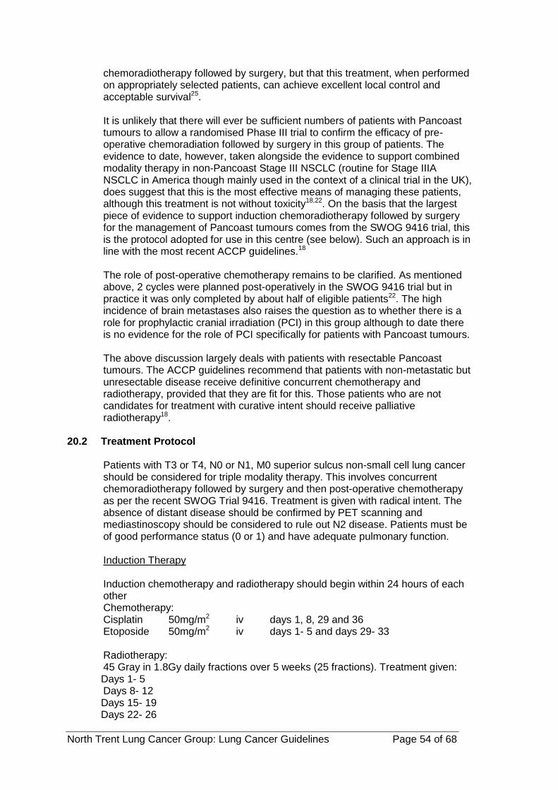
North Trent Lung Cancer Group: Lung Cancer Guidelines Page 54 of 68
chemoradiotherapy followed by surgery, but that this treatment, when performed on appropriately selected patients, can achieve excellent local control and acceptable survival25. It is unlikely that there will ever be sufficient numbers of patients with Pancoast tumours to allow a randomised Phase III trial to confirm the efficacy of pre-operative chemoradiation followed by surgery in this group of patients. The evidence to date, however, taken alongside the evidence to support combined modality therapy in non-Pancoast Stage III NSCLC (routine for Stage IIIA NSCLC in America though mainly used in the context of a clinical trial in the UK), does suggest that this is the most effective means of managing these patients, although this treatment is not without toxicity18,22. On the basis that the largest piece of evidence to support induction chemoradiotherapy followed by surgery for the management of Pancoast tumours comes from the SWOG 9416 trial, this is the protocol adopted for use in this centre (see below). Such an approach is in line with the most recent ACCP guidelines.18
The role of post-operative chemotherapy remains to be clarified. As mentioned above, 2 cycles were planned post-operatively in the SWOG 9416 trial but in practice it was only completed by about half of eligible patients22. The high incidence of brain metastases also raises the question as to whether there is a role for prophylactic cranial irradiation (PCI) in this group although to date there is no evidence for the role of PCI specifically for patients with Pancoast tumours. The above discussion largely deals with patients with resectable Pancoast tumours. The ACCP guidelines recommend that patients with non-metastatic but unresectable disease receive definitive concurrent chemotherapy and radiotherapy, provided that they are fit for this. Those patients who are not candidates for treatment with curative intent should receive palliative radiotherapy18.
20.2 Treatment Protocol
Patients with T3 or T4, N0 or N1, M0 superior sulcus non-small cell lung cancer should be considered for triple modality therapy. This involves concurrent chemoradiotherapy followed by surgery and then post-operative chemotherapy as per the recent SWOG Trial 9416. Treatment is given with radical intent. The absence of distant disease should be confirmed by PET scanning and mediastinoscopy should be considered to rule out N2 disease. Patients must be of good performance status (0 or 1) and have adequate pulmonary function. Induction Therapy Induction chemotherapy and radiotherapy should begin within 24 hours of each other Chemotherapy: Cisplatin 50mg/m2 iv days 1, 8, 29 and 36 Etoposide 50mg/m2 iv days 1- 5 and days 29- 33 Radiotherapy: 45 Gray in 1.8Gy daily fractions over 5 weeks (25 fractions). Treatment given: Days 1- 5 Days 8- 12
Days 15- 19 Days 22- 26

North Trent Lung Cancer Group: Lung Cancer Guidelines Page 55 of 68
Days 29- 33
Patients should be CT planned in the treatment position with arms by the side. The target volume should include the primary tumour and the ipsilateral supraclavicular region, but not the mediastinum or hilum. GTV should encompass the primary tumour mass and involved nodes, defined as nodes with short axis > 10 mm. CTV is arrived by expansion of GTV by a 5 mm and to include the ipsilateral supraclavicular region.
PTV is arrived by expansion of CTV by a 10 mm margin (15 mm margin in the cranio-caudal direction), allowance being made in planning for tumour and normal structure movement during quiet respiration. Hand modification of the PTV is permitted to avoid the spinal cord provided a minimum of 5 mm expansion over the CTV is maintained.
Critical normal structures should be considered as discussed in the ‘Normal Tissues’ section of Non-small cell lung cancer section of the Radiotherapy Handbook. For further details on radiotherapy see the ‘Radical’ section of the Non-small cell lung cancer chapter in the Radiotherapy handbook.
Evaluation after induction therapy Two to four weeks after induction therapy patients should have a CT scan of their thorax and upper abdomen to assess response to treatment. This should be discussed at the lung cancer MDT. Patients who do not have evidence of progressive disease should proceed to thoracotomy three to five weeks after induction treatment is completed. Lobectomy or pneumonectomy may be required. Patients unsuitable for surgery should have a further 2 cycles of chemotherapy as described below.
Boost chemotherapy Patients should receive a further 2 cycles of cisplatin and etoposide chemotherapy after recovery from surgery with the same regimen as above i.e.
Cisplatin 50mg/m2 iv days 1 and 8
Etoposide 50mg/m2 iv days 1 to 5
Cycle 2 begins on day 29
Patients who do not undergo surgery should also have these 2 cycles of chemotherapy.

North Trent Lung Cancer Group: Lung Cancer Guidelines Page 56 of 68
20.3 References
1. Pancoast HK. Superior pulmonary sulcus tumor. JAMA 1932; 99: 1391-1396 2. Chardack WM, MacCallum JD. Pancoast tumor (five year survival without
recurrence or metastases following radical resection and postoperative irradiation. J Thorac Surg 1956; 31: 535-542
3. Shaw RR. Pancoast’s tumour. Ann Thor Surg 1984; 37: 343-345 4. Shaw RR, Paulson DL, Kee JL Jr, et al. Treatment of the superior sulcus
tumor by irradiation followed by resection. Ann Surg 1961; 7: 29-40 5. Paulson DL. Carcinomas in the superior pulmonary sulcus. J Thorac
Cardiovasc Surg 1975; 70: 1095-1104 6. Hilaris BS, Martini N, Wong GY, et al. Treatment of superior sulcus tumor
(Pancoast tumor). Surg Clin North Am 1987; 67: 965-976 7. Sartori F, Rea F, Calabro F, et al. Carcinoma of the superior pulmonary
sulcus: Results of irradiation and radical resection. J Thorac Cardiovasc Surg 1992; 104: 679-683
8. Maggi G, Casadio C, Pischedda F, et al. Combined radiosurgical treatment of Pancoast tumor. Ann Thorac Surg 1994; 57: 198-202
9. Attar S, Krasna MJ, Sonnet JR, et al. Superior sulcus (Pancoast) tumor: Experience of 105 patients. Ann Thorac Surg 1998; 66: 193-198
10. Hagan MP, Choi NC, Mathisen DJ, et al. Superior sulcus lung tumors: impact of local control on survival. J Thorac Cardiovasc Surg 1999; 117:1086-1094
11. Komaki R, Roth JA, Walsh GL, et al. Outcome predictors for 143 patients with superior sulcus tumor treated by multidisciplinary approach at the University of Texas MD Anderson Cancer Centre. Int J Radiat Oncol Biol Phys 2000; 48: 347-354
12. Martinod E, D’Audiffret A, Thomas P, et al. Management of superior sulcus tumors: experience of 139 cases treated by surgical resection. Ann Thorac Surg 2002; 73: 1534-1540
13. van Geel AN, Jansen PP, van Klaveren RJ, et al. High relapse-free survival after pre-operative and intraoperative radiotherapy and resection for sulcus superior tumors. Chest 2003; 124: 1841-1846
14. Alifano M, D’Aiuto M, Magdeleinat P, et al. Surgical treatment of superior sulcus tumors: results and prognostic factors. Chest 2003; 124: 996-1003
15. Pfannschmidt J, Kugler C, Muley T, et al. Non-small-cell superior sulcus tumor: results of en bloc resection in fifty-six patients. Thorac Cardiovasc Surg 2003; 51: 332-337
16. Detterbeck FC, Jones DR, Rosenman JG. Pancoast tumors. In: Detterbeck FC, Rivera MP, Socinski MA, et al, eds. Diagnosis and treatment of lung cancer: an evidence-based guide for the practicing clinician. Philadelphia, PA: WB Saunders, 2001: 233-243
17. Rusch VW, Parekh KR, Leon L, et al. Factors determining outcome after surgical resection of T3 and T4 lung cancers of the superior sulcus. J Thorac Cardiovasc Surg 2000; 119: 1147-1153
18. Shen KR, Meyers BF, Larner JM and Jones DR. Special treatment issues in lung cancer: ACCP evidence-based clinical practice guidelines (2nd edition). Chest 2007; 132: 290-305
19. Anderson TM, Moy PM, Holmes EC. Factors affecting survival in superior sulcus tumors, J Clin Oncol 1986; 4: 1598-1603
20. Muscolino G, Valente M, Andreani S. Pancoast tumours. Clinical assessment and long-term results of combined radiosurgical treatment. Thorax 1997; 52 284-286

North Trent Lung Cancer Group: Lung Cancer Guidelines Page 57 of 68
21. Rusch VW, Giroux DJ, Kraut MJ, et al. Induction chemoradiation and surgical resection for superior sulcus non-small cell lung carcinomas: long-term results of Southwest Oncology Group Trial 9416 (Intergroup Trial 0160). J Clin Oncol 2008; 25: 313-318
22. Wright CD, Menard MT, Wain JC, et al. Induction chemoradiation compared with induction radiation for lung cancer involving the superior sulcus. Ann Thorac Surg 2002; 73: 1541-1544
23. Kwong K, Edelman MJ, Suntharalingam M, et al. High-dose radiotherapy in trimodality treatment of Pancoast tumors results in high pathologic complete response rates and excellent long-term survival. J Thorac Cardiovasc Surg 2005; 129: 1250-1257
24. De Leyn P, Vansteenkiste J, Lievens Y, et al. Survival after trimodality treatment for superior sulcus and central T4 non-small cell lung cancer. J Thorac Oncol 2009; 4: 62-68
25. Kappers I, Belderbos JSA, Burgers JA, et al. Non-small cell carcinoma of the superior sulcus: favourable outcomes of combined modality treatment in carefully selected patients. Lung Cancer 2008; 59: 385-390

North Trent Lung Cancer Group: Lung Cancer Guidelines Page 58 of 68
21 APPENDIX 4: REVIEW OF CHEMORADIOTHERAPY AND INDUCTION CHEMOTHERAPY IN NSCLC
21.1 Introduction
Patients with unresectable non-small cell lung cancer (NSCLC) should be considered for active anti-tumour treatment with radiotherapy and chemotherapy. Those with stage IV disease are incurable but can benefit from chemotherapy. Patients with stage I and II disease that decline or who are medically unsuitable for surgery have a good chance of cure and should receive optimal radiotherapy. Conventional radiotherapy (e.g. 60Gy in 30 fractions) results in 5-year survival rates of only 5-10% but the use of continuous hyperfractionated accelerated radiotherapy (CHART) can improve 2-year survival rates from 20% to 29%, albeit with added toxicity, particularly oesophagitis (Saunders et al, 1997; 1999).
21.2 Chemo-radiotherapy
For patients with stage III disease, the treatment options are less clear. The 5-year survival rates for stage IIIa are 9-13% and for stage IIb are 3-7%. While radiotherapy can improve the local tumour control rate, the majority of patients will die from distant relapse. Therefore, a number of studies have been performed to determine whether the use of chemo-radiotherapy (i.e. planned combination treatment with concurrent or sequential chemotherapy and radiotherapy) improves survival compared to radiotherapy alone.
A meta-analysis of the effects of chemotherapy in NSCLC included analysis of data from 3033 patients in 22 trials comparing radical radiotherapy to chemoradiotherapy (NSCLC Collaborative Group, 1995). It showed a small survival benefit from chemoradiotherapy corresponding to an absolute survival benefit of 3% at 2 years. Several more recent randomised studies using modern drug and radiation regimens have shown greater benefits (Dillman et al 1996; Jeremic et al 1995,1996; Cullen et al 1999). The largest, including 490 patients, showed that chemoradiation improved 2-year survival rates from 21% to 32% (Sause et al, 2000), a similar survival gain to that obtained with CHART. The use of hyperfractionated radiotherapy in several studies has not led to improved survival, and standard chemoradiotherapy was superior to hyperfractionated radiotherapy in one direct comparison (Sause et al, 2000). The optimal timing of chemotherapy in relation to radiotherapy remains unclear. Concomitant chemoradiotherapy is probably superior to sequential treatment, but there is concern that additive toxicities might compromise the ability to deliver full dose radiotherapy (Furuse et al, 1999). Indeed, recent local experience has confirmed combined modality treatments can lead to unexpected fatalities (Kirkbride et al, 2002).
Chemoradiotherapy is now the standard treatment for stage IIIa NSCLC in the USA (ASCO guidelines, 1998), but remains investigational in Europe.
21.3 Primary – (Induction) chemotherapy
In patients with regionally advanced disease that cannot be encompassed by a radical radiotherapy field, primary chemotherapy may be given with the intention of proceeding to radical treatment (surgery or radiotherapy) only if a significant tumour response is obtained. Patients with regionally advanced NSCLC (stage IIIa / IIIb) are twice as likely to respond to chemotherapy as patients with advanced disease. For example, in parallel studies of MIC chemotherapy in

North Trent Lung Cancer Group: Lung Cancer Guidelines Page 59 of 68
locally unresectable and advanced NSCLC, response rates were 54% and 32% respectively (Cullen et al, 1999). The strongest evidence for effect is with platinum-containing regimens (NSCLC Collaborative Group, 1995). Few studies have explored the role of primary chemotherapy for regionally advanced NSCLC. The European Lung Cancer Working Party study compared radiotherapy to maintenance chemotherapy in patients responding to primary chemotherapy. One hundred and fifteen NSCLC patients who responded to primary chemotherapy were randomly assigned further chemotherapy or chest irradiation, (60Gy over 6 weeks). Despite better control in the radiotherapy group, there was no significant difference in survival (Sculier et al, 1999). Patients with pathologically proven stage IIIA/N2 disease should be considered for study EORTC 08941, in which patients responding to primary chemotherapy are randomised between consolidation radiotherapy and surgery.
Interestingly, a recent study examined which patients with stage II-IIIB NSCLC benefited from treatment intensification with chemoradiotherapy and found that younger patients achieved greater benefit from intensive treatment, whereas older patients gained most quality-adjusted time from standard radiotherapy (Movsas et al, 1999).
21.4 Conclusion
Although hyperfractionated radiotherapy has gained popularity for treating locally advanced NSCLC in the UK (RCR-COIN, 1999), in the rest of the world chemoradiotherapy is the preferred treatment option for unresectable stage III disease (ASCO guidelines, 1998). The available data indicate that combined modality therapy can improve survival in locally advanced disease. Further studies are however required to determine the optimal regimen, doses, and scheduling of such treatments to maximise survival benefits and economic effectiveness while maintaining quality of life.
21.5 Recommendations
chemoradiotherapy for NSCLC is given only in the setting of a clinical trial. patients with pathologically confirmed stage IIIaN2 NSCLC would previously
have been registered in EORTC study 08941 and received primary chemotherapy. Responders were randomised between surgery and radical radiotherapy. This trial is now closed to registration. Patients of good performance status (0 or 1) with inoperable stage III NSCLC that cannot be encompassed by a radical radiotherapy field should be considered for primary chemotherapy provided they have adequate haematological and renal function. They should be reassessed after 2 treatment cycles and responders should receive a 3rd cycle of chemotherapy whilst MDT discussion and appropriate local treatment (surgery or radiotherapy) is arranged, including additional investigations. Radiotherapy should not start less than 3 weeks after the final cycle of chemotherapy. Patients who have symptomatic benefit should also receive a 3rd cycle. Non-responders should receive palliative radiotherapy or active supportive care as appropriate.
neoadjuvant chemoradiotherapy should not be offered routinely but there may be interest in formulating a research protocol.
Additional comment for 2005 guideline update: The results of EORTC 08491 are expected to be available later this year. In addition, as of 31st March 2005 we have 2 years of patient registrations and our

North Trent Lung Cancer Group: Lung Cancer Guidelines Page 60 of 68
experience to date is currently being reviewed as part of an M.Sc. project. This document will be updated in the light of the trial evidence and once our local review has been completed. We therefore anticipate that a revised version of this guideline will be available in 2006. Additional comment for 2008 guideline update: Both EORTC 08491 and the M.Sc. referred to in the 2005 comment are now completed. Abstracts from both are included below.
Randomized Controlled Trial of Resection Versus Radiotherapy after Induction Chemotherapy in Stage IIIA-N2 Non – Small-Cell Lung Cancer Jan P. van Meerbeeck , Gijs W. P. M . Kramer, Paul E. Y. Van Schil et al On behalf of the European Organisation for Research and Treatment of Cancer-Lung Cancer Group Background Induction chemotherapy before surgical resection increases survival compared with surgical resection alone in patients with stage IIIA-N2 non – small-cell lung cancer (NSCLC). We hypothesized that, following a response to induction chemotherapy, surgical resection would be superior to thoracic radiotherapy as locoregional therapy. Methods Selected patients with histologic or cytologic proven stage IIIA-N2 NSCLC were given three cycles of platinum based induction chemotherapy. Responding patients were subsequently randomly assigned to surgical resection or radiotherapy. Survival curves were estimated using Kaplan – Meier analyses from time of randomisation. Results Induction chemotherapy resulted in a response rate of 61% (95% confidence interval [CI] = 57% to 65%) among the 579 eligible patients. A total of 167 patients were allocated to resection and 165 to radiotherapy. Of the 154 (92%) patients who underwent surgery, 14% had an exploratory thoracotomy, 50% a radical resection, 42% a pathologic downstaging, and 5% a pathologic complete response; 4% died after surgery. Postoperative radiotherapy was administered to 62 (40%) of patients in the surgery arm. Among the 154 (93%) irradiated patients, overall compliance to the radiotherapy prescription was 55%, and grade 3/4 acute and late esophageal and pulmonary toxic effects occurred in 4% and 7%; one patient died of radiation pneumonitis. Median and 5-year overall survival for patients randomly assigned to resection versus radiotherapy were 16.4 versus 17.5 months and 15.7% versus 14%, respectively (hazard ratio = 1.06, 95% CI = 0.84 to 1.35). Rates of progression-free survival were also similar in both groups. Conclusion In selected patients with pathologically proven stage IIIA-N2 NSCLC and a response to induction chemotherapy, surgical resection did not improve overall or progression-free survival compared with radiotherapy. In view of its low morbidity and mortality, radiotherapy should be considered the preferred locoregional treatment for these patients. J Natl Cancer Inst 2007; 99: 442 – 50 Does primary chemotherapy in locally advanced non-small cell lung cancer allow down-staging and facilitate further treatment and, if so, are there any factors which predict response? Dr Sarah Elizabeth Lawless MBBS MRCP Background The aim of this local retrospective study was to ascertain the proportion of patients responding to primary chemotherapy for locally advanced non-small cell lung cancer (NSCLC) and who subsequently received radical treatment. In addition, radiological down-staging, time to progression (TTP) and overall

North Trent Lung Cancer Group: Lung Cancer Guidelines Page 61 of 68
survival (OS) were assessed. Possible predictive markers of response to primary chemotherapy and factors influencing survival were also analysed. Methods All 116 patients who received primary chemotherapy for locally advanced, histologically confirmed NSCLC, between 1st April 2003 and 31st March 2005, were analysed. Down-staging was assessed by review of both TNM (Tumour, Nodes, Metastases) and overall stage changes on pre and post chemotherapy CT scan reports. TTP and OS were analysed using the Kaplan-Meier method. Possible predictive markers of response and survival were assessed using univariate and multivariate analysis. Results Fifty-nine patients (50.9%) received radical treatment following primary platinum-based chemotherapy. Median TTP was 37.3 weeks (95% confidence interval (CI) 30.8-43.7 weeks). Median OS was 62.3 weeks (95% CI 46.4-78.2 weeks). OS was improved in those receiving radical treatment compared to those having no radical treatment post chemotherapy (94.9 weeks, 95% CI 73.3-116.4 weeks versus 39.4 weeks, 95% CI 33.5-45.3 weeks, P<0.001) with patients having surgery achieving the best OS (median OS not reached at the time of analysis). Of the 99 patients who had post chemotherapy CT scans, 27 (27.3%) were down-staged, 62 (62.6%) had no change in overall stage and 10 (10.1%) were upstaged post chemotherapy. There was a statistically significant improvement in T, N and overall stage post chemotherapy (P=0.011, P=0.01, P=0.003, respectively) with 3 patients having a complete radiological response. Number of chemotherapy cycles received and response on chest x-ray post 2 chemotherapy cycles, were significant factors predictive of being down-staged/unchanged stage versus upstaged (P<0.001, P=0.014 respectively). At multivariate analysis; performance status at diagnosis, type of chemotherapy received, chest x-ray response post 2 cycles of chemotherapy and pre chemotherapy neutrophil count, were all significant factors in predicting OS. Conclusion Primary chemotherapy in locally advanced NSCLC allows down-staging and facilitates further radical treatment in a significant proportion of patients, with a clear survival benefit if radical treatment can be delivered following chemotherapy. Performance status at diagnosis, pre chemotherapy neutrophil count and chest x-ray response post 2 chemotherapy cycles may be useful factors in predicting which patients are most likely to respond and have improved survival.
Appendix A: Chemoradiotherapy definitions
Adjuvant chemotherapy = chemotherapy given to high-risk
patients with no macroscopic disease following definitive surgery.
Neoadjuvant chemotherapy = pre-operative chemotherapy given to patients who are resectable and in whom definitive surgery is planned.
Chemoradiotherapy = planned combination treatment with chemotherapy and radiotherapy (concomitant or sequential).
Pr Primary chemotherapy = initial chemotherapy given with the intention of proceeding to definitive local treatment (surgery or
radiotherapy) in responding patients (i.e. about 25%).

North Trent Lung Cancer Group: Lung Cancer Guidelines Page 62 of 68
Appendix B: Chemoradiotherapy references
American Society of Clinical Oncology (1998) Clinical practice guidelines for the treatment of unresectable non-small-cell lung cancer. J Clin Oncol 15: 2996-3018. Cullen, M.H., Billingham, L.J., Woodroffe, C.M., et al (1999) Mitomycin, ifosfamide and cisplatin in unresectable non-small cell lung cancer: effects on survival and quality of life. J Clin Oncol 17: 3188-3194. Dillman, R. O., Seagren, S. L., Propert, K. L. et al (1990). A randomised trial of induction chemotherapy plus high-dose radiation versus radiation alone in stage III non-small cell lung cancer. N Engl J Med323: 940-945. Dillman, R. O., Herndon, J., Seagren, S. L., et al. (1996). Improved survival in stage III NSCLC: seven-year follow-up of cancer and leukaemia group B (CALGB) 8433 trial. J Natl Cancer Inst 88: 1210-1215. Furuse K, Fukuoka M, Kawahara M, et al. Phase III study of concurrent versus sequential thoracic radiotherapy in combination with mitomycin, vindesine and cisplatin in unresectable stage III non-small cell lung cancer. J Clin Oncol 1999;17:2692-2699. Jeremic, B., Shibamoto, Y., Acimovic, L., Djuric, L. (1995). Randomised trial of hyperfractionated radiation therapy with or without concurrent chemotherapy for stage III non-small cell lung cancer. J Clin Oncol 13: 452-458. Jeremic, B., Shibamoto, Y., Acimovic, L., et al (1996). Hyperfractionated radiotherapy with or without concurrent low-dose daily carboplatin/etoposide for stage III non-small-cell lung cancer: a randomised study. J Clin Oncol 14: 1065-70. Kirkbride, P., Hatton, M., Lorigan, P. et al. (2002). Fatal Pulmonary Fibrosis Associated with Induction Chemotherapy with Carboplatin and Vinorelbine followed by CHART Radiotherapy for Locally Advanced Non-small Cell Lung Cancer. Clin Oncol 14. 361-366. Movsas, B., Scott, C., Sause, W., et al. (1999). The benefit of treatment intensification is age and histology-dependent in patients with locally advanced non-small cell lung cancer (NSCLC): a quality-adjusted survival analysis of Radiation Therapy Oncology Group (RTOG) chemoradiation studies. Int J Radiat Oncol Biol Phys 45: 1143-9. Non-small cell lung cancer collaborative group (1995). Chemotherapy in non-small cell lung cancer: a meta-analysis using updated data on individual patients from 52 randomised trials. Br Med J 311: 899-909. Royal College of Radiologists. (1999). Guidelines for the non-surgical management of lung cancer. Clin Oncol 11: S18-S37. Saunders M, Dische S, Barrett A, et al, on behalf of the CHART Steering Committee (1999). Continuous hyperfractionated accelerated radiotherapy versus conventional radiotherapy in NSCLC: mature data from the randomised multi-centre trial. Radiother Oncol.52: 137-48. Sause W, Kolesar P, Taylor S, et al (2000). Final results of a phase III trial in regionally advanced unresectable non-small cell lung cancer: Radiation Therapy Oncology Group, Eastern Cooperative Oncology Group, and Southwest Oncology Group. Chest 117:358-64. Sause, W. T., Scott, C., Taylor, S. et al. (1995). Radiation Therapy Oncology Group (RTOG) 8808 and Eastern Co-operative Oncology Group (ECOG) 4588: Preliminary results of a phase III trial in regionally advanced unresectable non-small cell lung cancer. J Natl Cancer Inst 87: 198-205. Sculier JP, Paesmans M, Lafitte JJ, et al (1999). A randomised phase III trial comparing consolidation treatment with further chemotherapy to chest irradiation in patients with initially unresectable locoregional non-small-cell lung cancer responding to induction

North Trent Lung Cancer Group: Lung Cancer Guidelines Page 63 of 68
22 APPENDIX 5: ACKNOWLEDGEMENTS
Grateful thanks are given to all who have produced their knowledge, expertise and time to the development of this Referral and Clinical Guidelines for Lung Caner within North Trent, produced in May 2011 This document has been revised by a group including: - Dr Patricia Fisher - Dr Jennifer Hill - Mr John Edwards - Dr Trevor Rogers - Professor Penella Woll - Dr Kim Suvarna - Dr Sue Matthews - Sr Alison Bennett - Dr Caroline Lee Several of the above contributed to the previous guideline. Dr Trevor Cleveland and Dr. Matthew Bull contributed to Appendix 4 in previous guidelines which has remained unchanged. Dr Louise Murray wrote the papers outlining the treatment of Pancoast tumours in appendix 3.
23 APPENDIX 6: TEENAGERS AND YOUNG ADULTS
In January 2009 specific referral pathways were developed for teenagers (16-18 years) and young adults (19 -24 years) into the TYA MDT. The Lung NSSG has agreed age appropriate referral into these pathways set out below:- The North Trent Children and Young Peoples IOG (CYPIOG) Implementation Summary has been approved by the Specialist Commissioning Group and was submitted to the Cancer Action Team on September the 30th. This paper addresses referral to the Teenage and Young Adult (TYA) MDT, which is based at Weston Park Hospital. “The model for referral to the TYA MDT, has been agreed with the Cancer Network Chairs and MDT leads within the network and it is anticipated that all young people diagnosed with cancer within the network will be referred to the TYA MDT from April 2009. All patients in this age range will be flagged by the MDT co-ordinator and supported by IT systems to monitor that all appropriate patients are being referred to the TYA MDT.” (North Trent CYPIOG Action Plan). “Referral to the TYA MDT will be made by the site specific Consultant at their weekly MDT as per operational policy and communication will be two way, electronic and by way of videoconferencing where available to enhance clinical decision-making.” (North Trent CYPIOG Action Plan). It is clear that in order to implement the above actions there will be a requirement for NSSGs to incorporate the new Teenage and Young Adult Referral Guidelines into their existing Operational Policies. Illustrations are provided below on how these guidelines may look. Within the examples provided, there is some scope for local interpretation. 16 – 18 YR OLDS GP 2WW REFERRAL

North Trent Lung Cancer Group: Lung Cancer Guidelines Page 64 of 68
1. Referral straight to STHFT Site Specific MDT ROUTINE REFERRAL/HOSPITAL ADMISSION TO LOCAL TRUST 1. Diagnostic tests as appropriate. Positive Diagnosis/Strong suspicion of Cancer. Referral by local Consultant to STHFT for treatment via NHS.net electronic referral
form. 2. Patient seen by STHFT Site Specific Consultant. 3. Diagnostic tests as appropriate. 4 A joint Site Specific/TYA MDT discussion around the treatment plan must
take place. The means by which this will be achieved is to be locally determined but could include phone conversations/video conferencing/TYA CNS attendance at Site Specific MDT/e-mail correspondence. Whatever the means it must be documented and auditable.
Discussion at Site Specific MDT. Treatment plan generated. TYA
representative may or may not be in attendance depending on agreed joint discussion procedure.
Referral by Site Specific MDT coordinator to TYA MDT coordinator via
electronic referral form, using NHS.net, outlining diagnosis, treatment plan and any additional relevant information. May happen before or after SSMDT depending on agreed joint discussion procedure.
Clinical referral to TYA MDT by Site Specific Lead outlining diagnosis, treatment
plan and any additional relevant information. May happen before or after SSMDT meeting depending on agreed joint discussion procedure.
(Bold statements outline the “Must do’s”.) 5. Discussion with patient around treatment options to include TYA representation. This will most probably be the TYA Nurse Specialist/Key Worker. 6. Patient discussed at weekly TYA MDT with Site Specific input where appropriate. Clinical Psychologist or TYA CNS to produce supportive care plan for Site Specific
referring clinician. TYA MDT chair person to provide electronic outcome to Site Specific MDT
coordinator within 48hrs, using NHS.net. or letter . 7. Patient treatment. TYA key worker input in coordination of support services. 8. Review and follow up care coordinated by TYA Key Worker involving community,
secondary site, tertiary site and TYA MDT as appropriate and locally wherever possible.
Patient referred to TYA Late Effects MDT by TYA MDT via electronic referral form. 19 - 24 YR OLDS GP 2WW REFERRAL 1. Referral to Local Trust. Patient Seen by Site Specific Consultant. Diagnostic tests
as appropriate. Positive Diagnosis ROUTINE REFERRAL/HOSPITAL ADMISSION TO LOCAL TRUST 1. Diagnostic tests as appropriate. Positive Diagnosis/Strong suspicion of Cancer. Patient Seen by Site Specific Consultant Patients within this age group must be
offered a choice as to where they receive their cancer treatment and supportive care. This is wholly dependant on local service delivery
North Trent Lung Cancer Group: Lung Cancer Guidelines Page 65 of 68
2 A joint Site Specific/TYA MDT discussion around the treatment plan must take place. The means by which this will be achieved is to be locally determined but could include phone conversations/video conferencing/TYA attendance at Site Specific MDT/e-mail correspondence. Whatever the means it must be documented and auditable. Discussion at Site Specific MDT. Treatment plan generated. TYA representative may or may not be in attendance depending on agreed joint discussion procedure.
Referral by Site Specific MDT coordinator to TYA MDT coordinator via electronic referral form, using NHS.net, outlining diagnosis, treatment plan and any additional relevant information. May happen before or after SSMDT depending on agreed joint discussion procedure.
Clinical referral to TYA MDT by Site Specific Lead outlining diagnosis, treatment plan and any additional relevant information. May happen before or after SSMDT meeting depending on agreed joint discussion procedure.
(Bold statements outline the “Must do’s”.) 3. Discussion with patient around treatment options to include TYA representation.
This will most probably be the TYA Nurse Specialist/Key Worker. Patient has option of treatment in SCG designated adult services. 4. Patient discussed at weekly TYA MDT with Site Specific input where appropriate. Clinical Psychologist or TYA CNS to produce supportive care plan for Site Specific
referring clinician. TYA MDT chair person to provide electronic outcome to Site Specific MDT
coordinator within 48 hrs, using NHS.net. or letter 5. Patient treatment. TYA key worker input in coordination of support services. 6. Review and follow up care coordinated by TYA Key Worker involving community,
secondary site, tertiary site and TYA MDT as appropriate and locally wherever possible. Patient referred to TYA Late effects MDT by TYA MDT via electronic referral form.

North Trent Lung Cancer Group: Lung Cancer Guidelines Page 66 of 68
Clinical Referral to the Teenage & Young Adult Psycho/Social MDM
Patient Name
Hospital Number
NHS Number
Date of Birth
Age
Address
Social Circumstances
Lives with parents/carer Siblings 1 2 3 4 5
Lives with spouse Siblings ages
Cohabiting partner/friends Dependants 1 2 3 4 5
Lives Alone Dependants ages
Education/Work Status
Full time school Employed
Higher Education Unemployed
Referral Information & Diagnosis
Referring Clinician Contact No
Key Worker
Referring Hospital
Diagnosis
Site Specific MDT Date
MDT Outcome & Plan
Has patient been entered into a clinical trial
YES / No
If no, reason: Not eligible Too ill Not applicable Other
Referral Completed by ____________________ Date ______________
NORTH TRENT
Cancer Network

North Trent Lung Cancer Group: Lung Cancer Guidelines Page 67 of 68
Patient Name_______________________ DOB ______________ Any other relevant information
To be completed by TYA MDM TYA MDM Date ___________________
Has a joint agreement been made regarding treatment
Yes / No
If no, Action:
Outcome and Plan of TYA MDM
TYA Key Worker
Patient Name_______________________ DOB ______________
TYA Team involved in care: Name:

North Trent Lung Cancer Group: Lung Cancer Guidelines Page 68 of 68
Chaplaincy
Dietician
Education team
Fertility team
Occupational therapist
Physiotherapist
Psychologist
Social worker
Youth support worker
Name __________________ Signature__________________ Date______
Please email this referral via NHSnet to
[email protected] Or fax 0114 2265512 FAO Tricia Mitchell, WPH, Ex 65071