reducing pre-analytical errors - whitehat · pdf filereducing pre-analytical errors...
TRANSCRIPT

Reducing Pre-analytical Errors
Christopher R. McCudden, Ph.D., FACB, FCACB, DABCC
University of Ottawa The Ottawa Hospital
Eastern Ontario Regional Laboratory Association Ontario, Canada

What is the most common POC error?
• A. Patient misidentification
• B. Poor sample collection technique
• C. Deviation from analytical procedure
• D. Improper device maintenance (e.g QC, reagent storage)
• E. Improper/lack of recording results
• F. Safety (e.g. hand hygiene, device reuse)
• G. Other

Outline
• Introduction
• Pre-analytical Phase:
– Patient
– Sampling
– Transportation, Storage, and Mixing
– Summary and Key Points
Safe
ty

Objectives
• List three different phases of the testing process and identify which areas have the highest risk of error
• Describe strategies to minimize preanalytical error
• Explain methods to ensure safe practices for point of care testing
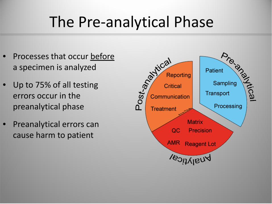
The Pre-analytical Phase
• Processes that occur before a specimen is analyzed
• Up to 75% of all testing errors occur in the preanalytical phase
• Preanalytical errors can cause harm to patient

Patient stability Patient identification
Tube/syringe labeling Site preparation
Sample collection
Specimen delivery to laboratory/storage
Specimen receipt Order/requisition processing
Mixing
Parts of the Pre-analytical Phase
Patient
Sampling
Transport
Processing
Safe
ty

Pre-analytical Challenges
• Many people involved: – Physicians: writing orders, instructing patients/staff – Nurses/Phlebotomists/RTs: patient ID, specimen collection – Runners: transport – Lab staff: receipt and processing
• More challenging in a teaching hospital
• Pre-analytical variables/errors are often unknown to testing personnel and the clinicians interpreting the results

Understanding Pre-analytical Issues
• Most steps
• Most people
• High urgency & stress
• Most variation in work environment, technique, and training
60% 25%
15%
% of Time Spent Pre-analysis
Analysis
Post-analysis

Patient stability Patient identification
Tube/syringe labeling Site preparation
Sample collection
Specimen delivery to laboratory/storage
Specimen receipt Order/requisition processing
Mixing
The Pre-analytical Process: POC
Patient
Sampling
Transport
Processing
Safe
ty

POC-Specific Pre-analytical Challenges
• Non-lab staff
– Limited Training & Experience
– Divided Focus
– Patient complexity

Steps of the Pre-analytical Phase Patient
Variation
Sampling
Transport
Processing

THE PATIENT
Patient Variation
Sampling
Transport
Processing

Starting on the Right Foot: Identify the Patient
• Incorrect/missing patient and sample IDs are frequent and critical pre-analytical errors
Approximately how much does a single misidentification error cost?
• A. 0-5 dollars
• B. >5 to 20 dollars
• C. >20 to 50 dollars
• D. >50 to 100 dollars
• E. >100 dollars

Consequences of Patient Misidentification
• Financial Implication of mislabeling*: • $500/incident • 250/month • Annual cost = USD 1.5 million
• Failure to provide proper and immediate care to a patient
• Inappropriate care to a patient
*Excluding medicolegal or liability costs

Avoiding Identification Errors
• Positive Patient Identification x2 • Correlate Orders with Patient Name • Identification on Sample Device at site of
Collection • Patient ID label attached • Pre-barcoded arterial syringe
• Enter a patient ID into the analyzer before analysis
• Use barcode readers

Test-Specific Advice: Patient Variables
• FIO2 and application of device – Mode of ventilation and Patient compliance with supplemental O2
• Duration of changes in vent settings
– Approximately 5-10 minutes post change up to 20% in stable Patient (Cakar, 2001, Intensive Care Medicine)
– Up to 30 minutes post change in Patient with Obstructive Lung Disease (Parsons, 2002)
• Patient's respiratory rate, temperature, position, activity
• Ease of (or difficulty with) blood sampling

SAFETY
Patient
Sampling
Transport
Processing Sa
fety

POC Testing and Safety
• POC testing != no risk
– Employee:
• Needle stick injury
• Blood exposure
– Patient:
• Nosocomial infection
– Drug resistant pathogens, Hepatitis

POC Testing and Safety
• Reports of multiple deaths for acute hepatitis B infection caused by poor practices with self-monitoring blood glucose meters
• 8/87 assisted living facility residents affected; 6 deaths
• Sharing of lancets
• Lack of disinfection
CDC Morb Mortal Wkly Rep 2011;60:182. http://www.cdc.gov/mmwr/preview/mmwrhtml/mm6006a5.htm

Reducing the Risk of POCT-related Infections*
• Discard finger-stick devices after each patient – Use autodisabling devices
• Assign POC devices to a single patient whenever possible
• Clean and disinfect POCT devices after every use
• Use proper hand-hygiene
*Safe and helps meet accreditation standards
Clinical Laboratory News (39):1 FDA Patient Safety News. Preventing infections while monitoring glucose.

Staff Safety
• Blood exposure and needlestick injuries are common – 23,908 injuries in 85 hospitals in 10
states (1995-2005)1
• All healthcare staff involved in patient care are affected – Medical technologists, Physicians,
Respiratory Therapists, and Nurses
1Percutaneous Injuries before and after the Needlestick Safety and Prevention Act. N Engl J Med 2012; 366:670-67 2Adapted from http://www.cdc.gov/niosh/stopsticks/sharpsinjuries.html
2

Exposure Causes and Consequences
• Causes: – Unavailability of safety devices – Lack of procedure for operator safety – Procedures for safety not known or followed
• Consequences:
– Needle-stick injury – Anxiety – Infection – Medical treatment

Risk Reduction Risk Reduction
• To avoid risks: – Use PPE
– Use a safety device that limits contact with patient blood
– Use a protection device for the safe removal of needles
– Ensure procedure for operator safety is established and followed

SAMPLING
Patient Variation
Sampling
Transport
Processing

Sampling
• Potential Issues:
– Site selection
– Site preparation
– Collection

Sampling: Arterial Puncture • Label the syringe with patient ID
• Choose Wisely
– Note location and direction of flow for IV fluids relative to draw site
– Confirm Arterial vs. Venous collection
– Adequate flushing of ports or lines
• Expel any air bubbles immediately after sampling
• Mix the sample thoroughly immediately after sampling

Poll
Type: Arterial pH: 6.975 pCO2: 8.2 pO2: 187 HCO3: <1.0 BE: -28.2 sO2: 98.9 tHgb: 13.8 K: 3.0 Na: 142 Glucose: 290
If unrecognized, what are the potential consequences of this error? A). Unnecessary blood transfusion B). Excess potassium supplementation C). Confusion & concern for misidentification D). Lack of appropriate insulin therapy
Type: Arterial pH: 6.923 pCO2: 12.4 pO2: 49.3 HCO3: 4.5 BE: -27.7 sO2: 83.5 tHgb: 7.0 K: 1.6 Na: 143 Glucose: 145
Contaminated sample
Accurate sample

Blood Gas Sampling To avoid errors: • Check the specific catheter package
for the exact volume of dead space
• Rule of thumb: discard at least three times the dead space – (CLSI recommends 6x)
• Draw the blood gas sample with a
dedicated blood gas syringe containing dry electrolyte-balanced heparin
• If in doubt, consider resampling
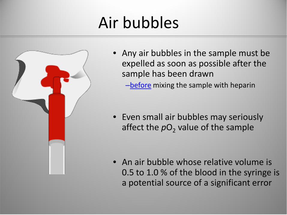
Air bubbles
• Any air bubbles in the sample must be expelled as soon as possible after the sample has been drawn –before mixing the sample with heparin
• Even small air bubbles may seriously affect the pO2 value of the sample
• An air bubble whose relative volume is 0.5 to 1.0 % of the blood in the syringe is a potential source of a significant error

Air bubble Effects depend on:
• Size of bubble
• Number of bubbles
• Initial oxygen status of sample
• Longer time
• Lower temperature
• Increased agitation
Effect on pO2
Surface area of air bubble

Effect of Air Bubbles
Type: Not specified pH: 7.37 pCO2: 56.7 pO2: 43.8 HCO3: 31.9 BE: 6.7 sO2: 81.1
Type: Not specified pH: 7.50 pCO2: 37.1 pO2: 163 HCO3: 28.9 BE: 5.6 sO2: 99.0
Sample was transferred between collection devices to inject low sample volume
Air Contaminated sample
Accurate sample
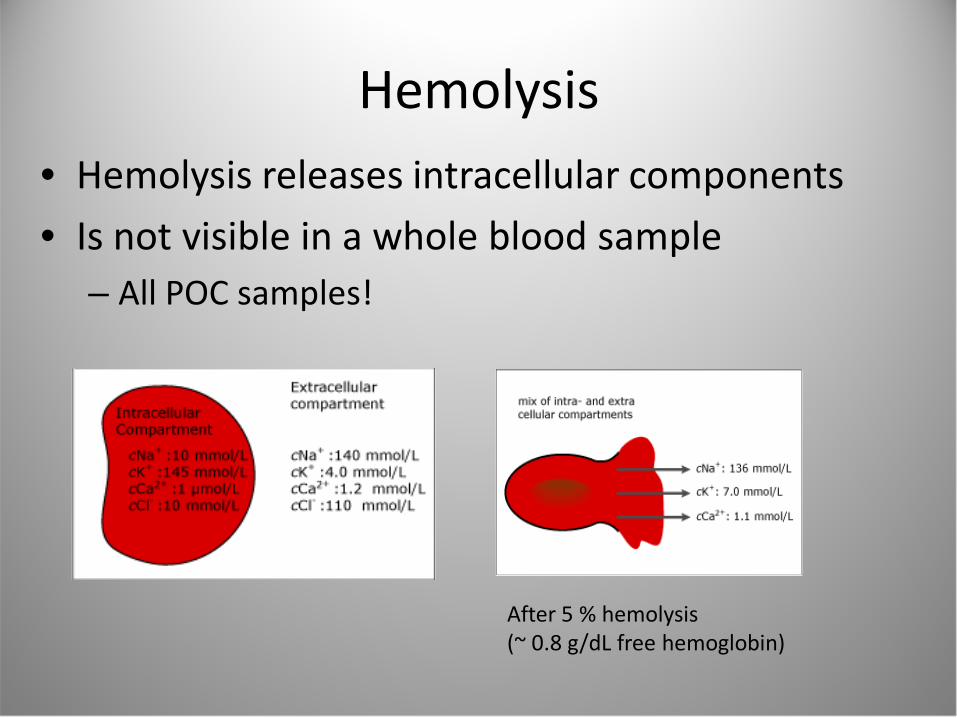
Hemolysis • Hemolysis releases intracellular components
• Is not visible in a whole blood sample – All POC samples!
After 5 % hemolysis (~ 0.8 g/dL free hemoglobin)

Hemolysis
• Hemolysis of the sample can lead to: – Biased results
– Possible misdiagnosis
– Possible erroneous patient treatment/lack of treatment
• To avoid errors: – Do not milk or massage the tissue during sampling
– Use self-filling syringes
– Use recommended procedures for mixing of samples

Patient Variation
Sampling
Transport
Processing
PROCESSING

Mixing and Clots
• Samples must be mixed after expelling air
• Before analyzing the sample, make a visual check of the blood
• Inspect for air bubbles
• Expel a few drops of blood from the syringe to inspect for clots

What Happens to the Instrument If a Clotted Sample is Analyzed?
• A). No effect, ABG instruments have a hemolyzer
• B). Instrument will be unusable until clot is removed
• C). Electrolyte results will decrease
• D). Electrolyte results will increase

What Happens to the Instrument If a Clotted Sample is Analyzed?
Error!!

Summary
• We’re all in this together Help the patient!
• Pre-analytical errors can lead to harm
• POC Testing has unique challenges
• A bad sample is worse than no sample

Thank you and Questions?

Additional Resources • Howanitz PJ, Howanitz JH. Quality control for the clinical laboratory. Clin
Lab Med. 1983;3:541-551.
• Bonini P, Plebani M, Ceriotti F, et al. Errors in laboratory medicine. Clin Chem. 2002;48(5):691-698.
• Grenache DG and Parker CM. Integrated and automatic mixing of whole blood: evaluation of a novel blood gas analyzer. Clinica Chimica Acta, 2007
• CLSI. Procedures for the Collection of Arterial Blood Specimens; Approved Standard—Fourth Edition. CLSI document H11-A4. Wayne, PA: Clinical and Laboratory Standards Institute 2004
• www.acutecaretesting.org
• Percutaneous Injuries before and after the Needlestick Safety and Prevention Act. N Engl J Med 2012; 366:670-67
• A discard volumes arterial blood gas sampling. Critical Care Medicine: June 2003 - Volume 31 - Issue 6 - pp 1654-1658
• http://www.cdc.gov/mmwr/preview/mmwrhtml/mm6006a5.htm

List of Potential Preanalytical Errors
• Missing or wrong patient/sample identification • Use of the wrong type or amount of anticoagulant
– dilution due to the use of liquid heparin – insufficient amount of heparin – binding of electrolytes to heparin
• Inadequate stabilization of the respiratory condition of the patient • Inadequate removal of flush solution in a-lines prior to blood collection • Mixture of venous and arterial blood during puncturing • Air bubbles in the sample • Insufficient mixing with heparin • Incorrect storage • Hemolysis of red blood cells • Not visually inspecting the sample for clots • Inadequate mixing of sample before analysis • Failure to identify the sample upon analysis