redox, amino acid, and fatty acid metabolism intersect ... · redox, amino acid, and fatty acid...
TRANSCRIPT
Redox, amino acid, and fatty acid metabolism intersectwith bacterial virulence in the gutReed Pifera,b,1, Regan M. Russella,b,1, Aman Kumara,b, Meredith M. Curtisa,b, and Vanessa Sperandioa,b,2
aDepartment of Microbiology, University of Texas (UT) Southwestern Medical Center, Dallas, TX 75390; and bDepartment of Biochemistry, UT SouthwesternMedical Center, Dallas, TX 75390
Edited by Jeff F. Miller, University of California, Los Angeles, CA, and approved September 27, 2018 (received for review August 8, 2018)
The gut metabolic landscape is complex and is influenced by themicrobiota, host physiology, and enteric pathogens. Pathogenshave to exquisitely monitor the biogeography of the gastrointes-tinal tract to find a suitable niche for colonization. To dissect theimportant metabolic pathways that influence virulence of enter-ohemorrhagic Escherichia coli (EHEC), we conducted a high-throughput screen. We generated a dataset of regulatory pathwaysthat control EHEC virulence expression under anaerobic conditions.This unraveled that the cysteine-responsive regulator, CutR, con-verges with the YhaO serine import pump and the fatty acid me-tabolism regulator FadR to optimally control virulence expression inEHEC. CutR activates expression of YhaO to increase activity of theYhaJ transcription factor that has been previously shown to directlyactivate the EHEC virulence genes. CutR enhances FadL, which is apump for fatty acids that represses inhibition of virulence expres-sion by FadR, unmasking a feedback mechanism responsive tometabolite fluctuations. Moreover, CutR and FadR also augmentmurine infection by Citrobacter rodentium, which is a murine path-ogen extensively employed as a surrogate animal model for EHEC.This high-throughput approach proved to be a powerful tool to mapthe web of cellular circuits that allows an enteric pathogen to mon-itor the gut environment and adjust the levels of expression of itsvirulence repertoire toward successful infection of the host.
enterohemorrhagic E. coli | EHEC | cutR | fadL
The gastrointestinal (GI) tract is a complex environment,where the availability of metabolites and signaling molecules
changes in different microenvironments, and is affected bymicrobiota composition, host physiology, and pathogenic insults(1). Enteric pathogens employ various metabolic and virulencestrategies to outcompete and/or exploit the resident microbiotato successfully colonize a GI niche. These strategies includeutilization of certain carbon and nitrogen sources as preferrednutrients and/or signals, exploitation of the host inflammation,and acquisition of metals, among others (1).Enterohemorrhagic Escherichia coli (EHEC) colonizes the
colon and causes severe diarrhea. EHEC virulence and intestinalcolonization, as well of its surrogate murine infection model,Citrobacter rodentium, is regulated by sugar, nitrogen, organicacid, short chain fatty acid, and oxygen availability (2). The locusof enterocyte effacement (LEE) of EHEC encodes a type IIIsecretion system (T3SS) that is essential for virulence. Effectorstranslocated through this T3SS induce cytoskeletal rearrange-ments on epithelial cells referred to as attaching and effacing(AE) lesions (3). Expression of the LEE is a significant meta-bolic burden for EHEC and must be carefully regulated (4). Weestablished a high-throughput method to define these LEEregulatory mechanisms and identified two transcription factors,CutR and FadR, that govern LEE expression.CutR (also known as YbaO/DecR) is a member of the feast/
famine regulatory protein (FFRP) family of transcription factors(5). Lrp, the canonical example of the FFRP family, regulatesthe LEE in response to butyrate levels (6). Butyrate is theprincipal microbiota-derived carbon source for colonic entero-cytes (7). CutR is a cysteine-responsive transcription factor (8).
Free L-cysteine cannot be detected in the cecal contents of adultspecific-pathogen-free (SPF) mice (9), but upon infection with C.rodentium, a bloom of L-cysteine is observed (10). Of note, cys-teine is important in the maintenance of the mucosal integritythrough its luminal redox status (11). In Salmonella, CutR isessential for the transcription of an adjacent cysteine desulfhy-drase, cdsH, and contributes to the detoxification of cysteine(12). CutR was identified as an activator of the yhaOM locus inthe E. coli K-12 strain BW25113 (8), and directly binds the yhaO(dslT) promoter in a cysteine-dependent manner. YhaO is aHAAAP family amino acid transporter of D- and L-serine (13),which activates expression of the LEE through YhaJ, a LysR-type transcription factor that directly binds the LEE regulatoryregion to drive its expression.FadR is a member of the GntR family of transcriptional reg-
ulators. FadR maintains a balance of expression of long chainfatty acid (LCFA) synthesis and catabolism, activating expressionof genes required for the former while repressing those of thelatter. FadR activity is regulated by the products of FadD, theenzyme facilitating the first step of β-oxidation, conversion ofLCFAs to Acyl-CoA derivatives. FadR–DNA interactions aredisrupted by binding to long Acyl-CoA molecules. LCFAs in-fluence the virulence of the enteric pathogens Vibrio choleraeand Salmonella enterica serovar Typhimurium. In V. cholera,FadR indirectly activates the expression of the master virulenceregulator toxT, which activates the expression of cholera toxinand toxin-coregulated pilus (14). FadR also participates in ToxTregulation by activating the expression of fabA, encoding anenzyme required for unsaturated fatty acid (UFA) synthesis. The
Significance
Enteric pathogens have to gauge the intestinal environmentand adapt their metabolism and virulence strategies to estab-lish themselves within the host. Here we show using a high-throughput screen that several metabolic pathways intertwinewith virulence gene expression in enterohemorrhagic Escher-ichia coli. This screen identified transcriptional regulators thatrespond to fluctuations in amino and fatty acids as playing animportant role in virulence gene expression both in vitro andduring mammalian infection. Our study has fundamental im-plications for how differential gut metabolite compositionsmay affect disease outcome and susceptibility to pathogens.
Author contributions: R.P., R.M.R., M.M.C., and V.S. designed research; R.P., R.M.R., A.K.,and M.M.C. performed research; R.P., R.M.R., A.K., M.M.C., and V.S. analyzed data; andR.P. and V.S. wrote the paper.
The authors declare no conflict of interest.
This article is a PNAS Direct Submission.
Published under the PNAS license.1R.P. and R.M.R. contributed equally to this work.2To whom correspondence should be addressed. Email: [email protected].
This article contains supporting information online at www.pnas.org/lookup/suppl/doi:10.1073/pnas.1813451115/-/DCSupplemental.
Published online October 22, 2018.
E10712–E10719 | PNAS | vol. 115 | no. 45 www.pnas.org/cgi/doi/10.1073/pnas.1813451115
Dow
nloa
ded
by g
uest
on
May
29,
202
0
UFA linoleic acid directly binds to ToxT to disrupt binding toDNA (15, 16). In S. Typhimurium, disruption of fadD decreasesexpression of the Salmonella pathogenicity island (SPI)-1 T3SStranscriptional activator, hilA (17). Exogenous LCFAs down-regulate hilA levels through a mechanism that is dependentupon the outer membrane LCFA transport protein, FadL, whilebeing independent of FadR. DNA binding of the hilA activator,HilD, can be abolished by the presence of the LCFA oleate (18).Here we designed a high-throughput screen to establish a
dataset of transcription regulators and metabolic pathways thataffect LEE gene regulation. Notably CutR, YhaO, and FadRintersect and converge to regulate the LEE in EHEC and C.rodentium. CutR and FadR regulation also occurs during mam-malian infection, with cutR and fadR C. rodentium mutants beingattenuated for pathogenesis. This links redox, amino acid, andfatty acid metabolism with virulence gene expression in an en-teric pathogen. In summary our findings indicate that a complexweb of metabolic interactions intersects with virulence regulationto promote enteric disease.
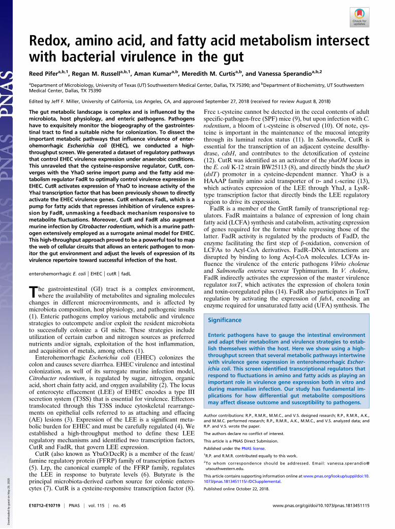
ResultsSurvey of E. coli Core Genome Metabolic and Transcription PathwaysThat Influence LEE Gene Expression. The LEE contains 41 genes,the majority of which are organized in five operons (LEE1–5).The first gene within LEE1 encodes the Ler transcriptional ac-tivator of all LEE genes (Fig. 1A). LEE4 encodes the EspBprotein that is part of the T3SS translocon (3, 19). We developedan ELISA-based approach for evaluating the expression of EspB(Fig. 1B). The pJAY1512 LEE encoding cosmid was trans-formed (20) into a subset of the E. coli strains from the BW25113Keio knockout library (21), generating a library of LEE-expressing K-12 strains. This library included 372 strains de-ficient in transcription factors, transcription antiterminators,two-component systems, and the DNA-binding type II antitoxinproteins (Fig. 1C and SI Appendix, Table S3). This library wasscreened in cysteine-supplemented DMEM under anaerobic
conditions, as oxygen availability is an important factor govern-ing LEE expression (22), and the pathways controlling LEEexpression under anaerobic conditions have been understudied.This screen yielded 58 strains that met the cutoff criteria of a
twofold change in EspB expression (Fig. 1D and SI Appendix,Table S3). As a testament to the validity of the screen, we alsoidentified several factors previously described to control LEEgene expression such as Fis, HNS, Hha, EutR, Fur, GadX, andLrp (23) (Fig. 1E). The majority of the genes involved in LEEregulation were in the other category. The second most commonwas antiterminators, followed by two-component systems.Transcription factors of the AraC and GntR family were foundat similar numbers, and the least common class of transcriptionfactors to regulate the LEE was LysRs (Fig. 1D and SI Appendix,Table S3). From these previously uncharacterized LEE regula-tors, three promising targets were selected for validation andfurther investigation: CutR, a FFRP family transcriptional reg-ulator; FadR, a GntR family transcription factor; and YehU, atwo-component system histidine sensor kinase. CutR is a cysteine-sensitive activator of the D-serine transporter YhaO, a previouslydescribed LEE-controlling protein (8, 13). FadR is the masterregulator of fatty acid synthesis and degradation, which has beenwell described with regards to metabolism, but has not beencharacterized as having a virulence-related function in pathogenicE. coli. YehU is a peptide and amino acid-sensitive histidinesensor kinase known to activate the expression of yjiY (24), thatencodes a transporter that is important during avian pathogenicE. coli infections (25). We generated deletion mutants of thesegenes in the 86-24 strain of EHEC. Upon rescreening these mu-tants by ELISA, we find that all three genes contribute to EspBregulation as suggested by our screen in K-12 (Fig. 1F).
CutR, FadR, and YehU in C. rodentium. To investigate whether therole of these transcription factors in virulence regulation trans-lated into in vivo phenotypes, we employed the C. rodentiummurine infection model. C. rodentium is an AE lesion-forming
A
B
C D
E
LEE1 LEE2 LEE3 LEE4
Ler
LEE5
F
escU escV �r/eae espA/espB
0 20 40 600
1
2
3
4
5
100
ELISA standard
EspB (ng/mL)
OD
450
0
123
45
OD450
0 20 40 60 100EspB (ng/ml)
EHEC LEE
K-12 Knockouts
-EspB
Total=372
Transcription factorsTwo-component systemsAntiterminatorsAntitoxins
Total=372
Transcrip�on factorsTwo component systemsAn�terminatorsAn�toxins
Total=58
AntiterminatorsAntitoxinsAraCAsnCGntRLuxRLysR
NagCTCSOther
Total=58
An�terminatorsAn�toxinsAraCAsnCGntRLuxRLysRNucleoid ProteinsNagCTCSOther
0
1
2
3
Rel
ati v
eEs
p BEx
p res
sion
(mut
ant/c
ont r
ol)
nd**
*
*ndnd
Ctr fis hnsB hha fur eutR gadX lrp
0
2
4
6
8
10
86-24 strain
Rel
ati v
eEs
p BEx
p res
sion
(mut
ant/c
ont r
ol)
**
****
WT cutR fadR yehU
Fig. 1. Screen for LEE regulators. (A) Schematic of the LEE operon arrangement. The LEE1-encoded Ler activates expression of all LEE operons. (B) Schematicof the screen for LEE regulators. BW25113 K-12 deletion strains were transformed with the LEE-encoding cosmid, pJAY1512. Strains were grown in DMEMunder anaerobic conditions in 96-well plate format and evaluated by ELISA for EspB production. (Inset) Validation of EspB-directed ELISA procedure byaddition of recombinant EspB to culture supernatants of BW25113. (C) Class makeup of the BW25113 deletion strains included in the screen. (D) Makeup ofgene families found in the screen with a twofold cutoff in change in EspB production. Statistical significance was calculated as ANOVA with Dunnett’s posthoc test. (E) K-12 knockouts identified in genes previously known to regulate the LEE. (F) EspB ELISA of EHEC (86-24) deletion strains used for validation ofscreen results. *P < 0.05, **P < 0.01; nd, results below limit of detection.
Pifer et al. PNAS | vol. 115 | no. 45 | E10713
MICRO
BIOLO
GY
Dow
nloa
ded
by g
uest
on
May
29,
202
0

pathogen that is extensively used as a surrogate organism for amouse model of EHEC infection. C. rodentium contains the LEEand forms AE lesions on murine colonocytes (26). We generatedΔcutR, ΔfadR, and ΔyehU strains of DBS770, a C. rodentiumstrain harboring the Shiga toxin encoding phage (27). Shiga toxin(Stx) is responsible for the hemolytic uremic syndrome in EHECinfections (3). The C. rodentium Stx model more closely resem-bles all of the facets of EHEC infection (27).Both the ΔcutR and the ΔfadR are attenuated for murine in-
fection compared with WT, and this phenotype could be com-plemented with cutR and fadR on a plasmid (Fig. 2 A–D and SIAppendix, Fig. S2 D and H) using both infectious doses of 109
(Fig. 2 A–D) and 107 cfu (SI Appendix, Fig. S2 D and H). TheΔyehU is not attenuated for murine infection (SI Appendix, Fig.S1), suggesting that this gene is not critical in this infectionmodel. The attenuation phenotypes of ΔcutR and ΔfadR cannotbe explained by an overt difference in bacterial burden, as these
strains colonized to levels equivalent to WT throughout the in-fection in stools at an infectious dose of 109 cfu (SI Appendix,Fig. S2 A and B). However, they did show a small one order ofmagnitude decrease at day 4 at an infectious dose of 107 cfu,suggesting that at a lower infectious dose, there is a slight de-crease in fitness (SI Appendix, Fig. S2 E and I). In terms of tissuecolonization WT and ΔcutR colonized the cecum at similarlevels, and ΔcutR showed a one order of magnitude decrease inthe colonization of the colon (SI Appendix, Fig. S2 F and G). TheΔfadR colonized both cecum and colon at the same levels as WT(SI Appendix, Fig. S2 J and K). This is not due to a growth defect,given that there is no significant difference in generation time forΔcutR DBS770 (82 ± 8 min) or ΔfadR DBS770 (71 ± 10 min)compared with WT (81 ± 9 min) (SI Appendix, Fig. S3).Both CutR and FadR influence LEE expression in C. roden-
tium in vitro. The ΔcutR has reduced secretion of EspB and LEEmRNA levels compared with WT (Fig. 2 E and G). The ΔfadR
A
B
C
D
E
F
I
G H
J
K L
Fig. 2. Representative screen hits in C. rodentium pathogenesis. All experiments were conducted using conventional mouse feed. Survival curves of C3H/HeJinfected with 109 cfu of WT, (A) ΔcutR, or (C) ΔfadR DBS770 C. rodentium or with PBS control (10 animals per infection group and 8 animals for PBS).Statistical significance calculated by Gehan–Breslow–Wilcoxon test. Weight of animals infected with (B) ΔcutR, or (D) ΔfadR DBS770, WT or mock (PBS). (E)Western for EspB from in vitro culture supernatants from WT, ΔcutR DBS770 and ΔcutR complemented (cultures grown in DMEM). (F) Western for EspB fromin vitro culture supernatants from WT, ΔfadR DBS770, and ΔfadR complemented (cultures grown in DMEM). (G) qRT-PCR of C. rodentium LEE mRNAs from invitro anaerobically grown WT, ΔcutR DBS770, and ΔcutR complemented strains (cultures grown in DMEM). (H) qRT-PCR quantification of C. rodentium LEEmRNAs from in vitro anaerobically grown WT, ΔfadR DBS770, and ΔfadR complemented strains (cultures grown in DMEM). (I) qRT-PCR of C. rodentium LEEmRNAs in murine cecum tissue of animals infected with WT, ΔcutR, and complemented strains. (J) qRT-PCR of C. rodentium LEE mRNAs in murine colon tissueof animals infected with WT, ΔcutR, and complemented strains. (K) qRT-PCR of C. rodentium LEE mRNAs in murine cecum tissue of animals infected with WT,ΔfadR, and complemented strains. (L) qRT-PCR of C. rodentium LEE mRNAs in murine colon tissue of animals infected with WT, ΔfadR, and complementedstrains. *P < 0.05, **P < 0.01, ***P < 0.001. P.I., postinfection.
E10714 | www.pnas.org/cgi/doi/10.1073/pnas.1813451115 Pifer et al.
Dow
nloa
ded
by g
uest
on
May
29,
202
0

secretes higher levels of EspB protein than WT (Fig. 2F) and is atranscriptional repressor of the LEE at the mRNA level, withΔfadR depicting higher LEE gene expression (Fig. 2H). Duringmurine infection ΔcutR has decreased LEE gene expression inthe cecum and the colon (Fig. 2 I and J). The ΔfadR has de-creased expression of the LEE genes in the cecum, and dysre-gulated expression of the LEE genes in the colon, with escUbeing up-regulated and tir and espA being down-regulated in thismutant (Fig. 2 K and L). It is noteworthy that the LEE4 andLEE5 operons that harbor espA and tir are highly post-transcriptionally regulated by the GlmY–GlmZ sRNAs (28).This indicates that deregulation of LEE expression, whether bydecreased or increased expression, affects C. rodentium patho-genesis. Overexpression of the LEE can be detrimental to thepathogen’s virulence because it creates an unnecessary energyburden that decreases fitness (4). Because EHEC has a low in-fectious dose of 50 cfu (3), it has to efficiently coordinate theright levels of expression of its virulence traits. This is especially
important to successfully compete with the dense and highlyadapted microbiota for a colonization niche.We have previously investigated the intestinal metabolic pro-
file of DBS770 infected or uninfected animals (10). Cysteine wasthe second most increased (155-fold) metabolite in C. rodentium-infected compared with uninfected (PBS control) animals, fol-lowing antibiotic pretreatment to deplete the resident micro-biota. As CutR requires cysteine to function (8), we investigatedthe course of infection for ΔcutR under depletion of the micro-biota to assess whether the bloom of cysteine was microbiotadependent. Following antibiotic treatment, ΔcutR was still atten-uated compared with WT, suggesting that changes in the cysteinelevels within the intestine are not being dictated by the microbiota(SI Appendix, Fig. S4) and may reflect the host immune responsesin attempting to restore mucosal integrity (11). Cysteine levels areincreased by antibiotic treatment in the lumen of both cecum andcolon, and in the tissues of colon and small intestine, while it re-mains unchanged in cecum tissues (SI Appendix, Fig. S5). This
eae (LEE5) mRNA
***
0.0
0.5
1.0
1.5
Rela
ve m
RNA
expr
essi
on
4mM Cys No Cys
***
espA (LEE4) mRNA
0.0
0.5
1.0
1.5
Rela
ve m
RNA
expr
essi
on
****
****
ler espA r eae0.0
0.5
1.0
1.5
Rela
ve m
RNA
Expr
essi
on
WTcutRcpl
**0.0
0.5
1.0
1.5
Rela
ve m
RNA
Expr
essi
onRe
lave
mRN
A Ex
pres
sion
** **
** **
0.0
0.5
1.0
1.5
Rela
ve m
RNA
Expr
essi
on
ler espA
** **0
2
4
6
Rela
ve m
RNA
Expr
essi
on
yhaO ler r espA
WT cutR cpl
yhaO
* **
***
0.00
0.01
0.02
0.03
0.04
0.05
Perc
ent o
f Inp
ut
rpoZ yhaO ybaO fadL ler ler-169+155 -372-102
***0.0
0.5
1.0
1.5
4mM Cys No Cys
ler (LEE1)mRNAcutRWT
A
E
B C
G
J
LK
F
I
H
4mM Cys No Cys
D
cutR
Fig. 3. CutR regulation of LEE expression in EHEC. (A) qRT-PCR of LEE genes from WT, ΔcutR, and complemented EHEC strains grown anaerobically in thepresence of cysteine (cultures grown in DMEM). (B) Western for EspB secreted from WT, ΔcutR, and complemented EHEC strains grown anaerobically in thepresence of cysteine (cultures grown in DMEM). (C) Western for EspB from whole cell lysates of WT, ΔcutR, and complemented EHEC strains (cultures grown inDMEM). (D) Schematic of the cutR locus from E. coli, Shigella dysenteriae, C. rodentium, and S. Typhimurium. (E–G) qRT-PCR of LEE transcripts for (E) ler, (F)eae, and (G) espA fromWT or ΔcutR grown anaerobically in DMEM either with or without cysteine. (H) Schematic representing a putative mechanism of cutR-dependent LEE regulation. CutR positively regulates the expression of yhaO, a serine transporter that positively regulates LEE expression via the LysR-typetranscription factor YhaJ. (I) qRT-PCR of yhaO mRNA from WT, ΔcutR, and complemented strains (cultures grown in DMEM). (J) qRT-PCR to assess LEE ex-pression from ΔcutRΔyhaO double mutant (cultures grown in DMEM). (K) qRT-PCR for complementation of LEE transcriptional phenotype by N-terminally V5-tagged CutR during preparation of cells for ChIP (cultures grown in DMEM). (L) ChIP-qPCR results for empty vector control or N-terminally tagged CutR.Probes are designed to amplify the promoter regions of yhaO (positive control), ybaO (cutR promoter), rpoZ (negative control), fadL, or overlapping frag-ments of the ler (LEE1) promoter, numbered from the proximal transcriptional start site. *P < 0.05, **P < 0.01, ***P < 0.001.
Pifer et al. PNAS | vol. 115 | no. 45 | E10715
MICRO
BIOLO
GY
Dow
nloa
ded
by g
uest
on
May
29,
202
0

increase in cysteine is due to the fact that antibiotic treatmentper se increases intestinal inflammation, as has been previouslyreported (29). Expression of most LEE genes in WT C. rodentiumis increased during murine infection in mice under a cystine (it isthe oxidized stable form of cysteine)-replete versus absent diet (SIAppendix, Fig. S6A). Moreover, ΔcutR is only attenuated for in-fection when the mice are under a cystine-replete diet (SI Ap-pendix, Fig. S6 B and D). This is because LEE regulation by CutRonly occurs in the presence of cysteine. However, in vivo tir reg-ulation was decreased in the presence of cystine, but did not reachstatistical significance, because tir is encoded within LEE5 which ishighly posttranscriptionally regulated; this discrepancy can be dueto that described in ref. 28 (SI Appendix, Fig. S6 C and E). Al-together C. rodentium is sensing cysteine in the gut to up-regulateLEE gene expression increasing its virulence potential, withoutaffecting pathogen expansion.
Intersection of CutR/YhaO and FadR LEE Regulation in EHEC. Con-gruent with the C. rodentium results, CutR is also a transcriptionactivator of LEE gene expression in EHEC (Fig. 3A). EspB se-cretion is decreased in ΔcutR (Fig. 3B), reflecting the overalldecreased transcription of the LEE-encoded T3SS (Fig. 3A).However, the levels of EspB in whole cell lysates are increased inΔcutR (Fig. 3C). The EspB transcript is highly posttranscrip-tionally regulated in EHEC (30), and the discrepancy in theoverall levels of EspB protein versus secreted EspB are probablya reflection of this regulation. This also explains the elevatedlevels of overall EspB protein in the ELISA performed with theEHEC ΔcutR (Fig. 1F). The cutR gene is located adjacent tothe cdsH gene in C. rodentium and S. Typhimurium, which en-codes the cysteine desulfhydrase (Fig. 3D). However, this gene isabsent in EHEC, in which cutR is immediately preceded by thethiamine pyrimidine pyrophosphate hydrolase, cof. CutR is acysteine-dependent transcription factor (8) and regulates the ex-pression of the cdsH gene that encodes cysteine desulfhydrasewhich contributes to the detoxification of cysteine (12). CutR isalso a cysteine-dependent transcription factor in the regulation ofits previously described target yhaO (8). Therefore, we attemptedto determine whether the CutR–LEE regulatory phenotype wasdependent upon the presence of cysteine by excluding this aminoacid from the growth media (no differences in growth in thepresence or absence of cysteine were observed). We observe thatthe ability of CutR to govern the LEE transcript level in EHECalso requires the presence of cysteine (Fig. 3 E–G), consistent withits dependency on cysteine for transcription activity.CutR is an activator of yhaO, which enhances serine import to
increase activity of the YhaJ transcription factor that directlyactivates LEE transcription (13) (Fig. 3 H and I). Therefore, it isconceivable that CutR-dependent LEE transcriptional activationacts through YhaO. We evaluated whether CutR-dependentLEE transcriptional activation occurs through yhaO by generat-ing a double knockout of ΔcutRΔyhaO. This double knockouthad an additive effect beyond the single yhaO deletion mutant,suggesting that CutR LEE transcriptional regulation also occursindependently from YhaO (Fig. 3J). YhaO exerts its control overthe LEE through the yhaJ-encoded transcription factor that di-rectly activates LEE transcription (13). Therefore, we hypothe-sized that cutR deficiency might diminish yhaJ expression as itdoes yhaO, and in turn diminish LEE transcript levels. However,we do not observe a decrease in yhaJ in ΔcutR, suggesting thatCutR is not involved in transcriptional regulation of this gene,probably modulating its activity through the levels of importedserine (SI Appendix, Fig. S7). Altogether, these data indicate thatthere is a CutR–YhaO arm and a CutR–YhaO independent armto control LEE transcription. These data suggest that CutR maydirectly regulate transcription of the LEE genes. Because wewere unable to purify soluble and folded CutR protein, toevaluate if CutR directly regulates transcription of the LEE, we
constructed a tet-inducible V5 N-terminal tagged CutR plasmidfor use as a ChIP construct. This construct is capable of com-plementing the cutRmutant (Fig. 3K). We then performed ChIP-qPCR to evaluate if segments of the LEE1 promoter (that en-codes the Ler master regulator of the LEE genes) are capable ofinteracting with CutR protein in vivo (Fig. 3L). We observed thatCutR is capable of interacting with the yhaO promoter asexpected, while not interacting with the negative control rpoZ.We observe that CutR interacts with the LEE1 regulatory region,suggesting that direct regulation of ler may be a mechanism bywhich cutR regulates T3S in EHEC (Fig. 3L).The CutR regulon has not been thoroughly explored; there-
fore, we performed transcriptomic studies to evaluate differen-tial gene regulation between ΔcutR and WT EHEC. Thesestudies showed that 121 genes were up-regulated, and 227 geneswere down-regulated in ΔcutR (SI Appendix, Fig. S8 A and B andTables S4 and S5). Notably, many of the up-regulated (10%) anddown-regulated (21%) genes fall into a general category oftransporters of metabolites, including fatty acids (fadL), poly-amines (ydcS), glycine betaine (yehY), arginine (artP), and D/L-serine (yhaO). This suggests that CutR may have a broad func-tion as a regulator of metabolite import. We confirmed themicroarray results for a subset of genes via qRT-PCR. Inagreement with previous reports, we observe that yhaO is one ofthe most strongly down-regulated genes in the cutR deletionmutant (SI Appendix, Fig. S8C).Because there is YhaO-independent CutR regulation of the
LEE genes (Fig. 3), we aimed to determine what other CutR-dependent processes may influence the LEE. We reasoned thatbecause the original CutR dependency of the LEE was observedin E. coli K-12, the mechanism of action must be at least partiallyconserved between K-12 and EHEC. Therefore, we decided toreutilize the LEE cosmid transformed Keio library to evaluatethe importance of 304 differentially expressed nonessential K-12 genes on EspB expression. We observe that 12 qRT-PCR-confirmed CutR-regulated genes (SI Appendix, Fig. S9) are capableof influencing EspB expression by at least fivefold. These datasuggest that CutR-dependent LEE regulation may be multifactorial.In these studies we observed that CutR activates expression of
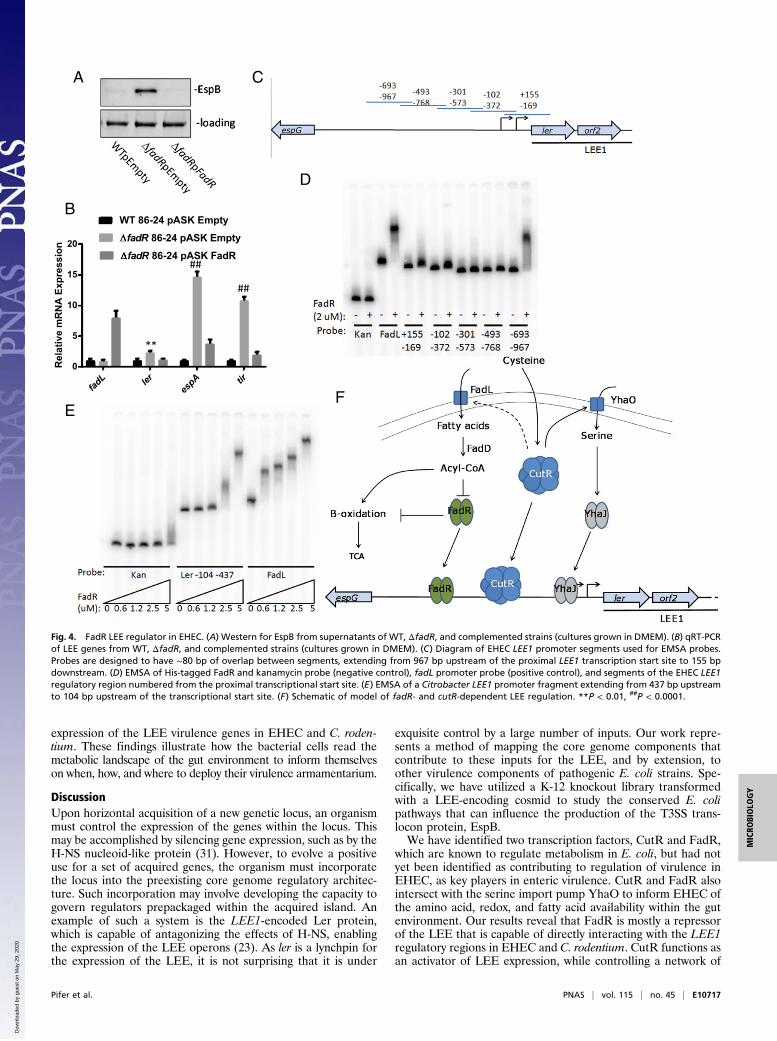
fadL (SI Appendix, Fig. S8C) that encodes a transporter for fattyacids. These fatty acids are converted to Acyl-CoA, which in-hibits the transcription factor FadR (Fig. 4F). In agreement withthe C. rodentium data (Fig. 2), FadR acts as a transcriptionalrepressor of the LEE genes, but this repression is alleviated whenCutR activates FadL which increases the levels of Acyl-CoA thatinhibit FadR function and repression of LEE transcription (Fig.4 A and B). These data are congruent with decreased tran-scription of the LEE ler and tir genes in the fadL mutant (SIAppendix, Fig. S8D). To determine whether FadR may serve as adirect regulator of the LEE, purified recombinant N-terminallyHis-tagged FadR protein (SI Appendix, Fig. S10) was used onelectrophoretic mobility shift assays (EMSAs) with overlappingfragments (Fig. 4C) of the LEE1 promoter. We observe thatFadR is capable of interacting with the fadL promoter while notinteracting with the kanamycin cassette of pRS551 (negativecontrol). FadR interacts with LEE1 fragments ranging from −967to −693 bp and −102 to −372 bp upstream of the proximalpromoter transcription start site (Fig. 4D). The Citrobacter LEE1regulatory region lacks the −967 to −693 bp region, as an in-sertional element has rendered the region significantly shorterthan that of EHEC. Therefore, we sought to determine whetherFadR is capable of interacting with a more proximal region ofCitrobacter’s LEE1. We find that FadR is capable of binding to afragment of the Citrobacter LEE1 region that is between −437and −104 bp upstream of the transcriptional start site (Fig. 4E).Our findings link CutR–cysteine-dependent regulation with
YhaO–serine regulation to fatty acid metabolism (FadL andFadR) being converged and interconnected to optimally regulate
E10716 | www.pnas.org/cgi/doi/10.1073/pnas.1813451115 Pifer et al.
Dow
nloa
ded
by g
uest
on
May
29,
202
0
expression of the LEE virulence genes in EHEC and C. roden-tium. These findings illustrate how the bacterial cells read themetabolic landscape of the gut environment to inform themselveson when, how, and where to deploy their virulence armamentarium.
DiscussionUpon horizontal acquisition of a new genetic locus, an organismmust control the expression of the genes within the locus. Thismay be accomplished by silencing gene expression, such as by theH-NS nucleoid-like protein (31). However, to evolve a positiveuse for a set of acquired genes, the organism must incorporatethe locus into the preexisting core genome regulatory architec-ture. Such incorporation may involve developing the capacity togovern regulators prepackaged within the acquired island. Anexample of such a system is the LEE1-encoded Ler protein,which is capable of antagonizing the effects of H-NS, enablingthe expression of the LEE operons (23). As ler is a lynchpin forthe expression of the LEE, it is not surprising that it is under
exquisite control by a large number of inputs. Our work repre-sents a method of mapping the core genome components thatcontribute to these inputs for the LEE, and by extension, toother virulence components of pathogenic E. coli strains. Spe-cifically, we have utilized a K-12 knockout library transformedwith a LEE-encoding cosmid to study the conserved E. colipathways that can influence the production of the T3SS trans-locon protein, EspB.We have identified two transcription factors, CutR and FadR,
which are known to regulate metabolism in E. coli, but had notyet been identified as contributing to regulation of virulence inEHEC, as key players in enteric virulence. CutR and FadR alsointersect with the serine import pump YhaO to inform EHEC ofthe amino acid, redox, and fatty acid availability within the gutenvironment. Our results reveal that FadR is mostly a repressorof the LEE that is capable of directly interacting with the LEE1regulatory regions in EHEC and C. rodentium. CutR functions asan activator of LEE expression, while controlling a network of
fadL ler
espA tir
0
5
Rel
ativ
e m
RN
A E
xpre
ssio
n
10
15
20
WT 86-24 pASK Empty
fadR 86-24 pASK Empty
fadR 86-24 pASK FadR
**
##
##
B
A C
D
EF
espG ler orf2
Fig. 4. FadR LEE regulator in EHEC. (A) Western for EspB from supernatants of WT, ΔfadR, and complemented strains (cultures grown in DMEM). (B) qRT-PCRof LEE genes from WT, ΔfadR, and complemented strains (cultures grown in DMEM). (C) Diagram of EHEC LEE1 promoter segments used for EMSA probes.Probes are designed to have ∼80 bp of overlap between segments, extending from 967 bp upstream of the proximal LEE1 transcription start site to 155 bpdownstream. (D) EMSA of His-tagged FadR and kanamycin probe (negative control), fadL promoter probe (positive control), and segments of the EHEC LEE1regulatory region numbered from the proximal transcriptional start site. (E) EMSA of a Citrobacter LEE1 promoter fragment extending from 437 bp upstreamto 104 bp upstream of the transcriptional start site. (F) Schematic of model of fadR- and cutR-dependent LEE regulation. **P < 0.01, ##P < 0.0001.
Pifer et al. PNAS | vol. 115 | no. 45 | E10717
MICRO
BIOLO
GY
Dow
nloa
ded
by g
uest
on
May
29,
202
0

genes which influence the LEE. Importantly, our transcriptomicsanalysis for ΔcutR EHEC suggests that this transcription factormay be important for maintaining a network of metabolitetransporters. This is interesting from a virulence perspective, asthe importance of small molecules in governing LEE expressionis being increasingly appreciated.Our dataset reveals other yet unexplored pathways that may
prove important for regulation of T3S in EHEC, comprising acomplex cellular web with intersecting circuits that remain to bemapped in detail. The metabolic landscape of the gut is dramat-ically changed by the presence of certain members of the micro-biota and upon enteric infection (10). EHEC is a remarkablyefficient pathogen, which is able to establish itself in the hostthrough a very small infectious dose. EHEC’s proficiency to in-tersect metabolic, signaling, redox, and oxygen sensing may be atthe core of its prowess as a successful enteric pathogen.
Materials and MethodsBacterial Strains and Plasmids. All strains and plasmids used in this study arelisted in SI Appendix, Table S1. Recombinant DNA and molecular biologytechniques were performed as previously described (32). All oligonucleotidesused are listed in SI Appendix, Table S2. Knockout strains were constructedusing lambda red (33). Plasmids pACYC184 (NEB) and pASK IBA32 (IBALifesciences) were used as complementation vectors. Cultures were grown inglucose DMEM (Gibco) with or without 4 mM cysteine. Generation timeswere calculated as [Log2]/slope of semilog plot of OD per time by GraphpadPrism 6. Cultures were grown at 37 °C under strict anaerobic conditions usinga ShelLabs Bactron chamber containing 5% H2, 5% CO2, and 90% N2.
EspB Production Screening in K-12. Keio library K-12 knockout strains weretransformed with pAY1512 and grown in 96-well triplicate plates in DMEMwith cysteine under anaerobic conditions. Upon reaching stationary phase,growth was halted by transfer to 4 °C and supplementation with 15 mg/mLsodium azide and Sigma Protease Inhibitor Mixture. They were diluted inPBS into Immulon Microtiter plates. EspB levels were determined with ELISAusing anti-EspB antisera. Absolute concentrations were interpolated fromstandard curves from titrations of recombinant EspB protein. The ΔlacA KeioK-12 knockout strain with and without pJAY1512 was used as controls fornormalization and background subtraction. All mutants were screened intriplicate; statistical significance was calculated in Graphpad Prism 6 byANOVA followed by Dunnett’s multiple comparison test.
Chromatin Immunoprecipitation. ChIP was performed using N-terminally V5-tagged CutR protein, cloned into PCR linearized pASK IBA32 using Gibsoncloning with primers described in SI Appendix, Table S2. WT and mutantstrains of 86-24 were transformed with pASK empty vector or pASKnV5Y-baO. They were grown with supplementation of 12 ng/mL anhydrotetracy-cline. The cells were harvested and split for use in evaluation of proteinexpression, mRNA expression, or for ChIP. ChIP samples were treated with1% formaldehyde for 20 min. Fixed cells were washed, resuspended in10 mM Tris pH 8, 150 mM NaCl, 1 mM EDTA, 1% Triton X-100, 0.1% sodiumdeoxycholate, 0.1% RNaseA, and Sigma Protease Inhibitor Mixture andsonicated to fragments of 100–600 bp by seven cycles of 30 s on/60 s off at95% power on a Qsonica Q125 sonicator. Lysed samples were cleared andquantified by nanodrop. Equivalent loadings of nucleic acids were used foreach ChIP replicate and coupled at 37 °C by adding 10 μg of anti-V5 antibody(Abcam) per reaction. These reaction were precipitated with 1.5 mg ProteinA Dynabeads and washed in 10 mM Tris pH 8, 500 mM LiCl, 1 mM EDTA,0.5% Nonidet P-40, 0.5% sodium deoxycholate. ChIP samples were decros-slinked for 18 h at 65 °C in 10 mM Tris pH 8, 50 mM NaCl, 10 mM EDTA, 1%SDS. Protein was degraded by the addition of 80 μg Proteinase K per re-action and incubation at 55 °C for 4 h, then 95 °C for 10 min. DNA waspurified with Qiagen MinElute kits. qRT-PCR was used to evaluate the per-cent of input of each sample captured during ChIP. Standard curves for eachinput sample were performed for each probe set (SI Appendix, Table S2) andused to calculate the percent of sequence precipitated.
Western Blotting. For Westerns of secreted proteins, all cultures were grownin DMEM to the same OD600 under anaerobiosis in the presence of cysteine at37 °C. A total of 10 μg BSA was added to secreted proteins before concen-tration and loading so that the efficiency of processing was known to beequivalent from sample to sample. Membranes were probed using an anti-EspB antiserum. Whole cell lysates were also run and probed with anti-EspB;
loading was evaluated using stain-free settings on a Bio-Rad ChemiDocimaging system.
Protein Purification and EMSA. FadR was cloned into the NdeI and BamHI sitesof pET28 by Gibson cloning (SI Appendix, Table S2) to create an N-terminalHis-tagged construct. This was transformed into NiCO21 (NEB) cells. His-tagged FadR was purified through nickel columns according to manufac-turer’s instructions. For EMSA, DNA probes were prepared by PCR (SI Ap-pendix, Table S2 for primers) from genomic templates. Probes were purifiedby gene electrophoresis and labeled with 32P y-ATP by T4 PNK (NEB). Labeledprobes were further purified by Qiagen PCR purification kit. EMSA reactionswere prepared as protein diluted into a 2× EMSA buffer (50 mM NaKPO4 pH7.5, 100 mM NaCl, 1.5 mM DTT, 100 μg/mL BSA, 250 μg/mL sonicated salmonsperm DNA). Binding was resolved on 5% polyacrylamide gels in Tris/borate/EDTA. Gels were dried onto filter paper and exposed to phosphoimagerscreens and assessed on a GE Typhoon scanner.
qRT-PCR and Microarray. Samples were grown in triplicate in 50-mL cultures asdescribed above. RNA was purified using an Ambion Bacterial RNA Extractionkit according to manufacturer’s instructions. qPCR was performed with Invi-trogen SYBR Green Real-Time PCR Master Mix according to manufacturer’sspecifications on an ABI QuantStudio 6 Flex instrument. Data were analyzedby the ΔΔCt method. Statistics were calculated as t test with GraphPad Prism.For microarrays, RNA samples were converted to cDNA and labeled as de-scribed in the Affymetrix Gene Expression manual. Samples were hybridized toAffymetrix E. coli 2.0 chips according to manufacturer’s recommendations.Data analysis was done with GeneSpring software, using MAS5 normalization.We report only genes found to be differentially expressed by twofold.
Animal Experiments. SPF female C3H/HeJ animals were used. We used 8–10 animals per experimental group that were between 4–6 wk of age at thetime of experiment. Animals were infected by oral gavage of 109 or 107 cfuof DBS770 or mutants in PBS or PBS alone. Animals were checked daily forsurvival, weight change, and stools and tissues were collected for analysis ofcolony-forming units and LEE gene expression. In some experiments, animalswere precleared of their microbiota by including in their water 1 g/L ampicillinsodium salt, 1 g/L neomycin trisulfate salt hydrate, 1 g/L metronidazole, and0.5 g/L vancomycin. This treatment was continued for 10 d, followed by asingle day without antibiotics before gavage. In some experiments animalswere given a cysteine-containing or a cysteine-absent feed. Statistical signifi-cance was determined by Prism 6. Survival was evaluated by Gehan–Breslow–
Wilcoxon test. Research involving animals has been approved by the UTSouthwestern Institutional Animal Care and Use Committee.
Cysteine Measurements. Mouse intestinal segments and stools from con-ventional and antibiotic-treated mice were collected. Tissue and content/stools were homogenized in a sixfold volume of 50:50 methanol:dH20containing 15 mM DTT using a polytron homogenizer (6× weight of tissue ing = vol of 50:50 MeOH/H20 added; total homogenate volume = 7×weight oftissue). The samples were spun twice and supernatant was collected. An al-iquot of each stool supernatant was pooled and diluted 50× in 50:50 meth-anol:dH20 to make background matrix for preparing the standard curve.A similar diluted homogenate was made for the intestinal tissue standardcurve. Standards were made by spiking 100 μL of the diluted homogenatewith 2 μL of various concentrations of cysteine prepared fresh from powder.Samples and standards prepared in the 50× diluted pooled homogenatewere then mixed 1:1 with crash solution (50:50 MeOH:H20 containing 15 mMDTT, 0.2% formic acid and 100 ng/mL DL-homocysteine d4). Standard con-centrations are based on final volume after addition of crash solution so a 2×correction is applied to sample concentrations. After addition of the crashsolution, samples were incubated for 10 min and samples were then spun5′ 16,100 × g, supernatant was collected, and spun a second time. The su-pernatant was then transferred to an HPLC vial with insert and analyzed byLC-MS/MS. The Qtrap 6500 + analytical conditions used were: buffer A:ddH20 + 0.1% formic acid; buffer B: MeOH + 0.1% formic acid; column:Resteck Ultra IBD column 5 μM, 150 × 4.6 mm; gradient conditions: 0–1.5 min3% B; 1.5–2.0 min gradient to 100% B; 2.0–3.5 min 100% B; 3.5–3.6 mingradient to 3% B; 3.6–4 min 3% B; flow rate: 1.5 mL/min; ion source/gas pa-rameters: CUR = 30, CAD = 9, IS = 5500, TEM = 600, GS1 = 60, GS2 = 60.Cysteine transition 121.952–75.9; DL-homocysteine-d4 139.96/94.0
ACKNOWLEDGMENTS. We thank James B. Kaper (University of MarylandMedical School) for the pJAY1512 LEE-encoding cosmid. This work wassupported by NIH Grants AI053067, AI77853, AI114511, and AI077613. R.P.was supported through NIH Training Grant 5 T32 AI7520-14.
E10718 | www.pnas.org/cgi/doi/10.1073/pnas.1813451115 Pifer et al.
Dow
nloa
ded
by g
uest
on
May
29,
202
0

1. Bäumler AJ, Sperandio V (2016) Interactions between the microbiota and pathogenicbacteria in the gut. Nature 535:85–93.
2. Cameron EA, Sperandio V (2015) Frenemies: Signaling and nutritional integration inpathogen-microbiota-host interactions. Cell Host Microbe 18:275–284.
3. Kaper JB, Nataro JP, Mobley HL (2004) Pathogenic Escherichia coli. Nat Rev Microbiol2:123–140.
4. Pacheco AR, et al. (2012) Fucose sensing regulates bacterial intestinal colonization.Nature 492:113–117.
5. Yokoyama K, et al. (2006) Feast/famine regulatory proteins (FFRPs): Escherichia coliLrp, AsnC and related archaeal transcription factors. FEMS Microbiol Rev 30:89–108.
6. Nakanishi N, et al. (2009) Regulation of virulence by butyrate sensing in enter-ohaemorrhagic Escherichia coli. Microbiology 155:521–530.
7. Roediger WE (1982) Utilization of nutrients by isolated epithelial cells of the rat colon.Gastroenterology 83:424–429.
8. Shimada T, Tanaka K, Ishihama A (2016) Transcription factor DecR (YbaO) controlsdetoxification of L-cysteine in Escherichia coli. Microbiology 162:1698–1707.
9. Sasabe J, et al. (2016) Interplay between microbial d-amino acids and host d-aminoacid oxidase modifies murine mucosal defence and gut microbiota. Nat Microbiol 1:16125.
10. Curtis MM, et al. (2014) The gut commensal Bacteroides thetaiotaomicron exacer-bates enteric infection through modification of the metabolic landscape. Cell HostMicrobe 16:759–769.
11. Circu ML, Aw TY (2012) Intestinal redox biology and oxidative stress. Semin Cell DevBiol 23:729–737.
12. Oguri T, Schneider B, Reitzer L (2012) Cysteine catabolism and cysteine desulfhydrase(CdsH/STM0458) in Salmonella enterica serovar typhimurium. J Bacteriol 194:4366–4376.
13. Connolly JP, et al. (2016) A highly conserved bacterial D-serine uptake system linkshost metabolism and virulence. PLoS Pathog 12:e1005359.
14. Kovacikova G, Lin W, Taylor RK, Skorupski K (2017) The fatty acid regulator FadRinfluences the expression of the virulence cascade in the El Tor biotype of Vibriocholerae by modulating the levels of ToxT via two different mechanisms. J Bacteriol199:e00762-16.
15. Thomson JJ, Plecha SC, Withey JH (2015) A small unstructured region in Vibrio chol-erae ToxT mediates the response to positive and negative effectors and ToxT pro-teolysis. J Bacteriol 197:654–668.
16. Plecha SC, Withey JH (2015) Mechanism for inhibition of Vibrio cholerae ToxT activityby the unsaturated fatty acid components of bile. J Bacteriol 197:1716–1725.
17. Lucas RL, et al. (2000) Multiple factors independently regulate hilA and invasion geneexpression in Salmonella enterica serovar typhimurium. J Bacteriol 182:1872–1882.
18. Golubeva YA, Ellermeier JR, Cott Chubiz JE, Slauch JM (2016) Intestinal long-chainfatty acids act as a direct signal to modulate expression of the Salmonella pathoge-nicity island 1 type III secretion system. MBio 7:e02170-e15.
19. Elliott SJ, et al. (1998) The complete sequence of the locus of enterocyte effacement(LEE) from enteropathogenic Escherichia coli E2348/69. Mol Microbiol 28:1–4.
20. Elliott SJ, Yu J, Kaper JB (1999) The cloned locus of enterocyte effacement from en-terohemorrhagic Escherichia coli O157:H7 is unable to confer the attaching and ef-facing phenotype upon E. coli K-12. Infect Immun 67:4260–4263.
21. Baba T, et al. (2006) Construction of Escherichia coli K-12 in-frame, single-geneknockout mutants: The Keio collection. Mol Syst Biol 2:2006.0008.
22. Carlson-Banning KM, Sperandio V (2016) Catabolite and oxygen regulation of en-terohemorrhagic Escherichia coli virulence. MBio 7:e01852-16.
23. Mellies JL, Lorenzen E (2014) Enterohemorrhagic Escherichia coli virulence generegulation. Microbiol Spectr 2:EHEC-0004-2013.
24. Behr S, Fried L, Jung K (2014) Identification of a novel nutrient-sensing histidine ki-nase/response regulator network in Escherichia coli. J Bacteriol 196:2023–2029.
25. Tuntufye HN, Lebeer S, Gwakisa PS, Goddeeris BM (2012) Identification of Avianpathogenic Escherichia coli genes that are induced in vivo during infection in chick-ens. Appl Environ Microbiol 78:3343–3351.
26. Deng W, et al. (2004) Dissecting virulence: Systematic and functional analyses of apathogenicity island. Proc Natl Acad Sci USA 101:3597–3602.
27. Mallick EM, et al. (2012) A novel murine infection model for Shiga toxin-producingEscherichia coli. J Clin Invest 122:4012–4024.
28. Gruber CC, Sperandio V (2014) Posttranscriptional control of microbe-induced re-arrangement of host cell actin. MBio 5:e01025-e13.
29. Ng KM, et al. (2013) Microbiota-liberated host sugars facilitate post-antibiotic ex-pansion of enteric pathogens. Nature 502:96–99.
30. Connolly JP, Finlay BB, Roe AJ (2015) From ingestion to colonization: The influence ofthe host environment on regulation of the LEE encoded type III secretion system inenterohaemorrhagic Escherichia coli. Front Microbiol 6:568.
31. Navarre WW, et al. (2006) Selective silencing of foreign DNA with low GC content bythe H-NS protein in Salmonella. Science 313:236–238.
32. Sambrook J, Fritsch EF, Maniatis T (1989) Molecular Cloning: A Laboratory Manual(Cold Spring Harbor Lab Press, Cold Spring Harbor, NY), 2nd Ed.
33. Datsenko KA, Wanner BL (2000) One-step inactivation of chromosomal genes in Es-cherichia coli K-12 using PCR products. Proc Natl Acad Sci USA 97:6640–6645.
Pifer et al. PNAS | vol. 115 | no. 45 | E10719
MICRO
BIOLO
GY
Dow
nloa
ded
by g
uest
on
May
29,
202
0