red blood cell - saylor · pdf filered blood cell 1 red blood cell ... very oxygen rich cold...
TRANSCRIPT
Red blood cell 1
Red blood cell
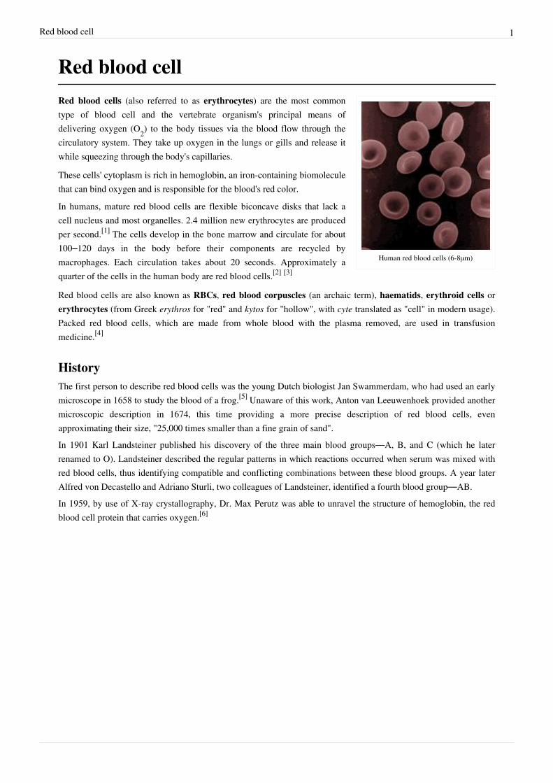
Human red blood cells (6-8μm)
Red blood cells (also referred to as erythrocytes) are the most commontype of blood cell and the vertebrate organism's principal means ofdelivering oxygen (O2) to the body tissues via the blood flow through thecirculatory system. They take up oxygen in the lungs or gills and release itwhile squeezing through the body's capillaries.
These cells' cytoplasm is rich in hemoglobin, an iron-containing biomoleculethat can bind oxygen and is responsible for the blood's red color.
In humans, mature red blood cells are flexible biconcave disks that lack acell nucleus and most organelles. 2.4 million new erythrocytes are producedper second.[1] The cells develop in the bone marrow and circulate for about100–120 days in the body before their components are recycled bymacrophages. Each circulation takes about 20 seconds. Approximately aquarter of the cells in the human body are red blood cells.[2] [3]
Red blood cells are also known as RBCs, red blood corpuscles (an archaic term), haematids, erythroid cells orerythrocytes (from Greek erythros for "red" and kytos for "hollow", with cyte translated as "cell" in modern usage).Packed red blood cells, which are made from whole blood with the plasma removed, are used in transfusionmedicine.[4]
HistoryThe first person to describe red blood cells was the young Dutch biologist Jan Swammerdam, who had used an earlymicroscope in 1658 to study the blood of a frog.[5] Unaware of this work, Anton van Leeuwenhoek provided anothermicroscopic description in 1674, this time providing a more precise description of red blood cells, evenapproximating their size, "25,000 times smaller than a fine grain of sand".In 1901 Karl Landsteiner published his discovery of the three main blood groups—A, B, and C (which he laterrenamed to O). Landsteiner described the regular patterns in which reactions occurred when serum was mixed withred blood cells, thus identifying compatible and conflicting combinations between these blood groups. A year laterAlfred von Decastello and Adriano Sturli, two colleagues of Landsteiner, identified a fourth blood group—AB.In 1959, by use of X-ray crystallography, Dr. Max Perutz was able to unravel the structure of hemoglobin, the redblood cell protein that carries oxygen.[6]

Red blood cell 2
Vertebrate erythrocytes
There is an immense size variation in vertebrateerythrocytes, as well as a correlation between cell andnucleus size. Mammalian erythrocytes, which do notcontain nuclei, are considerably smaller than those of
most other vertebrates.[7]
Erythrocytes consist mainly of hemoglobin, a complexmetalloprotein containing heme groups whose iron atomstemporarily bind to oxygen molecules (O2) in the lungs or gillsand release them throughout the body. Oxygen can easily diffusethrough the red blood cell's cell membrane. Hemoglobin in theerythrocytes also carries some of the waste product carbon dioxideback from the tissues; most waste carbon dioxide, however, istransported back to the pulmonary capillaries of the lungs asbicarbonate (HCO3
-) dissolved in the blood plasma. Myoglobin, acompound related to hemoglobin, acts to store oxygen in musclecells.[8]
The color of erythrocytes is due to the heme group of hemoglobin.The blood plasma alone is straw-colored, but the red blood cellschange color depending on the state of the hemoglobin: whencombined with oxygen the resulting oxyhemoglobin is scarlet, andwhen oxygen has been released the resulting deoxyhemoglobin isof a dark red burgundy color, appearing bluish through the vesselwall and skin. Pulse oximetry takes advantage of this color changeto directly measure the arterial blood oxygen saturation usingcolorimetric techniques.
The sequestration of oxygen carrying proteins inside specializedcells (rather than having them dissolved in body fluid) was animportant step in the evolution of vertebrates as it allows for lessviscous blood, higher concentrations of oxygen, and betterdiffusion of oxygen from the blood to the tissues. The size oferythrocytes varies widely among vertebrate species; erythrocytewidth is on average about 25% larger than capillary diameter and it has been hypothesized that this improves theoxygen transfer from erythrocytes to tissues.[9]
The only known vertebrates without erythrocytes are the crocodile icefishes (family Channichthyidae); they live invery oxygen rich cold water and transport oxygen freely dissolved in their blood.[10] While they do not usehemoglobin any more, remnants of hemoglobin genes can be found in their genome.[11]
NucleusErythrocytes in mammals are anucleate when mature, meaning that they lack a cell nucleus. In comparison, theerythrocytes of other vertebrates have nuclei; the only known exceptions are salamanders of the Batrachoseps genusand fish of the Maurolicus genus with closely related species.[12] [13]
Secondary functionsWhen erythrocytes undergo shear stress in constricted vessels, they release ATP which causes the vessel walls torelax and dilate so as to promote normal blood flow.[14]
When their hemoglobin molecules are deoxygenated, erythrocytes release S-nitrosothiols which also acts to dilatevessels,[15] thus directing more blood to areas of the body depleted of oxygen.

Red blood cell 3
It has been recently demonstrated that erythrocytes can also synthesize nitric oxide enzymatically, using L-arginineas substrate, just like endothelial cells.[16] Exposure of erythrocytes to physiological levels of shear stress activatesnitric oxide synthase and export of nitric oxide,[17] which may contribute to the regulation of vascular tonus.Erythrocytes can also produce hydrogen sulfide, a signalling gas that acts to relax vessel walls. It is believed that thecardioprotective effects of garlic are due to erythrocytes converting its sulfur compounds into hydrogen sulfide.[18]
Erythrocytes also play a part in the body's immune response: when lysed by pathogens such as bacteria, theirhemoglobin releases free radicals which break down the pathogen's cell wall and membrane, killing it.[19] [20]
Mammalian erythrocytes
Typical mammalian erythrocytes: (a) seen from surface; (b) in profile, formingrouleaux; (c) rendered spherical by water; (d) rendered crenate by salt. (c) and (d)
do not normally occur in the body.
Mammalian erythrocytes are unique amongthe vertebrates as they are non-nucleatedcells in their mature form. These cells havenuclei during early phases of erythropoiesis,but extrude them during development asthey mature in order to provide more spacefor hemoglobin. In mammals, erythrocytesalso lose all other cellular organelles such astheir mitochondria, golgi apparatus andendoplasmic reticulum. As a result of notcontaining mitochondria, these cells usenone of the oxygen they transport; insteadthey produce the energy carrier ATP bylactic acid fermentation of glucose. Becauseof the lack of nuclei and organelles, maturered blood cells do not contain DNA andcannot synthesize any RNA, and consequently cannot divide and have limited repair capabilities.[21]
Mammalian erythrocytes are typically shaped as biconcave disks: flattened and depressed in the center, with adumbbell-shaped cross section, and a torus-shaped rim on the edge of the disk. This distinctive biconcave shapeoptimises the flow properties of blood in the large vessels, such as maximization of laminar flow and minimizationof platelet scatter, which suppresses their atherogenic activity in those large vessels.[22] However, there are someexceptions concerning shape in the artiodactyl order (even-toed ungulates including cattle, deer, and their relatives),which displays a wide variety of bizarre erythrocyte morphologies: small and highly ovaloid cells in llamas andcamels (family Camelidae), tiny spherical cells in mouse deer (family Tragulidae), and cells which assume fusiform,lanceolate, crescentic, and irregularly polygonal and other angular forms in red deer and wapiti (family Cervidae).Members of this order have clearly evolved a mode of red blood cell development substantially different from themammalian norm.[7] [23] Overall, mammalian erythrocytes are remarkably flexible and deformable so as to squeezethrough tiny capillaries, as well as to maximize their apposing surface by assuming a cigar shape, where theyefficiently release their oxygen load.[24]
In large blood vessels, red blood cells sometimes occur as a stack, flat side next to flat side. This is known asrouleaux formation, and it occurs more often if the levels of certain serum proteins are elevated, as for instanceduring inflammation.The spleen acts as a reservoir of red blood cells, but this effect is somewhat limited in humans. In some othermammals such as dogs and horses, the spleen sequesters large numbers of red blood cells which are dumped into theblood during times of exertion stress, yielding a higher oxygen transport capacity.
Red blood cell 4
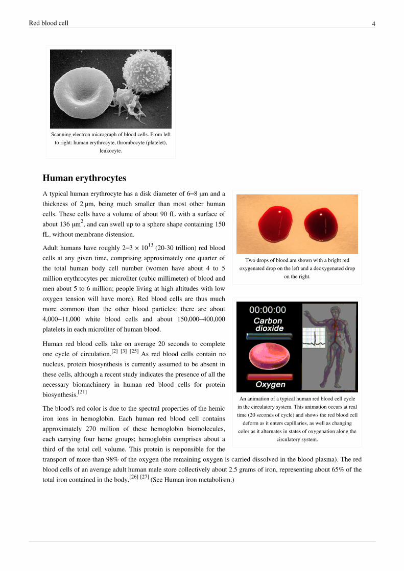
Scanning electron micrograph of blood cells. From leftto right: human erythrocyte, thrombocyte (platelet),
leukocyte.
Human erythrocytes
Two drops of blood are shown with a bright redoxygenated drop on the left and a deoxygenated drop
on the right.
An animation of a typical human red blood cell cyclein the circulatory system. This animation occurs at realtime (20 seconds of cycle) and shows the red blood cell
deform as it enters capillaries, as well as changingcolor as it alternates in states of oxygenation along the
circulatory system.
A typical human erythrocyte has a disk diameter of 6–8 µm and athickness of 2 µm, being much smaller than most other humancells. These cells have a volume of about 90 fL with a surface ofabout 136 μm2, and can swell up to a sphere shape containing 150fL, without membrane distension.
Adult humans have roughly 2–3 × 10¹³ (20-30 trillion) red bloodcells at any given time, comprising approximately one quarter ofthe total human body cell number (women have about 4 to 5million erythrocytes per microliter (cubic millimeter) of blood andmen about 5 to 6 million; people living at high altitudes with lowoxygen tension will have more). Red blood cells are thus muchmore common than the other blood particles: there are about4,000–11,000 white blood cells and about 150,000–400,000platelets in each microliter of human blood.
Human red blood cells take on average 20 seconds to completeone cycle of circulation.[2] [3] [25] As red blood cells contain nonucleus, protein biosynthesis is currently assumed to be absent inthese cells, although a recent study indicates the presence of all thenecessary biomachinery in human red blood cells for proteinbiosynthesis.[21]
The blood's red color is due to the spectral properties of the hemiciron ions in hemoglobin. Each human red blood cell containsapproximately 270 million of these hemoglobin biomolecules,each carrying four heme groups; hemoglobin comprises about athird of the total cell volume. This protein is responsible for thetransport of more than 98% of the oxygen (the remaining oxygen is carried dissolved in the blood plasma). The redblood cells of an average adult human male store collectively about 2.5 grams of iron, representing about 65% of thetotal iron contained in the body.[26] [27] (See Human iron metabolism.)

Red blood cell 5
Life cycleHuman erythrocytes are produced through a process named erythropoiesis, developing from committed stem cells tomature erythrocytes in about 7 days. When matured, these cells live in blood circulation for about 100 to 120 days.At the end of their lifespan, they become senescent, and are removed from circulation.
Erythropoiesis
Erythropoiesis is the development process in which new erythrocytes are produced, through which each cell maturesin about 7 days. Through this process erythrocytes are continuously produced in the red bone marrow of large bones,at a rate of about 2 million per second in a healthy adult. (In the embryo, the liver is the main site of red blood cellproduction.) The production can be stimulated by the hormone erythropoietin (EPO), synthesised by the kidney. Justbefore and after leaving the bone marrow, the developing cells are known as reticulocytes; these comprise about 1%of circulating red blood cells.
Functional lifetime
This phase lasts about 100–120 days, during which the erythrocytes are continually moving by the blood flow push(in arteries), pull (in veins) and squeezing through microvessels such as capillaries as they compress against eachother in order to move.
Senescence
The aging erythrocyte undergoes changes in its plasma membrane, making it susceptible to selective recognition bymacrophages and subsequent phagocytosis in the reticuloendothelial system (spleen, liver and bone marrow), thusremoving old and defective cells and continually purging the blood. This process is termed eryptosis, erythrocyteprogrammed cell death. This process normally occurs at the same rate of production by erythropoiesis, balancing thetotal circulating red blood cell count. Eryptosis is increased in a wide variety of diseases including sepsis, haemolyticuremic syndrome, malaria, sickle cell anemia, beta-thalassemia, glucose-6-phosphate dehydrogenase deficiency,phosphate depletion, iron deficiency and Wilson's disease. Eryptosis can be elicited by osmotic shock, oxidativestress, energy depletion as well as a wide variety of endogenous mediators and xenobiotics. Excessive eryptosis isobserved in erythrocytes lacking the cGMP-dependent protein kinase type I or the AMP-activated protein kinaseAMPK. Inhibitors of eryptosis include erythropoietin, nitric oxide, catecholamines and high concentrations of urea.Much of the resulting important breakdown products are recirculated in the body. The heme constituent ofhemoglobin are broken down into Fe3+ and biliverdin. The biliverdin is reduced to bilirubin, which is released intothe plasma and recirculated to the liver bound to albumin. The iron is released into the plasma to be recirculated by acarrier protein called transferrin. Almost all erythrocytes are removed in this manner from the circulation before theyare old enough to hemolyze. Hemolyzed hemoglobin is bound to a protein in plasma called haptoglobin which is notexcreted by the kidney.[28]
Membrane compositionThe membrane of the red blood cell plays many roles that aid in regulating their surface deformability, flexibility,adhesion to other cells and immune recognition. These functions are highly dependent on its composition, whichdefines its properties. The red blood cell membrane is composed of 3 layers: the glycocalyx on the exterior, which isrich in carbohydrates; the lipid bilayer which contains many transmembrane proteins, besides its lipidic mainconstituents; and the membrane skeleton, a structural network of proteins located on the inner surface of the lipidbilayer. In human erythrocytes, like in most mammal erythrocytes, half of the membrane mass is represented byproteins and the other half are lipids, namely phospholipids and cholesterol.[29]

Red blood cell 6
Membrane lipids
The most common erythrocyte cell membrane lipids, schematicallydisposed as they are distributed on the bilayer. Relative abundances
are not at scale.
The erythrocyte cell membrane comprises a typicallipid bilayer, similar to what can be found in virtuallyall human cells. Simply put, this lipid bilayer iscomposed of cholesterol and phospholipids in equalproportions by weight. The lipid composition isimportant as it defines many physical properties such asmembrane permeability and fluidity. Additionally, theactivity of many membrane proteins is regulated byinteractions with lipids in the bilayer.
Unlike cholesterol which is evenly distributed betweenthe inner and outer leaflets, the 5 major phospholipidsare asymmetrically disposed, as shown below:Outer monolayer
• Phosphatidylcholine (PC);• Sphingomyelin (SM).Inner monolayer
• Phosphatidylethanolamine (PE);• Phosphoinositol (PI) (small amounts).• Phosphatidylserine (PS);This asymmetric phospholipid distribution among thebilayer is the result of the function of severalenergy-dependent and energy-independentphospholipid transport proteins. Proteins called“Flippases” move phospholipids from the outer to theinner monolayer while others called “floppases” do the opposite operation, against a concentration gradient in anenergy dependent manner. Additionally, there are also “scramblase” proteins that move phospholipids in bothdirections at the same time, down their concentration gradients in an energy independent manner. There is stillconsiderable debate ongoing regarding the identity of these membrane maintenance proteins in the red cellmembrane.
The maintenance of an asymmetric phospholipid distribution in the bilayer (such as an exclusive localization of PSand PIs in the inner monolayer) is critical for the cell integrity and function due to several reasons:• Macrophages recognize and phagocytose red cells that expose PS at their outer surface. Thus the confinement of
PS in the inner monolayer is essential if the cell is to survive its frequent encounters with macrophages of thereticuloendothelial system, especially in the spleen.
• Premature destruction of thallassemic and sickle red cells has been linked to disruptions of lipid asymmetryleading to exposure of PS on the outer monolayer.
• An exposure of PS can potentiate adhesion of red cells to vascular endothelial cells, effectively preventing normaltransit through the microvasculature. Thus it is important that PS is maintained only in the inner leaflet of thebilayer to ensure normal blood flow in microcirculation.
• Both PS and phosphatidylinositol-4,5-bisphosphate (PIP2) can regulate membrane mechanical function, due to their interactions with skeletal proteins such as spectrin and protein 4.1R. Recent studies have shown that binding of spectrin to PS promotes membrane mechanical stability. PIP2 enhances the binding of protein band 4.1R to glycophorin C but decreases its interaction with protein band 3, and thereby may modulate the linkage of the

Red blood cell 7
bilayer to the membrane skeleton.The presence of specialized structures named "lipid rafts" in the erythrocyte membrane have been described byrecent studies. These are structures enriched in cholesterol and sphingolipids associated with specific membraneproteins, namely flotillins, stomatins (band 7), G-proteins, and β-adrenergic receptors. Lipid rafts that have beenimplicated in cell signaling events in nonerythroid cells have been shown in erythroid cells to mediate β2-adregenicreceptor signaling and increase cAMP levels, and thus regulating entry of malarial parasites into normal red cells.[30]
[31]
Membrane proteins
Red blood cell membrane proteins separated bySDS-Page and silverstained [32]
The proteins of the membrane skeleton are responsible for thedeformability, flexibility and durability of the red blood cell, enablingit to squeeze through capillaries less than half the diameter of theerythrocyte (7-8 μm) and recovering the discoid shape as soon as thesecells stop receiving compressive forces, in a similar fashion to anobject made of rubber.There are currently more than 50 known membrane proteins, whichcan exist in a few hundred up to a million copies per erythrocyte.Approximately 25 of these membrane proteins carry the various bloodgroup antigens, such as the A, B and Rh antigens, among many others.These membrane proteins can perform a wide diversity of functions,such as transporting ions and molecules across the red cell membrane,adhesion and interaction with other cells such as endothelial cells, assignaling receptors, as well as other currently unknown functions. Theblood types of humans are due to variations in surface glycoproteins oferythrocytes. Disorders of the proteins in these membranes areassociated with many disorders, such as hereditary spherocytosis, hereditary elliptocytosis, hereditarystomatocytosis, and paroxysmal nocturnal hemoglobinuria.[29] [30]
The red blood cell membrane proteins organized according to their function:
Red Blood Cell membrane major proteins
Transport
• Band 3 - Anion transporter, also an important structural componentof the erythrocyte cell membrane, makes up to 25% of the cellmembrane surface, each red cell contains approximately one millioncopies. Defines the Diego Blood Group;[33]
• Aquaporin 1 - water transporter, defines the Colton Blood Group;• Glut1 - glucose and L-dehydroascorbic acid transporter;• Kidd antigen protein - urea transporter;• RhAG - gas transporter, probably of carbon dioxide, defines Rh
Blood Group and the associated unusual blood group phenotype Rhnull;• Na+/K+ - ATPase;• Ca2+ - ATPase;• Na+ K+ 2Cl- - cotransporter;• Na+-Cl- - cotransporter;• Na-H exchanger;• K-Cl - cotransporter;• Gardos Channel.

Red blood cell 8
Cell adhesion
• ICAM-4 - interacts with integrins;• BCAM - a glycoprotein that defines the Lutheran blood group and also known as Lu or laminin-binding protein.Structural role - The following membrane proteins establish linkages with skeletal proteins and may play animportant role in regulating cohesion between the lipid bilayer and membrane skeleton, likely enabling the red cell tomaintain its favorable membrane surface area by preventing the membrane from collapsing (vesiculating).• Ankyrin-based macromolecular complex - proteins linking the bilayer to the membrane skeleton through the
interaction of their cytoplasmic domains with Ankyrin.• Band 3 - also assembles various glycolytic enzymes, the presumptive CO2 transporter, and carbonic anhydrase
into a macromolecular complex termed a “metabolon,” which may play a key role in regulating red cellmetabolism and ion and gas transport function);
• RhAG - also involved in transport, defines associated unusual blood group phenotype Rhmod.• Protein 4.1R-based macromolecular complex - proteins interacting with Protein 4.1R.
• Protein 4.1R - weak expression of Gerbich antigens;• Glycophorin C and D - glycoprotein, defines Gerbich Blood Group;• XK - defines the Kell Blood Group and the Mcleod unusual phenotype (lack of Kx antigen and greatly reduced
expression of Kell antigens);• RhD/RhCE - defines Rh Blood Group and the associated unusual blood group phenotype Rhnull;• Duffy protein - has been proposed to be associated with chemokine clearance;[34]
• Adducin - interaction with band 3;• Dematin- interaction with the Glut1 glucose transporter.
[29] [30]
Separation and blood dopingRed blood cells can be obtained from whole blood by centrifugation, which separates the cells from the bloodplasma. During plasma donation, the red blood cells are pumped back into the body right away and the plasma iscollected. Some athletes have tried to improve their performance by blood doping: first about 1 litre of their blood isextracted, then the red blood cells are isolated, frozen and stored, to be reinjected shortly before the competition.(Red blood cells can be conserved for 5 weeks at −79 °C.) This practice is hard to detect but may endanger thehuman cardiovascular system which is not equipped to deal with blood of the resulting higher viscosity.

Red blood cell 9
Artificially grown red blood cellsIn 2008 it was reported that human embryonic stem cells had been successfully coaxed into becoming erythrocytesin the lab. The difficult step was to induce the cells to eject their nucleus; this was achieved by growing the cells onstromal cells from the bone marrow. It is hoped that these artificial erythrocytes can eventually be used for bloodtransfusions.[35]
Diseases and diagnostic tools
Affected by Sickle-cell disease, red bloodcells alter shape and threaten to damage
internal organs.
Blood diseases involving the red blood cells include:• Anemias (or anaemias) are diseases characterized by low oxygen
transport capacity of the blood, because of low red cell count or someabnormality of the red blood cells or the hemoglobin.
• Iron deficiency anemia is the most common anemia; it occurs whenthe dietary intake or absorption of iron is insufficient, and hemoglobin,which contains iron, cannot be formed
• Sickle-cell disease is a genetic disease that results in abnormalhemoglobin molecules. When these release their oxygen load in thetissues, they become insoluble, leading to mis-shaped red blood cells.These sickle shaped red cells are less deformable and viscoelasticmeaning that they have become rigid and can cause blood vesselblockage, pain, strokes, and other tissue damage.
• Thalassemia is a genetic disease that results in the production of anabnormal ratio of hemoglobin subunits.
• Spherocytosis is a genetic disease that causes a defect in the red blood cell's cytoskeleton, causing the redblood cells to be small, sphere-shaped, and fragile instead of donut-shaped and flexible.
• Pernicious anemia is an autoimmune disease wherein the body lacks intrinsic factor, required to absorb vitaminB12 from food. Vitamin B12 is needed for the production of hemoglobin.
• Aplastic anemia is caused by the inability of the bone marrow to produce blood cells.• Pure red cell aplasia is caused by the inability of the bone marrow to produce only red blood cells.
Effect of osmotic pressure on blood cells
• Hemolysis is the general term for excessive breakdown of red bloodcells. It can have several causes and can result in hemolytic anemia.
• The malaria parasite spends part of its life-cycle in red bloodcells, feeds on their hemoglobin and then breaks them apart,causing fever. Both sickle-cell disease and thalassemia are morecommon in malaria areas, because these mutations convey someprotection against the parasite.
• Polycythemias (or erythrocytoses) are diseases characterized by asurplus of red blood cells. The increased viscosity of the blood can cause a number of symptoms.
• In polycythemia vera the increased number of red blood cells results from an abnormality in the bone marrow.• Several microangiopathic diseases, including disseminated intravascular coagulation and thrombotic
microangiopathies, present with pathognomonic (diagnostic) red blood cell fragments called schistocytes. Thesepathologies generate fibrin strands that sever red blood cells as they try to move past a thrombus.
• Inherited hemolytic anemias caused by abnormalities of the erythrocyte membrane comprise an important groupof inherited disorders. These disorders are characterized by clinical and biochemical heterogeneity and also

Red blood cell 10
genetic heterogeneity, as evidenced by recent molecular studies.• The Hereditary spherocytosis (HS) syndromes are a group of inherited disorders characterized by the presence
of spherical-shaped erythrocytes on the peripheral blood smear. HS is found worldwide. It is the most commoninherited anemia in individuals of northern European descent, affecting approximately 1 in 1000-2500individuals depending on the diagnostic criteria. The primary defect in hereditary spherocytosis is a deficiencyof membrane surface area. Decreased surface area may produced by two different mechanisms: 1) Defects ofspectrin, ankyrin, or protein 4.2 lead to reduced density of the membrane skeleton, destabilizing the overlyinglipid bilayer and releasing band 3-containing microvesicles. 2) Defects of band 3 lead to band 3 deficiency andloss of its lipid-stabilizing effect. This results in the loss of band 3-free microvesicles. Both pathways result inmembrane loss, decreased surface area, and formation of spherocytes with decreased deformability. Thesedeformed erythrocytes become trapped in the hostile environment of the spleen where splenic conditioninginflicts further membrane damage, amplifying the cycle of membrane injury.
• Hereditary elliptocytosis• Hereditary pyropoikilocytosis• Hereditary stomatocytosis[36]
• Hemolytic transfusion reaction is the destruction of donated red blood cells after a transfusion, mediated by hostantibodies, often as a result of a blood type mismatch.
Several blood tests involve red blood cells, including the RBC count (the number of red blood cells per volume ofblood), the hematocrit (percentage of blood volume occupied by red blood cells), and the erythrocyte sedimentationrate. The blood type needs to be determined to prepare for a blood transfusion or an organ transplantation.
References[1] Erich Sackmann, Biological Membranes Architecture and Function., Handbook of Biological Physics, (ed. R.Lipowsky and E.Sackmann,
vol.1, Elsevier, 1995[2] Laura Dean. Blood Groups and Red Cell Antigens (http:/ / www. ncbi. nlm. nih. gov/ books/ bv. fcgi?call=bv. View. . ShowTOC&
rid=rbcantigen. TOC& depth=2)[3] Pierigè F, Serafini S, Rossi L, Magnani M (January 2008). "Cell-based drug delivery". Advanced Drug Delivery Reviews 60 (2): 286–95.
doi:10.1016/j.addr.2007.08.029. PMID 17997501.[4] "Circular of Information for Blood and Blood Products" (http:/ / www. aabb. org/ resources/ bct/ Documents/ coi0809r. pdf) (pdf). American
Association of Blood Banks, American Red Cross, America's Blood Centers. . Retrieved 2010-11-01.[5] "Swammerdam, Jan (1637–1680)", McGraw Hill AccessScience, 2007. Accessed 27 December 2007.[6] Red Gold - Blood History Timeline (http:/ / www. pbs. org/ wnet/ redgold), PBS 2002. Accessed 27 December 2007.[7] Gulliver, G. (1875). "On the size and shape of red corpuscles of the blood of vertebrates, with drawings of them to a uniform scale, and
extended and revised tables of measurements". Proceedings of the Zoological Society of London 1875: 474–495.[8] Maton, Anthea; Jean Hopkins, Charles William McLaughlin, Susan Johnson, Maryanna Quon Warner, David LaHart, Jill D. Wright (1993).
Human Biology and Health. Englewood Cliffs, New Jersey, USA: Prentice Hall. ISBN 0-13-981176-1.[9] Snyder, Gregory K.; Sheafor, Brandon A. (1999). "Red Blood Cells: Centerpiece in the Evolution of the Vertebrate Circulatory System".
Integrative and Comparative Biology 39: 189. doi:10.1093/icb/39.2.189.[10] Ruud JT (May 1954). "Vertebrates without erythrocytes and blood pigment". Nature 173 (4410): 848–50. doi:10.1038/173848a0.
PMID 13165664.[11] Carroll, Sean (2006). The Making of the Fittest. W.W. Norton. ISBN 0393061639.[12] Cohen, W. D. (1982). "The cytomorphic system of anucleate non-mammalian erythrocytes". Protoplasma 113: 23.
doi:10.1007/BF01283036.[13] Wingstrand KG (1956). "Non-nucleated erythrocytes in a teleostean fish Maurolicus mülleri (Gmelin)" (http:/ / www. springerlink. com/
content/ j943833n74065634). Zeitschrift Für Zellforschung Und Mikroskopische Anatomie 45 (2): 195–200. doi:10.1007/BF00338830(inactive 2009-12-02). PMID 13402080. .
[14] Wan J, Ristenpart WD, Stone HA (October 2008). "Dynamics of shear-induced ATP release from red blood cells". Proceedings of theNational Academy of Sciences of the United States of America 105 (43): 16432–7. doi:10.1073/pnas.0805779105. PMC 2575437.PMID 18922780.
[15] Diesen DL, Hess DT, Stamler JS (August 2008). "Hypoxic vasodilation by red blood cells: evidence for an s-nitrosothiol-based signal".Circulation Research 103 (5): 545–53. doi:10.1161/CIRCRESAHA.108.176867. PMC 2763414. PMID 18658051.

Red blood cell 11
[16] Kleinbongard P, Schutz R, Rassaf T, et al (2006). "Red blood cells express a functional endothelial nitric oxide synthase". Blood 107 (7):2943–51. doi:10.1182/blood-2005-10-3992. PMID 16368881.
[17] Ulker P, Sati L, Celik-Ozenci C, Meiselman HJ, Baskurt OK (2009). "Mechanical stimulation of nitric oxide synthesizing mechanisms inerythrocytes". Biorheology 46 (2): 121–32. doi:10.3233/BIR-2009-0532. PMID 19458415.
[18] Benavides, Gloria A; Giuseppe L Squadrito, Robert W Mills, Hetal D Patel, T Scott Isbell, Rakesh P Patel, Victor M Darley-Usmar,Jeannette E Doeller, David W Kraus (2007-11-13). "Hydrogen sulfide mediates the vasoactivity of garlic" (http:/ / www. pnas. org/ content/104/ 46/ 17977. full). Proceedings of the National Academy of Sciences of the United States of America 104 (46): 17977–17982.doi:10.1073/pnas.0705710104. PMC 2084282. PMID 17951430. . Retrieved 2010-03-03.
[19] Red blood cells do more than just carry oxygen. New findings by NUS team show they aggressively attack bacteria too. (http:/ / www. dbs.nus. edu. sg/ eventlist/ happenings/ details/ 2007/ dingSTsep07. pdf), The Straits Times, 1 September 2007
[20] Jiang N, Tan NS, Ho B, Ding JL (October 2007). "Respiratory protein-generated reactive oxygen species as an antimicrobial strategy".Nature Immunology 8 (10): 1114–22. doi:10.1038/ni1501. PMID 17721536.
[21] Kabanova S, Kleinbongard P, Volkmer J, Andrée B, Kelm M, Jax TW (2009). "Gene expression analysis of human red blood cells" (http:/ /www. medsci. org/ v06p0156. htm). International Journal of Medical Sciences 6 (4): 156–9. PMC 2677714. PMID 19421340. .
[22] Uzoigwe C (2006). "The human erythrocyte has developed the biconcave disc shape to optimise the flow properties of the blood in the largevessels". Medical Hypotheses 67 (5): 1159–63. doi:10.1016/j.mehy.2004.11.047. PMID 16797867.
[23] Gregory TR (2001). "The bigger the C-value, the larger the cell: genome size and red blood cell size in vertebrates". Blood Cells, Molecules& Diseases 27 (5): 830–43. doi:10.1006/bcmd.2001.0457. PMID 11783946.
[24] Goodman SR, Kurdia A, Ammann L, Kakhniashvili D, Daescu O (December 2007). "The human red blood cell proteome and interactome".Experimental Biology and Medicine 232 (11): 1391–408. doi:10.3181/0706-MR-156. PMID 18040063.
[25] Hillman, Robert S.; Ault, Kenneth A.; Rinder, Henry M. (2005). Hematology in Clinical Practice: A Guide to Diagnosis and Management(4 ed.). McGraw-Hill Professional. p. 1. ISBN 0071440356.
[26] Iron Metabolism (http:/ / www. med-ed. virginia. edu/ courses/ path/ innes/ nh/ iron. cfm), University of Virginia Pathology. Accessed 22September 2007.
[27] Iron Transport and Cellular Uptake (http:/ / sickle. bwh. harvard. edu/ iron_transport. html) by Kenneth R. Bridges, Information Center forSickle Cell and Thalassemic Disorders. Accessed 22 September 2007.
[28] Föller M, Huber SM, Lang F (October 2008). "Erythrocyte programmed cell death". IUBMB Life 60 (10): 661–8. doi:10.1002/iub.106.PMID 18720418.
[29] Yazdanbakhsh K, Lomas-Francis C, Reid ME (October 2000). "Blood groups and diseases associated with inherited abnormalities of the redblood cell membrane". Transfusion Medicine Reviews 14 (4): 364–74. doi:10.1053/tmrv.2000.16232. PMID 11055079.
[30] Mohandas N, Gallagher PG (November 2008). "Red cell membrane: past, present, and future". Blood 112 (10): 3939–48.doi:10.1182/blood-2008-07-161166. PMC 2582001. PMID 18988878.
[31] Rodi PM, Trucco VM, Gennaro AM (June 2008). "Factors determining detergent resistance of erythrocyte membranes". BiophysicalChemistry 135 (1-3): 14–8. doi:10.1016/j.bpc.2008.02.015. PMID 18394774.
[32] Hempelmann E, Götze O (1984). "Characterization of membrane proteins by polychromatic silver staining". Hoppe Seyler's Z Physiol Chem365: 241–242.
[33] Iolascon A, Perrotta S, Stewart GW (March 2003). "Red blood cell membrane defects". Reviews in Clinical and Experimental Hematology 7(1): 22–56. PMID 14692233.
[34] Denomme GA (July 2004). "The structure and function of the molecules that carry human red blood cell and platelet antigens". TransfusionMedicine Reviews 18 (3): 203–31. doi:10.1016/j.tmrv.2004.03.006. PMID 15248170.
[35] First red blood cells grown in the lab (http:/ / www. newscientist. com/ article/ dn14565-first-red-blood-cells-grown-in-the-lab. html), NewScientist News, 19 August 2008
[36] An X, Mohandas N (May 2008). "Disorders of red cell membrane". British Journal of Haematology 141 (3): 367–75.doi:10.1111/j.1365-2141.2008.07091.x. PMID 18341630.
External links• Blood Groups and Red Cell Antigens (http:/ / www. ncbi. nlm. nih. gov/ books/ bv. fcgi?call=bv. View. .
ShowTOC& rid=rbcantigen. TOC& depth=2) by Laura Dean. Searchable and downloadable online textbook inthe public domain.
• Database of vertebrate erythrocyte sizes (http:/ / www. genomesize. com/ cellsize/ ).• Red Gold (http:/ / www. pbs. org/ wnet/ redgold), PBS site containing facts and history

Article Sources and Contributors 12
Article Sources and ContributorsRed blood cell Source: http://en.wikipedia.org/w/index.php?oldid=425001408 Contributors: (jarbarf), -df-, 03lausmi, AJim, ASEEMASEEM, Academic Challenger, Adambro, Addshore,Adhib, AdjustShift, Ahoerstemeier, Alansohn, Alec - U.K., Alfie66, Ali, Alksub, Alpha Quadrant, Amaher, Andre Engels, Andres, Andrew Delong, AndrewWTaylor, Andypandy.UK, AngelicWraith, Angrycrustacean, Ann Stouter, Anoko moonlight, Apers0n, Arcadian, Arisa, Arjun01, ArnoLagrange, Arvind0602, ArwinJ, Asparagirl, AssistantX, Atlantia, Aviad2001, Avoided,AxelBoldt, Baa, Bassbonerocks, Bbb2007, Beaumont, Benjamin.friedrich, Bensaccount, Bfigura, Bidabadi, Bignoter, Bobblewik, Bobo192, Bomac, Bongwarrior, Borgx, Boud, Brantonli,Brianski, BryanD, Burek, CIreland, Caeruleancentaur, Caltas, Can't sleep, clown will eat me, Caseydyck, Causa sui, Chaldor, Chanting Fox, ChongDae, Chris Capoccia, ClockworkSoul,Closedmouth, Corinne68, Corvus cornix, Countincr, Countmike, Cpl Syx, Crack98, Craig Pemberton, Craigmillar94, Crohnie, DARTH SIDIOUS 2, DRosenbach, Da monster under your bed,Dabomb87, Dan Gluck, Daniel5127, Danrudd, Darth Panda, Darthanakin, Davewild, DeadEyeArrow, Debresser, Deus Ex, Dietzel65, Discospinster, Dogcow, Dogposter, Dominus, DoubleBlue,Dr.Kane, DrMicro, Drmies, Dungodung, E Wing, ESkog, Eaefremov, Ed, Edcgooner, Egmontaz, Ekko, Ellywa, Emilyalbinoman, Epbr123, Everyking, Excirial, Ferkelparade, Fieldday-sunday,Fishyghost, Flamingspinach, Fr34k, Fraggle81, Franamax, Fratter, Fredil Yupigo, Fritzlein, Fryed-peach, Frymaster, Funnybunny, Fuzheado, Fvasconcellos, Gabbe, Gawaxay, Gene Nygaard,Giftlite, Gimboid13, Gobonobo, Golfandme, Grafen, Grandmasterka, GraybeardBiochemist, Greentryst, GregorB, Grim23, Guest9999, Guy Peters, Hadal, HaeB, Haham hanuka, HalfShadow,HamburgerRadio, Harthacnut, Haza-w, Hellbus, Hempelmann, Hnsampat, Hodja Nasreddin, Honette, Hu, Hunterd, Hydrogen Iodide, II MusLiM HyBRiD II, Ihope127, Immortal0king,ImplyingAuthority, Iridescent, J.delanoy, J04n, JNW, JWSchmidt, JaGa, Jakejakejake1313, Jaknouse, James Crippen, JamesTseng, JamillyM, JediLofty, Jeffq, Jfdwolff, Jfurr1981, Jnquach,JoanneB, John254, Johnbrownsbody, Jon salisbury, JonLS, Jordell 000, Josh3580, Jossi, Jsfouche, Juju Power, Jusdafax, Jyril, KC0ZHQ, Kander, Kariteh, Kassal, Katimawan2005, KaySL,Kazrak, Kennycub, Kerfern, Killerkris88, Kineticman, KingTT, Kingpin13, KnowledgeOfSelf, Kpjas, Kriptyk, Krylonblue83, Kubra, Kusma, KyraVixen, L Kensington, L33th4x0rguy, La gouttede pluie, Lahiru k, Landon1980, Lars Washington, LeaveSleaves, Lectonar, Lightdarkness, Linas, Linzhoo2u, Little Mountain 5, LittleOldMe old, LizardJr8, Looper5920, Lord of the Pit, Lotje,MC MasterChef, MZMcBride, Mad Max, Madhero88, Magister Mathematicae, Maias, Mailer diablo, Mandarax, Marek69, Markacohen, MartinGugino, Massimo Macconi, Masterjamie, MattDeres, Mav, McSly, Mdw123, Mephistophelian, Mexinadian, Mfero, Michael Devore, Mikael Häggström, Mike2vil, Mindspillage, Minna Sora no Shita, Mintleaf, Miquonranger03, Moe Epsilon,Monkey.invasion, Mrfickle, Mwilso24, Nakon, Natty1991, NeilN, Nemesis of Reason, Netalarm, NewEnglandYankee, Nifky?, Nlu, Novalincoln, Nsaa, Nscheffey, Nunh-huh, Orkybash,Oscarthecat, Oxymoron83, PDH, Pablo-flores, Pb30, Peak, Perspeculum, Peter Karlsen, Phantomsteve, Philip Trueman, Piano non troppo, PierreAbbat, Pinethicket, Pinkadelica, Policarp, Prari,Priyanath, Prolog, Prowster, Prvc, Pschwarzbaum, Puchiko, RUL3R, RainbowOfLight, Razimantv, Rcingham, Reaper X, Recognizance, Reconsider the static, Rettetast, Rgracesmyrna, Riana,Rich Farmbrough, Rob Maguire, Robo-rory, Rogeriopfm, Roshan baladhanvi, Roux, Rrburke, Rsabbatini, Ryulong, SDY, SGGH, SJP, Sakkura, Salgueiro, Samuel, Samwb123, Santoshpath,Sceptre, Schoolscienceteacher, Scootdive, Scottalter, Ser Amantio di Nicolao, SetRochelewsad, Sfmammamia, Shadowjams, Shanel, Sheitan, Sheogorath, Shirik, Shoeofdeath, Shyamdash, SirVicious, Sky Attacker, Slashme, Slowking Man, Smitz, Snowmanradio, Someone else, Spartan, Specs112, Spliffy, SquidSK, Squidnchips, StaticGull, Stephenb, Stevenfruitsmaak, Sundar,SuperHamster, SuperStingray, Sverdrup, SwammerBAM, THEN WHO WAS PHONE?, TYelliot, Tanthalas39, Tarquin, Taxman, Tchite, Teapotgeorge, Template namespace initialisation script,Tempodivalse, The Bushranger, The Rambling Man, The Thing That Should Not Be, TheEgyptian, Tide rolls, TimVickers, Tiptoety, Torchwoodwho, Tosta Dojen, Traderjohn, TrevorWennblom, Treygdor, Tsange, Turnstep, Tuspm, Tweetlebeetle367, Tyw7, Umheisman20, Uncle Dick, Valenciano, Vegfarandi, Vipinhari, Vrenator, WDM27, Wardface, Wars, Wavelength,Wayne Slam, Wayward, Wiki alf, WikipedianMarlith, Willking1979, Wimt, Wisdom89, Wknight94, WolfmanSF, Woohookitty, Xenobiologista, Yamaguchi先生, YetiSSM, Yuckfoo, Yurik,Zoomlines, 901 ,یدنقرمس ,24.يدماغ.دمحأ anonymous edits
Image Sources, Licenses and ContributorsImage:redbloodcells.jpg Source: http://en.wikipedia.org/w/index.php?title=File:Redbloodcells.jpg License: unknown Contributors: Dietzel65, Habj, Ranveig, Shizhao, Simon Shek, Solon,Sundar, ThuressonImage:Erythrocytes in vertebrates.jpg Source: http://en.wikipedia.org/w/index.php?title=File:Erythrocytes_in_vertebrates.jpg License: Public Domain Contributors: G Gulliver - Proceedingsof the Royal Society of LondonImage:Gray453.png Source: http://en.wikipedia.org/w/index.php?title=File:Gray453.png License: Public Domain Contributors: Henry GrayImage:Red White Blood cells.jpg Source: http://en.wikipedia.org/w/index.php?title=File:Red_White_Blood_cells.jpg License: Public Domain Contributors: Electron Microscopy Facility atThe National Cancer Institute at Frederick (NCI-Frederick)Image:NIK_3232-Drops_of_blood_medium.JPG Source: http://en.wikipedia.org/w/index.php?title=File:NIK_3232-Drops_of_blood_medium.JPG License: Creative Commons Attribution 3.0 Contributors: unkownImage:Erytrocyte_deoxy_to_oxy_v0.7.gif Source: http://en.wikipedia.org/w/index.php?title=File:Erytrocyte_deoxy_to_oxy_v0.7.gif License: Creative Commons Attribution-Sharealike 3.0 Contributors: User:RogeriopfmImage:Erythrocyte Membrane lipids.jpg Source: http://en.wikipedia.org/w/index.php?title=File:Erythrocyte_Membrane_lipids.jpg License: Creative Commons Attribution-Sharealike 3.0 Contributors: User:BorisTM, User:Likeitsmyjob, User:RogeriopfmFile:RBC Membrane Proteins SDS-PAGE gel.jpg Source: http://en.wikipedia.org/w/index.php?title=File:RBC_Membrane_Proteins_SDS-PAGE_gel.jpg License: Public Domain Contributors: Ernst HempelmannFile:RBC membrane major proteins.png Source: http://en.wikipedia.org/w/index.php?title=File:RBC_membrane_major_proteins.png License: Public Domain Contributors: User:TimVickersImage:Sicklecells.jpg Source: http://en.wikipedia.org/w/index.php?title=File:Sicklecells.jpg License: Public Domain Contributors: NIDDKImage:Osmotic pressure on blood cells diagram.svg Source: http://en.wikipedia.org/w/index.php?title=File:Osmotic_pressure_on_blood_cells_diagram.svg License: Public Domain Contributors: user:LadyofHats
LicenseCreative Commons Attribution-Share Alike 3.0 Unportedhttp:/ / creativecommons. org/ licenses/ by-sa/ 3. 0/