recruitment of mesenchymal stem cells to apoptotic tissue...
TRANSCRIPT

Aus dem Institut für Transplantationsdiagnostik und Zelltherapeutika
der Heinrich-Heine-Universität Düsseldorf
Kommissarischer Direktor: Dr. med. Johannes Fischer
Recruitment of mesenchymal stem cells to apoptotic tissue cells is mediated by hepatocyte growth factor
Dissertation
zur Erlangung des Grades eines Doktors der Medizin
der Medizinischen Fakultät der Heinrich-Heine-Universität Düsseldorf
vorgelegt von
Sebastian Vogel
(2013)

Als Inauguraldissertation gedruckt mit Genehmigung der Medizinischen Fakultät
der Heinrich-Heine-Universität Düsseldorf
gez. Univ.-Prof. Dr. med. Joachim Windolf
Dekan
Referent: Priv.-Doz. Dr. Rüdiger V. Sorg
Korreferent: Univ.-Prof. Dr. James A. Adjaye
I
ZusammenfassungMesenchymale Stammzellen (MSC) besitzen geweberegenerierende und immunmodulierende Eigenschaften, verbessern das Anwachsen hämato-poetischer Stammzellen nach deren Transplantation und sind gentherapeutisch als zelluläre Vehikel einsetzbar. Ein genaues Verständnis der Rekrutierung von MSC an den Ort des krankhaften Geschehens ist zentral für ihre Funktion wie auch deren therapeutische Ausnutzung. Der Hepatozytenwachstumsfaktor (HGF) ist ein pleiotropes Zytokin, das über seinen Rezeptor MET die Migration von MSC vermittelt. Es wird bei Gewebe-schäden und auch von einigen malignen Tumoren wie dem Glioblastoma multi-forme gebildet. Bei der Heilung von Gewebe hat HGF anti-apoptotische, zyto-protektive und pro-angiogenetische Effekte, im Glioblastom hingegen fördern diese Eigenschaften das invasive Wachstum des Tumors. In der vorliegenden Dissertation wurde (i) der Einfluss von Apoptose in Abgren-zung zu Nekrose von Herz- und Nervenzellen und (ii) die Auswirkung einer Aminolävulinsäure-vermittelten photodynamischen Behandlung von Glio-blastomzellen auf die Migration von MSC und die jeweilige Rolle der HGF/MET-Achse untersucht. HL-1 Kardiomyozyten und HT-22 hippokampale Neuronen wurden apoptotisch oder nekrotisch geschädigt, U87 und U251 Glioblastomzellen wurden einer photodynamischen Behandlung unterzogen. Annexin V/Propidiumjodid- und TUNEL-Färbungen dienten dem durchflusszytometrischen Nachweis von Apoptose und Nekrose. Die chemotaktische Aktivität der Zellen für MSC wurde in under-agarose chemotaxis assays bestimmt. Der jeweilige Beitrag von HGF zur Migration wurde in Neutralisationsstudien evaluiert. HGF und MET wurden durch Reverse Transkription-Polymerase Kettenreaktion, enzyme linked immunosorbent assays und Durchflusszytometrie nachgewiesen. Apoptotische Kardiomyozyten und neuronale Zellen induzierten die Rekru-tierung von MSC, während nekrotischer Untergang der gleichen Zellen zu einem Ausbleiben dieser Migrationsantwort führte. Auch Glioblastomzellen waren chemotaktisch für MSC, und eine photodynamische Behandlung der Tumorzellen verdreifachte in etwa die Zahl einwandernder MSC. Der HGF-Re-zeptor MET wurde von MSC exprimiert. HGF selbst konnte in apoptotischen, je-doch nicht in nekrotischen oder vitalen Herz- und Nervenzellen nachgewiesen werden. In Glioblastomzellen wurde eine vorhandene moderate Expression von HGF durch photodynamische Behandlung signifikant gesteigert, was mit einer Induktion von Apoptose durch die Therapie einherging. Die Neutralisation der Bioaktivität von HGF durch einen Antikörper führte zu einer signifikanten Hemmung der MSC-Migration zu allen untersuchten apoptotischen Gewebe-zellen. Apoptotischer Zelltod induziert somit die Attraktion von MSC über den HGF/ MET Signalweg. Auch Glioblastomzellen rekrutieren MSC über diese Achse, ein Mechanismus, der durch photodynamische Behandlung der Tumorzellen, abermals abhängig von der Induktion von Apoptose, verstärkt wird. Apoptose, nicht aber Nekrose, spielt also eine Schlüsselrolle in der Rekrutierung von MSC, was in der regenerativen Medizin wie auch in der Therapie von Glioblastomen von Relevanz sein kann.

Abkürzungsverzeichnis
ALA/PDT aminolaevulinic acid-mediated photodynamic therapy
CCR CC chemokine receptor
CD cluster of differentiation
CXCR CXC chemokine receptor
DAMP danger-associated molecular pattern
DC dendritische Zellen
GBM Glioblastoma multiforme
HGF Hepatozytenwachstumsfaktor
HLA humanes Leukozytenantigen
HMGB1 high mobility group box 1
HSC hämatopoetische Stammzellen
IFN Interferon
IL Interleukin
MET HGF-Rezeptor
MMP Matrix-Metalloproteinase
MSC mesenchymale Stammzellen
SDF-1 stromal-cell derived factor-1
TNF Tumornekrosefaktor
TRAIL tumor necrosis factor related apoptosis inducing ligand
II

Inhaltsverzeichnis
1 Einleitung 1
1.1 Mesenchymale Stammzellen und ihre Eigenschaften 1
1.2 Klinische Anwendungen von mesenchymalen Stammzellen 2
1.3 Rekrutierung mesenchymaler Stammzellen 3
1.4 Molekulare Mechanismen der Attraktion mesenchymaler
Stammzellen 5
1.5 HGF-vermittelter Tropismus mesenchymaler Stammzellen
und Induktion von Apoptose 6
2 Ziele der Arbeit 8
3 Originalarbeiten 9
3.1 Hepatocyte growth factor-mediated attraction of mesenchymal
stem cells for apoptotic neuronal and cardiomyocytic cells.
Vogel S, Trapp T, Börger V, Peters C, Lakbir D, Dilloo D,
Sorg RV (2010) Cellular and Molecular Life Sciences;
67(2):295-303 10
3.2 Migration of mesenchymal stem cells towards glioblastoma
cells depends on hepatocyte-growth factor and is enhanced
by aminolaevulinic acid-mediated photodynamic treatment.
Vogel S, Peters C, Etminan N, Börger V, Schimanski A,
Sabel MC, Sorg RV (2013) Biochemical and Biophysical
Research Communications; 431(3):428-432 19
4 Diskussion 24
5 Literaturverzeichnis 29
6 Danksagung 37
III

1
1 Einleitung
1.1 Mesenchymale Stammzellen und ihre Eigenschaften
Mesenchymale Stammzellen (MSC) wurden erstmals von Friedenstein und
Kollegen als stromale Vorläuferzellen im Knochenmark beschrieben [1]. Es
können mittlerweile auch in zahlreichen anderen Geweben MSC identifiziert
werden, etwa in Fett-, Muskel-, Periost- und Lungengewebe [2]. Sie werden
typischerweise als adhärent wachsende, Fibroblasten-ähnliche Zellen ge-
wonnen, die durch die Expression der Oberflächenmoleküle CD73, CD90 und
CD105 bei gleichzeitig fehlender Expression von CD14, CD34 und CD45
charakterisiert sind [3].
MSC sind nicht-hämatopoetische, somatische Stammzellen, die zu Osteo-
blasten, Chondroblasten und Adipozyten, also Zellen mesenchymalen Ur-
sprungs, differenzieren können [4]. Ob sie darüber hinaus ein keimblattüber-
schreitendes Differenzierungspotential besitzen [5], wie es unter anderem durch
ein Auswachsen in Herzmuskelzellen [6], neuronale Zellen [7] und Hepatozyten
[8] angedeutet wird, bedarf noch einer abschließenden Klärung [9]. Zweifellos
besitzen MSC geweberegenerierende Aktivität, die wahrscheinlich weniger auf
ihrem Differenzierungspotential, sondern vor allem auf parakrinen Mechanis-
men beruht [10-11].
MSC tragen ferner im Knochenmark als sogenannte mesenchymale Stroma-
zellen zur Bildung einer Nische für hämatopoetische Stammzellen (HSC) bei
[12-13]. Dieses spezifische Mikromilieu beeinflusst unter anderem über Wachs-
tumsfaktoren und Adhäsionsmoleküle die Proliferation der HSC sowie ihren Ein-
tritt in die Differenzierung und ihre Mobilisierung in das Blutsystem. Somit sind
MSC neben regenerativen Vorgängen als Stromazellen auch an der Regulation
hämatopoetischer Entwicklungsprozesse beteiligt.
Des Weiteren können MSC immunsupprimierende Aufgaben verrichten [14].
Auf der Seite der angeborenen Immunabwehr hemmen sie unter anderem die
Reifung dendritischer Zellen (DC), was sich insbesondere durch eine Vermin-
derung ihrer allostimulatorischen Potenz für Effektorzellen äußert [15]. MSC sti-
mulieren außerdem die Produktion des immunsuppressiven Interleukin (IL)-10

durch plasmazytoide DC [16]. Da DC als spezialisierte Antigen-präsentierende
Zellen eine zentrale Rolle in der Initiationsphase von T-Zell-Antworten spielen,
hat die Hemmung ihrer Ausreifung, wie auch die Induktion immunsuppressiver
Zytokine, Auswirkungen auf die Entwicklung der spezifischen, zellvermittelten
Immunität.
Auf der Seite der adaptiven Immunität hemmen MSC die Proliferation von T-
Zellen [17-18], einhergehend mit verminderter Interferon (IFN)-gamma Pro-
duktion und erhöhter IL-4 Produktion [16], was einem Shift der T-Helfer1/T-
Helfer2-Polarisierung von einem pro-inflammatorischen hin zu einem anti-
inflammatorischen T-Zell-Status gleichkommt. Auch konnte gezeigt werden,
dass die Aktivität CD8-positiver zytotoxischer T-Zellen durch MSC gehemmt
wird [19]. Dabei scheinen die Interaktionen zwischen MSC und T-Zellen
unabhängig vom HLA-Typ zu erfolgen; sie werden entweder über direkten
Zellkontakt [20] oder über lösliche Faktoren wie transforming growth factor beta
1 und Hepatozytenwachstumsfaktor (HGF) vermittelt [18].
1.2 Klinische Anwendungen von mesenchymalen Stammzellen
Aufgrund ihres Differenzierungspotentials, der geweberegenerierenden Eigen-
schaften, der Stromafunktion und der vielfältigen immunmodulatorischen Kom-
petenz, kommen MSC für eine ganze Reihe an klinischen Anwendungen in
Frage, insbesondere in der regenerativen Medizin, der Therapie von Auto-
immunerkrankungen, hämatologischen Erkrankungen und malignen Prozessen.
Geweberegenerierende Effekte von MSC konnten am Menschen unter
anderem bei Knochendefekten [21], kardialer Dysfunktion [22], insbesondere
bei Patienten mit akutem Myokardinfarkt nach intrakoronarer Injektion autologer
MSC aus dem Knochenmark [23-24], als auch bei zerebralem Insult [25] nach-
gewiesen werden. Im Rahmen der Transplantation allogener HSC bei hämato-
logischen Neoplasien wurden MSC zur Reduktion der Schwere einer graft-
versus host-disease [26-27] und des Risikos einer Transplantatabstoßung [28]
erfolgreich eingesetzt. Auch bei Multipler Sklerose scheint ihre immun-
supprimierende Aktivität therapeutisch vielversprechend. Eine durch ein Proteo-
lipid-Protein in der Maus verursachte autoimmune Enzephalomyelitis (ein Tier-
modell für die Multiple Sklerose) konnte durch eine MSC-Applikation in ihrer
2

Schwere gemildert werden, was auf eine Hemmung der T-Zell-Antwort gegen
das PLP-Peptid und eine verminderte Produktion spezifischer Antikörper zu-
rückgeführt wurde [29].
MSC als zelluläre Vektoren, insbesondere für anti-tumoral wirksame Substan-
zen, stellen in der Gentherapie oder onkolytischen Behandlung eine attraktive
Alternative zur direkten Verwendung von Viren dar, da sie aufgrund ihres Tro-
pismus für zahlreiche Tumoren das Therapeutikum mit hoher Spezifität zum
Tumor transportieren können [30]. Es konnte gezeigt werden, dass gen-
modifizierte, IFN-beta-exprimierende MSC in vitro die Proliferation von Mamma-
karzinom und Melanomzellen in Ko-Kulturen hemmen, was sich im Mausmodell
durch die Hemmung des Wachstums der entsprechenden Tumoren bestätigen
ließ [31]. Beim Glioblastoma multiforme (GBM), dem aggressivsten und
häufigsten malignen Hirntumor [32], gewinnt die zelluläre Gentherapie als alter-
native Therapieoption eine besondere Bedeutung. Das GBM ist ein astrozytärer
Tumor WHO Grad 4 mit einer nach wie vor desaströsen klinischen Prognose.
Nach Standardtherapieregime, bestehend aus einer Kombination von operativer
Entfernung des Tumors, fraktionierter Bestrahlung und chemotherapeutischer
Behandlung mit Temozolomid, einem alkylierenden Chemotherapeutikum, be-
tragen das mediane Überleben 14,6 Monate, die 2-Jahres Überlebensrate
27,2% und die 5-Jahres Überlebensrate 10% [33]. Insbesondere die Tatsache,
dass eine komplette chirurgische Resektion des Tumors aufgrund des stark in-
vasiven Wachstums mit Infiltration ins gesunde Hirngewebe unmöglich ist [34],
trägt zur schlechten Prognose bei. Die gentherapeutische Verwendung von
MSC als Transportvehikel für Zytokine, Enzyme/Prodrugs, Viruspartikel oder
Oberflächenantikörper erweist sich im Kampf gegen das GBM als vielver-
sprechend. Als Beispiele für in der GBM-Gentherapie relevante Zytokine seien
das anti-tumoral wirksame IFN-beta und tumor necrosis factor related apoptosis
inducing ligand (TRAIL), ein selektiv in Tumorzellen Apoptose-induzierender
Ligand, genannt [35].
1.3 Rekrutierung mesenchymaler Stammzellen
Die Migration von MSC an den Ort des krankhaften Geschehens ist eine Grund-
voraussetzung für das Erfüllen ihrer Aufgaben, vergleichbar mit der Ein-
3

wanderung von Immunzellen in entzündetes Gewebe. Die Migration von Zellen
ist ein komplexer Prozess, an dem die Ausbildung von Lamellipodien und Filo-
podien, das Entstehen neuer Adhärenzkontakte, eine durch Kontraktionen im
Zytoskelett hervorgerufene Translokation des Zellkörpers in die zu migrierende
Richtung und die Lösung der Adhärenz am hinteren Zellpol beteiligt sind [36].
Hierbei muss unterschieden werden zwischen Chemotaxis, also gerichteter
Migration entlang von Konzentrationsgradienten chemotaktisch wirksamer Sub-
stanzen, welche durch die Verteilung der jeweiligen Rezeptoren auf der Zell-
oberfläche der migrierenden Zelle erkannt werden, und Chemokinese, also un-
gerichtete Migration beziehungsweise Zellmotilität in zufällige Richtungen.
Gewebeschäden bewirken einen chemotaktischen Migrationsreiz für MSC [37].
In diesem Zusammenhang werden am häufigsten ischämisch bedingte Schä-
den in der Literatur beschrieben, beispielsweise am Herzen in Form eines
akuten Myokardinfarkts [38-39] oder im Hirn in Form eines zerebralen Insults
[25, 40].
Auch maligne Tumoren stellen einen chemotaktischen Migrationsreiz für MSC
dar [30]. Die Rekrutierung von MSC in den Tumor darf verglichen werden mit
dem Tropismus für Gewebeschäden. Ein maligner Prozess lässt sich als eine
„Wunde, die nicht heilt“ [41] beschreiben, die, ähnlich wie ischämisch bedingte
Gewebeschäden, entzündungsrelevante Mediatoren und Wachstumsfaktoren
ausschüttet, von denen einige nicht nur an der Rekrutierung von Immunzellen
beteiligt sind, sondern auch an der Migration von MSC [30]. Speziell bei der
zellulären Behandlung von malignen Hirntumoren wie dem GBM ergibt sich
durch den ausgeprägten Tumor-Tropismus der MSC, bei dem die Bluthirn-
schranke kein Hindernis darstellt, die Möglichkeit, MSC als Transportvehikel für
anti-tumoral wirksame Substanzen [42] oder ihre direkte zytotoxische Wirkung
auf maligne Zellen [43] therapeutisch auszunutzen. Dabei kann die Migration
von MSC hin zum GBM durch eine zusätzliche Radiotherapie des Tumors mit
Gamma-Strahlen verstärkt und somit optimiert werden [44].
4
1.4 Molekulare Mechanismen der Attraktion mesenchymaler Stamm-zellen
Die molekularen Mechanismen, die eine Rolle bei der Rekrutierung von MSC
spielen, sind vielfältig und nur teilweise verstanden. Es gibt viele verschiedene
Mediatoren, die in die Vermittlung von Migrationssignalen für MSC involviert
sind. Dies sind vor allem Wachstumsfaktoren wie platelet-derived growth factor,
insulin-like growth factor und HGF sowie eine Reihe von Chemokinen aus den
CC- und CXC-Familien [37]. So wurde die Expression von zahlreichen,
funktionell aktiven Chemokin-Rezeptoren auf MSC nachgewiesen: CC
chemokine receptor (CCR)1, CCR7, CCR9, CXC chemokine receptor (CXCR)4,
CXCR5 und CXCR6 [45]. Hieraus hervorzuheben sei CXCR4, dessen Ligand
CXCL12, auch stromal-cell derived factor-1 (SDF-1) genannt, eine zentrale
Rolle vor allem für die Steuerung der Rekrutierung von HSC spielt [46-47]. In
einer anderen Studie wurde neben diesen Chemokin-Rezeptoren auch die
Expression von CCR2, CCR3 und CCR4 auf MSC nachgewiesen, welche durch
vorherige Stimulation der Zellen mit Tumornekrosefaktor (TNF)-alpha verstärkt
werden konnte, was mit einer im Vergleich zu unstimulierten MSC signifikant er-
höhten Migration hin zu CCL5, CCL22 und CXCL12 einherging [48].
Die migrationsinduzierende Wirkung von HGF auf MSC [49-51], vermittelt durch
den HGF-Rezeptor MET [52], ist in vielerlei Hinsicht von therapeutischem
Interesse. HGF ist ein pleiotropes Zytokin, das bei Gewebeschäden vermehrt
gebildet und proteolytisch aktiviert wird [50, 53], was auch bei ischämisch be-
dingter Herz- [54] und Hirnschädigung [55] gezeigt werden konnte. Es hat eine
kardio- [56] und neuroprotektive [57] Wirkung, einhergehend mit anti-apopto-
tischen Eigenschaften [58-59]. HGF verstärkt auch die Proliferation und Migra-
tion von Endothelzellen, fördert die Angiogenese und trägt dadurch auch zur
Wundheilung bei [60].
Im GBM wird HGF, wie auch in vielen anderen malignen Tumoren, konstitutiv
gebildet [61-62]. Es ist hervorzuheben, dass HGF hier, vor allem durch seine
pro-angiogenetische Wirkung, das Wachstum und die Invasion des GBM fördert
[61-63] und, wie im Normalgewebe, zytoprotektiv auf die Tumorzellen wirkt [64].
5
1.5 HGF-vermittelter Tropismus mesenchymaler Stammzellen und Induktion von Apoptose
Apoptose, auch programmierter Zelltod genannt, ist ein energieabhängiger, fein
regulierter Prozess, der typischerweise zu einem Absterben nicht mehr be-
nötigter Zellen im Organismus führt [65]. Morphologisch imponieren apopto-
tische Zellen durch Kondensation des Zellkerns, Ausstülpung der Zellmembran
und Fragmentierung der Zelle in Vesikel, auch apoptotic bodies genannt. Im
Gegensatz zur Nekrose bleibt aufgrund einer anschließenden gezielten Phago-
zytose der apoptotic bodies durch Makrophagen in der Regel eine lokale
Entzündungsreaktion aus [65]. Apoptose kann allgemein durch eine Vielzahl
von Zellschädigungen, beispielsweise durch Sauerstoffmangel, Bestrahlung,
Radikale, Toxine, oder auch durch einfachen Entzug von Wachstumsfaktoren
verursacht werden. Zur Induktion von Apoptose kommt es entweder durch Akti-
vierung von Todesrezeptoren durch extrazelluläre Liganden wie Fas-Ligand,
TNF-alpha oder TRAIL, oder durch Freisetzung von Cytochrom c aus den
Mitochondrien ins Zytosol [66]. Letztere wird unter anderem durch anti- und pro-
apoptotische Varianten aus der Bcl-2-Proteinfamilie reguliert und führt zu einer
Aktivierung von apoptotic protease activating factor 1. Beide Wege führen
schließlich zu einer proteolytischen Aktivierung von Adapter- und Effektor-
caspasen, wodurch vor allem DNA und Zellproteine an charakteristischen
Stellen gespalten werden. Auch Stress am endoplasmatischen Retikulum durch
eine Anhäufung fehlgefalteter Proteine kann über einen Calcium-abhängigen
Signalweg zur Apoptose führen [66].
Die Induktion von Apoptose in neuronalen Zellen führt in vitro zu einer erhöhten
Expression von HGF [67], was eine Relevanz für den HGF/MET-vermittelten
Tropismus von MSC [49-51] haben könnte. Auch die Aminolävulinsäure-
vermittelte photodynamische Therapie (ALA/PDT), ein neuer Therapieansatz
beim GBM [68], verursacht Apoptose in den Hirntumorzellen [69-70]. Der zyto-
toxische Effekt kommt dabei so zustande: Oral aufgenommenes ALA wird als
Metabolit der Häm-Biosynthese zu Protoporphyrin IX umgewandelt, das sich
selektiv in GBM-Zellen aufgrund einer geringen Ferrochelatase-Aktivität an-
reichert [71-72]. Bestrahlung mit Licht einer Wellenlänge von 635 nm führt nun
zu einer Anregung von Protoporphyrin IX im Sinne einer photochemischen
6

Reaktion mit Bildung von reaktiven Sauerstoffspezies wie Singulett-Sauerstoff
[73], welche für das Absterben der Tumorzellen verantwortlich sind [71].
Da, wie bereits angesprochen, die Produktion und Freisetzung von HGF im Zu-
sammenhang mit Gewebeschäden [53] und malignen Prozessen wie dem GBM
[61] steht, und der HGF/MET-gesteuerte Tropismus von MSC [49-51] eine
therapeutische Relevanz im Hinblick auf eine Vermittlung von gewebe-
regenerierenden [10] und anti-tumoralen [35, 42-43] Effekten von MSC haben
kann, lag es nahe, die Bedingungen zu untersuchen, die diesen Tropismus
kontrollieren. Aufgrund der bekannten anti-apoptotischen Wirkung von HGF [58-
59] und MSC [74] stand bei der Untersuchung des Migrationsverhaltens von
MSC die Induktion von Apoptose, in Abgrenzung zum nekrotischen Zelltod, im
Mittelpunkt.
7

2 Ziele der Arbeit
Die Bedingungen, unter denen der HGF/MET-vermittelte Tropismus von MSC
eingeleitet wird, sind zum großen Teil unklar. In „Hepatocyte growth factor-
mediated attraction of mesenchymal stem cells for apoptotic neuronal and
cardiomyocytic cells“ wurde der Einfluss von apoptotischem in Abgrenzung zu
nekrotischem Zelltod auf die Rekrutierung von MSC und die jeweilige Rolle von
HGF ermittelt [75]. Dies geschah vor dem Hintergrund, dass
(i) HGF bei Gewebeschäden vermehrt gebildet und proteolytisch aktiviert
wird [50, 53]
(ii) HGF anti-apoptotische Eigenschaften besitzt [58]
(iii) HGF die Migration von MSC initiieren kann [49-51].
Ein besseres Verständnis der Vermittlung von geweberegenerierenden Effekten
von MSC, beispielsweise beim akuten Myokardinfarkt [22] oder zerebralem
Insult [25], stellte hierbei den klinischen Hintergrund dar.
In „Migration of mesenchymal stem cells towards glioblastoma cells depends on
hepatocyte-growth factor and is enhanced by aminolaevulinic acid-mediated
photodynamic treatment“ stand die Bedeutung einer Behandlung von GBM-
Zellen mit ALA/PDT, einem neuen Therapiekonzept, das unter anderem zu
einer Induktion von Apoptose in den Tumorzellen führt [69-70], für die Migration
von MSC im Mittelpunkt - auch hier wieder unter Berücksichtigung von HGF
[76]. In dieser Studie war der Hintergrund, dass
(i) HGF im GBM konstitutiv exprimiert wird [61-62]
(ii) HGF zytoprotektiv auf Tumorzellen wirkt [64], was dem Wachstum des
Tumors zugute kommt
(iii) MSC zum GBM migrieren [42].
Eine mögliche verstärkte Rekrutierung von MSC durch ALA/PDT-Behandlung
des GBM, vermittelt durch HGF, wurde vor dem Hintergrund der Funktion von
MSC als Transportvehikel für anti-tumorale Substanzen [42] und einer
möglichen direkten zytotoxischen Wirkung auf maligne Zellen [43] untersucht.
8

3 Originalarbeiten
3.1 Hepatocyte growth factor-mediated attraction of mesenchymal stem cells
for apoptotic neuronal and cardiomyocytic cells. Vogel S, Trapp T, Börger
V, Peters C, Lakbir D, Dilloo D, Sorg RV (2010) Cellular and Molecular
Life Sciences; 67(2):295-303.
3.2 Migration of mesenchymal stem cells towards glioblastoma cells
depends on hepatocyte-growth factor and is enhanced by
aminolaevulinic acid-mediated photodynamic treatment. Vogel S, Peters
C, Etminan N, Börger V, Schimanski A, Sabel MC, Sorg RV (2013)
Biochemical and Biophysical Research Communications; 431(3):428-
432.
9

RESEARCH ARTICLE
Hepatocyte growth factor-mediated attraction of mesenchymalstem cells for apoptotic neuronal and cardiomyocytic cells
Sebastian Vogel • Thorsten Trapp • Verena Borger •
Corinna Peters • Dalila Lakbir • Dagmar Dilloo •
Rudiger V. Sorg
Received: 29 June 2009 / Revised: 29 September 2009 / Accepted: 13 October 2009 / Published online: 3 November 2009
� Birkhauser Verlag, Basel/Switzerland 2009
Abstract Human bone marrow-derived mesenchymal
stem cells (MSC) home to injured tissues and have rege-
nerative capacity. In this study, we have investigated in vitro
the influence of apoptotic and necrotic cell death, thus dis-
tinct types of tissue damage, onMSCmigration. Concordant
with an increased overall motility, MSC migrated towards
apoptotic, but not vital or necrotic neuronal and cardiac
cells. Hepatocyte growth factor (HGF) was expressed by the
apoptotic cells only. MSC, in contrast, revealed expression
of the HGF-receptor, c-Met. Blocking HGF bioactivity
resulted in significant reduction of MSC migration. More-
over, recombinant HGF attracted MSC in a dose-dependent
manner. Thus, apoptosis initiates chemoattraction of MSC
via the HGF/c-Met axis, thereby linking tissue damage to the
recruitment of cells with regenerative potential.
Keywords Mesenchymal stem cells � Apoptosis �Tissue regeneration � Chemotaxis � Cell migration �HGF � c-Met
Introduction
Human mesenchymal stem cells (MSC) are adult multipotent
stem cells isolated from bone marrow (BM) and several other
tissues. Typically, they are plastic adherent, non-hemato-
poietic cells with fibroblastoid morphology, expressing
CD73, CD90, and CD105 but not the lineage markers CD14,
CD34, and CD45. They are capable of differentiating into
various types of mesenchymal cells, including osteoblasts,
adipocytes, and chondrocytes [1, 2]. Generation of other
tissue types including cardiomyocytes [3], neuronal cells [4],
and hepatocytes [5] has been reported as well. The multi-
potency of MSC and their beneficial effects on tissue repair,
which may also be due to paracrine mechanisms without
extensive engraftment and transdifferentiation, make MSC a
promising tool in regenerative medicine [6, 7].
In addition, MSC have stromal activity, support hemato-
poiesis, and may improve hematopoietic engraftment after
stem cell transplantation [8, 9]. Moreover, MSC have immu-
nosuppressive activities [7] which have already been exploited
therapeutically to reduce graft-versus-host disease after allo-
geneic hematopoietic stem cell transplantation [10, 11].
Although local transplantation of MSC under certain
conditions, like in the treatment of bone defects, may show
efficacy, homing to the respective tissues is key to the ther-
apeutic potential ofMSC. Tissue injury appears to generate a
strong chemoattractive signal [12]. Irradiation damage [13],
ischemia of heart [14] and brain [15], chemically induced
renal failure [16], and allograft rejection [17] have been
reported to attract MSC. Furthermore, MSC home to various
tumors, an activity which can be increased by radiotherapy
and which may allow for tumor site-directed delivery of
cytotoxic therapeutics using MSC as vectors [18].
The mechanisms underlying homing of MSC to the
various target tissues are not entirely understood. MSC
express a multitude of receptors which have been impli-
cated in chemo-attraction, including most chemokine
receptors and receptors for growth factors like platelet-
derived growth factor (PDGF), insulin-like growth factor
S. Vogel � T. Trapp � V. Borger � C. Peters � D. Lakbir �R. V. Sorg (&)
Institute for Transplantation Diagnostics and Cell Therapeutics,
Heinrich Heine University Medical Center, Moorenstrasse 5,
Bldg. 14.80, 40225 Dusseldorf, Germany
e-mail: [email protected]
D. Dilloo
Department of Pediatric Hematology and Oncology,
University Medical Center, Bonn, Germany
Cell. Mol. Life Sci. (2010) 67:295–303
DOI 10.1007/s00018-009-0183-3 Cellular and Molecular Life Sciences

(IGF), and hepatocyte growth factor (HGF) [12, 18]. This
variety of receptors may allow MSC to respond to distinct
signals and home to different tissues [12]. However, the
events generating these signals initially in the injured tis-
sues are less well characterized.
The receptor for HGF, c-Met, is expressed on MSC [19].
HGF has been reported to be produced following tissue
damage [19], including ischemia of brain [20] and heart [21],
and it may become proteolytically activated in response to
tissue injury [22]. It exerts neuroprotective [23] and cardio-
protective activities [24] which are at least partially due to
anti-apoptotic signals. However, HGF has also been shown to
attract MSC [19], and a beneficial effect of MSC after
myocardial infarction or stroke has been established [25, 26],
implying that attraction of MSC may contribute to the tissue
protective effects of HGF after ischemic injury.
In the present study, we examined whether different
modes of tissue cell death, apoptosis and necrosis, are
involved in initiation of MSC homing to injured tissues,
and whether they differently affect MSC migration. Fur-
thermore, we analyzed the respective contribution of HGF.
Materials and methods
Mesenchymal stem cells
Bone marrow was obtained from volunteer donors after
informed consent. BM mononuclear cells (BM-MNC) were
isolated by Ficoll (Biochrom, Berlin, Germany) gradient sep-
aration followed by ammonium chloride lysis of residual red
blood cells. 1 9 107 BM-MNCwere plated in 75-cm2 culture
flasks (Costar/Corning, Wiesbaden, Germany) and cultured at
37�C and 5% CO2 in a humidified atmosphere in DMEM
medium (Lonza, Verviers, Belgium) supplemented with
30% fetal calf serum (FCS; GIBCO/Invitrogen, Karlsruhe,
Germany), 100 U/ml penicillin, 100 lg/ml streptomycin and
2 mM L-glutamine (all from Lonza). Cells were left to adhere
for 48 h and then the non-adherent fraction was removed.
When adherently growingMSC reached 80%confluence, they
were detached with 0.25% trypsin (Lonza) and replated at 1:3.
All experiments were performedwith cells from passages 3–9.
All MSC preparations used showed a typical [1, 2] CD10?,
CD13?, CD29?, CD44?, CD71?, CD73?, CD90?, CD105?,
Lin-, and CD45- immunophenotype (data not shown). Fur-
thermore, they differentiated along osteogenic and adipogenic
pathways upon induction [1, 2] (data not shown).
Induction and detection of apoptosis and necrosis
in neuronal and cardiac cells
HT-22 murine hippocampal neurons [27] were cultured in
DMEM medium supplemented with 10% FCS, antibiotics,
and L-glutamine. HL-1 murine cardiac myocytes [28] were
cultured in Claycomb medium (Sigma–Aldrich, Taufkir-
chen, Germany) supplemented with 10% FCS, antibiotics,
L-glutamine and 0.1 mM norepinephrine (Sigma–Aldrich).
All cultures were performed at 37�C and 5% CO2 in a
humidified atmosphere.
To induce apoptosis, cells were treated with 300 nM
staurosporine (Calbiochem, Bad Soden, Germany) [29] or
10 mM sodium azide (Sigma–Aldrich) for 3, 6 and 10 h or
with 100 lg/ml poly (I:C) (Sigma–Aldrich) for 10 and
24 h. Necrotic cell death was initiated by incubation with
40 lM H2O2 (Sigma–Aldrich) for 10 h [29], 25% ethanol
(Sigma–Aldrich) for 1 h, or by incubation at 56�C for
30 min. To confirm apoptosis/necrosis, annexin V/propi-
dium iodide (PI) staining and flow cytometry were
performed as recommended by the manufacturer
(Beckman-Coulter, Krefeld, Germany), using a FACS
Canto flow cytometer with Diva software (BD Biosciences,
Heidelberg, Germany). For detection of DNA fragmen-
tation, a terminal-deoxynucleotidyl-transferase-mediated
dUTP nick end labeling (TUNEL)-kit was used (Fluores-
cein In Situ Cell Death Detection Kit; Roche Applied
Science, Mannheim, Germany) and data evaluated on a
FACS Canto flow cytometer.
After induction of apoptosis or necrosis, cells were
washed with PBS (Lonza) and incubated with fresh culture
medium for 12 h to produce conditioned medium (CM).
Alternatively, cells were used directly for experiments.
Scratch assay
After MSC had grown to confluence in 6-well plates
(Costar/Corning), a scratch was made in the cellular layer
with a sterile pipette tip over the total diameter of each
well. Migration of adjoining MSC into this ‘wound’ was
documented photographically after 0, 6, 12, and 24 h.
Scratch assays were performed in the absence or presence
of 100% CM derived from apoptotic or necrotic HT-22 and
HL-1 cells.
Under-agarose chemotaxis assay
Migration of MSC towards apoptotic or necrotic brain and
cardiac cells was analyzed in an under-agarose chemotaxis
assay [29, 30]. 0.8% agarose (Eurogentec, Cologne, Ger-
many) in DMEM medium was boiled, mixed with 0.5%
bovine serum albumin (BSA; Roth, Karlsruhe, Germany)
after cooling and poured into the wells of a 6-well plate.
After the agarose had solidified, three 2-mm-wide and 5-
mm-long slots 5 mm apart from each other were cut in the
agarose of each well using a specifically designed stamp.
Amounts of 8 9 104 MSC were then added to the central
slot of each well and incubated for 2 h at 37�C and 5% CO2
296 S. Vogel et al.
in a humidified atmosphere. During that time, left and right
slots of each well were filled with 0.5% BSA/DMEM to
prevent the agarose from drying-out; 70 ll of chemoat-
tractant were then added to the left slots. CM (100%) of
apoptotic and necrotic HT-22 or HL-1 cells, the cells
themselves (4 9 104 cells) or 20–60 ng/ml recombinant
HGF (R&D Systems, Wiesbaden, Germany) served as
chemoattractants. For certain experiments, 2 lg/ml of
neutralizing anti-mHGF goat polyclonal IgG antibody
(AF2207; R&D Systems) or normal goat IgG (Santa Cruz,
Heidelberg, Germany) were added; 0.5% BSA/DMEM in
the right slot of each well served as negative control. After
addition of chemoattractants, plates were incubated at 37�Cand 5% CO2 in a humidified atmosphere and migration of
MSC documented after 4, 8, and 12 h. For counting
migrated cells, the agarose between the center slot and the
left slot was divided into 4 equally sized segments, num-
bered 1–4 starting at the center slot. The total number of
migrated cells and the number of migrated cells in each
segment subtracted by the number of cells migrating to the
negative control slot/segments were documented. Only
those cells were counted as migrating cells that were
completely under the agarose.
RNA extraction and reverse transcription-polymerase
chain reaction (RT-PCR)
Total RNA was extracted using the RNeasy Mini Kit
(Qiagen, Hilden, Germany). Reverse transcription was
carried out with 1 lg of RNA using Transcriptor high
fidelity reverse transcriptase (Roche Applied Science) and
oligo-dT primers. Reverse transcription reactions without
the addition of enzyme served as negative controls. RT
reactions were carried out for 30 min at 50�C followed by
5 min incubation at 85�C. Resulting cDNA fragments were
amplified using Taq DNA polymerase (Qiagen) according
to the supplier’s instructions. The cycle profile was: 3 min
of denaturation at 94�C, 35 cycles of 30 s at 94�C, 30 s at
60�C, and 30 s at 72�C, followed by a final elongation step
for 7 min at 72�C and cooling to 4�C. Reactions were
carried out in a GeneAmp PCR System 9700 (Applied
Biosystems, Darmstadt, Germany). The following forward
and reverse PCR primers were used [29]: 50-GCACTGCTTTAATAGGACACT-30 and 50-CCACAACCTGCATGAAGCG-30 for human c-Met (215 bp fragment), 50-CATCAGCAATGCCTCCTGC-30 and 50-GTTCAGCTCAGGGATGACC-30 for human GAPDH (238 bp fragment),
50-GTGGACAAGATTGTTATCGTG-30 and 50-GTGTAGTATCTCCTTCACAAC-30 for mouse HGF (264 bp frag-
ment) and 50-GCAGTGGCAAAGTGGAGATTG-30 and
50-ATTTGCCGTGAGTGGAGTCAT-30 for mouse GAP-
DH (96 bp fragment). Results were evaluated after agarose
gel electrophoresis and ethidium bromide staining. HepG2
cells served as positive control for c-Met.
Detection of c-Met expression and production of HGF
Expression of c-Met on MSC was determined by flow
cytometry. MSC were labeled with anti-c-Met monoclonal
antibody (5 lg/ml; clone 95106, IgG1; R&D Systems)
followed by FITC-conjugated F(ab)2-goat-anti-mouse
IgG ? M (Beckman-Coulter). Flow cytometric analysis
was performed on a FACS Canto flow cytometer.
HGF levels in conditioned media derived from vital
HT-22 and HL-1 cells or harvested 12 h after induction of
apoptosis or necrosis (see above) were determined using an
ELISA kit (Gentaur, Brussels, Belgium).
Statistical analysis
All data are presented as mean ± SEM for n C 3 unless
stated otherwise. Statistical significance was determined
with the Student’s t test using Graph Pad Prism software
(GraphPad, San Diego, CA, USA).
Results
Apoptotic but not necrotic neuronal and cardiac cells
increase overall motility of MSC
To investigate the influence of apoptotic and necrotic brain
and cardiac tissues on overall MSC motility, a scratch
assay was used. After a scratch was made in a confluent
layer of MSC, cultures were continued in the presence of
CM derived from either staurosporine-induced apoptotic or
H2O2-induced necrotic neuronal HT-22 or cardiac HL-1
cells. Recolonization of the scratch by adjoining MSC was
already observed after 6 h in the presence of CM derived
from apoptotic cells (data not shown), and after 24 h, MSC
completely covered the scratch (Fig. 1a, c). In contrast,
MSC cultured in the presence of CM derived from necrotic
HT-22 (Fig. 1b) or HL-1 cells (Fig. 1d) for 24 h failed to
recolonize the scratch.
MSC migrate towards apoptotic but not vital or necrotic
neuronal and cardiac cells
Using an under-agarose chemotaxis assay, the specific
target-directed migration of MSC was assessed. Treatment
of either HT-22 or HL-1 cells with staurosporine to induce
apoptosis resulted in a strong chemoattractive activity for
MSC (Fig. 2a). In contrast, CM derived from vital or
necrotic HT-22 and HL-1 cells obtained after treatment
with H2O2 induced no target-directed migration of MSC
Homing of MSC to apoptotic tissues 297

(Fig. 2a). Similar results were obtained when sodium azide
or poly (I:C) were used to induce apoptosis and ethanol or
incubation at 56�C to induce necrosis: only CM derived
from apoptotic HT-22 and HL-1 cells constituted a che-
moattractant for MSC (Fig. 2a). When the influence of the
length of induction of apoptosis on the migratory response
of MSC was evaluated, there was no difference between 3-,
6- or 10-h treatment with staurosporine or sodium azide
(Fig. 2b) or 12- and 24-h treatment with poly (I:C) (data
not shown) of HT-22 and HL-1 cells.
Total numbers of MSC migrating specifically towards
the chemoattractant continuously increased over the 12-h
investigation period (Fig. 2c). There was no difference
whether CM or the apoptotic cells themselves were used as
chemoattractant, neither in the total number of migrating
cells (Fig. 2c) nor in the fraction of cells covering a certain
distance within the 12-h period (Fig. 2d), indicating a
comparable speed of migration.
To assess the influence of passage number on the
migratory capacity of MSC, cells from passages 3, 6, and 9
were compared regarding their migration towards stauro-
sporine-induced apoptotic HT-22 cells. There was a
decrease in the total number of migrating MSC associated
with increasing passage number. After 3 passages, the
number of migrated MSC (1.330 ± 54 cells) was signifi-
cantly higher than after 9 passages (1.010 ± 62 cells,
n = 4; p = 0.008). Furthermore, the number of migrated
MSC in distant agarose segments (segments 3 and 4) was
higher for MSC with lower passage number. After 3 pas-
sages, 184 ± 22 MSC reached segment 4 compared to
91 ± 23 MSC (n C 3; p = 0.049) after 9 passages.
Extent of apoptosis and necrosis of HT-22 and HL-1
cells
To evaluate the extent of induction of apoptosis and
necrosis by the various procedures, annexin V/PI and
TUNEL staining were performed (Fig. 3). Induction of
apoptosis in HT-22 and HL-1 cells by staurosporine or
sodium azide for 10 h or by poly (I:C) for 24 h revealed
Annexin V?/PI- early apoptotic cells as well as a smaller
population ranging from 12.7 to 18.3% of Annexin V?/PI?
secondary necrotic cells. These secondary necrotic cells
were not observed at earlier time points of induction (data
not shown). At all time points analyzed, TUNEL staining
detected DNA fragmentation in the majority of apoptotic
cells, ranging from 90.0 to 97.3%.
Induction of necrosis by H2O2, ethanol or heat resulted
in Annexin V-/PI? necrotic cells (range 96.7–97.4%).
DNA fragmentation was only detected in a minor popula-
tion of the necrotic cells (range 0.3–7.1%).
The HGF/c-Met pathway mediates chemoattraction
of MSC to apoptotic brain and cardiac cells
To analyze the molecular mechanism underlying MSC
migration towards apoptotic brain and cardiac tissue, we
determined expression of HGF in HT-22 and HL-1 cells by
RT-PCR and HGF protein levels in CM derived from vital,
apoptotic, and necrotic cells. Only apoptotic but not vital or
necrotic HT-22 and HL-1 cells revealed HGF expression
(Fig. 4a). Consistent with this observation, significant
levels of HGF protein were detected only in CM of HT-22
Fig. 1 Motility of MSC in
response to apoptotic and
necrotic cells. A scratch was
made in a confluent monolayer
of MSC, and cells cultured in
the presence of conditioned
media derived from apoptotic
(300 nM staurosporine, 10 h;
a,c) or necrotic (40 lM H2O2,
10 h; b,d) HT-22 hippocampal
neurons (a,b) or HL-1 cardiac
myocytes (c,d). Recolonizationof the scratch by adjoining MSC
was documented after 24 h
298 S. Vogel et al.

and HL-1 cells after induction of apoptosis with stauro-
sporine, sodium azide, or poly (I:C), but not for vital and
necrotic cells (Fig. 4b). Moreover, HGF levels only
slightly increased with increasing length of apoptosis-
inducing treatment (data not shown), and HGF levels
measured were in good agreement with the extent of
migration observed (compare Figs. 2a, 4b).
The respective receptor of HGF, c-Met, was detected in
MSC by RT-PCR (Fig. 5a) as well as by flow cytometry
(Fig. 5b).
Thus, there is a correlation between expression of HGF/
c-Met and the migratory response of MSC to apoptotic
cells. To confirm that this axis is responsible for chemo-
attraction of MSC to apoptotic cells, neutralization studies
were performed. In the presence of a neutralizing anti-HGF
polyclonal antibody, migration of MSC towards CM
derived from apoptotic HT-22 and HL-1 cells was inhibited
significantly (p\ 0.001; n C 3) irrespective of whether
staurosporine (78.4 ± 4.1 and 78.5 ± 5.6% inhibition,
respectively), sodium azide (80.1 ± 2.6 and 80.5 ± 2.7%
inhibition, respectively), or poly (I:C) (81.9 ± 2.2 and
81.7 ± 2.6% inhibition, respectively) were used to induce
apoptosis (Fig. 6a). Furthermore, CM derived from
apoptotic cells could be replaced by recombinant HGF.
Graded doses of HGF stimulated a dose-dependent
migratory response of MSC (Fig. 6b).
Discussion
MSC were shown to migrate towards apoptotic but not
necrotic brain and cardiac cells in vitro. Migration corre-
lated with c-Met expression on MSC and induction of HGF
by apoptosis in the target tissues. Vital and necrotic neu-
rons or cardiomyocytes showed no HGF expression.
Moreover, blocking of HGF with a neutralizing antibody
inhibited migration of MSC nearly completely. These data
indicate that the HGF/c-Met axis is a key pathway involved
in attracting MSC to damaged neuronal and cardiac tissues,
and that tissue apoptosis constitutes a requirement to
initiate these processes.
Homing of MSC to injured tissues is well established
[12]. Following ischemia of heart and brain, MSC
migrate to the injured tissue and contribute to tissue
regeneration, although the regenerative capacity may be
rather due to paracrine mechanisms than to a direct
Fig. 2 Chemoattractive activity of apoptotic and necrotic neuronal
and cardiac cells for MSC. Conditioned media derived from vital,
apoptotic [300 nM staurosporine, 10 h; 10 mM sodium azide 10 h;
100 lg/ml poly (I:C), 24 h] or necrotic (40 lM H2O2, 10 h; 25%
ethanol, 1 h; 56�C, 30 min) HT-22 hippocampal neurons (a–d) or
HL-1 cardiac myocytes (a,b) or the cells themselves (c,d) were used
as targets in an under-agarose chemotaxis assay. The total number of
specifically migrating cells (a–c) and the number of cells reaching the
migration segments 1–4 (d) were determined after 12 h (a,b,d) or
over a 12-h period (c). Data are presented as mean ± SEM for n C 3.
Vital, apoptotic, and necrotic cells are indicated by grey, black, andwhite columns, respectively
Homing of MSC to apoptotic tissues 299

differentiation of MSC to the target tissue cell types [6,
14, 15, 25, 26]. HGF is produced and proteolytically
activated in response to tissue injury [19, 22]. During
cardiac damage, a rapid increase in HGF plasma levels
has been observed [31], and cardioprotective properties
of HGF have been attributed to anti-apoptotic and pro-
angiogenic activities [24, 32]. Similarly, HGF production
in the ischemic brain [20, 29] and neuroprotective
activity of HGF due to the protection against apoptotic
death of cerebral endothelial cells [33] have been
reported, and this may also be true for other tissues [19,
34]. However, HGF is a potent chemo-attractant for
MSC [19], and the c-Met/HGF axis is also used by other
cells contributing to tissue regeneration, including endo-
thelial progenitor cells [35], neural stem cells [36], and
cord blood-derived unrestricted somatic stem cells [29].
Moreover, up-regulation of the HGF receptor c-Met on
MSC, e.g., by hypoxia, is associated with increased
migration towards and accelerated restoration of hind
limb injury [37]. Therefore, the HGF-mediated guiding
of MSC towards sites of tissue damage may also con-
tribute to the cytoprotective activity of HGF.
Only apoptotic cells produced HGF and induced HGF-
dependent migration of MSC. Necrotic cell death failed to
result in HGF production and no migration of MSC
towards necrotic neurons or cardiomyocytes was observed.
Thus, the type of cell death, i.e., the type of tissue damage,
may allow for distinct cellular response patterns to occur.
Apoptosis resulted in production of HGF which on the one
side has anti-apoptotic activities and on the other side
attracts cells with regenerative potential, providing the
basis for limiting apoptosis and initiating tissue repair.
Different cellular responses on apoptotic versus
necrotic cell death have also been reported for the
immune system. Necrotic cell death constitutes an
immunostimulatory signal resulting in activation of den-
dritic cells (DC) and induction of immunity whereas
apoptotic cell death appears to be associated with
immunological tolerance although the microenvironment
may modulate this outcome [38]. HGF [39] and MSC [7]
Fig. 3 Extent of apoptosis and
necrosis in HT-22 and HL-1
cells. After induction of
apoptosis [staurosporine, 10 h;
sodium azide, 10 h; poly (I:C),
24 h] or necrosis (H2O2, 10 h;
ethanol, 1 h; 56�C, 30 min), the
frequencies of apoptotic
(annexin V?/PI-), necrotic
(annexin V-/PI?), and
secondary necrotic cells
(annexin V?/PI?) as well as of
cells showing DNA
fragmentation (fluorescein?)
were determined by flow
cytometry. Quadrants were set
according to isotype controls or
to untreated cells. FSC Forward
scatter
300 S. Vogel et al.

have immunosuppressive properties and affect dendritic
cell as well as T-cell activation and function. Therefore,
apoptosis appears not only to set off processes to limit
apoptosis and to favor tissue regeneration but also
immunological tolerance. In contrast, necrosis is associ-
ated with pro-inflammatory signals which generate an
immunostimulatory microenvironment and result in
recruitment of dendritic cells, thereby laying the path to
immunity.
Interestingly, although in this study no migration
towards necrotic cells was detected, Meng et al. [40]
reported migration of MSC towards high mobility group
box 1 (HMGB-1), a nuclear protein passively released
upon necrotic cell death [41]. Whether this reflects heter-
ogeneity of MSC, subsets of cells responding to distinct
signals, or unique tissue-specific signals is currently
unknown. c-Met was expressed on all cells homogenously
and a negative subset was not identified. Nevertheless,
contribution of other factors cannot be ruled out com-
pletely, because neutralization of HGF bioactivity inhibited
migration of MSC only by about 75%. Other reports
indicated the CXCL12/CXCR4 axis to be involved in
homing of MSC to the injured heart [42] and brain [43].
However, blocking the CXCL12–CXCR4 interaction did
not inhibit homing completely either [42], or it caused no
inhibition at all [44]. Homing to tissue injuries is a complex
process which may use different pathways for distinct tis-
sues, and there may also be redundancy in chemoattractants
guiding MSC to individual tissues. MSC express a multi-
tude of chemokine and growth factor receptors involved in
chemo-attraction [12, 18], some of which are expressed on
subsets of cells only like CXCR4, CCR1, and CX3CR1
[45]. Other molecules including ligands of selectins may
also contribute to tissue-specific homing [46]. Furthermore,
culture-dependent differences in expression of receptors as
well as in migration of MSC [47] have been reported, and
the source of cells, e.g., bone marrow, placenta and cord
blood, influences migratory potential [48]. In the present
study after longer culture periods, MSC showed the same
chemotaxis pattern towards apoptotic neurons and cardio-
myocytes. However, a decline in migration with increasing
passage number was observed.
In conclusion, we have identified a mechanism, apop-
tosis, which initiates migration of MSC after neuronal and
Fig. 4 Expression of HGF in HT-22 and HL-1 cells. mRNA
expression of HGF in vital, apoptotic (300 nM staurosporine, 10 h)
and necrotic (40 lM H2O2, 10 h) HT-22 hippocampal neurons and
HL-1 cardiac myocytes was determined by RT-PCR (a). GAPDHserved as positive control, reactions without reverse transcriptase (all
negative, data not shown) served as negative controls. A 100-bp
ladder was used as size marker. HGF levels in conditioned media
derived from vital, apoptotic [300 nM staurosporine, 10 h; 10 mM
sodium azide 10 h; 100 lg/ml poly (I:C), 24 h] or necrotic HT-22 and
HL-1 cells (40 lM H2O2, 10 h; 25% ethanol, 1 h; 56�C, 30 min)
were measured by ELISA (b). Vital, apoptotic, and necrotic cells are
indicated by grey, black, and white columns, respectively. Data are
expressed as mean ± SEM for n C 2
Fig. 5 Expression of the HGF receptor c-Met in MSC. Expression of
c-Met in 4 MSC lines (MSC A–D) was detected by RT-PCR (a).GAPDH and RNA from the cell line HepG2 served as positive
controls and reactions without reverse transcriptase (all negative, data
not shown) served as negative controls. A 100-bp ladder was used as
size marker. Surface expression of c-Met on MSC (b) was detected by
staining with a c-Met specific monoclonal antibody and flow
cytometry (open histogram). Negative control staining is indicated
by a gray histogram
Homing of MSC to apoptotic tissues 301
cardiac tissue injury and could show that the HGF/c-Met
axis is the key pathway involved.
Acknowledgments This work has been supported by a grant from the
German Federal Ministry for Education and Research (BMBF,
01GN0951). The authors would like to thank Ms. Nathalie Walter and
Heike Loffler for excellent technical assistance. We are grateful to
Dr. A. Methner (Department of Neurology, Heinrich Heine University
Medical Center, Dusseldorf, Germany) for HT-22 cells and to Prof. Dr.
A. Godecke (Institute of Heart and Circulation Physiology, Heinrich
Heine University Medical Center, Dusseldorf, Germany) for HL-1 cells.
References
1. Pittenger MF, Mackay AM, Beck SC, Jaiswal RK, Douglas R,
Mosca JD, Moorman MA, Simonetti DW, Craig S, Marshak DR
(1999) Multilineage potential of adult human mesenchymal stem
cells. Science 284:143–147
2. Dominici M, Le Blanc K, Mueller I, Slaper-Cortenbach I, Marini
F, Krause D, Deans R, Keating A, Prockop D, Horwitz E (2006)
Minimal criteria for defining multipotent mesenchymal stromal
cells. The international society for cellular therapy position
statement. Cytotherapy 8:315–317
3. Makino S, Fukuda K, Miyoshi S, Konishi F, Kodama H, Pan J,
Sano M, Takahashi T, Hori S, Abe H, Hata J, Umezawa A,
Ogawa S (1999) Cardiomyocytes can be generated from marrow
stromal cells in vitro. J Clin Invest 103:697–705
4. Woodbury D, Schwarz EJ, Prockop DJ, Black IB (2000) Adult rat
and human bone marrow stromal cells differentiate into neurons.
J Neurosci Res 61:364–370
5. Sato Y, Araki H, Kato J, Nakamura K, Kawano Y, Kobune M,
Sato T, Miyanishi K, Takayama T, Takahashi M, Takimoto R,
Iyama S, Matsunaga T, Ohtani S, Matsuura A, Hamada H, Niitsu
Y (2005) Human mesenchymal stem cells xenografted directly to
rat liver are differentiated into human hepatocytes without fusion.
Blood 106:756–763
6. Phinney DG, Prockop DJ (2007) Concise review: mesenchymal
stem/multipotent stromal cells: the state of transdifferentiation
and modes of tissue repair—current views. Stem Cells 25:2896–
2902
7. Uccelli A, Moretta L, Pistoia V (2008) Mesenchymal stem cells
in health and disease. Nat Rev Immunol 9:726–736
8. Dazzi F, Ramasamy R, Glennie S, Jones SP, Roberts I (2006) The
role of mesenchymal stem cells in haemopoiesis. Blood Rev
20:161–171
9. Ball LM, Bernardo ME, Roelofs H, Lankester A, Cometa A,
Egeler RM, Locatelli F, Fibbe WE (2007) Cotransplantation of ex
vivo expanded mesenchymal stem cells accelerates lymphocyte
recovery and may reduce the risk of graft failure in haploidentical
hematopoietic stem–cell transplantation. Blood 110:2764–2767
10. Le Blanc K, Frassoni F, Ball L, Locatelli F, Roelofs H, Lewis I,
Lanino E, Sundberg B, Bernardo ME, Remberger M, Dini G,
Egeler RM, Bacigalupo A, Fibbe W, Ringden O (2008) Mesen-
chymal stem cells for treatment of steroid-resistant, severe, acute
graft-versus-host disease: a phase II study. Lancet 371:1579–
1586
11. von Bonin M, Stolzel F, Goedecke A, Richter K, Wuschek N,
Holig K, Platzbecker U, Illmer T, Schaich M, Schetelig J, Kiani
A, Ordemann R, Ehninger G, Schmitz M, Bornhauser M (2009)
Treatment of refractory acute GVHD with third-party MSC
expanded in platelet lysate-containing medium. Bone Marrow
Transplant 43:245–251
12. Chamberlain G, Fox J, Ashton B, Middleton J (2007) Concise
review: mesenchymal stem cells: their phenotype, differentiation
capacity, immunological features, and potential for homing. Stem
Cells 25:2739–2749
13. Chapel A, Bertho JM, Bensidhoum M, Fouillard L, Young RG,
Frick J, Demarquay C, Cuvelier F, Mathieu E, Trompier F,
Dudoignon N, Germain C, Mazurier C, Aigueperse J, Borneman J,
Gorin NC, Gourmelon P, Thierry D (2003) Mesenchymal stem
cells home to injured tissues when co-infused with hematopoietic
cells to treat a radiation-induced multi-organ failure syndrome.
J Gene Med 5:1028–1038
14. Ma J, Ge J, Zhang S, Sun A, Shen J, Chen L, Wang K, Zou Y
(2005) Time course of myocardial stromal cell-derived factor 1
expression and beneficial effects of intravenously administered
bone marrow stem cells in rats with experimental myocardial
infarction. Basic Res Cardiol 100:217–223
15. Chen J, Li Y, Wang L, Zhang Z, Lu D, Lu M, Chopp M (2001)
Therapeutic benefit of intravenous administration of bone marrow
stromal cells after cerebral ischemia in rats. Stroke 32:1005–1011
16. Herrera MB, Bussolati B, Bruno S, Morando L, Mauriello-
Romanazzi G, Sanavio F, Stamenkovic I, Biancone L, Camussi G
(2007) Exogenous mesenchymal stem cells localize to the kidney
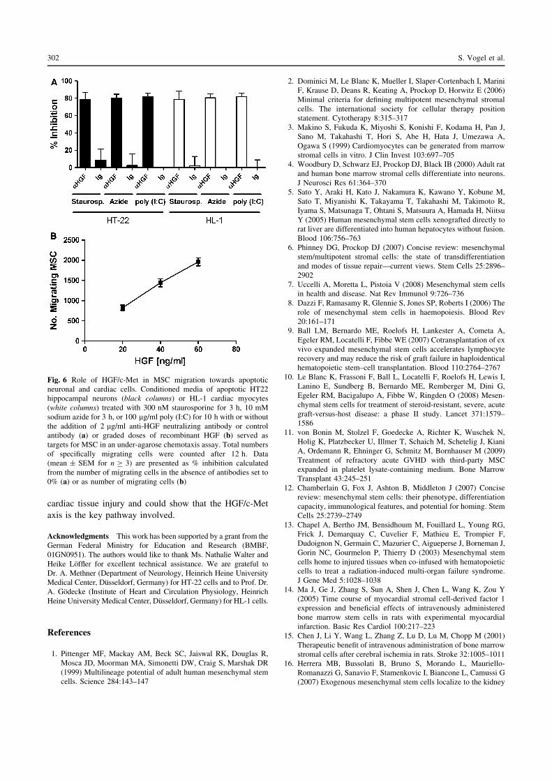
Fig. 6 Role of HGF/c-Met in MSC migration towards apoptotic
neuronal and cardiac cells. Conditioned media of apoptotic HT22
hippocampal neurons (black columns) or HL-1 cardiac myocytes
(white columns) treated with 300 nM staurosporine for 3 h, 10 mM
sodium azide for 3 h, or 100 lg/ml poly (I:C) for 10 h with or without
the addition of 2 lg/ml anti-HGF neutralizing antibody or control
antibody (a) or graded doses of recombinant HGF (b) served as
targets for MSC in an under-agarose chemotaxis assay. Total numbers
of specifically migrating cells were counted after 12 h. Data
(mean ± SEM for n C 3) are presented as % inhibition calculated
from the number of migrating cells in the absence of antibodies set to
0% (a) or as number of migrating cells (b)
302 S. Vogel et al.
by means of CD44 following acute tubular injury. Kidney Int
72:430–441
17. Wu GD, Nolta JA, Jin YS, Barr ML, Yu H, Starnes VA, Cramer
DV (2003) Migration of mesenchymal stem cells to heart allo-
grafts during chronic rejection. Transplantation 75:679–685
18. Spaeth E, Klopp A, Dembinski J, Andreeff M, Marini F (2008)
Inflammation and tumor microenvironments: defining the
migratory itinerary of mesenchymal stem cells. Gene Ther
15:730–738
19. Neuss S, Becher E, Woltje M, Tietze L, Jahnen-Dechent W
(2004) Functional expression of HGF and HGF receptor/c-met in
adult human mesenchymal stem cells suggests a role in cell
mobilization, tissue repair, and wound healing. Stem Cells
22:405–414
20. Honda S, Kagoshima M, Wanaka A, Tohyama M, Matsumoto K,
Nakamura T (1995) Localization and functional coupling of HGF
and c-Met/HGF receptor in rat brain: implication as neurotrophic
factor. Brain Res Mol Brain Res 32:197–210
21. Ono K, Matsumori A, Shioi T, Furukawa Y, Sasayama S (1997)
Enhanced expression of hepatocyte growth factor/c-Met by
myocardial ischemia and reperfusion in a rat model. Circulation
95:2552–2558
22. Miyazawa K, Shimomura T, Naka D, Kitamura N (1994) Pro-
teolytic activation of hepatocyte growth factor in response to
tissue injury. J Biol Chem 269:8966–8970
23. Miyazawa T, Matsumoto K, Ohmichi H, Katoh H, Yamashima T,
Nakamura T (1998) Protection of hippocampal neurons from
ischemia-induced delayed neuronal death by hepatocyte growth
factor: a novel neurotrophic factor. J Cereb Blood Flow Metab
18:345–348
24. Nakamura T, Mizuno S, Matsumoto K, Sawa Y, Matsuda H,
Nakamura T (2000) Myocardial protection from ischemia/reper-
fusion injury by endogenous and exogenous HGF. J Clin Invest
106:1511–1519
25. Nesselmann C, Ma N, Bieback K, Wagner W, Ho A, Konttinen
YT, Zhang H, Hinescu ME, Steinhoff G (2008) Mesenchymal
stem cells and cardiac repair. J Cell Mol Med 12:1795–1810
26. Dharmasaroja P (2009) Bone marrow-derived mesenchymal stem
cells for the treatment of ischemic stroke. J Clin Neurosci 16:12–20
27. Davis JB, Maher P (1994) Protein kinase C activation inhibits
glutamate-induced cytotoxicity in a neuronal cell line. Brain Res
652:169–173
28. White SM, Constantin PE, Claycomb WC (2004) Cardiac phys-
iology at the cellular level: use of cultured HL-1 cardiomyocytes
for studies of cardiac muscle cell structure and function. Am J
Physiol Heart Circ Physiol 286:H823–H829
29. Trapp T, Kogler G, El-Khattouti A, Sorg RV, Besselmann M,
Focking M, Buhrle CP, Trompeter I, Fischer JC, Wernet P (2008)
Hepatocyte growth factor/c-met-axis mediated tropism of cord
blood derived unrestricted somatic stem cells for neuronal injury.
J Biol Chem 283:32244–32253
30. Laevsky G, Knecht DA (2001) Under-agarose folate chemotaxis
of Dictyostelium discoideum amoebae in permissive and
mechanically inhibited conditions. Biotechniques 31:1140–1142
31. Sato T, Yoshinouchi T, Sakamoto T, Fujieda H, Murao S, Sato H,
Kobayashi H, Ohe T (1997) Hepatocyte growth factor (HGF): a
new biochemical marker for acute myocardial infarction. Heart
Vessels 12:241–246
32. Aoki M, Morishita R, Taniyama Y, Kida I, Moriguchi A, Mat-
sumoto K, Nakamura T, Kaneda Y, Higaki J, Ogihara T (2000)
Angiogenesis induced by hepatocyte growth factor in non-
infarcted myocardium and infarcted myocardium: up-regulation
of essential transcription factor for angiogenesis, ets. Gene Ther
7:417–427
33. Date I, Takagi N, Takagi K, Kago T, Matsumoto K, Nakamura T,
Takeo S (2004) Hepatocyte growth factor attenuates cerebral
ischemia-induced learning dysfunction. Biochem Biophys Res
Commun 319:1152–1158
34. Forte G, Minieri M, Cossa P, Antenucci D, Sala M, Gnocchi V,
Fiaccavento R, Carotenuto F, De Vito P, Baldini PM, Prat M, Di
Nardo P (2006) Hepatocyte growth factor effects on mesenchy-
mal stem cells: proliferation, migration, and differentiation. Stem
Cells 24:23–33
35. Zhu G, Huang L, Song M, Yu Z, Wu X, Zhao X, Jin J, Zhao G,
Chen J, Yu S (2008) Over-expression of hepatocyte growth factor
in smooth muscle cells regulates endothelial progenitor cells
differentiation, migration and proliferation. Int J Cardiol. doi:
10.1016/j.ijcard.2008.10.042
36. Heese O, Disko A, Zirkel D, Westphal M, Lamszus K (2005)
Neural stem cell migration toward gliomas in vitro. Neuro Oncol
7:476–484
37. Rosova I, Dao M, Capoccia B, Link D, Nolta JA (2008) Hypoxic
preconditioning results in increased motility and improved ther-
apeutic potential of human mesenchymal stem cells. Stem Cells
26:2173–2182
38. Skoberne M, Beignon AS, Larsson M, Bhardwaj N (2005)
Apoptotic cells at the crossroads of tolerance and immunity. Curr
Top Microbiol Immunol 289:259–292
39. Okunishi K, Dohi M, Nakagome K, Tanaka R, Mizuno S, Mat-
sumoto K, Miyazaki J, Nakamura T, Yamamoto K (2005) A
novel role of hepatocyte growth factor as an immune regulator
through suppressing dendritic cell function. J Immunol
175:4745–4753
40. Meng E, Guo Z, Wang H, Jin J, Wang J, Wu C, Wang L (2008)
High mobility group box 1 protein inhibits the proliferation of
human mesenchymal stem cells and promotes their migration and
differentiation along osteoblastic pathway. Stem Cells Dev
17:805–813
41. Raucci A, Palumbo R, Bianchi ME (2007) HMGB1: a signal of
necrosis. Autoimmunity 40:285–289
42. Abbott JD, Huang Y, Liu D, Hickey R, Krause DS, Giordano FJ
(2004) Stromal cell-derived factor-1alpha plays a critical role in
stem cell recruitment to the heart after myocardial infarction but
is not sufficient to induce homing in the absence of injury.
Circulation 110:3300–3305
43. Ji JF, He BP, Dheen ST, Tay SS (2004) Interactions of chemo-
kines and chemokine receptors mediate the migration of
mesenchymal stem cells to the impaired site in the brain after
hypoglossal nerve injury. Stem Cells 22:415–427
44. Ip JE, Wu Y, Huang J, Zhang L, Pratt RE, Dzau VJ (2007)
Mesenchymal stem cells use integrin beta1 not CXC chemokine
receptor 4 for myocardial migration and engraftment. Mol Biol
Cell 18:2873–2882
45. Sordi V, Malosio ML, Marchesi F, Mercalli A, Melzi R, Giord-
ano T, Belmonte N, Ferrari G, Leone BE, Bertuzzi F, Zerbini G,
Allavena P, Bonifacio E, Piemonti L (2005) Bone marrow mes-
enchymal stem cells express a restricted set of functionally active
chemokine receptors capable of promoting migration to pancre-
atic islets. Blood 106:419–427
46. Sackstein R, Merzaban JS, Cain DW, Dagia NM, Spencer JA, Lin
CP, Wohlgemuth R (2008) Ex vivo glycan engineering of CD44
programs human multipotent mesenchymal stromal cell traffick-
ing to bone. Nat Med 14:181–187
47. Honczarenko M, Le Y, Swierkowski M, Ghiran I, Glodek AM,
Silberstein LE (2006) Human bone marrow stromal cells express
a distinct set of biologically functional chemokine receptors.
Stem Cells 24:1030–1041
48. Li G, Zhang XA, Wang H,Wang X, Meng CL, Chan CY, Yew DT,
Tsang KS, Li K, Tsai SN, Ngai SM, Han ZC, Lin MC, He ML,
Kung HF (2009) Comparative proteomic analysis of mesenchymal
stem cells derived from human bone marrow, umbilical cord, and
placenta: implication in the migration. Proteomics 9:20–30
Homing of MSC to apoptotic tissues 303

Migration of mesenchymal stem cells towards glioblastoma cells dependson hepatocyte-growth factor and is enhanced by aminolaevulinic acid-mediatedphotodynamic treatment
Sebastian Vogel a, Corinna Peters a,b,1, Nima Etminan b, Verena Börger a, Adrian Schimanski a,b,Michael C. Sabel b, Rüdiger V. Sorg a,⇑a Institute for Transplantation Diagnostics and Cell Therapeutics, Heinrich-Heine University Hospital, Moorenstrasse 5, 40225 Düsseldorf, GermanybDepartment of Neurosurgery, Heinrich-Heine University Hospital, Moorenstrasse 5, 40225 Düsseldorf, Germany
a r t i c l e i n f o
Article history:Received 18 December 2012Available online 16 January 2013
Keywords:Hepatocyte-growth factorMigrationMesenchymal stem cellsGlioblastomaAminolaevulinic-acidPhotodynamic therapy
a b s t r a c t
Hepatocyte-growth factor (HGF) is expressed by glioblastomas and contributes to their growth, migrationand invasion. HGF also mediates migration of mesenchymal stem cells (MSC) to sites of apoptotic celldeath. Moreover, MSC show tropism for glioblastomas, which is exploited in gene therapy to deliverthe therapeutics to the tumor cells. Here, we have studied whether HGF contributes to the recruitmentof MSC by glioblastoma cells and whether aminolaevulinic acid-mediated photodynamic therapy (ALA/PDT), a novel therapeutic approach that induces apoptosis in glioblastoma cells, affects HGF releaseand this migratory response. MSC expressed the HGF receptor MET and migrated towards U87 andU251 glioblastoma spheroids. Migration increased significantly when spheroids were subjected to ALA/PDT, which was associated with induction of apoptosis and up-regulation of HGF. Neutralizing HGFresulted in significant inhibition of MSC migration towards untreated as well as ALA/PDT-treatedspheroids. Thus, glioblastoma cells express HGF, which contributes to the attraction of MSC. ALA/PDTinduces apoptosis and augments HGF release causing enhanced MSC migration towards the tumor cells.ALA/PDT may therefore be exploited to improve targeting of MSC delivered gene therapy, but it may alsoconstitute a risk in terms of beneficial effects for the tumor.
� 2013 Elsevier Inc. All rights reserved.
1. Introduction
Glioblastoma is the most frequent and aggressive malignantprimary brain tumor [1]. Despite multimodal therapy combiningsurgery, radiotherapy and alkylating chemotherapy, prognosis ofpatients is dismal: median survival is 14.6 months, the 2-year sur-vival rate 27.2% [2]. Aminolaevulinic acid-mediated photodynamictherapy (ALA/PDT) is a novel therapeutic approach for glioblas-toma, and early clinical results are promising [3,4]. ALA is an inter-mediate of the heme biosynthesis pathway. Oral uptake of ALAresults in preferential accumulation of protoporphyrin IX (PPIX)in glioblastoma cells, mainly due to low ferrochelatase activity[5,6]. This preferential accumulation can be exploited for intraop-
erative identification of the tumor during fluorescence-guided sur-gery using light of 400 nmwavelength for illumination, allowing toincrease the extent of resection, associated with higher progres-sion-free survival [7]. However, when exposed to light of 635 nmwavelength, PPIX acts as a potent photosensitizer [6]. Its excitationinitiates a photochemical reaction, which kills the tumor cells viathe generation of singlet oxygen [5]. In addition to this direct photo-toxic effect, there appears to be an immunological component to ALA/PDT, and it has been shown to influence the tumor vasculature aswell as the migratory and invasive behavior of tumor cells [5,8–11].
We and others have previously shown that ALA/PDT inducesapoptosis of glioblastoma cells [11,12]. The type of cell death hasconsequences besides killing of the tumor cells, e.g. it may affectthe development of immunity [13]. Furthermore, apoptosis butnot necrosis of neurons has been shown to result in up-regulationof hepatocyte-growth factor (HGF) [14,15]. HGF is a pleiotropiccytokine with anti-apoptotic activity [16]. It promotes glioblas-toma growth, invasiveness and angiogenesis [17–19], and its tyro-sine kinase receptor MET may represent a marker formesenchymal and proneural glioblastoma stem cell subtypes[20]. Therefore, the HGF–MET pathway is currently evaluated as
0006-291X/$ - see front matter � 2013 Elsevier Inc. All rights reserved.http://dx.doi.org/10.1016/j.bbrc.2012.12.153
Abbreviations: ALA/PDT, aminolaevulinic acid-mediated photodynamic therapy/treatment; HGF, hepatocyte-growth factor; MSC, mesenchymal stem cell(s).⇑ Corresponding author. Address: Institute for Transplantation Diagnostics and
Cell Therapeutics, Heinrich-Heine University Hospital, Moorenstrasse 5, Bldg. 14.88,40225 Düsseldorf, Germany. Fax: +49 211 81 04340.
E-mail address: [email protected] (R.V. Sorg).1 Current address: Department of Neurology, Heinrich-Heine University Hospital,
Düsseldorf, Germany.
Biochemical and Biophysical Research Communications 431 (2013) 428–432
Contents lists available at SciVerse ScienceDirect
Biochemical and Biophysical Research Communications
journal homepage: www.elsevier .com/locate /ybbrc

a potential therapeutic target in glioblastoma [21]. On the otherhand, HGF is a chemoattractant for mesenchymal stem cells(MSC) [14,22,23]. MSC are multipotent stem cells found in bonemarrow and other tissues. They are capable of differentiating intovarious types of mesenchymal cells, including osteoblasts, adipo-cytes and chondrocytes [24]. It is well established that MSC hometo glioblastomas and this tropism is utilized to target therapeuticsto the tumors, including cytokines, enzymes/pro-drugs, oncolyticviruses, toxins and others [25].
Expression of HGF by glioblastoma cells [17,19] and its activityas a chemotactic stimulus for MSC [14,22,23] as well as the obser-vation that apoptosis, thus, the type of cell death, which is inducedby ALA/PDT [11,12], is associated with induction of HGF in neurons[14,15], raises the questions whether HGF is involved in the migra-tion of MSC towards glioblastoma cells and whether ALA/PDT-induced apoptosis results in increased HGF release by glioblastomacells and an enhanced migratory response of MSC.
2. Materials and methods
2.1. Mesenchymal stem cells
Human bone marrow was obtained from volunteer donors afterinformed consent with the ethical approval of the local ethicalcommittee. MSC were isolated from bone marrow as describedpreviously [14]. Briefly, bone marrow cells were plated in 75-cm2
culture flasks (Greiner, Nürtingen, Germany) and cultured at37 �C and 5% CO2 in a humidified atmosphere in DMEM (Lonza,Verviers, Belgium) supplemented with 30% fetal calf serum (FCS;GIBCO/Invitrogen, Karlsruhe, Germany), 50 lg/ml gentamycinand 2 mM L-glutamine (all from Lonza). After 48 h, non-adherentcells were removed and cultures continued. When reaching 80%confluence, cells were harvested with trypsin (Lonza) and re-platedat 1:3. All MSC preparations showed the immunophenotype, andosteogenic and adipogenic differentiation typical of MSC [24].
2.2. Glioblastoma spheroids and ALA/PDT
Culture and generation of spheroids as well as ALA/PDT of U87and U251 glioblastoma cell lines were performed as described pre-viously [10,11]. Briefly, tumor cells were maintained in DMEM sup-plemented with 10% FCS, 100 U/ml penicillin, 100 lg/mlstreptomycin and 2 mM L-glutamine. To generate spheroids, cellswere plated in agar-coated culture flask. After 3 days of culture, tu-mor spheroids with a diameter of approximately 250 lm hadformed. For ALA/PDT, spheroid cultures were supplemented with12.5 lg/ml ALA (Merck, Darmstadt, Germany) and incubated for4 h. Spheroids were collected under microscopic control and trans-ferred (25 spheroids/well) into agar-coated flat-bottom 96-wellplates (Greiner) containing 100 ll/well of DMEM without phenolred. Exposure to laser light was performed for 625 s with an energyof 30 mW/cm2 (equivalent to 25 J/s or 1 W on 33 cm2) using a Cer-alas 633-nm PDT diode laser (Biolitec, Jena, Germany). After laserlight exposure, spheroids were used in the experiments. TheseALA/PDT conditions result in induction of apoptosis in about 60%of cells [11]. Untreated spheroids and spheroids treated with ALAonly or exposed to laser-light only served as controls.
2.3. Under-agarose chemotaxis assay
Chemoattraction of MSC by glioblastoma spheroids was studiedusing an under-agarose chemotaxis assay as described [14,15].Briefly, 0.8% agarose (Eurogentec, Cologne, Germany) in PBS wasboiled, mixed after cooling with 0.5% bovine serum albumin(BSA; Roth, Karlsruhe, Germany) in DMEM and poured into 6-well
plates (Costar/Corning, Wiesbaden, Germany). Three 2 mm wideand 5 mm long slots 5 mm apart from each other were cut in theagarose of each well. 8 � 104 MSC were then added to the centralslots of each well, 70 ll of chemoattractant (25 treated or un-treated spheroids) in the left target slots and 70 ll of 0.5% BSA inDMEM in the right control slots. The number of cells, which mi-grated towards the target slot subtracted by the number of cells,which migrated towards the control slot, was determined for eachwell after a 12 h migration period.
To study the contribution of HGF to MSC migration towards thespheroids, neutralization studies were performed by adding neu-tralizing anti-HGF polyclonal antibody (2 lg/ml; goat IgG; R&DSystems, Wiesbaden) or normal goat IgG (2 lg/ml; Santa Cruz,Heidelberg, Germany) to the targets.
2.4. Detection of MET and HGF expression
MET expression on MSC was determined by flow cytometryusing an anti-MET monoclonal antibody (5 lg/ml; clone 95106,IgG1; R&D Systems) and Fluorescein isothiocyanate (FITC)-conjugated F(ab)2-goat-anti-mouse IgG + M (Beckman-Coulter,Krefeld, Germany) as secondary antibody. Cells were analyzed ona FACS Canto flow cytometer (BD Biosciences, Heidelberg,Germany).
Intracellular HGF and apoptosis were detected by double label-ing with FITC-conjugated Annexin V (Beckman-Coulter) and anti-HGF rabbit polyclonal antibody (15 lg/ml; Abgent, San Diego,CA) 12 h after ALA/PDT or control treatments. Phycoerythrin(PE)-conjugated goat-anti-rabbit IgG (Jackson Immuno Research,Newmarket, UK) served as secondary antibody for anti-HGF. Cellswere stained first with Annexin V according to the manufacturer’sprotocol. Subsequently, they were fixed and permeabilized usingthe IntraPrep Fix/Perm kit (Beckman-Coulter), stained with anti-HGF polyclonal antibody and finally labeled with the secondaryantibody prior to flow cytometric analysis.
HGF protein in conditionedmedia of glioblastoma spheroids ob-tained 12 h after ALA/PDT or control treatments was quantified byenzyme-linked immunosorbent assay (ELISA; Shino Test Corpora-tion, Kanagawa, Japan) according to the manufacturer’s protocol.
Detection of HGF transcripts by reverse transcription-polymerase chain reaction (RT-PCR) was performed as describedrecently [14,15]. Glycerinaldehyde-3-phosphate dehydrogenase(GAPDH) served as positive control.
2.5. Statistical analysis
All data are presented as mean ± SEM for nP 3 unless statedotherwise. Statistical significance was determined with the Stu-dent’s t-test using Prism Software (GraphPad, San Diego, CA).
3. Results
3.1. Attraction of MSC by glioblastoma spheroids is enhanced by ALA/PDT
To assess migration of MSC towards U87 and U251 glioblastomaspheroids, an under-agarose migration assay was performed. Inthree independent experiments using different MSC preparations,MSC migrated towards the spheroids (Fig. 1). Migration was signif-icantly enhanced after ALA/PDT compared to the untreated spher-oids (U87, 3.6 ± 0.6-fold, p = 0.0096; U251, 3.8 ± 0.4-fold,p = 0.0018). In contrast, control spheroids treated with exposureto laser light or incubation with ALA only showed no such effect.
S. Vogel et al. / Biochemical and Biophysical Research Communications 431 (2013) 428–432 429
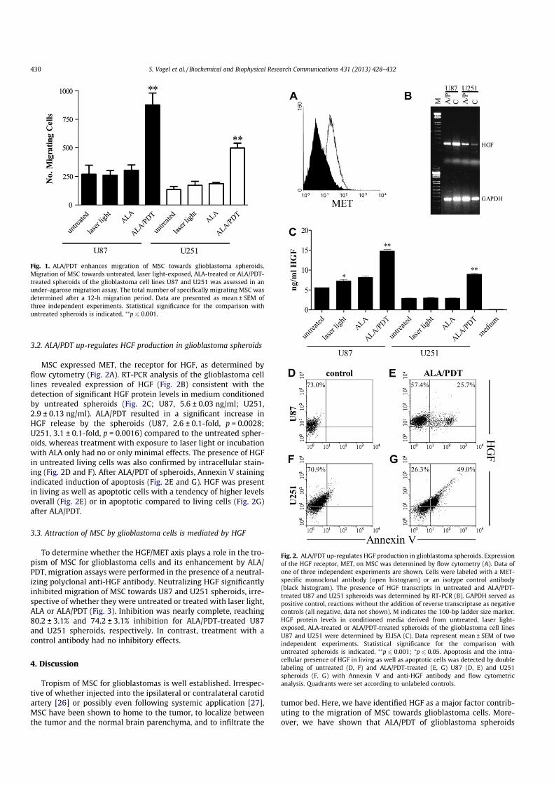
3.2. ALA/PDT up-regulates HGF production in glioblastoma spheroids
MSC expressed MET, the receptor for HGF, as determined byflow cytometry (Fig. 2A). RT-PCR analysis of the glioblastoma celllines revealed expression of HGF (Fig. 2B) consistent with thedetection of significant HGF protein levels in medium conditionedby untreated spheroids (Fig. 2C; U87, 5.6 ± 0.03 ng/ml; U251,2.9 ± 0.13 ng/ml). ALA/PDT resulted in a significant increase inHGF release by the spheroids (U87, 2.6 ± 0.1-fold, p = 0.0028;U251, 3.1 ± 0.1-fold, p = 0.0016) compared to the untreated spher-oids, whereas treatment with exposure to laser light or incubationwith ALA only had no or only minimal effects. The presence of HGFin untreated living cells was also confirmed by intracellular stain-ing (Fig. 2D and F). After ALA/PDT of spheroids, Annexin V stainingindicated induction of apoptosis (Fig. 2E and G). HGF was presentin living as well as apoptotic cells with a tendency of higher levelsoverall (Fig. 2E) or in apoptotic compared to living cells (Fig. 2G)after ALA/PDT.
3.3. Attraction of MSC by glioblastoma cells is mediated by HGF
To determine whether the HGF/MET axis plays a role in the tro-pism of MSC for glioblastoma cells and its enhancement by ALA/PDT, migration assays were performed in the presence of a neutral-izing polyclonal anti-HGF antibody. Neutralizing HGF significantlyinhibited migration of MSC towards U87 and U251 spheroids, irre-spective of whether they were untreated or treated with laser light,ALA or ALA/PDT (Fig. 3). Inhibition was nearly complete, reaching80.2 ± 3.1% and 74.2 ± 3.1% inhibition for ALA/PDT-treated U87and U251 spheroids, respectively. In contrast, treatment with acontrol antibody had no inhibitory effects.
4. Discussion
Tropism of MSC for glioblastomas is well established. Irrespec-tive of whether injected into the ipsilateral or contralateral carotidartery [26] or possibly even following systemic application [27],MSC have been shown to home to the tumor, to localize betweenthe tumor and the normal brain parenchyma, and to infiltrate the
tumor bed. Here, we have identified HGF as a major factor contrib-uting to the migration of MSC towards glioblastoma cells. More-over, we have shown that ALA/PDT of glioblastoma spheroids
Fig. 1. ALA/PDT enhances migration of MSC towards glioblastoma spheroids.Migration of MSC towards untreated, laser light-exposed, ALA-treated or ALA/PDT-treated spheroids of the glioblastoma cell lines U87 and U251 was assessed in anunder-agarose migration assay. The total number of specifically migrating MSC wasdetermined after a 12-h migration period. Data are presented as mean ± SEM ofthree independent experiments. Statistical significance for the comparison withuntreated spheroids is indicated, ⁄⁄p 6 0.001.
Fig. 2. ALA/PDT up-regulates HGF production in glioblastoma spheroids. Expressionof the HGF receptor, MET, on MSC was determined by flow cytometry (A). Data ofone of three independent experiments are shown. Cells were labeled with a MET-specific monoclonal antibody (open histogram) or an isotype control antibody(black histogram). The presence of HGF transcripts in untreated and ALA/PDT-treated U87 and U251 spheroids was determined by RT-PCR (B). GAPDH served aspositive control, reactions without the addition of reverse transcriptase as negativecontrols (all negative, data not shown). M indicates the 100-bp ladder size marker.HGF protein levels in conditioned media derived from untreated, laser light-exposed, ALA-treated or ALA/PDT-treated spheroids of the glioblastoma cell linesU87 and U251 were determined by ELISA (C). Data represent mean ± SEM of twoindependent experiments. Statistical significance for the comparison withuntreated spheroids is indicated, ⁄⁄p 6 0.001; ⁄p 6 0.05. Apoptosis and the intra-cellular presence of HGF in living as well as apoptotic cells was detected by doublelabeling of untreated (D, F) and ALA/PDT-treated (E, G) U87 (D, E) and U251spheroids (F, G) with Annexin V and anti-HGF antibody and flow cytometricanalysis. Quadrants were set according to unlabeled controls.
430 S. Vogel et al. / Biochemical and Biophysical Research Communications 431 (2013) 428–432

induces apoptosis, augments HGF release and thereby causes an in-crease in MSC attraction.
Expression of HGF in glioblastoma cells, including U87 andU251 cell lines, has been described before [17,19,28,29]. Moreover,Chu et al. reported increased HGF production following gammairradiation or hypoxia of U251 cells [29,30], and Kim and col-leagues showed that irradiation of glioblastoma cells enhancesattraction of MSC [31]. Thus, the ALA/PDT induced up-regulationof HGF in glioblastoma spheroids described here may represent astress response, similar to that in response to gamma irradiationand hypoxia [29–31] and to the induction of apoptosis in neurons,which results in attraction of MSC in an HGF-dependent fashion[14]. Indeed, we could show recently [11] and confirm here thatALA/PDT of glioblastoma spheroids causes apoptosis, thus the typeof cell death, which in neurons is associated with production ofHGF. Tropism of other stem cell populations for glioblastomas ap-pears to be regulated similarly: hematopoietic stem and progenitorcells [32] and neural stem cells [33] show enhanced migration to-wards glioblastoma cells in vitro as well as in vivo when these hadbeen previously irradiated or subjected to hypoxic conditions be-fore. Such stress responses, however, may also result in attractionof immune cells: we could recently show migration of immaturedendritic cells towards ALA/PDT-treated glioma spheroids [10].Thus, stress responses appear to initiate a concerted action byrecruiting various cell types with regenerative or immunologicalactivities. Interestingly, ALA/PDT appears to affect HGF releasenot only by the apoptotic, but also by the surviving cells. Whetherthis effect on surviving cells is direct or mediated indirectly byneighboring apoptotic cells requires further investigations.
Tropism of hematopoietic stem and progenitor cells for glio-blastomas appears to be mediated by the CXCL12/CXCR4 axis[32], whereas for neural stem cells the CXCL12/CXCR4, vascularendothelial growth factor/vascular endothelial growth factorreceptor 2 (VEGF/VEGFR2), urokinase-type plasminogen activa-tor/urokinase-type plasminogen activator receptor and the HGF/MET axes appear to contribute [33]. HGF has been described aschemoattractant for MSC [14,22] as well as other cell types includ-ing microglia cells [34], neural stem cells [33,35] and glioblastomacells [17,36]. However, its role in driving migration of MSC towardsglioblastomas has not been addressed before, and tropism has beenattributed to other factors produced by glioblastoma cells, particu-larly VEGF [37], platelet-derived growth factor [38,39], CXCL12[40] and CXCL-8 [31,41]. MSC express a multitude of chemokineand growth factor receptors involved in chemoattraction [42],some of which are expressed on subsets of cells only like CXCR4,CCR1 and CX3CR1 [43]. Moreover, there may be an interplay be-tween the individual pathways, e.g. Tu et al. reported an up-regulation of CXCL12 in U251 glioblastoma cells after stimulationwith HGF [44]. Thus, several pathways may contribute to the tro-pism of MSC for glioblastomas. So far, our observations are limitedto in vitro studies and the analysis of cell lines. Although a three-dimensional spheroid model has been used, which mimics microtumors more closely than monolayer cultures [45], differences be-tween cell lines and primary tumor cells as well as differences inthe micro milieu of the tumor in vivo may influence the results.Therefore, the effect of ALA/PDT on the HGF-driven tropism ofMSC for glioblastomas has to be confirmed in vivo.
The increasing local concentration of HGF following ALA/PDT inglioblastomas and attraction of MSC to the tumors may have ben-eficial as well as detrimental effects. HGF contributes to glioblas-toma growth, migration and invasion, and has cytoprotective,anti-apoptotic, immunosuppressive and angiogenic activity [16–19,46]. However, we could document that irrespective of the HGFexpression described here, ALA/PDT causes a long-lasting inhibi-tion of glioblastoma cell migration and invasiveness [11] and pro-motes anti-tumor immunity by initiating the initial steps of anadaptive immune response [10]. Moreover, Xie et al. have recentlyshown that expression of HGF by glioblastoma cells correlates withhigh levels of MET phosphorylation and predicts their sensitivity totherapeutic inhibition of the MET pathway [47]. Thus, there may bea rational for combining ALA/PDT with MET–HGF targeted therapy.Similarly, the recruitment of MSC by HGF may also have positiveand negative consequences. They may contribute to angiogenesis,immunosuppression and tumor cell survival [48], but may also in-duce glioblastoma cell death directly [39]. Moreover, MSC are usedto deliver therapeutics such as cytokines, enzymes/pro-drugs, onco-lytic viruses and toxins to glioblastomas [25], and this strategy maybe improved when combined with ALA/PDT, due to an enhancedtropism of the MSC. Although the net effect of ALA/PDT is currentlyunknown and requires further analyses, the beneficial effects ofALA/PDT may prevail as suggested by early clinical studies [3,4].
In summary, we have shown that HGF contributes to the attrac-tion of MSC towards glioblastoma cells and that ALA/PDT enhancesHGF production resulting in increased migration of MSC, whichmay have direct therapeutic consequences or may be exploitedto improve targeted gene therapy with MSC serving as therapeuticvectors.
Acknowledgment
The authors would like to thank Heike Löffler and Brigitte Sen-ger for excellent technical assistance. This work has been sup-ported by a grant from the Research Committee of the MedicalFaculty of the Heinrich-Heine University, Düsseldorf, Germany.The authors declare no conflict of interest.
Fig. 3. MSC migrate towards glioblastoma spheroids in an HGF-dependent fashion.Migration of MSC towards untreated (open bars), laser light-exposed (hatchedbars), ALA-treated (grey bars) or ALA/PDT-treated spheroids (black bars) of theglioblastoma cell lines U87 (A) and U251 (B) in the presence of a neutralizingpolyclonal anti-HGF antibody or control IgG was determined. Data are presented asmean ± SEM of three independent experiments. Statistical significance for compar-ison with untreated and treated spheroids in the absence of antibodies is indicated,⁄⁄p 6 0.001; ⁄p 6 0.05.
S. Vogel et al. / Biochemical and Biophysical Research Communications 431 (2013) 428–432 431

References
[1] J.L. Fisher, J.A. Schwartzbaum, M. Wrensch, et al., Epidemiology of braintumors, Neurol. Clin. 25 (2007) 867–890.
[2] R. Stupp, M.E. Hegi, W.P. Mason, et al., Effects of radiotherapy withconcomitant and adjuvant temozolomide versus radiotherapy alone onsurvival in glioblastoma in a randomised phase III study: 5-year analysis ofthe EORTC-NCIC trial, Lancet Oncol. 10 (2009) 459–466.
[3] M.S. Eljamel, C. Goodman, H. Moseley, ALA and Photofrin fluorescence-guidedresection and repetitive PDT in glioblastoma multiforme: a single centre phaseIII randomised controlled trial, Lasers Med. Sci. 23 (2008) 361–367.
[4] W. Stummer, T. Beck, W. Beyer, et al., Long-sustaining response in a patientwith non-resectable, distant recurrence of glioblastoma multiforme treated byinterstitial photodynamic therapy using 5-ALA: case report, J. Neurooncol. 87(2008) 103–109.
[5] A. Juzeniene, Q. Peng, J. Moan, Milestones in the development of photodynamictherapy and fluorescence diagnosis, Photochem. Photobiol. Sci. 6 (2007) 1234–1245.
[6] B. Krammer, K. Plaetzer, ALA and its clinical impact, from bench to bedside,Photochem. Photobiol. Sci. 7 (2008) 283–289.
[7] W. Stummer, U. Pichlmeier, T. Meinel, et al., Fluorescence-guided surgery with5-aminolevulinic acid for resection of malignant glioma: a randomisedcontrolled multicentre phase III trial, Lancet Oncol. 7 (2006) 392–401.
[8] A.P. Castano, P. Mroz, M.R. Hamblin, Photodynamic therapy and anti-tumourimmunity, Nat. Rev. Cancer 6 (2006) 535–545.
[9] H. Hirschberg, C.H. Sun, T. Krasieva, et al., Effects of ALA-mediatedphotodynamic therapy on the invasiveness of human glioma cells, LasersSurg. Med. 38 (2006) 939–945.
[10] N. Etminan, C. Peters, D. Lakbir, et al., Heat-shock protein 70-dependentdendritic cell activation by 5-aminolevulinic acid-mediated photodynamictreatment of human glioblastoma spheroids in vitro, Br. J. Cancer 105 (2011)961–969.
[11] N. Etminan, C. Peters, J. Ficnar, et al., Modulation of migratory activity andinvasiveness of human glioma spheroids following 5-aminolevulinic acid-based photodynamic treatment, J. Neurosurg. 115 (2011) 281–288.
[12] H. Inoue, Y. Kajimoto, M.A. Shibata, et al., Massive apoptotic cell death ofhuman glioma cells via a mitochondrial pathway following 5-aminolevulinicacid-mediated photodynamic therapy, J. Neurooncol. 83 (2007) 223–231.
[13] L. Zitvogel, O. Kepp, G. Kroemer, Decoding cell death signals in inflammationand immunity, Cell 140 (2010) 798–804.
[14] S. Vogel, T. Trapp, V. Borger, et al., Hepatocyte growth factor-mediatedattraction of mesenchymal stem cells for apoptotic neuronal andcardiomyocytic cells, Cell. Mol. Life Sci. 67 (2010) 295–303.
[15] T. Trapp, G. Kogler, A. El-Khattouti, et al., Hepatocyte growth factor/c-METaxis-mediated tropism of cord blood-derived unrestricted somatic stem cellsfor neuronal injury, J. Biol. Chem. 283 (2008) 32244–32253.
[16] D.C. Bowers, S. Fan, K.A. Walter, et al., Scatter factor/hepatocyte growth factorprotects against cytotoxic death in human glioblastoma viaphosphatidylinositol 3-kinase- and AKT-dependent pathways, Cancer Res. 60(2000) 4277–4283.
[17] S. Koochekpour, M. Jeffers, S. Rulong, et al., Met and hepatocyte growth factor/scatter factor expression in human gliomas, Cancer Res. 57 (1997) 5391–5398.
[18] T. Moriyama, H. Kataoka, R. Hamasuna, et al., Up-regulation of vascularendothelial growth factor induced by hepatocyte growth factor/scatter factorstimulation in human glioma cells, Biochem. Biophys. Res. Commun. 249(1998) 73–77.
[19] R. Abounader, J. Laterra, Scatter factor/hepatocyte growth factor in braintumor growth and angiogenesis, Neuro-oncology 7 (2005) 436–451.
[20] F. De Bacco, E. Casanova, E. Medico, et al., The MET oncogene is a functionalmarker of a glioblastoma stem cell subtype, Cancer Res. 72 (2012) 4537–4550.
[21] P.Y. Wen, D. Schiff, T.F. Cloughesy, et al., A phase II study evaluating theefficacy and safety of AMG 102 (rilotumumab) in patients with recurrentglioblastoma, Neuro-oncology 13 (2011) 437–446.
[22] S. Neuss, E. Becher, M. Woltje, et al., Functional expression of HGF and HGFreceptor/c-met in adult human mesenchymal stem cells suggests a role in cellmobilization, tissue repair, and wound healing, Stem cells 22 (2004) 405–414.
[23] G. Forte, M. Minieri, P. Cossa, et al., Hepatocyte growth factor effects onmesenchymal stem cells: proliferation, migration, and differentiation, Stemcells 24 (2006) 23–33.
[24] M. Dominici, K. Le Blanc, I. Mueller, et al., Minimal criteria for definingmultipotent mesenchymal stromal cells, The International Society for CellularTherapy position statement, Cytotherapy 8 (2006) 315–317.
[25] E. Binello, I.M. Germano, Stem cells as therapeutic vehicles for the treatment ofhigh-grade gliomas, Neuro-oncology 14 (2012) 256–265.
[26] A. Nakamizo, F. Marini, T. Amano, et al., Human bone marrow-derivedmesenchymal stem cells in the treatment of gliomas, Cancer Res. 65 (2005)3307–3318.
[27] X. Wu, J. Hu, L. Zhou, et al., In vivo tracking of superparamagnetic iron oxidenanoparticle-labeled mesenchymal stem cell tropism to malignant gliomasusing magnetic resonance imaging, Lab. Invest., J. Neurosurg. 108 (2008) 320–329.
[28] E.M. Rosen, J. Laterra, A. Joseph, et al., Scatter factor expression and regulationin human glial tumors, Int. J. Cancer 67 (1996) 248–255.
[29] S.H. Chu, Y.B. Ma, Z.A. Zhu, et al., Radiation-enhanced hepatocyte growth factorsecretion in malignant glioma cell lines, Surg. Neurol. 68 (2007) 610–613.
[30] S.H. Chu, D.F. Feng, Y.B. Ma, et al., Stabilization of hepatocyte growth factormRNA by hypoxia-inducible factor 1, Mol. Biol. Rep. 36 (2009) 1967–1975.
[31] S.M. Kim, J.H. Oh, S.A. Park, et al., Irradiation enhances the tumor tropism andtherapeutic potential of tumor necrosis factor-related apoptosis-inducingligand-secreting human umbilical cord blood-derived mesenchymal stem cellsin glioma therapy, Stem Cells 28 (2010) 2217–2228.
[32] G. Tabatabai, B. Frank, R. Mohle, et al., Irradiation and hypoxia promotehoming of haematopoietic progenitor cells towards gliomas by TGF-beta-dependent HIF-1alpha-mediated induction of CXCL12, Brain 129 (2006) 2426–2435.
[33] D. Zhao, J. Najbauer, E. Garcia, et al., Neural stem cell tropism to glioma: criticalrole of tumor hypoxia, Mol. Cancer Res. 6 (2008) 1819–1829.
[34] B. Badie, J. Schartner, J. Klaver, et al., In vitro modulation of microglia motilityby glioma cells is mediated by hepatocyte growth factor/scatter factor,Neurosurgery 44 (1999) 1077–1082.
[35] O. Heese, A. Disko, D. Zirkel, et al., Neural stem cell migration toward gliomasin vitro, Neuro-oncology 7 (2005) 476–484.
[36] T. Moriyama, H. Kataoka, K. Seguchi, et al., Effects of hepatocyte growth factor(HGF) on human glioma cells in vitro: HGF acts as a motility factor in gliomacells, Int. J. Cancer 66 (1996) 678–685.
[37] C. Schichor, V. Albrecht, B. Korte, et al., Mesenchymal stem cells and gliomacells form a structural as well as a functional syncytium in vitro, Exp. Neurol.234 (2012) 208–219.
[38] P. Cheng, Z.Q. Gao, Y.H. Liu, et al., Platelet-derived growth factor BB promotesthe migration of bone marrow-derived mesenchymal stem cells towards C6glioma and up-regulates the expression of intracellular adhesion molecule-1,Neurosci. Lett. 451 (2009) 52–56.
[39] C.S. Gondi, K.K. Veeravalli, B. Gorantla, et al., Human umbilical cord blood stemcells show PDGF-D-dependent glioma cell tropism in vitro and in vivo, Neuro-oncology 12 (2010) 453–465.
[40] F. Xu, J. Shi, B. Yu, et al., Chemokines mediate mesenchymal stem cellmigration toward gliomas in vitro, Oncol. Rep. 23 (2010) 1561–1567.
[41] T. Birnbaum, J. Roider, C.J. Schankin, et al., Malignant gliomas actively recruitbone marrow stromal cells by secreting angiogenic cytokines, J. Neurooncol.83 (2007) 241–247.
[42] E. Spaeth, A. Klopp, J. Dembinski, et al., Inflammation and tumormicroenvironments: defining the migratory itinerary of mesenchymal stemcells, Gene Ther. 15 (2008) 730–738.
[43] V. Sordi, M.L. Malosio, F. Marchesi, et al., Bone marrow mesenchymal stemcells express a restricted set of functionally active chemokine receptorscapable of promoting migration to pancreatic islets, Blood 106 (2005) 419–427.
[44] H. Tu, Z. Zhou, Q. Liang, et al., CXCR4 and SDF-1 production are stimulated byhepatocyte growth factor and promote glioma cell invasion, Onkologie 32(2009) 331–336.
[45] R.M. Sutherland, Cell and environment interactions in tumor microregions: themulticell spheroid model, Science 240 (1988) 177–184.
[46] K. Okunishi, M. Dohi, K. Nakagome, et al., A novel role of hepatocyte growthfactor as an immune regulator through suppressing dendritic cell function, J.Immunol. 175 (2005) 4745–4753.
[47] Q. Xie, R. Bradley, L. Kang, et al., Hepatocyte growth factor (HGF) autocrineactivation predicts sensitivity to MET inhibition in glioblastoma, Proc. Natl.Acad. Sci. USA 109 (2012) 570–575.
[48] A. Uccelli, L. Moretta, V. Pistoia, Mesenchymal stem cells in health and disease,Nat. Rev. Immunol. 8 (2008) 726–736.
432 S. Vogel et al. / Biochemical and Biophysical Research Communications 431 (2013) 428–432

24
4 Diskussion
Es konnte gezeigt werden, dass die HGF/MET-Achse eine zentrale Bedeutung
für die Rekrutierung von MSC zu geschädigten Kardiomyozyten und neuronalen
Zellen hat. Auch für die Migration von MSC zu Glioblastomzellen spielt sie eine
entscheidende Rolle. Apoptose, in Herz- und Nervenzellen direkt induziert, in
GBM-Zellen durch ALA/PDT verursacht, konnte in direkten Zusammenhang mit
diesen HGF-vermittelten Tropismen gebracht werden, was aufgrund der ge-
weberegenerativen und gentherapeutisch ausnutzbaren Eigenschaften von
MSC klinische Relevanz haben kann.
Die Rekrutierung von MSC zu geschädigtem Gewebe [37] mit anschließender
Ausübung regenerierender Aktivität, die wahrscheinlich eher auf parakrinen
Mechanismen und nicht auf einer gewebespezifischen Differenzierung beruht
[10-11], ist in der Literatur ausführlich beschrieben. Auch die Bildung und
proteolytische Aktivierung von HGF im Zusammenhang mit Gewebeschäden ist
bekannt [50, 53]. HGF wird bei ischämischer Herz- [54] und Hirnschädigung
[55] vermehrt gebildet und hat eine kardio- [56] und neuroprotektive [57]
Wirkung aufgrund von anti-apoptotischen [58-59] und pro-angiogenetischen [60,
77] Eigenschaften. Weiterhin wurde die Rekrutierung von MSC [49-51], un-
restringierten somatischen Stammzellen [67], endothelialen Progenitorzellen
[78], kardialen [79] sowie neuralen Stammzellen [80] in der Literatur mit der
HGF/MET-Achse in Verbindung gebracht. Somit trägt die Migration solcher Vor-
läuferzellen zu Gewebeschäden zusätzlich zur direkten zytoprotektiven Wirkung
von HGF vermutlich zur Regeneration bei. Eine Steigerung der Effektivität des
HGF-vermittelten Tropismus von MSC, welche ein erstrebenswertes Ziel bei
deren Transplantation im Hinblick auf regenerative Therapien darstellt, ließe
sich möglicherweise durch verstärkte Produktion von HGF im geschädigten
Milieu erzielen oder durch erhöhte Expression von MET auf MSC. Tatsächlich
konnten Rosova et al. zeigen, dass hypoxische Bedingungen bei der Kulti-
vierung von MSC zu einer verstärkten Expression von MET führten, und eine
kritische Ischämie in vivo assoziert war mit lokal erhöhtem HGF. Beides zu-
sammen trug zu einer verbesserten Migration der MSC und einer beschleu-
nigten Wiederherstellung der Durchblutungssituation bei [81].

In „Hepatocyte growth factor-mediated attraction of mesenchymal stem cells for
apoptotic neuronal and cardiomyocytic cells“ produzierten ausschließlich
apoptotische Zellen HGF und bewirkten eine HGF/MET-vermittelte Rekru-
tierung von MSC. Dies traf auf nekrotische und vitale Zellen nicht zu. Die Art
des Zelltodes kann über unterschiedliche Signalwege somit zu einer fein regu-
lierten Auslösung von Zelltropismen führen, die grundlegend verschiedene
Konsequenzen für das Schicksal des geschädigten Gewebes haben können.
HGF ist neben seinen erwähnten anti-apoptotischen Eigenschaften auch
immunsuppressiv, was unter anderem durch Hemmung der Antigen-präsen-
tierenden Aktivität von DC [82] und Induktion immunologischer Toleranz nach
Herztransplantation im Mausmodell [83] gezeigt werden konnte. MSC besitzen
neben ihrer geweberegenerierenden Aktivität [10] ebenfalls immun-
supprimierende Eigenschaften [14]. Sie hemmen unter anderem die Reifung
von DC [15] und deren zelluläre Sekretion von TNF-alpha [16]. Da TNF-alpha
neben der Leukozyten-Rekrutierung auch an der Induktion von Apoptose durch
Aktivierung von Todesrezeptoren beteiligt ist [84], steht dies im Einklang mit
einer zusätzlichen anti-apoptotischen Aktivität von MSC [74]. Die Apoptose-
induzierte Rekrutierung von MSC vermittelt durch HGF ist also in ein Setting
einzuordnen, in dem Limitierung von Apoptose, Geweberegeneration und
immunologische Toleranz vorherrscht.
In einem Mikromilieu, in dem vorwiegend nekrotischer Zelltod stattfindet, be-
steht eine grundlegend andere Situation. Nekrose ist assoziiert mit einer Viel-
zahl von immunstimulierenden Signalen, wie beispielsweise high mobility group
box 1 (HMGB1), ein Kernprotein, das entweder passiv von nekrotischen Zellen
freigesetzt [85] oder aktiv unter anderem von Monozyten und Makrophagen
nach vorheriger Stimulation sezerniert wird [86-87]. HMGB1 und andere immun-
stimulierende Mediatoren aus sterbenden Zellen, sogenannte danger-
associated molecular patterns, kurz DAMP [88], führen zu einer Rekrutierung
von Immunzellen, wie beispielsweise DC [89]. Da in der ersten Studie der vor-
liegenden Arbeit die Induktion von nekrotischem Zelltod zu keiner Migration von
MSC führte, einhergehend mit dem Ausbleiben einer Produktion von HGF, liegt
die Annahme nahe, dass das entsprechende Mikromilieu bei Nekrose einen
Weg Richtung Immunität einschlägt, in dem geweberegenerierende und den
25

zellulären Schaden begrenzende Antworten in den Hintergrund geraten. Tat-
sächlich konnte kürzlich bestätigt werden, dass nekrotischer Zelltod über die
Freisetzung von HMGB1 Monozyten und DC rekrutiert (Manuskript in Vorbe-
reitung). Interessanterweise beschreiben Meng et al. die Migration von MSC zu
HMGB1 [90]. Da die Freisetzung von HMGB1 typischerweise mit nekrotischem
Zelltod in Verbindung gebracht wird [85], suggeriert die Studie von Meng et al.
somit einen möglichen Zusammenhang zwischen Nekrose und der Migrations-
induktion von MSC. Andererseits wurde in einer neueren Studie die Freisetzung
von HMGB1 auch aus apoptotischen Zellen beschrieben. In diesem Fall war
HMGB1 jedoch durch mitochondriale reaktive Sauerstoffspezies oxidiert und
somit immunologisch inaktiviert worden [91]. Der Einfluss von HMGB1 und
anderen DAMP auf einen möglichen Migrationsanstoß von MSC ist im je-
weiligen Mikromilieu daher nur schwer vorhersehbar. Keine der drei in der vor-
liegenden Arbeit gewählten Bedingungen für die Induktion von Nekrose (H2O2,
Hitze, Ethanol) resultierten in einer Migration von MSC. Kulturen von MSC
können desweiteren verschiedene Subpopulation beinhalten, die auf unter-
schiedliche Migrationssignale unterschiedlich stark (oder eben gar nicht)
reagieren können. Die im Rahmen dieser Arbeit verwendeten MSC-Kulturen
waren durchweg positiv für MET. Da in den HGF-Inhibitionsstudien dieser Ar-
beit jedoch niemals eine vollständige Hemmung der MSC-Migration erreicht
wurde, könnte dies ein Hinweis dafür sein, dass ein Teil der Zellen trotz der An-
wesenheit migrationsinduzierender Mediatoren nicht rekrutiert wird. Für die
Migration von MSC zu geschädigtem Herz [92] und Hirn [93] stellt die SDF-
1/CXCR4-Interaktion einen alternativen Signalweg dar. Der Rezeptor CXCR4
lässt sich jedoch nur auf Subpopulationen von MSC nachweisen, was auch auf
andere Chemokin-Rezeptoren, wie CCR1 oder CX3CR1, zutrifft [94]. Das Zu-
sammenspiel von chemotaktischen Signalen für MSC erscheint somit komplex
und ist unzureichend verstanden.
In „Migration of mesenchymal stem cells towards glioblastoma cells depends on
hepatocyte-growth factor and is enhanced by aminolaevulinic acid-mediated
photodynamic treatment” wurde gezeigt, dass MSC HGF-abhängig zu
Glioblastomzellen migrieren. Dies konnte durch ALA/PDT-Behandlung der
26

GBM-Zellen signifikant verstärkt werden, was mit einer Induktion von Apoptose
und einer vermehrten Expression von HGF durch die Tumorzellen einherging.
Die Migration von MSC zum GBM nach deren Injektion in die Arteria carotis
[42], systemisch in eine Vene [95] oder intranasal appliziert [96] ist in der
Literatur gut dokumentiert. Auch die Expression von HGF in GBM-Zellen ist be-
kannt [61-62] und lässt sich durch Bestrahlung oder hypoxische Bedingungen
noch steigern [97-98]. In diesem Zusammenhang kann die ALA/PDT-verur-
sachte erhöhte Freisetzung von HGF als Bestandteil einer Stress-Antwort inter-
pretiert werden, analog zu der nach Bestrahlung und Hypoxie [97-98] oder nach
Induktion von Apoptose in Nervenzellen [67]. Eine Konsequenz dieser Stress-
antwort stellt die Rekrutierung von MSC dar.
Aufgrund der immunsupprimierenden Wirkung von HGF [82-83] und MSC [14]
würde man in diesem Setting eigentlich erwarten, dass immunologische To-
leranz nach ALA/PDT im GBM vorherrscht. Es kommt jedoch zu einer akuten
Entzündungsreaktion mit Hitzeschockprotein-abhängiger Einwanderung von
Leukozyten in den Tumor und einer Aktivierung von DC, welche in einer anti-
tumoralen Immunantwort resultiert [99-100]. Ob und inwiefern die pro-inflamma-
torische Antwort nach ALA/PDT in Konkurrenz steht zu potentiell immun-
hemmenden Effekten durch die Einwanderung von MSC in den Tumor, muss
daher in weiteren Studien geklärt werden. Interessanterweise gibt es auch
einen immunogenen apoptotischen Zelltod, wie es beispielsweise bei der In-
duktion von Apoptose von Tumorzellen mittels Antrazyklinen gezeigt wurde
[101]. Auch ALA/PDT verursacht Apoptose im GBM [70], und die in vitro-
Untersuchungen von Etminan et al. weisen auf eine immunogene Form der
ALA/PDT-induzierten Apoptose hin [100]. Desweiteren werden in der Literatur
neben den bereits beschriebenen immuninhibierenden Effekten von HGF kon-
troverserweise auch immunstimulierende Aktivitäten dokumentiert. Insbe-
sondere eine fördernde Wirkung von HGF auf die Rekrutierung von Monozyten
[102] und T-Zellen [103] wurde berichtet. Die beobachtete gesteigerte HGF-Pro-
duktion in der zweiten Studie der vorliegenden Arbeit könnte so also auch im
Zusammenhang stehen mit der beschriebenen Immunantwort auf den Tumor
nach ALA/PDT.
27

HGF fördert, insbesondere durch seine pro-angiogenetischen [60, 77] und auf
GBM-Zellen zytoprotektiven Effekte [64], Wachstum und Invasion des GBM [61,
63]. Weiterhin induziert es die Expression von Matrix-Metalloproteinase (MMP)-
2 in GBM-Zellen [104], was ebenfalls der Invasion des Tumors zugutekommen
kann. Da in der vorliegenden Arbeit eine ALA/PDT-Behandlung von GBM-
Zellen mit einer erhöhten HGF-Freisetzung einherging, suggerieren diese Daten
potentiell tumorfördernde Effekte nach photodynamischer Behandlung. Ein
direkter therapeutischer Nutzen von ALA/PDT bei Patienten mit GBM ist jedoch
wahrscheinlich [105-106]. Außerdem wurden tumorhemmende Effekte von ALA/
PDT in Verbindung gebracht mit einer verminderten Expression von MMP-7 und
MMP-8 [69]. Gründe für die scheinbar im Konflikt stehenden Daten könnten
darin bestehen, dass die beobachtete Hemmung von MMP-7 und MMP-8 nach
ALA/PDT in einem Setting unabhängig von HGF stattfindet. Dies würde be-
deuten, dass der fördernde Einfluss von HGF auf MMP nach ALA/PDT-Be-
handlung eine untergeordnete Rolle spielt, beispielsweise durch andere ALA/
PDT-induzierte Mechanismen, die mit HGF und MMP interferieren. Zum
anderen ist es möglich, dass HGF und ALA/PDT auf jeweils verschiedene Sub-
typen von MMP in den Tumorzellen wirken; Interferenzen in der Wirkung dieser
Subtypen untereinander sind nicht auszuschließen. Obwohl nachteilige Effekte
durch eine gesteigerte Produktion von HGF in den GBM-Zellen nach ALA/PDT
möglich, und weitere Studien deshalb notwendig sind, kann eine anti-tumorale
Wirkung der HGF-vermittelten verstärkten Migration von MSC zum Tumor über-
wiegen, sei sie durch gentherapeutisch modifizierte MSC, die beispielsweise
IFN-beta oder TRAIL exprimieren [35], oder durch eine direkte zytotoxische
Wirkung auf den Tumor [43] hervorgerufen.
Apoptotischer Zelltod und der HGF/MET Signalweg spielen also eine Schlüssel-
rolle in der Rekrutierung von MSC. Dies kann von Relevanz sein für die Ver-
wendung von MSC in der regenerativen Medizin wie auch in der zellulären
Therapie von Glioblastomen.
28
5 Literaturverzeichnis
1. Friedenstein, A.J., Chailakhyan, R.K., Latsinik, N.V., Panasyuk, A.F., Keiliss-Borok, I.V., Stromal cells responsible for transferring the microenvironment of the hemopoietic tissues. Cloning in vitro and retransplantation in vivo. Transplantation, 1974. 17(4): p. 331-340.
2. da Silva Meirelles, L., Caplan, A.I., Nardi, N.B., In search of the in vivo identity of mesenchymal stem cells. Stem Cells, 2008. 26(9): p. 2287-2299.
3. Dominici, M., Le Blanc, K., Mueller, I., Slaper-Cortenbach, I., Marini, F., Krause, D., Deans, R., Keating, A., Prockop, D., Horwitz, E., Minimal criteria for defining multipotent mesenchymal stromal cells. The International Society for Cellular Therapy position statement. Cytotherapy, 2006. 8(4): p. 315-317.
4. Pittenger, M.F., Mackay, A.M., Beck, S.C., Jaiswal, R.K., Douglas, R., Mosca, J.D., Moorman, M.A., Simonetti, D.W., Craig, S., Marshak, D.R., Multilineage potential of adult human mesenchymal stem cells. Science, 1999. 284(5411): p. 143-147.
5. Grove, J.E., Bruscia, E., Krause, D.S., Plasticity of bone marrow-derived stem cells. Stem Cells, 2004. 22(4): p. 487-500.
6. Makino, S., Fukuda, K., Miyoshi, S., Konishi, F., Kodama, H., Pan, J., Sano, M., Takahashi, T., Hori, S., Abe, H., Hata, J., Umezawa, A., Ogawa, S., Cardiomyocytes can be generated from marrow stromal cells in vitro. J Clin Invest, 1999. 103(5): p. 697-705.
7. Woodbury, D., Schwarz, E.J., Prockop, D.J., Black, I.B., Adult rat and human bone marrow stromal cells differentiate into neurons. J Neurosci Res, 2000. 61(4): p. 364-370.
8. Sato, Y., Araki, H., Kato, J., Nakamura, K., Kawano, Y., Kobune, M., Sato, T., Miyanishi, K., Takayama, T., Takahashi, M., Takimoto, R., Iyama, S., Matsunaga, T., Ohtani, S., Matsuura, A., Hamada, H., Niitsu, Y., Human mesenchymal stem cells xenografted directly to rat liver are differentiated into human hepatocytes without fusion. Blood, 2005. 106(2): p. 756-763.
9. Hombach-Klonisch, S., Panigrahi, S., Rashedi, I., Seifert, A., Alberti, E., Pocar, P., Kurpisz, M., Schulze-Osthoff, K., Mackiewicz, A., Los, M., Adult stem cells and their trans-differentiation potential--perspectives and therapeutic applications. J Mol Med (Berl), 2008. 86(12): p. 1301-1314.
10. Phinney, D.G., Prockop, D.J., Concise review: mesenchymal stem/multipotent stromal cells: the state of transdifferentiation and modes of tissue repair--current views. Stem Cells, 2007. 25(11): p. 2896-2902.
11. Prockop, D.J., "Stemness" does not explain the repair of many tissues by mesenchymal stem/multipotent stromal cells (MSCs). Clin Pharmacol Ther, 2007. 82(3): p. 241-243.
12. Dazzi, F., Ramasamy, R., Glennie, S., Jones, S.P., Roberts, I., The role of mesenchymal stem cells in haemopoiesis. Blood Rev, 2006. 20(3): p. 161-171.
13. Kiel, M.J., Morrison, S.J., Uncertainty in the niches that maintain haematopoietic stem cells. Nat Rev Immunol, 2008. 8(4): p. 290-301.
14. Uccelli, A., Moretta, L., Pistoia, V., Mesenchymal stem cells in health and disease. Nat Rev Immunol, 2008. 8(9): p. 726-736.
15. Jiang, X.X., Zhang, Y., Liu, B., Zhang, S.X., Wu, Y., Yu, X.D., Mao, N., Human mesenchymal stem cells inhibit differentiation and function of monocyte-derived dendritic cells. Blood, 2005. 105(10): p. 4120-4126.
16. Aggarwal, S., Pittenger, M.F., Human mesenchymal stem cells modulate allogeneic immune cell responses. Blood, 2005. 105(4): p. 1815-1822.
17. Bartholomew, A., Sturgeon, C., Siatskas, M., Ferrer, K., McIntosh, K., Patil, S., Hardy, W., Devine, S., Ucker, D., Deans, R., Moseley, A., Hoffman, R.,
29

Mesenchymal stem cells suppress lymphocyte proliferation in vitro and prolong skin graft survival in vivo. Exp Hematol, 2002. 30(1): p. 42-48.
18. Di Nicola, M., Carlo-Stella, C., Magni, M., Milanesi, M., Longoni, P.D., Matteucci, P., Grisanti, S., Gianni, A.M., Human bone marrow stromal cells suppress T-lymphocyte proliferation induced by cellular or nonspecific mitogenic stimuli. Blood, 2002. 99(10): p. 3838-3843.
19. Rasmusson, I., Ringden, O., Sundberg, B., Le Blanc, K., Mesenchymal stem cells inhibit the formation of cytotoxic T lymphocytes, but not activated cytotoxic T lymphocytes or natural killer cells. Transplantation, 2003. 76(8): p. 1208-1213.
20. Krampera, M., Glennie, S., Dyson, J., Scott, D., Laylor, R., Simpson, E., Dazzi, F., Bone marrow mesenchymal stem cells inhibit the response of naive and memory antigen-specific T cells to their cognate peptide. Blood, 2003. 101(9): p. 3722-3729.
21. Quarto, R., Mastrogiacomo, M., Cancedda, R., Kutepov, S.M., Mukhachev, V., Lavroukov, A., Kon, E., Marcacci, M., Repair of large bone defects with the use of autologous bone marrow stromal cells. N Engl J Med, 2001. 344(5): p. 385-386.
22. Nesselmann, C., Ma, N., Bieback, K., Wagner, W., Ho, A., Konttinen, Y.T., Zhang, H., Hinescu, M.E., Steinhoff, G., Mesenchymal stem cells and cardiac repair. J Cell Mol Med, 2008. 12(5B): p. 1795-1810.
23. Strauer, B.E., Brehm, M., Zeus, T., Kostering, M., Hernandez, A., Sorg, R.V., Kogler, G., Wernet, P., Repair of infarcted myocardium by autologous intracoronary mononuclear bone marrow cell transplantation in humans. Circulation, 2002. 106(15): p. 1913-1918.
24. Wollert, K.C., Meyer, G.P., Lotz, J., Ringes-Lichtenberg, S., Lippolt, P., Breidenbach, C., Fichtner, S., Korte, T., Hornig, B., Messinger, D., Arseniev, L., Hertenstein, B., Ganser, A., Drexler, H., Intracoronary autologous bone-marrow cell transfer after myocardial infarction: the BOOST randomised controlled clinical trial. Lancet, 2004. 364(9429): p. 141-148.
25. Dharmasaroja, P., Bone marrow-derived mesenchymal stem cells for the treatment of ischemic stroke. J Clin Neurosci, 2009. 16(1): p. 12-20.
26. Le Blanc, K., Frassoni, F., Ball, L., Locatelli, F., Roelofs, H., Lewis, I., Lanino, E., Sundberg, B., Bernardo, M.E., Remberger, M., Dini, G., Egeler, R.M., Bacigalupo, A., Fibbe, W., Ringden, O., Mesenchymal stem cells for treatment of steroid-resistant, severe, acute graft-versus-host disease: a phase II study. Lancet, 2008. 371(9624): p. 1579-1586.
27. von Bonin, M., Stolzel, F., Goedecke, A., Richter, K., Wuschek, N., Holig, K., Platzbecker, U., Illmer, T., Schaich, M., Schetelig, J., Kiani, A., Ordemann, R., Ehninger, G., Schmitz, M., Bornhauser, M., Treatment of refractory acute GVHD with third-party MSC expanded in platelet lysate-containing medium. Bone Marrow Transplant, 2009. 43(3): p. 245-251.
28. Ball, L.M., Bernardo, M.E., Roelofs, H., Lankester, A., Cometa, A., Egeler, R.M., Locatelli, F., Fibbe, W.E., Cotransplantation of ex vivo expanded mesenchymal stem cells accelerates lymphocyte recovery and may reduce the risk of graft failure in haploidentical hematopoietic stem-cell transplantation. Blood, 2007. 110(7): p. 2764-2767.
29. Gerdoni, E., Gallo, B., Casazza, S., Musio, S., Bonanni, I., Pedemonte, E., Mantegazza, R., Frassoni, F., Mancardi, G., Pedotti, R., Uccelli, A., Mesenchymal stem cells effectively modulate pathogenic immune response in experimental autoimmune encephalomyelitis. Ann Neurol, 2007. 61(3): p. 219-227.
30. Spaeth, E., Klopp, A., Dembinski, J., Andreeff, M., Marini, F., Inflammation and tumor microenvironments: defining the migratory itinerary of mesenchymal stem cells. Gene Ther, 2008. 15(10): p. 730-738.
30

31. Studeny, M., Marini, F.C., Dembinski, J.L., Zompetta, C., Cabreira-Hansen, M., Bekele, B.N., Champlin, R.E., Andreeff, M., Mesenchymal stem cells: potential precursors for tumor stroma and targeted-delivery vehicles for anticancer agents. J Natl Cancer Inst, 2004. 96(21): p. 1593-1603.
32. Fisher, J.L., Schwartzbaum, J.A., Wrensch, M., Wiemels, J.L., Epidemiology of brain tumors. Neurol Clin, 2007. 25(4): p. 867-890.
33. Stupp, R., Hegi, M.E., Mason, W.P., van den Bent, M.J., Taphoorn, M.J., Janzer, R.C., Ludwin, S.K., Allgeier, A., Fisher, B., Belanger, K., Hau, P., Brandes, A.A., Gijtenbeek, J., Marosi, C., Vecht, C.J., Mokhtari, K., Wesseling, P., Villa, S., Eisenhauer, E., Gorlia, T., Weller, M., Lacombe, D., Cairncross, J.G., Mirimanoff, R.O., Effects of radiotherapy with concomitant and adjuvant temozolomide versus radiotherapy alone on survival in glioblastoma in a randomised phase III study: 5-year analysis of the EORTC-NCIC trial. Lancet Oncol, 2009. 10(5): p. 459-466.
34. Donaldson, S.S., Laningham, F., Fisher, P.G., Advances toward an understanding of brainstem gliomas. J Clin Oncol, 2006. 24(8): p. 1266-1272.
35. Binello, E., Germano, I.M., Stem cells as therapeutic vehicles for the treatment of high-grade gliomas. Neuro Oncol, 2012. 14(3): p. 256-265.
36. Lauffenburger, D.A., Horwitz, A.F., Cell migration: a physically integrated molecular process. Cell, 1996. 84(3): p. 359-369.
37. Chamberlain, G., Fox, J., Ashton, B., Middleton, J., Concise review: mesenchymal stem cells: their phenotype, differentiation capacity, immunological features, and potential for homing. Stem Cells, 2007. 25(11): p. 2739-2749.
38. Ma, J., Ge, J., Zhang, S., Sun, A., Shen, J., Chen, L., Wang, K., Zou, Y., Time course of myocardial stromal cell-derived factor 1 expression and beneficial effects of intravenously administered bone marrow stem cells in rats with experimental myocardial infarction. Basic Res Cardiol, 2005. 100(3): p. 217-223.
39. Nagaya, N., Fujii, T., Iwase, T., Ohgushi, H., Itoh, T., Uematsu, M., Yamagishi, M., Mori, H., Kangawa, K., Kitamura, S., Intravenous administration of mesenchymal stem cells improves cardiac function in rats with acute myocardial infarction through angiogenesis and myogenesis. Am J Physiol Heart Circ Physiol, 2004. 287(6): p. 2670-2676.
40. Chen, J., Li, Y., Wang, L., Zhang, Z., Lu, D., Lu, M., Chopp, M., Therapeutic benefit of intravenous administration of bone marrow stromal cells after cerebral ischemia in rats. Stroke, 2001. 32(4): p. 1005-1011.
41. Dvorak, H.F., Tumors: wounds that do not heal. Similarities between tumor stroma generation and wound healing. N Engl J Med, 1986. 315(26): p. 1650-1659.
42. Nakamizo, A., Marini, F., Amano, T., Khan, A., Studeny, M., Gumin, J., Chen, J., Hentschel, S., Vecil, G., Dembinski, J., Andreeff, M., Lang, F.F., Human bone marrow-derived mesenchymal stem cells in the treatment of gliomas. Cancer Res, 2005. 65(8): p. 3307-3318.
43. Gondi, C.S., Veeravalli, K.K., Gorantla, B., Dinh, D.H., Fassett, D., Klopfenstein, J.D., Gujrati, M., Rao, J.S., Human umbilical cord blood stem cells show PDGF-D-dependent glioma cell tropism in vitro and in vivo. Neuro Oncol, 2010. 12(5): p. 453-465.
44. Kim, S.M., Oh, J.H., Park, S.A., Ryu, C.H., Lim, J.Y., Kim, D.S., Chang, J.W., Oh, W., Jeun, S.S., Irradiation enhances the tumor tropism and therapeutic potential of tumor necrosis factor-related apoptosis-inducing ligand-secreting human umbilical cord blood-derived mesenchymal stem cells in glioma therapy. Stem Cells, 2010. 28(12): p. 2217-2228.
31

45. Honczarenko, M., Le, Y., Swierkowski, M., Ghiran, I., Glodek, A.M., Silberstein, L.E., Human bone marrow stromal cells express a distinct set of biologically functional chemokine receptors. Stem Cells, 2006. 24(4): p. 1030-1041.
46. Mohle, R., Bautz, F., Rafii, S., Moore, M.A., Brugger, W., Kanz, L., The chemokine receptor CXCR-4 is expressed on CD34+ hematopoietic progenitors and leukemic cells and mediates transendothelial migration induced by stromal cell-derived factor-1. Blood, 1998. 91(12): p. 4523-4530.
47. Petit, I., Szyper-Kravitz, M., Nagler, A., Lahav, M., Peled, A., Habler, L., Ponomaryov, T., Taichman, R.S., Arenzana-Seisdedos, F., Fujii, N., Sandbank, J., Zipori, D., Lapidot, T., G-CSF induces stem cell mobilization by decreasing bone marrow SDF-1 and up-regulating CXCR4. Nat Immunol, 2002. 3(7): p. 687-694.
48. Ponte, A.L., Marais, E., Gallay, N., Langonne, A., Delorme, B., Herault, O., Charbord, P., Domenech, J., The in vitro migration capacity of human bone marrow mesenchymal stem cells: comparison of chemokine and growth factor chemotactic activities. Stem Cells, 2007. 25(7): p. 1737-1745.
49. Forte, G., Minieri, M., Cossa, P., Antenucci, D., Sala, M., Gnocchi, V., Fiaccavento, R., Carotenuto, F., De Vito, P., Baldini, P.M., Prat, M., Di Nardo, P., Hepatocyte growth factor effects on mesenchymal stem cells: proliferation, migration, and differentiation. Stem Cells, 2006. 24(1): p. 23-33.
50. Neuss, S., Becher, E., Woltje, M., Tietze, L., Jahnen-Dechent, W., Functional expression of HGF and HGF receptor/c-met in adult human mesenchymal stem cells suggests a role in cell mobilization, tissue repair, and wound healing. Stem Cells, 2004. 22(3): p. 405-414.
51. Son, B.R., Marquez-Curtis, L.A., Kucia, M., Wysoczynski, M., Turner, A.R., Ratajczak, J., Ratajczak, M.Z., Janowska-Wieczorek, A., Migration of bone marrow and cord blood mesenchymal stem cells in vitro is regulated by stromal-derived factor-1-CXCR4 and hepatocyte growth factor-c-met axes and involves matrix metalloproteinases. Stem Cells, 2006. 24(5): p. 1254-1264.
52. Zarnegar, R., Michalopoulos, G.K., The many faces of hepatocyte growth factor: from hepatopoiesis to hematopoiesis. J Cell Biol, 1995. 129(5): p. 1177-1180.
53. Miyazawa, K., Shimomura, T., Naka, D., Kitamura, N., Proteolytic activation of hepatocyte growth factor in response to tissue injury. J Biol Chem, 1994. 269(12): p. 8966-8970.
54. Ono, K., Matsumori, A., Shioi, T., Furukawa, Y., Sasayama, S., Enhanced expression of hepatocyte growth factor/c-Met by myocardial ischemia and reperfusion in a rat model. Circulation, 1997. 95(11): p. 2552-2558.
55. Honda, S., Kagoshima, M., Wanaka, A., Tohyama, M., Matsumoto, K., Nakamura, T., Localization and functional coupling of HGF and c-Met/HGF receptor in rat brain: implication as neurotrophic factor. Brain Res Mol Brain Res, 1995. 32(2): p. 197-210.
56. Nakamura, T., Mizuno, S., Matsumoto, K., Sawa, Y., Matsuda, H., Myocardial protection from ischemia/reperfusion injury by endogenous and exogenous HGF. J Clin Invest, 2000. 106(12): p. 1511-1519.
57. Miyazawa, T., Matsumoto, K., Ohmichi, H., Katoh, H., Yamashima, T., Nakamura, T., Protection of hippocampal neurons from ischemia-induced delayed neuronal death by hepatocyte growth factor: a novel neurotrophic factor. J Cereb Blood Flow Metab, 1998. 18(4): p. 345-348.
58. Bardelli, A., Longati, P., Albero, D., Goruppi, S., Schneider, C., Ponzetto, C., Comoglio, P.M., HGF receptor associates with the anti-apoptotic protein BAG-1 and prevents cell death. EMBO J, 1996. 15(22): p. 6205-6212.
59. Xiao, G.H., Jeffers, M., Bellacosa, A., Mitsuuchi, Y., Vande Woude, G.F., Testa, J.R., Anti-apoptotic signaling by hepatocyte growth factor/Met via the
32
phosphatidylinositol 3-kinase/Akt and mitogen-activated protein kinase pathways. Proc Natl Acad Sci U S A, 2001. 98(1): p. 247-252.
60. Bussolino, F., Di Renzo, M.F., Ziche, M., Bocchietto, E., Olivero, M., Naldini, L., Gaudino, G., Tamagnone, L., Coffer, A., Comoglio, P.M., Hepatocyte growth factor is a potent angiogenic factor which stimulates endothelial cell motility and growth. J Cell Biol, 1992. 119(3): p. 629-641.
61. Abounader, R., Laterra, J., Scatter factor/hepatocyte growth factor in brain tumor growth and angiogenesis. Neuro Oncol, 2005. 7(4): p. 436-451.
62. Koochekpour, S., Jeffers, M., Rulong, S., Taylor, G., Klineberg, E., Hudson, E.A., Resau, J.H., Vande Woude, G.F., Met and hepatocyte growth factor/scatter factor expression in human gliomas. Cancer Res, 1997. 57(23): p. 5391-5398.
63. Moriyama, T., Kataoka, H., Hamasuna, R., Yokogami, K., Uehara, H., Kawano, H., Goya, T., Tsubouchi, H., Koono, M., Wakisaka, S., Up-regulation of vascular endothelial growth factor induced by hepatocyte growth factor/scatter factor stimulation in human glioma cells. Biochem Biophys Res Commun, 1998. 249(1): p. 73-77.
64. Bowers, D.C., Fan, S., Walter, K.A., Abounader, R., Williams, J.A., Rosen, E.M., Laterra, J., Scatter factor/hepatocyte growth factor protects against cytotoxic death in human glioblastoma via phosphatidylinositol 3-kinase- and AKT-dependent pathways. Cancer Res, 2000. 60(15): p. 4277-4283.
65. Elmore, S., Apoptosis: a review of programmed cell death. Toxicol Pathol, 2007. 35(4): p. 495-516.
66. Vermeulen, K., Van Bockstaele, D.R., Berneman, Z.N., Apoptosis: mechanisms and relevance in cancer. Ann Hematol, 2005. 84(10): p. 627-639.
67. Trapp, T., Kogler, G., El-Khattouti, A., Sorg, R.V., Besselmann, M., Focking, M., Buhrle, C.P., Trompeter, I., Fischer, J.C., Wernet, P., Hepatocyte growth factor/c-MET axis-mediated tropism of cord blood-derived unrestricted somatic stem cells for neuronal injury. J Biol Chem, 2008. 283(47): p. 32244-32253.
68. Beck, T.J., Kreth, F.W., Beyer, W., Mehrkens, J.H., Obermeier, A., Stepp, H., Stummer, W., Baumgartner, R., Interstitial photodynamic therapy of nonresectable malignant glioma recurrences using 5-aminolevulinic acid induced protoporphyrin IX. Lasers Surg Med, 2007. 39(5): p. 386-393.
69. Etminan, N., Peters, C., Ficnar, J., Anlasik, S., Bunemann, E., Slotty, P.J., Hanggi, D., Steiger, H.J., Sorg, R.V., Stummer, W., Modulation of migratory activity and invasiveness of human glioma spheroids following 5-aminolevulinic acid-based photodynamic treatment. Laboratory investigation. J Neurosurg, 2011. 115(2): p. 281-288.
70. Inoue, H., Kajimoto, Y., Shibata, M.A., Miyoshi, N., Ogawa, N., Miyatake, S., Otsuki, Y., Kuroiwa, T., Massive apoptotic cell death of human glioma cells via a mitochondrial pathway following 5-aminolevulinic acid-mediated photodynamic therapy. J Neurooncol, 2007. 83(3): p. 223-231.
71. Juzeniene, A., Peng, Q., Moan, J., Milestones in the development of photodynamic therapy and fluorescence diagnosis. Photochem Photobiol Sci, 2007. 6(12): p. 1234-1245.
72. Krammer, B., Plaetzer, K., ALA and its clinical impact, from bench to bedside. Photochem Photobiol Sci, 2008. 7(3): p. 283-289.
73. Dolmans, D.E., Fukumura, D., Jain, R.K., Photodynamic therapy for cancer. Nat Rev Cancer, 2003. 3(5): p. 380-387.
74. Li, J.H., Zhang, N., Wang, J.A., Improved anti-apoptotic and anti-remodeling potency of bone marrow mesenchymal stem cells by anoxic pre-conditioning in diabetic cardiomyopathy. J Endocrinol Invest, 2008. 31(2): p. 103-110.
75. Vogel, S., Trapp, T., Borger, V., Peters, C., Lakbir, D., Dilloo, D., Sorg, R.V., Hepatocyte growth factor-mediated attraction of mesenchymal stem cells for
33

apoptotic neuronal and cardiomyocytic cells. Cell Mol Life Sci, 2010. 67(2): p. 295-303.
76. Vogel, S., Peters, C., Etminan, N., Borger, V., Schimanski, A., Sabel, M.C., Sorg, R.V., Migration of mesenchymal stem cells towards glioblastoma cells depends on hepatocyte-growth factor and is enhanced by aminolaevulinic acid-mediated photodynamic treatment. Biochem Biophys Res Commun, 2013. 431(3): p. 428-432.
77. Aoki, M., Morishita, R., Taniyama, Y., Kida, I., Moriguchi, A., Matsumoto, K., Nakamura, T., Kaneda, Y., Higaki, J., Ogihara, T., Angiogenesis induced by hepatocyte growth factor in non-infarcted myocardium and infarcted myocardium: up-regulation of essential transcription factor for angiogenesis, ets. Gene Ther, 2000. 7(5): p. 417-427.
78. Zhu, G., Huang, L., Song, M., Yu, Z., Wu, X., Zhao, X., Jin, J., Zhao, G., Chen, J., Yu, S., Over-expression of hepatocyte growth factor in smooth muscle cells regulates endothelial progenitor cells differentiation, migration and proliferation. Int J Cardiol, 2010. 138(1): p. 70-80.
79. Urbanek, K., Rota, M., Cascapera, S., Bearzi, C., Nascimbene, A., De Angelis, A., Hosoda, T., Chimenti, S., Baker, M., Limana, F., Nurzynska, D., Torella, D., Rotatori, F., Rastaldo, R., Musso, E., Quaini, F., Leri, A., Kajstura, J., Anversa, P., Cardiac stem cells possess growth factor-receptor systems that after activation regenerate the infarcted myocardium, improving ventricular function and long-term survival. Circ Res, 2005. 97(7): p. 663-673.
80. Heese, O., Disko, A., Zirkel, D., Westphal, M., Lamszus, K., Neural stem cell migration toward gliomas in vitro. Neuro Oncol, 2005. 7(4): p. 476-484.
81. Rosova, I., Dao, M., Capoccia, B., Link, D., Nolta, J.A., Hypoxic preconditioning results in increased motility and improved therapeutic potential of human mesenchymal stem cells. Stem Cells, 2008. 26(8): p. 2173-2182.
82. Okunishi, K., Dohi, M., Nakagome, K., Tanaka, R., Mizuno, S., Matsumoto, K., Miyazaki, J., Nakamura, T., Yamamoto, K., A novel role of hepatocyte growth factor as an immune regulator through suppressing dendritic cell function. J Immunol, 2005. 175(7): p. 4745-4753.
83. Yamaura, K., Ito, K., Tsukioka, K., Wada, Y., Makiuchi, A., Sakaguchi, M., Akashima, T., Fujimori, M., Sawa, Y., Morishita, R., Matsumoto, K., Nakamura, T., Suzuki, J., Amano, J., Isobe, M., Suppression of acute and chronic rejection by hepatocyte growth factor in a murine model of cardiac transplantation: induction of tolerance and prevention of cardiac allograft vasculopathy. Circulation, 2004. 110(12): p. 1650-1657.
84. Bhardwaj, A., Aggarwal, B.B., Receptor-mediated choreography of life and death. J Clin Immunol, 2003. 23(5): p. 317-332.
85. Scaffidi, P., Misteli, T., Bianchi, M.E., Release of chromatin protein HMGB1 by necrotic cells triggers inflammation. Nature, 2002. 418(6894): p. 191-195.
86. Lotze, M.T., Tracey, K.J., High-mobility group box 1 protein (HMGB1): nuclear weapon in the immune arsenal. Nat Rev Immunol, 2005. 5(4): p. 331-342.
87. Wang, H., Bloom, O., Zhang, M., Vishnubhakat, J.M., Ombrellino, M., Che, J., Frazier, A., Yang, H., Ivanova, S., Borovikova, L., Manogue, K.R., Faist, E., Abraham, E., Andersson, J., Andersson, U., Molina, P.E., Abumrad, N.N., Sama, A., Tracey, K.J., HMG-1 as a late mediator of endotoxin lethality in mice. Science, 1999. 285(5425): p. 248-251.
88. Chen, G.Y., Nunez, G., Sterile inflammation: sensing and reacting to damage. Nat Rev Immunol, 2010. 10(12): p. 826-837.
89. Yang, D., Chen, Q., Yang, H., Tracey, K.J., Bustin, M., Oppenheim, J.J., High mobility group box-1 protein induces the migration and activation of human dendritic cells and acts as an alarmin. J Leukoc Biol, 2007. 81(1): p. 59-66.
90. Meng, E., Guo, Z., Wang, H., Jin, J., Wang, J., Wu, C., Wang, L., High mobility group box 1 protein inhibits the proliferation of human mesenchymal stem cells
34
and promotes their migration and differentiation along osteoblastic pathway. Stem Cells Dev, 2008. 17(4): p. 805-813.
91. Kazama, H., Ricci, J.E., Herndon, J.M., Hoppe, G., Green, D.R., Ferguson, T.A., Induction of immunological tolerance by apoptotic cells requires caspase-dependent oxidation of high-mobility group box-1 protein. Immunity, 2008. 29(1): p. 21-32.
92. Abbott, J.D., Huang, Y., Liu, D., Hickey, R., Krause, D.S., Giordano, F.J., Stromal cell-derived factor-1alpha plays a critical role in stem cell recruitment to the heart after myocardial infarction but is not sufficient to induce homing in the absence of injury. Circulation, 2004. 110(21): p. 3300-3305.
93. Ji, J.F., He, B.P., Dheen, S.T., Tay, S.S., Interactions of chemokines and chemokine receptors mediate the migration of mesenchymal stem cells to the impaired site in the brain after hypoglossal nerve injury. Stem Cells, 2004. 22(3): p. 415-427.
94. Sordi, V., Malosio, M.L., Marchesi, F., Mercalli, A., Melzi, R., Giordano, T., Belmonte, N., Ferrari, G., Leone, B.E., Bertuzzi, F., Zerbini, G., Allavena, P., Bonifacio, E., Piemonti, L., Bone marrow mesenchymal stem cells express a restricted set of functionally active chemokine receptors capable of promoting migration to pancreatic islets. Blood, 2005. 106(2): p. 419-427.
95. Wu, X., Hu, J., Zhou, L., Mao, Y., Yang, B., Gao, L., Xie, R., Xu, F., Zhang, D., Liu, J., Zhu, J., In vivo tracking of superparamagnetic iron oxide nanoparticle-labeled mesenchymal stem cell tropism to malignant gliomas using magnetic resonance imaging. Laboratory investigation. J Neurosurg, 2008. 108(2): p. 320-329.
96. Danielyan, L., Schafer, R., von Ameln-Mayerhofer, A., Buadze, M., Geisler, J., Klopfer, T., Burkhardt, U., Proksch, B., Verleysdonk, S., Ayturan, M., Buniatian, G.H., Gleiter, C.H., Frey, W.H., 2nd, Intranasal delivery of cells to the brain. Eur J Cell Biol, 2009. 88(6): p. 315-324.
97. Chu, S.H., Feng, D.F., Ma, Y.B., Zhu, Z.A., Zhang, H., Qiu, J.H., Stabilization of hepatocyte growth factor mRNA by hypoxia-inducible factor 1. Mol Biol Rep, 2009. 36(7): p. 1967-1975.
98. Sheng-Hua, C., Yan-Bin, M., Zhi-An, Z., Hong, Z., Dong-Fu, F., Zhi-Qiang, L., Xian-Hou, Y., Radiation-enhanced hepatocyte growth factor secretion in malignant glioma cell lines. Surg Neurol, 2007. 68(6): p. 610-3; discussion 613-614.
99. Castano, A.P., Mroz, P., Hamblin, M.R., Photodynamic therapy and anti-tumour immunity. Nat Rev Cancer, 2006. 6(7): p. 535-545.
100. Etminan, N., Peters, C., Lakbir, D., Bunemann, E., Borger, V., Sabel, M.C., Hanggi, D., Steiger, H.J., Stummer, W., Sorg, R.V., Heat-shock protein 70-dependent dendritic cell activation by 5-aminolevulinic acid-mediated photodynamic treatment of human glioblastoma spheroids in vitro. Br J Cancer, 2011. 105(7): p. 961-969.
101. Casares, N., Pequignot, M.O., Tesniere, A., Ghiringhelli, F., Roux, S., Chaput, N., Schmitt, E., Hamai, A., Hervas-Stubbs, S., Obeid, M., Coutant, F., Metivier, D., Pichard, E., Aucouturier, P., Pierron, G., Garrido, C., Zitvogel, L., Kroemer, G., Caspase-dependent immunogenicity of doxorubicin-induced tumor cell death. J Exp Med, 2005. 202(12): p. 1691-1701.
102. Beilmann, M., Vande Woude, G.F., Dienes, H.P., Schirmacher, P., Hepatocyte growth factor-stimulated invasiveness of monocytes. Blood, 2000. 95(12): p. 3964-3969.
103. Adams, D.H., Harvath, L., Bottaro, D.P., Interrante, R., Catalano, G., Tanaka, Y., Strain, A., Hubscher, S.G., Shaw, S., Hepatocyte growth factor and macrophage inflammatory protein 1 beta: structurally distinct cytokines that induce rapid cytoskeletal changes and subset-preferential migration in T cells. Proc Natl Acad Sci U S A, 1994. 91(15): p. 7144-7148.
35
104. Hamasuna, R., Kataoka, H., Moriyama, T., Itoh, H., Seiki, M., Koono, M., Regulation of matrix metalloproteinase-2 (MMP-2) by hepatocyte growth factor/scatter factor (HGF/SF) in human glioma cells: HGF/SF enhances MMP-2 expression and activation accompanying up-regulation of membrane type-1 MMP. Int J Cancer, 1999. 82(2): p. 274-281.
105. Eljamel, M.S., Goodman, C., Moseley, H., ALA and Photofrin fluorescence-guided resection and repetitive PDT in glioblastoma multiforme: a single centre Phase III randomised controlled trial. Lasers Med Sci, 2008. 23(4): p. 361-367.
106. Stummer, W., Beck, T., Beyer, W., Mehrkens, J.H., Obermeier, A., Etminan, N., Stepp, H., Tonn, J.C., Baumgartner, R., Herms, J., Kreth, F.W., Long-sustaining response in a patient with non-resectable, distant recurrence of glioblastoma multiforme treated by interstitial photodynamic therapy using 5-ALA: case report. J Neurooncol, 2008. 87(1): p. 103-109.
36

6 Danksagung
Ich danke allen Menschen, die ich liebe. Dafür, dass sie in meinem Leben sind
oder waren. Sie haben mich zu dem Menschen gemacht, der ich heute bin.
An Herrn PD Dr. Rüdiger Sorg, meinem Doktorvater, sind folgende Worte ge-
richtet: Ich erinnere mich noch sehr gut an Deine Immunologie-Vorlesung, die
ich 2007 gehört hatte. Und daran, wie ich Doktorand in Deiner Arbeitsgruppe
wurde. Seitdem ist viel passiert für mich. Von Dir habe ich das wissenschaft-
liche Arbeiten gelernt. Nicht nur an meiner fachlichen, sondern auch an meiner
persönlichen Weiterentwicklung hat der Kontakt zu Dir stark beigetragen. Vor
deiner fachlichen Kompetenz, deiner peniblen wie auch kreativen Art wissen-
schaftlich zu arbeiten und deiner Menschlichkeit habe ich tiefste Hochachtung.
Du bist ein großartiger Doktorvater und Mentor für mich. Dafür empfinde ich
tiefe Dankbarkeit.
Ganz herzlich danke ich Herrn Prof. Dr. James Adjaye für die Übernahme des
Koreferates meiner Promotion.
Großer Dank an Herrn Dr. Thorsten Trapp für die anfängliche Einleitung in die
Forschungsthematik Apoptose/Nekrose neuronaler Zellen und zahlreiche stimu-
lierende Gespräche.
Meinem lieben Vater, Herrn Prof. Dr. Peter Vogel: Danke für alles, was Du für
mich getan hast. Für die damaligen Gespräche über mathematische Frage-
stellungen im Rahmen meines Mathematik-Leistungskurses während meiner
Schulzeit, in denen ich logisches Denken gelernt habe. Dafür, dass Du immer
für mich da warst, vor allem in meinen Stunden persönlicher Not. Dafür, dass
Du mein Studium, meine Examenszeit und meine Doktorarbeit begleitet hast,
und mich bei meinem bevorstehenden Umzug begleiten wirst. Dafür, dass Du
immer an mich geglaubt hast, und das auch immer tun wirst. Danke, dass Du
ein solcher Vater bist.
Meiner verstorbenen Mutter, Margarete Vogel, danke ich für alles, was sie für
mich getan hat. Sie liebte mich sehr, und sie wollte immer nur mein Bestes.

Meinen Großeltern Anni und Hermann Hoss großer Dank für Ihre bedingungs-
lose Unterstützung.
Sebastian, Wolfram, Zoya, Michael, Thomas, Mounier und Christa: Danke, dass
ich so tiefe Freundschaften mit Euch führen darf.
Ich danke gegenwärtigen und ehemaligen Mitarbeitern des ITZ für eine schöne
Zeit im Labor, insbesondere Corinna, Verena, Teja, Dalila, Adrian, Frederike,
Malrun und Kathrin. Unserer ehemaligen MTA Nathalie Walter und unserer
gegenwärtigen MTA Heike Löffler danke ich ganz besonders für Ihre wertvolle
Arbeit und Unterstützung im Labor.
Ich danke außerdem Holger Gressmann und Murat Aktas für zahlreiche nette
und interessante Unterhaltungen in den letzten Jahren.
Patientenversorgung, Forschung und Lehre stellen für mich eine sehr hohe
Sinnhaftigkeit dar. Ich danke allen Menschen in meinem Leben, die mich in mei-
nem Wunsch einer akademischen Laufbahn unterstützen.

Eidesstattliche Versicherung
Ich versichere an Eides statt, dass die Dissertation selbständig und ohne
unzulässige fremde Hilfe erstellt worden ist, und die hier vorgelegte Dissertation
nicht von einer anderen medizinischen Fakultät abgelehnt worden ist.
04.03.2013, Sebastian Vogel