recent progress toward surface modification of bone/dental implants with titanium … · ·...
TRANSCRIPT
JScholar Publishers
Recent Progress toward Surface Modification of Bone/Dental Implants with Titanium and Zirconia Dioxide NanotubesAzhang Hamlekhan1, Christos Takoudis2, 3, Cortino Sukotjo4, Mathew T Mathew5, Amarjit Virdi6, Reza Shahbazian-Yassar1, 7, 8, Tolou Shokuhfar1, 7, 8,*
1 Mechanical Engineering–Engineering Mechanics, Michigan Technological University, Houghton, MI, 499312 Department of Chemical Engineering, University of Illinois at Chicago, Chicago, IL, 606073 Department of Bioengineering, University of Illinois at Chicago, Chicago, IL, 606074 Department of Restorative Dentistry, College of Dentistry, University of Illinois at Chicago, Chicago, IL, 606125 Department of Orthopedics, Rush University Medical Center, Chicago, IL, 606126 Department of Anatomy and Cell Biology, Orthopedic Surgery, Rush University, Chicago, IL, 606127 Department of Physics, University of Illinois at Chicago, Chicago, IL, 606078 Department of Mechanical and Industrial Engineering, University of Illinois at Chicago, Chicago, IL, 60607
Review Open Access
Received Date: May 02, 2014 Accepted Date: June 04, 2014 Published Date: July 29, 2014
Citation: Tolou Shokuhfar, et al. (2014) Recent Progress toward Surface Modification of Bone/Dental Implants with Titanium and Zirconia Dioxide Nanotubes. J Nanotech Smart Mater 1: 1-14
*Corresponding author: Tolou Shokuhfar, Department of Mechanical and Industrial Engineering, University of Illinois at Chicago, USA E-mail: [email protected]
©2013 The Authors. Published by the JScholar under the terms of the Crea-tive Commons Attribution License http://creativecommons.org/licenses/by/3.0/, which permits unrestricted use, provided the original author and source are credited.
Introduction
Abstract
Fabrication of titanium dioxide nanotubes (TNTs) and zirconia dioxide nanotubes (ZrNTs), through electrochemical ano-dization method on metal substrate, has shown great potential in biomedical purposes. As a modified surface, nanotubular surfaces promote cellular interaction compared with conventional flat or polished surfaces. In this study we review different aspects of improvements achieved by growing metal oxide nanotubes. ZrNTs and TNTs have been shown to be promising candidates for application as orthopedic or dental implants. This paper presents an overview of anodization techniques used to produce nanotubular structures (specifically TNTs), subsequent properties of these anodized surfaces, and eventually in vitro as well as in vivo biological responses pertinent to clinical applications.
Journal ofNanotechnology and Smart Materials
J Nanotech Smart Mater 2014 | Vol 1: 301
Keywords: TiO2; Nanotube; ZrO2 nanotube; Anodization; Drug loading; Drug release; Dental application; Ortho-pedics; Osseointegration; Cellular response; Cytotoxicity; Surface modification; Surface treatment; Bone implants; Dental Implants; Oral and maxillofacial implants.
Different categories of biomaterials have been employed to repair bone injuries including metals, polymers, ceramics, as well as their composites and natural materials. Although polymers have shown appropriate primary fixation, they are potential to release monomers in the body which results in inflammation and degradation of implant [1]. Ceramics and bioglasses provide higher biocompatibility and compres-sion strength compared to biopolymers; however, they suf-
fer from low fracture toughness and higher elastic modulus compared to bone. Ceramics, in the form of nanoparticles, are employed in polymeric matrix in order to fabricate com-posites that benefit from advantages of both ceramics and polymers at the same time [2-4]. Biometals, such as stainless steel and cobalt chromium alloy, have high mechanical prop-erties but they release nickel and are potential to cause al-lergenic response and adverse reaction due to corrosion [5].
Compared with other biometals used as implants, titanium and its alloys have recently attracted attention [6] as they provide great biocompatibility in terms of low ion release [7], excellent corrosion resistance [8], great mechani-cal properties in terms of high hardness, low elastic modulus and low density [9-14]. Surface characteristics initiate from
2
JScholar Publishers J Nanotech Smart Mater 2014 | Vol 1: 301
presence of a native oxide layer on the surface. When titanium is exposed to air, a layer of titania (TiO2) with thickness of 2–5 nm is formed on its surface that protects the bulk mate-rial from corrosion [15] and makes it bioinert [16]. However, in minor cases they are encapsulated by fibrous tissue in vivo and lack osteointegrity [17]. In addition low pH and presence of lipopolysaccharide in saliva enhances corrosion rate of tita-nium dental implant [18]. In order to develop bioactivity and osteointegration, different surface modifications have been performed.
Anodization technique of titanium leading to the for-mation of Titanium Oxide Nanotubes (TNTs) on the surface has attracted much attention lately [19-25]. The anodized TNT surface possesses promising potentials for biomedical applica-tion [26], since it has shown to be able to increase osteoblast cell adhesion and desirable functions [19-21], increase growth of hydroxyapatite, [27, 28] and influence cellular behavior to enhance tissue integration [29].
In this study, different mechanisms of formation of TNT are reviewed and anodization technique is elaborated as the most investigated method. Cellular response to TNTs, with an emphasis on bone cells behavior, is taken into considera-tion. It is discussed how cellular response can be controlled by different parameters including crystallinity, roughness, wetta-bility and TNTs dimension. Anodization of titanium alloys is briefly explained and finally success of nanotubular implants in in vivo experiments and their potential for drug release pur-pose is overviewed.
Nanotube DevelopmentIn order to develop bioactivity and osteointegration,
different surface modifications of titanium have been done in-cluding hydroxyapatite and calcium phosphate coating [30]. However these coatings result in delamination at hydroxyapa-tite and titanium interface because of difference in mechanical moduli [31]. Later studies demonstrated that early healing of pre-implant soft tissue is affected by topography of titanium surface [32]. Recently surface of implants have been modified by taking advantage of nanotechnology. Titanium nanostruc-tured surfaces provide more surface area for protein adsorp-tion and as a result more cellular interaction [33]. Being in-tegrated with bulk substrate, they also prevent delamination deficiency [34] and improve osteointegration of the implant.
Fabrication of TiO2 nanotubes
Assisted-template method
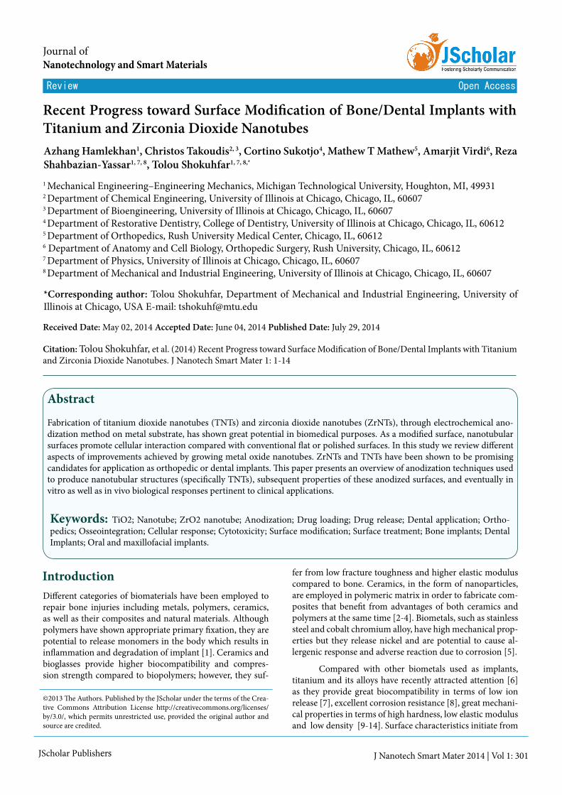
Assisted-template method is performed either by posi-tive or negative templates. A positive template is used for coat-ing oxide layer on the outer surface of template while a nega-tive template is used to coat its inside porosities [35]. For both types of templates, Anodic Aluminum Oxide (AAO) mem-brane is commonly used as template which holds scattered cylindrical pores with uniform dimensions in its structure (Figure 1) [36-39].
Figure 1: Fabrication of nanotubes via a porous hard template such as AAO.
Hydrothermal treatmentHydrothermal treatment method is started by NaOH
treatment of TiO2 nanoparticles. Electrostatic repulsion of the charge on sodium results in extension of TiO2 nanopar-ticles to form nano-sheets. After washing with HCl, electro-static charges are removed and sheets scroll to become TiO2 nanotubes [37]. A major advantage of this method is obtaining pure phase TiO2 nanotubes with good crystallinity [36]. Dis-advantages include long reaction times and the application of NaOH, which can cause production of nanotubes that are in powder form of random alignment [36, 37].
AnodizationIn this review we have focused on anodization as the
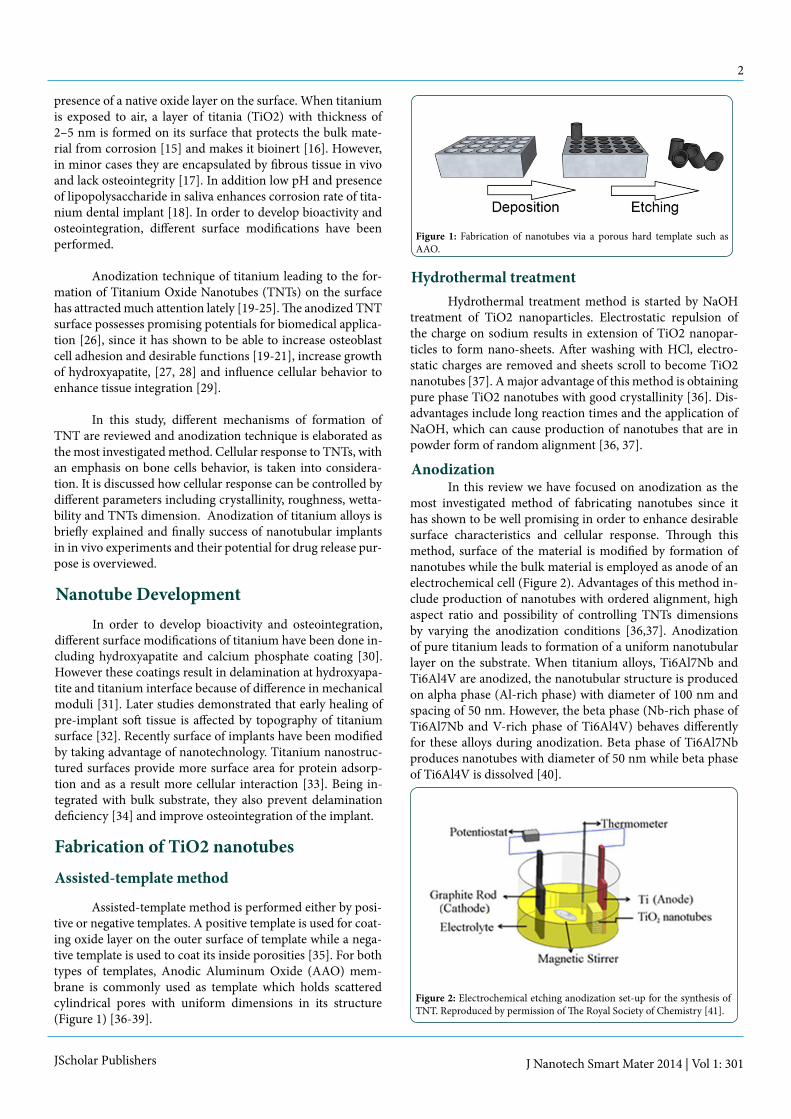
most investigated method of fabricating nanotubes since it has shown to be well promising in order to enhance desirable surface characteristics and cellular response. Through this method, surface of the material is modified by formation of nanotubes while the bulk material is employed as anode of an electrochemical cell (Figure 2). Advantages of this method in-clude production of nanotubes with ordered alignment, high aspect ratio and possibility of controlling TNTs dimensions by varying the anodization conditions [36,37]. Anodization of pure titanium leads to formation of a uniform nanotubular layer on the substrate. When titanium alloys, Ti6Al7Nb and Ti6Al4V are anodized, the nanotubular structure is produced on alpha phase (Al-rich phase) with diameter of 100 nm and spacing of 50 nm. However, the beta phase (Nb-rich phase of Ti6Al7Nb and V-rich phase of Ti6Al4V) behaves differently for these alloys during anodization. Beta phase of Ti6Al7Nb produces nanotubes with diameter of 50 nm while beta phase of Ti6Al4V is dissolved [40].
Figure 2: Electrochemical etching anodization set-up for the synthesis of TNT. Reproduced by permission of The Royal Society of Chemistry [41].
3
JScholar Publishers J Nanotech Smart Mater 2014 | Vol 1: 301
Mechanism of nanotube formation during anodi-zation
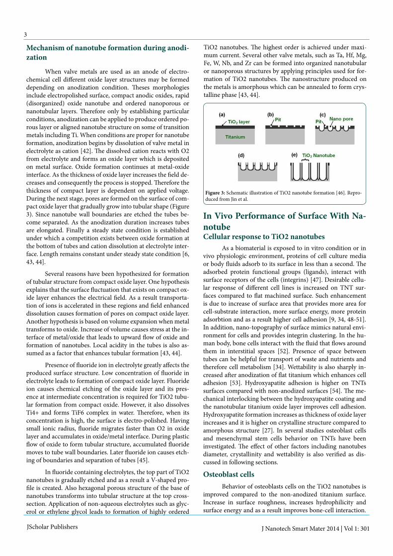
When valve metals are used as an anode of electro-chemical cell different oxide layer structures may be formed depending on anodization condition. Theses morphologies include electropolished surface, compact anodic oxides, rapid (disorganized) oxide nanotube and ordered nanoporous or nanotubular layers. Therefore only by establishing particular conditions, anodization can be applied to produce ordered po-rous layer or aligned nanotube structure on some of transition metals including Ti. When conditions are proper for nanotube formation, anodization begins by dissolution of valve metal in electrolyte as cation [42]. The dissolved cation reacts with O2 from electrolyte and forms an oxide layer which is deposited on metal surface. Oxide formation continues at metal-oxide interface. As the thickness of oxide layer increases the field de-creases and consequently the process is stopped. Therefore the thickness of compact layer is dependent on applied voltage. During the next stage, pores are formed on the surface of com-pact oxide layer that gradually grow into tubular shape (Figure 3). Since nanotube wall boundaries are etched the tubes be-come separated. As the anodization duration increases tubes are elongated. Finally a steady state condition is established under which a competition exists between oxide formation at the bottom of tubes and cation dissolution at electrolyte inter-face. Length remains constant under steady state condition [6, 43, 44].
Several reasons have been hypothesized for formation of tubular structure from compact oxide layer. One hypothesis explains that the surface fluctuation that exists on compact ox-ide layer enhances the electrical field. As a result transporta-tion of ions is accelerated in these regions and field enhanced dissolution causes formation of pores on compact oxide layer. Another hypothesis is based on volume expansion when metal transforms to oxide. Increase of volume causes stress at the in-terface of metal/oxide that leads to upward flow of oxide and formation of nanotubes. Local acidity in the tubes is also as-sumed as a factor that enhances tubular formation [43, 44].
Presence of fluoride ion in electrolyte greatly affects the produced surface structure. Low concentration of fluoride in electrolyte leads to formation of compact oxide layer. Fluoride ion causes chemical etching of the oxide layer and its pres-ence at intermediate concentration is required for TiO2 tubu-lar formation from compact oxide. However, it also dissolves Ti4+ and forms TiF6 complex in water. Therefore, when its concentration is high, the surface is electro-polished. Having small ionic radius, fluoride migrates faster than O2 in oxide layer and accumulates in oxide/metal interface. During plastic flow of oxide to form tubular structure, accumulated fluoride moves to tube wall boundaries. Later fluoride ion causes etch-ing of boundaries and separation of tubes [45].
In fluoride containing electrolytes, the top part of TiO2 nanotubes is gradually etched and as a result a V-shaped pro-file is created. Also hexagonal porous structure of the base of nanotubes transforms into tubular structure at the top cross-section. Application of non-aqueous electrolytes such as glyc-erol or ethylene glycol leads to formation of highly ordered
TiO2 nanotubes. The highest order is achieved under maxi-mum current. Several other valve metals, such as Ta, Hf, Mg, Fe, W, Nb, and Zr can be formed into organized nanotubular or nanoporous structures by applying principles used for for-mation of TiO2 nanotubes. The nanostructure produced on the metals is amorphous which can be annealed to form crys-talline phase [43, 44].
Figure 3: Schematic illustration of TiO2 nanotube formation [46]. Repro-duced from Jin et al.
In Vivo Performance of Surface With Na-notubeCellular response to TiO2 nanotubes
As a biomaterial is exposed to in vitro condition or in vivo physiologic environment, proteins of cell culture media or body fluids adsorb to its surface in less than a second. The adsorbed protein functional groups (ligands), interact with surface receptors of the cells (integrins) [47]. Desirable cellu-lar response of different cell lines is increased on TNT sur-faces compared to flat machined surface. Such enhancement is due to increase of surface area that provides more area for cell-substrate interaction, more surface energy, more protein adsorbtion and as a result higher cell adhesion [9, 34, 48-51]. In addition, nano-topography of surface mimics natural envi-ronment for cells and provides integrin clustering. In the hu-man body, bone cells interact with the fluid that flows around them in interstitial spaces [52]. Presence of space between tubes can be helpful for transport of waste and nutrients and therefore cell metabolism [34]. Wettability is also sharply in-creased after anodization of flat titanium which enhances cell adhesion [53]. Hydroxyapatite adhesion is higher on TNTs surfaces compared with non-anodized surfaces [54]. The me-chanical interlocking between the hydroxyapatite coating and the nanotubular titanium oxide layer improves cell adhesion. Hydroxyapatite formation increases as thickness of oxide layer increases and it is higher on crystalline structure compared to amorphous structure [27]. In several studies osteoblast cells and mesenchymal stem cells behavior on TNTs have been investigated. The effect of other factors including nanotubes diameter, crystallinity and wettability is also verified as dis-cussed in following sections.
Osteoblast cellsBehavior of osteoblasts cells on the TiO2 nanotubes is
improved compared to the non-anodized titanium surface. Increase in surface roughness, increases hydrophilicity and surface energy and as a result improves bone-cell interaction.

4
JScholar Publishers J Nanotech Smart Mater 2013 | Vol 1: 301
Experiments also show that filopodia of the osteoblasts grow into nanotube porosities and provides an integrated structure (Figure 4) [53]. Effect of nanotube structure on attachment, growth and differentiation of human osteoblast is investigated by 3-(4,5-Dimethylthiazol-2-Yl)-2,5-Diphenyltetrazolium Bromide (MTT) assay and Alkaline phosphatase (ALP) meas-urement in previous experiments. Results show that surface modification increases cell adhesion, cell proliferation and os-teoblast expression. Cells seeded on nanotube structure show filamentous network structure and formation of nodules and increased Extracellular Matrix (ECM) [10]. The possibility of enhancing the TiO2 nanotubes bioactivity is verified by expo-sure to NaOH solution. The findings indicate that the sodium titanate nanostructure formed on the edge of the anodized TiO2 nanotubes can increase in vitro hydroxyapatite forma-tion [48].
Figure 4: Human osteoblast cell attaches to nanotubular surface while filopodia penetrates into porosities as anchorage sites [10]. Reproduced with permission from John Wiley and Sons Inc.
Mesenchymal stem cellBeing derived from bone marrow, Mesenchymal Stem
Cells ( MSCs) are pluripotent cells that have the potential to differentiate into different cell types including osteoblasts [55]. It is shown that nanotubes with size range between 15 to 30 nm provide proper substrate for MSC interaction. An increase of focal contact formation was observed on nanotubes smaller than 30 nm and increased cell proliferation and osteoblast dif-ferentiation was observed on 15 nm nanotubes. Also differen-tiation of Hematopoietic Stem Cells (HSC) into osteoclasts in-creased in nanotubes below 30 nm. Similar observations were found from differentiation of MSCs to osteoblasts [53].
Thus the diameter of nanotubes drastically affects cellu-lar response. Previous studies conclude that stem cells dramat-ically respond to change in size of TiO2 nanotubes in range of 15 to 100 nm. More importantly they suggest that small nano-tubes having diameter less than 30 nm, increase cell adhesion, proliferation, migration and integrin clustering/focal contact formation. These reactions tend to decline significantly with increasing pore size [49, 56-59].
Experiments by Schumki et al. led to the conclusion that differentiation, protein aggregation, lamellipodia exten-sion and filopodia extension was higher on smaller nanotubes
[49, 57-59]. However, a recent study concludes that differentia-tion, protein aggregation, lamellipodia extension and filipodia extension increases as nanotube diameter increases [56]. Au-thors of this study assume that on 100 nm nanotubes, hMSCs need to struggle to find TiO2 region where more protein ag-gregates have been deposited. Therefore they form more elon-gated shape and their filopodia is extended. These results are compatible with results of McBeath et al. who reported that decreasing cell density increases osteoblastic differentiation. These data are also compatible with the hypothesized concept that increasing physical stress increases stem cell differentia-tion.
Bauer et al. investigated the effect of change in dimen-sion of nanotubes on MSCs response attachment and prolif-eration. They concluded that change in size is more effective on cell response compared to change in surface chemistry and length size [36].
Chondrocyte
Similar to osteoblasts, chondrocytes attachment on anodized TiO2 nanotubes increases compared with unano-dized Ti. Nanotubular structure increases surface area and initial protein adsorption, therefore interaction and adhesion of chondrocyte is increased. Glycosaminoglycan secretion in the culture medium is reported to increase and chondrogenic markers such as aggrecan and collagen type II are also shown in higher level. Although the cells produced dense ECM fibrils, they retained their circular morphology [34, 60].
Fibroblast and keratinocyte
Biomaterials that are implanted as transcutaneous device interact with both the fibroblasts of dermal (internal) layer of the skin, and keratinocytes of the epidermal (exter-nal) layer. The responses of fibroblasts and keratinocytes on TiO2 nanotubes have been investigated to verify its potential for transcutaneous application. Studies indicate that the nano-tube topography provides a proper substrate for interaction of fibroblasts cells but not for keratinocytes cells [61,62].
Compared to the smooth titanium substrate, adhesion of fibroblasts is increased on nanotubular surface while ke-ratinocytes is decreased. Similarly MTT assays show increases of cell proliferation rate for fibroblasts but decreased of prolif-eration for keratinocytes on TiO2 nanotubes substrate com-pared with the smooth substrate. In addition, cytoskeleton re-organization improvement and membrane protein expressions were observed for fibroblasts cells while keratinocytes cells showed lack of cytoskeleton reorganization [61, 62]. Indirect immunofluorescence staining characterizing was performed for specific marker proteins to investigate cell proliferation. The results show an increase in specific marker expression of fibroblasts cells and decrease in specific marker of keratino-cytes [61, 62].
Fibroblast and keratinocyte
TiO2 nanotube structure has the potential to be used as a vascular stent material. As a vascular stent device, a biomate-rial interacts with smooth muscle cells and endothelial cells.
5
JScholar Publishers J Nanotech Smart Mater 2014 | Vol 1: 301
Endothelial cell growth is enhanced on nanotubular morphol-ogy while MOVAS smooth muscle cell show little tendency to proliferate. Nanotubes maintain the differentiated state of muscle cells and their non-proliferative phenotype while they allow arranged changes in endothelial cell locomotion, cy-toskeleton organization and cell-to-cell communication [63]. In order to assess thrombogenicity nanotubular surface, the release of nitrogen oxide and endothelin-1 is investigated in presence of nanotubular structure. Nitrogen oxide causes va-sodilatation and inhibits platelet aggregation while endothe-lin-1 causes vasoconstriction and enhances platelet aggrega-tion. The results show that nitrogen oxide and endothelin-1 release are balanced in a way that nanotubular structure have antithrombotic effect [63].
Schmuki et al [64] assessed differentiation of mesen-chymal cells to endothelial cells and smooth muscle cells on TiO2 nanotubes. In agreement with their previous studies, they concluded that 15 nm diameter maximizes differentiation of mesenchymal cells to endothelial cells and smooth muscle cells.
Antibacterial effects
Anodization of pure titanium and Ti6Al4V alloy sur-faces, results in decrease of bacterial attachment and biofilm formation compared to non-anodized surfaces in vitro and in vivo. Application of higher voltages leads to enhanced an-tibacterial effects. Treatment with high voltage also results in increased proliferation of osteoblasts and fibroblasts [65]. The most robust antibacterial response of TNT surface is reported to be achieved on 80 nm diameter nanotubes after heat treat-ment [66]. Antibacterial property of TNTs is enhanced follow-
ing exposure to UV light illumination [67]. In addition, TNTs can be loaded with antibiotics in order to further reduce bacte-rial adhesion [68].
Effect of anodization parameters
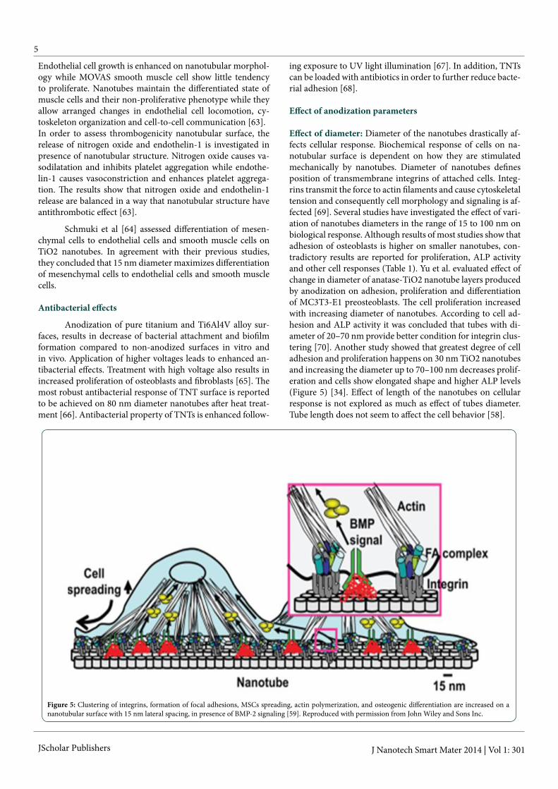
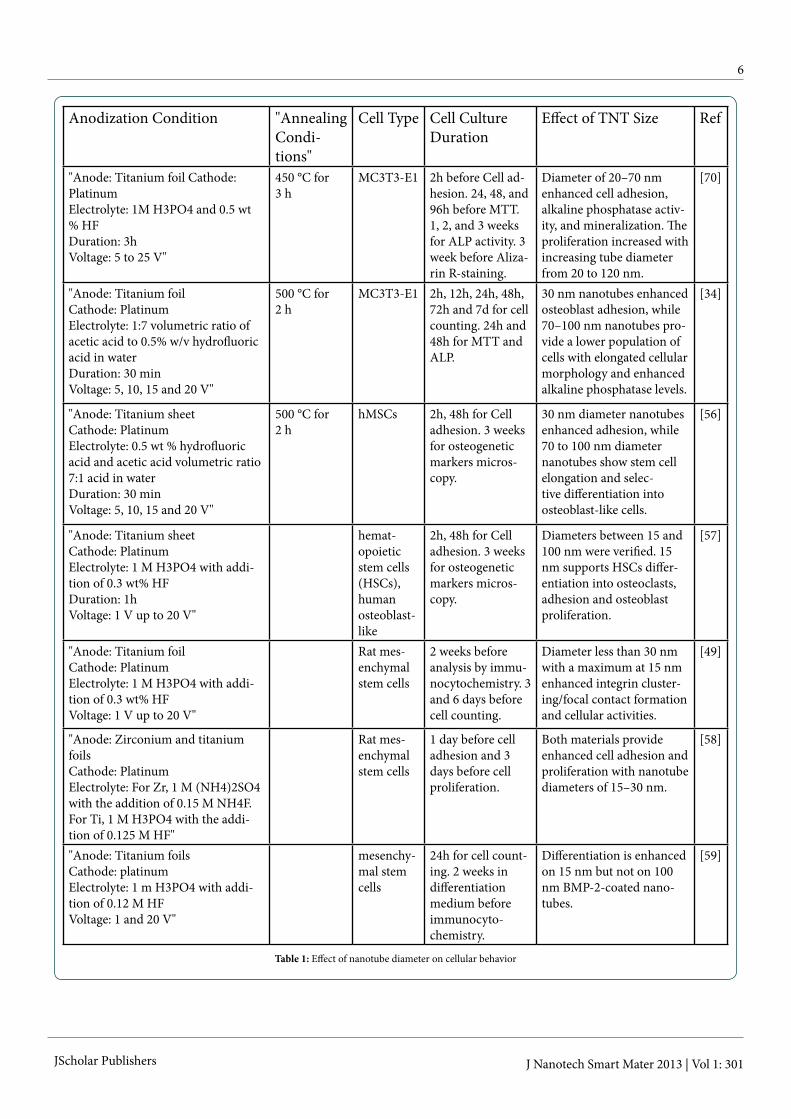
Effect of diameter: Diameter of the nanotubes drastically af-fects cellular response. Biochemical response of cells on na-notubular surface is dependent on how they are stimulated mechanically by nanotubes. Diameter of nanotubes defines position of transmembrane integrins of attached cells. Integ-rins transmit the force to actin filaments and cause cytoskeletal tension and consequently cell morphology and signaling is af-fected [69]. Several studies have investigated the effect of vari-ation of nanotubes diameters in the range of 15 to 100 nm on biological response. Although results of most studies show that adhesion of osteoblasts is higher on smaller nanotubes, con-tradictory results are reported for proliferation, ALP activity and other cell responses (Table 1). Yu et al. evaluated effect of change in diameter of anatase-TiO2 nanotube layers produced by anodization on adhesion, proliferation and differentiation of MC3T3-E1 preosteoblasts. The cell proliferation increased with increasing diameter of nanotubes. According to cell ad-hesion and ALP activity it was concluded that tubes with di-ameter of 20–70 nm provide better condition for integrin clus-tering [70]. Another study showed that greatest degree of cell adhesion and proliferation happens on 30 nm TiO2 nanotubes and increasing the diameter up to 70–100 nm decreases prolif-eration and cells show elongated shape and higher ALP levels (Figure 5) [34]. Effect of length of the nanotubes on cellular response is not explored as much as effect of tubes diameter. Tube length does not seem to affect the cell behavior [58].
Figure 5: Clustering of integrins, formation of focal adhesions, MSCs spreading, actin polymerization, and osteogenic differentiation are increased on a nanotubular surface with 15 nm lateral spacing, in presence of BMP-2 signaling [59]. Reproduced with permission from John Wiley and Sons Inc.
6
JScholar Publishers J Nanotech Smart Mater 2013 | Vol 1: 301
Anodization Condition "Annealing Condi-tions"
Cell Type Cell Culture Duration
Effect of TNT Size Ref
"Anode: Titanium foil Cathode: Platinum Electrolyte: 1M H3PO4 and 0.5 wt % HF Duration: 3h Voltage: 5 to 25 V"
450 °C for 3 h
MC3T3-E1 2h before Cell ad-hesion. 24, 48, and 96h before MTT. 1, 2, and 3 weeks for ALP activity. 3 week before Aliza-rin R-staining.
Diameter of 20–70 nm enhanced cell adhesion, alkaline phosphatase activ-ity, and mineralization. The proliferation increased with increasing tube diameter from 20 to 120 nm.
[70]
"Anode: Titanium foil Cathode: Platinum Electrolyte: 1:7 volumetric ratio of acetic acid to 0.5% w/v hydrofluoric acid in water Duration: 30 min Voltage: 5, 10, 15 and 20 V"
500 °C for 2 h
MC3T3-E1 2h, 12h, 24h, 48h, 72h and 7d for cell counting. 24h and 48h for MTT and ALP.
30 nm nanotubes enhanced osteoblast adhesion, while 70–100 nm nanotubes pro-vide a lower population of cells with elongated cellular morphology and enhanced alkaline phosphatase levels.
[34]
"Anode: Titanium sheet Cathode: Platinum Electrolyte: 0.5 wt % hydrofluoric acid and acetic acid volumetric ratio 7:1 acid in water Duration: 30 min Voltage: 5, 10, 15 and 20 V"
500 °C for 2 h
hMSCs 2h, 48h for Cell adhesion. 3 weeks for osteogenetic markers micros-copy.
30 nm diameter nanotubes enhanced adhesion, while 70 to 100 nm diameter nanotubes show stem cell elongation and selec-tive differentiation into osteoblast-like cells.
[56]
"Anode: Titanium sheet Cathode: Platinum Electrolyte: 1 M H3PO4 with addi-tion of 0.3 wt% HF Duration: 1h Voltage: 1 V up to 20 V"
hemat-opoietic stem cells (HSCs), human osteoblast-like
2h, 48h for Cell adhesion. 3 weeks for osteogenetic markers micros-copy.
Diameters between 15 and 100 nm were verified. 15 nm supports HSCs differ-entiation into osteoclasts, adhesion and osteoblast proliferation.
[57]
"Anode: Titanium foil Cathode: Platinum Electrolyte: 1 M H3PO4 with addi-tion of 0.3 wt% HF Voltage: 1 V up to 20 V"
Rat mes-enchymal stem cells
2 weeks before analysis by immu-nocytochemistry. 3 and 6 days before cell counting.
Diameter less than 30 nm with a maximum at 15 nm enhanced integrin cluster-ing/focal contact formation and cellular activities.
[49]
"Anode: Zirconium and titanium foils Cathode: Platinum Electrolyte: For Zr, 1 M (NH4)2SO4 with the addition of 0.15 M NH4F. For Ti, 1 M H3PO4 with the addi-tion of 0.125 M HF"
Rat mes-enchymal stem cells
1 day before cell adhesion and 3 days before cell proliferation.
Both materials provide enhanced cell adhesion and proliferation with nanotube diameters of 15–30 nm.
[58]
"Anode: Titanium foils Cathode: platinum Electrolyte: 1 m H3PO4 with addi-tion of 0.12 M HF Voltage: 1 and 20 V"
mesenchy-mal stem cells
24h for cell count-ing. 2 weeks in differentiation medium before immunocyto-chemistry.
Differentiation is enhanced on 15 nm but not on 100 nm BMP-2-coated nano-tubes.
[59]
Table 1: Effect of nanotube diameter on cellular behavior

7
JScholar Publishers J Nanotech Smart Mater 2013 | Vol 1: 301
Effect of crystallinity: Crystallinity, the degree of structural order, is a surface factor that affects cell behavior. Under most anodization conditions as formed TiO2 nanotube have amor-phous structure. Annealing at 450 °C and 600 °C for 3h leads to formation of different crystalline phases of anatase and ru-tile respectively [71-73]. Relative amount of anatase forma-tion is higher for the samples anodized with a higher voltage compared to the samples anodized at lower voltage [65]. Crys-tallized phase of substrate increases hydrophilicity [74]; and consequently, enhances desirable responses of cells cultured on it [75]. MC3T3-E1 preosteoblasts activity and tendency to spread increases as nanotubes amorphous structure changes to pure anatase and is maximized when pure anatase transforms to anatase-rutile. Not only cell proliferation increases with increasing annealing temperature but also apatite mineraliza-tion and corrosion-resistance is maximized on rutile structure (Figure 6) [9, 76]. Highest amount of filopodia extension oc-curs on anatase structure [76] while filopodia formation is maximized on anatase [9]. Cell adhesion increases as amount of present fluoride increases. Annealing nanotubes decreases the amount of fluoride and cell numbers [64].
Figure 6: Schematic illustration of TiO2 nanotube formation [46]. Re-produced from Jin et al.
Transformation from amorphous to anatase structure slightly increases Yang modulus and hardness while trans-formation from anatase to rutile sharply increases theses me-chanical properties. Since high hardness and low Yang modu-lus is desirable for biomedical application, an anatase/rutile structure is suggested to be utilized to optimize mechanical properties [76]. Yang modulus is also influenced by diameter and wall thickness of nanotubes [22].
Effect of roughness: Roughness is increased on nanotube structure compared to smooth titanium as measured by AFM [9]. Effect of surface topography is shown to be higher com-pared to crystallinity and surface chemistry [64]. Increasing voltage of anodization slightly increases surface roughness and biological response is affected to some extent by surface roughness variance in nano scale [65].
Roughened surface of titanium in micro scale, com-pared to flat surface, is anticipated to provide mechanical in-terlocking for long time. In addition cell functions such as cell
adhesion and gene expression are promoted after acid etching [77]. The micro-nano scale structure produced by anodization of roughened titanium surface mimics structure of natural bone and has shown to increase hydroxyapatite formation and protein adsorption [78].
Effect of wettability: Wettability is another factor that affects osteoblast behavior. Water contact angle is decreased after anodization [65]. Surface of titanium becomes hydrophilic after anodization and hydrophilicity further increases when anodized surface in annealed. Interestingly, nanotubular sur-face loses part of its hyrophilicity when it is exposed to air for a period of three months. Ambient atmosphere affects wet-tability probably through alkane contamination and organic contaminants [79]. Super-hydrophilic TiO2 nanotube become hydrophobic when coated with a monolayer of octadecylphos-phonic acid. Comparison of mesenchymal stem cells adhe-sion, spreading and growth on the unmodified nanotubes with modified nanotubes shows that coating diminishes effect of tube diameter and hydrophobicity causes decrease of prolif-eration [80].
Figure 7: Staining actin cytoskeleton of MC3T3-E1osteoblast on: (a) smooth surface, (b) non-annealed nanotubular surface, nanotubular surfaces annealed at (c) 450 °C and (d) 550 °C. Compared with the smooth and the non-annealed nanotubular surfaces, annealed surfaces show higher regular arrangement [9]. Reproduced with permission from John Wiley and Sons Inc.
Controlling TNTs DimensionsTNT dimensions can be controlled by optimizing dif-
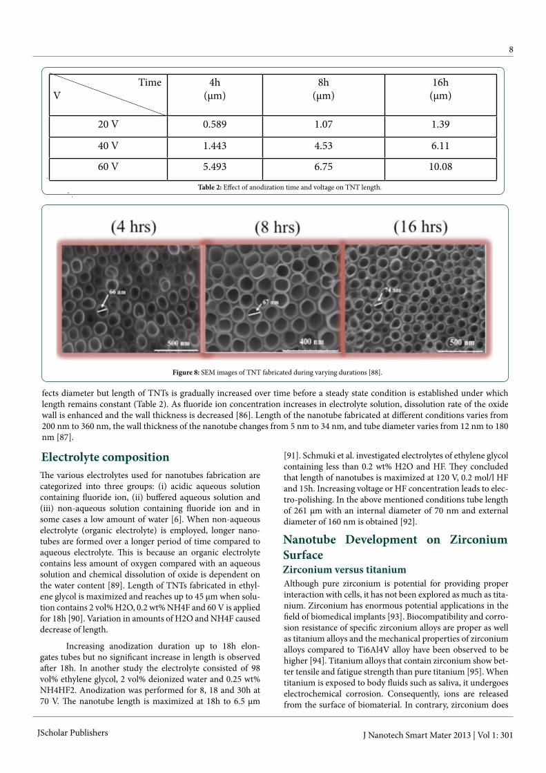
ferent parameters including electrolyte composition, electro-lyte pH, type of electrolyte, voltage magnitude and anodiza-tion duration [34, 81-83]. Also agitation speed of electrolyte, temperature and the ratio of cathode-to-anode surface area affect morphology of TNTs [84]. Diameter of TNTs increases as either applied voltage or anodization duration increases (Figure 8). Length of TNTs can be increased decreasing acidity and fluoride concentration [44]. As pH is increased the time taken for nanotube formation increases; therefore, fabricated nanotubes are longer [85]. Anodization duration slightly af-
8
JScholar Publishers J Nanotech Smart Mater 2013 | Vol 1: 301
TimeV
4h(μm)
8h(μm)
16h(μm)
20 V 0.589 1.07 1.39
40 V 1.443 4.53 6.11
60 V 5.493 6.75 10.08
Table 2: Effect of anodization time and voltage on TNT length.
fects diameter but length of TNTs is gradually increased over time before a steady state condition is established under which length remains constant (Table 2). As fluoride ion concentration increases in electrolyte solution, dissolution rate of the oxide wall is enhanced and the wall thickness is decreased [86]. Length of the nanotube fabricated at different conditions varies from 200 nm to 360 nm, the wall thickness of the nanotube changes from 5 nm to 34 nm, and tube diameter varies from 12 nm to 180 nm [87].
Figure 8: SEM images of TNT fabricated during varying durations [88].
Table 2: Effect of anodization time and voltage on TNT length.
Electrolyte compositionThe various electrolytes used for nanotubes fabrication are categorized into three groups: (i) acidic aqueous solution containing fluoride ion, (ii) buffered aqueous solution and (iii) non-aqueous solution containing fluoride ion and in some cases a low amount of water [6]. When non-aqueous electrolyte (organic electrolyte) is employed, longer nano-tubes are formed over a longer period of time compared to aqueous electrolyte. This is because an organic electrolyte contains less amount of oxygen compared with an aqueous solution and chemical dissolution of oxide is dependent on the water content [89]. Length of TNTs fabricated in ethyl-ene glycol is maximized and reaches up to 45 µm when solu-tion contains 2 vol% H2O, 0.2 wt% NH4F and 60 V is applied for 18h [90]. Variation in amounts of H2O and NH4F caused decrease of length.
Increasing anodization duration up to 18h elon-gates tubes but no significant increase in length is observed after 18h. In another study the electrolyte consisted of 98 vol% ethylene glycol, 2 vol% deionized water and 0.25 wt% NH4HF2. Anodization was performed for 8, 18 and 30h at 70 V. The nanotube length is maximized at 18h to 6.5 µm
[91]. Schmuki et al. investigated electrolytes of ethylene glycol containing less than 0.2 wt% H2O and HF. They concluded that length of nanotubes is maximized at 120 V, 0.2 mol/l HF and 15h. Increasing voltage or HF concentration leads to elec-tro-polishing. In the above mentioned conditions tube length of 261 μm with an internal diameter of 70 nm and external diameter of 160 nm is obtained [92].
Nanotube Development on Zirconium SurfaceZirconium versus titaniumAlthough pure zirconium is potential for providing proper interaction with cells, it has not been explored as much as tita-nium. Zirconium has enormous potential applications in the field of biomedical implants [93]. Biocompatibility and corro-sion resistance of specific zirconium alloys are proper as well as titanium alloys and the mechanical properties of zirconium alloys compared to Ti6Al4V alloy have been observed to be higher [94]. Titanium alloys that contain zirconium show bet-ter tensile and fatigue strength than pure titanium [95]. When titanium is exposed to body fluids such as saliva, it undergoes electrochemical corrosion. Consequently, ions are released from the surface of biomaterial. In contrary, zirconium does
9
JScholar Publishers J Nanotech Smart Mater 2013 | Vol 1: 301
not show undesirable electrochemical characteristics. Also zirconium color is similar to tooth while titanium has a gray shine [96].
In a comparative animal study, zirconium implants demonstrated identical osseointegration as titanium implants. In addition, no significant difference was observed between healing of the tissues interacting with the materials [96].
ZrO2 nanotube formationSince presence of nanostructure on implant surface is
shown to enhance desirable cellular response, fabrication of ZrO2 nanotubes by anodization is studied, and the influence of various electrochemical factors have been evaluated includ-ing potential of power source and its sweep rate, electrolyte composition and anodization time [93]. Pre-anodization of zirconium is reported to be beneficent to form highly ordered ZrO2 nanotubes. Also ZrO2 nanotube structures that are fab-ricated on electropolished zirconium show more uniformity [93]. When organic electrolytes are employed for anodization, the nanostructure formed is thicker, more regular and less wavy [97-100].
Microstructure of substrate influences anodic oxidiza-tions and eventually affects fabrication of ZrO2 nanotubes. Following surface mechanical attrition treatment, commer-cially pure zirconium has nanocrystallized surface layers with high density of grain boundaries compared with non-treated zirconium. Nanocrystallized zirconium is beneficent to the formation of ZrO2 nanotubes and grain boundaries are effec-tive in accelerating reaction rate. ZrO2 nanotube layer formed on the treated zirconium is considerably thicker than that formed on the non-treated zirconium. Thickness increases also with increase in anodization duration and follows a para-bolic function [93].
The SEM images show that nanotubes are gradually formed on the flat surface similar to formation of TiO2 nano-tubes. Self-organization is suggested to be product of compe-tition between growing pores and elsewhere is suggested to be result of local surface perturbations. Localized dissolution of ZrO2 causes formation of pores and reduction of oxide layer thickness. As a result, the electrical field intensity is increased at the base of pore and creation of new oxide is induced. In ad-dition, similar to growth mechanism of nanotube on titanium, ZrO2 nanotube formation in electrolytes that contain fluoride is the outcome of a competition between oxide formation and its chemical dissolution by fluoride ions [93].
Different elements that are present in Ti15Nb4Ta4Zr (TNTZ) alloy have different electrochemical oxidation rates. Therefore reaction rate of TNTZ anodization depends on its composition. Different sizes of self-organized nanotubes are formed on the surface. Eight tubes with small diameter sur-round a tube of larger diameter [101]. Anodization of Ti28Z-r8Nb alloy leads to formation of tubes with 98 nm diameter that surround a larger tube of 175 nm diameter [102]. Nano-tubes that formed on TiZr alloy exhibit uniform arrays; how-ever, as the zirconium content was increased the diameter of the tubes decreased and the length increased [103].
ZrO2 nanotube formationFor the osteoblasts cells cultured on ZrO2 nanotubes
the initial adhesion, spreading, growth, functionality in terms of alkaline phosphatase activity and the formation of extracel-lular matrix is reported to considerably improve as compared with smooth zirconium surface. The cells attached on the nanotube surface demonstrated a high cytoskeleton organi-zation, which was lacking on the flat zirconium [94]. Mesen-chymal stem cells respond identical to ZrO2 nanotubes, TiO2 nanotubes and AuPd-coated TiO2 nanotubes. Cell response is chiefly based on nano-topographical features rather than a certain surface chemistry related to TiO2 [94].
Bone Implant Contact (BIC) and Bone Mineral Deposition (BMD)
Frandsen et al. compared osseointegration of titanium-zirconium (TiZr) with pure titanium and Ti6Al4V alloy. Al-though the formation of new bone inside the implant grooves increased over time regardless of the implant material; how-ever, the amount of Bone to Implant Contact (BIC) was shown to be a function of the implant material. For TiZr and pure ti-tanium implants, the BIC increased gradually but for Ti6Al4V implants the BIC peaked after 2 weeks followed by a decline after 8 weeks. On surface of Ti6Al4V implants, considerably more coverage by multinucleated giant cells was observed. Briefly, TiZr and pure titanium implants showed earlier osse-ointegration compared with Ti6Al4V implants. Maturation of bone marrow next to Ti6Al4V implants was observed to be less advanced compared to TiZr and pure titanium implants [94]. Lack of considerable difference between BIC of titanium and zirconium were also detected in another in vivo study while BIC was affected by roughness. A considerably higher BIC was observed for zirconium implants with regular rough-ness compared with low and high roughness implants [96]. In addition, bone mineral density (BMD) around TNTZ alloy is observed to be similar to Ti6Al4V [104].
Bone Implant Contact (BIC) and Bone Mineral Deposition (BMD)
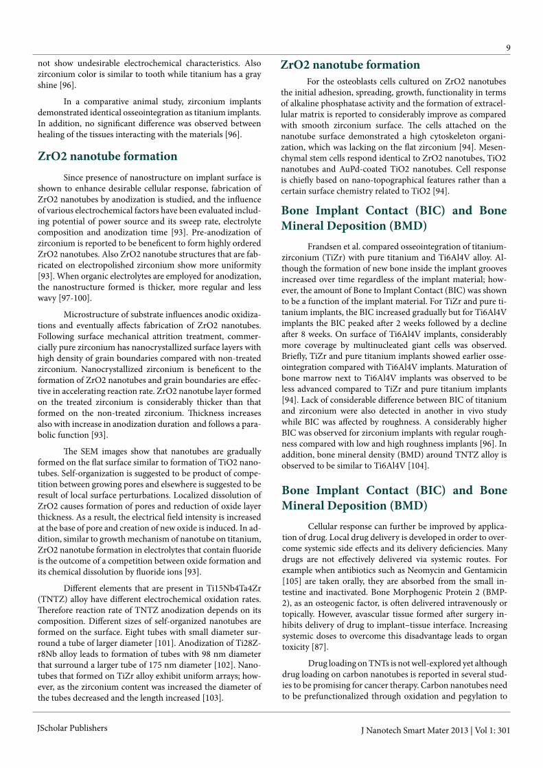
Cellular response can further be improved by applica-tion of drug. Local drug delivery is developed in order to over-come systemic side effects and its delivery deficiencies. Many drugs are not effectively delivered via systemic routes. For example when antibiotics such as Neomycin and Gentamicin [105] are taken orally, they are absorbed from the small in-testine and inactivated. Bone Morphogenic Protein 2 (BMP-2), as an osteogenic factor, is often delivered intravenously or topically. However, avascular tissue formed after surgery in-hibits delivery of drug to implant–tissue interface. Increasing systemic doses to overcome this disadvantage leads to organ toxicity [87].
Drug loading on TNTs is not well-explored yet although drug loading on carbon nanotubes is reported in several stud-ies to be promising for cancer therapy. Carbon nanotubes need to be prefunctionalized through oxidation and pegylation to
10
JScholar Publishers J Nanotech Smart Mater 2013 | Vol 1: 301
become water soluble and biocompatible. Then further func-tionalization also is needed to attach the drug onto carbon nanotubes and target them toward cancer tissue. After being taken up by the cell, carbon nanotubes release their cargo and later they are eliminated from the body [106-112]. However, application of carbon nanotubes is restricted since they show toxicity and inflammation [113, 114].
Not only TNTs are potential to be loaded with drug and antibacterial agents, but also they are biocompatible and hydrophilic in contrast to carbon nanotubes. Although local drug delivery provides targeted release of drug, control of drug release over time remains to be a challenge. Preventing sudden release of drug after implantation avoids denaturation of drug and enhances its efficiency [115]. Formation of nanotubular structure on machined surfaces increases the amount of load-ed BMP-2 and prolongs drug release [116]. TNTs loaded with BMP-2 were coated with multi-layers of gelatin and chitosan to further retard release [115]. Although controlled release of
drug from TNT was successfully achieved through this tech-nique, the polymeric coating that is used in this method pre-vents cellular interaction with surface nanostructure. When titania nanotube arrays are loaded with polymer micelles as drug carriers [117], surface nanostructure can induce cellular response. Since degradation of the polymer may induce in-flammation; it is preferable to avoid its application and change the nanotube dimension for optimizing drug release. Anodi-zation of Ti4Zr22Nb2Sn at different potentials, concentration of NH4F and anodization time shows that longer nanotubes prolong drug release [118]. Release of the antibiotic from elongated TNT formed in an electrolyte based on ethylene glycol is reported to be longer in comparison to release from shorter TNT that are formed in an aqueous solution [91]. The surface of titania nanotubes has a small negative charge due to presence of terminal hydroxyl groups. Therefore positively charged drugs are released slower compared to negatively charged agents [87]. Osteoblast response is also improved when nanotubes are loaded with antibiotics [119].
Figure 9: Intercalation of drug inside the nanotubes. Reproduced by permission of The Royal Society of Chemistry [41].
SummarySince titanium and zirconium provide several ben-
efits compared to other biometals, they are widely used as a biomaterial for fabrication of bone and orthopedic implants. Desirable characteristics of titanium and zirconium alloys in-clude biocompatibility, corrosion resistance and proper me-chanical properties. Biocompatibility and corrosion resistance of specific zirconium alloys are as well as titanium alloys and the mechanical properties of zirconium alloys are higher than Ti alloys. Despite all these benefits, metal implants occasion-ally become loose or infectious after surgery which eventually results in implant failure. In order to overcome this problem, nanotechnology is used to modify the surface and increase osseointegration. Specifically, fabrication of TiO2 nanotubes
through anodization technique on surface of titanium has shown great potential to promote desirable cellular behavior such as adhesion, proliferation and differentiation. In addi-tion, hydroxyapatite mineralization is increased and bacterial adhesion is decreased on nanotubular surfaces compared with conventional smooth surfaces. Recent studies have success-fully optimized properties of nanotubular surfaces to further increase osseointegration. In summary, it is concluded that nanotubes dimensions, heat treatment and drug loading can individually dictates cellar fate.
SummaryThe authors would like to thank Prof. Craig Friedrich,
MTU, Multi-Scale Technologies Institute for all of his contri-butions. The author would like to acknowledge the support of-
11
JScholar Publishers J Nanotech Smart Mater 2013 | Vol 1: 301
19) Webster TJ, Ejiofor JU (2004) Increased osteoblast adhesion on nanophase metals: Ti, Ti6Al4V, and CoCrMo. Biomaterials 25: 4731-4739.20) Rajyalakshmi A, Ercan B, Balasubramanian K, Webster TJ (2011) Reduced adhesion of macrophages on anodized titanium with select nanotube surface features. Int J Nanomedicine 6: 1765-1771.21) Yao Chang, Perla Venu, McKenzie Janice L, Slamovich Elliot B, Webster Thomas J (2005) Anodized Ti and Ti(6)Al(4)V Possessing Nanometer Surface Features Enhances Osteoblast Adhesion. Journal of Biomedical Nanotechnology 1: 68-73.22) Shokuhfar T1, Arumugam GK, Heiden PA, Yassar RS, Friedrich C (2009) Direct Compressive Measurements of Individual Titanium Dioxide Nanotubes. Acs Nano 3: 3098-3102. 23) Asthana A, Shokuhfar T, Gao Q, Heiden P, Friedrich C, et al. (2010) A study on the modulation of the electrical transport by me-chanical straining of individual titanium dioxide nanotube. Applied Physics Letters 97.24) Shokuhfar T, Gao Q, Ashtana A, Walzack K, Heiden P, et al. (2010) Structural instabilities in TiO2 nanotubes. Journal of Applied Physics 108. 25) Asthana A, Shokuhfar T, Gao Q, Heiden PA, Yassar RS (2012) De-formation-driven electrical transport in amorphous TiO2 nanotubes. Applied Physics a-Materials Science & Processing 109: 127-132.26) Debmalya Ganguly, Tolou Shokuhfar, Reza Shahbazian-Yassar (2014) Recent Advances in Nanotubes for Orthopedic Implants. J Nanotech Smart Mater 1: 201.27) Tsuchiya H, Macak JM, Müller L, Kunze J, Müller F et al (2006) Hydroxyapatite growth on anodic TiO2 nanotubes. J Biomed Mater Res A 77: 534-541.28) Oh SH, Finõnes RR, Daraio C, Chen LH, Jin S (2005) Growth of nano-scale hydroxyapatite using chemically treated titanium oxide nanotubes. Biomaterials 26: 4938-4943.29) von Wilmowsky C, Bauer S, Roedl S, Neukam FW, Schmuki P, et al. (2012) The diameter of anodic TiO2 nanotubes affects bone forma-tion and correlates with the bone morphogenetic protein-2 expres-sion in vivo. Clin Oral Implants Res 23: 359-366.30) Kim HW, Koh YH, Li LH, Lee S, Kim HE (2004) Hydroxyapatite coating on titanium substrate with titania buffer layer processed by sol-gel method. Biomaterials 25: 2533-2538.31) Bjursten LM, Rasmusson L, Oh S, Smith GC, Brammer KS, et al. (2010) Titanium dioxide nanotubes enhance bone bonding in vivo. J Biomed Mater Res A 92: 1218-1224.32) Yamano S, Al-Sowygh ZH, Gallucci GO, Wada K, Weber HP, et al. (2011) Early peri-implant tissue reactions on different titanium sur-face topographies. Clin Oral Implants Res 22: 815-819.33) Simchi A, Tamjid E, Pishbin F, Boccaccini AR (2011) Recent pro-gress in inorganic and composite coatings with bactericidal capability for orthopaedic applications. Nanomedicine 7: 22-39.34) Brammer KS, Oh S, Cobb CJ, Bjursten LM, van der Heyde H, et al. (2009) Improved bone-forming functionality on diameter-controlled TiO2 nanotube surface. Acta Biomater 5: 3215-3223.35) Changdeuck Bae, Hyunjun Yoo, Sihyeong Kim, Kyungeun Lee, Jiyoung Kim, et al. (2008) Template-directed synthesis of oxide nano-tubes: Fabrication, characterization, and applications. Chemistry of Materials 20: 756-767.36) Nathan Swami, Zhanwu Cui, Lakshmi S Nair (2011) Titania Na-notubes: Novel Nanostructures for Improved Osseointegration. Jour-nal of Heat Transfer-Transactions of the Asme 133.
fered by Dr. William Hendrickson at University of Illinois at Chicago.
References1) Navarro M, Michiardi A, Castaño O, Planell JA (2008) Biomateri-als in orthopaedics. J R Soc Interface 5: 1137-1158. 2) Hamlekhan A, Mozafari M, Nezafati N, Azami M, Hadipour H (2010) A proposed fabrication method of novel PCL-GEL-HAp na-nocomposite scaffolds for bone tissue engineering applications. Ad-vanced Composites Letters 19: 123-130.3) Hamlekhan A, Moztarzadeh F, Mozafari M, Azami M, Nezafati N (2011) Preparation of laminated poly(epsilon-caprolactone)-gelatin-hydroxyapatite nanocomposite scaffold bioengineered via compound techniques for bone substitution. Biomatter 1: 91-101.4) Azhang Hamlehkhan, MM, Nader Nezafati, Mahmoud Azami, Ali Samadikuchaksaraei (2011) Novel Bioactive Poly(ε-caprolactone)-Gelatin-Hydroxyapatite Nanocomposite Scaffolds for Bone Regen-eration. Key Engineering Materials 493 - 494: 909-915.5) Niinomi M (2008) Metallic biomaterials. J Artif Organs 11: 105–110.6) Minagar S, Berndt CC, Wang J, Ivanova E, Wen C (2012) A review of the application of anodization for the fabrication of nanotubes on metal implant surfaces. Acta Biomater 8: 2875-2888.7) Niespodziana K, Jurczyk K, Jurczyk M (2008) The synthesis of titanium alloys for biomedical applications. Rev Adv Mater Sci 18: 236-240.8) Barão VA, Mathew MT, Assunção WG, Yuan JC, Wimmer MA, et al. (2012) Stability of cp-Ti and Ti-6Al-4V alloy for dental implants as a function of saliva pH - an electrochemical study. Clin Oral Implants Res 23: 1055-1062.9) Yu WQ, Zhang YL, Jiang XQ, Zhang FQ (2010) In vitro behavior of MC3T3-E1 preosteoblast with different annealing temperature ti-tania nanotubes. Oral Dis 16: 624-630.10) Das K, Bose S, Bandyopadhyay A, (2009) TiO2 nanotubes on Ti: Influence of nanoscale morphology on bone cell-materials interac-tion. J Biomed Mater Res A 90: 225-237. 11) Chen ZX, Takao Y, Wang WX, Matsubara T, Ren LM (2009) Sur-face characteristics and in vitro biocompatibility of titanium anodized in a phosphoric acid solution at different voltages. Biomed Mater 4.12) Yu WQ, Qiu J, Xu L, Zhang FQ (2009) Corrosion behaviors of TiO2 nanotube layers on titanium in Hank’s solution. Biomed Mater 4.13) Chen GJ, Wang Z, Bai H, Li JM, Cai H (2009) A preliminary study on investigating the attachment of soft tissue onto micro-arc oxidized titanium alloy implants. Biomed Mater 4: 015017.
14) Kim H, Choi SH, Ryu JJ, Koh SY, Park JH (2008) The biocompat-ibility of SLA-treated titanium implants. Biomed Mater 3: 025011.
15) Lausmaa J (1996) Surface spectroscopic characterization of ti-tanium implant materials. Journal of Electron Spectroscopy and Re-lated Phenomena. 81: 343-361.
16) Xuanyong Liua, Paul K Chub, Chuanxian Ding (2004) Surface modification of titanium, titanium alloys, and related materials for biomedical applications. Materials Science & Engineering R-Reports. 47: 49-121.
17) Xixue Hua, Hong Shen, Kegang Shuai, Enwei Zhanga, Yanjie Bai, Yan Cheng , et al. (2011) Surface bioactivity modification of titanium by CO2 plasma treatment and induction of hydroxyapatite: In vitro and in vivo studies. Applied Surface Science 257: 1813-1823.
18) Barão VA, Mathew MT, Assunção WG, Yuan JC, Wimmer MA, et al. (2011) The Role of Lipopolysaccharide on the Electrochemical Behavior of Titanium. Journal of Dental Research 90: 613-618.
37) Hsin-Hung Ou, Shang-Lien Lo (2007) Review of titania nano-tubes synthesized via the hydrothermal treatment: Fabrication, modi-fication, and application. Separation and Purification Technology 58: 179-191.

12
JScholar Publishers J Nanotech Smart Mater 2013 | Vol 1: 301
57) Park J, Bauer S, Schlegel KA, Neukam FW, von der Mark K, et al. (2009) TiO2 Nanotube Surfaces: 15 nm - An Optimal Length Scale of Surface Topography for Cell Adhesion and Differentiation. Small 5: 666-671.58) Sebastian Bauer, Jung Park, Josef Faltenbacher, Steffen Berger, Klaus von der Mark, et al. (2009) Size selective behavior of mesen-chymal stem cells on ZrO2 and TiO2 nanotube arrays. Integrative Biology 1: 525-532.59) Park J, Bauer S, Pittrof A, Killian MS, Schmuki P, et al. (2012) Synergistic Control of Mesenchymal Stem Cell Differentiation by Na-noscale Surface Geometry and Immobilized Growth Factors on TiO2 Nanotubes. Small 8: 98-107.60) Karla Brammera S, Seunghan Oha, Christine Frandsena J, Shyni Varghesec, Sungho Jin (2010) Nanotube surface triggers increased chondrocyte extracellular matrix production. Materials Science & Engineering C-Materials for Biological Applications 30: 518-525.61) Niinomi M (2008) Mechanical biocompatibilities of titanium al-loys for biomedical applications. J Mech Behav Biomed Mater 1: 30-42.62) Kathy Wang (1996) The use of titanium for medical applications in the USA. Materials Science and Engineering a-Structural Materials Properties Microstructure and Processing 213: 134-137.63) Peng L, Eltgroth ML, LaTempa TJ, Grimes CA, Desai TA (2009) The effect of TiO2 nanotubes on endothelial function and smooth muscle proliferation. Biomaterials 30: 1268-1272.64) Park J, Bauer S, Schmuki P, von der Mark K (2009) Narrow Win-dow in Nanoscale Dependent Activation of Endothelial Cell Growth and Differentiation on TiO2 Nanotube Surfaces. Nano Letters 9: 3157-3164.65) Giordano C, Saino E, Rimondini L, Pedeferri MP, Visai L, et al. (2011) Electrochemically induced anatase inhibits bacterial coloni-zation on Titanium Grade 2 and Ti6Al4V alloy for dental and ortho-pedic devices. Colloids Surf B Biointerfaces 88: 648-655.66) Ercan B, Taylor E, Alpaslan E, Webster TJ (2011) Diameter of titanium nanotubes influences anti-bacterial efficacy. Nanotechnol-ogy 29: 11.67) Giordano C, et al. (2004) Titanium for osteointegration: Com-parison between a novel biomimetic treatment and commercially exploited surfaces. Journal of applied biomaterials & biomechanics 2: 35-44.68) Christensen GD , WA Simpson, Younger JJ, BaddourLM, Bar-rett FF, et al. (1985) Adherence of coagulase-negative staphylococci to plastic tissue culture plates: a quantitative model for the adherence of staphylococci to medical devices. J Clin Microbiol 22: 996-1006.69) Chen CS (2008) Mechanotransduction - a field pulling together? (2008) J Cell Sci 121: 3285-3292.70) Yu WQ, Jiang XQ, Zhang FQ, Xu L (2010) The effect of anatase TiO2 nanotube layers on MC3T3-E1 preosteoblast adhesion, prolif-eration, and differentiation. J Biomed Mater Res A 94: 1012-1022.71) Azhang Hamlekhan, Sweetu Patel AB, Dmitry Royhman, Chris-tos Takoudis, Cortino Sukotjo, (2014) Optimization of Anodization and Annealing Condition Enhances TiO2 Nanotubular Surface Hy-drophilicity. TMS 2014 143rd Annual Meeting & Exhibition Annual Meeting Supplemental Proceedings 2014: 221.
38) Chenglin Yan, Jun Liu, Fei Liu, Junshu Wu, Kun Gao , et al. (2008) Tube formation in nanoscale materials. Nanoscale Res Lett 3: 473-480.39) Chenglin Yan, Jun Liu, Dongfeng Xue (2008) Tube Formation in Nanoscale Materials. Nanoscale Research Letters 3: 473-480.40) Macak JM, Tsuchiya H, Taveira L, Ghicov A, Schmuki P (2005) Self-organized nanotubular oxide layers on Ti-6A1-7Nb and Ti-6A1-4V formed by anodization in NH4F solutions. J Biomed Mater Res A 75: 928-933.41) Tolou Shokuhfar,Suman Sinha-Ray, Cortino Sukotjoc, Alexander Yarin L (2013) Intercalation of anti-inflammatory drug molecules within TiO2 nanotubes. Rsc Advances 3: 17380-17386.42) Macak JM, Tsuchiya H, Ghicov A, Yasuda K, Hahn R, et al. (2007) TiO2 nanotubes: Self-organized electrochemical formation, proper-ties and applications. Current Opinion in Solid State & Materials Sci-ence 11: 3-18.43) Poulomi Roy, Steffen Berger, Patrik Schmuki (2011) TiO2 Nano-tubes: Synthesis and Applications. Angewandte Chemie-Internation-al Edition 50: 2904-2939.44) Andrei Ghicova, Patrik Schmuki (2009) Self-ordering electro-chemistry: a review on growth and functionality of TiO2 nanotubes and other self-aligned MOx structures. Chemical Communications 20: 2791-2808.45) Sergiu Albu P, Andrei Ghicov, Saule Aldabergenova, Peter Drech-sel, Darren LeClere , et al. (2008) Formation of Double-Walled TiO2 Nanotubes and Robust Anatase Membranes. Advanced Materials 20: 4135.46) Karla S. Brammer, Seunghan Oh, Christine J. Frandsen, Sungho Jin (2011) Biomaterials and Biotechnology Schemes Utilizing TiO2 Nanotube Arrays. 947) Stephen Massia P (1999) Cell-extracellular matrix interactions relevant to vascular tissue engineering. Tissue Engineering Prosthetic Vascular Grafts. 48) Seunghan Oh, Sungho Jin (2006) Titanium oxide nanotubes with controlled morphology for enhanced bone growth. Materials Science & Engineering C-Biomimetic and Supramolecular Systems 26: 1301-1306.49) Park J, Bauer S, von der Mark K, Schmuki P (2007) Nanosize and vitality: TiO2 nanotube diameter directs cell fate. Nano Letters 7: 1686-1691.50) Nourmohammadzadeh M, Lo JF, Bochenek M, Mendoza-Elias JE, Wang Q (2013) Microfluidic Array with Integrated Oxygenation Control for Real-Time Live-Cell Imaging: Effect of Hypoxia on Physi-ology of Microencapsulated Pancreatic Islets. Analytical Chemistry 85: 11240-11249.51) Eshkeitia A, Narakathua BB, Reddya ASG, Moorthia A, Atashbar MZ (2012) Detection of heavy metal compounds using a novel inkjet printed surface enhanced Raman spectroscopy (SERS) substrate. Sensors and Actuators B-Chemical 171: 705-711.52) Tami AE, Schaffler MB, Knothe Tate ML (2003) Tate, Probing the tissue to subcellular level structure underlying bone’s molecular siev-ing function. Biorheology 40: 577-590.
72) Asthana A, Shokuhfar T, Gao Q, Heiden PA, Friedrich C, et al. (2010) A Real Time Observation of Phase Transition of Anatase TiO2 Nanotubes Into Rutile Nanoparticles by In-Situ Joule Heating Inside Transmission Electron Microscope. Advanced Science Letters 3: 557-562.
53) Tana AW, Pingguan-Murphya B, R. Ahmadb, S.A. Akbar (2012) Review of titania nanotubes: Fabrication and cellular response. Ce-ramics International 38: 4421-4435.54) Oh S, Daraio C, Chen LH, Pisanic TR, Fiñones RR, et al. (2006) Significantly accelerated osteoblast cell growth on aligned TiO2 na-notubes. J Biomed Mater Res A 78: 97-103.55) Brammer KS, Frandsen CJ, Jin S (2012) TiO2 nanotubes for bone regeneration. Trends Biotechnol 30: 315-322.56) Oh S, Brammer KS, Li YS, Teng D, Engler AJ, et al. (2009) Stem cell fate dictated solely by altered nanotube dimension. Proc Natl Acad Sci U S A 106: 2130-2135.
73) Butt A, Hamlekhan A, Patel SB, Royhman D, Sukotjo C (2014) A Novel Investigation of the Formation of TiO₂ Nanotubes on Ther-mally Formed Oxide of Ti-6Al-4V. J Oral Implantol .

13
JScholar Publishers J Nanotech Smart Mater 2013 | Vol 1: 301
93) Lan Zhang, Jianmin Shao, Yong Han (2011) Enhanced anodiza-tion growth of self-organized ZrO2 nanotubes on nanostructured zir-conium. Surface & Coatings Technology 205: 2876-2881.94) Christine J Frandsen, Karla S Brammer, Kunbae Noh, Laura S Connelly, Seunghan Oh, et al.(2011) Zirconium oxide nanotube sur-face prompts increased osteoblast functionality and mineralization. Materials Science & Engineering C-Materials for Biological Applica-tions 31: 1716-1722.95) Saulacic N, Bosshardt DD, Bornstein MM, Berner S, Buser D (2012) Bone apposition to a titanium-zirconium alloy implant, as compared to two other titanium-containing implants. Eur Cell Mater 23: 273-288.96) Mueller CK, Solcher P, Peisker A, Mtsariashvilli M, Schlegel KA, et al. (2013) Analysis of the influence of the macro- and microstruc-ture of dental zirconium implants on osseointegration: a minipig study. Oral Surg Oral Med Oral Pathol Oral Radiol 116: E1-E8.97) Hiroaki Tsuchiyaa, Jan Macaka, Schmuki P (2006) Formation of Self-Organized Zirconia Nanostructure. ECS Trans 1: 351-357.98) Steffen Berger, Josef Faltenbacher, Sebastian Bauer, Patrik Schmu-ki (2008) Enhanced self-ordering of anodic ZrO2 nanotubes in in-organic and organic electrolytes using two-step anodization. Physica Status Solidi-Rapid Research Letters 2: 102-104.99) Steffen Berger, Florian Jakubka, Patrik Schmuki (2008) Forma-tion of hexagonally ordered nanoporous anodic zirconia. Electro-chemistry Communications 10: 1916-1919.100) Yeonmi Shin, Seonghoon Lee (2009) A freestanding membrane of highly ordered anodic ZrO2 nanotube arrays. Nanotechnology 20.101) Tsuchiya H, Macak JM, Ghicov A, Schmuki P (2006) Self-or-ganization of anodic nanotubes on two size scales. Small 2: 888-891.102) Feng XJ, Macak JM, Albu SP, Schmuki P (2008) Electrochemi-cal formation of self-organized anodic nanotube coating on Ti-28Zr-8Nb biomedical alloy surface. Acta Biomaterialia 4: 318-323.103) Hiroaki Tsuchiyaa, Toshifumi Akakia, Junji Nakataa, Daisuke Teradab, Nobuhiro Tsuji, et al. (2009) Metallurgical aspects on the formation of self-organized anodic oxide nanotube layers. Electro-chimica Acta 54: 5155-5162.
104) Hiroshi Nakada, Yasuko Numata, Toshiro Sakae, Yoshimitsu Okazaki, Yasuhiro Tanimoto, et al. (2008) Comparison of Bone Min-eral Density and Area of Newly Formed Bone Around Ti-15%Zr-4%Nb-4%Ta Alloy and Ti-6%Al-4%V Alloy Implants. Journal of Hard Tissue Biology 17: 99-108.
105) Gulati K, Aw MS, Losic D (2011) Drug-eluting Ti wires with titania nanotube arrays for bone fixation and reduced bone infection. Nanoscale Res Lett 6.
74) Hamlekhan A, Butt A, Patel S, Royhman D, Takoudis C, et al. (2014) Fabrication of Anti-Aging TiO2 Nanotubes on Biomedical Ti Alloys. PLOS ONE 2014.75) Sweetu B Patel, Azhang Hamlekhan, Dmitry Royhman, Arman Butt, Judy Yuan (2014) Enhancing Surface Characteristics of Ti-6Al-4V for Bio-implants Using Integrated Anodization and Thermal Oxi-dation. J Mater Chem.
76) Yu Bai, Song Park, Hyeoung Ho Park, Min Ho Lee, Tae Sung Bae, et al. (2011) The effect of annealing temperatures on surface prop-erties, hydroxyapatite growth and cell behaviors of TiO2 nanotubes. Surface and Interface Analysis 43: 998-1005.77) Zhao L, Mei S, Chu PK, Zhang Y, Wu Z (2010) The influence of hierarchical hybrid micro/nano-textured titanium surface with tita-nia nanotubes on osteoblast functions. Biomaterials 31: 5072-5082.78) Gao L, Feng B, Wang J, Lu X, Liu D, et al. (2009) Micro/Nano-structural Porous Surface on Titanium and Bioactivity. J Biomed Ma-ter Res B Appl Biomater 89: 335-341.79) Shin DH, Shokuhfar T, Choi CK, Lee SH, Friedrich C (2011) Wettability changes of TiO2 nanotube surfaces. Nanotechnology 22.80) Bauer S, Park J, von der Mark K, Schmuki P (2008) Improved attachment of mesenchymal stem cells on super-hydrophobic TiO2 nanotubes. Acta Biomaterialia 4: 1576-1582.81) Dawei Gonga, Craig A Grimes, Oomman K Varghese, Wenchong Hu, Singh RS, et al. (2001) Titanium oxide nanotube arrays prepared by anodic oxidation. Journal of Materials Research 16: 3331-3334.82) Andrei Ghicov, Hiroaki Tsuchiya, Jan M Macak, Patrik Schmuki (2005) Titanium oxide nanotubes prepared in phosphate electrolytes. Electrochemistry Communications 7: 505-509.83) Sebastian Bauer, Sebastian Kleber, Patrik Schmuki (2006) TiO2 nanotubes: Tailoring the geometry in H3PO4/HF electrolytes. Elec-trochemistry Communications 8: 1321-1325.84) Young-Taeg Sul, Carina B Johansson, Yongsoo Jeong, Tomas Al-brektsson (2001) The electrochemical oxide growth behaviour on titanium in acid and alkaline electrolytes. Medical Engineering & Physics 23: 329-346.85) Cai QY, Yang LX, Yu Y (2006) Investigations on the self-organized growth of TiO2 nanotube arrays by anodic oxidization. Thin Solid Films 515: 1802-1806.86) Kouji Yasuda, Patrik Schmuki (2007) Control of morphology and composition of self-organized zirconium titanate nanotubes formed in (NH4)(2)SO4/NH4F electrolytes. Electrochimica Acta. 52: 4053-4061.
106) Maurizio Prato, Kostas Kostarelos, Alberto Bianco (2008) Func-tionalized carbon nanotubes in drug design and discovery. Accounts of Chemical Research 41: 60-68.
87) Popat KC, Eltgroth M, LaTempa TJ, Grimes CA, Desai TA (2007) Titania nanotubes: A novel platform for drug-eluting coatings for medical implants? Small 3: 1878-1881.88) Shokuhfar T (2010) Structural and surface property characteriza-tion of titanium dioxide nanotubes for orthopedic implants. Michi-gan Technological University.89) Narayanana R, Tae-Yub Kwona,Kyo-Han Kim (2009) TiO2 na-notubes from stirred glycerol/NH4F electrolyte: Roughness, wetting behavior and adhesion for implant applications. Materials Chemistry and Physics 117: 460-464.90) Jun Wana, Xia Yan, Junjie Ding, Meng Wang, Kongcheng Hu (2009) Self-organized highly ordered TiO2 nanotubes in organic aqueous system. Materials Characterization 60: 1534-1540.
107) Liu Z, Sun X, Nakayama-Ratchford N, Dai H (2007) Supramo-lecular chemistry on water-soluble carbon nanotubes for drug load-ing and delivery. Acs Nano 1: 50-56.
91) Claus Moseke, Felix Hage, Elke Vorndran, Uwe Gbureck (2012) TiO2 nanotube arrays deposited on Ti substrate by anodic oxidation and their potential as a long-term drug delivery system for antimicro-bial agents. Applied Surface Science 258: 5399-5404.92) Sergiu P. Albu, Andrei Ghicov, Jan M. Macak, Patrik Schmuki (2007) 250 mu m long anodic TiO2 nanotubes with hexagonal self-ordering. Physica Status Solidi-Rapid Research Letters 1: R65-R67.
108) Liu Z, Chen K, Davis C, Sherlock S, Cao Q, et al. (2008) Drug delivery with carbon nanotubes for in vivo cancer treatment. Cancer Res 68: 6652-6660.
109) Rodney P Feazell, Nozomi Nakayama-Ratchford,Hongjie Dai,Stephen J Lippard (2007) Soluble single-walled carbon nano-tubes as longboat delivery systems for Platinum(IV) anticancer drug design. J Am Chem Soc 129: 8438-8439.
110) Kam NW, Liu Z, Dai H (2005) Functionalization of carbon na-notubes via cleavable disulfide bonds for efficient intracellular deliv-ery of siRNA and potent gene silencing. J Am Chem Soc 127: 12492-12493.
14
JScholar Publishers J Nanotech Smart Mater 2013 | Vol 1: 301
111) Chen J, Chen S, Zhao X, Kuznetsova LV, Wong SS (2008) Func-tionalized Single-Walled Carbon Nanotubes as Rationally Designed Vehicles for Tumor-Targeted Drug Delivery. J Am Chem Soc 130: 16778-16785.112) Sun X, Liu Z, Welsher K, Robinson JT, Goodwin A, et al. (2008) Nano-Graphene Oxide for Cellular Imaging and Drug Delivery. Nano Res 1: 203-212.113) Zhao XC, Liu RT (2012) Recent progress and perspectives on the toxicity of carbon nanotubes at organism, organ, cell, and biomacro-molecule levels. Environ Int 40: 244-255.114) Craig Poland A, Rodger Duffin, Ian Kinloch, Andrew Maynard, William Wallace AH, et al. (2008) Carbon nanotubes introduced into the abdominal cavity of mice show asbestos-like pathogenicity in a pilot study. Nature Nanotechnology 3: 423-428.115) Hu Y, Cai K, Luo Z, Xu D, Xie D, et al. (2012) TiO2 nanotubes as drug nanoreservoirs for the regulation of mobility and differentiation of mesenchymal stem cells. Acta Biomaterialia 8: 439-448.116) Bae IH, Yun KD, Kim HS, Jeong BC, Lim HP, et al. (2010) An-odic Oxidized Nanotubular Titanium Implants Enhance Bone Mor-phogenetic Protein-2 Delivery. J Biomed Mater Res B Appl Biomater 93: 484-491.117) Aw MS, Addai-Mensah J, Losic D (2012) A multi-drug delivery system with sequential release using titania nanotube arrays. Chem Commun (Camb) 48: 3348-3350.118) Liang YQ, Cui ZD, Zhu SL, Yang XJ, et al. (2011) Characteri-zation of self-organized TiO2 nanotubes on Ti-4Zr-22Nb-2Sn alloys and the application in drug delivery system. J Mater Sci Mater Med 22: 461-467.119) Popat KC, Eltgroth M, Latempa TJ, Grimes CA, Desai TA (2007) Decreased Staphylococcus epidermis adhesion and increased osteo-blast functionality on antibiotic-loaded titania nanotubes. Biomateri-als 28: 4880-4888.
Submit your manuscript to JScholar journals and benefit from:
¶ Convenient online submission ¶ Rigorous peer review ¶ Immediate publication on acceptance ¶ Open access: articles freely available online ¶ High visibility within the field ¶ Better discount for your subsequent articles
Submit your manuscript at http://www.jscholaronline.org/submit-manuscript.php