reading speed as an objective measure of improvement
TRANSCRIPT
HAL Id: hal-02946884https://hal.inria.fr/hal-02946884
Submitted on 23 Sep 2020
HAL is a multi-disciplinary open accessarchive for the deposit and dissemination of sci-entific research documents, whether they are pub-lished or not. The documents may come fromteaching and research institutions in France orabroad, or from public or private research centers.
L’archive ouverte pluridisciplinaire HAL, estdestinée au dépôt et à la diffusion de documentsscientifiques de niveau recherche, publiés ou non,émanant des établissements d’enseignement et derecherche français ou étrangers, des laboratoirespublics ou privés.
Reading Speed as an Objective Measure of ImprovementFollowing Vitrectomy for Symptomatic Vitreous
OpacitiesEdwin Ryan, Linda Lam, Christine Pulido, Steven Bennett, Aurelie Calabrese
To cite this version:Edwin Ryan, Linda Lam, Christine Pulido, Steven Bennett, Aurelie Calabrese. Reading Speed asan Objective Measure of Improvement Following Vitrectomy for Symptomatic Vitreous Opacities.Ophthalmic Surgery, Lasers and Imaging Retina, Slack 2020. �hal-02946884�
Reading speed as a reliable outcome measure to assess visual 1
improvement following vitrectomy for symptomatic vitreous 2
opacities in patients with clear lenses 3
4
Short title: Reading speed as an objective measure of improvement following vitrectomy for 5
symptomatic vitreous opacities 6
7
Edwin H. Ryan, MD1, Linda A. Lam, MD2, Christine M. Pulido, MD1,3, Steven R. Bennett, 8
MD1, Aurélie Calabrèse, PhD 4 9
10
1- VitreoRetinal Surgery PA, Edina, MN, USA 11
2- USC Keck School of Medicine, Los Angeles, CA, USA 12
3- Feinberg School of Medicine, Chicago, IL, USA 13
4- Université Côte d’Azur, Inria, Sophia-Antipolis, France 14
15
Correspondence to: Aurélie Calabrèse, PhD 16
Université Côte d’Azur, Inria, Sophia Antipolis, France 17
------ 18
2004, route des Lucioles - BP 93 19
06902 Sophia Antipolis Cedex France 20
------ 21
+33 7 61 57 30 37 23
Authors’contribution:24
25
• Study concept and design: Edwin H. Ryan, Aurèlie Calabrèse, Linda Lam 26
• Acquisition of data: Christine Pulido, Edwin H. Ryan, Linda Lam, Steven R. Bennett 27
• Analysis and interpretation of data: Aurèlie Calabrèse, Edwin H. Ryan 28
• Statistical expertise: Aurèlie Calabrèse 29
• Drafting of the manuscript: Aurèlie Calabrèse, Edwin H. Ryan, Linda A. Lam 30
• Critical revision of the manuscript: Aurèlie Calabrèse, Edwin H. Ryan, Linda A. Lam, 31
Christine Pulido, Steven R. Bennett 32
33
Funding34
Dr. Calabrèse reports grants from National Eye Institute/NIH (grant EY002934) and Fondation 35
de France, during the conduct of the study.36
37
DeclarationofConflictingInterests38
The authors declare no potential conflicts of interest with respect to the research, authorship or 39
publication of this article. In addition, Dr. Calabrese reports receiving royalties for sales of the 40
MNREAD iPad app through a licensing agreement between the University of Minnesota and 41

Precision Vision outside of the present work. Dr. Ryan reports royalties for patents related to 42
vitrectomy surgery from Alcon Surgical. 43
44
45
46
47
Abstract48
Background and Objective: There is currently no objective measure of the visual deficits 49
experienced by patients with symptomatic vitreous opacities (SVO) that would also correlate 50
with the functional improvement they report following vitrectomy. This study aims to determine 51
whether reading speed can be used as a reliable outcome measure to assess objectively the 52
impact of both SVO and vitrectomy on patients’ visual performance. 53
Study Design/Materials and Methods: 20 adult patients seeking surgery for SVO were 54
included. Measures of visual function were obtained before and after vitrectomy using the 55
ETDRS acuity chart, the NEI-VFQ and the MNREAD. 56
Results: In patients with non-opacified lenses (N=10), maximum reading speed increased 57
significantly from 138 to 159 words per minute after complete removal of SVO by vitrectomy 58
(95%CI = [14, 29], p < 0.001). 59
Conclusion: Reading speed is impaired with SVO, and improves following vitrectomy in phakic 60
and pseudophakic eyes with clear lenses. Reading speed is a valid objective measure to assess 61
the positive effect of vitrectomy for SVO on near-distance daily life activities. 62
63
Keywords64
65
Vitrectomy, symptomatic vitreous opacities, lens opacity, reading speed, daily-life function, functional 66
improvement, objective measurement 67
68
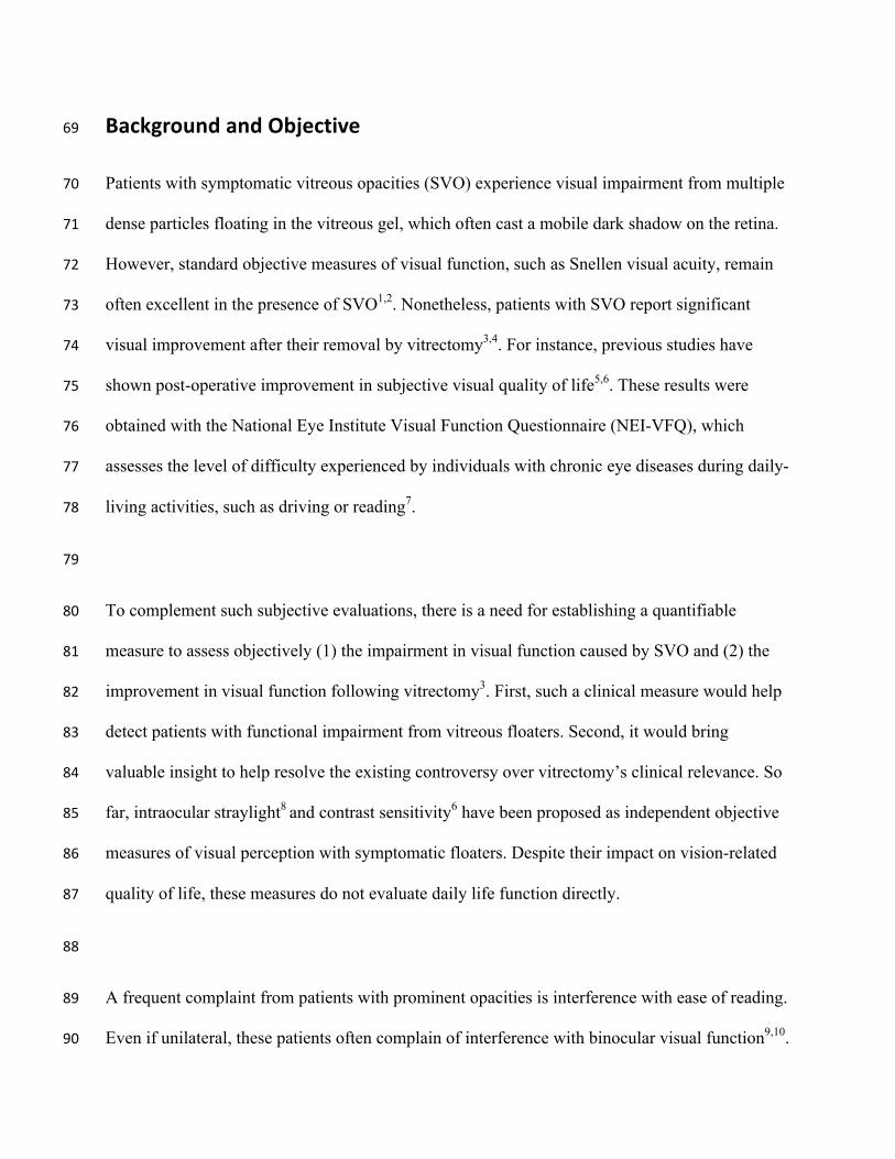
BackgroundandObjective69
Patients with symptomatic vitreous opacities (SVO) experience visual impairment from multiple 70
dense particles floating in the vitreous gel, which often cast a mobile dark shadow on the retina. 71
However, standard objective measures of visual function, such as Snellen visual acuity, remain 72
often excellent in the presence of SVO1,2. Nonetheless, patients with SVO report significant 73
visual improvement after their removal by vitrectomy3,4. For instance, previous studies have 74
shown post-operative improvement in subjective visual quality of life5,6. These results were 75
obtained with the National Eye Institute Visual Function Questionnaire (NEI-VFQ), which 76
assesses the level of difficulty experienced by individuals with chronic eye diseases during daily-77
living activities, such as driving or reading7. 78
79
To complement such subjective evaluations, there is a need for establishing a quantifiable 80
measure to assess objectively (1) the impairment in visual function caused by SVO and (2) the 81
improvement in visual function following vitrectomy3. First, such a clinical measure would help 82
detect patients with functional impairment from vitreous floaters. Second, it would bring 83
valuable insight to help resolve the existing controversy over vitrectomy’s clinical relevance. So 84
far, intraocular straylight8 and contrast sensitivity6 have been proposed as independent objective 85
measures of visual perception with symptomatic floaters. Despite their impact on vision-related 86
quality of life, these measures do not evaluate daily life function directly. 87
88
A frequent complaint from patients with prominent opacities is interference with ease of reading. 89
Even if unilateral, these patients often complain of interference with binocular visual function9,10. 90
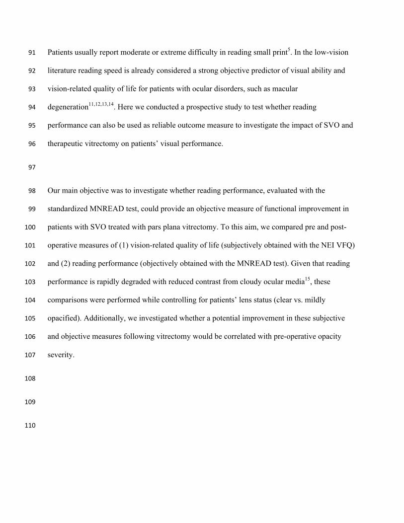
Patients usually report moderate or extreme difficulty in reading small print5. In the low-vision 91
literature reading speed is already considered a strong objective predictor of visual ability and 92
vision-related quality of life for patients with ocular disorders, such as macular 93
degeneration11,12,13,14. Here we conducted a prospective study to test whether reading 94
performance can also be used as reliable outcome measure to investigate the impact of SVO and 95
therapeutic vitrectomy on patients’ visual performance. 96
97
Our main objective was to investigate whether reading performance, evaluated with the 98
standardized MNREAD test, could provide an objective measure of functional improvement in 99
patients with SVO treated with pars plana vitrectomy. To this aim, we compared pre and post-100
operative measures of (1) vision-related quality of life (subjectively obtained with the NEI VFQ) 101
and (2) reading performance (objectively obtained with the MNREAD test). Given that reading 102
performance is rapidly degraded with reduced contrast from cloudy ocular media15, these 103
comparisons were performed while controlling for patients’ lens status (clear vs. mildly 104
opacified). Additionally, we investigated whether a potential improvement in these subjective 105
and objective measures following vitrectomy would be correlated with pre-operative opacity 106
severity. 107
108
109
110

Patients/MaterialsandMethods111
Patients 112
Patients over 21 years old were included in the present work if they (1) elected to undergo 113
vitrectomy, (2) presented symptoms consistent with examination findings of dense opacities for 114
at least 6 months, (3) had visual acuity of 20/80 (0.6 logMAR) or better in both eyes before 115
surgery and (4) did not experience a significant drop in acuity in the non-operated eye between 116
the pre- and post-surgery measurements. Phakic and pseudophakic patients were included, as 117
well as patients with or without a vitreous detachment. History of scleral buckle for retinal 118
detachment (RD) was acceptable if the macula was not involved. If an epiretinal membrane was 119
noted on OCT but not clinically visible or deemed significant, patients were included in the 120
study. Patients were excluded if they had history of cognitive impairment, macula-off RD, severe 121
glaucoma, macular degeneration, diabetic macular edema, or other confounding ocular disorders. 122
A total of 20 patients were recruited, tested and treated at two different sites: 11 at a private 123
retina practice in Minnesota and 9 at an academic retina practice in California. Figure 1 124
illustrates the protocol sequence. Institutional Review Board (IRB)/Ethics Committee approval 125
was obtained and written informed consent was obtained before the study from each patient 126
according to IRB guidelines. The study also complied with tenets of the Declaration of Helsinki 127
and HIPAA. 128
129
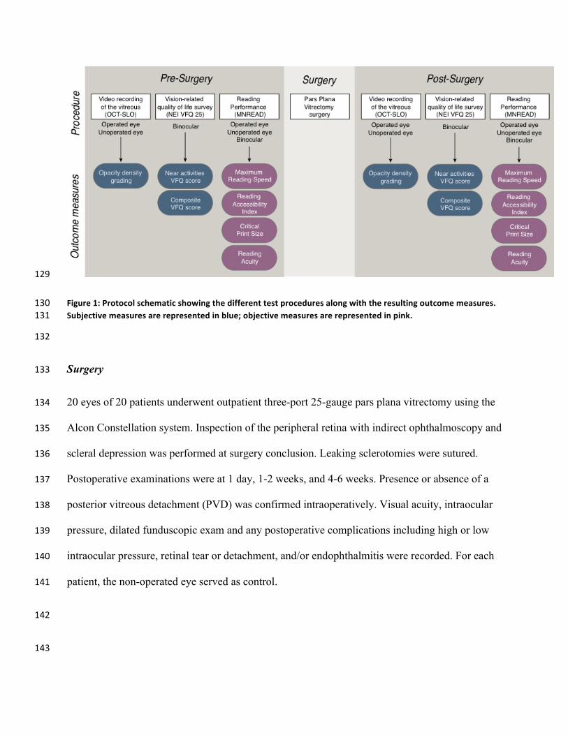
Figure1:Protocolschematicshowingthedifferenttestproceduresalongwiththeresultingoutcomemeasures.130Subjectivemeasuresarerepresentedinblue;objectivemeasuresarerepresentedinpink. 131
132
Surgery 133
20 eyes of 20 patients underwent outpatient three-port 25-gauge pars plana vitrectomy using the 134
Alcon Constellation system. Inspection of the peripheral retina with indirect ophthalmoscopy and 135
scleral depression was performed at surgery conclusion. Leaking sclerotomies were sutured. 136
Postoperative examinations were at 1 day, 1-2 weeks, and 4-6 weeks. Presence or absence of a 137
posterior vitreous detachment (PVD) was confirmed intraoperatively. Visual acuity, intraocular 138
pressure, dilated funduscopic exam and any postoperative complications including high or low 139
intraocular pressure, retinal tear or detachment, and/or endophthalmitis were recorded. For each 140
patient, the non-operated eye served as control. 141
142
143
Subjective grading of opacity density 144
Before and after surgery, patients went through video recording of the vitreous using the infrared 145
confocal scanning laser ophthalmoscope (SLO) combined with optical coherence tomography 146
(OCT)16,17. The movie created with this technique reveals motion of the shadows projected by 147
the opacities onto the retinal surface, enabling a subjective grading of the opacity density. 148
Recording was performed in each eye. Patients were instructed to look to one side and then re-149
fixate, which set the vitreous opacities in motion. This step was repeated to each side several 150
times. The pre and post-surgery videos were assessed by two experienced, masked surgeons and 151
given a score of 0-3, with 0 corresponding to no floaters and 3 corresponding to very dense 152
floaters (see supplementary material for a pre-op movie graded as 2 and a post-op movie graded 153
as 0). 154
155
Subjective measure of vision-related quality-of-life 156
Before and after surgery, patients were interviewed with the NEI-VFQ-25, the 25-item version of 157
the VFQ test7. Data were scored using the standard method to calculate: 1-the near activities 158
VFQ score (involving reading) and 2- the composite VFQ score (encompassing many vision-159
related functions). Scores ranged from 0 to 100, with higher scores representing better function. 160
161
Objective measure of reading speed 162
Before and after surgery, patients’ reading performance was measured with the MNREAD acuity 163
chart, a standardized test designed to measure binocular and monocular reading performance18. 164
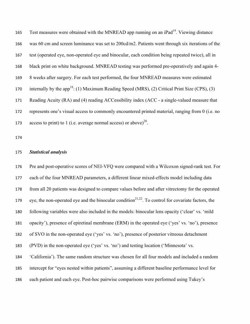
Test measures were obtained with the MNREAD app running on an iPad19. Viewing distance 165
was 60 cm and screen luminance was set to 200cd/m2. Patients went through six iterations of the 166
test (operated eye, non-operated eye and binocular, each condition being repeated twice), all in 167
black print on white background. MNREAD testing was performed pre-operatively and again 4-168
8 weeks after surgery. For each test performed, the four MNREAD measures were estimated 169
internally by the app18: (1) Maximum Reading Speed (MRS), (2) Critical Print Size (CPS), (3) 170
Reading Acuity (RA) and (4) reading ACCessibility index (ACC - a single-valued measure that 171
represents one’s visual access to commonly encountered printed material, ranging from 0 (i.e. no 172
access to print) to 1 (i.e. average normal access) or above)20. 173
174
Statistical analysis 175
Pre and post-operative scores of NEI-VFQ were compared with a Wilcoxon signed-rank test. For 176
each of the four MNREAD parameters, a different linear mixed-effects model including data 177
from all 20 patients was designed to compare values before and after vitrectomy for the operated 178
eye, the non-operated eye and the binocular condition21,22. To control for covariate factors, the 179
following variables were also included in the models: binocular lens opacity (‘clear’ vs. ‘mild 180
opacity’), presence of epiretinal membrane (ERM) in the operated eye (‘yes’ vs. ‘no’), presence 181
of SVO in the non-operated eye (‘yes’ vs. ‘no’), presence of posterior vitreous detachment 182
(PVD) in the non-operated eye (‘yes’ vs. ‘no’) and testing location (‘Minnesota’ vs. 183
‘California’). The same random structure was chosen for all four models and included a random 184
intercept for “eyes nested within patients”, assuming a different baseline performance level for 185
each patient and each eye. Post-hoc pairwise comparisons were performed using Tukey’s 186

correction. In the Results section, mean values estimated by the models and post-hoc pairwise 187
comparisons are reported with their 95% confidence intervals (95%CI) and p-values. 188
189
190
Results191
Patients 192
Preoperative clinical examination revealed the presence of SVO and clinical evidence of PVD in 193
all patients. Thirteen patients had bilateral but asymmetric opacities noted on clinical 194
examination, and were asymptomatic in the fellow eye. Six patients had concurrent minimally 195
significant epiretinal membrane. One patient had a history of scleral buckling for a macula-196
sparing retinal detachment. Vitreous opacities symptoms had been present for 6 to 24 months. 197
Table 1 presents the patients’ preoperative individuals characteristics. 198
Patient ID Location Gender Age Lens opacity
in both eyes
Operated eye Non-operated eye
Pathology SVO Acuity OCT-SLO Opacity grading
Pathology SVO Acuity
P1 Minnesota M 58 Clear PVD Yes 20/25 2 ERM No 20/25
P2 Minnesota M 59 Clear PVD Yes 20/20 1.5 -- Yes 20/25
P3 California M 61 Clear PVD Yes 20/20 3 ERM No 20/40
P4 Minnesota M 62 Clear PVD+ ERM Yes 20/20 1.5
Scleral buckling + ERM
No 20/20
P5 Minnesota M 64 Clear PVD+ ERM Yes 20/15 2 PVD Yes 20/25
P6 Minnesota F 64 Clear PVD Yes 20/25 2.5 -- Yes 20/20
P7 Minnesota F 64 Clear PVD Yes 20/20 2.5 PVD Yes 20/25
P8 Minnesota F 68 Clear PVD Yes 20/30 1 PVD Yes 20/15
P9 California M 69 Clear PVD Yes 20/20 2.5 PVD Yes 20/20
P10 Minnesota F 72 Clear PVD Yes 20/25 2 PVD Yes 20/25
P11 California F 32 Mild opacity PVD Yes 20/25 1 PVD Yes 20/80

P12 California M 52 Mild opacity PVD+ ERM Yes 20/25 2.5 Vitreous Syneresis No 20/20
P13 California M 54 Mild opacity PVD+ ERM Yes 20/40 3 NPDR No 20/20
P14 California F 54 Mild opacity PVD Yes 20/25 2.5 PVD Yes 20/80
P15 Minnesota M 63 Mild opacity PVD+ ERM Yes 20/40 2.5 ERM Yes 20/25
P16 California M 63 Mild opacity PVD+ ERM Yes 20/80 2 ERM No 20/25
P17 Minnesota M 64 Mild opacity PVD Yes 20/20 2.5 Vitreous Syneresis Yes 20/20
P18 California F 65 Mild opacity PVD Yes 20/30 2.5 PVD Yes 20/25
P19 California F 67 Mild opacity PVD Yes 20/30 2.5 ERM No 20/25
P20 Minnesota M 68 Mild opacity PVD Yes 20/20 1.5 PVD Yes 20/25
199
Table 1: Patients’ individual characteristics prior to surgery. SVO stands for symptomatic vitreous 200opacities; ERM stands for epiretinal membrane. PVD stands for posterior vitreous detachment; 201NPDR stands for non-proliferative diabetic retinopathy; Visual acuity is given in Snellen notation. 202
203
Surgery 204
No complications were seen. No cataract progression was observed in phakic patients during the 205
short period of follow-up (6 weeks). Complete removal of the central vitreous opacities was 206
documented by examination and video SLO in all 20 cases. Prior to surgery, OCT-SLO grading 207
of opacity was on average 2.2, ranging from 0 to 3 (Table 1). After vitrectomy, opacity grading 208
score was 0 for all 20 patients. 209
210
Visual function 211
In the operated eye, mean visual acuity was 0.11±0.16 logMAR before surgery and 0.09±0.16 212
logMAR after surgery. The difference between pre- and post-op visual acuity was not significant 213
(p = 0.36). Both NEI-VFQ scores improved significantly after vitrectomy, but this improvement 214
was dependent on the lens opacity (Figure 2). Among patients with clear lenses (N=10), the 215
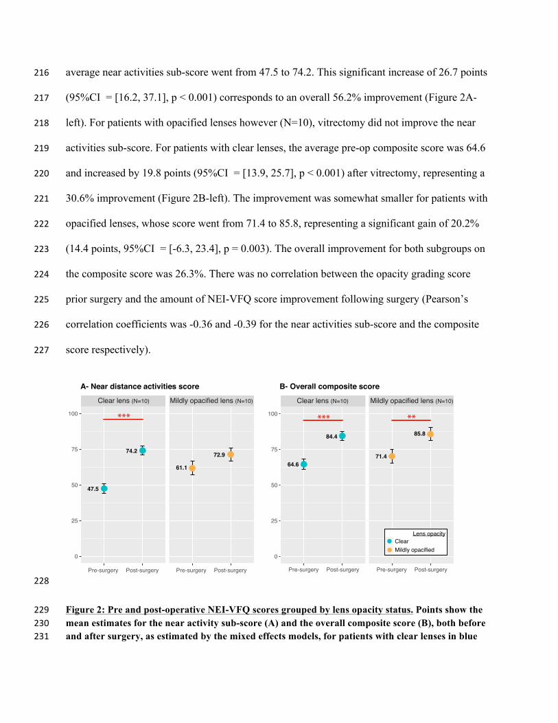
average near activities sub-score went from 47.5 to 74.2. This significant increase of 26.7 points 216
(95%CI = [16.2, 37.1], p < 0.001) corresponds to an overall 56.2% improvement (Figure 2A-217
left). For patients with opacified lenses however (N=10), vitrectomy did not improve the near 218
activities sub-score. For patients with clear lenses, the average pre-op composite score was 64.6 219
and increased by 19.8 points (95%CI = [13.9, 25.7], p < 0.001) after vitrectomy, representing a 220
30.6% improvement (Figure 2B-left). The improvement was somewhat smaller for patients with 221
opacified lenses, whose score went from 71.4 to 85.8, representing a significant gain of 20.2% 222
(14.4 points, 95%CI = [-6.3, 23.4], p = 0.003). The overall improvement for both subgroups on 223
the composite score was 26.3%. There was no correlation between the opacity grading score 224
prior surgery and the amount of NEI-VFQ score improvement following surgery (Pearson’s 225
correlation coefficients was -0.36 and -0.39 for the near activities sub-score and the composite 226
score respectively). 227
228
Figure 2: Pre and post-operative NEI-VFQ scores grouped by lens opacity status. Points show the 229mean estimates for the near activity sub-score (A) and the overall composite score (B), both before 230and after surgery, as estimated by the mixed effects models, for patients with clear lenses in blue 231
●
●
●
●
Clear lens (N=10) Mildly opacified lens (N=10)
0
25
50
75
100
Clear Mildly opacified
Lens opacity
B- Overall composite score
Pre-surgery Post-surgery Pre-surgery Post-surgery
●
●
●
●
Clear lens (N=10) Mildly opacified lens (N=10)
0
25
50
75
100
Pre-surgery Post-surgery Pre-surgery Post-surgery
A- Near distance activities score
*** *** **
61.1
72.974.2
47.5
64.6
84.4
71.4
85.8
(N=10) and patients with mildly opacified lenses in orange (N=10). Error bars represent the 95% 232confidence intervals. 233
234
Reading performance 235
Maximum Reading Speed (MRS) 236
First, we included data from all 20 patients in the mixed-effects model without any distinction on 237
their lens opacity status. MRS before surgery was on average 137 words/minute (wpm) for the 238
operated eye (95%CI = [125, 149]). It was significantly higher by 13 wpm in the non-operated 239
eye (95%CI = [5, 22], p = 0.003) and by 15 wpm in the binocular condition (95%CI = [7, 24], p 240
< 0.001). After surgery, MRS in the operated eye increased significantly to 146 wpm (i.e. a 9 241
wpm increase; 95%CI = [3, 15], p = 0.007). Post-operatively, MRS did not change significantly 242
in the non-operated eye (1 wpm increase; 95%CI = [-12, 14]; p = 0.8) or in the binocular 243
condition (3 wpm increase; 95%CI = [-9, 17]; p = 0.23). 244
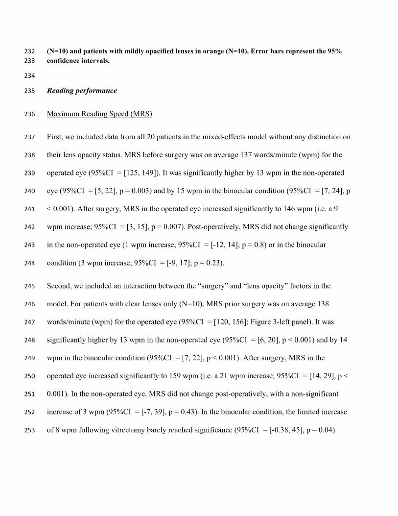
Second, we included an interaction between the “surgery” and “lens opacity” factors in the 245
model. For patients with clear lenses only (N=10), MRS prior surgery was on average 138 246
words/minute (wpm) for the operated eye (95%CI = [120, 156]; Figure 3-left panel). It was 247
significantly higher by 13 wpm in the non-operated eye (95%CI = [6, 20], p < 0.001) and by 14 248
wpm in the binocular condition (95%CI = [7, 22], p < 0.001). After surgery, MRS in the 249
operated eye increased significantly to 159 wpm (i.e. a 21 wpm increase; 95%CI = [14, 29], p < 250
0.001). In the non-operated eye, MRS did not change post-operatively, with a non-significant 251
increase of 3 wpm (95%CI = [-7, 39], p = 0.43). In the binocular condition, the limited increase 252
of 8 wpm following vitrectomy barely reached significance (95%CI = [-0.38, 45], p = 0.04). 253
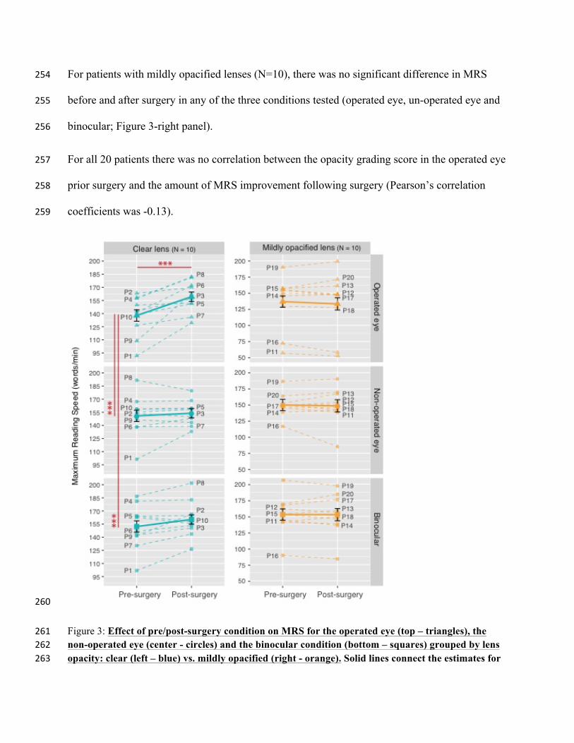
For patients with mildly opacified lenses (N=10), there was no significant difference in MRS 254
before and after surgery in any of the three conditions tested (operated eye, un-operated eye and 255
binocular; Figure 3-right panel). 256
For all 20 patients there was no correlation between the opacity grading score in the operated eye 257
prior surgery and the amount of MRS improvement following surgery (Pearson’s correlation 258
coefficients was -0.13). 259
260
Figure 3: Effect of pre/post-surgery condition on MRS for the operated eye (top – triangles), the 261non-operated eye (center - circles) and the binocular condition (bottom – squares) grouped by lens 262opacity: clear (left – blue) vs. mildly opacified (right - orange). Solid lines connect the estimates for 263
each sub-group as given by the mixed-effects model. Errors bars (black) represent their standard 264errors. Dashed lines connect the MRS values for each patient, numbered from P1 to P20. 265 266
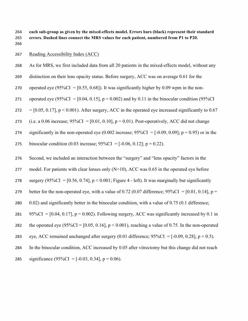
Reading Accessibility Index (ACC) 267
As for MRS, we first included data from all 20 patients in the mixed-effects model, without any 268
distinction on their lens opacity status. Before surgery, ACC was on average 0.61 for the 269
operated eye (95%CI = [0.55, 0.68]). It was significantly higher by 0.09 wpm in the non-270
operated eye (95%CI = [0.04, 0.15], p = 0.002) and by 0.11 in the binocular condition (95%CI 271
= [0.05, 0.17], p < 0.001). After surgery, ACC in the operated eye increased significantly to 0.67 272
(i.e. a 0.06 increase; 95%CI = [0.01, 0.10], p = 0.01). Post-operatively, ACC did not change 273
significantly in the non-operated eye (0.002 increase; 95%CI = [-0.09, 0.09]; p = 0.95) or in the 274
binocular condition (0.03 increase; 95%CI = [-0.06, 0.12]; p = 0.22). 275
Second, we included an interaction between the “surgery” and “lens opacity” factors in the 276
model. For patients with clear lenses only (N=10), ACC was 0.65 in the operated eye before 277
surgery (95%CI = [0.56, 0.74], p < 0.001; Figure 4 - left). It was marginally but significantly 278
better for the non-operated eye, with a value of 0.72 (0.07 difference; 95%CI = [0.01, 0.14], p = 279
0.02) and significantly better in the binocular condition, with a value of 0.75 (0.1 difference; 280
95%CI = [0.04, 0.17], p = 0.002). Following surgery, ACC was significantly increased by 0.1 in 281
the operated eye (95%CI = [0.05, 0.16], p < 0.001), reaching a value of 0.75. In the non-operated 282
eye, ACC remained unchanged after surgery (0.01 difference; 95%CI = [-0.09, 0.28], p = 0.5). 283
In the binocular condition, ACC increased by 0.05 after vitrectomy but this change did not reach 284
significance (95%CI = [-0.03, 0.34], p = 0.06). 285
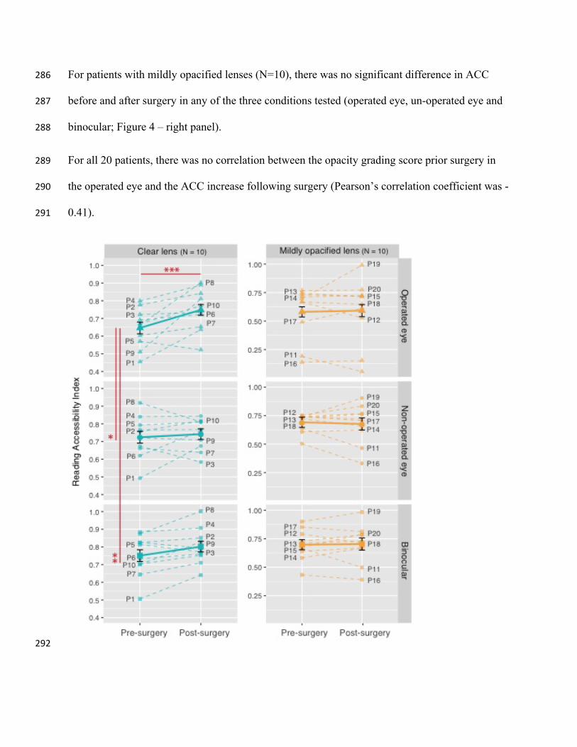
For patients with mildly opacified lenses (N=10), there was no significant difference in ACC 286
before and after surgery in any of the three conditions tested (operated eye, un-operated eye and 287
binocular; Figure 4 – right panel). 288
For all 20 patients, there was no correlation between the opacity grading score prior surgery in 289
the operated eye and the ACC increase following surgery (Pearson’s correlation coefficient was -290
0.41). 291
292
Figure 4: Effect of pre/post-surgery condition on ACC for the operated eye (top – triangles), the 293non-operated eye (center - circles) and the binocular condition (bottom – squares) grouped by lens 294opacity: clear (left – blue) vs. mildly opacified (right - orange). Solid lines connect the estimates for 295each sub-group as given by the mixed effects model. Errors bars (black) represent their standard 296errors. Dashed lines connect the MRS values for each patient, numbered from P1 to P20. 297 298
Critical Print size (CPS) and Reading Acuity (RA) 299
For both CPS and RA, we found no significant difference between the operated eye and the non-300
operated eye or the binocular condition before surgery. None of these measures changed 301
significantly after surgery in the tested eyes. 302
303
Correlation between reading performance change and daily life visual function improvement 304
Lastly, we inspected the correlation between the improvement in reading performance and the 305
improvement in NEI-VFQ near activities sub-score in the operated eye of all 20 patients. We 306
found no correlation between the percentage of improvement in MRS and the increase in NEI-307
VFQ near activities sub-score (r = 0.4, 95%CI = [-0.12, 0.75], p = 0.12). On the other hand, the 308
improvement in ACC was significantly correlated with the near activities sub-score (r = 0.74, 309
95%CI = [0.39, 0.90], p = 0.001; Figure 5). 310
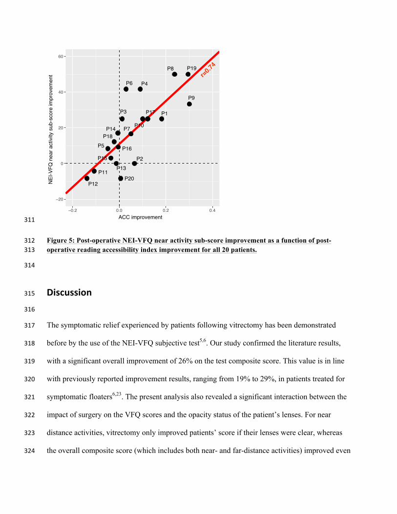
311
Figure 5: Post-operative NEI-VFQ near activity sub-score improvement as a function of post-312operative reading accessibility index improvement for all 20 patients. 313
314
Discussion315
316
The symptomatic relief experienced by patients following vitrectomy has been demonstrated 317
before by the use of the NEI-VFQ subjective test5,6. Our study confirmed the literature results, 318
with a significant overall improvement of 26% on the test composite score. This value is in line 319
with previously reported improvement results, ranging from 19% to 29%, in patients treated for 320
symptomatic floaters6,23. The present analysis also revealed a significant interaction between the 321
impact of surgery on the VFQ scores and the opacity status of the patient’s lenses. For near 322
distance activities, vitrectomy only improved patients’ score if their lenses were clear, whereas 323
the overall composite score (which includes both near- and far-distance activities) improved even 324
●
●
●
●
●
●
●
●
●
●
●
●
●
●
●
●
●
●
●
●
−20
0
20
40
60
−0.2 0.0 0.2 0.4ACC improvement
NEI
-VFQ
nea
r act
ivity
sub
-sco
re im
prov
emen
t
P19P8
P9
P4P6
P1P17
P10
P3
P7
P5P18
P13
P12P20
P2
P11
P14
P15P16
r=0.74

if the lenses were mildly opacified. To our knowledge, this result was never reported before and 325
suggests that the removal of SVO may have a significant impact on near-distance daily life 326
activities, but only in the absence of cataract or lens opacification. Because near distance 327
activities rely on fine central vision, for which performance is rapidly degraded past a critical 328
contrast threshold24, SVO removal may not be sufficient to help improve performance if contrast 329
sensitivity is still reduced from lens opacification. 330
331
Our second result is the poor MRS achieved in all 10 patients with SVO and clear lenses (138 332
wpm on average in the operated eye prior surgery) compared to normal values. According to 333
Calabrèse et al., 2016, normal readers between 58 to 68 years old should reach a MRS comprised 334
between 183.2 and 189.2 wpm when reading with one or both eyes25,26. This 35% decrease 335
suggests that reading speed may be considered as an objective measure of functional impairment 336
in the presence of SVO. However, this finding should be interpreted with caution, given that 337
other confounding clinical factors (e.g. cognitive or visual) may have also contributed to 338
reducing reading speed. 339
340
Our third outcome is the significant change in MRS, measured after vitrectomy in patients with 341
clear lenses, with a 15% improvement in the operated eye. For these patients, the non-operated 342
eye served as control and showed no improvement post-surgery, confirming that the 343
improvement measured in the fellow eye was not due to a practice effect. More importantly, this 344
improvement did not occur in eyes with mildly opacified lenses, either from cataract (phakic 345
eyes) or posterior capsule opacification (pseudophakic eyes). Taken all together, these results 346
suggest that reading speed may be a valid objective measure to quantify the positive impact of 347

vitrectomy on visual function, but only if contrast sensitivity is not still altered by lens 348
opacification. There is evidence that a main effect of vitrectomy is to restore normal contrast 349
sensitivity function for individuals with clear lenses6,27. We hypothesize that for patients with 350
mildly opacified lenses, who experienced no post-surgery improvement in MRS, reduced 351
contrast sensitivity from cloudy ocular media created a bottleneck for any potential increase in 352
reading speed. We noted that binocular MRS was not improved post-surgery. Since, our 353
population was not restricted to patients with non-pathological fellow eyes, we did not expect to 354
see monocular vitrectomy having a significant impact on binocular performance. 355
356
ACC showed the same pattern as MRS, suggesting that this measure, which is potentially 357
quicker to obtain (in terms of testing and calculation time), could be a good alternative in clinical 358
settings where time is often limited. More interestingly, the improvement in ACC induced by 359
vitrectomy was significantly correlated with the improvement in near distance activities score 360
measured with the NEI-VFQ. This result alone suggests that improved reading performance 361
following vitrectomy will also have a positive impact on the overall patients’ quality of life. The 362
simple objective assessment of ACC post-operatively may therefore provide some insight to the 363
patient and his/her care team about his/her overall quality of life improvement. 364
365
Surprisingly, neither CPS nor RA were sensitive to the presence of dense floaters. Even more, 366
we found no effect of vitrectomy on any of these measures. In their study of 110 treated eyes, 367
Nie et al., 2013 reported that 71% of their patients had difficulty in reading small print, which 368
markedly improved after surgery5. Based on their results, we had hypothesized that RA (i.e. the 369
smallest print one can read) would improve following vitrectomy. However, our results do not 370
support this hypothesis and suggest that these reading measures may not be valid to quantify the 371
impact of floaters on daily visual function. 372
373
We had expected patients with the eyes having the most prominent vitreous opacities to exhibit 374
the greatest improvement in both NEI-VFQ scores and reading performance. This was not the 375
case. In clinical practice, patients with a wide range of vitreous debris are seen, and often 376
individuals with very substantial opacities can be essentially asymptomatic (as in asteroid 377
hyalosis)28. Our result, as well as the wide variability in dysfunction among patients with similar 378
vitreous opacities, suggests that the location and motion characteristics of the opacities may be 379
more significant drivers than the level of opacity itself in the decision to seek symptomatic relief 380
with surgery. However, the ability to show the degree of vitreous opacification using the video 381
SLO was found to be helpful for educational purposes, both pre- and post-operatively. First, to 382
show family members dynamically what the patients were seeing. Second, to help persuading 383
patients with significant complaints but mild opacities on SLO testing that surgery would not be 384
prudent. Finally, to document the absence of the opacities post-surgery. 385
386
Our work presents some limitations. The main one is the restricted number of patients. In the 387
future, our results should be replicated with larger sets of patients to confirm our findings. 388
Another limitation is that, given the nature of the MNREAD, the current study only measured 389
fluent reading for short sentences. Therefore, it remains to be determined whether speed is also 390
improved (and to what extent) for spot reading (i.e. for isolated words, such as tag labels) and 391
sustained reading (i.e. for long texts). 392
393
394
Acknowledgments395
The authors would like to thank Gordon E. Legge for his help in the earlier stages of this study. 396
397
398
EthicalApproval 399
This study was conducted in accordance with the Declaration of Helsinki. Ethical approval for 400
this study was obtained from the Institutional Review Board (IRB) at the University of South 401
Carolina. The collection and evaluation of all protected patient health information was performed 402
in a Health Insurance Portability and Accountability Act (HIPAA)-compliant manner. 403
404
StatementofInformedConsent405
Written informed consent was obtained before the study from each patient according to IRB 406
guidelines, including permission for publication of all videos included herein.407
408
409
References410
1. Schulz-Key S, Carlsson J, Crafoord S (2011) Longterm follow-up of pars plana vitrectomy for 411
vitreous floaters: complications, outcomes and patient satisfaction. Acta Ophthalmol 89: 159-65. 412
2. Tan HS, Mura M, Lesnik Oberstein SY, Bijl HM (2011) Safety of vitrectomy for floaters. Am 413
J Ophthalmol 151: 995-8. 414
3. Ivanova T, Jalil A, Antoniou Y, et al. (2016) Vitrectomy for primary symptomatic vitreous 415
opacities: an evidence-based review. Eye (Lond) 30: 645-55. 416
4. Schiff WM, Chang S, Mandava N, Barile GR (2000) Pars plana vitrectomy for persistent, 417
visually significant vitreous opacities. Retina 20: 591-6. 418
5. de Nie KF, Crama N, Tilanus MAD, Klevering BJ, Boon CJF (2013) Pars plana vitrectomy 419
for disturbing primary vitreous floaters: clinical outcome and patient satisfaction. Graefes Arch 420
Clin Exp Ophthalmol 251: 1373-82. 421
6. Sebag J, Yee KMP, Nguyen JH, Nguyen-Cuu J (2018) Long-Term Safety and Efficacy of 422
Limited Vitrectomy for Vision Degrading Vitreopathy Resulting from Vitreous Floaters. 423
Ophthalmology Retina 2: 881--887. 424
7. Mangione CM, Lee PP, Gutierrez PR, Spritzer K, Berry S, Hays R (2001) Development of the 425
25-list-item national eye institute visual function questionnaire. Archives of Ophthalmology 119: 426
1050-1058. 427
8. Castilla-Marti M, van den Berg TJTP, de Smet MD (2015) Effect of vitreous opacities on 428
straylight measurements. Retina 35: 1240-6. 429
9. Delaney YM, Oyinloye A, Benjamin L (2002) Nd:YAG vitreolysis and pars plana vitrectomy: 430
surgical treatment for vitreous floaters. Eye (Lond) 16: 21-6. 431
10. Mason JO 3rd, Neimkin MG, Mason JO 4th, et al. (2014) Safety, efficacy, and quality of life 432
following sutureless vitrectomy for symptomatic vitreous floaters. Retina 34: 1055-61. 433
11. Hazel CA, Petre KL, Armstrong RA, Benson MT, Frost NA (2000) Visual function and 434
subjective quality of life compared in subjects with acquired macular disease. Invest Ophthalmol 435
Vis Sci 41: 1309-15. 436

12. McClure ME, Hart PM, Jackson AJ, Stevenson MR, Chakravarthy U (2000) Macular 437
degeneration: do conventional measurements of impaired visual function equate with visual 438
disability?. Br J Ophthalmol 84: 244-50. 439
13. Murro V, Sodi A, Giacomelli G, et al. (2017) Reading Ability and Quality of Life in 440
Stargardt Disease. Eur J Ophthalmol 27: 740-745. 441
14. Rubin GS (2013) Measuring reading performance. Vision Res 90: 43-51. 442
15. Rubin GS, Legge GE (1989) Psychophysics of reading VI - The role of contrast in low 443
vision. Vision Res 29: 79-91. 444
16. Kakehashi A, Ishiko S, Konno S, Akiba J, Kado M, Yoshida A (1995) Vitreous videography 445
using the scanning laser ophthalmoscope. Jpn J Ophthalmol 39: 377-83. 446
17. Mojana F, Kozak I, Oster SF, et al. (2010) Observations by spectral-domain optical 447
coherence tomography combined with simultaneous scanning laser ophthalmoscopy: imaging of 448
the vitreous. Am J Ophthalmol 149: 641-50. 449
18. Mansfield JS, Ahn SJ, Legge GE, Luebker A (1993) A new reading-acuity chart for normal 450
and low vision. Ophthalmic and Visual Optics/Noninvasive Assessment of the Visual System 451
Technical Digest, (Optical Society of America, Washington, DC., 1993.) 3: 232--235. 452
19. Calabrèse A, To L, He Y, Berkholtz E, Rafian P, Legge GE (2018a) Comparing performance 453
on the MNREAD iPad application with the MNREAD acuity chart. J Vis 18: 8. 454
20. Calabrèse A, Owsley C, McGwin G, Legge GE (2016a) Development of a Reading 455
Accessibility Index Using the MNREAD Acuity Chart. JAMA Ophthalmol 134: 398-405. 456
21. R Core Team (2018) R: A Language and Environment for Statistical Computing. Vienna, 457
Austria: R Foundation for Statistical Computing. , https://www.R-project.org/. 458
22. Zuur AF, Ieno EN, Elphick CS (2010) A protocol for data exploration to avoid common 459
statistical problems. Methods in Ecology and Evolution 1: 3-14. 460
23. Sebag J, Yee KMP, Wa CA, Huang LC, Sadun AA. Vitrectomy For Floaters: Prospective 461
Efficacy Analyses And Retrospective Safety Profile. Retina 2014;34:1062-8. 462
24. Owsley C (2003) Contrast sensitivity. Ophthalmol Clin North Am 16: 171-7. 463
25. Calabrèse A, Cheong AMY, Cheung S, et al. (2016c) Baseline MNREAD Measures for 464
Normally Sighted Subjects From Childhood to Old Age. Invest Ophthalmol Vis Sci 57: 3836-43. 465
26. Johansson J, Pansell T, Ygge J, Seimyr GÖ (2014) Monocular and binocular reading 466
performance in subjects with normal binocular vision. Clin Exp Optom 97: 341-8. 467
27. Garcia GA, Khoshnevis M, Yee KMP, Nguyen-Cuu J, Nguyen JH, Sebag J (2016) 468
Degradation of Contrast Sensitivity Function Following Posterior Vitreous Detachment. Am J 469
Ophthalmol 172: 7-12. 470
28. Khoshnevis M, Rosen S, Sebag J (2019) Asteroid Hyalosis - A comprehensive review. Surv 471
Ophthalmol. 472
473
474
Figurelegends475
Figure 1: Protocol schematic showing the different test procedures along with the resulting 476
outcome measures. Subjective measures are represented in blue; objective measures are 477
represented in pink. 478
479

Figure 2: Pre and post-operative NEI-VFQ scores grouped by lens opacity status. Points show 480
the mean estimates for the near activity sub-score (A) and the overall composite score (B), both 481
before and after surgery, as estimated by the mixed effects models, for patients with clear lenses 482
in blue (N=10) and patients with mildly opacified lenses in orange (N=10). Error bars represent 483
the 95% confidence intervals. 484
485
Figure 3: Effect of pre/post-surgery condition on MRS for the operated eye (top – triangles), the 486
non-operated eye (center - circles) and the binocular condition (bottom – squares) grouped by 487
lens opacity: clear (left – blue) vs. mildly opacified (right - orange). Solid lines connect the 488
estimates for each sub-group as given by the mixed-effects model. Errors bars (black) represent 489
their standard errors. Dashed lines connect the MRS values for each patient, numbered from P1 490
to P20. 491
492
Figure 4: Effect of pre/post-surgery condition on ACC for the operated eye (top – triangles), the 493
non-operated eye (center - circles) and the binocular condition (bottom – squares) grouped by 494
lens opacity: clear (left – blue) vs. mildly opacified (right - orange). Solid lines connect the 495
estimates for each sub-group as given by the mixed effects model. Errors bars (black) represent 496
their standard errors. Dashed lines connect the MRS values for each patient, numbered from P1 497
to P20. 498
499
Figure 5: Post-operative NEI-VFQ near activity sub-score improvement as a function of post-500
operative reading accessibility index improvement for all 20 patients. 501

502
Tables503
Patient ID Location Gender Age Lens opacity
in both eyes
Operated eye Non-operated eye
Pathology SVO Acuity OCT-SLO Opacity grading
Pathology SVO Acuity
P1 Minnesota M 58 Clear PVD Yes 20/25 2 ERM No 20/25
P2 Minnesota M 59 Clear PVD Yes 20/20 1.5 -- Yes 20/25
P3 California M 61 Clear PVD Yes 20/20 3 ERM No 20/40
P4 Minnesota M 62 Clear PVD+ ERM Yes 20/20 1.5
Scleral buckling + ERM
No 20/20
P5 Minnesota M 64 Clear PVD+ ERM Yes 20/15 2 PVD Yes 20/25
P6 Minnesota F 64 Clear PVD Yes 20/25 2.5 -- Yes 20/20
P7 Minnesota F 64 Clear PVD Yes 20/20 2.5 PVD Yes 20/25
P8 Minnesota F 68 Clear PVD Yes 20/30 1 PVD Yes 20/15
P9 California M 69 Clear PVD Yes 20/20 2.5 PVD Yes 20/20
P10 Minnesota F 72 Clear PVD Yes 20/25 2 PVD Yes 20/25
P11 California F 32 Mild opacity PVD Yes 20/25 1 PVD Yes 20/80
P12 California M 52 Mild opacity PVD+ ERM Yes 20/25 2.5 Vitreous Syneresis No 20/20
P13 California M 54 Mild opacity PVD+ ERM Yes 20/40 3 NPDR No 20/20
P14 California F 54 Mild opacity PVD Yes 20/25 2.5 PVD Yes 20/80
P15 Minnesota M 63 Mild opacity PVD+ ERM Yes 20/40 2.5 ERM Yes 20/25
P16 California M 63 Mild opacity PVD+ ERM Yes 20/80 2 ERM No 20/25
P17 Minnesota M 64 Mild opacity PVD Yes 20/20 2.5 Vitreous Syneresis Yes 20/20
P18 California F 65 Mild opacity PVD Yes 20/30 2.5 PVD Yes 20/25
P19 California F 67 Mild opacity PVD Yes 20/30 2.5 ERM No 20/25
P20 Minnesota M 68 Mild opacity PVD Yes 20/20 1.5 PVD Yes 20/25
504
Table 1: Patients’ individual characteristics prior to surgery. SVO stands for symptomatic 505
vitreous opacities; ERM stands for epiretinal membrane. PVD stands for posterior vitreous 506
detachment; NPDR stands for non-proliferative diabetic retinopathy; Visual acuity is given in 507
Snellen notation. 508
509
Supplementarymaterial510
Movie 1. Video 1, preoperative video SLO. The video is live streaming of scanning laser 511
ophthalmoscopic images from the Heidelberg OCT machine. This is recorded in "Movie Max" 512
mode, (in avi) then converted to .mov. A patient with prominent vitreous opacities months post 513
scleral buckling was instructed to look left and re-fixate, then look right and re-fixate. Shadows 514
from mobile vitreous opacities were projected on the stabilized retinal surface and thus imaged 515
with the infrared camera. 516
517
Movie 2. Video 2, postoperative video SLO. The video is recorded in the same manner 518
(with saccades) as Video 1, and of the same eye one week post vitrectomy. Absence of 519
shadowing from vitreous opacities is noted. 520