rapid epidemiologic analysis of cytomegalovirus by using
TRANSCRIPT

JOURNAL OF CLINICAL MICROBIOLOGY, Apr. 1992, p. 839-844 Vol. 30, No. 40095-1137/92/040839-06$02.00/0Copyright (C 1992, American Society for Microbiology
Rapid Epidemiologic Analysis of Cytomegalovirus by UsingPolymerase Chain Reaction Amplification
of the L-S Junction RegionDAWN M. SOKOL,"14 GAIL J. DEMMLER,l12,3,4* AND GREGORY J. BUFFONE" 2'4
Departments of Pediatrics, 1 Pathology, 2 and Microbiology and Immunology, 3 Baylor College ofMedicine, and Te-xas Children's Hospital,4 Houston, Texas 77030
Received 27 August 1991/Accepted 3 January 1992
A technique based on polymerase chain reaction (PCR) amplification was developed to facilitate the study ofthe epidemiology of cytomegalovirus (CMV). Consensus oligonucleotide primers from repetitive DNAsequences were designed to amplify interspersed repetitive sequences in an area of heterogeneity within the L-Sjunction region of the CMV genome, and PCR products were detected by gel electrophoresis. Purified CMVDNAs from 25 CMV isolates, 13 from members of five families in which person-to-person transmission wasdocumented, 9 random clinical isolates of CMV, and 3 laboratory reference strains of CMV (Towne, Davis,and AD169), were analyzed. The gel electrophoretic patterns of DNA bands, or PCR profiles, produced byamplification with the L-S primers were unique for epidemiologically unrelated strains and laboratoryreference strains, yet similar patterns were observed for epidemiologically related strains isolated frommembers of the same family. This method of rapid fingerprinting of CMV DNA within the hypervariable L-Sjunction region by PCR to produce strain-specific, variably sized PCR products should simplify the molecularepidemiologic analysis of CMV.
Molecular epidemiologic analysis of cytomegalovirus(CMV) has enhanced the understanding of its transmissionamong immunocompromised hosts, neonates, and families,as well as in day-care and hospital settings (1, 3, 8, 14, 23, 29,33-35). However, the standard molecular techniques used inthese studies have several limitations that preclude theirwidespread application. For example, restriction enzymeanalysis to detect polymorphisms arising from the absenceor acquisition of restriction enzyme recognition sites re-quires relatively large quantities of purified viral DNA,which is produced by multiple in vitro passages of virus, andmultiple viral passages, in turn, produce minor geneticchanges that can alter the restriction enzyme profile (14, 15,33). In addition, restriction enzyme analysis is limited be-cause CMV strains have been shown to have approximately80% homology in their nucleotide sequences, and this ho-mology results in similarities in restriction enzyme profiles,even among unrelated strains (16). Epidemiologic analysis ofCMV also has been accomplished by the use of Southernblot hybridization with cloned subgenomic CMV probes thatdefine differences across the entire genome or alternativelyat the heterogeneous L-S junction region (1, 3, 8, 9, 29).While relatively less viral DNA is needed for Southern blotmethods, they have proven to be cumbersome and time-consuming procedures which require radiolabeled CMVDNA.DNA amplification by the polymerase chain reaction
(PCR) has been successfully applied to the detection of CMVin various specimens for diagnostic purposes (2, 5, 13, 26).More recently, PCR has been explored as a potentiallyuseful tool for the epidemiologic analysis of CMV (4, 6, 36).Amplification has been accomplished at target sequences inregions encoding CMV DNA polymerase, envelope glyco-proteins, and major immediate-early and structural proteins,
* Corresponding author.
839
as well as short fragments in the L-S junction region of theCMV genome. However, all of these reported methodsrequire multiple PCR amplifications and restriction enzymeanalysis and are only able to define minimal differencesbetween CMV strains.
In this study, PCR with consensus primers complemen-tary to degenerate repetitive DNA sequences was applied tothe rapid epidemiologic analysis of CMV isolates. Thismethod required minute quantities of viral DNA and norestriction enzyme digestion. A region of the CMV genomeknown as the L-S junction was chosen for amplification byPCR because duplications and deletions frequently occur inthis region during virus replication and produce interstrainnucleotide sequence variations (18, 27, 32). Degeneraterepeat sequences in the L-S junction region selected frompublished sequence data for laboratory reference strainsCMV Towne and AD169 were used to derive consensusoligonucleotide primers anticipated to provide amplificationof strain-specific, variably sized PCR products (22, 31, 32).Amplification of a portion of the L-S junction region con-taining these degenerate direct repeats was successfullyapplied to distinguish both epidemiologically related andunrelated CMV isolates. Because of its relative simplicity,this method should facilitate further study of the molecularepidemiology of CMV.
(This work was presented in part at the 30th InterscienceConference on Antimicrobial Agents and Chemotherapy,Atlanta, Ga., 21 to 24 October 1990.)
MATERIALS AND METHODS
CMV isolates. All CMV strains were isolated and propa-gated in human foreskin fibroblast cell cultures (Viromed,Minnetonka, Minn.) by standard techniques (30). The DNAfrom 16 isolates was extracted from low-passage CMV-infected fibroblasts contained in a 25-cm2 flask and exhibit-ing 80 to 95% cytopathic effects, and this DNA was used for
on January 4, 2019 by guesthttp://jcm
.asm.org/
Dow
nloaded from
840 SOKOL ET AL.
both Southern blot analysis and PCR amplification. TheDNA from nine random clinical isolates was extracted fromone or two cell culture tubes containing cells exhibiting lowcytopathic effects. Extraction methods included either amanual modified Hirt procedure or the use of an automatednucleic acid extractor (Applied Biosystems, Foster City,Calif.) (11). Extracted DNA was resuspended in Tris-EDTAbuffer (pH 8.0) and quantitated on a fluorescence spectro-photometer (Perkin-Elmer Cetus, Norwalk, Conn.). No fur-ther purification of viral DNA from cellular DNA wasrequired prior to analysis.
Epidemiologically related CMV strains included 13 iso-lates from members of five families in which person-to-person transmission was documented by serial serology andviral culturing (7). Epidemiologically unrelated CMV strainswere randomly selected from clinical specimens submittedto the Baylor College of Medicine CMV Virology Labora-tory for viral isolation. These nine clinical isolates were fromfive congenitally infected infants, three transplant recipients,and a hospitalized patient with febrile illness. Laboratoryreference strains AD169, Towne, and Davis were obtainedfrom the American Type Culture Collection (Rockville, Md.)and were maintained in our laboratory by standard tech-niques (30).
Southern blot analysis. Isolates of CMV were analyzed bySouthern blot hybridization with 32P-labeled EcoRI junctionfragments F and H (generously provided by D. Spector andS. Spector, University of California at San Diego, SanDiego) to determine whether the molecular profiles of theCMV strains isolated from family members were similar.Southern blot analysis was performed with 6 pLg of purifiedviral DNA as previously described by Spector et al., withminor modifications (28).
Primers. Oligonucleotide primers were developed on thebasis of published sequence data for the L-S junction regionsof strains AD169 and Towne with the assistance of acomputer program (OLIGO; National Biosciences, Hamil,Minn.) for selecting optimal oligonucleotide primers for invitro amplification (25). Oligonucleotide primer LS-1 wasderived from a consensus sequence of the degenerate directrepeat sequences present within the L-S junction region.Because of the variable numbers and locations of the degen-erate direct repeats, primer LS-1 had the potential to bind tomultiple sites within the L-S junction region (Fig. 1). Asecond consensus oligonucleotide primer, LS-2, was se-lected from a more constant region near the terminal se-quence of the L-S junction region. The sequences of theconsensus primers are as follows: CMV LS-1, 5'-GGGTGTGKYGGYIGGGTGTGTCRG-3', and CMV LS-2, 5'-ACGCCYGACATCCATTCCGGGCCG-3', where K = G+T, Y =
C+T, R = A+G, and I = inosine.Amplification. Each PCR was carried out with a microcen-
trifuge tube and a final total volume of 50 ,ul. The reactionmixture contained 0.1 ,ug of purified DNA, 50 pmol of eachprimer, 200 ,uM each deoxynucleotide triphosphate (dATP,dTTP, dCTP, and dGTP), and 5 [LI of 10x PCR buffer 50 mMKCl, 10 mM Tris-Cl, 1.5 mM MgCl2, 0.01% gelatin), withsterile H20 added to bring the volume to 49 ,ul. The samplewas overlaid with 50 ,ul of mineral oil and heated to 100°C for7 min in a heating block (VWR Scientific, Media, Pa.). Twounits (1 ,ul) of Thennus aquaticus (Taq) polymerase wasadded to the reaction mixture. Laboratory precautions weretaken to minimize sample carryover as described by Kwok(19). CMV strain AD169 was used as a positive control, anduninfected human foreskin fibroblasts were used as a nega-tive control. Each CMV isolate was run in duplicate.
L-S lunctionb' a'c'
./ 1770 1780 1790 1800 1810 18iOACGCGG cCcCCACCGGCGG
1830 1840 1850 -- 1860 1870 1880GTGGGGC GGCGG GGGT GTGC G C GCGGGCTGT GTTGGTG
1890 1900 1910 1920 1930 1940IGS- GGAGG TGTGTCALFG GCGTGGCCGGGT O-qGTGTGTCGT
1950 1960 1970 19t' 1990 2000GC9~GTGTG TCGCGGGCGT GTGGCGGpTG GGT WG
2010 2020 2030 2040 2050 2060GGCGGTGTGC G--- CTCGG GGTGTGCGGC CGAOTGTGTG GCCTCGCGGC
2070 2080 2090 2100 2110 2120aGTTATCTCC Cc~GcGGC cCGQGGGCCGS OTCcCTCGC CCCGGQ TGCTTTTCGT
2130 2140 2150 2160 2170 2180_ SG GGA&TGC TGCccCC ~QGGAWTC CT C CGGGAATCK
2190 2200 2210 2220 2230 2240CACAGACAC& CACACGCGTC TTCTTTTCGC GC TTTGCT CGCC
2250 2260 2270 2280 2290 2300GTCG~C TccTCCCCAC ACGCAACTAG TCGCC&CACACGC TCCAAGTTTC
2310 2320 2330 2340 231% 2360Ac CC TAAAAACcC CC:CCW= TCGhGGAC
2370 2380 2390 2400 2410 2420CC&CCTAGT GGGTCGCG cCGGGC GC CT=CAGTGGTAC
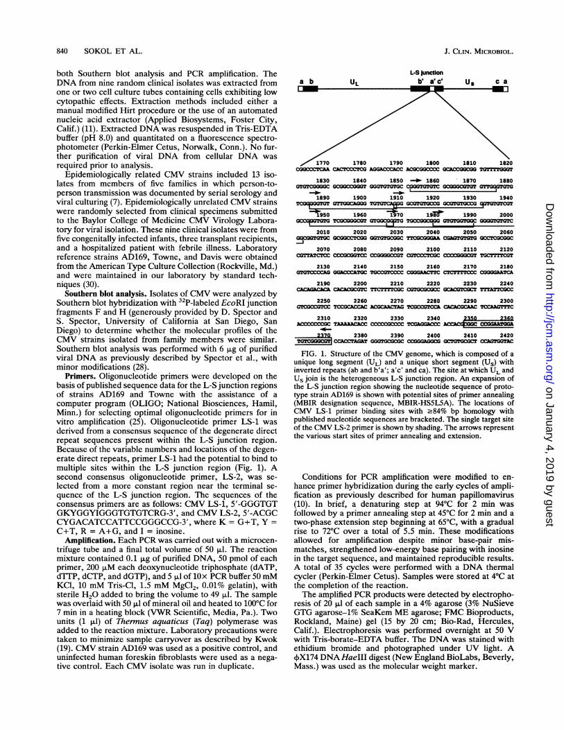
FIG. 1. Structure of the CMV genome, which is composed of aunique long segment (UL) and a unique short segment (Us) withinverted repeats (ab and b'a'; a'c' and ca). The site at which UL andUs join is the heterogeneous L-S junction region. An expansion ofthe L-S junction region showing the nucleotide sequence of proto-type strain AD169 is shown with potential sites of primer annealing(MBIR designation sequence, MBIR-HS5L5A). The locations ofCMV LS-1 primer binding sites with .84% bp homology withpublished nucleotide sequences are bracketed. The single target siteof the CMV LS-2 primer is shown by shading. The arrows representthe various start sites of primer annealing and extension.
Conditions for PCR amplification were modified to en-hance primer hybridization during the early cycles of ampli-fication as previously described for human papillomavirus(10). In brief, a denaturing step at 94°C for 2 min wasfollowed by a primer annealing step at 45°C for 2 min and atwo-phase extension step beginning at 65°C, with a gradualrise to 72°C over a total of 5.5 min. These modificationsallowed for amplification despite minor base-pair mis-matches, strengthened low-energy base pairing with inosinein the target sequence, and maintained reproducible results.A total of 35 cycles were performed with a DNA thermalcycler (Perkin-Elmer Cetus). Samples were stored at 4°C atthe completion of the reaction.The amplified PCR products were detected by electropho-
resis of 20 ,ul of each sample in a 4% agarose (3% NuSieveGTG agarose-1% SeaKem ME agarose; FMC Bioproducts,Rockland, Maine) gel (15 by 20 cm; Bio-Rad, Hercules,Calif.). Electrophoresis was performed overnight at 50 Vwith Tris-borate-EDTA buffer. The DNA was stained withethidium bromide and photographed under UV light. A,X174 DNA HaeIII digest (New England BioLabs, Beverly,Mass.) was used as the molecular weight marker.
J. CLIN. MICROBIOL.
on January 4, 2019 by guesthttp://jcm
.asm.org/
Dow
nloaded from

CMV L-S JUNCTION PCR 841
<coo)< m 0 LO DnWl T- r- y- y-
o0 e t 0LL LL lL LL LL
U)(0T-
<: tM'Y" 'U a < U)I 4 C( (N. ..4CO CY)
I± EL IL LIL IL
.-*
~_ _mb_~~~O 40 __ a_ _m 4 .
roW
_ *, *..:::J:
B 1" < CD
X._ c CDX LU. U
FIG. 2. Representative Southern blot hybridization of 32P-la-beled CMV junctional fragment F to EcoRI-digested CMV DNAfrom members of four families (FS1 family members A, B, and C;FS15 family members C and D; FS21 family members A, B, C, andD; and FS30 family members A and C). Laboratory reference strainAD169 is included for comparison.
RESULTS
Southern blot analysis. Hybridization of the F and Hprobes to the L-S junction region confirmed that the strainstransmitted within families were molecularly related (Fig. 2).These strains were therefore used to evaluate the applicationof this PCR method to determining strain relatedness.PCR analysis. Target sequences within the L-S junction
region for primer pair LS-1-LS-2 were successfully ampli-fied in viral DNA extracted from all 25 CMV isolates.Electrophoresis of the amplified products revealed strain-specific PCR profiles consisting of three to eight bandsranging in size from 100 to 1,000 bp. No amplificationoccurred in DNA extracted from uninfected human foreskinfibroblasts, and reproducible PCR profiles were seen forpositive control sample AD169 as well as for the other CMVisolates.
Epidemiologically related CMV strains isolated frommembers of the same family showed remarkably similar PCRprofiles, with strains demonstrating between five and eightbands (Fig. 3A and B). Families FS1, FS15, and NN2-HS180showed identical PCR profiles among their members. ThePCR profiles were nearly identical for families FS21 andFS30, with only one minor band variation demonstratedamong some members. Minor molecular variations withinthese two families also were noted when the strains were
analyzed by Southern blot hybridization with the F and Hfragments as probes for the L-S junction region (Fig. 4).
Laboratory reference strains Towne, Davis, and AD169,as well as the random clinical isolates of CMV, all producedunique PCR profiles (Fig. 5A and B).
872 bp-
310 bp-
118 bp-
0 a < C)
en cn en U3 ZLL LL LLLLZ
r~~~~~
o7r4'noD
FIG. 3. PCR products from epidemiologically related CMVstrains amplified with primers CMV LS-1 and CMV LS-2, analyzedby gel electrophoresis, and visualized with ethidium bromide stain.(A) FS1 family members A, B, and C; FS15 family members C andD. (B) FS21 family members A, B, C, and D; FS30 family membersA and C. NN2 and HS180 are a husband-wife pair. HFF, DNAextracted from uninfected human foreskin fibroblasts (HFF) andused as a negative control; AD169, laboratory reference strain ofCMV used as a positive control; 4$X174, molecular weight marker.
DISCUSSION
CMV oligonucleotide primers LS-1 and LS-2 successfullyamplified the heterogeneous L-S junction region of the CMVgenome and identified epidemiologically related isolates ofCMV from five families (total number of isolates, 13) and 12epidemiologically unrelated CMV isolates from infants andchildren. The similarities of the PCR profiles obtained for theepidemiologically related CMV strains demonstrated thatthe numbers and locations of the degenerate repeats in thehypervariable region of the genome were conserved betweenstrains, even though the strains had undergone both in vitropassage in the laboratory and transmission between personsin the same family. Also, the unique patterns observed forthe epidemiologically unrelated CMV strains, as well as thelaboratory reference strains, confirmed that the locationsand numbers of degenerate repeats in the L-S junction region
a._T-1L
A
872 bp -
< im 0un en en<coo.U UL)LO 0
en coDLLLL IL < I
310 bp-118 bp-72 bp-
VOL. 30, 1992
on January 4, 2019 by guesthttp://jcm
.asm.org/
Dow
nloaded from
842 SOKOL ET AL.
< 0 c< uCl)
LL LL L LL
I"1'1
a t.
A(.0 C (A1
< F C
r-
X
872 bp-
310 bp-
118 bp-
B
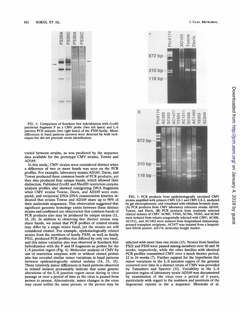
FIG. 4. Comparison of Southern blot hybridization with EcoRIjunctional fragment F as a CMV probe (two left lanes) and L-Sjunction PCR analysis (two right lanes) of the FS30 family. Minordifferences in band patterns (arrows) were detected by both tech-niques but did not preclude strain identification.
varied between strains, as was predicted by the sequencedata available for the prototype CMV strains, Towne andAD169.
In this study, CMV strains were considered distinct whena difference of two or more bands was seen on the PCRprofiles. For example, laboratory strains AD169, Davis, andTowne produced three common bands of PCR products, yetthey also produced four unique bands, which allowed theirdistinction. Published EcoRI and HindIII restriction enzymeanalysis profiles also showed comigrating DNA fragmentswhen CMV strains Towne, Davis, and AD169 were com-pared, and reciprocal DNA-DNA reassociation kinetics in-dicated that strains Towne and AD169 share up to 90% oftheir nucleotide sequences. This observation suggested thatsignificant genomic homology exists between these distinctstrains and confirmed our observation that common bands ofPCR products also may be produced by unique strains (11,18, 24). In addition to observing that distinct strains mayshare bands, we noted that PCR profiles of related strainsmay differ by a single minor band, yet the strains are stillconsidered related. For example, epidemiologically relatedstrains from the members of family FS30, as well as familyFS21, produced PCR profiles that differed by only one band,and this minor variation also was observed in Southern blothybridization with the F and H fragments as probes for theL-S junction region (Fig. 4). Molecular analysis of CMV byuse of restriction enzymes with or without cloned probesalso has revealed similar minor variations in band patternsbetween epidemiologically related isolates (14, 23, 33).These relatively minor differences in band patterns observedin related isolates presumably indicate that some geneticalterations of the L-S junction region occur during in vitropassage or over a period of time as the virus is passed fromperson to person. Alternatively, minor changes in the virusmay occur within the same person, or the person may be
qr ll- V- F t s40 Ot) -to' (0 (0 0o
X tv r- X In) ' CV, CO
E UC ))0V OuO ODEL < en < >< m<I >-
872 bp -
310 bp-
118 bp-
FIG. 5. PCR products from epidemiologically unrelated CMVstrains amplified with primers CMV LS-1 and CMV LS-2, analyzedby gel electrophoresis, and visualized with ethidium bromide stain.(A) PCR products from CMV laboratory reference strains AD169,Towne, and Davis. (B) PCR products from randomly selectedclinical isolates of CMV. SC905, Y9304, SC566, Y8303, and SC849were isolated from infants congenitally infected with CMV; AC406,AC1511, and AC1462 were isolated from hospitalized immunosup-pressed transplant recipients; AC1477 was isolated from a hospital-ized febrile patient. (X174, molecular weight marker.
infected with more than one strain (15). Strains from familiesFS21 and FS30 were passed among members over 60 and 36weeks, respectively, while the other families with identicalPCR profiles transmitted CMV over a much shorter period,12 to 16 weeks (7). Further support for the hypothesis thatminor variations in the L-S junction region of the genomeoccurred over time in a distinct strain of CMV was providedby Tamashiro and Spector (32). Variability in the L-Sjunction region of laboratory strain AD169 was documentedby examination of the virus over a period of 3 years,particularly with regard to the numbers and positions of thedegenerate repeats in the (x sequence. Mocarski et al.
J. CLIN. MICROBIOL.
on January 4, 2019 by guesthttp://jcm
.asm.org/
Dow
nloaded from

CMV L-S JUNCTION PCR 843
reported less variability of the a sequence in CMV strainTowne on passage, but over an undefined period of time (22).Analysis of the Towne or AD169 strain by the PCR tech-nique described here could further define the stability of thisregion over time.While no previous reports have delineated differences
between CMV strains by use of PCR with consensus primerscomplementary to degenerate repetitive DNA sequences,the analysis of CMV strain variability by use of several sitesof PCR amplification followed by restriction enzyme diges-tion was recently evaluated (4, 6, 36). Chou amplified theCMV genome in conserved areas of the unique long segmentand, using restriction enzymes in a stepwise manner, at-tempted to produce distinct profiles among CMV strains (4).The epidemiologic analysis ofCMV by this method appearedless rapid and more complex than ours, as it requiredamplification with a minimum of three sets of PCR primersand digestion with six restriction enzymes to distinguish 20strains. A recent analysis by Zaia et al. of differences amongCMV strains in the L-S junction region and at two sites in theunique long sequence of the CMV genome involved PCRamplification followed by restriction enzyme digestion (36).Primers for the L-S junction region amplified a conservedregion of the a sequence, with a PCR product of only about138 bp. The use of seven restriction enzymes demonstratedslight fragment size differences in unrelated CMV isolates,while some unrelated isolates remained identical in the PCRanalysis. In addition, several clinical isolates and referencestrain Davis could not be amplified. In fact, our initialattempts at the epidemiologic analysis of CMV by PCRinvolved a similar segment of the a sequence and yielded anamplified product of approximately 100 bp (6). We found thatrestriction enzyme digestion with SmaI yielded identicalpatterns among related CMV strains. However, unrelatedCMV strains were difficult to distinguish as unique becauserestriction enzyme digestion yielded PCR products thatoften differed in size by less than 10 bp. We thereforedesigned the primers described in this report to amplify amuch larger segment of the variable L-S junction region;these primers incorporated direct repetitive sequences tooptimize strain differences. A similar strategy of using PCRamplification with primers complementary to interspersedrepetitive DNA sequences also has proven valuable in theanalysis of yeast artificial chromosome clones and somaticcell hybrid lines for the presence of human genomic DNA, aswell as fingerprinting of DNA extracted from biologicalevidence in the forensic identification of individuals (9, 12,17, 20, 21).
In summary, the novel consensus PCR primers CMV LS-1and LS-2 successfully amplified a portion of the L-S junctionregion containing interspersed degenerate repeat sequencesand produced variably sized PCR products that permittedstrain identification of CMV. In addition, this method pro-vided an advantage over previous molecular techniques inthat significantly less viral DNA was required for evaluationby PCR (0.1 ,ug) than for Southern hybridization (6 ,ug), andno radioisotope or restriction enzyme digestion was re-quired. This approach allowed for more rapid, more conve-nient, and less hazardous processing of samples. Because ofthese advantages, L-S junction region PCR may provide anexpedient means for analyzing the large number of speci-mens usually required for epidemiology studies.
ACKNOWLEDGMENTSThis work was supported by NIH Biomedical Research Support
Grant RR-054-25, the Vale-Asche Foundation, the National Foun-
dation for Infectious Diseases-Burroughs-Welcome Fund, a TexasAdvanced Technology Program Award (project number 004949029),and the Myers Black-Melon Infectious Diseases Section.
REFERENCES1. Adler, S. P. 1985. The molecular epidemiology of cytomegalo-
virus transmission among children attending a day care center.J. Infect. Dis. 152:760-768.
2. Cassol, S. A., M. C. Poon, R. Pal, M. J. Naylor, J. Culver-James,T. J. Bowen, J. A. Russel, S. A. Krawetz, R. T. Pon, and D. I.Hoar. 1989. Primer-mediated enzymatic amplification of cyto-megalovirus (CMV) DNA: application to the early diagnosis ofCMV infection in marrow transplant recipients. J. Clin. Invest.83:1109-1115.
3. Chou, S. 1986. Acquisition of donor strains of cytomegalovirusby renal-transplant recipients. N. Engl. J. Med. 22:1418-1423.
4. Chou, S. 1990. Differentiation of cytomegalovirus strains byrestriction analysis of DNA sequences amplified from clinicalspecimens. J. Infect. Dis. 162:738-742.
5. Demmler, G. J., G. J. Buffone, C. M. Schimbor, and R. A. May.1988. Detection of cytomegalovirus in urine from newborns byusing polymerase chain reaction DNA amplification. J. Infect.Dis. 158:1177-1184.
6. Demmler, G. J., A. M. Major, R. A. May, and G. J. Buffone.1989. Restriction enzyme analysis (REA) of polymerase chainreaction (PCR) products to study the epidemiology of cytomeg-alovirus (CMV), abstr. 797. Program Abstr. 29th Intersci. Conf.Antimicrob. Agents Chemother.
7. Demmler, G. J., S. G. Reis, R. A. May, G. J. Buffone, L. H.Taber, and M. D. Yow. 1989. Transmission of cytomegaloviruswithin families: a risk to women of childbearing age, abstr. 3.Program Abstr. 2nd Int. CMV Workshop.
8. Demmler, G. J., M. D. Yow, S. A. Spector, S. G. Reis, M. T.Brady, D. C. Anderson, and L. H. Taber. 1989. Nosocomialcytomegalovirus infections within two hospitals caring for in-fants and children. J. Infect. Dis. 156:9-16.
9. Erlich, H. A., D. Gelfand, and J. J. Sninsky. 1991. Recentadvances in the polymerase chain reaction. Science 252:1643-1651.
10. Gregoire, L., M. Arella, J. Campione-Piccardo, and W. D.Lancaster. 1989. Amplification of human papillomavirus DNAsequences by using conserved primers. J. Clin. Microbiol.27:2660-2665.
11. Grillner, L., and I. Blomberg. 1984. Restriction enzyme analysisof human cytomegalovirus using DNA extracted from infectedcells. J. Med. Virol. 14:313-322.
12. Higuchi, R., C. H. Von Beroldingen, G. F. Sensabaugh, andH. A. Erlich. 1988. DNA typing from single hairs. Nature(London) 332:543-546.
13. Hsia, K., D. H. Spector, J. Lawrie, and S. A. Spector. 1989.Enzymatic amplification of human cytomegalovirus sequencesby polymerase chain reaction. J. Clin. Microbiol. 27:1802-1809.
14. Huang, E. S., C. A. Alford, D. W. Reynolds, S. Stagno, andR. F. Pass. 1980. Molecular epidemiology of cytomegalovirusinfections in women and their infants. N. Engl. J. Med. 303:958-962.
15. Huang, E. S., S. M. Huong, G. E. Tegtmeier, and C. A. Alford.1980. Cytomegalovirus: genetic variation of viral genomes.Ann. N.Y. Acad. Sci. 354:332-346.
16. Huang, E. S., B. A. Kilpatrick, Y. T. Huang, and J. S. Pagano.1976. Detection of cytomegalovirus and analysis of strain vari-ation. Yale J. Biol. Med. 49:29-43.
17. Jeffreys, A. J., V. Wilson, and S. L. Thein. 1985. Individual-specific "fingerprints" of human DNA. Nature (London) 316:76-79.
18. Kemble, G. W., and E. S. Mocarski. 1989. A host cell proteinbinds to a highly conserved sequence element (pac-2) within thecytomegalovirus a sequence. J. Virol. 63:4715-4728.
19. Kwok, S. 1990. Procedures to minimize PCR-product carry-over, p. 142-145. In M. A. Innis, D. H. Gelfand, J. J. Sninsky,and T. J. White (ed.), PCR protocols, 1st ed. Academic Press,Inc., San Diego, Calif.
20. Ledbetter, S. A., D. L. Nelson, S. T. Warren, and D. H.
VOL. 30, 1992
on January 4, 2019 by guesthttp://jcm
.asm.org/
Dow
nloaded from

844 SOKOL ET AL.
Ledbetter. 1990. Rapid isolation of DNA probes within specificchromosome regions by interspersed repetitive sequence poly-merase chain reaction. Genomics 6:475-481.
21. Lichter, P., S. A. Ledbetter, D. H. Ledbetter, and D. C. Ward.1990. Fluorescence in situ hybridization with Alu and Lipolymerase chain reaction probes for rapid characterization ofhuman chromosomes in hybrid cell lines. Proc. Natl. Acad. Sci.USA 87:6634-6638.
22. Mocarski, E. S., A. C. Liu, and R. R. Spaete. 1987. Structureand variability of the a sequence in the genome of humancytomegalovirus (Towne strain). J. Gen. Virol. 68:2223-2230.
23. Pass, R. F., E. A. Little, S. Stagno, W. J. Britt, and C. A. Alford.1987. Young children as a probable source of maternal andcongenital cytomegalovirus infection. N. Engl. J. Med. 316:1366-1370.
24. Pritchelt, R. F. 1980. DNA nucleotide sequence heterogeneitybetween the Towne and AD169 strains of cytomegalovirus. J.Virol. 350:152-161.
25. Rychlik, W., and R. E. Rhoads. 1989. A computer program forchoosing optimal oligonucleotides for filter hybridization, se-quencing and in vitro amplification of DNA. Nucleic Acids Res.117:8543-8551.
26. Shibata, D., W. J. Martin, M. D. Appleman, D. M. Causey,J. M. Leedom, and N. Arnheim. 1988. Detection of cytomega-lovirus DNA in peripheral blood of patients infected with humanimmunodeficiency virus. J. Infect. Dis. 158:1185-1192.
27. Spaete, R. R., and E. S. Mocarski. 1985. The a sequence of thecytomegalovirus genome functions as a cleavage/packaging sig-nal for herpes simplex virus defective genomes. J. Virol. 54:817-824.
28. Spector, S. A., T. R. Neuman, and K. K. Hirata. 1985. Rapiddetermination of molecular relatedness of isolates of humancytomegalovirus. J. Infect. Dis. 152:755-759.
29. Spector, S. A., and D. H. Spector. 1982. Molecular epidemiologyof cytomegalovirus infections in premature twin infants andtheir mother. Pediatr. Infect. Dis. J. 1:405-409.
30. Stagno, S., W. J. Britt, and R. F. Pass. 1989. Cytomegalovirus,p. 321-378. In N. J. Schmidt and R. W. Emmons (ed.),Diagnostic procedures for viral, rickettsial, and chlamydialinfections, 6th ed. American Public Health Association, Wash-ington, D.C.
31. Tamashiro, J. C., D. Filpula, T. Friedmann, and D. H. Spector.1984. Structure of the heterogeneous L-S junction region ofhuman cytomegalovirus strain AD169 DNA. J. Virol. 52:541-548.
32. Tamashiro, J. C., and D. H. Spector. 1986. Terminal structureand heterogeneity of human cytomegalovirus strain AD169. J.Virol. 59:591-604.
33. Wilfert, C. M., E. S. Huang, and S. Stagno. 1982. Restrictionendonuclease analysis of cytomegalovirus deoxyribonucleicacid as an epidemiologic tool. Pediatrics 70:717-721.
34. Winston, D. J., E. S. Huang, M. J. Miller, C. H. Lin, W. G. Ho,R. P. Gale, and R. E. Champlin. 1985. Molecular epidemiologyof cytomegalovirus infections associated with bone marrowtransplant. Ann. Intern. Med. 102:16-20.
35. Yow, M. D., A. D. Lakeman, S. Stagno, R. B. Reynolds, andF. J. Plavidal. 1982. Use of restriction enzymes to investigatethe source of a primary cytomegalovirus infection in a pediatricnurse. Pediatrics 70:713-716.
36. Zaia, J. A., G. Gallez-Hawkins, M. A. Churchill, A. Morton-Blackshere, H. Pande, S. P. Adler, G. M. Schmidt, and S. J.Forman. 1990. Comparative analysis of human cytomegalovirusax-sequence in multiple clinical isolates by using polymerasechain reaction and restriction fragment length polymorphismassays. J. Clin. Microbiol. 28:2602-2607.
J. CLIN. MICROBIOL.
on January 4, 2019 by guesthttp://jcm
.asm.org/
Dow
nloaded from