raman and thermal analysis of indomethacin pvp solid dispersion enteric microparticles
TRANSCRIPT
Available online at www.sciencedirect.com
www.elsevier.com/locate/ejpb
European Journal of Pharmaceutics and Biopharmaceutics 70 (2008) 409–420
Research paper
Raman and thermal analysis of indomethacin/PVPsolid dispersion enteric microparticles
Adamo Fini a,*, Cristina Cavallari b, Francesca Ospitali c
a Dipartimento SMETEC, University of Bologna, Bologna, Italyb Dipartimento di Scienze Farmaceutiche, University of Bologna, Bologna, Italy
c Dipartimento di Chimica Fisica e Inorganica, University of Bologna, Bologna, Italy
Received 9 January 2008; accepted in revised form 12 March 2008Available online 8 April 2008
Abstract
Indomethacin (IMC) and three types of poly-(vinylpyrrolidone) (PVP 12PF, PVP K30 and PVP K90) were studied in the form of soliddispersion, prepared with the solvent evaporation method, by spectroscopic (Raman, FT-IR, X-ray diffraction), thermal (differential scan-ning calorimetry, thermogravimetry, hot-stage microscopy), fractal and image analysis. Raman and FT-IR micro-spectroscopy indicatedthe occurrence of drug/polymer interaction and the presence of an amorphous form of IMC, as also resulting from X-ray diffractometry.Hot-stage microscopy suggested that the interaction between IMC and the polymer occurring on heating of a physical mixture, is commonto other acidic compounds and causes a depression of the temperature of the appearance of a molten phase. Co-evaporated particles werecoated by spray-congealing process with molten stearic acid for gastroprotection, but also for stabilization of the amorphous structure of thedrug: the final particles were spherically shaped. Dissolution tests carried out on the final microparticles showed that the coating with stearicacid prevents IMC release at acidic pH and also protects against recovery of the IMC crystallinity, at least after 9 months of aging: the extentand mode of the release, before and after aging, overlap perfectly. The test revealed a notable improvement of the drug release rate from thesolid dispersion at suitable pH, with respect to pure IMC. The comparison of the present solid dispersion with IMC/PVP (surface) soliddispersion obtained by freeze-drying of an aqueous suspension, where IMC maintained its crystalline state, revealed that there was no dif-ference concerning the release rate, but suggested a superior quality of this last process as a mean of improving IMC availability for the eas-iness of preparation and stability, due to the absence of the amorphous state of the drug, as a possible instability source of the system. Finally,the coating with stearic acid is discussed as a determining process for the practical application of solid dispersions.� 2008 Published by Elsevier B.V.
Keywords: Indomethacin/poly-(vinylpyrrolidone) co-evaporated particles; Raman and FT-IR micro-spectroscopy; Thermal analysis; Co-evaporated andfreeze-dried system comparison
1. Introduction
The improvement of the release rate of poorly solubledrugs represents an important challenge for researchers,who have designed a variety of formulations for this pur-pose. Poorly soluble drugs may benefit from formulationapproaches that overcome poor solubility and/or a dissolu-
0939-6411/$ - see front matter � 2008 Published by Elsevier B.V.
doi:10.1016/j.ejpb.2008.03.016
* Corresponding author. Dipartimento SMETEC, University of Bolo-gna, Via San Donato 15, I-40127 Bologna, Italy. Tel.: +39 051 2095655;fax: +39 051 2095652.
E-mail address: [email protected] (A. Fini).
tion rate-limited bioavailability and, for this purpose, soliddispersions are frequently proposed. The main objective ofthe preparation of solid dispersions is to enhance drug dis-solution rates via dispersion of the drug within water-solu-ble polymer matrices: in fact, because the crystallinity ofdrugs is often responsible for their low solubility in water,crystallization of drugs embedded in a polymer matrixshould be avoided, since it has a favourable impact onthe dissolution rate and the therapeutic value of such prod-ucts is consequently affected. However, challenges andopportunities in the preparation of solid dispersions ofpoorly water-soluble drugs have recently been critically
410 A. Fini et al. / European Journal of Pharmaceutics and Biopharmaceutics 70 (2008) 409–420
revised and discussed in some detail and several limitationsto their commercial diffusion have been outlined [1].
These systems can be prepared with a variety of methods(solvent evaporation, melt extrusion and mechanical treat-ment of the mixtures (co-grinding) [2] and materials (poly-mers and lipid substances).
As a consequence of the important results that can beobtained with these systems, it could be rather interestingto carefully evaluate the preparative aspects of these formu-lations and to suggest variations to the proposed methodswith a view to promoting their practical and commercialapplications. We therefore compared two methods forobtaining a solid dispersion, employing indomethacin(IMC) and poly-(vinylpyrrolidone) (PVP), and evaluatedthe differences but also the advantages of each process, alsodiscussing a process enabling stabilization of the solid disper-sions. IMC represents a model drug practically insoluble inwater and PVP, a soluble hydrophilic polymer able to forma complex and drive dissolution of the drug.
In this paper IMC and PVP were closely associated by thepreparation of a solid dispersion, dissolving both drug andpolymer in a common solvent (ethanol), and leading to ahomogeneous system after the evaporation of the solvent.The weight ratio of the IMC/PVP physical mixtures was keptconstant to 1:1 and three types of PVP were employed: 12PF,K30 and K90, differing for their MW and solubility in etha-nol. The IMC/PVP solid dispersion system obtained by thesolvent method is referred to as co-evaporated and was ana-lyzed by a variety of techniques from Raman and ATR FT-IR micro-spectroscopy to differential scanning calorimetry(DSC), thermogravimetry (TG) and hot-stage microscopy(HSM) analysis, from SEM to X-ray diffractometry.
In a previous paper [3] we proposed a different methodto couple IMC and PVP in a solid dispersion: an aqueoussuspension where IMC, very poorly soluble in water, repre-sented the solid phase dispersed, while hydrosoluble PVPwas present as a solute in the dispersing medium. The finalsystem represented a surface solid dispersion and wasobtained by freeze-drying, where PVP covers the IMC crys-tals as a fine precipitate and is referred to as freeze-dried.This mode of preparation does not involve a change tothe IMC phase that remains crystalline inside the solid dis-persion. As a consequence it should display a notable sta-bility, higher than that containing an amorphous phaseinside the dispersion.
Powders of the two different systems were finally pro-cessed by a spray-congealing apparatus, where the startingparticles of both types were coated by a thin film of stearicacid and are referred to as co-evaporated and freeze-dried
microparticles.
2. Experimental part
2.1. Materials
Indomethacin (IMC) was a commercial sample of phar-maceutical grade (Sigma Chemical Company, St. Louis,
MO) with a melting point of 161 �C (pure gamma form);IMC was sieved and only the <200 lm fraction was used.PVP 12PF, K30 and K90 of different molecular weights(2.5 � 102, 5 � 104, 1 � 106) were gifts from BASF (Lud-wigshafen, Germany) and were used as received. Stearicacid (SA) of pharmaceutical grade (69–71 �C) was pur-chased from Fluka (Buchs, Switzerland). Other solventswere commercial samples of reagent grade.
2.2. Co-evaporated particles
IMC/PVP solid dispersion was prepared using the sol-vent evaporation technique. Equi-ponderal amounts (5 g)of IMC and PVP were dissolved in 95% v/v ethanol at65 �C and filtered, obtaining a yellow solution. The solventwas then removed at reduced pressure (14 mm Hg) at50 �C, using a rotary evaporator. The final residue was keptat reduced pressure (1 mm Hg) for 24 h for complete elim-ination of the solvent. It was then milled and the size frac-tion 100 > � > 200 lm, used for further tests, was stored ina desiccator over silica gel to prevent humidity absorption.
2.3. Spray-congealing
The microparticles were produced using an ultrasonicatomizer [3]: the sonotrode was a type UIP 250 (Hielscher,Berlin, Germany), the frequency was 25 kHz and the poweroutput was 2.2 kW. A sample of stearic acid (SA), meltedand kept at 60 �C, was added to an amount of IMC/PVPco-evaporated particles up to a 20% w/w SA. The suspensionturned to a pale yellow due to partial dissolution of the IMCin the SA. The suspension was poured into a thermostatedreservoir (preset at 80 �C) surrounding the sonotrode. Thereservoir was not stirred, because the US cavitation effectprevented the settling of the suspended material. The mix-ture, dropped on the sonotrode plate vibrating at US fre-quency, was fragmented into small droplets and atomizedby US energy, without acquiring kinetic energy. Falling for1.5 m, the droplets solidified at room temperature and werecollected in a cylindrical chamber and stored in a vacuumdesiccator, over silica gel at room temperature. Each formu-lation was produced in duplicated experiments.
The particles displayed an evident spherical shape andshowed a pale yellow colour. According to accepted defini-tions, the final particles can be referred to as microcapsules,since the external coating and the nucleus are chemicallydifferent. Using the same method, we also prepared micro-particles containing only IMC/SA, without PVP, forcomparison.
2.4. SEM analysis
The morphological examination (shape and surfacecharacteristics) of each formulation was performed byscanning electron microscopy (SEM, Philips XL-4) aftergold coating (10 nm) of the particles. The following threeparameters, which are all normalised (defined to have val-

A. Fini et al. / European Journal of Pharmaceutics and Biopharmaceutics 70 (2008) 409–420 411
ues lying in the range 0–1), were determined to quantify thedifferent aspects of particle shape: circularity, elongation,and fractal dimension of the particle contour.
Sphericity or ‘‘circularity” of particles ranges between 0and 1, with 1 representing a perfect circle. It is determinedfrom measured area A and perimeter P, using the equation4pA/P2. A perfect circle has a sphericity of 1, while a very‘spiky’ or irregular object has a circularity closer to 0.
Elongation is a measurement of the length/width ratio:it is defined as 1 � [width/length]. Shapes symmetrical inall axes, such as circles or squares, have similar lengthand width (elongation close to 0).
Fractal dimension of the particle contour can beobtained by SEM by means of suitable programs: it rangesfrom 1, for the regular contour of a Euclidean geometricalobject, to 2 for a very irregular perimeter of a particle.
3. Thermal analysis
3.1. Differential scanning calorimetry (DSC) analysis
Thermal characteristics of the pure materials, the phys-ical mixtures and solid dispersions were determined by anautomatic thermal analyzer system (Mettler 821e). Thedata processing system (Mettler 821e/500/847 thermo-cryo-stat) was connected to the thermal analyzer. Holed alumi-num pans were used for the measurements of all thesamples. Temperature calibrations were made using indiumas standard. The thermograms were run at a scanning of10 �C/min, from 30 to 200 �C.
3.2. Thermogravimetric analysis (TGA)
Thermogravimetric analysis was performed with a Met-tler Toledo automatic thermal analyzer system TGA/SDTA851e/SF/1100). Open alumina crucibles were usedfor analysis in the temperature range 30–300 �C at 10 �C/min scanning rate, under nitrogen stream.
3.3. Hot stage microscopy (HSM)
Thermomicroscopy employed a hot-stage MettlerToledo p90 coupled to an optical microscope NikonEclipse E400, equipped with Nikon PU 100 Digital Netcolour camera to acquire images. The different sampleswere observed under the microscope by using a scanningspeed of 1 �C/min. Changes in sample morphology werenoted as a function of temperature. Data were importedinto a computer for real-time observation and storage.
4. Spectroscopic analysis
4.1. Raman micro-spectroscopy
Spectra were recorded by means of a Renishaw RamanInvia interfaced with a Leica DMLM microscope (maxi-mum spatial resolution: 1 lm2). Experimental details:
sources: Laser Ar+ (514.5 nm), Diode Laser (780.0 nm);monochromator: 1800 and 1200 lines/mm; detector: CCD(Charge-Coupled Device) (cooled at 203 K); spectral reso-lution: 2 cm�1; power on the sample 0.03–3 mW; accumu-lation time: 20 s; scan number = 4.
4.2. Micro-FT-IR
ATR and near-normal reflection–absorption spectrawere recorded by a Nicolet FT-IR Nexus 470 connectedto a Nicolet Continuum microscope. Experimental details:source globar (SiC candle); beam splitter m-IR: KBr; detec-tor: MCT (CdTe, doped by Hg); spectral window: 4000–650 cm�1; lateral resolution: 7–80 lm; spectral resolution:4 cm�1.
4.3. X-ray diffractometric analysis (XRD)
To perform X-ray diffractometric analysis a Philips PW3719 diffractometer was used, controlled by a computer.Experimental conditions were as follows: Cu Ka radiation(k = 1.78896 A); 40 kV and 30 mA. Scanning interval: 5�–50� 2h; Time for step: 1 s; Graphite monochromator onthe diffracted beam.
4.4. Dissolution study
The final particles were sieved and different fractionswere tested for in vitro IMC release using a USP paddleapparatus with 1000 ml of pH 2.0 ± 0.1 buffer solution(artificial gastric juice) as the dissolution medium at37 �C and at 100 rpm; and with pH 7.2 ± 0.1 buffer (artifi-cial intestinal juice) in similar experimental conditions.
5. Discussion
5.1. IMC/PVP systems
The IMC/PVP system was studied in the form of soliddispersion prepared with the solvent method. The twocomponents were dissolved in ethanol, as a common andsafe solvent. Due to the different solubility of the threepolymers, the volume of the solvent needed to providecomplete PVP dissolution increased in the order12PF < K30 < K90: complete dissolution of the IMC/PVP system is required to achieve a homogeneous distribu-tion of the drug inside the various size fractions of the co-evaporated material. After evaporation of the solvent, amonophasic solid system was thus obtained, where thedrug is dispersed as a solute inside the polymer that actsas a (solid) solvent at molecular level. The possibility ofobtaining an amorphous and hydrophilic final materialallows the prediction of a high dissolution rate and thusavailability of the drug. The presence of PVP, with a highglass transition temperature, increases that of the wholesystem, since it decreases the phase mobility and reduces

412 A. Fini et al. / European Journal of Pharmaceutics and Biopharmaceutics 70 (2008) 409–420
the tendency of the drug to crystallize, and this fact isknown to guarantee a certain level of stabilization.
The same IMC/PVP association was proposed in a previ-ous paper [3], obtained by freeze-drying an aqueous suspen-sion: the final system consisted of IMC crystals covered by aPVP layer, since the starting system actually contained IMCparticles in the dispersing aqueous medium formed by a PVPsolution. On sublimation of the ice from the frozen sample,the solute PVP deposited on the surface of the insolubleIMC crystal surface: the result was a surface solid dispersionof PVP onto IMC crystals. In this case a close associationbetween PVP and IMC was obtained where each componentretained its physical phase, as in a physical mixture; in thissolid dispersion, however, the two components are no longerspatially separated, but are closely contiguous, since thepolymer coats the drug crystals as a film. Due to the absenceof an amorphous phase of the drug, the dissolution ratecould be expected to be lower, but the stability to be deci-sively higher, than that of the previous system.
5.2. Effect of the PVP type
To study the effect of the polymer chemistry on the sta-bility of solid dispersion three types of PVP were chosen toprepare the solid dispersion of different molecular weightgrades (K90, K30 and 12PF) and different glass transitiontemperatures (Tg): PVP K90 and PVP K30 have high Tg
values (176 �C and 171.5 �C, respectively, for the dry mate-rial), while PVP 12PF has a Tg value around 100 �C [4,5].
In the light of the obtained results, the nature of thepolymers seems to have a minor role on the overall behav-iour of both systems: this agrees with what was reported[6], that it, is not the PVP type that inhibits the crystalliza-tion process of IMC during dissolution but its content inthe mixture. Elsewhere it was also reported [7] that themajor basis for the crystal inhibition of amorphous IMCin molecular dispersions with PVP is not related to thepolymer molecular weight, but rather to the ability of thepolymer to form a hydrogen bond with IMC, inhibitingthe formation of IMC dimers of the crystalline form [8].It is evident that, where crystalline IMC is present, PVPplays a role only with its hydrophilicity improving therelease of the drug.
5.3. SEM analysis of co-evaporated particles
SEM offers images of the particle shape of the differentmaterials employed. PVP, due to the process employed forits polymerization, presents particles as irregularly roundedspheroids which on fracture reveal empty and rich in cracksand fissures, at least for polymers of lower MW, (PVP12PF and K30) (Fig. 1A and B), while PVP K90 particlesare irregularly shaped (Fig. 1C). IMC is formed by platecrystals with smooth borders, but irregularly shaped andthis morphology is retained also at low size (Fig. 1D).
The particles of the present study are formed by multiplestratified thin layers stacked on each other, as a result of
evaporation of the solvent: this is particularly evident inthe case of the sample containing PVP K90 (Fig. 1E).XRD analysis (see below) suggests that co-evaporatedmicroparticles are dominantly amorphous. Co-freeze-driedparticles recall the plated IMC morphology: the IMC crys-tal surface, during the freeze-drying process, is simplycoated by PVP film deposited on it (Fig. 1F).
5.4. Thermal analysis
The association between the drug and the polymer oper-ates through a hydrogen bond formed between the PVPamide carbonyl and the IMC carboxylic acid hydroxyl,thus disrupting the IMC dimers in the solid state, responsi-ble for the low solubility and dissolution rate of the drug.Occurrence of the interaction, previously examined byFT-IR and Raman spectra [9], was also demonstrated hereby thermal analysis. The IMC thermogram shows a uniquenarrow and symmetric melting endotherm, as a conse-quence of the crystalline nature of the drug. It was reported[3] that a physical mixture or a solid dispersion displays acommon thermal behaviour since their thermograms recallthat of pure PVP considered, irrespectively of MW, with-out any melting endotherm of IMC. This can be explainedeither by amorphization of the drug during the preparationof the solid dispersion or by its dissolution into the carrieras temperature scans in the case of heterogeneous systems.The PVP thermogram shows only a broad and asymmetricendotherm associated with a weight loss of about 6% in thetemperature range 74–110 �C, recorded by TGA: this canbe attributed to dehydration of the polymer, since PVP isa hygroscopic material and, likewise many polymers, isreported not to melt. However, HSM reveals an unex-pected aspect (Fig. 2), since starting from 100 �C (Fig. 2Band C) vesicles of homogeneous molten material are pres-ent on the observation plate. For comparison we examinedtwo other NSAIDs different from IMC. In the presence ofan acidic, either lower or higher melting point, drug (e.g.,ibuprofen or diclofenac, respectively), either in the physicalform of a mixture or as a solid dispersion, droplets of mol-ten materials appear at a lower temperature than that ofthe melting of the drug. In the case of ibuprofen, meltingat 78 �C, PVP particles appear to dissolve into molten ibu-profen, while in the case of diclofenac, vesicles of moltenmaterial are again visible at temperatures lower than thatof diclofenac (m.p. 179 �C). These findings support the ideathat the interaction described for PVP and IMC couldactually be general for acidic drugs: the hydrogen bondinteraction, observed in the case of PVP systems, inhibitsthe preferential formation of carboxylic acid dimers, whichrepresent the usual solid state structure for these acidicdrugs. The ‘‘solution” nature [9] of the amorphous stateproduced during the preparation of a solid dispersionbetween PVP and IMC was thus confirmed. The same sit-uation, occurring in the formation of a solid dispersion bythe solvent method, can be reached simply by heating thetwo components together: the presence of melting material

Fig. 1. SEM micrographs of the starting materials used to prepare the co-evaporated system. (A) PVP 12PF; (B) PVP K30; (C) PVP K90; (D)indomethacin; (E) IMC/PVP K90 co-evaporated sample; (F) IMC/PVP K90 co-freeze-dried sample.
A. Fini et al. / European Journal of Pharmaceutics and Biopharmaceutics 70 (2008) 409–420 413
(Fig. 2D) supports the occurrence of a strong interactionbetween PVP and an acidic compound, such as IMC,favouring the depression of the melting point.
5.5. Raman analysis
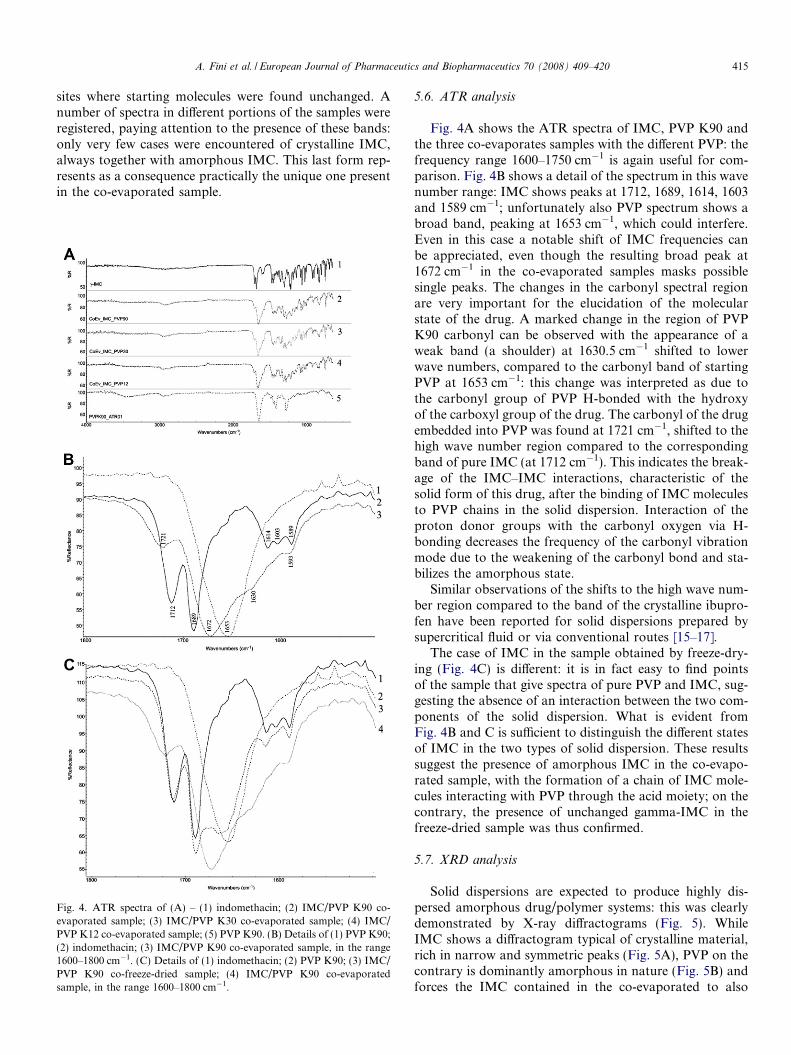
Vibrational spectroscopy provides an excellent methodfor probing solid state interactions between molecules,and the use of IR micro-spectroscopy for this purpose iswidely documented, while Raman micro-spectroscopy isless frequently employed in the field of pharmaceuticalresearch [10–13]. Moreover, modern equipment providesa very narrow spatial resolution and this aspect can beself-defeating when mixtures are analyzed, since the result-ing spectrum depends on the position of the beam; but atthe same time, this fact can offer evidence of heterogeneity.In this way, spectroscopy can be employed as a tool todescribe a mixture and reveal the situation of the compo-nents (e.g., amorphous or crystalline; free or bonded)[5,14].
Fig. 3A shows the Raman spectra of pure gamma-IMCand pure PVP together with that of the co-evaporated sys-tem. From the comparison of the three spectra it emergedthat the frequencies in the range 1500–1700 cm�1 are usefulto evaluate changes. The IMC molecule contains two car-bonyl groups of the benzoyl and carboxyl moieties. Taylorand Zografi [9] also suggested that the peaks present in thisrange must be attributed to the asymmetric benzoyl stretch-ing, while the peaks associated with the symmetrical stretch-ing vibrations of the cyclic dimer formed by the carboxylgroups are not active in the Raman spectra. There is a verynarrow and symmetric peak at 1700 cm�1: the same authorsprovided an explanation for the higher frequency withrespect to common amide groups. Three other peaks of lowerintensity are observed at lower frequencies: 1580, 1590 and1621 cm�1. This is clearly visible in Fig. 3B where a detailedand magnified portion of the spectrum is shown.
PVP shows only a broad and rounded peak (centred at1666 cm�1) in this range, attributed to the amide carbonylof the PVP units. The three types of PVP employed to pre-
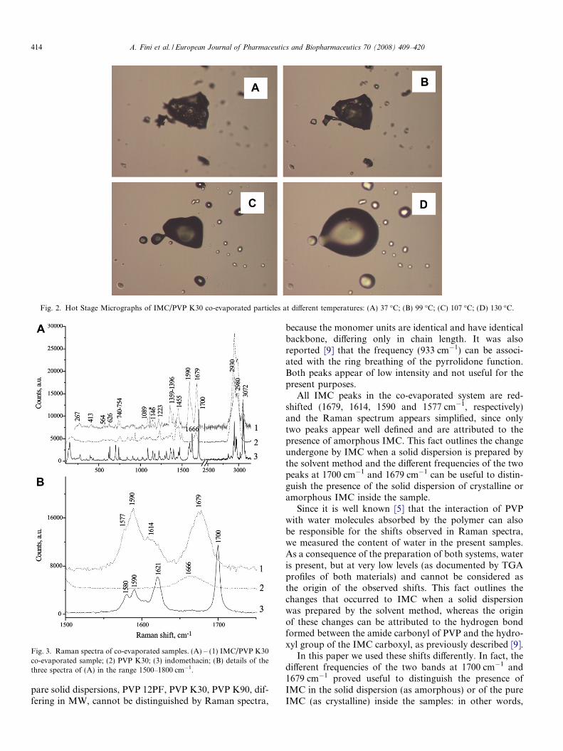
Fig. 2. Hot Stage Micrographs of IMC/PVP K30 co-evaporated particles at different temperatures: (A) 37 �C; (B) 99 �C; (C) 107 �C; (D) 130 �C.
Fig. 3. Raman spectra of co-evaporated samples. (A) – (1) IMC/PVP K30co-evaporated sample; (2) PVP K30; (3) indomethacin; (B) details of thethree spectra of (A) in the range 1500–1800 cm�1.
414 A. Fini et al. / European Journal of Pharmaceutics and Biopharmaceutics 70 (2008) 409–420
pare solid dispersions, PVP 12PF, PVP K30, PVP K90, dif-fering in MW, cannot be distinguished by Raman spectra,
because the monomer units are identical and have identicalbackbone, differing only in chain length. It was alsoreported [9] that the frequency (933 cm�1) can be associ-ated with the ring breathing of the pyrrolidone function.Both peaks appear of low intensity and not useful for thepresent purposes.
All IMC peaks in the co-evaporated system are red-shifted (1679, 1614, 1590 and 1577 cm�1, respectively)and the Raman spectrum appears simplified, since onlytwo peaks appear well defined and are attributed to thepresence of amorphous IMC. This fact outlines the changeundergone by IMC when a solid dispersion is prepared bythe solvent method and the different frequencies of the twopeaks at 1700 cm�1 and 1679 cm�1 can be useful to distin-guish the presence of the solid dispersion of crystalline oramorphous IMC inside the sample.
Since it is well known [5] that the interaction of PVPwith water molecules absorbed by the polymer can alsobe responsible for the shifts observed in Raman spectra,we measured the content of water in the present samples.As a consequence of the preparation of both systems, wateris present, but at very low levels (as documented by TGAprofiles of both materials) and cannot be considered asthe origin of the observed shifts. This fact outlines thechanges that occurred to IMC when a solid dispersionwas prepared by the solvent method, whereas the originof these changes can be attributed to the hydrogen bondformed between the amide carbonyl of PVP and the hydro-xyl group of the IMC carboxyl, as previously described [9].
In this paper we used these shifts differently. In fact, thedifferent frequencies of the two bands at 1700 cm�1 and1679 cm�1 proved useful to distinguish the presence ofIMC in the solid dispersion (as amorphous) or of the pureIMC (as crystalline) inside the samples: in other words,
A. Fini et al. / European Journal of Pharmaceutics and Biopharmaceutics 70 (2008) 409–420 415
sites where starting molecules were found unchanged. Anumber of spectra in different portions of the samples wereregistered, paying attention to the presence of these bands:only very few cases were encountered of crystalline IMC,always together with amorphous IMC. This last form rep-resents as a consequence practically the unique one presentin the co-evaporated sample.
Fig. 4. ATR spectra of (A) – (1) indomethacin; (2) IMC/PVP K90 co-evaporated sample; (3) IMC/PVP K30 co-evaporated sample; (4) IMC/PVP K12 co-evaporated sample; (5) PVP K90. (B) Details of (1) PVP K90;(2) indomethacin; (3) IMC/PVP K90 co-evaporated sample, in the range1600–1800 cm�1. (C) Details of (1) indomethacin; (2) PVP K90; (3) IMC/PVP K90 co-freeze-dried sample; (4) IMC/PVP K90 co-evaporatedsample, in the range 1600–1800 cm�1.
5.6. ATR analysis
Fig. 4A shows the ATR spectra of IMC, PVP K90 andthe three co-evaporates samples with the different PVP: thefrequency range 1600–1750 cm�1 is again useful for com-parison. Fig. 4B shows a detail of the spectrum in this wavenumber range: IMC shows peaks at 1712, 1689, 1614, 1603and 1589 cm�1; unfortunately also PVP spectrum shows abroad band, peaking at 1653 cm�1, which could interfere.Even in this case a notable shift of IMC frequencies canbe appreciated, even though the resulting broad peak at1672 cm�1 in the co-evaporated samples masks possiblesingle peaks. The changes in the carbonyl spectral regionare very important for the elucidation of the molecularstate of the drug. A marked change in the region of PVPK90 carbonyl can be observed with the appearance of aweak band (a shoulder) at 1630.5 cm�1 shifted to lowerwave numbers, compared to the carbonyl band of startingPVP at 1653 cm�1: this change was interpreted as due tothe carbonyl group of PVP H-bonded with the hydroxyof the carboxyl group of the drug. The carbonyl of the drugembedded into PVP was found at 1721 cm�1, shifted to thehigh wave number region compared to the correspondingband of pure IMC (at 1712 cm�1). This indicates the break-age of the IMC–IMC interactions, characteristic of thesolid form of this drug, after the binding of IMC moleculesto PVP chains in the solid dispersion. Interaction of theproton donor groups with the carbonyl oxygen via H-bonding decreases the frequency of the carbonyl vibrationmode due to the weakening of the carbonyl bond and sta-bilizes the amorphous state.
Similar observations of the shifts to the high wave num-ber region compared to the band of the crystalline ibupro-fen have been reported for solid dispersions prepared bysupercritical fluid or via conventional routes [15–17].
The case of IMC in the sample obtained by freeze-dry-ing (Fig. 4C) is different: it is in fact easy to find pointsof the sample that give spectra of pure PVP and IMC, sug-gesting the absence of an interaction between the two com-ponents of the solid dispersion. What is evident fromFig. 4B and C is sufficient to distinguish the different statesof IMC in the two types of solid dispersion. These resultssuggest the presence of amorphous IMC in the co-evapo-rated sample, with the formation of a chain of IMC mole-cules interacting with PVP through the acid moiety; on thecontrary, the presence of unchanged gamma-IMC in thefreeze-dried sample was thus confirmed.
5.7. XRD analysis
Solid dispersions are expected to produce highly dis-persed amorphous drug/polymer systems: this was clearlydemonstrated by X-ray diffractograms (Fig. 5). WhileIMC shows a diffractogram typical of crystalline material,rich in narrow and symmetric peaks (Fig. 5A), PVP on thecontrary is dominantly amorphous in nature (Fig. 5B) andforces the IMC contained in the co-evaporated to also

416 A. Fini et al. / European Journal of Pharmaceutics and Biopharmaceutics 70 (2008) 409–420
become amorphous (Fig. 5C): these materials exhibit dif-fused spectra typical of amorphous structures. On the con-trary the system previously studied was prepared by freeze-drying a suspension of IMC crystals (insoluble in water) inan aqueous solution of PVP: the final microparticles there-fore actually consisted of crystalline IMC covered by PVPmultiple layers. Diffractograms of this last system show thepatterns of crystalline IMC, even though of reduced inten-sity due to the composition of the system [3]. The presenceof an amorphous phase in the co-evaporated system intro-duces a source of instability, even though stabilized by thepresence of PVP: as a consequence, co-freeze-dried parti-cles, containing the crystalline phase of the drug, can beconsidered a more stable formulation.
6. Microcapsules
6.1. Spray-congealing
Particles obtained by the two methods of preparing soliddispersion were suspended in molten SA and spray-con-gealed obtaining a coating with a film of stearic acid.Fig. 5D suggests the microcapsule nature of the particleobtained by spray-congealing process: the external coating
Fig. 5. X-ray diffractograms. (A) Indomethacin; (B) PVP K30; (C) IMC/PVcongealing of IMC/PVP K30 co-evaporated sample (stearic acid of the surfac
is formed by crystalline stearic acid, without any peaks ofdifferent substances. The final particles have a sphericalshape, with a not perfectly smooth surface (Fig. 6): thepresence of IMC complexed by PVP seems to hindersmoothness, since IMC/SA microparticles, obtained atthe same weight ratio and experimental conditions, aresmoother. Possibly the mutual solubility of SA and IMCin the molten phase forces the solid mixture to a less crys-talline structure. In some cases, the SA coating reveals thedifficulty to obtain a perfect shape (Fig. 6A and B). More-over, SA crystallization does not leave the particle surfaceperfectly smooth (Fig. 6C) like those prepared by a soliddispersion formed by IMC and SA in the absence ofPVP: in this last case the mutual solubility of IMC andSA in the molten phase allows the formation of smoothand perfectly shaped microspheres (Fig. 6D).
6.2. Image analysis of microcapsules
Image analysis, carried out by means of SEM, extractsinformation contained in the microscopic observation bymeans of digital image processing analysis and presenta-tion of the shape and size parameters of the particles.The following three parameters, which are all normalised
P K30 co-evaporated sample; (D) particle obtained by ultrasound spraye).
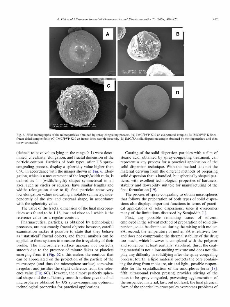
Fig. 6. SEM micrographs of the microparticles obtained by spray-congealing process. (A) IMC/PVP K30 co-evaporated sample; (B) IMC/PVP K30 co-freeze-dried sample (first); (C) IMC/PVP K30 co-freeze-dried sample (second). (D) IMC/SA solid dispersion sample obtained by melting method and thenspray-congealed.
A. Fini et al. / European Journal of Pharmaceutics and Biopharmaceutics 70 (2008) 409–420 417
(defined to have values lying in the range 0–1) were deter-mined: circularity, elongation, and fractal dimension of theparticle contour. Particles of both types, after US spray-congealing process, display a sphericity value higher than0.90, in accordance with the images shown in Fig. 6. Elon-gation, which is a measurement of the length/width ratio, isdefined as 1 � [width/length]: shapes symmetrical in allaxes, such as circles or squares, have similar lengths andwidths (elongation close to 0): final particles show verylow elongation values indicating a notable symmetry, inde-pendently of the size and external shape, in accordancewith the sphericity value.
The value of the fractal dimension of the final micropar-ticles was found to be 1.16, low and close to 1 which is thereference value for a regular contour.
Pharmaceutical particles, as obtained by technologicalprocesses, are not exactly fractal objects: however, carefulexamination makes it possible to state that they behaveas ‘‘statistical” fractal objects, and fractal analysis can beapplied to these systems to measure the irregularity of theirprofile. The microsphere surface appears not perfectlysmooth due to the presence of minute flakes or plateletsemerging from it (Fig. 6C): this makes the contour thatcan be appreciated on the projection of the particle of themicroscope (and thus the microsphere surface) somewhatirregular, and justifies the slight difference from the refer-ence value (Fig. 6C). However, the almost perfectly spher-ical shape and the sufficiently smooth surface gave the finalmicrospheres obtained by US spray-congealing optimumtechnological properties for practical applications.
Coating of the solid dispersion particles with a film ofstearic acid, obtained by spray-congealing treatment, canrepresent a key process for a practical application of thesolid dispersion technique. With this method it is not thematerial deriving from the different methods of preparingsolid dispersion that is handled, but spherically shaped par-ticles, with excellent technological properties of hardness,stability and flowability suitable for manufacturing of thefinal formulation [19].
The process of spray-congealing to obtain microspheresthat follows the preparation of both types of solid disper-sions also displays important functions in terms of practi-cal applications of solid dispersions, since it overcomesmany of the limitations discussed by Serajuddin [1].
First, any possible remaining traces of solvent,employed in the solvent method of preparation of solid dis-persion, could be eliminated during the mixing with moltenSA; second, the temperature of molten SA is relatively lowand does not compromise the thermal stability of the drugtoo much, which however is complexed with the polymerand somehow, at least partially, stabilized; third, the coat-ing material is not a low-melting mixture and does not dis-play any difficulty in solidifying after the spray-congealingprocess; fourth, a lipid material protects the core contain-ing the drug from moisture, air and light, possible respon-sible for the crystallization of the amorphous form [18];fifth, ultrasound (when present) provides stirring of themass to be spray-congealed, preventing agglomeration ofthe suspended material; last, but not least, the final physicalform of the spherical microcapsules overcomes problems of

418 A. Fini et al. / European Journal of Pharmaceutics and Biopharmaceutics 70 (2008) 409–420
preparing the final formulation in terms, e.g., of direct hardgelatine capsule filling.
Finally, the drug solubility and miscibility in meltedlipid, which determine the loading capacity of drug in thelipid particles, are less important in the present case, sincethe lipid material represents only the encapsulation layerexternal to the preformed IMC/PVP nuclei. Solid lipid par-ticles, composed of a physiological and well-tolerated lipid,combine several advantages: the possibility of controlleddrug release and drug targeting; the protection of incorpo-rated active compounds against changes or degradationand possibly of re-crystallization of the amorphous phaseincluding, by contact with moisture and with gastric envi-ronment, by enzymes or acidic fluids.
However, an analysis of the final material reveals thatthere are still some problems. Fig. 7 shows the Ramanspectra of materials present on the microsphere surfaceobtained for both types of materials. Obviously, the mostfrequently encountered material is SA, whose spectrum isshown in Fig. 7A. The two samples can be distinguishedby the presence of IMC peaks that refer either to the crys-
Fig. 7. Raman spectra of microparticles where the following substances werepeaks); (C) stearic acid and amorphous IMC (italic bold peaks); (D) stearic apeaks).
talline (Fig. 7B) or amorphous structure (Fig. 7C) of IMC,according to the preparation method of the solid disper-sion. Both IMC forms could be detected in some instancesalso in the freeze-dried sample (Fig. 7D), looking at thepresence of typical bands of one or of the other type ofIMC: it can be hypothesized that IMC particles, not com-pletely covered by PVP during the freeze-drying, could dis-solve into the molten SA, originating a limited amount ofamorphous portion. IMC particles could be observed atthe microcapsule surface as distinct crystals also in theco-evaporated samples. This can be a consequence of theinstability of the amorphous phase during the spray-con-gealing process of the formation of droplets. In fact, soliddispersion in the present study contains IMC and PVP at50/50 w/w ratio: according to calculated values [9], thisratio corresponds to a 1:2 mole ratio IMC/PVP (mono-mers), which is exceedingly high and guarantees hydrogenbinding of all the IMC molecules present in PVP chains,in other words to ensure complete amorphousness of thesystem. However, a low glass transition temperature forIMC was reported (Tg = 42.3 �C) [20]: in the presence of
detected: (A) stearic acid; (B) stearic acid and crystalline IMC (underlinedcid, crystalline IMC (underlined peaks) and amorphous IMC (italic bold

A. Fini et al. / European Journal of Pharmaceutics and Biopharmaceutics 70 (2008) 409–420 419
an IMC/PVP binary mixture this value increases up to78.1 �C (for PVP K30 and possibly this could be practicallythe same for other PVP types). As a consequence, this valuecould easily be overcome during the US atomization pro-cess, since the solid dispersion particles are in contact withmolten SA to produce final microspheres: at the workingtemperature for this process (80 �C), stabilization of theamorphous state can no longer be assured, even by thepresence of PVP. This can account for the presence of crys-talline IMC on the surface of the final microcapsules, afterthe coating with a film of stearic acid.
These facts again outline the potential instability of theamorphous phase and the great importance of Raman andFT-IR micro-analysis in exploring even small particles togather information on the structure of the sample andthe possible interactions existing among the componentsof a given formulation. It is worth noting that the resultsof the Raman analysis of the sample surface have statisticalvalue since only a limited number of points can beexamined.
0%
1%
2%
3%
4%
5%
6%
7%
8%
9%
10%
0 20 40 60 80 100 120
Time (min.)
Dis
solv
ed IM
C
0%
10%
20%
30%
40%
50%
60%
70%
80%
90%
0 2 4 6 8 10Time (min.)
Dis
solv
ed IM
C
97531
Fig. 8. Dissolution profiles. (A) Microparticles obtained by spray-congealing athe particle size at pH 2.0. �: 50 < x < 100 lm; j: 100 < x < 200 lm; N: 200 <congealing a suspension of IMC/PVP K30 particles with molten stearic acid: effN: 200 < x < 355 lm; �: 355 < x < 500 lm; j: unsieved; : pure indomethacinIMC/PVP K30 particles with molten stearic acid: effect of the particle size at psymbols) � e: 50 < x < 100 lm; j h: 100 < x < 200 lm; N M: 200 < x < 355between microparticles (100 < x < 200 lm) obtained by spray-congealing a suspfreeze-dried sample; N: co-evaporated sample; j: pure IMC 100 < x < 200 lm
6.3. Dissolution test
Fig. 8 shows the release profiles of the drug frommicrospheres.
Beside inhibiting the contact with water, SA coatingoffers protection against the gastric pH: dissolution atlow pH was observed to a very low extent (lower than5% after 2 h, with the exception of the smallest size frac-tion considered) (Fig. 8A). Differences appear markedonly when the first moments of the dissolution are con-sidered or when pH increased up to that of the intestinalenvironment: in this last case, release is complete in fewminutes (Fig. 8B). Behaviour as a function of time ismore important: aging appears not to have any influenceon the release and the dissolution profiles readily overlapafter 1 week and after 9 months (Fig. 8B). These factsshow the importance of the protection by stearic acidthat stabilizes the amorphous phase formed by the sol-vent evaporation method against the absorption of wateras plasticizer.
0%
10%
20%
30%
40%
50%
60%
70%
80%
90%
0 3 8 10
Time (min.)
Dis
solv
ed IM
C
0%
10%
20%
30%
40%
50%
60%
70%
80%
90%
0 2 6 9 10Time (min.)
Dis
solv
ed IM
C
9765421
875431
suspension of IMC/PVP K30 particles with molten stearic acid: effect ofx < 355 lm; �: 355 < x < 500 lm. (B) Microparticles obtained by spray-
ect of the particle size at pH 7.4 �: 50 < x < 100 lm; j: 100 < x < 200 lm;<200 lm. (C) Microparticles obtained by spray-congealing a suspension ofH 7.4 and of aging: after a week (black symbols) and after 9 months (openlm; � s: 355 < x < 500 lm. (D) Comparison of IMC dissolution profilesension of IMC/PVP K30 particles with molten stearic acid at pH 7.4 �: co-(for comparison).

420 A. Fini et al. / European Journal of Pharmaceutics and Biopharmaceutics 70 (2008) 409–420
Finally, the comparison between the release profiles ofboth systems reveals practically no difference and this sug-gests that the two methods of association between IMCand PVP behave in the same way to improve the releaseof the drug and that the choice between the two methodsmust be made taking into account the parameters otherthan release, such as stability of the final system and easi-ness of the process (Fig. 8C).
7. Conclusions
Although solid dispersions offer a significant improve-ment in drug release with respect to a neat amorphousstate, there is still a potential risk of crystallization of thesesystems, when an amorphous phase is formed. As an alter-native, the solid dispersion obtained by co-freeze-dryingallows IMC to be associated with the hydrophilic PVP,though maintaining its crystalline phase and avoiding anyrisk of changes on aging. Despite this, the formulationgreatly improves the availability of the anti-inflammatorydrug through rapid dissolution. This process can be pro-posed as a first and general choice for other anti-inflamma-tory drugs that, like IMC, are poorly soluble in water: theprocess is quite simple and suitable for practical applica-tions, since it does not require organic solvents, heatingor complex apparatus, but only a water-soluble polymer.Finally, particles of the solid dispersion, protected by astearic acid coating, are stabilized against possible degrada-tion caused by the external environment. This process canalso be suggested as a general way of stabilizing an amor-phous phase that could be protected against the recovery ofthe crystalline state, maintaining the advantages of amor-phization obtained with both solvent and melting methodsto prepare solid dispersions.
Acknowledgement
This study was partially supported by MIUR Funds.
References
[1] A.T.M. Serajuddin, Solid dispersion of poorly water-soluble drugs:early promises, subsequent problems, and recent breakthroughs, J.Pharm. Sci. 88 (1999) 1058–1066.
[2] T. Watanabe, S. Hasegawa, N. Wakiyama, A. Kusai, M. Senna,Comparison between polyvinylpyrrolidone and silica nanoparticles ascarriers for indomethacin in a solid state dispersion, Int. J. Pharm. 20(2003) 283–286.
[3] C. Cavallari, B. Luppi, A.M. Di Pietra, L. Rodriguez, A. Fini,Enhanced release of indomethacin from PVP/stearic acid microcap-
sules prepared coupling co-freeze-drying and ultrasound assistedspray-congealing process, Pharm. Res. 24 (2007) 521–529.
[4] K. Kougaz, S.D. Clas, Crystallization inhibition in solid dispersion ofMK-0591 and poly(vinylpyrrolidone) polymers, J. Pharm. Sci. 89(2000) 1325–1334.
[5] L.S. Taylor, F.W. Langkilde, G. Zografi, Fourier transformationraman spectroscopy study for the interaction of water vapour withamorphous polymers, J. Pharm. Sci. 90 (2001) 888–901.
[6] K. Takayama, N. Nambu, T. Nagai, Dissolution kinetics of copre-cipitates of indomethacin with poly-vinylpyrrolidone, Chem. Pharm.Bull. 28 (1980) 3304–3309.
[7] M. Yoshioka, B.C. Hancock, G. Zografi, Inhibition of indomethacincrystallization in poly(vinylpyrrolidone) coprecipitates, J. Pharm. Sci.84 (1995) 983–986.
[8] T. Matsumoto, G. Zografi, Physical properties of solid moleculardispersion of indomethacin with poly(vinylpyrrolidone-co-vinyl ace-tate) in relation to indomethacin crystallization, Pharm. Res. 16(1999) 1722–1728.
[9] L.S. Taylor, G. Zografi, Spectroscopic characterization of interactionbetween PVP and indomethacin in amorphous molecular dispersion,Pharm. Res. 14 (1997) 1691–1698.
[10] S.C. Park, M. Kim, J. Noh, H. Chung, Y. Woo, J. Lee, M.S. Kemper,Reliable and fast quantitative analysis of active ingredient inpharmaceutical suspension using Raman spectroscopy, Anal. Chim.Acta 593 (2007) 46–53.
[11] M. Kim, H. Chung, Y. Woo, M.S. Kemper, A new non invasive,quantitative Raman technique for determination of an active ingre-dient in pharmaceutical liquids by direct measurements through aplastic bottle, Anal. Chim. Acta 587 (2007) 200–207.
[12] C.J. Strachan, T. Rades, K.C. Gordon, J. Rantanen, Ramanspectroscopy for quantitative analysis of pharmaceutical solids, J.Pharm. Pharmacol. 59 (2007) 179–192.
[13] G. Zoppetti, N. Puppini, F. Ospitali, A. Fini, Solid state character-ization of progesterone in a freeze dried 1:2 progesterone/HPBCDmixture, J. Pharm. Sci. 96 (2007) 1729–1736.
[14] L.S. Taylor, G. Zografi, The quantitative analysis of crystallinityusing FT-Raman spectroscopy, Pharm. Res. 15 (1998) 755–761.
[15] S.G. Kazarian, G.G. Martirosyan, Spectroscopy of polymer/drugformulations processed with supercritical fluids: in situ ATR-IR andRaman study of impregnation of ibuprofen into PVP, Int. J. Pharm.232 (2002) 81–90.
[16] T.P. Shakhtshneider, M.A. Vasiltchenko, A.A. Politov, V.V. Boldy-rev, The mechanochemical preparation of solid disperse systems ofibuprofen–polyethylene glycol, Int. J. Pharm. 130 (1996) 25–32.
[17] Y. Shibata, M. Fujii, M. Kokudai, S. Noda, H. Okada, M. Kondoh,Y. Watanabe, Effect of characteristics of compounds on maintenanceof an amorphous state in solid dispersion with crospovidone, J.Pharm. Sci. 96 (2007) 1537–1547.
[18] K.J. Crowley, G. Zografi, Water vapor absorption into amorphoushydrophobic drug/poly(vinylpyrrolidone) dispersions, J. Pharm. Sci.91 (2002) 2150–2165.
[19] L. Rodriguez, N. Passerini, C. Cavallari, M. Cini, P. Sancin, A. Fini,Description and preliminary evaluation of a new ultrasonic atomizerfor spray-congealing processes, Int. J. Pharm. 183 (1999) 133–143.
[20] R.J. Chokshi, H.K. Sandhu, R.M. Iyer, N.H. Shah, A.W. Malick, H.Zia, Characterization of physico-mechanical properties of indometh-acin and polymers to assess their suitability for hot melt extrusionprocess as a means to manufacture solid dispersion solution, J.Pharm. Sci. 94 (2005) 2463–2474.