rady 403 case presentation katherine i. jicha,...
TRANSCRIPT
Katherine I. Jicha, MS3May 2019
RADY 403 Case Presentation
Focused patient history and workup
• 1 month old boy in neonatal ICU presented with worsening respiratory status with frequent apneic events
• Urine culture positive for methicillin susceptible Staph aureus
• Born prematurely by C-section due to decreased fetal movement and non-reactive Non-Stress Test (NST)• Completed 29 weeks gestation
• Other conditions: congenital anomaly of the brain, perinatal intraventricular hemorrhage, congenital CMV, cleft lip and palate, chronic lung disease
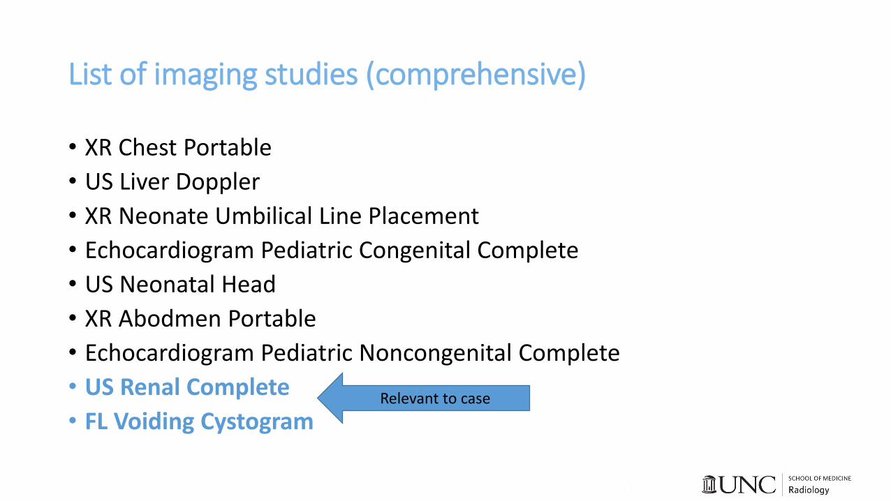
List of imaging studies (comprehensive)
• XR Chest Portable
• US Liver Doppler
• XR Neonate Umbilical Line Placement
• Echocardiogram Pediatric Congenital Complete
• US Neonatal Head
• XR Abodmen Portable
• Echocardiogram Pediatric Noncongenital Complete
• US Renal Complete
• FL Voiding CystogramRelevant to case
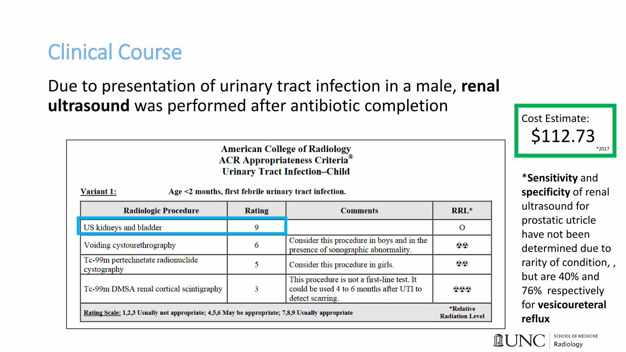
Clinical Course
Due to presentation of urinary tract infection in a male, renal ultrasound was performed after antibiotic completion
*Sensitivity and specificity of renal ultrasound for prostatic utricle have not been determined due to rarity of condition, , but are 40% and 76% respectively for vesicoureteral reflux
Cost Estimate:
$112.73*2017
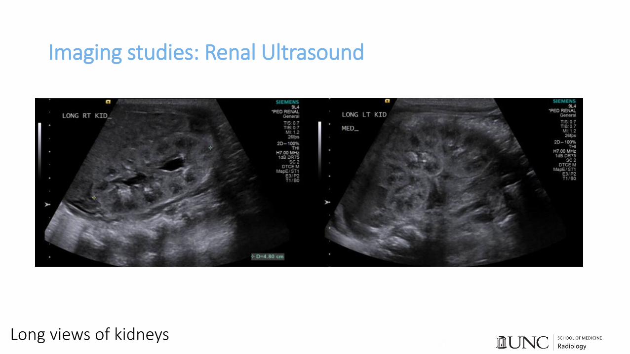
Imaging studies: Renal Ultrasound
Long views of kidneys
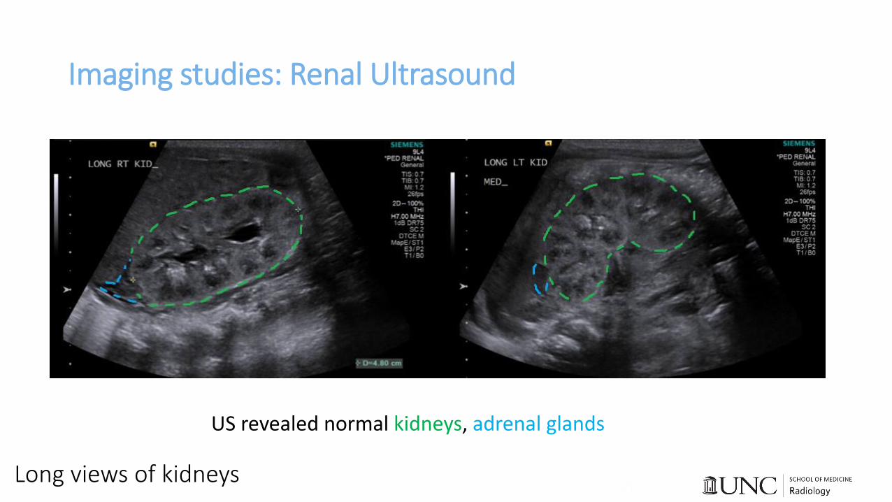
Imaging studies: Renal Ultrasound
Long views of kidneys
US revealed normal kidneys, adrenal glands
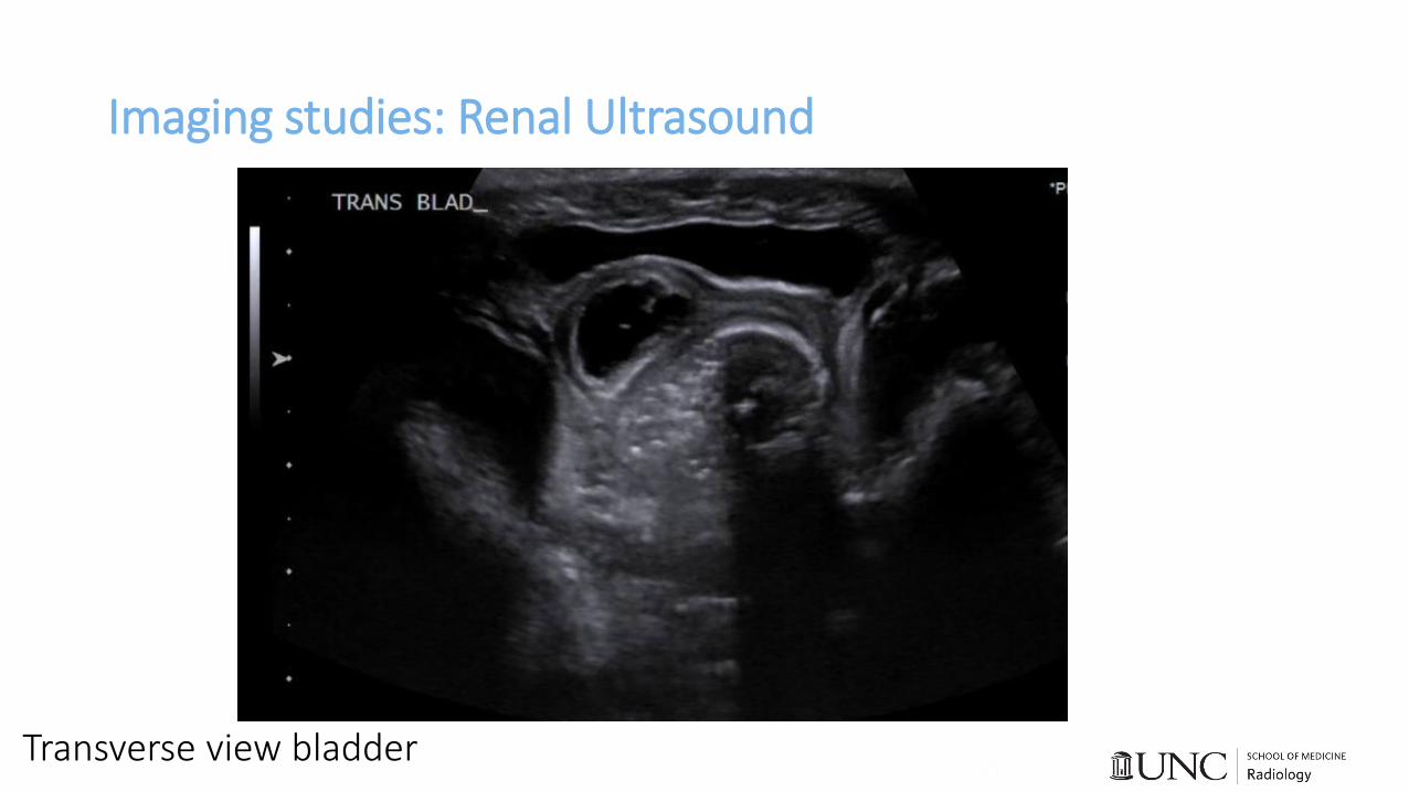
Imaging studies: Renal Ultrasound
Transverse view bladder
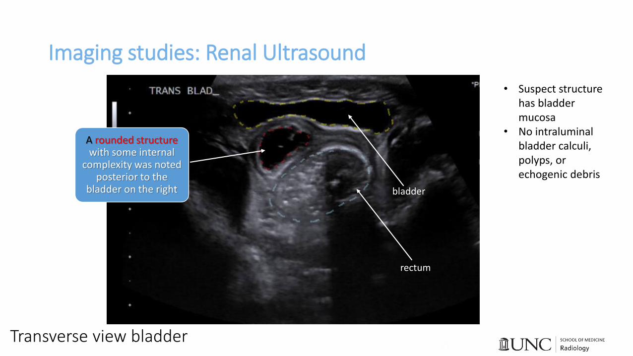
Imaging studies: Renal Ultrasound
Transverse view bladder
bladder
A rounded structure with some internal
complexity was noted posterior to the
bladder on the right
• Suspect structure has bladder mucosa
• No intraluminal bladder calculi, polyps, or echogenic debris
rectum
Imaging studies: Renal Ultrasound
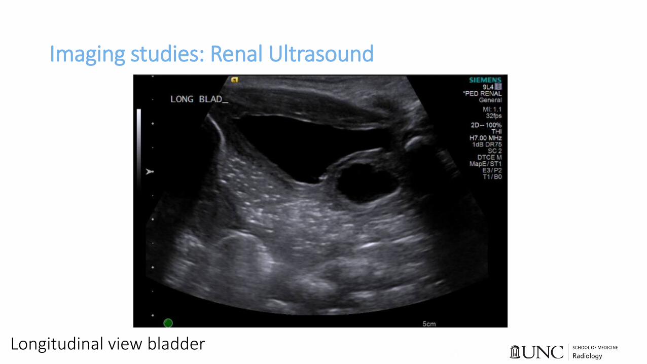
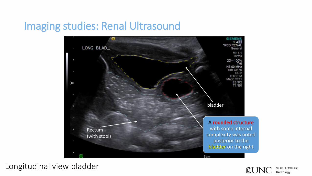
Longitudinal view bladder
Imaging studies: Renal Ultrasound
Longitudinal view bladder
A rounded structure with some internal
complexity was noted posterior to the
bladder on the right
Rectum(with stool)
bladder

Clinical CourseUltrasound showed normal kidneys and adrenal glands, but also an irregular cystic structure behind the bladder prompting concern for ureterocele. Next step? Voiding Cystourethrogram (VCUG)
*Sensitivity and specificity of VCUG for prostatic utriclehave not been determined due to rarity of condition, but sensitivity is 88% for vesicoureteral reflux
$500-1000Cost Estimate:

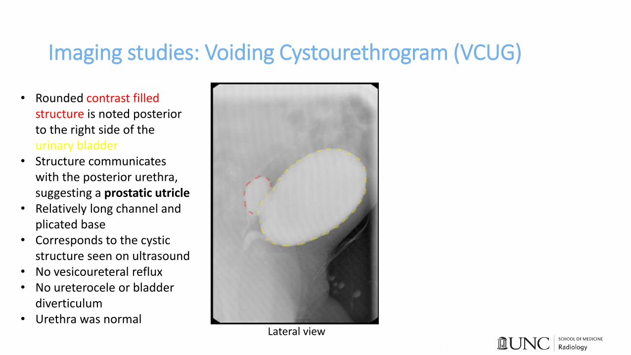
Imaging studies: Voiding Cystourethrogram (VCUG)
Lateral view
Imaging studies: Voiding Cystourethrogram (VCUG)
Lateral view
• Rounded contrast filled structure is noted posterior to the right side of the urinary bladder
• Structure communicates with the posterior urethra, suggesting a prostatic utricle
• Relatively long channel and plicated base
• Corresponds to the cystic structure seen on ultrasound
• No vesicoureteral reflux• No ureterocele or bladder
diverticulum• Urethra was normal
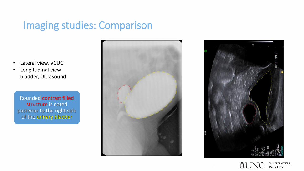
Imaging studies: Comparison
Rounded contrast filled structure is noted
posterior to the right side of the urinary bladder
• Lateral view, VCUG• Longitudinal view
bladder, Ultrasound
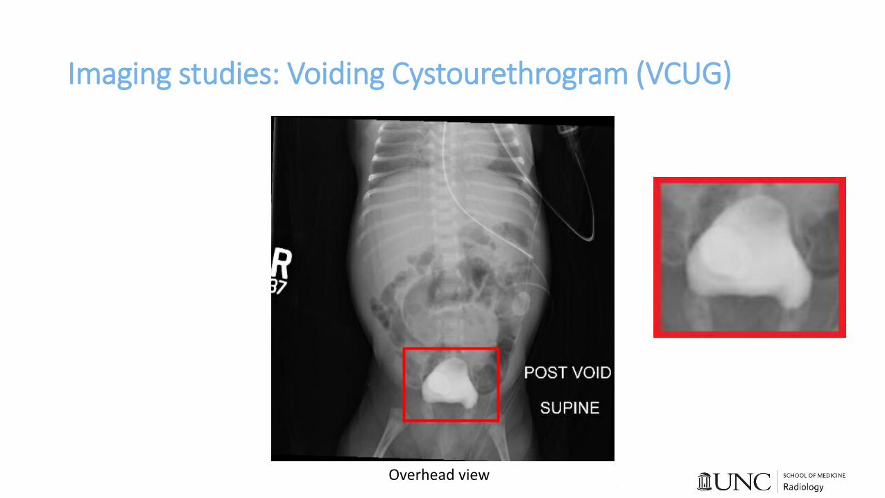
Imaging studies: Voiding Cystourethrogram (VCUG)
Overhead view
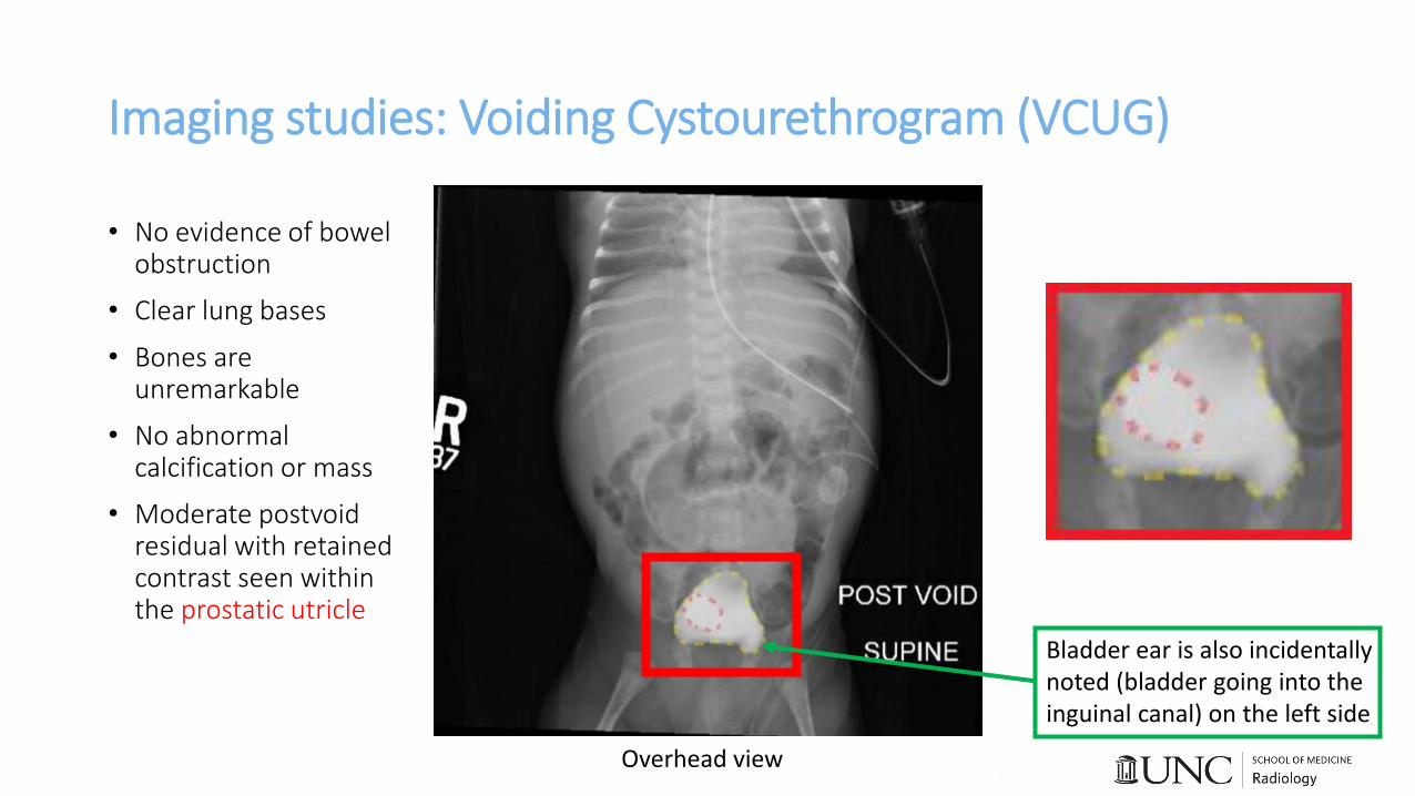
Imaging studies: Voiding Cystourethrogram (VCUG)
• No evidence of bowel obstruction
• Clear lung bases
• Bones are unremarkable
• No abnormal calcification or mass
• Moderate postvoid residual with retained contrast seen within the prostatic utricle
Overhead view
Bladder ear is also incidentally noted (bladder going into the inguinal canal) on the left side
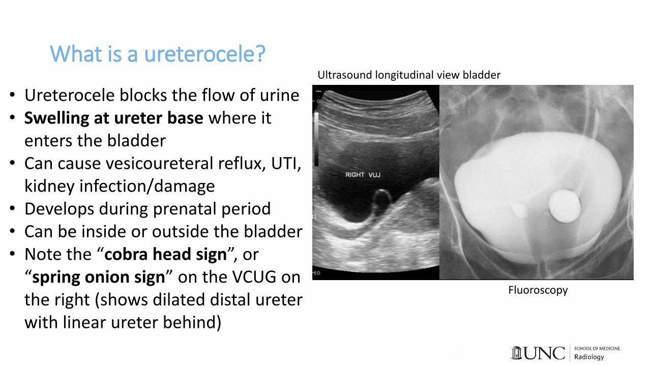
What is a ureterocele?Ultrasound longitudinal view bladder
Fluoroscopy
• Ureterocele blocks the flow of urine• Swelling at ureter base where it
enters the bladder• Can cause vesicoureteral reflux, UTI,
kidney infection/damage• Develops during prenatal period• Can be inside or outside the bladder• Note the “cobra head sign”, or
“spring onion sign” on the VCUG on the right (shows dilated distal ureter with linear ureter behind)

What is a ureterocele?Ultrasound longitudinal view bladder
Fluoroscopy
bladder
ureterocele
• Dilatation at ureter base where it enters the bladder
• Can cause vesicoureteral reflux, UTI, kidney infection/damage
• Develops during prenatal period• Can be inside (trigone or bladder
base), or outside the bladder (everting)
• Note the “cobra head sign”, or “spring onion sign” on the VCUG on the right (shows dilated distal ureter with linear ureter behind, at trigone)

Patient treatment or outcome
• Based on the findings of the VCUG (moderate-sized prostatic utricle), urology did not recommend UTI antibiotic prophylaxis
• Per urology, no additional imaging or follow up will be required in the future
• Patient remained in neonatal ICU due to numerous other medical complications
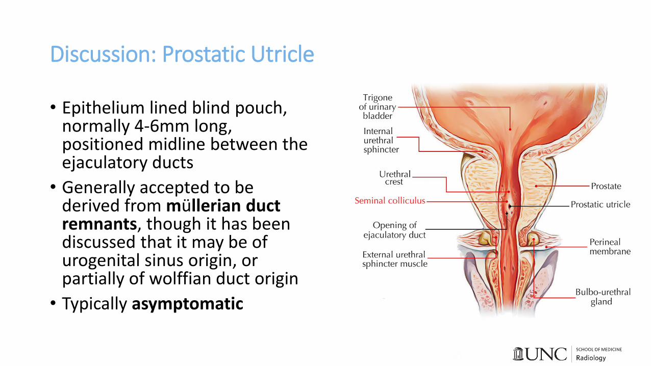
Discussion: Prostatic Utricle
• Epithelium lined blind pouch, normally 4-6mm long, positioned midline between the ejaculatory ducts
• Generally accepted to be derived from müllerian duct remnants, though it has been discussed that it may be of urogenital sinus origin, or partially of wolffian duct origin
• Typically asymptomatic
Discussion: Enlarged Prostatic Utricle
• Enlarged prostatic utricles may become symptomatic• Lower urinary tract irritation / repeated UTIs• Post micturition dribbling• Urethral discharge• Stone formation within the utricle• Urine retention• Hematospermia• Epididymitis
• Can cause infertility• Utricles may become malignant (3%) secondary to repeated UTI• >50% incidence in patients with perineal hypospadias (opening is behind
the scrotal sac); also seen in patients with posterior urethral valves, and Eagle Barrett syndrome (prune belly)
Discussion: Enlarged Prostatic Utricle
• Treatment is generally reserved for symptomatic patients
• Surgery is the definitive treatment• Endoscopic and laparoscopic approaches have been described
• Open excision is recommended in pediatric age group
• Surgery is challenging because it is close to many important structures; most severe adverse effect is damage to pelvic nervescausing incontinence
Take Home Point
• Prostatic utricle is a rare underlying cause of UTI in a young male
• Treatment is reserved for symptomatic patients only as surgery can be challenging
References• 1. Shapiro E, Huang H, McFadden DE, et al. The prostatic utricle is not a müllerian duct remnant: Immunohistochemical evidence for a distinct urogenital
sinus origin. In: Journal of Urology. ; 2004. doi:10.1097/01.ju.0000140267.46772.7d
• 2. Oh CS, Chung IH, Won HS, Kim JH, Nam K Il. Morphologic variations of the prostatic utricle. Clin Anat. 2009. doi:10.1002/ca.20759
• 3. Ramachandra M, Bendre P, Redkar R, Taide D. Isolated prostatic utricle. J Indian Assoc Pediatr Surg. 2010. doi:10.4103/0971-9261.59610
• 4. Desautel MG, Stock J, Hanna MK. Mullerian duct remnants: Surgical management and fertility issues. In: Journal of Urology. ; 1999. doi:10.1016/S0022-5347(01)68050-9
• 5. IKOMA F, SHIMA H, YABUMOTO H. Classification of Enlarged Prostatic Utricle in Patients with Hypospadias. Br J Urol. 1985. doi:10.1111/j.1464-410X.1985.tb06356.x
• 6. Willetts IE, Roberts JP, MacKinnon AE. Laparoscopic excision of a prostatic utricle in a child. Pediatr Surg Int. 2003. doi:10.1007/s00383-003-0993-6
• 7. Mostafa IA, Woodward MN, Shalaby MS. Cystoscopic-assisted laparoscopic excision of prostatic utricle. J Pediatr Urol. 2018. doi:10.1016/j.jpurol.2017.09.024
• 8. Krstić ZD, Smoljanić Ž, Mićović Ž, Vukadinović V, Sretenović A, Varinac D. Surgical treatment of the Müllerian duct remnants. J Pediatr Surg. 2001. doi:10.1053/jpsu.2001.23958
• 9. Coppens L, Bonnet P, Andrianne R, De Leval J. Adult müllerian duct or utricle cyst: Clinical significance and therapeutic management of 65 cases. J Urol. 2002. doi:10.1016/S0022-5347(05)65190-7
• 10. Devine CJ, Gonzalez-Serva L, Stecker JF, Horton CE. Utricular configuration in hypospadias and intersex. J Urol. 1980. doi:10.1016/S0022-5347(17)55959-5
• 11. Meisheri I V., Motiwale SS, Sawant V V. Surgical management of enlarged prostatic utricle. Pediatr Surg Int. 2000. doi:10.1007/s003830050722
• 12. Schuhrke TD, Kaplan GW. Prostatic utricle cysts (Mullerian duct cysts). J Urol. 1978. doi:10.1016/S0022-5347(17)57627-2

References• 13. Husmann DA, Allen TD. Endoscopic management of infected enlarged prostatic utricles and remnants of rectourethral fistula tracts of high imperforate
anus. J Urol. 1997. doi:10.1016/S0022-5347(01)64898-5
• 14. Priyadarshi V, Singh JP, Mishra S, Vijay MK, Pal DK, Kundu AK. Prostatic utricle cyst: a clinical dilemma. APSP J Case Rep. 2013.
• 15. Chung BI, Sommer G, Brooks JD. Anatomy of the Lower Urinary Tract and Male Genitalia. In: Campbell-Walsh Urology. ; 2012. doi:10.1016/b978-1-4160-6911-9.00002-5
• 16. Volker, Joseph H. “Easy Notes On 【Verumontanum or Seminal Colliculus】 – Earth's Lab.” Earth's Lab, 30 Aug. 2018, www.earthslab.com/anatomy/verumontanumseminal-colliculus/.
• 17. “ACR Appropriateness Criteria®.” ® | American College of Radiology, www.acr.org/Clinical-Resources/ACR-Appropriateness-Criteria.
• 18. GE, Healthcare. “Reimbursement Informationfor Diagnostic Elastography.” GetHealthcare, Aug. 2017, www.gehealthcare.com/-/media/ad0a303c8f5e4eeb9cf92e00bb8dcb00.pdf?la=en&hash=6FBD89883D56A4D5AED66FFED06C669CA7D28A5D.
• 19. Mahant S, Friedman J, MacArthur C. Renal ultrasound findings and vesicoureteral reflux in children hospitalised with urinary tract infection. Arch Dis Child. 2002. doi:10.1136/adc.86.6.419
• 20. Kis É, Nyitrai A, Várkonyi I, et al. Voiding urosonography with second-generation contrast agent versus voiding cystourethrography. Pediatr Nephrol. 2010. doi:10.1007/s00467-010-1618-7
• 21. MD Save. “How Much Does a Cystogram (VCUG) Cost Near Me?” MDsave, 2019, www.mdsave.com/procedures/cystogram-vcug/d785fcc4.
• 22. Gaillard, Frank. “Ureterocoele | Radiology Case.” Radiopaedia Blog RSS, 2010, radiopaedia.org/cases/ureterocoele?lang=us.
• 23. Patel, Maulik S. “Ureterocoele | Radiology Case.” Radiopaedia Blog RSS, 2010, radiopaedia.org/cases/ureterocoele-6?lang=us.
• 24. “What Is a Ureterocele?” What Is a Ureterocele? - Urology Care Foundation, www.urologyhealth.org/urologic-conditions/ureterocele.
• 25. Radswiki. “Cobra Head Sign (Ureter) | Radiology Reference Article.” Radiopaedia Blog RSS, radiopaedia.org/articles/cobra-head-sign-ureter?lang=us.