radiotherapy in prostate cancer - intech -...
TRANSCRIPT

8
Radiotherapy in Prostate Cancer
Tatjana Arsenijević, Kata Dabić Stanković, Miodrag Aćimović and Ljiljana Radošević Jelić
Institute for Oncology and Radiology of Serbia, Institute for Urology Serbia
1. Introduction
Therapeutic management of prostate cancer has become complex, multidisciplinary and
stage-specific. (Heidenereich et al., 2011) Based on PSA level, histopathological grading
and clinical staging, prostate cancer is classified as low-, intermediate- and high risk for
disease recurrence. The risk status often plays a major role in deciding further therapy.
(Kirby & Madhavan, 2010) It is usually impossible to state that one therapy is superior to
another because of the lack of randomized controlled trials. However some
recommendations can be made. (Heidenereich et al., 2011, Aus et al. 2001) Based on
European Association of Urology recommendations in 2010, patients with low-risk (PSA
≤10 ng/ml, Gleason score <6 and cT1c-cT2a) or intermediate risk prostate cancer (PSA
10.1-20 ng/ml, Gleason score 7 or cT2b-c) are to be treated interdisciplinary with an
urologist and a radiation oncologist. Treatment options for these patients vary from
watchful waiting and active surveillance to radical prostatectomy or definitive
radiotherapy. Multidisciplinary tumor board is needed when discussing neoadjuvant and
adjuvant treatment options in high-risk prostate cancer patients (PSA>20ng/ml, Gleason
score 8-10 or ≥cT3a) (Heidenereich et al., 2011, Choe & Liauw, 2010).
Radiotherapy is widely used as curative treatment modality for prostate cancer. There is a
diverse array of radiotherapeutic strategies that can be effectively used to treat both organ-
confined and locally advanced disease, alone or in combination with androgen-deprivation
therapy. Furthermore, it has also a significant role in post-prostatectomy setting, as adjuvant
or salvage radiotherapy.
In recent decades, radiotherapy in prostate cancer has undergone significant clinical and
technological advances that aim to optimize cancer control outcomes while minimizing
treatment morbidity. (Choe & Liauw, 2010, Hayden et al., 2010)
2. External-beam radiotherapy in prostate cancer
External-beam radiotherapy has a very long history in the curative treatment of prostate
cancer. It is proven and most extensively used radiation modality. As a flexible,
noninvasive, outpatient therapy, external-beam radiotherapy can be used in all stages of
prostate cancer. (Hayden et al., 2010) It is based on daily delivery of radiation to a target
volume using high-energy radiation beams from linear accelerators (or cobalt machines)
over a course of 7 to 9 weeks.
www.intechopen.com

Prostate Cancer – Diagnostic and Therapeutic Advances
170
In the last 30 years it has undergone a long, improving path from conventional, two-dimensional radiotherapy to intensity-modulated and image-guided radiotherapy and onwards. In low-risk prostate cancer its efficacy appears to be comparable to that of radical prostatectomy but with different toxicities (Choe & Liauw, 2010). For patients not suitable for surgery, external-beam radiotherapy will be the treatment of choice in most cases, alone or combined with androgen-deprivation therapy.
2.1 Conventional radiotherapy Introducing high-energy radiotherapy machines it become possible to deliver tumoricidal doses to target volume while minimizing damage to the skin and adjacent organs. Historically, various techniques have been used, ranging from parallel anteroposterior-/posteroanterior (AP/PA) portals to lateral portals (box technique) or rotational fields to irradiate or supplement the dose to the prostate. (Chao et al, 2002) Due to the difficulty of localizing the prostate gland, a large volume was treated to ensure proper coverage. More of the surrounding tissues were included in the treated volume so the safely applicable dose was limited to 60-65Gy. (Choe & Liauw, 2010) The treatment fields for prostate cancer were simulated and designed on plane films and using bony markers with the patient in the supine position. Rectum and bladder were marked by intraluminal injection of iodinated contrast. Small intestine was marked with barium contrast ingested per os one hour prior to simulation. If the external-beam radiotherapy was applied to the prostate only (local technique), the field
size was approximately 8x10 cm for T1 and T2 tumors, 10x12 cm or 12x14 cm for T3 and T4
prostate cancer. Patients younger than 71 years of age with clinical T1c, T2a, and Gleason score
more than 7 and PSA 20ng/ml or more, as well as patients with T2b, c T3 and T4 were treated
to the whole pelvis with the field size of 15x15 cm, or 15x18 cm to cover the common iliac
nodes. The inferior margin of the field usually was 1.5 cm distal to the junction of the prostatic
and membranous urethra that is at or caudal to the bottom of the ischial tuberosities. The
lateral margins were approximately 1 to 2 cm from the lateral bony pelvis.
(a) (b)
Fig. 1. Simulation AP radiograph for field verification (a) size 12x12 cm and (b) 14x14 cm Low field border is on the lower border of ishiadic bone and the field centre on the symphisis. Barium contrast in bowels (With thanks to the Institute for oncology and radiology of Serbia, Belgrade)
www.intechopen.com

Radiotherapy in Prostate Cancer
171
The initial lateral fields included a volume similar to that treated with AP/PA portals. The anterior margin was 1.5 cm posterior to the projection of the anterior cortex of the pubic symphisis. Posteriorly, the fields included the pelvic and presacral lymph nodes above the S3 segment, which allowed some sparing of posterior rectal wall distal to this level. Large treated volume was obtained since the average variation of prostate position relative to bony markers was approximately 8 mm in the superior and posterior positions, 7 mm in the inferior, 5 mm in the lateral and 4 mm in anterior position. The seminal vesicles are located high in the pelvis, and posterior to the bladder, which was very critical when reducing treated volume in T3 patients. When indicated, the periaortic lymph nodes could be treated through extended AP/PA
portals or separate periaortic fields placed above the pelvic fields. The superior margin of
the periaortic field was at the Th12-L1 vertebral interspace with the width usually above 10
cm (determined by lymphangiogram or CT scan). (Chao et al., 2002, Dobbs et al., 1999)
Conventional external-beam radiotherapy required daily radiation delivery to target
volume using high-energy beams (more than 10MV). In most institutions a standard
fractionation was used with 1.8 to 2Gy per day. In radical approach, initially a dose of 45-
50Gy was applied to the whole pelvis or to the prostate through two parallel opposed
fields (AP/PA). Than addition of a boost dose was delivered, up to a total dose of 65-66Gy
through lateral opposed fields, two anterior or two posterior oblique fields. (Chao et al.,
2002, Dobbs et al., 1999)
2.2 Two-dimensional radiotherapy (2D-RT) Once a decision is being made to treat prostate cancer with external-beam radiotherapy, the radiotherapy plan is defined either to limit treatment to the gland or to extend treatment field to include the periprostatic tissues, seminal vesicles and pelvic lymph nodes. (Hayden et al. 2010) CT or MRI of the abdomen and pelvis is used to assess the involvement of surrounding structures. MRI is particularly useful for distinguishing capsular invasion, seminal vesicle involvement and periapical extension. CT scanning for treatment planning is performed to every patient, which means that two-dimensional (2D) radiotherapy is a step forward comparing to conventional radiotherapy. The patient is immobilized in supine position with skin tattoos over the pubic symphisis
and laterally over the iliac crests to prevent lateral rotation. CT scans of pelvis are
obtained with slice thickness of 4-5 mm. No oral, rectal or intravenous contrast is used.
The CT section at the centre of the volume is used as the main planning slice to outline
patient contour, target volume, rectum, and bladder. The margins of the target volume are
determined by the tumor extent. The gross tumor volume contains entire prostate gland,
but if there is a risk of seminal vesicles involvement, they must be included in target
volume too. The gross tumor volume is outlined on the central slice only. To allow
position variation, an additional margin is added to gross tumor volume (1-1.5 cm in all
directions) defining planning target volume (PTV). For two-dimensional planning, PTV is
outlined on multiple sections to ensure that the entire tumor is encompassed. The rectal
outline must be transposed on to the central section so that the dose can be adequately
calculated. Shaping the target volume by shielding blocks or multileaf collimators reduces
the dose to normal tissues.
Treatment technique depends on the target volume size and shape. Three-field technique using an anterior and two posterior oblique or opposed lateral fields give a high dose to the
www.intechopen.com
Prostate Cancer – Diagnostic and Therapeutic Advances
172
prostate but spare the posterior rectal wall. Four-field (box) technique may result in better dose distribution when seminal vesicles are included in target volume, but increases the dose posteriorly. The patient is than treated daily, 1.8 to 2Gy per fraction, on linear accelerator. The correct position is assured with skin tattoos. The field centre is marked with a tattoo also. All fields are treated isocentrically with shielding as instructed. The recommended dose is 64Gy in 32 fractions given in 6.5 to 7 weeks. (Dobbs et al., 1999)
2.3 Three-dimensional conformal radiotherapy (3D-CRT) Introducing CT-based radiotherapy simulations by the mid 1980s and multileaf collimator in new aged linear accelerators, it became possible to arrange treatment fields to individually match prostate target volume minimizing high dose exposure to adjacent normal tissues. This led to dose escalation without inducing more toxicity and implementing three-dimensional conformal radiotherapy in clinical practice as a gold standard in prostate cancer radiotherapy treatment. Practically, the aim is to minimize treatment toxicity for patients with more favorable disease, and to maximize locoregional tumor control for those with less favorable disease. (Hayden et al., 2010, Dearnaley, 2001, Gazdda et al., 1996/97) For 3D-CRT treatment planning a multi-slice CT scan and 3D planning system is used. In
order to minimize random and systemic setup error, prior to CT scan, patient should be
positioned and immobilized in a fashion that position obtained can be maintained and
reproduced. This is secured by the use of alpha cradles, shells or by positioning the patient
in supine position with leg restraints. In all this cases a midline and lateral laser lights are
used for set up. These markers are tattooed on the skin over the pubic symphisis and
laterally over the iliac crests. This is very important for daily positioning of the patient and
prevention of lateral rotation. The position is later verified by portal image. (Dobbs et al.,
1999, Hayden et al., 2010, Malone et al., 2000)
CT scan of pelvis is performed in a treatment position. Patient should empty the rectum
and the bladder should be comfortably full both on the simulation and on the radiation.
This standard should be followed because the variation in bladder and rectum distension
results in significant prostate displacement. A great controversy still remains regarding
prostate apex. MRI of pelvis and CT/MRI fusion is recommended to reduce inter-
observer variability in contouring, improving target delineation accuracy, particularly the
prostate apex. This fusion is also recommended were significant CT artifact is present i.e.
from hip prosthesis.
Once the CT and/or MRI scans are obtained on each slice a target volume and organs at risk
are delineated. Organs at risk are adjacent structures endangered by high radiation dose
delivered to treated volume. These organs include bladder, rectum, femoral heads and small
bowel when it is in the treatment field. (Fiorino et al., 2009)
In 3D-CRT of prostate cancer target volume consists of clinical target volume (CTV) and planning target volume (PTV). CTV includes prostate only, or prostate with seminal vesicles and lymph nodes depending on risk category of the disease. In low-risk prostate cancer, the risk of seminal vesicles involvement is less than 5%, so the CTV should be restricted to prostate only. But, for intermediate risk patients the risk of seminal vesicle involvement is higher (over 15%) hence the proximal third of the seminal vesicles (1 cm) should be included in CTV. For high-risk patients proximal 2 cm of seminal vesicles should be in encompassed with CTV. In
www.intechopen.com

Radiotherapy in Prostate Cancer
173
cases were seminal vesicle involvement is proven, whole of them should be included in CTV. Any extracapsular extension is also delineated under the CTV, and even a margin of 2-5 mm (excluding rectum) should be considered in high–risk and T3 disease. (Hayden et al., 2010, Koh et al., 2003, Boehmer et al., 2006) According to RTOG guidelines in 2009, pelvic lymph node irradiation may be considered in high-risk patients judged by the treating clinician. The risk of lymph node involvement approaches the risk of distant metastases so the benefit of lymph node irradiation remains controversial. (Lawton et al., 2009) PTV is a margin added to CTV to reduce the impact of set-up error and organ motion on
CTV displacement. PTV also covers inter-observer variability in both delineating of
mentioned structures and verification process. According to RTOG recommendation, a
PTV is determined by institutional set-up and verification protocol, and measurement of
institutional random and systemic errors of prostate position. (Lawton et al., 2009) In
many institutions the acceptable CTV-PTV margin ranges from 5 do 10 mm. (Hayden et
al., 2010) (Figure 2.)
(a) (b)
(c) (d)
Fig. 2. Delineation of target volume (prostate-pink and seminal vesicles-purple) and organs at risk (bladder-green, rectum-red and femoral heads-light green and dark green) delineation. PTV margin-magenta (a, b, c). Sagital reconstruction (d). (With thanks to the Institute for oncology and radiology of Serbia, Belgrade)
www.intechopen.com
Prostate Cancer – Diagnostic and Therapeutic Advances
174
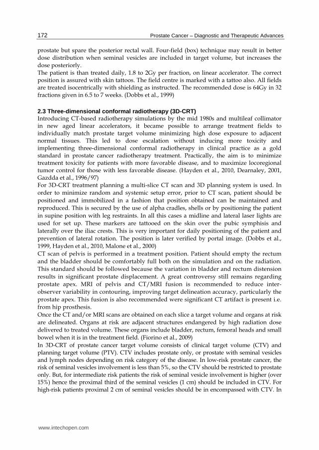
When the delineation process is completed, the medical physicists arrange beam angles and
adjust them to maximize target coverage and minimize high-dose exposure to normal
organs. (Choe & Liauw, 2010) (Figure 3.)
Fig. 3. Field arrangement for four-field (box) treatment (With thanks to the Institute for oncology and radiology of Serbia, Belgrade)
Digitally reconstructed radiograph (DRR) is created by computer program transforming the
CT slices into a radiograph image. DRR represents the referent image to which the later
portal films of treatment field position are compared. (Figure 4.)
Dose-volume-histogram (DVH) is also created and it shows the percent of prescribed dose
to every delineated structure. For organs at risk the ALARA principle (as low as reasonably
achievable) is recommended following the tolerant dose of each organ. But, although DVH
gives valuable information on the dose to each structure, it is calculated on a single
pretreatment pelvic organs position, and they are mobile. That is the reliability on a single
pretreatment DVH is limited, and does not have to correlate with late toxicity (Fiorino et al.,
2009) (Figure 5.)
www.intechopen.com

Radiotherapy in Prostate Cancer
175
(a) (b)
Fig. 4. Digitaly reconstructed radiographs (DRR) for AP (a) and right lateral (b) field (With thanks to the Institute for oncology and radiology of Serbia, Belgrade)
Fig. 5. Dose-volume-histogram (DVH) showing the doses delivered to each delineated structure (With thanks to the Institute for oncology and radiology of Serbia, Belgrade)
www.intechopen.com

Prostate Cancer – Diagnostic and Therapeutic Advances
176
According to EAU guidelines in prostate cancer in 2010, for external radiotherapy, a dose of at least 74Gy to PTV is recommended for low-risk prostate cancer because the biochemical disease-free survival is significantly higher when compared to a dose of under 72Gy (69% vs. 63%; P=0.046). For intermediate-risk prostate cancer the dose is ranging from 76Gy to 81Gy, and for high-risk prostate cancer a combination with androgen deprivation is recommended regardless dose escalation since the risk of systemic relapse has to be covered. (Heidenereich et al., 2011) The patient is treated daily, 1.8 to 2Gy per fraction, on linear accelerator in a position that matches the position taken during CT simulation. The correct position is obtained by immobilizing devices and by setting up the skin tattoo markers to treatment room wall lasers. Once the radiotherapy has started, portal films of arranged fields are taken on the accelerator, in treatment position and compared with digitally reconstructed radiograph (DRR) for set-up or other errors several times during radiotherapy course.
2.4 Intensity-modulated radiotherapy (IMRT) Intensity-modulated radiotherapy is considered a grate improvement in radiation oncology. It is a form of 3D-CRT in which the optimization of the dose prescribed to the target volume is achieved by x-ray beam intensity changes. By using computer algorithms the intensity of the beam is changed in order to increase the dose difference between the target volume and organs at risk. Radiotherapy treatment planning using IMRT is based on a CT slices on which the physician delineates target volumes as for the 3D-CRT, as well as organs at risk. For each delineated structure the tolerant dose in imputed in the mathematical algorithms and than the beam intensity is calculated as a function of beam angle. The beam angle is individually adjusted by inclination of the radiotherapy bed, collimator or gentry. (Valicenti et al., 2000) Unlike 3D-CRT, in IMRT the dose from each beam is not delivered all at once. At each beam angle, the intensity of the beam is modulated by multiple smaller subfields that change in time. This allows a high degree of dose conformity around complex and irregularly shaped tumors (Choe & Liauw, 2010) and dose escalation up to 86Gy. (Zelefsky et al., 2002)
2.5 Organ motion tracking and image-guided radiotherapy (IGRT) Highly conformal radiation therapy requires precise localization of the prostate. Since the prostate gland is a movable organ due to breathing and distention of rectum and bladder, a variety of strategies have been developed to account prostate motion including transabdominal ultrasound-based imaging, on-line CT and implantation of radiopaque markers. The prostate gland can not be visualized on portal images, but radiopaque fiducial markers can be placed within the prostate. These markers can be visualized on portal imaging so prior to every treatment a correction of targeting can be made. (Choe & Liauw, 2010) A minimum of three markers should be implanted under ultrasound guidance in the ipsilateral apex, base and contra-lateral mid-gland one week prior to simulation. (Hayden et al., 2010) This technique is used to track interfraction movement (prostate movement between treatments), but it cannot account prostate movement during treatment (intrafractional movement). Nowadays there is a growing interest in real-time tracking of the prostate. There is a special system that uses a real-time tracking of the radiofrequency transponders implanted into the prostate and if they present outside of the predetermined margins the radiation stops.
www.intechopen.com

Radiotherapy in Prostate Cancer
177
These new technologies are still under investigation, but first results are optimistic. (Choe & Liauw, 2010) When IGRT is used prostate displacement caused by rectal distension is largely corrected. (Hayden et al., 2010)
2.6 Acute and late toxicity of external-beam radiotherapy in prostate cancer Radiation-induced complications can be acute and late. Acute adverse events occur during treatment and late may develop months to years after treatment. When we irradiate the prostate, acute and late toxicity are a consequence of high dose given to the surrounding organs i.e. bladder, rectum and skin. The severance of these side-effects largely depends on the tissue volume irradiated and relates to the treatment technique. During conventional radiotherapy of the prostate acute toxicity include acute proctitis followed by rectal discomfort, tenesmus and diarrhea, and rarely rectal bleeding. It is mostly mild and resolves after symptomatic therapy with hydration and antidiarrheal and anti-inflammatory medication. Skin reactions include erythema, dry and humid desquamation. According to RTOG scale (RTOG, 1999) acute toxicity has four grades of severity stated in table 1.
Grade 0 1 2 3 4
Dermatitis No complications
Mild erythema or dry desquamation
Moderate erytherma or incipient moist desquamation, mild skid edema
Confluent moist desquamation more than 1.5 cm of the skin, moderate edema
Ulcerations or skin necrosis
Colitis No complications
Asymptomatic
Abdominal pain, mucus and/or blood in stool
Abdominal pain, fever, peritoneal signs or ileus
Perforation
Diarrhea No complications
Up to 4 stools per day
4-6 stools per day, night stools
More than 7 stools per day and/or incontinency or parentheral substitution due to dehydrations
Hemodynamic collapse
Cystitis, dysuria
No complications
Mild dysuria
Moderate dysuria that need symptomatic therapy
Symptoms not relieved on symptomatic therapy
Table 1. Acute toxicity in radical radiotherapy of prostate cancer-RTOG scale
www.intechopen.com

Prostate Cancer – Diagnostic and Therapeutic Advances
178
The most frequent adverse event on the urinary tract is radiation cystitis producing dysuria,
nocturia, frequency and urgency. It is low graded in most cases (7.7%) while severe urinary
complications are seen in less than 0.5% of patients. According to RTOG study on over 1000
prostate cancer patients treated with external-beam radiotherapy, acute toxicity occurs in 70-
90% of the patients with mild symptoms. Moderate symptoms are developed in 20-45% of
the patients while 1-4% has severe or prolonged reactions. (Dearnaley, 2001)
Although acute toxicity is very unpleasant for the patient it usually resolves after the
treatment. Late toxicity is of much more concern since it is unpredictable and very often
irreversible. According to RTOG, late toxicity also has four grades of severity (table 2).
Mostly it is mild and does not influence quality of life, but sever late toxicity is reported in 4-
8% of patients. Most common genitourinary side effects are chronic cystitis (5%),
incontinency, urethral stricture (5%, mostly patients with previous transurethral resection of
the prostate), bladder ulceration and impotency (30-40%). Late toxicity on rectum affects 3%
of the patients and includes tenesmus, sphincter dysfunction, occasional bleeding, strictures
or ulcerations. (Dearnaley, 2001)
Grade 0 1 2 3 4
Bladder No complications
Mild epithelia atrophy, discreet teleangiectasia
Diffuse teleangiectasia, macroscopic hemathuria
Frequent urinating, dysuria and hemathuria, bladder capacity less than 150 ml
Bladder necrosis, capacity less than 100 ml, hemorrhagic cystitis
Skin No complications
Mild skin atrophy, hyperpigmentation, hair loss
Moderate skin atrophy, teleangiectasis, total hair loss
Severe skin atrophy, severe teleangiectasia
Ulceration
Bowels No complications
Mild diarrhea or increased bowel motion, or mild rectal bleeding
Moderate diarrhea, abdominal pain, rectal mucus or harder bleeding
Ileus or bleeding that requires surgical treatment
Necrosis, perforation, fistula
Table 2. Late toxicity in radical radiotherapy of prostate cancer-RTOG scale
Introducing 3D-CRT and IMRT the volume of bladder and rectum irradiated is limited, but
the dose escalation can still induce significant toxicity. In trials of dose escalation, reported
rates of acute toxicity are very similar to those of conventional radiotherapy. Late toxicity
however is still considered high. MD Anderson trial has shown a significant gastrointestinal
toxicity (grade 2 or more) in 25% of patients with escalated dose comparing to 13% in low
dose group (78Gy vs. 70Gy). (Kuban et al., 2008) The Dutch trial reported 26% of late rectal
www.intechopen.com

Radiotherapy in Prostate Cancer
179
toxicity grade 2, and Medical research council 33% in dose escalated group. (Al-Mamgani et
al., 2008, Dearnaley et al., 2007) These results compare with reports of IMRT treatment with
81Gy (3% of patients experienced late rectal toxicity at grade 2 or grater) and treatment with
IGRT with 79.8Gy (12% of patients with grade 2 rectal toxicity). (Hayden et al. 2010) For
urinary toxicity none of these trials found significant correlation between late adverse events
and radiation dose. Although improved radiotherapy techniques appear to enable dose
escalation with less toxicity, the optimal dose that can eradicate the disease without the risk
of toxicity is jet to be defined. (Choe & Liauw, 2010)
2.7 Results of external-beam radiotherapy of prostate cancer Following external-beam radiotherapy, long-term clinically assessed local tumor control is
good for patients with stage T1 cancers (83% at 15 years), but it is falling to 65-68% for T2
and 44-75% for T3 cancers. Reported incidence of positive biopsy after external-beam
radiotherapy vary from 18 to 45% and increases with disease bulk from 15% for T1 disease,
to 68-79% for men with T2 and T3 cancers. Regarding biochemical control, Hanks et al.
reported a long-term biochemical control in 72% of T1 cancers, falling to 22% and 28% for
bulky T2 and T3 cancers in a mean follow-up of 12.6 years.
PSA level and Gleason score are powerful predictors of outcome. In patients with low
Gleason score the rate of biochemical control ranges about 75%, compared to only 18% for
Gleason score 7 and 0% for Gleason score 8 or 9. In patients with pretreatment PSA more
than 20 ng/ml only 28% remained biochemically free of progression at 4 years in the results
of Hanks et al (Dearnaley, 2001)
Several randomized controlled trials and one meta-analysis shown that improved
biochemical outcomes (biochemical failure free survival) are associated with dose escalation.
(Kuban et al., 2008, Al-Mmgani et al., 2008, Dearnaley et al., 2007, Zietman et al., 2010, Viani
et al., 2009). Radiation Therapy Oncology Group trials have even shown that higher
radiation dose improves disease-specific and overall survival in high-grade prostate
cancer. (Valicenti et al., 2000) Despite promising results of dose escalation there are still
uncertainties regarding routine application of dose escalation especially. The subgroup of
patients that will benefit the most from dose escalation is not clearly defined. These trials
enrolled men in all risk groups of localized prostate. Only U.K. Medical Research Council
trial divided patients in risk groups showing the benefit of dose escalation in all groups.
But statistically significance was reached only in high-risk group. In the Dutch trial the
benefit from dose escalation from 68 to 78Gy was also registered in intermediate and
high-risk patients. These results led to a question whether a higher dose is required for
low-risk prostate cancer. Although MD Anderson trial shown the benefit form dose
escalation in high-risk group of patients, it also reported longer follow-up of 8.7 years that
can indirectly demonstrate a benefit of dose escalation even in the low-risk group. While
the improved biochemical outcomes are practically proven with dose escalation, there is
still no sufficient evidence of improvement in cancer-specific survival and overall
survival. (Choe & Liauw, 2010)
Regarding overall survival, radiotherapy is efficient method for many cases of localized
prostate cancer. Five-year overall survival for T1 and T2 stage ranges about 70-80%, and 90%
of local tumor control. Locally advanced stages have poorer prognosis with 5-year overall
survival of 40-50%. High Gleason score is the most significant negative prognostic marker
www.intechopen.com

Prostate Cancer – Diagnostic and Therapeutic Advances
180
since it is associated with higher malignant potential. Cancer related death for high Gleason
score tumors (8-10) is about 60-80% in 15 years. (Hadzi-Djokic, 2005)
3. Brachytherapy in prostate cancer
In the age of the developed imaging technology (CT, MRI, PET, US), and advanced
biochemical markers (tumor-specific and nonspecific), a large number of tumors, including
prostate cancer, are being diagnosed in the early stages. In early stages of prostate cancer
(T1-T2 N0 M0, PSA <10ng/ml, Gleason Score <6, prostate volume <50ml, maximum urinary
flow>15ml/s), in addition to the conventional, laser and robotic prostatectomy,
hyperthermia, hormone therapy and brachytherapy is often applied.
In brachytherapy sealed sources of radiation are placed either in direct contact or in
proximity of the tumor, so the interstitial brachytherapy for localized prostate cancer often
represents the method of choice, and its efficiency is not far behind the effectiveness of
surgery, with less morbidity. Brachytherapy allows local application of extremely high
doses (up to 160Gy, even more). The effectiveness of prostate cancer brachytherapy is
directly correlated with the total dose and precision of administration, which represents its
advantage over transcutaneous radiotherapy.
Prostate cancer is usually multicentric, so the brachytherapy target is the entire prostate, and
the total dose has to cover the area of about 2-5 mm beyond the prostate capsule. From the
point of radiotherapy, particularly brachytherapy, the initial prostate cancer (prostate itself,
with or without vesicles involved) represents the ideal target, with adequate spare of the
urethra, rectum, bladder and perineal area. Moreover, brachytherapy can be applied within
a combined radiotherapy (brachytherapy + transcutaneous radiotherapy) by the additional
dose (boost), as well as in the case of local recurrence or rest after prostatectomy or
transcutaneous radiotherapy.
Brachytherapy for prostate cancer is not a new therapy method, though in the history of
medicine it experienced its ups and downs, mainly due to the previous imperfections of
visualization techniques and applications, as well as, imperfection in radiation and
dosimetric characteristics of the radiation sources, while today it is a routine method of
treatment. With the introduction of transrectal ultrasound (TRUS), CT and development of
new sources of radiation (103Pd, and 192Ir), techniques of radioisotope implantation (loading
and afterloading) and computer systems for brachytherapy planning in routine clinical
practice, interstitial brachytherapy for prostate cancer began to experience another upswing,
but with markedly better results and lower morbidity.
Today, two modalities for interstitial brachytherapy are applied:
1. low dose rate (LDR), about 2Gy/day, with low activity 125I sources (from 0.03 to 1.5GBq
per seed, max. photon energy of 35.5 keV, the half-life about 60 days in the form of
cylinders /height 4.5 mm, 0.8 mm diameter, encapsuled in titanium sleeve/) or 103Pd
(Blasko et al., 2000) (dose rate to about 5Gy/day; activity around 0.07GBq per source,
the maximum photon energy of 21 keV, the half-life 17 days in the form of cylinder/
height 4.5 mm, 0.8 mm diameter /or spheres/ diameter of about 1 mm /) – a
permanent implant;
2. (high) dose rate (HDR) about 0.2 to 3Gy / min with radioactive 192Ir source in the form
of cylinder /dimensions around 0.6 x3,5 mm / (initial activity of about 370GBq; time
www.intechopen.com

Radiotherapy in Prostate Cancer
181
half-life of 74.2 days, mean photon energy of about 380keV,) - a temporary
implantation.
3.1 Low dose rate brachytherapy Strictly speaking, from the point of view of radiobiology, the only differences (not great) in the indications for applying permanent (LDR) or temporary implantation (HDR) are mostly related to tumor grade. For example, with low-grade and low-risk tumors (eg Stage<T2a, PSA<10, Gleason Score 2-4) and slow-growing tumors, we expect greater efficiency in the application of permanent implants, while with tumors of high grade and higher risk, higher efficiency is expected from temporary implantation. Although there are relative differences in the indications for application of LDR or HDR brachytherapy, it seems that the predominant technique is the one with permanent implants (LDR). Selection of isotopes (125I or 103Pd) is in the favor of the cheaper iodine, so if otherwise not indicated it's considered that radioisotope 125I is being used in LDR brachytherapy for treating prostate cancer. Permanent implants are rarely used in cases of rest or local recurrence after prostatectomy or transcutaneous radiotherapy. In this case a HDR brachytherapy with 192Ir of initial activity over 370 GBq, by using afterloading device, is applied. Prostate brachytherapy requires a multidisciplinary approach, which assumes collaboration between urologists, radiation oncologists (brachytherapists), anesthesiologists and brachytherapy physicists. Regardless of which brachytherapy modality is implemented in prostate brachytherapy, and with the aim to providing a top quality treatment, all steps are strictly determined: 1. Assessing the stage and spread of the disease (laboratory-biochemical, prostate
morphology / palpatory findings, prostate size - US and TRUS /, histopathological
verification and determination of Gleason Score, a CT/MRI, scintigraphy,
determination of TNM disease stage)
2. Assessing the possibility of applications (talk with the patient, the patient’s state,
maximum urinary flow rate, the presence of residual urine, previous TUR, assessment
of cardiovascular conditions, the possibility of implanting, anatomy of the pelvis,
prostate size, type of implant LDR vs. HDR)
3. Preparing the patient prior to implantation - 24 hours in advance (admission of the
patient, medical therapy, anti-coagulant and antibiotic, preparation of the patient,
rectum cleaning)
4. Preparation of instrumentarium (selection and sterilization of instruments, calibration
of stepper, network and TRUS ) (Figure 6.)
5. Patient’s positioning and anesthesia (in lithotomic position on the movable therapy
table; spinal or general anesthesia)
6. Placing markers of critical radiosensitive structures (Folly catheter for marking the
bladder and urethra)
7. Setting TRUS probes, template and steper, and prostate visualisation, and visualisation
of urethra and rectum (in steps of max. 5 mm from base to apex (Grey et al., 2000),
(Figure 7. a, c)
8. Pre-planning (determinatuion of the number and location of radiation sources and
methods of implantation)
9. The application (placing the radiation source guides/needles; in the LDR technique
inserting a radiation source - seeds) - TRUS-guided (Figure 8. b)
www.intechopen.com

Prostate Cancer – Diagnostic and Therapeutic Advances
182
10. Verification and correction (cistoscopic, fluoroscopic, x-ray (Figure 7. c) - optional CT
and/or MRI; in case of LDR brachytherapy adding of seeds, if necessary)
11. Computer reconstruction (seeds / LDR / - needles / HDR /, prostate capsule, the
position of the urethra and rectum), optimization, plan analysis (the determination of
DVH - Dose Volume Histogram, ie. minimum volume of prostate receiving 90%, 100%,
150% and 200% of the total dose, and the dose and volumes that receive critical
radiosensitive structures), (Figure 7. d, e);
12. Irradiation; for HDR brachytherapy: deapplication of needles or observation of the
patient (LDR)
13. Transport of patient to the patient room
14. Patient care (analgesic and antibiotics therapy, toilet)
15. Check-up (of dysuric problems and estimate the degree of proctitis) and discussion
with the patient, the patient's discharge
16. Regular checks (during treatment /HDR/ and in 2, 3, 6, 12 and 18 months, then
annually - the level of PSA, rectal examination, assessing the level of dysuric problems
and evaluation of potency)
Fig. 6. Schematic representation of TRUS-guided brachytherapy with radioactive seeds.
www.intechopen.com

Radiotherapy in Prostate Cancer
183
(a) (b) (c)
(d) (e)
(a) TRUS guided needles application (b) Administrated needles /guides of 192Ir source (c) Radiographic verification of the needles’ positions (d) Isodose transversal view (e) 3D treatment volume (With the thanks of the General Hospital Medical System, Belgrade)
Fig. 7. Steps in HDR prostate cancer brachytherapy - real patient
When using permanent implantation (monotherapy) radiation sources (seeds) remain in the prostate of the patient. It is therefore recommended to the patient a minimal two-week sexual abstinence and avoidance of contact with pregnant women and small children for a minimum of 2 months. A typical therapeutic dose of radiation, when 125I is implanted, is in the range 140-160Gy, and when 103Pd is implanted to about 125Gy. In a case of a "boost" dose, for 125I a somewhat lower dose of 80-120 is applied or to about 90-100Gy for 103Pd. (Blasko et al., 2000, Beyer, 2001)
3.1.1 Results of low dose rate brachytherapy Prostate cancer with its characteristics and different biological behavior represents a problem in the analysis of brachytherapy final outcomes in terms of overall survival or local control. We are aware that many patients with untreated prostate cancer can survive tens of years, ie. do not die from prostate cancer. Therefore to evaluate the effectiveness of treatment of patients with prostate cancer, as for overall survival and local control, the PSA level was adopted. Greem and associates (Greem et al., 1997 )provide representation of the results of treatment of patients with permanent implants (monotherapy), showing a five-year survival of NED (no evidence of disease) of 94%, 84% and 54% for low risk, medium risk and higher risk
www.intechopen.com

Prostate Cancer – Diagnostic and Therapeutic Advances
184
group of 403 patients, respectively . Blasko and associates (Blasko et al, 2000) show the results of a five-year biochemical PSA control: 94%, 82% and 65% for low risk, medium risk and higher risk respectively. Slightly worse, but comparable results are displayed by other authors: 85-94%, 33-82% and 5-65% for low risk, medium risk and high-risk group, respectively (Beyer, 2001). Based on these results, most urologists and brachytherapists exclude a group of patients at high risk from the LDR brachytherapy and consider them patients with advanced disease in which the tumor penetrated the prostate capsule. Association of American Brachytherapy Society has established a Low Dose-Rate Task Group (Merrick, G., Zelefsky, M., Sylvester, E., Nag, S., Bice, W) with the task of defining the general criteria for inclusion of patients with prostate cancer in the group for the treatment by permanent implants (LDR): expected survival > 5 years, clinical stage T1b-T2c and selected T3, Gleason Score<10, PSA<50ng/ml, without involved lymph nodes, no distant metastases, Karnofsky performance status> 70%. Criteria for exclusion are: inflammatory prostate disease, severe urinary obstruction, the middle lobe hyperplasia, extensive TUR defects, a prostate size greater than 60x50 mm, the extension of disease to the seminal vesicles and bladder, involved lymph nodes, previously conducted EBRT. Absolute contraindications are distant metastatic disease, the inability of anesthesia (general, spinal, epidural), no possibility of peaceful lying, expected survival <5 years. It is obvious that the consistent application of these criteria for inclusion and exclusion would ensure consistent application of LDR brachytherapy in this roup of patients, with the results of treatment (five-and even ten-year PSA control NED> 85%).
3.1.2 Toxicity of low dose rate brachtherapy Mild and transient acute urinary symptoms (LENT SOMA, 1995) (hematuria and dysuria grade G1-G2) often accompany LDR brachytherapy, while the acute symptoms on the rectum (proctitis, tenesms and bleeding) are rare and mainly a result of edema and hematoma. All of the symptoms can be associated to radiation or trauma during the application procedure. As late symptoms of bladder neck irradiation, chronic irritative urinary symptoms may occur and in a small number of cases due to urethral scarring, obstruction or incontinence may occur. Late effects on the rectum are mild, and manifested by periodic bleeding and proctitis. Erectile function was preserved for over 70% of treated patients whose erectile function was satisfactory before the implementation of brachytherapy. In order to preserve erectile function, it is important to avoid positioning of seeds in the perineal area, outside the prostate apex, which prevents the occurrence of fibrosis and consequent devascuralisation of this region. Most patients (over 2/3) treated with permanent implants, estimate their quality of life as good, which is just as important as biochemical and clinical course of disease. However, due to the relatively limited fixation of radioactive seeds (which are smooth on the surface) there is a possibility of their inter-prostate migration and the creation of "hot" and "cold" zones, as well as, their migration to other organs (rectum, bladder and pelvic veins and even lungs), which sometimes requires serious surgical treatment and drastically reduces the quality of patient's life.
3.2 High dose rate brachytherapy HDR temporary prostate cancer brachytherapy with 192Ir entered into clinical practice mainly in combination with transcutaneous radiotherapy about 20 years ago. Obviously, it was necessary to acquire certain technical and technological advances for its application
www.intechopen.com

Radiotherapy in Prostate Cancer
185
Soon, the advantages of HDR regime compared to LDR brachytherapy of prostate cancer were noticed, which are reflected in: the high precision of application, the possibility of subsequent dose optimization, more accurate dose planning and application, sparing the surrounding tissues and organs, more favorable radiobiological effect, the fact that after treatment there are no sources of radiation in the patient and therefore no possibility of source "seeds" migration into the bladder, rectum or surrounding larger blood vessels, there is no irradiation of staff, there is the possibility of extending indicational scope of application and easier possibility of combination with transcutaneous radiotherapy. However, when it comes to temporary HDR brachytherapy of prostate cancer as monotherapy, less obvious results and experiences are presented in the literature, so one can get the wrong impression that this modality of brachytherapy is either less efficient or at very least, least mystified. To clarify this problem to an extent, if not totally resolve, the association of American Brachytherapy Society has formed a High-Dose Rate Task Group (Hsu, C., Yamada, Y., Vigneaut, E., Pouliout, J.) with the aim to define general criteria for inclusion of patients with prostate cancer in the treatment group Temporary implants (HDR), which was formed following the general criteria for inclusion: clinical stageT1-T3 and selected T4, Gleason Score any, PSA no upper limit, no distant metastases (T1-3N0M0). Criteria for relative exclusion are: severe urinary obstruction, extensive TUR defects, the TUR within 6 months, vascular disease. Absolute contraindications are impossibility to anesthesia and no possibility of peaceful lying Given all the above-mentioned recommendations, for inclusion and exclusion, and in particular those given by the American Brachytherapy Society, extended operational criteria and opinions can be formed related to the application of HDR brachytherapy in different patient groups, including recommendations on the total dose and method of its fractions, and as: monotherapy - with tumor stage T1-T2 N0 M0, Gleason Score <7 and prostate volume
to 70 cc and T3 confined to the prostate (TD - 31.5 Gy/3 fr. To TD45/6 hypofractioned (Duchesne & Peters, 1999) or hyperfractioned / pause between the fraction of at least 6 hours or even up to 54Gy/9 fr./5 days (Yoshioka et al., 2006) (where the stage T3 N0 M0 may include seminal vessicles); boost in the combined radiotherapy approach (TD - 12 (Mate et al., 1998) -24 (Demanes et al., 2009) Gy/1-4 fr. EBRT ± 36-50Gy or 9-15Gy/1-2 fr. EBRT ± 65Gy ); for recurrent disease confined to the prostate (depending on the previousl therapy TD> Gy/1-2 7-14 fr. to 8Gy/4 fr.) further: to have no contraindications (no indication for exclusion).
3.2.1 Results of high dose rate brachytherapy Yoshioka et al (Yoshioka et al., 2006) presented the results of 111 patients (15 low, 28 medium and 68 high risk, according to the ASTRO criteria (ASTRO, 1997) treated with HDR temporary implantation (monotherapy), showing a three-year and five-year survival without signs of biochemical disease of 83% and 70%, and overall survival of 100% and 97%, respectively. For the 17 patients from high risk groups, in which biochemical relapse was observed, in 9 patients the presence of distant metastases were confirmed, of which 4 patients died. Given that this group of patients is not stratified based on risk, it is clear that the shown results after the HDR brachytherapy are comparable or even better than in patients treated with permanent implants (monotherapy) (Blasko et al., 2000, Beyer, 2001, Greem et al., 1997).
www.intechopen.com

Prostate Cancer – Diagnostic and Therapeutic Advances
186
3.2.2 Toxicity of high dose rate brachytherapy Mild and transient acute urinary symptoms (dysuria and hematuria of grade G1) followed
HDR brachytherapy in about 50% of patients in the first week or two, and persist for up to
6 months in less than 35% of treated patients, while grade G2 symptoms occurred in about
11 % of patients in the first weeks after treatment and after 6 months they completely
disappeared. Pronounced symptoms of grade G3 (urethral stricture at the level of bladder
base) are very rare (less than 2%), and they require a retention of urinary catheter, and
usually occur immediately after irradiation, and disappear within ten days. Acute
symptoms of grade G1 rectum (proctitis, tenesma and bleeding) are rare and mainly are
result of edema and hematoma, and occur in less than 25% of patients in the first weeks
and decrease to about 8% in the 6-month after conducted therapy. Acute complications of
grade G3 were not observed.
All listed acute symptoms may be connected to radiation and trauma during application
procedure, although problems have not been noted during the application itself.
The frequency and severity of late complications after HDR brachytherapy is similar to the
permanent implant brachytherapy (LDR), except that complications associated with the
migration of radiation sources do not occur.
When HDR brachytherapy (22-24 Gy/5-6 fr.) is applied in combination with transcutaneous radiotherapy (EBRT to 40Gy), the results of treatment (five-year survival NED/biochemical/and overall survival) of 63% in patients with high risk are comparable and slightly better than with the application of brachytherapy (LDR and HDR) as a monotherapy. (Deamens et al., 2009) In the same paper, the authors conclude that no benefit was noted when applying deprivate androgen therapy in relation to combined radiotherapy. Acute and late effects on the bladder and rectum are more pronounced in cases of combined radiotherapy, which can be expected.
4. Postoperative radiotherapy in prostate cancer
Radical prostatectomy is proven treatment modality for prostate cancer control for a long time. Some authors report 10-year cancer-specific survival of 85-90% in localized prostate cancer after radical prostatectomy, and 82% at 15 years. Survival is better if the tumor is low grade i.e. low Gleason score and low stage. The risk of surgical margins positivity is of great concern after radical prostatectomy. Positive margins are noticed in 28% of patients with T1-T2 prostate cancer and prostate apex is the most common site. For T3 cancers this percent is even higher-up to 52%. High tumor stage (T3a and T3b) and a positive surgical margin are strong predictors of local recurrence, biochemical and clinical failure. In general, it is considered that the percent of local recurrence after radical prostatectomy ranges about 15% for T2 prostate cancers, and 50-70% for T3 tumors. In order to analyze the impact of predictive factors on development of local recurrence, univariate and multivariate analyzes were performed. In univariate analyzes strong predictors of local relapse are high-grade cancer, positive surgical margins and involvement of seminal vesicles. In multivariate analyzes these predictors are high-grade tumor, positive surgical margin and elevate prostatic phosphatase. Identification of patients that are candidates for adjuvant therapies after radical prostatectomy is still a great issue. The adequate treatment modality for these patients is an open question too. There is no consensus yet.
www.intechopen.com
Radiotherapy in Prostate Cancer
187
After radical prostatectomy, the application of radiotherapy can lower the incidence of local relapses, but its effect on distant metastases appearance is not confirmed. Alternative regimen is the use of androgen-deprivation therapy alone or in combination with radiotherapy which can also improve local control and eradication of distant metastases. (Hadzi-Djokic, 2005) Three randomized trials (Bolla, Wiegel & Thompson), have shown an advantage in biochemical relapse-free survival with postoperative radiotherapy for men with positive surgical margins or pT3 disease. These trials compared postoperative radiotherapy with 60-Gy to the prostatic fossa to radical prostatectomy alone in men with high-risk prostate cancer. With the use of postoperative irradiation the 5-year biochemical progression-free survival was significantly improved, as well as clinical progression-free survival. Thompson’s trial has also show an advantage of overall survival (10.3 years for irradiated patients after prostatectomy to 3.1 year for prostatectomy only). Bolla failed to demonstrate this advantage. That means that not all men with adverse prognostic factors will relapse. Also, not all men treated with postoperative radiotherapy will be cured. Combined treatment is also associated with greater toxicity than radiotherapy or prostatectomy alone. So what will the optimal treatment be? (Hayden et al., 2010, Bolla et al., 2005, Wiegel et al., 2009, Thompson et al., 2009) Eastham et al. managed to give actually 4 possible scenarios for post-prostatectomy setting: (1) there is no residual disease and adjuvant radiation is not necessary; (2) persistent disease is present in the prostatic fossa only and adjuvant irradiation may provide long-term cure; (3) there is a residual local disease as well as microscopic disseminated disease and adjuvant irradiation may eradicate local disease but will have no impact on the systemic component; (4) disease is only systemic and adjuvant local irradiation is not necessary. Salvage radiotherapy in the setting of a rising PSA after prostatectomy is unproven and still controversial. This is most likely the result of inadequately selected patients for post-prostatectomy irradiation. Many of them already have systemic recurrence so a detail diagnostic workout is necessary. (Eastham et al., 2010) The special issue is the use of adjuvant hormone therapy. Some studies show that the application of adjuvant hormone therapy reduces the risk of positive surgical margins, improves local disease control, eradicates micro metastases and prolongs time to progression and overall survival. On the other hand, neoadjuvant hormone therapy, prior to surgery, in order to downstage the disease, has not been proven for successes neither in preventing biochemical or clinical relapse nor in improving survival so in most centres it is deserted. (Dearnaley, 2005)
5. Androgen-derprivation therapy combined with radiotherapy of the prostate cancer
Radiotherapy is traditionally the treatment of choice in locally advanced prostate cancer. Unfortunately the results of radical radiotherapy regarding 10-years and 15-years overall survivals are not satisfactory (Zlotecki, 2001) (Table 3.) Androgen deprivation therapy is used routinely in combination with radiotherapy for locally advanced prostate cancer, but recent studies show that it improves treatment result is localized and intermediate-risk disease.(Milecki et al., 2010). Hormone therapy, that is in fact androgen deprivation, can be realized in several ways: orchyectomy, blockade of the hypothalamus-hypophysis-gonade path (with gonadothropin
www.intechopen.com

Prostate Cancer – Diagnostic and Therapeutic Advances
188
realizing hormone agonist) or by direct blockade of androgen receptors with androgen antagonists. (Anderson, 2003) Although, it is generally thought that androgen deprivation combined with radiotherapy influence the results of treatment in local and systemic way, it is uncertain whether that action is the result of radiosensitizing, systemic micro metastases eradication or both. Androgen deprivation leads to the shrinkage of the entire prostate gland reducing the irradiated volume to which the higher dose can be applied. In several studies this prostate shrinkage is ranging form 30% to 40%. Some authors say that it improves radiotherapy effectiveness by oxygenation of hypoxic cancer cells and that it even induces apoptosis and tumoricidal immune system response.
Study/Institution Clinical T stage 10 Year Survival
(%) 15 Year Survival
(%)
Pattern of Care T3-T4 33 23
RTOG 75-06 T3-T4 38 No results
Mallinckrodt T3 38 No results
Stanford T3 35 18
Stanford T4 15 15
M.D. Anderson T3 45 31
Table 3. 10 and 15-year survival rate for patients with locally advanced prostate cancer treated with radical RT
The role of androgen deprivation therapy is unclear in men with low risk prostate cancer
but some patients still received it as primary or neoadjuvant treatment. The Radiation
Therapy Oncology Group (RTOG) 94-08 randomized trial included almost 2000 men with
T1b-T2 prostate cancer and PSA less than 20ng/ml. Androgen deprivation therapy was
administered 4 months prior or concomitantly with radiotherapy. Overall survival at 8 years
was 76% vs. 73% for combined treatment and radiotherapy only, respectively. The disease-
specific survival was 98% for hormone and irradiation vs. 99% for radiotherapy alone. This
study did not bring solid results, as well as many others, because some patients were
clinically in a higher risk stage than deemed low risk according to National Comprehensive
Network classification. Retrospective studies of Bolla et al. and Cietzki at al. are among few
that have shown the advantage of radiotherapy combine with androgen-deprivation in low
risk prostate cancer (Milecki et al., 2010)
For intermediate and high-risk patients many randomized studies were performed such as
RTOG 85-31 (977 patients T1-T2, T3, N+, adjuvant hormone therapy vs. radiotherapy alone),
RTOG 86-10 (456 patients, T2-T4, neoadjuvant/concomitant hormone therapy for 4 months vs.
radiotherapy alone), EORTC 22863 (415 patients, T1-T4 prostate cancer, concomitant/adjuvant
hormone therapy for 36 months vs. radiotherapy alone). In 5-years follow-up all of them have
shown a statistically significant difference in improved local disease control, reduction of
www.intechopen.com
Radiotherapy in Prostate Cancer
189
distant metastases and longer progression free survival in hormone therapy-radiotherapy
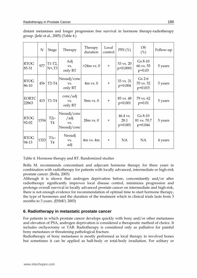
group. (Jelić et al., 2005) (Table 4.)
N Stage Therapy Therapy duration
Local control
PFS (%) OS (%)
Follow-up
RTOG 85-31
977 T1-T2, N+,T3
Adj vs.
only RT >24m vs. 0 +
53 vs. 20 p<0.0001
Gs 8-10 66 vs. 55 p=0.03
5 years
RTOG 86-10
456 T2-T4 Neoadj/conc
vs. only RT
4m vs. 0 + 33 vs. 21 p=0.004
Gs 2-6 70 vs. 52 p=0.015
5 years
EORTC 22863
415 T1-T4 conc/adj
vs. only RT
36m vs. 0 + 85 vs. 48 p<0.001
79 vs. 62 p=0.01
5 years
RTOG 92-02
1554 T2c-T4
Neoadj/conc/adj vs.
Neoadj/conc
28m vs. 0 + 46.4 vs.
28.1 p<0.001
Gs 8-10 81 vs. 70.7
p=0.044 5 years
RTOG 94-13
1323 T1c-T4
Neoadj vs. adj
4m vs. 4m + NA NA 4 years
Table 4. Hormone therapy and RT. Randomized studies
Bolla M. recommends concomitant and adjuvant hormone therapy for three years in combination with radiotherapy for patients with locally advanced, intermediate or high-risk prostate cancer. (Bolla, 2003) Although it is shown that androgen deprivation before, concomitantly and/or after radiotherapy significantly improves local disease control, minimizes progression and prolongs overall survival in locally advanced prostate cancer on intermediate and high-risk, there is not enough evidence for recommendation of optimal time to start hormone therapy, the type of hormones and the duration of the treatment which in clinical trials lasts from 3 months to 3 years. (ESMO, 2003)
6. Radiotherapy in metastatic prostate cancer
For patients in which prostate cancer develops quickly with bony and/or other metastases and elevation of PSA, androgen deprivation is considered a therapeutic method of choice. It includes orchyectomy or TAB. Radiotherapy is considered only as palliative for painful bony metastases or threatening pathological fracture. Radiotherapy of bony metastases is mostly performed as local therapy to involved bones but sometimes it can be applied as half-body or total-body irradiation. For solitary or
www.intechopen.com

Prostate Cancer – Diagnostic and Therapeutic Advances
190
localized bony metastases radiotherapy is applied through simple fields (two opposed fields, single direct field etc). A higher daily dose of 2.5 Gy or 3 Gy is given. Single dose of 8 Gy (single shoot), 20 Gy in 5 fractions or 30Gy in 10 fractions are considered to have the same results regarding pain relief and survival. Half-body irradiation is performed when there are many disseminated bony metastases and
probably as many occult. The dose of 8 Gy is applied to lower half, and 6 Gy to upper half of
the body. If we treat the whole body, the gap between irradiation of upper and lower half of
the body is four weeks. (Samija et al., 1996)
On the other hand, for patients with extensive locoregional prostate cancer, radiotherapy
can be applied to pelvis with a total dose of 50-60Gy in order to reduce pain, hemathuria,
urethral obstruction or lymphedema (Jelić, 2005)
7. New radiation techniques
The biological effect of ionizing radiation to cancer cells and normal tissues it based on the
fact that the cancer cells are more susceptible to radiation because the lack of normal
repair mechanism. Hypofractionated radiotherapy can reduce the duration of treatment
since larger dose is given per day and the cumulative dose is adjusted to a lower dose.
The randomized trials that compared hypofractionation with conventional RT did not
have long enough follow-up and used to low total dose for current standards. But the
investigation is still on. The extreme form of hypofractionation is stereotactic body
radiotherapy (radiosurgery). This method uses only few fractions but with very high
doses applied to a target volume in a very precise fashion. Although these techniques are
attractive for theoretical advantage in radiobiology, the risk of late toxicity is
considerable.
Another possible radiotherapy approach to enhance radiotherapeutic ratio for prostate
cancer is to utilize charged particles such as protons and carbon ions. The clinical benefit is
still unclear but the optimism is based upon the fact that charged particles deposit most of
their energy at a given depth followed by a dose fall-of with almost no dose deposition
beyond the point of maximal dose called Bragg peak which can lead to increased normal
tissue sparing. For proton therapy, a dosimetric analysis did not show a significant
improvement in conformity to spare normal tissues over photon IMRT. Clinical outcome
and toxicity is also similar. Carbon ion has the same dose distribution as proton beams with
Bragg peak and following dose fall-of. But the relative biological effectiveness of carbon ion
is four times higher than protons and photons that can lead to improved local tumor control
without causing more toxicity. However these technologies are still developing and results
are yet to be seen. (Choe & Liauw, 2010)
8. Conclusion
Radiotherapy is widely used as curative treatment modality for many cases of prostate
cancer. There is a diverse array of radiotherapeutic strategies that can be effectively used to
treat both organ-confined and locally advanced disease, alone or in combination with
androgen-deprivation therapy. In recent decades it has undergone significant clinical and
technological advances that aim to optimize cancer control outcomes while minimizing
treatment morbidity.
www.intechopen.com

Radiotherapy in Prostate Cancer
191
9. References
Al-Mamgani, A., Van Putten, WL., Heemsbergen, WD. et al. (2008). Update of Dutch
multicenter dose-escalation trial of radiotherapy for localized prostate cancer.
International Journal of Radiation Oncology Biology Physics, vol. 72, pp. (980-988)
American Society for Theapeutic Radiology and Oncology Consensus Panel, Consensus
statement: Guidelines for PSA following radiation therapy. (1997). International
Journal of Radiation Oncology Biology Physics, vol. 37, pp. (1035-1041)
Anderson, J. (2003). Treatment of Prostate Cancer. The Role of Primary Hormonal
Therapy. EAU Update Series, vol. 1, pp. (32-39)
Aus, G., Abbou, C., Bolla, M. et al. (2001). Guidelines on prostate cancer. European Urology,
vol. 40, no. 2, pp. (97-101)
Beyer, D. (2001). The evolving role of prostate brachytherapy. Cancer Control, vol. 8, pp.
(163-170)
Blasko, J., Grimm, D., Sylvester, E. et al. (2000). Paladium 103 brachytherapy for prostate
carcinoma. International Journal of Radiation Oncology Biology Physics, vol. 46, pp.
(839-850)
Blasko, J., Grimm, D., Sylvester, E. et al. (2000). The role of external beam radiotherapy
with I-125/Pd-103 brachytherapy for prostate carcinoma. Radiotherapy and
Oncology, vol. 57, pp. (273-278)
Boehmer, D., Maingon, P., Poortman, P. et al. (2006). Guidelines for primary radiotherapy
of patients with prostate cancer. Radiotherapy and Oncology, vol. 79, pp. (259-269)
Bolla, M. (2003). Treatment of Locally Advanced Prostate Cancer. The Clinical Use of
Radiotherapy. EAU Update Series, vol. 1, pp. (23-31)
Bolla, M., van Poppel, H., Collette, L. et al. (2005). Postoperative radiotherapy after radical
prostatectomy: a randomized controlled trial (EORTC trial 22911). Lancet, vol.
366, pp. (572-578)
Chao, KSC., Perez, C., & Brady, LW. (2002). Prostate, In: Radiation oncology management
decision, pp. (447-467), Lippincot-Williams&Wilkins, ISBN 0-7817-3222-0,
Philadelphia
Choe, KS., & Liauw, SL. (2010). Radiotherapeutic strategies in the management of low-risk
prostate cancer. The Scientific World JOURNAL: TSW Urology, vol. 10, pp. (1854-
1869)
Common Toxicity Criteria Version 2.0, DCTD, NCI, NIH, DHHS. (1999). Cancer Therapy
Graduation Programme. RTOG: Late radiation morbidity scoring system.
Deamens, J., Brandt, D., Schour, L. et al. (2009). Excellent results from high dose rate
brachytherapy and external beam for prostate cancer are not improved by
androgen deprivation. American Journal of Clinical Oncology, vol. 32, pp. (342-347)
Dearnaley, DP., (2001). Radiotherapy and combined modality approaches in localized
prostate cancer. Prostate Cancer. Educational Book. ECCO 11, pp. (137-140), Lisbon
Dearnaley, DP., (2005). Radiotherapy in locally advanced prostate cancer. EJC Education
Book. ECCO 13, pp. (317-330), Paris
Dearnaley, DP., Sydes, MR., Graham, JD. et al. (2007). Escalated-dose versus standard-
dose conformal radiotherapy in prostate cancer: first results from MRC RT01
randomized controlled trial. Lancet Oncology, vol. 8, pp. (475-487)
www.intechopen.com

Prostate Cancer – Diagnostic and Therapeutic Advances
192
Dobbs, J., Barrett, A., & Ash, D., (1999). Prostate, In: Practical radiotherapy planning, pp.
(271-280), Arnold, ISBN 0-340-70-631-7, Great Britain
Duchesne, G., & Peters, L. (1999). What is the alpha/beta ratio for prostate cancer?
Rationale for hypofractionated high-dose-rate brachytherapy. International Journal
of Radiation Oncology Biology Physics, vol. 44, pp. (747-748)
Eastham, JA., Chirstopher, PE., & Zietman, A. (2010). What is the optimal management of
high risk, clinically localized prostate cancer? Urologic Oncology, vol. 28, pp. (557-
567)
ESMO Minimum Clinical Recommendations for Diagnosis, Treatment and Follow up of
Prostate Cancer. (2003). Annals of Oncology, vol. 14, pp. (1010-1011)
Fiorino, C., Valdagni, R., Rancati, T. et al. (2009). Dose-volume effects for normal tissues in
external radiotherapy: pelvis. Radiotherapy and Oncology, vol. 93, pp. (153-167)
Gazdda, MJ., Lawrence, R., & Coia, MD. (1996/97). Radiation treatment planning and
techniques. Cancer Management: A Multidisciplinary Approach. Medical, Surgical &
Radiation Oncology, pp. (593-604), Huntington, New York
Greem, P., Blasko, J., Ragde, H. et al. (1997). Transperineal ultrosund guided 1251/103Pd
brachytherapy for early stage prostate cancer: Update on clinical experience at
seven years. International Journal of Radiation Oncology Biology Physics, vol. 39, pp.
(219)
Grey, J., Merricks, S., Beyer, D. et al. (2000). Comparartive analysis of prostate
brachytherapy pre-planing. Radiotherapy and Oncology, vol. 55, pp. (42-43)
Hadži-Đokić, J. (2005). Lokalizovani karcinom prostate, Elit. Medica, Beograd
Hayden, AJ., Catton, C., & Pickles, T. (2010). Radiotherapy in prostate cancer: a risk risk-
adapted strategy. Urologic Oncology, vol. 17, no. 2, pp. (18-24)
Hayden, AJ., Martin, JM., Kneebone, AB. et al. (2010). Australian & New Zealand faculty
of radiation oncology genito-urinary group; 2010 consensus guidelines for
definitive external beam radiotherapy for prostate carcinoma. Journal of Medical
Imaging and Radiation Oncology, vol. 54, pp. (513-525)
Heidenereich, A., Bellmunt, J., Bolla, M. et al. (2011). EAU guidelines on prostate cancer.
Part 1: screening, diagnosis and treatment of clinically localized prostate cancer.
European Urology, vol. 59, pp. (61-71)
Jelić, Lj., Stojanović, S., & Popov, I. (2005). Radiotherapy in prostate cancer treatment. Acta
Chirurgica Iugoslavica, vol. 2, no. 4, pp. (93-102)
Jelić, Lj., & Stojanović, S. (2005). Radioterapija karcinoma prostate, In: Lokalizovani
karcinom prostate, Hadži-Đokić J., pp. (109-118), Elit. Medica, Beograd
Kirby, R., & Madhavan, SG. (2010). Prostate cancer. Renal and urology II, In: Surgery, vol.
28, no. 12, pp. (594-598)
Koh, H., Kattan, MW., Scardino, PT. et al. (2003). A nomogram to predict seminal vesicles
invasion by the extent and location of cancer in systemic biopsy results. Journal of
Urology, vol. 170, pp. (1203-1208)
Kuban, DA., Tucker, SL., Dong, L. et al. (2008). Long-term results of the M. D. Anderson
randomized dose escalation trial for prostate cancer. International Journal of
Radiation Oncology Biology Physics, vol. 70, pp. (67-74)
www.intechopen.com

Radiotherapy in Prostate Cancer
193
Lawton, CA., Michalski, J., El-Naqa, I. et al. (2009). RTOG GU radiation oncology
specialists reach consensus on pelvic lymph node volumes for high risk prostate
cancer. International Journal of Radiation Oncology Biology Physics, vol. 74, pp. (383-
387)
LENT SOMA scales for all anatomic sites. (1995). International Journal of Radiation Oncology
Biology Physics, vol. 31, pp. (1049-1091)
Malone, S., Szanto, J., Perry, G. et al. (2000). A prospective comparison of three systems of
patient immobilization for prostate radiotherapy. International Journal of Radiation
Oncology Biology Physics, vol. 48, pp. (657-665)
Mate,T., Gottesman, J., Hatton, J. et al. (1998). High-dose-rate afterloading Ir-192
brachytherapy: Feasibility report. International Journal of Radiation Oncology
Biology Physics, vol. 41, pp. (522-533)
Milecki, P., Martenka, P., Antczak, A. et al. (2010). Radiotherapy combined with hormonal
therapy in prostate cancer: the state of art. Cancer Management and Research, vol. 2,
pp. (243-253)
Thompson, IM., Tangen, CM., Paradelo, J. et al. (2009). Adjuvant radiotherapy for
pathological T3N0M0 prostate cancer significantly reduces risk of metastases and
improves survival: long term follow-up of a randomized clinical trial. Journal of
Urology, vol. 181, pp. (956-962)
Valicenti, R., Lu, J., Pilepich, M. et al. (2000). Survival advantage from higher-dose
radiation therapy for clinically localized prostate cancer treated on Radiation
Oncology Group trials. Journal of Clinical Oncology, vol. 18, pp. (2740-2749)
Viani, GA., Stefano, EJ., & Alfonso, SL. (2009). Higher than conventional radiation doses
in localized prostate cancer treatment: a meta-analysis of randomized controlled
trials. International Journal of Radiation Oncology Biology Physics, vol. 74, pp. (1405-
1418)
Wiegel, T., Bottke, D., Steiner, U. et al. (2009). Phase III postoperative adjuvant
radiotherapy after radical prostatectomy compared with radical prostatectomy
alone in T3 prostate cancer with postoperative undetectable prostate-specific
antigen: ARO 96-02/AUO AP 09/95. Journal of Clinical Oncology, vol. 27, pp.
(2924-2930)
Zelefsky, MJ., Fuks, Z., Hunt, M. et al. (2002). High-dose intensity modulated radiation
therapy for prostate cancer: early toxicity and biochemical outcome in 772
patients. International Journal of Radiation Oncology Biology Physics, vol. 53, pp.
(1111-1116)
Zietman, AL., Bac, K., Slater, JD. et al. (2010). Randomized trial comparing conventional-
dose with high-dose conformal radiotherapy in early-stage adenocarcinoma of
the prostate: long-term results from photon, radiation oncology group/American
college of radiology. Journal of Clinical Oncology, vol. 28, pp. (1106-1111)
Zivkovic, M., & Deponte, V. (1996.) Palijativna radioterapija, In: Radioterapija, Samija, M.,
Krajina, Z., Purisic, A., pp. (363-378), Nakladni zavod Globus, Zagreb
Zlotecki, RA. (2001). External-Beam Radiation in the Management of Carcinoma of the
Prostate. Cancer Control, vol. 8, pp. (503-510)
www.intechopen.com

Prostate Cancer – Diagnostic and Therapeutic Advances
194
Yoshioka, Y., Konishi, K., Oh, R-J. et al. (2006). High-dose-rate brachytherapy without
external beam irradiation for locally advanced prostate cancer. Radiotherapy and
Oncology, vol. 80, pp. (62-68)
www.intechopen.com

Prostate Cancer - Diagnostic and Therapeutic AdvancesEdited by Dr. Philippe E. Spiess
ISBN 978-953-307-319-4Hard cover, 378 pagesPublisher InTechPublished online 25, November, 2011Published in print edition November, 2011
InTech EuropeUniversity Campus STeP Ri Slavka Krautzeka 83/A 51000 Rijeka, Croatia Phone: +385 (51) 770 447 Fax: +385 (51) 686 166www.intechopen.com
InTech ChinaUnit 405, Office Block, Hotel Equatorial Shanghai No.65, Yan An Road (West), Shanghai, 200040, China
Phone: +86-21-62489820 Fax: +86-21-62489821
In this book entitled "Prostate Cancer - Diagnostic and Therapeutic Advances", we highlight many of thesignificant advances made in our treatment armamentarium of prostate cancer. The book is subdivided intofour sections termed: 1) novel diagnostic approaches, 2) surgical treatments options, 3) radiation therapy andits potential sequelae, and 4) medical management and its treatment complications. After reading the presentbook , readers will be very familiar with the major clinical advances made in our multifaceted treatmentapproach to prostate cancer over the past decade.This book is a tribute to our pioneering urologists and alliedhealthcare professionals who have continually pushed forward our traditional therapeutic envelope.
How to referenceIn order to correctly reference this scholarly work, feel free to copy and paste the following:
Tatjana Arsenijevic ́, Kata Dabic ́ Stankovic ́, Miodrag Ac ́imovic ́ and Ljiljana Rados ̌evic ́ Jelić (2011). Radiotherapyin Prostate Cancer, Prostate Cancer - Diagnostic and Therapeutic Advances, Dr. Philippe E. Spiess (Ed.),ISBN: 978-953-307-319-4, InTech, Available from: http://www.intechopen.com/books/prostate-cancer-diagnostic-and-therapeutic-advances/radiotherapy-in-prostate-cancer

© 2011 The Author(s). Licensee IntechOpen. This is an open access articledistributed under the terms of the Creative Commons Attribution 3.0License, which permits unrestricted use, distribution, and reproduction inany medium, provided the original work is properly cited.