radiopharmacy diagnostic nuclear medicine 4 continue 7
TRANSCRIPT

RADIOPHARMACY
Diagnostic Nuclear Medicine 4continue
7
BRAIN IMAGING Cerebral perfusion brain- imaging agents. Radiopharmaceuticals for evaluating brain perfusion must possess a lipophilic partition coefficient sufficient to diffuse passively across the blood-brain barrier (BBB) almost completely within one pass of the cerebral circulation, as well as being sufficiently retained to permit data collection. The regional uptake of these agents is proportional to cerebral blood flow. This class of radiopharmaceuticals is useful in the diagnosis of altered regional blood perfusion as in stroke.
Tc-99m HMBAO is a neutral , lipid-soluble complex that freely crosses the BBB. This is a relatively unstable complex, which rapidly converts to a secondary, less lipophilic complex incapable of penetrating into the brain. The in vitro addition of a methylene blue/phosphate buffer stabilizing solution after preparing the Tc-99m HMBAO will stabilize the lipid-soluble complex for 4 hr . Administration and dosage. IV, 20 mCi (740 MBq)
Tc-99m HMBAO hexamethylpropyleneamine oxine


INFECTION AND INFLAMMATION.
Evaluation of sites of infection include the use of agents that can associate with components of the natural defence mechanisms and can accumulate where they localize.
Gallium (Ga-67) citrate
It is supplied as a sterile, pyrogen-free radiopharmaceutcal with preservatives.The mechanism of localization depends on the format ion of a gallium transferrin complex in the blood and on binding to transferrin receptors associatedwith infection and inflammation. It accumulates in areas of white blood cell (WBC) localization.
Biodistribution After administration, the highest concentration of Ga-67 citrate, other than at thesite of infection, is in the liver and spleen, renal cortex and bone.
Physical half - life: 78 hr Decay emissions: 185 kev γ photons Administration and dosage. IV, 3 mCi (111 MBq) . A daily laxative or an enema should be used by the patient after the injection and before the images to cleanse the bowel of radioactivity that may interfere with the images and possibly lead to a false positive result.

WBC labelling agents
Radiolabeled WBCs are used in the detect ion of a wide variety of infectious and inflammatory processes.Current use includes the diagnosis of intra-abdominal abscesses, inflammatory bowel disease, appendicitis, fever of unknown origin, and osteomyelitis.
WBCs can be radiolabeled with In-111 oxine or Tc-HMPAO.


Tc-99mHMPAO
Tc-99m HMPAO is used to radiolabel leukocytes after reconstitution withsodium pertechnetate. Tc-99m HMPAO is a neutral , lipid-soluble complex that is able to penetrate the WBC membrane. This lipophilic complex is relatively unstable and rapidly converts to a secondary complex incapable of penetrating (leaving) the WBCs. The methylene blue/phosphate buffer stabilized solution is not able to radiolabel cells and should not be used. Biodistribution(1) After IV injection, the radiolabeled cells localize in the lungs, liver , spleen, blood pool , bone marrow, and bladder as well as the sites of WBC accumulation.(2) Elimination is primarily via the liver .
Administration and dosage. IV, for infection, 20 mCi (740 MBq)
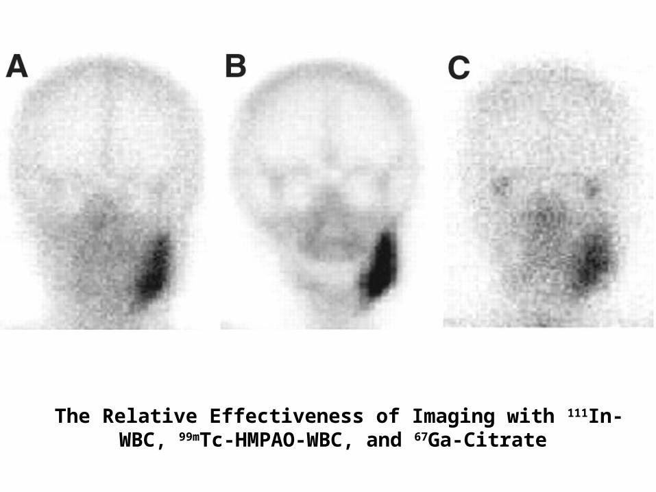
The Relative Effectiveness of Imaging with 111In-WBC, 99mTc-HMPAO-WBC, and 67Ga-Citrate
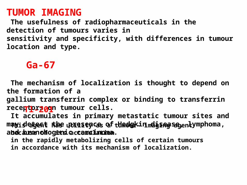
TUMOR IMAGING The usefulness of radiopharmaceuticals in the detection of tumours varies insensitivity and specificity, with differences in tumour location and type.
Ga-67
The mechanism of localization is thought to depend on the formation of a gallium transferrin complex or binding to transferrin receptors on tumour cells. It accumulates in primary metastatic tumour sites and may detect the presence of Hodgkin disease, lymphoma, and bronchogenic carcinoma.
Tl-201
This agent has utility as a tumour- imaging agent because of its accumulationin the rapidly metabolizing cells of certain tumours in accordance with its mechanism of localization.

Tc-99m MIBI Tc-99m MIBI is used for both tumour and cardiac imaging.Tc-99mMIBI is indicated for imaging as a second line of evaluating breast lesions in patients with an abnormal mammogram or a palpable breast mass.
NB. Tumour imaging agent are not used to screen breast cancer, to confirm the presence or absence of malignancy, or to replace a biopsy.

99mTC-sestamibi studies of a breast cancer patient before and after chemotherapy treatment

I-131 MIBG (meta-iodobenzylguanidine)
It is supplied as a sterile, pyrogen- free radiopharmaceut ical for use as an adjunctive diagnostic agent for the localization of primary and metastatic pheochromocytomas and neuroblastomas.
MIBG (meta-iodobenzylguanidine) labeled with I-131 acts as a physiological analog of norepinephrine and is transported and accumulated in the adrenal medulla.
This allows for the detection of neuroendocrine tumours via the specific uptake of labeled MIBG.
Because of its physiological similarities to norepinephrine, many classes ofdrugs that interfere with catecholamine transport and function may affect the uptake and localization of labeled MIBG .
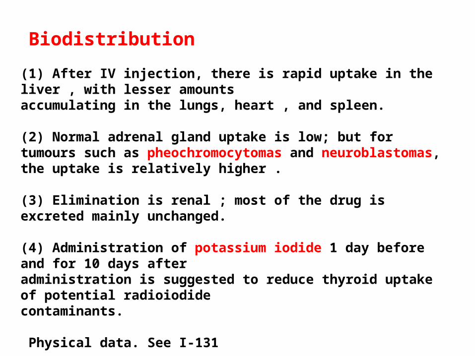
Biodistribution
(1) After IV injection, there is rapid uptake in the liver , with lesser amountsaccumulating in the lungs, heart , and spleen.
(2) Normal adrenal gland uptake is low; but for tumours such as pheochromocytomas and neuroblastomas, the uptake is relatively higher .
(3) Elimination is renal ; most of the drug is excreted mainly unchanged.
(4) Administration of potassium iodide 1 day before and for 10 days afteradministration is suggested to reduce thyroid uptake of potential radioiodidecontaminants.
Physical data. See I-131
Administration and dosage. IV, 1 mCi ( 37 MBq )
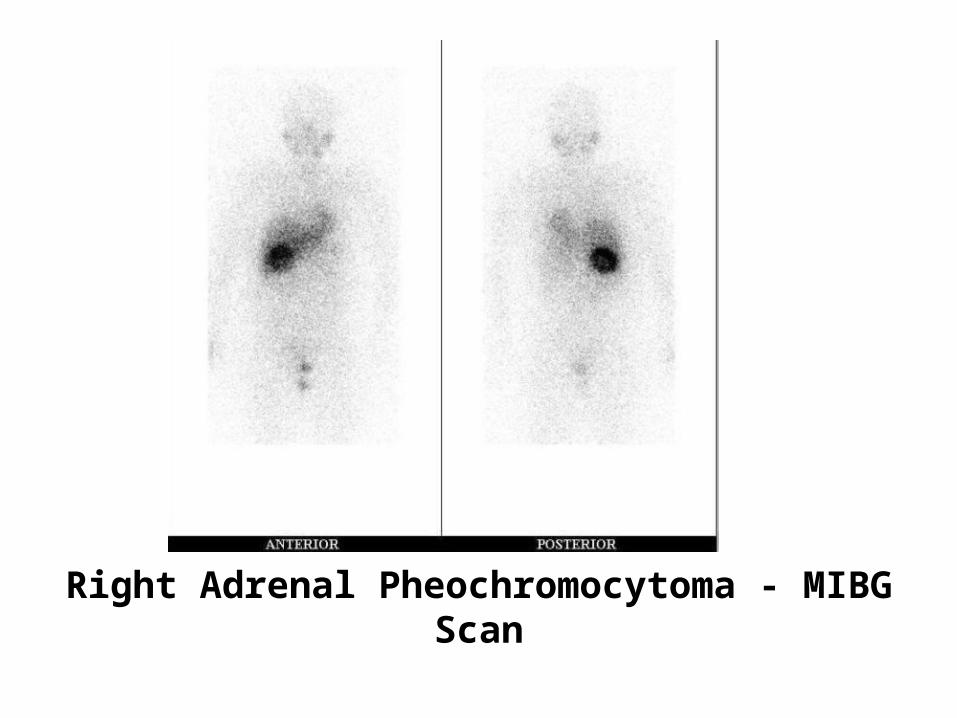
Right Adrenal Pheochromocytoma - MIBG Scan

I-131 MIBG scan demonstrated a large focus of tracer accumulation that corresponds to ganglioneuroblastoma

Thank you and Good Luck
Prof. Dr. Omar Shebl Zahra