radiographic anatomical landmarks by dr. armaan singh
TRANSCRIPT

RADIOGRAPHIC ANATOMICAL LANDMARKS

CONTENTS
Tooth anatomy
Supporting structures
Anatomical landmarks

Tooth Anatomy Teeth are composed primarily of dentin, with an enamel cap over
the coronal portion and a thin layer of cementum over the root surface.
Radiographic Appearance of Enamel
ENAMEL appears more radio-opaque than other tissues.
It is 90% mineral causes greator attenuation of X-ray photons.

RADIOGRAPHIC APPEARANCE OF DENTIN
75% mineral content less radiopaque than enamel. Radiopacity similar to bone.
ENAMELODENTINAL JUNCTION appears as a distinct interface separating these two structures.

Radiographic appearance of CEMENTUM
50%mineral content and it appears as a very thin layer on the root surface.
It is usually not so apparent radiographically.
CERVICAL BURNOUT
Radiographs sometimes show diffuse radiolucent areas with ill defined borders present on the mesial or distal aspects of the teeth in the cervical region.
These regions appear between the edge of the enamel cap and the crest of the alveolar ridge.

REASON FOR CERVICAL BURNOUTNormal configuration of the affected teeth, results in
decreased X-ray absorption in the areas in question.
Perception of these areas is due to contrast with the adjacent ,relatively radiopaque enamel and alveolar –bone.
It should not be confused with root caries which has similar appearance.

Radiographic Appearance of the Pulp
It is composed of soft tissues so it appears radiolucent.
Pulp chambers and root canals extend from the interiors of the chamber till the root apices.
It is seen radiographically also as apical foramen.
In some cases, it may exit on the side of the canal.
Lateral canals may end at the apex as a discernible foramen or may exit at the side of the root.

ENAMEL
PULP
DENTIN

The pulp canals of a developing tooth root diverge and walls of the root taper to a knife edge.
A radiolucent area is seen surrounding it in the trabecular bone. It is surrounded by the hyperostotic bone.
IT IS THE DENTAL PAPILLA WITH ITS BONY CRYPT.
Its radiographic evaluation helps in determining the stage of maturation of the developing tooth.

Supporting Structures
Periodontal ligament space
Lamina dura
Alveolar crest
Trabecular bone

RADIOGRAPHIC FEATURES OF THE PERIODONTAL LIGAMENT SPACE
It is composed of collagen so appears as a radiolucent space between the root and lamina dura.
It is thinner in the middle of the root and slightly wider near the alveolar crest and the apex ,suggesting that the fulcrum of the physiologic movements is in the region where PDL is thinnest.

RADIOGRAPHIC FEATURES OF LAMINA DURA
It is a thin radiopaque layer of dense bone surrounding the tooth socket.
Its radiographic appearance is due to attenuation of the X-ray beam as it passes tangentially through the thickness of the bone.
It is thicker than the surrounding trabecular bone and thickness increases with increase in amount of occlusal stress.

RADIOGRAPHIC FEATURES OF ALVEOLAR CREST
It is the radiopaque gingival margin of the alveolar process which surrounds the teeth.
It is considered normal if it is 1.5mm or less from the CEJ.
It shows apical recession with the age or periodontal disease.

RADIOGRAPHIC FEATURES OF THE CANCELLOUS BONE
Also called as the trabecular bone or the spongiosa.
Lies between the cortical plates in both the jaws.
It is composed of thin radiopaque plates and rods surrounding many small radiolucent pockets of marrow.
In posterior maxilla, it is similar to anterior maxilla but marrow spaces are larger.

ANATOMIC LANDMARKS OF MAXILLA
Intermaxillary suture
Anterior nasal spine
Nasal fossa and Nasal septum
Incisive foramen
Superior foramina of nasopalatine canal
Lateral fossa
Nose
Nasolacrimal canal
Maxillary sinus
Zygoma & zygomatic process of maxilla
Nasolabial fold
Pterygoid plates

RADIOGRAPHIC FEATURES INTERMAXILLARY SUTURE Also called as median suture.
In IOPA, it appears as a thin radiolucent line in the midline between the two portions of premaxilla.
It extends from the alveolar crest between the central incisors superiorly through the anterior nasal spine and continues posteriorly between the maxillary palatine process to the posterior aspect of the hard palate.

RADIOGRAPHIC FEATURE Anterior Nasal Spine
Mostly seen on IOPA of maxillary central incisors.Located in midline1.5-2cm above the alveolar crest. It is radiopaque and usually V-shaped.

RADIOGRAPHIC FEATURES NASAL FOSSA & NASAL SEPTUMThe nasal cavity shows the hazy shadow of the
inferior nasal conchae extending from the right and left lateral walls
Floor of Nasal Fossa
Nasal Septum

RADIOGRAPHIC FEATURES INCISIVE FORAMEN
Also called as NASOPALATINE or ANTERIOR PALATINE FORAMEN.
It is the oral terminatus of the nasopalatine canal. It transmits the nasopalatine vessels and nerves.Lies in the midline of palate behind the central incisors at
the junction of the median palatine and incisive sutures.Radiographic image variability is due to:
1.Different angles of the X-ray beam.2.Variability in its anatomic size.
IT IS FREQUENTLY THE POTENTIAL SITE OF CYST FORMATION.

Radiographic features of Superior Foramina of the Nasopalatine canal
The nasopalatine canal originates at two foramina in floor of the nasal cavity.
Radiographically, it can be recognized as two radiolucent areas above the apices of the central incisors in floor of the nasal cavity near its anterior border and both the sides of the septum.
Lateral wall of nasopalatine canalSuperior
foramina

RADIOGRAPHIC FEATURES OF THE LATERAL FOSSAAlso called as INCISIVE FOSSA.Appears as depression in the maxilla near the
apex of the lateral incisor .Appears diffusely radiolucent in the IOPA.

RADIOGRAPHIC FEATURES OF THE NASOLACRIMAL CANALThe nasal and maxillary bones form the
nasolacrimal canal.
It runs from the medial aspect of the antero inferior border of the orbit inferiorly, to drain under the inferior conchae into the nasal cavity.

RADIOGRAPHIC FEATURES OF NOSEThe soft tissue of the nose is frequently seen in the
projections of the maxillary central and lateral incisors ,superimposed over the roots of these teeth.
Image appears uniformly opaque with a sharp border.

RADIOGRAPHIC FEATURES NASOLABIAL FOLDAn oblique line demarcating a region that
appears to be covered by a slight radio opacity
frequently traverses periapical radiographs of
the premolar region.

RADIOGRAPHIC FEATURES OF MAXILLARY SINUSMAXILLARY SINUS is an air containing cavity
lined by mucous membrane.Appears as the three sided pyramid .
Base -formed by mesial wall adjacent to nasal cavity.
Apex –extending laterally into the zygomatic process of maxilla.

MAXILLARY SINUSOn the IOPA, maxillary sinus appears as a thin
,delicate radiopaque line.
It extends from the distal aspect of the canine to the posterior wall of the maxilla above the tuberosity.
Around the age of puberty, its floor coincides with the floor of the nasal cavity.

MAXILLARY SINUS In response to the loss of function (associated with loss
of posterior teeth) the sinus may expand further into the alveolar bone , occasionally extending to the alveolar ridge.
Thin radiolucent lines of the uniform width are found within the image of the maxillary sinus.
These are shadows of the neuro -vascular canals that accommodate the posterior superior vessels and nerves.

RADIOGRAPHIC FEATURES ZYGOMATIC PROCESS AND ZYGOMATIC BONEThe zygomatic process of the maxilla is an extension of
the lateral maxillary surface that arises in the region of the apices of the first and the second molars and serves as the articulation for the zygomatic bone.
Appears as a U-shaped radiopaque line with rounded ends projected in the apical region of the first and second molars.

RADIOGRAPHIC FEATURES PTERYGOID PLATES The medial and lateral pterygoid plates lie immediately
posterior to the tuberosity of maxilla.
They cast a single radiopaque shadow without any evidence of
trabeculation.
Extending inferiorly from the medial pterygoid plate, the hamular process may be seen.

Anatomic Landmarks of Mandible
SymphysisGenial tuberclesLingual foramenMental ridgeMental fossaMental foramenMandibular canalNutrient canalsMylohyoid ridgeSubmandibular gland fossaExternal oblique ridgeInferior border of mandibleCoronoid process

RADIOGRAPHIC FEATURES SYMPHYSISThe region of mandibular symphysis in infants
demonstrate a radiolucent line through the midline of the jaw between the images of the forming deciduous central incisors.
The suture usually fuses by the end of 1st year of life and is no longer radiographically apparent.

RADIOGRAPHIC FEATURE GENIAL TUBERCLES
These are tiny bumps of bone that serve as attachment for the genioglossus and geniohyoid muscles.
Present on lingual side.
On IOPA, appears as ring shaped radiopacity below the apices of mandibular incisors.

RADIOGRAPHIC FEATURE LINGUAL FORAMEN
It is a hole or tiny opening located on the internal surface of mandible and surrounded by the genial tubercles.
Radiographically, appears as a radiolucent dot inferior to the apices of the mandibular incisors.

RADIOGRAPHIC FEATURES MENTAL RIDGE It is a linear prominence of cortical bone located on the external
surface extending from the premolar region to the midline and slopes upward.
Radiographically, appears as a radiopaque band that extends from the premolar region to the incisor region.

RADIOGRAPHIC FEATURE MENTAL FOSSALocated above the mental ridge.
On peri apical radiograph, appears as a radiolucent area above the mental ridge.

RADIOGRAPHIC FEATURE MENTAL FORAMEN
Located on the external surface of the mandible as an opening in the region of the mandibular premolars.
Mental nerves and blood vessels exit through it.
Radiogarphically, it appears as a small ovoid radiolucent area located below the apices of the premolars.

RADIOGRAPHIC FEATURES MANDIBULAR CANAL
Tube like passage extending from the mandibular foramen to the mental foramen and contains inf.alv. Nerves and blood vessels.
Appears as a radiolucent band outlined by two radiopaque lines of cortical plate.

RADIOGRAPHIC FEATURES NUTRIENT CANALSNutrient canals are tube like passage-ways
through bone that contains nerves and blood vessels that supply the teeth.
Radiographically seen as vertical radiolucent lines.
More prominent in anterior mandible where bone is thin.

RADIOGRAPHIC FEATURES MYLOHYOID RIDGE
Linear prominence of bone located on the internal surface of mandible.
Extends from the molar region downward and forward towards the lower border of mandibular symphysis.
On IOPA, appears as radiopaque band extending downward from molars.

RADIOGRAPHIC FEATURES EXTERNAL OBLIQUE RIDGELinear prominence of bone located on external
surface of mandible extending downwards and is a continuation of anterior border of ramus.
It appears as a radiopaque band extending downwards and forwards from ant. border of mandible & ends in 3rd molar region.

RADIOGRAPHIC FEATURES SUBMANDIBULAR GLAND FOSSA
Depressed area of bone located on the internal surface of mandible.
Submandibular salivary gland lies in this fossa.
It appears as a radiolucent area in the molar region below the mylohyoid ridge.

RADIOGRAPHIC FEATURES INTERNAL OBLIQUE RIDGE
Linear prominence of bone located on internal surface of mandible extending downwards and forwards from ramus.
It appears as a radiopaque band extending downwards from ramus.
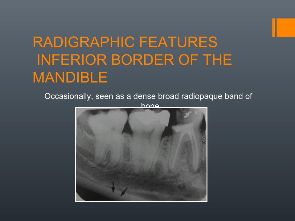
RADIGRAPHIC FEATURES INFERIOR BORDER OF THE MANDIBLE
Occasionally, seen as a dense broad radiopaque band of bone.

RADIOGRAPHIC FEATURES CORONOID PROCESS
It is a marked prominence of bone on the ant. ramus of the mandible.
Not seen on a mandibular IOPA but appears on a maxillary molars IOPA.
It is seen as a triangular radiopacity superimposed over or inferior to maxillary tuberosity.
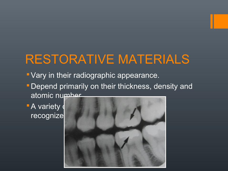
RESTORATIVE MATERIALSVary in their radiographic appearance.Depend primarily on their thickness, density and
atomic number.A variety of restorative materials may be
recognized on intra oral radiographs.

Thank You