radiation therapy of head and neck cancer with …
TRANSCRIPT

2
Department of Oncology
University of Helsinki Finland
RADIATION THERAPY OF HEAD AND NECK CANCER WITH SPECIAL
EMPHASIS ON LOCOREGIONAL RECURRENCE AND ADVERSE
EVENTS
Kauko Saarilahti
ACADEMIC DISSERTATION
To be publicly discussed, by permission of the Medical Faculty of the University of Helsinki, in the
Auditorium of the Department of Oncology, Helsinki University Hospital, Haartmaninkatu 4, on
November 5th, 2004, at 12 o’clock noon.
Helsinki 2004

3
SUPERVISED BY:
Professor Heikki Joensuu M.D.
Department of Oncology
University of Helsinki
and
Docent Mikael Kajanti M.D.
Department of Oncology
University of Helsinki
REVIEWED BY:
Professor Reidar Grénman M.D.
Department of Otorhinolaryngology - Head and Neck Surgery
University of Turku
and
Professor Pirkko Kellokumpu-Lehtinen M.D.
Department of Oncology
University of Tampere
OPPONENT:
Docent Antti Ojala M.D.
Department of Oncology
University of Tampere
ISBN 952-91-7892-1
Helsinki 2004
Yliopistopaino

4
To Irene

5
TABLE OF CONTENTS
1. LIST OF ORIGINAL PUBLICATIONS 4
2. ABBREVIATIONS 5
3. INTRODUCTION 7
4. REVIEW OF THE LITERATURE 12
4.1. Locoregional recurrence of head and neck cancer following radiotherapy 12
4.1.1. Therapy of recurred head and neck cancer 14
4.2. Factors influencing the frequency of locoregional recurrence following radiotherapy 16
4.2.1. Pretreatment evaluation of head and neck cancer 16
4.2.2. Fractionation in head and neck radiotherapy 18
4.2.3. Assessment of tumor cell proliferation rate in radiation therapy of head and neck cancer 22
4.2.4. Chemotherapy combined with radiotherapy in the treatment of head and neck cancer 25
4.2.5. Advances in the radiation therapy delivery techniques 31
4.3. Radiotherapy-related adverse events following treatment of head and neck cancer 34
4.3.1. Acute adverse events 34
4.3.2. Late adverse events 36
4.3.3. Radiation mucositis 38
4.3.4. Xerostomia 39
4.4. Prevention of radiation-associated mucositis and xerostomia 40
4.4.1. Prevention of mucositis 40
4.4.2. Prevention of xerostomia 42
5. AIMS OF THE STUDY 45
6. PATIENTS AND METHODS 46
6.1. Patients (I-V) 46
6.2. Treatment (I-V) 48
6.3. Immunohistochemistry (I) 51

6
6.4. Randomization (IV) 52
6.5. Assessment of mucositis (III-V) 53
6.6. Assessment of laryngeal function (III) 54
6.7. Assessment of xerostomia (V) 54
6.8. Statistical analyses 54
7. RESULTS 56
7.1. Effect of total treatment time and cell repopulation on the frequency of locoregional
recurrence (I-III) 56
7.2. Correlation of cyclin A and Ki-67 with other tumour-related factors and survival (I ) 57
7.3. Safety and feasibility of mitomycin C given concomitantly with accelerated, hyperfractionated
radiotherapy (III) 58
7.4. Granulocyte-macrophage colony-stimulating mouthwashes in prevention of radiation-induced
mucositis (IV) 59
7.5. Laryngeal function (III) 60
7.6. Effect of intensity modulated radiotherapy on radiation-related xerostomia (V) 60
8. DISCUSSION 62
9. CONCLUSIONS 69
10. ACKNOWLEDGEMENTS 70
11. REFERENCES 71

7
1. LIST OF ORIGINAL PUBLICATIONS
This thesis is based on the following original articles referred to in the text by their Roman
numerals:
I Saarilahti K, Kajanti M, Kouri M, Aaltonen L-M, Franssila K, Joensuu H. Cyclin A and Ki-67
expression as predictors for locoregional recurrence and outcome in laryngeal cancer patients
treated with surgery and postoperative radiotherapy. Int J Radiat Oncol Biol Phys 2003; 57:
986-995.
II Saarilahti K, Kajanti M, Lehtonen H, Hämäläinen T, Joensuu H. Repopulation during radical
radiotherapy for T1 glottic cancer. Radiother Oncol 1998; 47: 155-159.
III Saarilahti K, Kajanti M, Atula T, Mäkitie A, Aaltonen L-M, Kouri M, Mäntylä M. Biweekly
escalated, accelerated hyperfractionated radiotherapy with concomitant single-dose mitomycin
C results in a high rate of local control in advanced laryngeal and hypopharyngeal cancer. Am J
Clin Onc 2004; in press.
IV Saarilahti K, Kajanti M, Joensuu T, Kouri M, Joensuu H. Comparison of granulocyte-
macrophage colony-stimulating factor and sucralfate mouthwashes in the prevention of
radiation-induced mucositis. a double-blind prospective randomized phase III study. Int J Radiat
Oncol Biol Phys 2002; 54: 479-485.
V Saarilahti K, Kouri M, Collan J, Hämäläinen T, Atula T, Joensuu H, Tenhunen M. Intensity
modulated radiotherapy for head and neck cancer: evidence for preserved salivary gland
function. Submitted.

8
2. ABBREVIATIONS
CDK cyclin-dependent kinase
CDKI cyclin-dependent kinase inhibitor
CHART continuous hyperfractionated accelerated radiotherapy
CT computed tomography
D50 the dose for 50% complication probability 3-D three-dimensional DFS disease-free survival
DMLC dynamic multileaf collimator
DNA deoxyribonucleic acid
EORTC European Organization for Research and Treatment of Cancer
5-FU 5-fluorouracil
GM-CSF granulocyte-macrophage colony-stimulating factor
Gy Gray
ICRU International Commission on Radiation Units
IMRT intensity-modulated radiotherapy
MMC mitomycin C
MRI magnetic resonance image
OAR organ at risk
PEG percutaneous endoscopic gastrostomy
PET positron emission tomography
PTV planning target volume
RTOG Radiation Therapy Oncology Group
SCCHN squamous cell cancer of head and neck region

9
TNM Tumour node metastasis
UICC International Union Against Cancer
VAS visual analogue scale
WHO World Health Organization

10
3. INTRODUCTION
Based on the incidence and mortality data available, the global number of new cancers of the oral
cavity, nasopharynx and other pharyngeal sites has been estimated to be 455 000, and the number of
new laryngeal cancers 161 000 in the year 2000. Annually, these tumours are responsible for over
300 000 cancer deaths [1, 2]. Most patients (75 %) presenting with these tumours are men. In
Finland in 2001 there were 306 new oral and pharyngeal cancers (excluding lip cancer) and 108
laryngeal cancers, and these cancers were the primary cause of death in 206 cases [3]. In the recent
Eurocare-3 study, the survival of cancer patients patients diagnosed from 1990 to 1994 in 22
European countries was analysed [4, 5]. The European average 5-year relative survivals for head
and neck cancers were strongly dependent on the primary site of the cancer, varying from over 60%
for laryngeal cancer to only 23% for hypopharyngeal cancer. The 5-year survival figures for all
head and neck cancers were 32.6% for men and 50.7% for women; in Finland, the corresponding
figures were 42.9% and 57.5%. A survival advantage of ≥15% at 5 years was observed in women
for four cancers arising in the head and neck areas, namely cancers of the salivary glands, tongue,
oral cavity and oropharynx. These large differences were thought to be in part due to earlier
diagnosis in women. Only for laryngeal cancer was a slight survival advantage noted for men.
Histologically, most head and neck cancers are squamous cell carcinomas. In laryngeal cancer,
these comprise over 90% of all tumours; other histological types, including neoplasms with
neuroendocrine differentiation, salivary gland adenocarcinomas and sarcomas, are rare.
Approximately 90% of malignant tumours of the oral cavity and oropharynx are also squamous cell
carcinomas. Most of the remaining 10% are carcinomas of minor salivary glands, which are futher
subtyped according to the WHO classification [6]. Rarely, lymphomas, melanoma or sarcomas may

11
arise in the oral cavity. The nasopharyngeal carcinomas are divided into three main types:
keratinizing squamous cell carcinoma, differentiated non-keratinizing carcinoma and
undifferentiated carcinoma [7].
The strongest risk factor for squamous cell carcinoma of head and neck (SCCHN) region is
cigarette smoking. Population-based studies of male cigarette smokers have reported relative risks
of 3-13 for ever-smokers [8-10]. The risk associated with smoking is related to the number of
cigarettes smoked per day and the length of exposure [10]. This has been suggested to be relatively
higher in women than in men. The relative risk of light smokers, adjusted for alcohol consumption,
has been estimated to be 1.6 in men and 3.0 in women, and the corresponding figures for heavy
smokers 4.4 and 10.2 [9, 11]. In most case-control studies a relationship between smokeless tobacco
products and oral cancer has been found [12], although not all investigators agree [13, 14]. Betel-
quid chewing, practised in some Asian cultures, is associated with an increased risk of oral
cancer[15]. Alcohol consumption is another risk factor for SCCHN, and there is evidence of a
synergistic effect between smoking and alcohol consumption [10, 16]. The use of alcohol-based
mouthwashes can also lead to an increased risk of oral cancer [17]. Human papillomavirus (HPV)
has been identified as an risk factor of head and neck cancer (odds ratio 3.0-3.5) [18, 19]. Especially
HPV types 16 and 18 are associated with an increased risk of SCCHN [20-22].
The main treatment modalities used in the treatment of head and neck cancer are surgery,
radiotherapy and chemotherapy. Most early-stage head and neck squamous cell cancers can be
cured by either radical surgery or radiotherapy. Also in advanced stages of head and neck cancer
radiotherapy can be used as an alternative to surgical treatment. When radiation therapy is given
for curative intent, a total dose of 65-70 Gy in 6-7 weeks is able to produce local control rates of 80-
90% in T1 and T2 lesions [23-27]. However, the control rates are much lower in large (T3 or T4)

12
cancers [28-30], and for massive cancers doses ranging from 75 to 80 Gy or even more may be
needed [31]. Escalation of tumour doses can produce higher local control figures, but at the price of
increased radiation-induced toxicity [32, 33].
In the treatment of advanced stages of head and neck cancer, combined surgery and radiotherapy
has been the most widely accepted standard therapy. Postoperative radiation therapy is usually
considered when the risk of recurrence above the clavicles exceeds 10-20 %. The main clinical
indications for postoperative radiotherapy are positive or close tumour resection margins, advanced
primary tumour, presence of metastatic lymph nodes and possible extracapsular nodal or perineural
spread [34, 35]. A total dose of 50 Gy with conventional fractionation (2 Gy per day and five
fractions per week), is generally sufficient to control occult disease in 90% of cases [36]. In cases
with marginal resection or extracapsular spread of nodal metastasis, doses in the range of 60-70 Gy
are needed to prevent tumour recurrence. Another approach to combine surgery and radiotherapy is
preoperative radiotherapy, where the primary intention is to prevent marginal recurrence, to control
subclinical disease or convert inoperable tumours into operable ones. The main arguments
presented against preoperative radiotherapy are the delay in surgery, loss of knowledge of the exact
tumour extent at surgery, and possibly more frequent surgical complications following preoperative
radiotherapy. In a prospective randomized study carried out by the Radiation Therapy Oncology
Group (RTOG) in the 1970’s that compared preoperative and postoperative radiotherapy in
supraglottic laryngeal cancer and hypopharyngeal cancer, the local control rate was significantly
better for the postoperative radiotherapy group, but overall survival was unaffected [37].
Only about one third of patients with squamous cell head and neck cancer present with T1 or T2
node-negative lesions. The remaining patients at diagnosis have locally or regionally advanced
disease (T3-T4, N1-N3, M0). The survival rates for patients with advanced disease (stage III-IV),

13
have been disappointing with conventional therapy, within the range of 30-40%, and the majority of
these patients will eventually die of cancer [38, 39]. There have been numerous attempts to make
treatment of these tumours more effective by modifying fractionation in radiotherapy schedules and
by combining radiotherapy with chemotherapeutic agents. In head and neck cancer patients, the
ultimate cause of death is most often locoregional recurrence of cancer, and therefore, it is of utmost
importance to develop treatment protocols that are able to produce maximal local control figures.
Optimal fractionation in radiotherapy is still under investigation. In head and neck cancer, both
hyperfractionationated and accelerated radiotherapy schedules have been able to produce better
local control figures [40]. This has not, however, led to improved survival in these patients. Another
approach has been to combine radical radiotherapy with chemotherapy, and evidence has emerged,
that concurrent chemoradiotherapy can achieve not only better local control but also increased
survival in advanced head and neck cancer [30, 41-45].
More intense combination therapy leads to intensification of acute radiation- and chemotherapy-
related adverse events in normal tissues. In head and neck radiotherapy of special interest are acute
radiation-related mucosal reactions because they render the patient more vulnerable to infections
and malnutrition. Mucosal reactions are also a major cause of disruptions in the course of
radiotherapy, which can lead to inferior local control [46, 47]. The efficacy of drugs used in the
prophylaxis and treatment of radiation-related mucositis has been disappointingly low and more
efficient medications are needed. In addition the delayed effects of radiotherapy, such as radiation-
induced xerostomia, can be distressing for patients. Current radiotherapeutic techniques are able to
reduce radiotherapy- related toxicity by lowering the dose to healthy normal tissues. In the
prevention of radiation- induced xerostomia, some progress has also been achieved by
radioprotectants such as amifostine [48]. The main limitations of the use of amifostine are adverse
events, laborious use, and the costs involved.

14
During the past years technical developments in radiotherapy have been rapid. Computer-based
three-dimensional radiotherapy planning programs have made the targeting and dose prescription
more accurate. In the radiotherapy of head and neck cancer, modern patient fixation systems, such
as thermoplastic masks and stereotactic head and neck immobilization devices, can minimize the
effect of set-up errors in radiotherapy [49]. Novel radiotherapy techniques, such as conformal
radiotherapy and intensity-modulated radiotherapy (IMRT), enable escalating the radiotherapy
doses given to advanced tumours and simultaneously reducing the doses to healthy normal tissues,
thus significantly improving the therapeutic ratio of radiotherapy [50].
Achieving better local control figures in head and neck cancer is not possible without intensifying
treatment protocols, which in turn is associated with increased acute and late side-effects of
radiotherapy. The aim of this work was to study the effect of fractionation and concomitant
chemotherapy on the outcome of SCCHN and to enhance the therapeutic ratio of therapy by
identifying ways to reduce radiotherapy-related toxicity in treatment of these cancers.

15
4. REVIEW OF THE LITERATURE
4.1. Locoregional recurrence of head and neck cancer following radiotherapy
The great majority of local failures in head and neck cancer occur within 2 years of treatment [51].
In a study by Eckardt et al., 36.4% of all recurrences were detected within 1 year and 79.8% within
2 years of primary therapy [52]. In another study, local recurrences were noted in 26% (n=67) of
257 patients treated with surgery and radiotherapy for head and neck cancer, and in only 6 patients
did the recurrences become evident after more than 2 years [53].
In a study by Pigot et al., the exact site of failure was determined in 89 head and neck cancer
patients with recurrent tumour after radiotherapy given with curative intent. Of the 73 patients who
failed at the primary site, 71 (97%) did so within the site of the original tumour; only 2 patients
developed marginal recurrence. Of the 30 patients with N1-3 nodal disease who later showed failure
in the lymph nodes, 28 (93%) did so at their original site of disease [54]. Thus, when radiotherapy
fails, it usually does so at the site of the primary tumour; this is in contrast to surgical failures,
where marginal recurrences are common.
The frequency of locoregional recurrence in head and neck cancer is greatly affected by such
tumour-related factors as size of the primary tumour and presence of nodal metastases. The
probability of control of a tumour at a given radiotherapy dose level is a function of the number of
clonogenic cancer cells that need to be eliminated [51]. In general, the clonogenic cell number is
closely correlated with the tumour volume. Thus, with an increasing tumour volume, higher doses
of radiotherapy are needed to ensure local control. For subclinical disease, a dose of 50 Gy given
over 5 weeks is sufficient for achieving local control in 90-95% of cases. In tumours 2 to 4 cm in

16
diameter, a dose of 70 Gy in 7 weeks is recommended to achieve a 90 % probability of local
control, and in massive tumours over 6 cm in diameter even larger doses (75 to 80 Gy) are needed
[31].
The T stage of the primary tumour has proved to be an important determinant of local control. In a
study by Johnson et al., where T stages were grouped T1 to T3 vs. T4, the 36-month local control
rates were 73% and 41%, respectively, (p=0.03). In the same study the N stage grouping of N0-1
vs. N2-3 was also associated with significant outcome difference (78% vs. 41%, p= 0.009). In a
retrospective study of 476 patients with head and neck cancer, a multivariate analysis revealed that
T stage, maximum tumour diameter, cancer differentiation grade, N stage, tumour site and overall
radiotherapy treatment time correlated with locoregional control, in decreasing order of significance
[55]. In a third large study consisting of 1000 patients with head and neck cancer, the incidence of
local recurrence for T1 and T2 tumours was 28.9%, whereas in the T3 and T4 group it was 44.6%
[52]. Muriel et al. reported the local control rate following surgery and postoperative radiotherapy
to be 83% for T2 and 57% for T4 tumours, and within each stage, the N status was the major
determinant for recurrence[56]. In several studies, the precence of nodal involvement has been
described as the most important tumour-related prognostic factor for local control. The number of
positive nodes [57, 58] and extracapsular spread of nodal metastasis [59, 60] have also been found
to be important in predicting tumour recurrence. Nodal metastasis is not only an important
determinant of local recurrence but also of distant metastasis. When Ellis et al. examined 455 head
and neck cancer patients with nodal metastasis, a close correlation of the nodal stage and location
was found with development of distant metastasis. Subsequent distant metastasis was observed in
11% of N1 patients, in 18% of N2 patients and in 27% of N3 patients, and the incidence was greater
for those patients with metastatic adenopathy in the lower neck [61]. Some studies have reported a

17
close correlation between the total tumour volume measured in pretreatment CT scans and local
control following radiotherapy for head and neck cancer [62, 63].
In postoperative radiotherapy for head and neck cancer, the radicality of surgery also has an
important impact on the local control. Patients with positive surgical margins have greatly inferior
local control figures [64-67], and larger postoperative radiotherapy doses are recommended for this
group to achieve local control [53, 56]. Furthermore, perineural spread in histological specimens
has an influence on local control in head and neck cancer treated by surgery and postoperative
radiotherapy[68, 69].
The site of the primary tumour also has an important impact on local and distant tumour recurrence
following therapy for head and neck cancer. This has been attributed to the rich vascular and
lymphatic network present in certain areas, such as the base of the tongue, and absent in others such
as the glottic larynx. Small, biologically aggressive tumours of the nasopharynx, the tonsilla fossa,
base of the tongue or the pyriform sinus may present with extensive neck disease. In contrast, nodal
disease is extremely rare in small T1 and T2 glottic cancers. The ultimate local control figures are
generally low for primary tumour sites that tend to be associated with early nodal spread. The
patterns of spread and the frequency of nodal metastasis at each T stage for different primary sites
of head and neck cancers are presented in several textbooks of radiation oncology [70-72].
The main therapeutic modalities for local recurred head and neck cancer are surgery, irradiation and
chemotherapy. After primary radiotherapy or combined modality therapy has failed, surgical
4.1.1. Therapy of recurred head and neck cancer

18
salvage is in most cases preferentially offered, if feasible. In small T1 and T2 laryngeal cancers, the
local control figures after salvage surgery are high, 75% to 86% [25, 26, 73], and even for T3
laryngeal tumours the salvage rates are relatively high [74]. In more advanced, T4 tumours salvage
surgery is less successful. Davidson et al. Reported a 3-year survival rate of 22% following
treatment of recurrent advanced laryngeal cancer [75]. Although total laryngectomy is normally
warranted in most patients with recurrent laryngeal cancer after failure of radiotherapy, in selected
recurrent tumours (rT1 or rT2) larynx preserving surgery may be possible [73]. For most other
primary sites, salvage surgery is far less successful, the salvage rates ranging from 24% to 32% for
tumours of the oral cavity, including the tonsils, base of the tongue and the hypopharynx [76-78].
Salvage neck dissection may also be effective after radiotherapy in some patients [79-81]. Re-
irradiation may occasionally be attempted after failure of primary radiotherapy. In studies on re-
irradiation of recurrent local head and neck cancer, long-term survival has been reported in 13% to
20% of patients [82, 83]. In this situation, conformal radiotherapy, especially intensity modulated
radiotherapy, may be valuable in restricting most of the re-irradiation dose to the site of relapse,
thus allowing the radiotherapy dose to the recurred tumour to be escalated [84, 85]. Stereotactic
radiotherapy can also be used in the treatment of small ( rT1-rT2 ) recurrences and can, in selected
patients, produce 1-year local control in up to 82% of the treated patients [86]. In a recent study by
Ashamalla et al. radioactive gold implants were used to treat recurrent head and neck cancer, and a
complete local control was achieved in 33% of recurrent tumours smaller than 2.5 cm in diameter.
When the longest tumour diameter was greater than 2.5 cm, the local control rate was, however,
only 11% [87]. Concomitant chemotherapy and radiotherapy has also been attempted, with results
differing little from radiotherapy alone; some of the most recent data do, however, support the use
of chemoradiotherapy [88]. In a study by De Crevoisier et al. a 5-year survival rate of 6% was
achieved with reirradiation alone and a rate of 14% with concomitant treatment with 5-fluorouracil
and hydroyurea [89]. With chemotherapy complete responses are achieved in less than 20% of

19
patients, and partial responses in 50-60%. Nevertheless, the duration of responses achieved with
chemotherapy is usually only a few months, and the median survival time following chemotherapy
is approximately one year [90-92].
As locoregional recurrence of cancer of the head and neck is associated in most instances with a
relatively low probability of achieving a permanent cure, every attempt should be made to plan the
primary treatment so that it is as effective as possible.
4.2. Factors influencing the frequency of locoregional recurrence following radiotherapy
Several tumour- and radiotherapy-related factors can cause local failure after radiotherapy of head
and cancer. A crude geographic miss or tumour underdosage will unavoidably lead to local
recurrence. With present radiotherapy technology, these are seldom causes of failure. Computer-
based radiotherapy planning offers tools to estimate tumour doses with precision, and the use of
modern immobilization devices and localization systems make gross geographic misses unlikely
[49, 93-96]. Moreover, radiobiological factors related to cancer volume, hypoxia, tumour cell
kinetics, intrinsic cellular radiosensitivity and tumour repair capacity may have an impact on
therapy outcome [51, 97].
4.2.1. Pretreatment evaluation of head and neck cancer
Because the prognosis in head and neck squamous cell cancer is highly dependent on tumour stage,
the treatment decisions are based on the exact staging of each tumour. In evaluation of prognosis in
head and neck cancer, tumour node metastasis (TNM) staging is the most important. There is no

20
curative treatment for patients presenting with distant metastasis, and it is therefore important to
rule this out at the time of diagnosis. Both primary tumour size and presence of nodal metastasis are
associated with the ultimate outcome in head and neck cancer, the nodal stage being more important
[98-101].
The initial staging of head and neck cancer usually includes physical examination, panendoscopy
and computed tomography (CT) or magnetic resonance imaging (MRI) to evaluate the extent of the
primary tumour and metastatic nodal disease [102]. Fine-needle aspiration cytology performed with
ultrasound-guiding may provide additional information about the nature of enlarged lymph nodes,
and it may be used to diagnose malignancy in small lymph nodes not found by other methods [103,
104]. In advanced stages of the disease, assessment of possible metastatic disease is also necessary
[102]. Because of the importance of primary staging in head and neck cancer, new surgical and
radiological methods have been developed for accurate staging. A sentinel node biopsy may be
useful in staging of a clinically negative neck [105-108]. In one such study upstaging of the
clinically negative neck occurred in as many as 5 (25%) out of the 20 patients with T1 cancer, 5
(42%) of the 12 patients with T2 cancer and in 5 (45%) of the 11 patients with T3 or T4 oral or
oropharyngeal cancer [105]. Positron emission tomography (PET) as an initial staging procedure
may also be helpful in this respect [109], and can also be used to identify multiple level disease in a
clinically positive neck [110]. In a recent study by Schmid et al., whole-body PET was able to
assess lymph node involvement, distant metastasis and second primaries in a single study, and the
authors concluded that even after a routine staging PET leads to a change in the treatment plan in
8% of patients [111].
In addition, the histological grade of differentation has been used to assess the clinical behaviour of
a head and neck cancer. In a study of 1266 consecutive patients with head and neck cancer treated

21
with definitive or postoperative radiotherapy by Fortin et al., grade was found to be a strong and
independent factor associated with distant metastasis and survival [112]. In grades 1, 2 and 3, the
respective distant metastasis-free survival rates were 97%, 92% and 76% for patients treated with
radiotherapy, and 97%, 87% and 76% for patients treated with surgery and postoperative
radiotherapy. However, the literature concerning the prognostic significance of the histological
grade is contradictionary in head and neck cancer.
There is, however, significant prognostic variation within each TNM class and each tumour
histological grade; some small cancers considered to have a low risk of recurrence eventually recur
and, many large tumours can be cured with locoregional therapy only. Hence, prognostic tools
more refined than tumour stage or grade are needed to estimate prognosis and help in treatment
decisions.
4.2.2.. Fractionation in head and neck radiotherapy
Fractionation is one of the most important factors determining the outcome of radiotherapy. In
conventional fractionated radiotherapy of head and neck cancer, a daily dose of 1.8-2.0 Gy is given
5 times a week for a total dose of 60 to 70 Gy over 6 to 7 weeks. The main types of unconventional
fractionation are hypofractionation, hyperfractionation and accelerated fractionation. The
radiotherapy can be given as a continuous treatment or with a mid-course pause (split-course
radiotherapy). Hypofractionation in head and neck cancer has resulted in decreased tumour control
and increased complication rates and has therefore been abandoned [113-115]. In split-course
radiotherapy, the potential gain is the possible better oxygenation of the remaining tumour cells

22
after the treatment gap, leading to better radiosensitivity, and the split also gives time for the acute
radiation-related side-effects to heal, thus making the treatment easier on the patient. Clinical
experience, however, indicates that tumour control is consistently lower than with continuous
radiotherapy [116-119]. Because of evidence indicating that hypofractionation and split-course
radiotherapy are less beneficial in head and neck radiotherapy, the fractionation models left for
further development are hyperfractionation and accelerated fractionation.
In hyperfractionation, multiple small fractions are given 2 to 3 times a day (e.g. 1.15 to 1.2 Gy
twice a day), while the overall treatment time remains unchanged as compared with treatment times
in conventional fractionation. The rationale is that because of the higher fractionation sensitivity of
late-responding tissues the use of small fractions makes it possible to administer higher total doses
within the tolerance of late-responding normal tissues. A higher biologically effective dose can be
given to the tumour since the α/β ratio for the tumour is greater than that for the dose-limiting
normal tissues. Hyperfractionation also gives a greater opportunity for cells that are in a radio-
resistant phase to be redistributed to a sensitive phase during the radiotherapy, and the influence of
tumour hypoxia may be reduced with small fractional doses [120]. Data from hyperfractionation
regimens applying a 10 to 15% total dose increment over the standard 66 to 70 Gy have revealed a
10 to 15% improvement in local control rates without increasing the incidence of radiation-related
late complications [32, 121].
In accelerated fractionation, conventional-sized fractions are given in shorter treatment span. The
intention is to counteract clonogenic cell repopulation during the radiotherapy course. A briefer
overall treatment time reduces chances of tumour cell repopulation, thus increasing the probability
of local tumour control. Repopulation of the surviving clonogenic cells during fractionated
radiotherapy is one of the most important factors determining the probability of cure. The radiation

23
dose needed to compensate for repopulation has been suggested to be larger than what is necessary
to compensate for tumour growth if tumours maintained their preirradiation growth rate. This extra
dose can be interpreted as accelerated repopulation of clonogenic tumour cells during radiation
therapy[46, 122]. Accelerated repopulation has been estimated to begin about 2 to 4 weeks after the
beginning of radiotherapy [123-125]. The dose needed to compensate for repopulation during
fractionated radiotherapy in T2 and T3 cancers has been estimated to be 0.5-0.8 Gy/day [126-130].
Measurements of potential doubling times (Tpot ) have shown a value ranging from 3 to 7 days for
head and neck cancer [122, 131]. Because of the relatively short doubling times, several doublings
of clonogenic cells could occur during a break of a few weeks in radiotherapy, and to compensate
for this, the total dose should be raised significantly, especially as evidence indicates that the
tumour repopulation rate might be even faster during treatment gaps than during the days of
irradiation (required compensatory dose 0.75 vs. 0.2 Gy/day )[124].
One of the most commonly used accelerated fractionation schedules is to give a concomitant boost
to the primary lesion site along with a conventional fractionated radiotherapy programme [132-
135]. Another method is to increase the amount of weekly fractions. In a Danish study, 1485
patients with head and neck cancer were randomly assigned to receive either 6 fractions per week in
the experimental arm or 5 fractions per week in the conventional RT arm; a significantly better local
control, but not overall survival, was observed in the accelerated treatment group [136].
Continuous hyperfractionated accelerated radiotherapy (CHART) is a hybrid form of accelerated
radiotherapy. In a large trial of 918 patients with advanced head and neck cancer, the patients were
randomized to receive either three 1.5-Gy daily fractions to a total dose of 54 Gy in 12 days or
conventional radiotherapy in 2-Gy daily fractions to a total dose of 66 Gy in 6.5 weeks. No
differences were found in local control, disease-free survival or overall survival [137]. Another

24
variant of accelerated radiotherapy is escalated, accelerated, hyperfractionated radiotherapy in
which the daily fractions are escalated during the course of radiotherapy to counteract the
accelerated repopulation of clonogenic cells [138, 139]. In this method, the radiotherapy is begun
with, for example, 1.2 Gy twice a day, then after 2 weeks the fraction size is raised to 1.4 Gy, and
after another 2 weeks to 1.6 Gy twice a day, such that the fraction size keeps rising towards the last
weeks of radiotherapy, when repopulation of the remaining clonogenic tumour cells is thought to be
fastest.
The largest clinical study so far that directly compared different modalities of fractionation in head
and neck cancer is the RTOG 9003 study. The design of this clinical trial is presented in Figure 1.

25
In this study, local control was significantly better in patients treated with hyperfractionation
(54.4% vs. 46%, p=0.045) and those who received accelerated fractionation with a concomitant
boost (54.5% vs 46%, p=0.050) than in patients treated with standard fractionation. No difference
was observed between the accelerated split-course regimen and standard radiotherapy. There was a
trend towards better disease-free survival in the hyperfractionated (p=0.067) and accelerated
fractionation with concomitant boost (p=0.054) arms as compared with standard fractionation, but
no significant difference was present in overall survival. Acute side-effects were significantly
greater in all three groups of altered fractionation; in late effects, no difference was observed [40].
The main pitfall of the numerous trials with altered fractionation is that, although many of them
have been able to produce better figures of local control, this has not led to improved survival in
these patients. There are only a few clinical trials in which any effect on survival was found. One of
these is the EORTC 22851 randomized trial, where a trend (p=0.06) towards better survival was
observed in the accelerated treatment group [140]. Moreover, in most publications, altered
fractionation has been found to be associated with increased acute radiation-induced reactions, such
as mucositis [141-143], thus making the treatment more inconvenient for the patient and more
complicated and demanding for the radiotherapy units.
Not only the overall radiotherapy treatment time, but also the time from surgery to the beginning of
radiotherapy may influence the outcome of head and neck cancer patients. A systematic review by
Huang et al. [144] summarized the results of seven studies involving a total of 851 patients treated
by surgery and postoperative radiotherapy for head and neck cancer. The overview analysis showed
that the locoregional recurrence rate was significantly higher among patients who received
postoperative RT for head and neck cancer more than 6 weeks after surgery than among those
treated within 6 weeks of surgery (OR = 2.89; 95% CI 1.60-5.21). The study found little evidence

26
suggesting that delay in initiation of RT might influence the risk of distant recurrence or the
probability of long-term survival.
4.2.3. Assessment of tumour cell proliferation rate in radiation therapy of head and neck cancer
It would be interesting to determine whether the response to radiotherapy could be estimated based
on tumour proliferative markers and whether the tumour repopulation rate during a RT course could
be predicted by these markers. Theoretically, accelerated treatment is most important in tumours
with a high proliferative capacity. One of the most studied factors in this respect is the Ki-67
proliferation antigen. The results from studies on the influence of Ki-67 antigen expression on
radiotherapy response, local control and patient survival have been conflicting. Two studies have
suggested that patients with a tumours containing a high proportion of Ki-67-positive cells (>20%)
have better local control than those with a lower expression of Ki-67 [145, 146]. However, no such
association after radiotherapy could be found in a study on patients treated for oral cavity cancer
[147]. In assessing the prognosis of patients after chemoradiotherapy for head and neck cancer, Ki-
67 was associated with overall survival but not with locoregional recurrence [148]. Lazaris et al.
reported high Ki-67 expression to be associated with nodal metastasis and early recurrence in
laryngeal cancer [149], in contrast to another study, where the Ki-67 index had no value in
predicting treatment outcome in SCCHN [150]. Fortin et al. found tumour histological grade to
correlate closely with Ki-67 expression levels [112]. Thus, the association of Ki-67 with local
control and survival needs to be confirmed in a larger series of patients. Another measure of tumour
proliferative capacity used is the tumour potential doubling time (Tpot). Research results have been
variable, some showing none or borderline significance of Tpot, while others have indicated that the
Tpot measurement is a strong prognostic parameter [151]. In a multicentre analysis reported by Begg

27
et al., head and neck cancers with a low labelling index (LI < 5%) had significantly better local
control than tumours with a high LI [55].
Mutations or amplifications of genes that regulate cell growth or apoptosis may also lead to
enhancement of the tumour growth capacity. Many SCCHNs express the receptor to epidermal
growth factor (EGF) receptor (erbB1) in increased quantities. This may lead to aggressive growth
and poor prognosis in patients with such tumours [152]. Mutations of the tumour supressor gene
TP53 have also been linked in some studies with poor prognosis in SCCHN; most studies have,
however, found the TP53 expression status to be of llimited prognostic significance [152, 153]. In a
study by Hirvikoski et al. overexpression of p53 protein was associated with favourable disease-
free and overall survival [154].
The sensitivity of cells to radiation varies widely depending on in which phase the cells are during
radiation. Cells in the G2 and M phases are about three times more sensitive to irradiation than cells
in the S phase [155]. The cell cycle is regulated by sequential activation of cyclin-dependent kinases
(CDKs) by their partner cyclins. The cyclin-CDK complexes are involved in the initiation of both
DNA replication and mitosis, and they control cell-cycle progression through various cell-cycle
transition points [156-158]. In addition, CDK inhibitors (CDKIs) act to inhibit the cyclin-CDK
complexes [159, 160]. In general, cell proliferation is balanced by stimulatory and inhibitory
proteins and the transcription of genes regulating their synthesis. Evaluation of cyclins and their
regulatory functions may aid in assessing prognosis and making treatment decisions in various
human cancers.
Because of the close relation of the cell cycle phase of tumour cells to their radiosensitivity,
attempts have been made to find correlations between regulators of the cell cycle and response to

28
radiotherapy, and eventually patient outcome. In head and neck squamous cell cancer, the most
studied cyclin is the cyclin D1, and 35 to 64% of head and neck cancers have been reported to
overexpress cyclin D1 or have CCND1 (cyclin D1 gene) amplification [161-165]. Some studies
have concluded that high expression of cyclin D1 is associated with poor outcome in laryngeal
cancer [166-168]. Cyclins, cyclin-dependent kinases and the genes regulating their synthesis may
also provide targets for cancer therapy in head and neck cancer. For example, the CDK inhibitor
flavopiridol has inhibited transcription of cyclin D in preclinical studies, and induces a cell-cycle
arrest at the transitions between the G2 and M phases and the G1 and S phases. Flavopiridol may
induce p53-independent apoptosis [169] and is now being tested in clinical trials in head and neck
carcinoma [170].
Cyclin A has a dual role in the control of the cell cycle. It is required for DNA replication during
the S phase and is also expressed at high levels in the early mitotic phase [171, 172]. The cyclin A-
CDK2 complex is a rate-limiting component required for cell entry into mitosis and the progression
of the cell through mitosis until the late prophase, and the complex may be the target of the
prophase checkpoint [173]. Cyclin A overexpression has been found to be an adverse prognostic
factor in several cancers, including non-small-cell lung cancer [174, 175], breast cancer [176],
colorectal cancer [177], renal cancer [178] and soft-tissue sarcomas [179]. In head and neck cancer,
the role of cyclin A has not been defined, but cyclin A expression could potentially also be a useful
prognostic marker in these tumours, facilitating treatment decisions. At present, however, none of
the studied proliferation markers or cyclins has reached wide acceptance, and further studies are
needed to define their roles in clinical practice.

29
Many chemotherapeutic agents have antitumour activity in the treatment of advanced SCCHN. As
single-agent therapy, these drugs are generally able to generate response rates of 30% or less; the
most extensively studied agents in this respect are cisplatin [180-182], carboplatin [183, 184],
methotrexate [185, 186], 5-fluorouracil [181], ifosfamide [187] and the taxanes paclitaxel [188,
189] and docetaxel [189, 190]. The most commonly used combinations in the treatment of advanced
head and neck cancer include cisplatinum and 5-FU [181, 182, 191], carboplatin and 5-FU [182,
183] and combinations of cisplatin or carboplatin with the taxanes [90, 189, 192]. Chemotherapy of
advanced, locally or distantly recurrent, SCCHN may prolong survival by about only 10 weeks over
the best supportive care alone [193]. Combination chemotherapy is able to produce higher response
rates than single agents but does not improve survival as compared to single-agent therapy[193].
Chemotherapy can be combined with radiotherapy in several ways. In induction chemotherapy, the
aim is to reduce the number of clonogenic cells and to cause reoxygenation of the surviving hypoxic
cells, thus rendering tumours more easily controllable by radiotherapy [194]. The results from
studies on induction chemotherapy have, however, generally been disappointing. The reasons for
this may include accelerated repopulation of tumours induced by chemotherapy and selection or
induction of drug-resistant cell lines cross-resistant to radiation [194]. Adjuvant chemotherapy
designates a treatment modality where chemotherapy is given some time following radiotherapy.
The main objective is to eradicate subclinical disseminated disease. In concurrent
chemoradiotherapy chemotherapeutic agents are given simultaneuously with radiotherapy. In this
form of therapy, the intention is to enhance both locoregional control and to eradicate possible
micrometastatic disease outside the radiation fields.
Chemotherapeutic agents may enhance radiosensitivity of tumours by different mechanisms of
action. Taxanes can block the transition of cells through mitosis, resulting in accumulation of cells
4.2.4. Chemotherapy combined with radiotherapy in the treatment of head and neck cancer

30
in the radiosensitive G2 and M phases of the cell cycle [195]. Nucleoside analogues, such as
fludarabine and gemcitabine, become incorporated into radioresistant S-phase cells with subsequent
elimination of these cells by apoptosis [196, 197]. The radiosensitizing properties of 5-fluorouracil
(5-FU) are based on its incorporation into DNA and RNA, which leads to disruption of DNA and
RNA function, and inhibition of thymidylate synthetase function and of direct incorporation of 5-
FU into DNA. The effect of platinum-based compounds is based on inhibition of DNA synthesis,
inhibition of transcription elongation by DNA interstrand cross-links and inhibition of repair of
radiation-induced DNA-damage [194]. Some chemotherapeutic agents, such as mitomycin C and
tirapazamine, are known to sensitize hypoxic cells to radiation [198, 199]. Topoisomerase I
inhibitors (e.g. irinotecan and topotecan) inhibit the repair of radiation-induced DNA strand breaks
and also redistribute cells into the more radiosensitive G2 phase [200, 201].
Despite numerous randomized trials, the impact of adjuvant or neoadjuvant chemotherapy as an
adjunct to locoregional treatment of head and neck cancer has been disappointing. In a recent meta-
analysis by Pignon et al., which consisted of 63 randomized trials conducted between 1965 and
1993 and included 10 741 patients with locoregional squamous cell cancer of the oropharynx, oral
cavity, larynx or hypopharynx, no significant survival benefit was found with adjuvant or
neoadjuvant chemotherapy [41]. In contrast a significant (p<0.0001) survival benefit of 8% at 5
years was observed with concomitantly given chemotherapy. The value of the concomitant
chemotherapy was assessed from 14 very heterogeneous trials which included only 11% of the
patients in the meta-analysis; thus, the size of the benefit remained uncertain. When the concomitant
chemotherapy trials were grouped according to the number of chemotherapy agents used, the effect
of concomitant chemotherapy turned out to be significantly greater with multiagent chemotherapy
than with single-agent chemotherapy (hazard ratio 0.69 vs. 0.87, p<0.01).
31
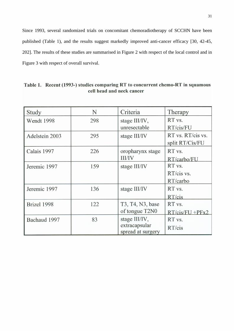
Since 1993, several randomized trials on concomitant chemoradiotherapy of SCCHN have been
published (Table 1), and the results suggest markedly improved anti-cancer efficacy [30, 42-45,
202]. The results of these studies are summarised in Figure 2 with respect of the local control and in
Figure 3 with respect of overall survival.

32

33
In a prospective randomized multicentre trial by Wendt et al., 298 previously untreated patients
with locoregionally advanced head and neck cancer were treated with either radiotherapy alone or
simultaneous radiotherapy plus chemotherapy consisting of cisplatin, fluorouracil and leucovorin
administered three times during the course of radiotherapy. Radiotherapy was identical in both arms
(70.2 Gy given in 1.8 Gy fractions). Concomitant chemotherapy resulted in improved local control
(48% vs 24%) and survival (36% vs. 17%) [42]. In another trial reported by Calais et al., a total of
226 patients with advanced oropharyngeal carcinoma participated in a phase III multicentre,
randomized trial comparing radiotherapy alone with radiotherapy plus concomitant chemotherapy.
Radiotherapy was identical in the two arms, consisting of conventional fractionation up to 70 Gy in
35 fractions. In the experimental arm, patients received during the course of radiotherapy three 4-
day cycles of carboplatin and 5-fluorouracil. In this trial, too, a significant improvement in both

34
local tumour control and overall survival was observed [202]. In a third large trial, reported by
Adelstein et al., 295 patients with unresectable SCCHN were randomly assigned to receive: 1)
single daily fractionated radiation (70 Gy, 2 Gy/day), 2) identical radiation therapy with concurrent
bolus of cisplatin, given on days 1, 22 and 43 of the radiotherapy course or 3) a split course
radiation, where three cycles of concurrent fluorouracil and bolus cisplatin chemotherapy were
given; two of the cycles were given concomitantly with radiation. In this trial, the addition of
concurrent high-dose, single-agent cisplatin to conventional once daily fractionated radiation
significantly improved survival, although it also increased acute toxicity. The loss of efficacy
resulting from split-course radiation was not offset by multiagent chemotherapy or by midcourse
surgery proved in the split-course arm [30].
A trial published by Brizel et al. in 1998, compared the efficacy of hyperfractionated irradiation
plus concurrent chemotherapy (combined treatment) to hyperfractionated irradiation alone. A total
of 122 patients with advanced head and neck cancer were randomized to receive either
hyperfractionated irradiation (a total dose of 75 Gy; 1.25 Gy given twice a day) or hyperfractionated
therapy (70 Gy; 1.25 Gy given twice a day) and 5 days of treatment with cisplatin and fluorouracil
during weeks 1 and 6 of irradiation. Two cycles of cisplatin and fluorouracil were given to most
patients after the completion of radiotherapy. At 3 years, both overall survival (55% vs 34%,
p=0.07) and locoregional control of the disease (70% vs 44%, p=0.01) were superior in the
combined therapy group [44].
Postoperatively given concurrent chemoradiotherapy improves local control and survival among
high risk patients with resected head and neck cancer. In a study reported by Cooper et al., 459
patients with head and cancer were randomly assigned to to receive either 1) postoperative
radiotherapy alone or 2) postoperative radiotherapy and cisplatin 100mg/m2 on days 1, 22 and 43

35
during the radiotherapy course in the experimental arm. High risk characteristics for recurrence
were defined as presence of one or more of the following features: histologic evidence of cancer
invasion to two or more regional lymph nodes, extracapsular extension of the nodal disease, or
microscopically involved mucosal margins of resection. Both locoregional control and disease free
survival were significantly better in the combined therapy group [203]. Similar results were
obtained from another randomized trial by Bernier et al [204].
Although not leading to improved survival rates, induction chemotherapy followed by radiotherapy
may have some role in attempts at organ preservation. In a study by the Veterans’ Affairs Laryngeal
Cancer Study Group, a total of 332 patients were randomly assigned either to the standard therapy
arm consisting of laryngectomy with postoperative radiotherapy or to an experimental arm
consisting of three cycles of cisplatin and fluorouracil, followed by RT. The larynx was preserved
in 64% of patients in the experimental group. No difference was found in survival [205]. In another
study concluded by the EORTC Head and Neck Cancer Co-operative Group, the larynx was
preserved in 42% of patients receiving induction chemotherapy followed by radiotherapy for
hypopharnyx cancer [206]. Neither of the studies mentioned above had a radiotherapy-only arm,
which makes it difficult to assess, whether the same results could have been achieved without
chemotherapy. In a recently published randomized study by Forastiere et al., radiotherapy given
with concurrent chemotherapy was compared to radiotherapy alone or to induction chemotherapy
followed by radiotherapy in patients with laryngeal cancer. After a follow-up of two years, the rate
of locoregional control and the proportion of patients with preserved larynx was higher in the
chemoradiotherapy group than in the other two groups [207]. On the basis of this study one can
question, whether induction chemotherapy followed by radiotherapy can be recommended even in
laryngeal cancer. Since the chemoradiotherapy protocols have improved locoregional control rates,
distant metastasis failures are becoming a more important cause of treatment failure than local

36
recidives. As a consequence, renewed interest has arisen on the possibility of eradicating
micrometastasis by modern induction chemotherapy or by adjuvant chemotherapy in head and neck
cancer patients, and thus, in improving overall survival [193]. The concept of induction
chemotherapy, however, remains experimental.
Results from both in vitro and in vivo studies have suggested that mitomycin C is preferentially
cytotoxic for hypoxic cells as compared with well-oxygenated cells [198, 208, 209]. Theoretically,
this might be of value when treating advanced head and neck tumours, which often contain poorly
oxygenated, radioresistant clonogenic cells.
Data from randomized studies suggest that infusions of mitomycin C (MMC) during radiation
therapy may improve the outcome of fractionated radiotherapy in head and neck cancer (Table 2).

37
Two consecutive randomized trials were performed at the Yale University School of Medicine
between 1980 and 1992. A total of 203 patients were enrolled in these trials. Intravenous MMC (15
mg/m²) or MMC (15 mg/m²) and dicumarol were given as an adjunct to conventional fractionated
radiotherapy (the total cumulative dose ranged from 60 to 68 Gy). A significant benefit was
achieved in the MMC arms with respect to cause-specific survival (74% vs. 51%; p=0.005), local
recurrence-free survival (85% vs. 66%; p=0.002), and loco-regional recurrence-free survival (76%
vs. 54%; p=0.003). No significant difference in overall survival was found between the MMC arms
and radiation alone [210]. Another large randomized study by Dobrowsky and Naudé supports the
efficacy of MMC given concomitantly with fractionated radiotherapy. In this study, 239 patients
with squamous cell cancer originating in the head and neck region were randomized to receive
either 1) conventionally fractionated radiation therapy to 70 Gy in 35 fractions given over 7 weeks,
2) continuous hyperfractionated accelerated radiotherapy to a total dose of 55.3 Gy in 33 fractions
over 17 consecutive days (V-CHART) or 3) V-CHART with concomitant administration of 20
mg/m² MMC on day 5 of treatment. A significant improvement in local tumour control and survival
was found in the accelarated fractionated treatment plus MMC arm as compared to the two RT
arms, but no difference was observed between the two RT arms [211]. In another large trial, 212
patients with previously untreated advanced squamous carcinoma of the larynx or hypopharynx
were randomized to receive either 1) initial treatment with radiotherapy, 50 Gy in 20 fractions given
over 28 days, or 2) to split-course radiotherapy, where 50 Gy was given in 20 fractions with a 4-
week break in the middle of the radiotherapy course and with concurrent MMC given on days 1 and
43, and 5-FU continuous infusions given on days 1 to 4 and days 43 to 46 of the radiotherapy
course. The result of the trial showed no advantage in local control or survival for the experimental
treatment arm of split-course radiotherapy and concurrent chemotherapy with MMC and 5-FU

38
compared with radiotherapy alone [212]. This is in line with the observation from the previously
mentioned trial by Adelstein et al. [30], where no effect was achieved with chemotherapy in the
split-course radiotherapy group. In a recent multicenter trial reported by Grau et al. that compared
fractionated radiotherapy (66 Gy in 33 fractions) with or without MMC 15 mg/m² given at the end
of the first week of treatment, no benefit was observed with concomitant mitomycin except in N0
patients, where locoregional control was significantly enhanced [213].
In concurrent chemoradiotherapy of head and neck cancer, the best documented single agent is at
present cis-platinum, and the most studied drug combination is cis-platinum combined with 5-FU.
In future chemoradiotherapy trials, potential new drugs should probably be compared to these
agents.
4.2.5. Advances in the radiation therapy delivery techniques
Developments in imaging technologies, including computed tomography (CT) and magnetic
resonance imaging (MRI), together with rapid advancements in computer systems have greatly
improved radiotherapy planning procedures recently. CT and MRI are capable of providing a full
3D model of the anatomy, thus enabling tumour volumes and their relationships to normal, healthy
tissues to be estimated more accurately. Novel functional imaging techniques, such as positron
emission tomography (PET), may in selected cases also be helpful in delineating radiotherapy target
volumes [214-218]. By using fusion of PET and CT/MRI images, it may be possible to enhance
treatment accuracy even further [219].

39
Three-dimensional CT-based treatment planning enables the use of conformal radiotherapy (CRT),
in which the dose distribution to the target volume is adjusted to conform to the shape of the tumour
to better avoid irradiation of the critical normal tissues. With this method, the dose to the tumour
can be increased, and doses received by normal tissues reduced. To improve the conformality of the
dose distribution, conventional beam modifiers, such as wedges or compensating filters, can be
used. The new generation of linear accelerators are equipped with computer-controlled multileaf
collimators (MLCs), which enable irregular shaping of treatment volumes and are thus very useful
in 3D-CRT [220].
Intensity-modulated radiotherapy (IMRT) is a novel form of CRT planning and delivery
technology. It represents one of the most important technical advances in the development of
radiotherapy. IMRT is based on the use of optimized non-uniform radiation beam intensities
incident on the patient. IMRT allows production of concave and irregular target volume dose
distributions, and thus has the potential to reduce the volume of healthy tissues irradiated to a high
dose [50]. The basis for development of IMRT was the rapid advancement in computer hardware
and software techniques during the past decade. Modern IMRT planning is generally based on
inverse treatment planning to achieve an optimal dose distribution in the target volume, while
simultaneously sparing sensitive normal structures [50, 221, 222]. In radiotherapy planning, the
target volumes and the organs-at-risk must first be defined similarly as in conventional 3D-CRT
planning. The dose constraints for all prescribed volumes must then be defined [221, 223, 224].
When this has been accomplished, a computer optimization system is used to adjust the beam
parameters to achieve the desired outcome [50]. In IMRT of head and neck tumours and cervical
node areas 5 to 9 coplanar fields are usually used [225-232], although occasionally as few as three
fields may be sufficient [229]. The radiation distribution within the target volume is most often

40
accomplished by multileaf collimators by programming the leafs to move dynamically during the
radiation, thus modifying the radiotherapy dose at different points of the target volume [233-240].
Clinical implementation of the IMRT technique requires novel methods for quality control of the
equipment and for verification of the treatment plans. For quality assurance, the dynamic multileaf
collimator (DMLC) boundaries are verified on a localization port film, the leaf motions are verified
to produce the planned dose distribution, the dose distributions produced by DMLC are verified to
produce the dose distribution that is consistent with the treatment plan, the leaf motions are
compared with those implemented for the treatment and the initial and final positions of the MLC
for each field are confirmed, and the actual doses are verified by in vivo dose measurements [241-
243]. In a study by Van Esch et al., the final correspondence between the calculated and measured
dose was found to be satisfactory in all five participating radiotherapy centres [243]. During the
actual treatment the most important cause for error was found to be in patient positioning rather
than dosimetry. In IMRT of head and neck cancer, keeping the patient’s position unchanged
throughout the entire treatment course is thus critical. This can be accomplished by using a
conventional thermoplastic head and neck fixation mask or a stereotactic head and neck
immobilization device. Patient positioning and field localization must be confirmed by repeated
simulations during the treatment course.
Clinical benefits of IMRT are expected to be most pronounced at body sites where sensitive normal
tissues surround or are located close to a target with a complex 3D shape. In the radiotherapy of
head and neck cancer, the irradiation doses needed for tumour control are often much higher than
the tolerance of the surrounding structures such as the spinal cord, the optic nerve, the eyes and the
salivary glands. IMRT provides a tool to reduce the dose to such sensitive normal structures or,
alternatively, to target dose escalation at a given level of normal tissue damage [228, 244-246].

41
IMRT has been successfully used in the treatment of head and neck cancer. In nasopharyngeal
cancer, excellent tumour target coverage with higher tumour doses has been achieved than using
traditional 3D planning with significant sparing of the salivary glands, the spinal cord, the brain
stem and other critical normal tissues [244, 245, 247-249]. In most of these studies, the follow-up is
not yet sufficiently long to allow comparisons with the results achieved by normal CRT, but good
local and locoregional control has achieved with IMRT [249, 250]. In the treatment of parotid
neoplasms, IMRT can be used to lower the irradiation doses to the brain, the spinal cord, the
cochlea and the oral cavity. The dose to the contralateral parotid gland delivered with IMRT is
dependent on the field arrangements and can be minimized by optimization of beam directions
[225, 229]. IMRT is also useful in minimizing the radiation dose to the optic pathways in the
treatment of maxillary sinus tumours and other paranasal sinus tumours, as well as orbital and
paraorbital tumours [226, 251-253].
4.3. Radiotherapy-related adverse events following treatment of head and neck cancer
4.3.1. Acute adverse effects
The adverse effects of radiotherapy can be chronologically divided into acute and late effects. Acute
adverse effects are defined as changes in tissues or associated symptoms noted within 90 days from
the date of initiation of radiotherapy [254, 255]. The most widely used grading system for acute
radiation-induced adverse events until recently has been the RTOG Acute Radiation Morbidity
Scoring Criteria [255]. In this classification, acute morbidity is classified from grade 0 (no change
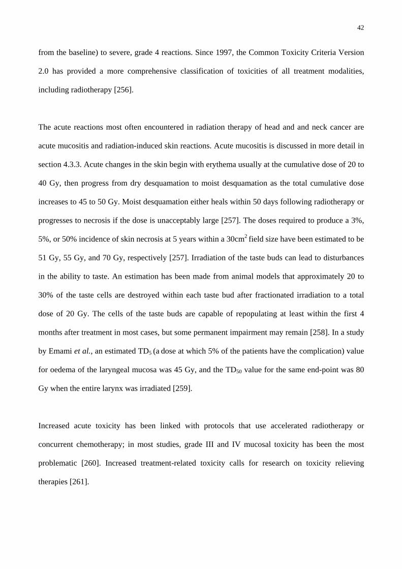
42
from the baseline) to severe, grade 4 reactions. Since 1997, the Common Toxicity Criteria Version
2.0 has provided a more comprehensive classification of toxicities of all treatment modalities,
including radiotherapy [256].
The acute reactions most often encountered in radiation therapy of head and and neck cancer are
acute mucositis and radiation-induced skin reactions. Acute mucositis is discussed in more detail in
section 4.3.3. Acute changes in the skin begin with erythema usually at the cumulative dose of 20 to
40 Gy, then progress from dry desquamation to moist desquamation as the total cumulative dose
increases to 45 to 50 Gy. Moist desquamation either heals within 50 days following radiotherapy or
progresses to necrosis if the dose is unacceptably large [257]. The doses required to produce a 3%,
5%, or 50% incidence of skin necrosis at 5 years within a 30cm2 field size have been estimated to be
51 Gy, 55 Gy, and 70 Gy, respectively [257]. Irradiation of the taste buds can lead to disturbances
in the ability to taste. An estimation has been made from animal models that approximately 20 to
30% of the taste cells are destroyed within each taste bud after fractionated irradiation to a total
dose of 20 Gy. The cells of the taste buds are capable of repopulating at least within the first 4
months after treatment in most cases, but some permanent impairment may remain [258]. In a study
by Emami et al., an estimated TD5 (a dose at which 5% of the patients have the complication) value
for oedema of the laryngeal mucosa was 45 Gy, and the TD50 value for the same end-point was 80
Gy when the entire larynx was irradiated [259].
Increased acute toxicity has been linked with protocols that use accelerated radiotherapy or
concurrent chemotherapy; in most studies, grade III and IV mucosal toxicity has been the most
problematic [260]. Increased treatment-related toxicity calls for research on toxicity relieving
therapies [261].

43
4.3.2. Late adverse events
Late effects are defined as changes in tissue or associated symptoms that occur more than 3 months
from the beginning of radiotherapy [255]. In grading of these adverse effects, the RTOG/EORTC
late radiation morbidity scoring scheme is commonly used [255]. The risk of radiation-induced late
complications of various organs is highly dependent on the dose given and the treatment volume
and fractionation used. For conventional fractionation, the minimal tolerance dose of each tissue is
defined as TD5/5, which represents the dose of radiation that would cause no more than a 5% severe
complication rate within 5 years after radiotherapy [254]. Correspondingly, the TD50/5 value is the
dose of radiation that would cause a 50% severe complication rate at 5 years. Care should be taken
when using unconventional fractionation schedules, because the TD5/5 values for late normal tissue
damage are valid only for conventional fractionation. To express an equal biological effect
produced by different fractionation schemes, isoeffect lines have been generated [254]. Slopes for
tumour curability and for normal tissue late effects have been calculated. In general, the slope for
tumour curability is less steep than that for normal tissue reactions [254].
When estimating the late radiation effects, an organ can be considered to consist of multiple
functional subunits (FSUs) that are arranged serially or in parallel [254]. In serially arranged organs,
such as the spinal cord, damage to one portion of the organ may render the entire organ
dysfunctional. In organs with parallel function, such as salivary glands, the surviving FSUs operate
independently of the damaged group, and thus, organ function is maintained if the proportion of the
functioning FSUs is large enough [262].

44
The capacity for repair of sublethal injury is the most important biological phenomenon influencing
the fractionation response of tissue [97]. Slowly responding tissues consistently show a greater
capacity for repair than rapidly responding tissues [263]. Large dose fractions have been
demonstrated to be more harmful for late-responding tissues, and thus, a therapeutic gain may be
achieved by hyperfractionation [97]. When using hyperfractionation or accelerated fractionation,
the interfraction time period must be at least 6 hours to allow complete repair of sublethal damage
in late-responding tissues. In a phase I-II RTOG trial on hyperfractionation a short interfraction
interval (<4.5 hours) was found to be a major determinant of late effects when twice-a-day
irradiation schedules were used [264].
Most late effects develop within the first 3 years following radiotherapy for head and neck cancer,
and a few progress beyond 3 years [260]. Data derived from RTOG trials indicate that 85% of
patients who received conventional radiation alone experienced some form of late toxicity.
Approximately 12% suffered from grade 3 or 4 reactions, the most common of which were
xerostomia, dysphagia and laryngeal toxicity [260]. Some mucosal atrophy and loss of mucosal
mobility are common after conventional fractionated radiotherapy to a total dose of 60 to 70 Gy, but
bone exposure seldom occurs unless dose delivery is accelerated or the total dose exceeds 70 Gy in
7 weeks [265]. The TD5/5 for telangiectasia of the skin is about 45 Gy. Higher doses lead to an
increased incidence of telangiectasia, fibrosis and atrophy [257]. The TD5/5 and TD50/5 values for
laryngeal cartilage necrosis are estimated to be 70 and 80 Gy, respectively. Another possible severe
late complication of head and neck radiotherapy is mandibular osteoradionecrosis; the TD5/5 value
for this complication, when the entire organ is irradiated is 60 Gy [259].
The late effects of radiation to the central nervous system must also be taken into account. The 5%
incidence of radiation myelopathy has been suggested to be between a total dose of 57 and 61 Gy

45
with conventional fractionation [266]. The severe complication rate of the spinal cord using the
conventional dose limit of 40 to 45 Gy given in 1.8-2 Gy fractions over 4 to 5 weeks is practically
nil. Brain necrosis is seldom noted at doses of 60 Gy or less with conventional fractionation in
adults. Neurocognitive changes may, however, take place at lower doses, especially in children
[266]. When the hypothalamic-pituitary axis is included in the treatment field, neuroendocrine
disturbances are observed at doses exceeding 40 Gy [267]. Late ocular, vestibular and hearing
adverse effects need also be considered when planning radiotherapy for head and neck cancer [268,
269].
4.3.3. Radiation mucositis
Oropharyngeal mucositis is the most common and clinically significant acute adverse effect of
radiotherapy for head and neck cancer. With conventional fractionation, the first signs of mucositis
normally appear during the second week of radiotherapy and progress towards the end of
radiotherapy from enanthema to spotted or confluent pseudomembranous mucositis [270-272].
Recovery occurs within 2.5 to 3 weeks after completion of radiotherapy, and within one month the
mucosa heals in 90 to 95% of patients [260, 271]. Acute mucosal reactions cause pain, with
concomitant difficulties in swallowing and speaking. Difficulties in eating may lead to worsening of
the nutritional status and weight loss, and mucositis also predisposes to local and systemic
infections. Severe mucositis is the most common cause for interruption of the radiotherapy course
for head and neck cancer, which in turn can lead to significant loss of local tumour control
probability [47, 270].

46
With hyperfractionated and accelerated radiotherapy, mucositis appears earlier and tends to be more
severe than with conventional fractionation [137, 271-273]. The incidence of grade ≥3 mucositis
increased from 20 to 50% to 66 to 86% in several trials where accelerated fractionation was used
[260]. In chemoradiotherapy trials, the most problematic acute toxicity is also increased mucosal
toxicity [260]. Drugs that often cause mucosal side-effects include 5-FU, capecitabine,
methotrexate, bleomycin and doxorubicin. In contrast, drugs such as cisplatin and carboplatin
seldom cause mucosal problems as single agents and are therefore preferable as chemoradiotherapy
agents because they have little overlapping toxicity with ionizing radiation [265]. Efforts have been
made to counteract the increased toxicity associated with intensified treatment protocols with
various toxicity ameliorating drugs [261].
4.3.4. Xerostomia
One of the most common and distressing long-term adverse effects of head and neck radiotherapy is
permanent xerostomia resulting from salivary gland damage. Xerostomia predisposes to infections
and dental caries and disturbs swallowing and speech [265, 274]. Saliva is normally produced at a
rate of 1000 to 1500 mL per day. Over 90% of this amount is secreted by three pairs of major
salivary glands; the parotid, the submandibular and the sublingual glands. The minor salivary
glands scattered over the mucosal surfaces of the mouth and pharynx account for less than 10% of
the total salivary production [275]. In one study the submandibular glands produced 69% of
unstimulated salivary flow and the parotid and sublingual glands 26% and 5%, respectively,
whereas the parotid glands produced two-thirds of stimulated saliva [275]. Less than 0.1 mL/minute
of unstimulated salivary flow and 0.5 to 0.7 mL/minute of stimulated salivary flow are generally
considered as abnormally low [276].

47
The most important treatment-related factors that contribute to radiation-induced xerostomia are the
volume of the salivary glands included in the irradiation fields and the total dose [265]. Parotid
gland salivary flow is markedly reduced when a cumulative dose of 30 to 50 Gy is given using
conventional fractionation [265, 277-282]; this dose level is often exceeded in conventional CRT of
head and neck cancer.
4.4. Prevention of radiotherapy-associated mucositis and xerostomia
4.4.1. Prevention of mucositis
The progress made in studies of altered fractionation and chemoradiotherapy in head and neck
carcinoma offers tools for achieving better local control and, with chemoradiotherapy, also better
overall survival than that achieved with conventional treatment. However, because these treatment
modalities are associated with increased local toxicity, effective treatments of local acute reactions
would be valuable.
Numerous medical agents, such as beta-carotene [283], chlorhexidine [284, 285], prostaglandin E1
[286], benzyldamine [287], glutamine [288], povidone-iodine [289], hydrogen peroxide rinses
[290], sucralfate [291, 292] and local antibiotics [293, 294], have been tested for their ability to
alleviate radiation-induced mucositis. The results have been disappointing, and in a recent meta-
analysis of randomized clinical trials on mucositis prophylaxis reported by Sutherland and
Browman, the conclusion was that at present insufficient evidence exists to support the
development of recommendations for the prevention of oral mucositis in clinical practice, with the
possible exception of narrow-spectrum antibacterial agents [295]. In a recent double-blind,

48
randomized phase III study, antibiotic lozenges did not have a significant impact on the severity of
radiation- induced mucositis [296].
Over the last few decades the radioprotective activity of thiol-containing compounds has been under
investigation. The most promising of these compounds has been amifostine. Its mechanism of
action in radioprotection is thought to take place through scavenging of radiation-induced free
radicals [297]. In small clinical trials, amifostine was found to protect against radiation-induced
salivary gland damage as well as against radiation-induced acute mucositis [298, 299]. In a phase
III randomized trial by Brizel et al., 315 patients with head and neck cancer were randomized to
radiation treatment with or without amifostine at a daily infusion of 200 mg/m² 15 to 30 minutes
before radiotherapy. A significant reduction in radiation-induced xerostomia was observed, but
amifostine did not diminish the severity of acute mucositis [48].
Based on preliminary reports with small patient numbers, granulocyte-macrophage colony-
stimulating factor (GM-CSF, molgramostin) was considered as one of the most promising new
agents for the prevention of radiation-induced mucositis [300-302]. GM-CSF is a glycoprotein with
a molecular weight of 22 kD. GM-CSF acts by enhancing colony formation of granulocytes,
macrophages and eosinophils and it also regulates several functions of mature leucocytes,
macrophages and dendritic cells in the dermis and submucosa [303, 304]. In addition, GM-CSF
enhances keratinocyte and fibroblast growth and improves healing of cuts, leg ulcers and skin grafts
[305-310]. In early reports, subcutaneously given GM-CSF was found to significantly alleviate
radiation-induced mucositis [311, 312]. This could not, however, be confirmed in a small
randomized study, where subcutaneous GM-CSF was associated with moderate toxicity [312]. A
few small, non-randomized studies have suggested that GM-CSF mouthwashes during
oropharyngeal radiotherapy might reduce the severity of radiation-induced mucositis [300-302].

49
Before being adopted in clinical practice, the influence of topical GM-CSF must be confirmed in
adequately powered randomized studies.
In the absence of specific anti-mucositic agents for radiation-induced acute mucositis, the mainstays
in the care of head and neck cancer patients who receive radiotherapy are careful oral hygiene
during radiotherapy and adequate nutritional protocols to maintain a good nutritional status during
the radiotherapy course. In recent trials, percutaneous endoscopic gastrostomy (PEG) has been
demonstrated to be feasible and effective in preventing dehydration and malnutrition during a
course of head and neck radiotherapy [313-315].
4.4.2. Prevention of xerostomia
The most important aspect in the prevention of radiation-induced salivary gland damage is careful
treatment planning to minimize the volumes of salivary glands included in the irradiation fields.
Based on the normal secretion patterns of saliva, most of the stimulated saliva flow will be
maintained if the parotid gland dose can be limited, and avoidance of submandibular gland
irradiation at high doses will help to maintain the unstimulated saliva production.
As salivary glands are considered to function as parallel organs with respect to late radiation-
induced damage, preservation of the salivary function is to be expected if irradiation of large
volumes of the major salivary glands can be avoided. In a study by Roesink et al., an increase of
the irradiated parotid gland volume from 0-40% to 90-100% while maintaining a mean parotid
dose of 35 to 45 Gy resulted in a decrease in the saliva flow ratio from approximately 100% to less
than 10% following irradiation [316]. CRT and especially IMRT provide new possibilities for

50
defining the volumes of salivary glands irradiated and the doses received by each gland. Computer-
based radiotherapy planning enables the use of dose-volume histograms (DVHs), which allow for
more accurate estimations of the target volumes and doses received by each organ [97, 254].
Eisbruch et al. recently reported that cumulative doses of less than 24 to 26 Gy to the main salivary
glands are associated with substantial preservation of saliva flow rates [317]. In another study, no
treshold dose was found, and limiting the cumulative parotid gland dose to 39 Gy or less was
recommended [316]. Not only restricting the dose to the major salivary glands but also limiting the
dose to the minor salivary glands is considered important, since the minor glands produce up to
70% of the total mucins secreted by salivary glands [318].
The most effective radioprotective compound studied in prevention of radiation-induced xerostomia
has been amifostine. Amifostine has a cytoprotective activity only when present during exposure of
cells to radiation or cytotoxic agents [297]. Amifostine is administered as an inactive prodrug and is
activated extracellularly by alkaline phosphatase, which is present at high concentrations in normal
but not in malignant tissues [319-321]. The effectiveness of amifostine in prevention of radiation-
induced xerostomia was shown in a large randomized trial by Brizel et al. In this study, the
incidence of xerostomia at one year post-treatment was significantly reduced in patients who
received amifostine (34% vs. 57%) [48]. The side-effects associated with amifostine are nausea,
vomiting and hypotension. Allergic reactions, sometimes resulting in life-threatening anaphylactoid
reactions or toxic epidermal necrolysis, have been described [322, 323]. In a phase III study by
Rades et al. [324] intravenous amifostine was given to 39 patients receiving radiotherapy for head
and neck cancer; nine of the patients received also concurrent chemotherapy. Intravenous
amifostine had to be discontinued in 16 (41%) out of the 39 patients and in 7 (78%) out of the 9
patients who received concomitant chemoradiotherapy, which led to discontinuation of the study.

51
Based on this study and other data from the literature, discontinuation of amifostine occurs in about
24% of the patients who receive amifostine concurrently with radiotherapy. Subcutaneous
administration of aminofostine has been reported to be associated with less toxicity than
intravenous administration. The frequency of severe adverse events remains nevertheless over 10%
[325, 326].
Another widely investigated drug in prevention and treatment of radiation-induced xerostomia is
pilocarpine. Pilocarpine is a muscarine cholinerginic agonist that increases salivary output in
normal conditions [261]. In a randomized, placebo-controlled trial by Johnson et al., pilocarpine
was shown to significantly improve the sensation of oral dryness following radiation therapy, but it
did not have any permanent impact on quantitative measures of salivary flow [327]. In a non-
randomized study by Zimmerman et al., the use of 5 mg oral pilocarpine four times a day during
radiotherapy for head and neck cancer and 3 months afterwards was found to be associated with
significantly less subjective xerostomia than reported in retrospective controls [328]. Similar results
have obtained from one small randomized study [329].
Theoretically, by combining IMRT and amifostine, greater sparing of the salivary function might be
achieved than by either treatment modality alone [330, 331]. A randomized trial is ongoing on
subcutaneously delivered amifostine in patients who receive IMRT for head and neck cancer, but
the results are not yet available [331].

52
5. AIMS OF THE STUDY
The general objective of this study was to improve treatment efficacy and to reduce treatment-
related adverse effects in squamous cell head and neck cancer. The specific aims were as follows:
1. To study expression of cyclin A and the Ki-67 antigen as predictors for locoregional recurrence
and outcome in laryngeal cancer patients treated with surgery and postoperative radiotherapy.
2. To evaluate the effects of tumour cell repopulation and radiotherapy treatment time on local
control in T1 laryngeal cancer treated with radiotherapy.
3. To assess the efficacy of biweekly escalated, accelerated hyperfractionated radiotherapy with
concomitant single-dose mitomycin C in the treatment of advanced laryngeal and
hypopharyngeal cancers with respect to tumour control and organ preservation.
4. To estimate the effect of granulocyte-macrophage colony-stimulating factor in the
prevention of radiation-induced mucositis during postoperative radiotherapy of squamous cell
cancer head and neck.
5. To study intensity-modulated radiotherapy in the prevention of radiation-induced
salivary gland damage and subsequent xerostomia

53
6. PATIENTS AND METHODS
6.1. PATIENTS ( STUDIES I-V )
The patient population involved in these studies comprised of 285 patients with squamous cell head
and neck cancer treated by definitive or postoperative radiotherapy at the Department of
Radiotherapy and Oncology, Helsinki University Central Hospital in 1982 to 2002. The patients
involved in Studies I to V are given in Table 3.
Table 3. Patients and the treatments given in Studies I-V. Study
Number of Patients (F, female; M, male)
Year of Diagnosis
Clinical Stage
Type of Radiotherapy
Scheduled concomitant medication
I
90 ( F 12, M 78 )
1982 to 1998
Stage II-IV, T1-4 N0-2 M0
Postoperative RT (split-course, N=56; continuous, N=34)
None
II
117 ( F 11, M 106 )
1982 to 1993
Stage I, all T1N0 M0
Definitive RT (split-course, N=89; continuous, N=28)
None
III
21 ( F 3, M 18 )
1998 to 2001
Stage III-IV, T2-4 N0-3 M0
Escalated, accelerated, hyperfractionated RT
Mitomycin C 10mg iv given on d. 30
IV
40 ( F 21, M 19 )
1999 to 2001
Stage II-IV, T1-4 N0-N2 M0
Postoperative RT
GM-CSF 37.5µgx 4 vs. Sucralfate 1g x4 mouthwashes
V
17 ( F 10, M 7 )
2000 to 2002
Stage II-IV, T1-4 N0-2 M0
Intensity modulated Radiotherapy (IMRT)
None
54
In Study I, paraffin-embedded tumour samples from 90 laryngeal cancer patients (78 men, 12
women) were stained for cyclin A and the Ki-67 antigen using immunohistochemistry. All of the
patients were treated with partial or total laryngectomy followed by postoperative radiotherapy to a
total dose of 50 Gy or greater. The median age at diagnosis was 63 (range 35 to 85 years). The
median post-treatment follow-up time was 91 months (minimum 48 months).
Study II was an analysis of the effect of radiotherapy treatment time on the local tumour control in
patients with T1 laryngeal cancer patients. The patient series consisted of 117 consecutive patients
(106 men, 11 women) with T1 laryngeal cancer treated between 1982 and 1993 by radical
radiotherapy alone, given either as continuous (n=28) or split-course treatment (n=89). The median
age of patients was 66 (range, 42 to 87 years). Eighty cancers were located in the mobile vocal cord,
33 involved the anterior commissure and four involved both cords.
In Study III, 21 patients (18 men, 3 women) with stage III to IV laryngeal or hypopharyngeal
squamous cell cancer were treated with a biweekly escalated, accelerated hyperfractionated
radiotherapy schedule with a concomitant single dose of mitomycin C. Ten of the patients had
laryngeal and 11 hypopharyngeal cancer. Their median age was 59 (range, 27 to 71) years.
Study IV was a double-blind, prospective, randomized study that compared granulocyte-
macrophage colony-stimulating factor (GM-CSF) mouthwashes with sucralfate mouthwashes in the
prevention of radiation-induced mucositis. Forty patients (27 men, 13 women) with radically
operated head and neck cancer were randomly allocated to use either GM-CSF (n=21) or sucralfate
(n=19) mouthwash during postoperative radiotherapy. Patients with prior chemotherapy or
radiotherapy, chronic autoimmune or inflammatory disease or the World Health Organization
(WHO) performance status >2 were not eligible for the trial.

55
In Study V, 17 patients (10 women, 7 men) with primary squamous cell cancer of the head and neck
were treated with IMRT. A decision to use IMRT instead of conventional conformal radiotherapy
was made in these cases, because the use of conventional radiation therapy would have resulted in
irradiation of all major salivary glands to a cumulative dose greater than 45 Gy with a high risk of
subsequent post-irradiation xerostomy. The mean age of the patients was 55 (range, 32 to 78) years.
The WHO performance status was 0 (normal) in 11 cases and 1 in six patients. Six patients had a
nasopharyngeal primary tumour and the remaining 11 had oropharyngeal cancer. Three of the
patients had stage II, four had stage III, and 10 stage IV cancer. Eleven patients received definitive
radiotherapy with a curative intent, and 6 patients received postoperative radiotherapy.
In studies III and IV, which required scheduled concomitant medication, the study protocol was
approved by an Ethical Committee of the Helsinki University Central Hospital before enrolling any
patients in the trials, and the patients provided a written informed consent. Studies I, II and V were
based on retrospective analyses of hospital case records and data collected along with standard care
of patients.
6.2. TREATMENT (I-V )
The patients and treatments in Studies I-V are summarized in Table 3. All 90 patients in Study I
underwent surgery followed by postoperative radiotherapy. Total laryngectomy was performed in
72 (80%), supraglottic laryngectomy in 14 (16%), and hemilaryngectomy in 4 (4%) cases. Neck
dissection was done in 25 cases (28%) where nodal metastasis was suspected. In 23 of these,

56
presence of nodal metastases was confirmed histologically. After surgery all patients received
postoperative radiotherapy. The median time from surgery to the beginning of radiation therapy was
47 (range, 23 to 83) days. The first 26 patients were treated with 60Co between 1981 and 1987, and
the following 64 patients with a 6 MV linear accelerator between 1988 and 1993. The upper neck
was irradiated through two lateral portals, and the lower neck from a separate anterior field. The
total dose varied from 50 to 66 Gy depending on the size of the primary tumour and involvement of
surgical margins. The fraction size ranged from 1.8 to 2.0 Gy, and in all patients, the treatment was
given in one daily fraction. Between 1981 and 1993, the treatment for 56 cases was given as split-
course radiotherapy with a 14- to 21-day planned gap in the middle of the radiotherapy, between
1994 an 1998 as continuous radiotherapy for the remaining 34 cases. The mean total treatment time
in patients treated with split-course radiotherapy was 66 days (range, 58 to 71), and in those treated
with continuous radiotherapy 41 days (range, 33 to 41).
All 117 patients included in Study II received definitive radiotherapy for TI laryngeal cancer. Sixty-
six of these patients were treated with 60Co, and 51 with a 6 MV linear accelerator. Two lateral
opposing fields with compensatory wedges were used in all patients. The target dose was calculated
by a computer-based radiotherapy planning program. The first 89 patients treated between 1982 and
1987 received split-course radiotherapy with a planned 2- to 3 week gap in the middle of the
radiotherapy course. The mean total dose in this group was 66 Gy (range, 60 to 68.2) and the
overall treatment time varied from 53 to 79 days (mean, 65). The fraction size was increased by
10% from 2.0 to 2.2 Gy to compensate for the planned gap. Since 1988, 28 patients were treated
with a continuous radiotherapy course. In these patients, the mean total dose was 62 Gy (range, 60
to 66), and the mean overall treatment time was 44 days ( range, 39 to 50). All patients received the
therapy in 2-Gy daily fractions.

57
In Study III a hyperfractionated, escalated radiotherapy schedule with concomitant MMC was used
in all 21 cases. An individually made thermoplastic mask (Sinmed®) was used for head and neck
fixation. Treatment planning was carried out using a CT-based treatment planning computer
program (Cadplan®, version R.6.2.7., Varian Medical Systems). Radiotherapy doses were
prescribed according to the ICRU 50 specifications [332], where the ICRU reference point was
chosen centrally within the planning target volume (PTV). At the beginning of the radiotherapy
course, the treatment volume encompassed the primary tumour and the locoregional lymphnodes.
Radiotherapy was given in 2 daily fractions with an interfraction interval of at least 6 hours. The
fractionation schedule is presented in Table 1 of the original contribution of Study III. After a
cumulative dose of 52 Gy, the fields were reduced and the macroscopic tumour plus 1- to 2 cm
margins were boosted to a total cumulative dose of 74.4 Gy. A single dose of MMC 10mg/m2 was
given intravenously 2 hours before irradiation on day 30 of the radiation course. A part from a
diagnostic biopsy, none of the patients underwent primary surgery. According to the treatment
protocol, surgery on the primary site was required only when the tumour persisted for longer than 2
months after the chemoradiotherapy or when the tumour recurred locally. A radical neck dissection
was to be carried out after chemoradiotherapy when the clinical nodal classification was N3 or if the
enlarged lymph nodes persisted 2 months after the completion of chemoradiotherapy.
Study IV was a double-blind, prospective randomized, phase III study comparing the effectiveness
of granulocyte-macrophage colony-stimulating factor (GM-CSF) and sucralfate mouthwashes in the
treatment of radiation-induced mucositis. All patients had undergone radical surgery for head and
neck cancer and were scheduled to receive postoperative radiotherapy. Radiotherapy dose planning
was performed with a CT-based planning program (Cadplan®). Treatment to the primary resection
area and upper neck lymphatics was usually given through two parallel opposed fields; the lower
neck lymphatics were, whenever necessary, treated from a separate anterior field. The dose to the

58
medulla was restricted to 38 Gy, following which, the radiation therapy to the dorsal neck was
completed with 9 MeV electrons. The total dose to the primary tumour site was 50 to 60 Gy, and 50
Gy was delivered to the locoregional lymphatics. Radiotherapy was given to all patients as
continuous therapy in 2-Gy daily fractions 5 times a week.
The GM-CSF mouthwash solution was prepared by dissolving 150 µg of dry drug powder into 100
ml of sterile water, and the sucralfate solution by dissolving 4.0 g of sucralfate in the same amount
of water. The respective mouthwashes were started once a cumulative total dose of 10 Gy was
reached (after the first week of radiotherapy) and continued until the end of the radiotherapy.
Mouthwashes were used during treatment days; their use was interrupted on Saturdays and
Sundays. In both treatment groups, the patients were instructed to use 100 ml of the treatment
solution divided into 4 equal 25 ml doses during each day of radiotherapy. The dose of GM-CSF
per one mouthwash thus was 37.5 µg, and that of sucralfate 1 g.
In Study V intensity-modulated radiotherapy (IMRT) was used to avoid the permanent xerostomia
often accompanying radiotherapy of head and neck cancer. The patients were immobilized during
the radiotherapy using either a conventional thermoplastic device or, in the last 7 patients of the
study using a stereotactic head and neck immobilization device. The IMRT technique used to
irradiate the primary tumour and the cervical lymph nodes consisted of 5 to 7 coplanar fields. In
most cases (n=14) 6 fields were used. The arrangement of the fields is described in Table 2 of the
original contribution. The treatment plans were generated using an inverse planning software. The
dose constraints were adjusted according to the clinical situation. The cumulative dose to the spinal
cord was kept under 40 Gy. Those salivary glands that were excluded from the primary target
volume were included in the optimization procedure using a maximum dose constraint of 25 Gy in
the first 5 patients, and 16 to 20 Gy in the remaining 11 patients. In addition, a volume of healthy
59
tissue was delineated outside the PTV in each CT slice, and in CT slices located immediately
cranial and caudal to the slices containing the PTV, to prevent hot spots outside the PTV. The
contralateral parotid gland was treated in the optimization procedure as an organ-at-risk (OAR) in
all patients, and in 6 patients the contralateral submandibular gland was also excluded from the
target volume.
6.3. IMMUNOHISTOCHEMISTRY ( STUDY I )
Immunohistological analysis was performed on tissue sections prepared from formalin-fixed,
paraffin-embedded archival tissue of the excised primary tumour; no needle biopsy samples were
included. The tissue sections were deparaffined in xylene, and the samples were rehydrated using an
ethanol series. Antigen demasking was carried out by heating the samples in a microwave oven in
0.1 M citric acid buffer at pH 6, 4 times for 5 minutes. For immunohistochemistry, the specimens
were incubated overnight at room temperature with an anti-human cyclin A mouse monoclonal
antibody at a dilution of 1:100 (Novocastra Laboratories Ltd., Newcastle, UK). The sections were
counterstained with haematoxylin and eosin, and mounted. Hyperplastic human tonsillar tissue was
used as a positive control; the primary antibody was omitted in the negative control samples.
Immunostaining for the Ki-67 antigen was performed similarly to staining for cyclin A. A rabbit
anti-human antibody (A 0047; DAKO A/S, Glostrup, Denmark) at a dilution of 1:100 was used as
the primary antibody.
The assessment of the staining was done similarly as described elsewhere[179]. In brief, in each
case, a total of 5 fields were assessed at a magnification of 10 x 40 using a Leitz Laborlux D
microscope (Wetzlar, Germany). The fields were chosen from the tumour areas showing the highest

60
density of positive nuclear staining when scanned at a low magnification. To determine the
percentage of positively staining nuclei, an ocular grid consisting of 100 (10x10) squares was used.
All positive nuclei from the grid area were counted. To estimate the number of negative nuclei
within the same 100-square field, we counted the number of nuclei in 3 non-adjacent rows of 10
squares, and multiplied the mean score by 10. For both cyclin A and Ki-67 staining the results are
reported as the percentage of tumour cells with positive nuclear staining.
6.4. RANDOMIZATION ( STUDY IV )
Study IV was a double-blind, prospective, randomized phase III study in which 40 patients were
scheduled to receive postoperative radiotherapy for head and neck cancer. They were randomized to
receive either GM-CSF or sucrafate mouthwashes during the radiotherapy. Randomization was
done using computer-generated random digits; 21 patients were assigned to GM-CSF mouthwashes
and 19 to sucralfate mouthwashes to be given during radiotherapy.
After the patients had provided a written informed consent, they were assigned to a treatment group.
The patient’s name and social security number were provided for unequivocal indentification of
each patient. The patients were stratified before randomization by the volume of oral cavity and
oropharyngeal mucosa involved in the radiotherapy target volume to guarantee that the mucosal
areas irradiated were roughly equal in both study arms. The stratification was done on whether 50 to
75% or over 75% of the oropharyngeal mucosa was included in the target volume.

61
The Radiation Therapy Oncology Group acute radiation morbidity scoring criteria were used in the
estimation of the degree of radiation mucositis in Studies III-V [255]. In Study III, the examination
of the degree of mucositis and assessment of mucositis-related symptoms was done before the
beginning of radiotherapy, weekly during therapy, and 1, 2 and 4 weeks after therapy. Oral
mucositis-related pain was estimated by the patients themselves using a linear visual analogue scale
(VAS) [333] from 0 to 10, where 0 represents absence of pain and 10 maximal pain. Grave
mucositis is associated with worsening of the patients’ nutritional status, and therefore patients’
weight and serum prealbumin levels were monitored in addition to the clinical status to obtain an
objective measure of the nutritional status during the radiotherapy course. The biological half-time
of prealbumin is short (about 2 days), and when the protein balance is negative, the serum
prealbumin level falls rapidly [334, 335]. The use of local anaesthetics (lidocaine mouthwashes)
and systemic analgesic drugs was registered for each patient as was the use of antibiotics or
antimycotics for mucositis-related infections. Mucositis-related interruptions in radiotherapy and
possible hospitalization of trial patients during radiotherapy were also recorded.
6.6. ASSESSMENT OF LARYNGEAL FUNCTION ( STUDY III )
Laryngeal function following radiotherapy was assessed by performing videolaryngoscopy and the
voice quality was evaluated perceptually at the Department of Otorhinolaryngology – Head and
Neck Surgery of the Helsinki University Central Hospital.
6.5. ASSESSMENT OF MUCOSITIS ( III-V )

62
In Study V, the salivary gland function of patients treated with IMRT was assessed before the
beginning of radiotherapy and at 6 and 12 months after radiotherapy had ended. Both the basal and
the stimulated saliva secretion rates were measured. The basal rate was obtained by measuring the
total saliva secretion over 15 minutes, after which saliva secretion was stimulated by chewing a
piece of paraffin wax, and collected for 15 minutes. The patients prepared the paraffin wax by
chewing for 2 minutes before saliva collection. The patients were advised not to eat, drink or smoke
for one hour before collection of stimulated saliva was initiated. In addition to measuring saliva
secretion, the degree of xerostomia was also assessed by grading according to the Radiation
Therapy Oncology Group (RTOG) and the European Organization for Research and Treatment of
Cancer (EORTC) classification systems [255].
6.8. STATISTICAL ANALYSES
Data in Studies I and III to V were analysed using the Number Cruncher Statistical System (NCSS)
2000 program. In Study II, the BMDP (Biomedical Computer Programs-P series) statistical package
was used.
The Kaplan-Meier product-limit method was used to estimate the survival distributions for disease-
free survival and overall survival (I to III). Comparisons of the survival rate between groups were
done using the log-rank test or univariate Cox regression analysis (I). The relative importance of
prognostic factors was analysed using Cox’s proportional regression model (I). The association
between Ki-67 and cyclin A expression was investigated using the Spearman correlation test, and
associations of cyclin A and Ki-67 expression with categorical parameters were assessed with the
Mann-Whitney test or the Kruskal-Wallis analysis of variance (I). The Mann-Whitney test was also
6.7. ASSESSMENT OF XEROSTOMIA (V)

63
used in Studies IV and V to test differences between treatment groups. Frequency tables were
analysed using either chi-square or Fisher’s exact test (I, IV). In study IV, repeated observations
were analysed with repeated measures analysis of variance. All p-values are 2-sided.
The analysis of the effect of the overall treatment time on local control and the estimation of
proliferation rates are presented in Section 2.5. of the original contribution (II). The mathematics for
modelling of salivary flow as a function of dose is presented in detail in the Appendix of Study V.

64
7. RESULTS
7.1. EFFECT OF THE OVERALL TREATMENT TIME AND CELL REPOPULATION ON
THE FREQUENCY OF LOCOREGIONAL RECURRENCE ( I-III )
In Study I, the effect of the overall radiotherapy treatment time on tumours with varying cell
proliferation rate was examined. Cancers with high Ki-67 expression (>34% nuclei positive, the
highest tertile) recurred more frequently locoregionally when treated with split-course radiotherapy
than when treated with a continuous course of therapy (p=0.035), whereas with lower Ki-67
expression, a break in treatment had no influence on the frequency of locoregional recurrences
(p=0.93). Seventeen (30%) of the 56 patients treated with split-course radiotherapy and 6 (18%) of
the 34 patients treated with a continuous radiation therapy course had a local recurrence (p=0.18).
When the proliferative fraction of the cancers was taken into account, planned gaps in the
radiotherapy course turned out to be more deleterious for patients who had cancer with a large
proliferative fraction. Only one locoregional recurrence was observed among the 12 (8%) patients
who had high Ki-67 expression cancer when treated with continuous radiotherapy as compared with
8 (44%) among the 18 patients treated with planned split-course radiotherapy. In contrast, the
proportion of locoregional recurrences was roughly similar in patients with cancer with low Ki-67
expression irrespective of whether a planned break was held or not (24% vs. 23%, respectively,
p=0.93).
Patients with a high cyclin A expression level had somewhat more locoregional recurrences when
treated with split-course radiotherapy, but this difference did not reach statistical significance.
When both tumour cyclin A and Ki-67 expression were within the highest tertile, only one of such
patients (17%) treated with continuous radiotherapy had a locoregional recurrence, whereas as

65
many as 8 (73%) of 11 of such patients treated with split-course radiotherapy had a local
recurrence (p=0.050). No significant associations were found between the duration of the surgery-
to-radiotherapy interval, tumor proliferation fraction, and the frequence of locoregional
recurrences.
The impact of tumour cell repopulation on outcome of T1 laryngeal cancers treated with
radiotherapy was evaluated in Study II. The local control rate was 95% (range, 94 to 96%) for the
continuous and 81% (range, 75 to 91%) for the split-course therapy groups, respectively. During
follow-up a total of 18 recurrences took place, all within the first 3 years following radiotherapy.
The median overall duration of radiotherapy was 65 days for patients who had recurrence as
compared with 59 days among those with no recurrence. The Dprolif value at the steepest part of the
response versus time curve was 0.48 Gy/day for local control. The dose required to compensate for
a one-week increase in treatment time for local control at the 90% level at 3 years was 3.5 Gy.
In Study III, the treatment schedule was planned to compensate accelerated tumour cell
repopulation during radiotherapy. At the end of chemoradiotherapy 10 (48%) patients had complete
primary tumour clearance and 11 (52%) patients had a partial response. When the responses were
evaluated 2 months after therapy by clinical examination and by laryngomicroscopy, all patients
showed a complete response with no residual primary tumour. With a median follow-up of 48
(range, 28 to 61) months, a local control rate of 70% and a disease-free survival rate of 60% were
achieved in the laryngeal cancer patients; whereas in patients with hypopharyngeal cancer, the
corresponding figures were 64% (82% after salvage surgery) and 36%.

66
Cyclin A and Ki-67 expression as predictors of locoregional recurrence and survival in laryngeal
cancer treated with surgery and postoperative radiotherapy was evaluated in Study I. The median of
14% (range, 0 to 81%) of tumour cells expressed cyclin A and 25% (range, 0 to77%) expressed the
Ki-67 antigen. A strong positive correlation was found between immunostaining for cyclin A and
Ki-67 (rs = 0.79, p=0.002). Supraglottic cancers had a higher frequency of cells staining positively
for cyclin A and Ki-67 than glottic cancers, suggesting that, in general, the proliferative
compartment is larger in supraglottic cancer (p=0.008 and p=0.006, respectively). A high
percentage of nuclear Ki-67 staining was associated with a poor histological grade of differentiation
(p= 0.0009), and a similar trend was found for cyclin A (p=0.09). Neither cyclin A nor Ki-67
showed a significant association with the primary tumour size, nodal status, clinical stage or
presence of positive margins at surgery.
High cyclin A expression (>19% positive cancer cell nuclei, the highest tertile) was found to be
associated with a high rate of locoregional tumour recurrence and unfavourable disease-free and
overall survival as compared with cases with a lower expression (p=0.025, p=0.032, and p=0.042,
respectively). In a multivariate analysis, high cyclin A expression was an independent predictor of
poor disease-free survival (RR 2.4, 95% CI 1.2-4.9, p = 0.013) and overall survival (RR 2.1, 95%
CI 1.2-3.6, p=0.012) together with a Karnofsky's performance status and the presence of positive
margins at surgery. Thus, these findings suggest that cyclin A may be a novel prognostic factor in
laryngeal cancer.
7.2. CORRELATION OF CYCLIN A AND Ki-67 EXPRESSION WITH OTHER
CLINICOPATHOLOGICAL FACTORS AND SURVIVAL ( I )

67
Although toxicity of the combination therapy was substantial, only one grade 4 toxic adverse event
was encountered (skin necrosis, size 2x2 cm). In 6 patients, the skin reactions were recorded as
grade 3, and in the rest of the patients as grade 1 or 2. The skin reactions were observed mainly at
the site of the primary tumour and in the upper neck, where the radiation dose was highest. No
grade 4 mucosal reactions were noted (grade 3, 62%; grade 2, 38%). Twelve (57%) patients were
hospitalized for nutritional support, and 5 of these needed a nasogastric tube, which could be
removed in all cases within a few weeks following the radiotherapy. Acute reactions healed within
3 months from the last day of radiotherapy. No MMC- related haematological toxicity was
registered. In general, the overall toxicity was considered to be acceptable, and the treatment
regimen feasible to administer.
7.4. GRANULOCYTE-MACROPHAGE COLONY-STIMULATING MOUTHWASHES IN
PREVENTION OF RADIATION-INDUCED MUCOSITIS ( IV )
Oral mucositis tended to be less severe in the group of patients who received GM-CSF than those
given sucralfate mouthwashes (p=0.072). Complete (n=1) or partial (n=4) healing of mucositis
occurred during the radiotherapy course in 5 (24%) patients in the GM-CSF group, but in none in
the sucralfate group (p=0.049). Patients who received GM-CSF had less mucosal pain (p=0.058)
and were less often prescribed opioids for pain (p=0.042). Three patients in the sucralfate group
needed hospitalization for mucositis during radiotherapy compared with none in the GM-CSF
group. Four (21%) patients in the sucralfate group and none in the GM-CSF group required an
interruption in the radiotherapy course (p=0.042). No significant differences in weight, the
prealbumin level or the blood cell counts were found between the groups, and both mouthwashes
7.3. SAFETY AND FEASIBILITY OF MITOMYCIN C GIVEN CONCOMITANTLY WITH
ACCELERATED, HYPERFRACTIONATED RADIOTHERAPY ( III )

68
were well tolerated. These findings suggest that GM-CSF mouthwashes may decrease the severity
of radiotherapy-induced mucositis when administered during radiotherapy.
7.5. LARYNGEAL FUNCTION ( III )
All patients with hypopharyngeal cancer had a well functioning larynx including a good voice
quality after radiation therapy. Of the 6 laryngeal cancer patients surviving with a preserved larynx,
the voice quality of was deemed to be good in 3, slightly hoarse in one and poor in 2. Although
based on small patient numbers, these findings suggest that many hypopharyngeal cancer patients
have useful laryngeal function following larynx-preserving therapy.
7.6. EFFECT OF SALIVARY GLAND SPARING BY INTENSITY-MODULATED
RADIOTHERAPY ON RADIATION RELATED XEROSTOMIA ( V )
The median basal saliva flow rate was 0.13 ml/min prior to the radiotherapy course, 0.04 ml/min at
6 months and 0.07 ml/min at 12 months after completion of IMRT in 17 patients treated with IMRT
for head and neck cancer. The decline in the basal saliva flow within the first 12 months after
receiving IMRT as compared with the baseline was 42% (p=0.065, paired t-test). The
corresponding values for stimulated saliva secretion were 0.49 ml/min, 0.33 ml/min and 0.45
ml/min. This decline observed within the first 12 months following irradiation in the stimulated
saliva flow rate was not significant (p=0.32). High cumulative mean parotid gland doses were
associated with low stimulated saliva flow rates measured following radiotherapy. A D50 value of
26 Gy was calculated from the dose-response curve for the stimulated parotid gland saliva flow rate.

69
No unexpected adverse effects occurred during or after the IMRT course. All patients had mucositis
during the radiotherapy (grade 1, n=1; grade 2, n=9; grade 3, n=7). Two patients were hospitalized
because of mucositis. The skin reactions were mild.
During a median follow-up time of 24 (range, 12 to 40) months, none of the patients had a local
cancer recurrence. Two patients developed distant metastases at 10 and 21 months following the
treatment; local control was, however, maintained also in these patients.
These results suggest that the salivary gland function may be partially preserved by IMRT in the
majority of patients who are at risk for developing severe xerostomia after conventional therapy
without compromizing the local control rate. Hovever, the non-randomized nature of the study and
the relatively short follow-up prevent making firm conclusions.

70
8. DISCUSSION
Optimal results in the treatment of head and neck squamous cell cancer cannot be achieved without
careful selection of the treatment modalities for each patient. This is not possible without estimation
of the factors that determine the prognosis. Although the TNM staging of tumours remains an
important method in outcome estimation and treatment selection, evaluation of new tumour- and
patient-related factors that can help to classify patients into precisely defined prognostic subgroups
is a priority.
High cyclin A expression was associated with an elevated risk of local recurrence and poor disease-
free and overall survival in univariate analyses in patients treated with laryngectomy and
postoperative radiotherapy for laryngeal cancer. In addition, high cyclin A expression proved to be
an independent predictor of DFS and overall survival in a multivariate analysis. These observations
are in line with results obtained in earlier studies on a few other histological types of human cancer
[174-179, 336]. Cyclin A was found to be a stronger predictor of prognosis than Ki-67 in the
present study.
Cyclins, cyclin-dependent kinases and the genes regulating their synthesis may also become targets
for cancer therapy in head and neck cancer. For example, a CDK-inhibitor, flavopiridol, is currently
being tested in clinical trials [169, 170]. Another CDK inhibitor, CCI-779, decreases the kinase
activity of the CDK4-cyclin D complex in a p53-independent fashion [337]. In line with other
experiments, suppression of endogenous cyclin D1 expression in a human head and neck squamous
cell carcinoma line was found to suppress in vitro cell growth and tumourigenicity in athymic nude
mice, and antisense cyclin D1 transfection to enhance tumour cell chemosensitivity to cisplatin

71
[338]. Transfection of antisense cyclin D1 to CLL23 cells enhanced responsiveness to cisplatin in
one study[339].
The associations between Ki-67 expression with tumour clinical characteristics and patient outcome
appear to be weak and conflicting in head and neck cancer [145-147, 149]. In our study, Ki-67 did
not have an association with prognosis in laryngeal cancer treated with surgery and postoperative
radiotherapy. A strong correlation was, however, found between the histological grade and Ki-67
expression, and Ki-67 expression level was also significantly higher in supraglottic than in glottic
cancers. In addition, our results suggest that planned gaps in the radiotherapy course may be more
deleterious in the treatment of cancers with a large tumour proliferative fraction as estimated by
high expression of Ki-67, or both high Ki-67 and cyclin A. Breaks in a course of radiotherapy have
an adverse effect on local control in laryngeal cancer [124, 126, 128, 340]. In addition to
radiotherapy treatment time, the time from surgery to the beginning of radiotherapy also has been
identified to influence the outcome of head and neck cancer patients[144]. The lack of such
correlation in the present study may have been due to the relatively small number of patients
studied. In head and neck cancer, prolonged overall treatment times appear to worsen the outcome
also in postoperative radiotherapy [121, 341]. Discovery of tumour-related factors that can predict
the outcome associated with various fractionation schedules may be helpful in determining when to
use the accelerated treatment protocols.
The negative impact of radiotherapy gaps on tumour control has been considered to be result from
repopulation of clonogenic tumour cells. The repopulation rate picks up speed over the course of
fractionated radiotherapy [46, 122]. This accelerated repopulation probably begins about 2 weeks
after the start of radiotherapy [123]. For T2 to T4 laryngeal cancer, it has been estimated that due to
tumour cell repopulation an additional dose of 0.5 to 0.8 Gy is needed to compensate for each day

72
of treatment interruption [126-130]. In our study on 117 patients with T1 glottic cancers treated
with radiotherapy alone, the results showed a mean Dprolif value at the steepest part of the response
versus time curve of 0.48 Gy for local control at 3 years. The dose required to compensate for a
one-week increase in treatment time for local control at the 90% level was 3.5 Gy.
The recommendations for the optimal radiotherapy treatment duration in head and neck cancer
vary; according to Wang and Efird, the overall treatment time should be shorter than 6 weeks [342],
and Fowler suggested a duration of 4 to 5.5 weeks [343]. A few trials of accelerated radiotherapy
suggest that shortening of the overall treatment time may provide therapeutic gains in the treatment
of head and neck cancer [40, 136]. The local control rates can be improved by hyperfractionated or
accelerated radiotherapy protocols as compared with standard fractionation, but this has resulted to
suprisingly little, if any, improvement in the overall survival of these patients. This might be due to
the relatively small improvements in the local control figures rates achieved and to increased
toxicity of the treatment. Patients with SCCHN also often have concomitant diseases that strongly
influence the overall survival. Adding concomitant chemotherapy to a fractionated radiotherapy
schedule is likely to enhance the therapeutic gain even further [41-43, 45, 202, 344]. This advantage
is probably achieved only, when the radiotherapy is given as a continuous treatment; planned gaps
may negate the therapeutic effect gained by chemoradiotherapy [30, 212].
One of the chemotherapeutic agents tested in chemoradiotherapy of head and neck squamous cell
cancer is mitomycin C [210-213]. MMC has been shown to be preferentially cytotoxic for hypoxic
cells as compared with well-oxygenated cells [198, 208, 209]. This may be of value when treating
advanced head and neck cancers, which often contain poorly oxygenated, radioresistant clonogenic
cells. In our trial, single-dose MMC was added to an escalated, accelerated radiotherapy schedule to
treat advanced laryngeal and hypopharyngeal cancers. Theoretically, this approach might be

73
effective in counteracting repopulation during a radiotherapy course and might provide a better
chance of eradicating the radioresistant, hypoxic cells within these tumours. A high percentage of
local control was achieved, although many patients in the hypopharyngeal cancer group ultimately
died of local recurrence or distant metastases; in laryngeal cancer, the survival figures were better.
This treatment schedule may also provide an opportunity to preserve the laryngeal function in most
patients. No clinical trial has directly compared the effectiveness of MMC with other
chemotherapeutic agents in chemoradiotherapy of head and neck cancer. The most studied agent at
present is cisplatin, and a randomized trial comparing the relative effectiveness of cisplatin- and
MMC-based chemoradiotherapy would be valuable. There are, however, also newer agents that
have the capability of sensitizing hypoxic cells to radiation, and these may replace MMC in the
future. One of these agents is tirapazamine, which is a benzotiazine bioreductive compound that has
shown differential toxicity for hypoxic cells [199]. In preclinical studies, an additive effect was
demonstrated when tirapazamine was combined with radiation [345]. Tirapazamine was also shown
to markedly potentiate the cytotoxicity of cisplatin [346]. An early clinical report indicates that
tirapazamine might be effective as a part of multiagent chemoradiotherapy regimen for head and
neck cancer [347].
Accelerated radiotherapy protocols and chemoradiotherapy are capable of producing significant
improvement in local control, and the latter also in survival of head and neck cancer patients. Their
main disadvantage is an increase in radiation-induced acute and late normal tissue reactions. If these
reactions can not be treated properly, the therapeutic gain produced by these more intense treatment
protocols is reduced.
Oral and pharyngeal mucositis is the most common and clinically significant acute adverse effect of
radiotherapy for head and neck cancer. When using conventional fractionation, radiation-induced

74
mucositis usually appears during the second week of radiation and then proceeds from enanthema to
spotted or confluent pseudomembranous mucositis [142, 260, 270, 271]. Acute mucosal reactions
cause pain, and difficulties in swallowing and speaking. Difficulties in eating may lead to a poor
nutritional status and weight loss. Mucositis also predisposes to local and systemic infections.
Severe mucosal reactions are the predominant cause for interruption of radiotherapy for head and
neck cancer, which can result in significant loss of the tumour control probability [46, 47]. Thus far,
none of the numerous agents tested for prophylaxis of radiation-related mucositis has demonstrated
satisfactory efficacy [295]. Parenterally administered GM-CSF has been effective for oral mucositis
occurring in association with cancer chemotherapy and myeloablation [348, 349]. In a prospective,
randomized trial, GM-CSF mouthwashes resulted in a significantly shorter duration and quicker
resolution of oral mucositis after cancer chemotherapy than the combined topical use of an
antiseptic agent and amphotericin B [350]. When used for radiation-induced mucositis,
subcutaneous GM-CSF failed to prove effective [312]. A few non-randomized trials have suggested
that GM-CSF mouthwashes might be effective in the prevention of radiation-induced mucositis
[300, 311].
In our study on GM-CSF mouthwashes versus sucralfate washes, radiation mucositis-related
symptoms, body weight, serum prealbumin levels, and the blood cell counts were monitored
weekly. Oral mucositis tended to be less severe in the GM-CSF group. Complete or partial healing
of mucositis occurred during the radiotherapy course in 5 patients in the GM-CSF group and in
none in the sucralfate group. Patients who received GM-CSF also had less mucosal pain and were
less often prescribed opioids for pain. Three patients in the sucralfate group needed hospitalization
for mucositis during radiotherapy compared as with none in the GM-CSF group. Four patients in
the sucralfate group and none in the GM-CSF group required an interruption in the radiotherapy
course. No significant differences in weight, prealbumin level or the blood cell counts were found

75
between the groups, and both mouthwashes were well tolerated. A similar degree of weight loss and
comparable changes in the serum prealbumin levels between the two groups may suggest a lack of
efficacy for GM-CSF mouthwashes in correcting the nutritional status despite some effect on
mucositis. The best method to administer GM-CSF mouthwashes has not yet been described. The 3-
minute mouth rinsing time used in this trial may have been suboptimal, resulting in too brief
exposure of the mucosal membranes to GM-CSF. In addition, the medication was started after the
first week of RT; it might have been more efficient to start it at the same time with radiotherapy.
We do not know whether using the medication also on weekends might have improved the results.
The GM-CSF rinses were well tolerated with no observed toxicity. The main disadvantage of the
medication may be economical; at the dosages applied, the use of GM-CSF adds approximately
1000 euros to the costs of five weeks of fractionated radiation therapy in Finland in 2004 (3250€ vs.
2250€).
In high-dose radiotherapy of advanced tumours, the late reactions of the surrounding normal tissues
are dose-restricting, and the late sequelae of radiotherapy can cause the patient considerable
distress. The therapeutic ratio of radiotherapy may be improved by new radiotherapy techniques
such as IMRT. Clinical benefits of IMRT are expected to be most pronounced at the body sites
where sensitive normal tissues surround or are located close to a target with a complex 3D shape. In
the head and neck region, the tolerance of many organs, including the spinal cord, the optic nerve,
the eyes and the salivary glands, is much lower than the dose needed to eradicate squamous cell
cancer. IMRT provides a new tool to reduce the dose to the surrounding sensitive normal structures
or, alternatively, to allow dose escalation at a given level of normal tissue damage.
One of the most common and distressing adverse effects of head and neck radiotherapy is
permanent xerostomia resulting from radiation-induced salivary gland damage. Parotid gland

76
salivary flow is markedly reduced following a cumulative dose of 30 to 50 Gy using conventional
fractionation [265, 277, 278]. In Study V we observed that marked sparing of the parotid gland
function can be obtained with IMRT without compromising locoregional control in the treatment of
locally advanced oropharyngeal and nasopharyngeal carcinomas. The dose-response curve for
stimulated parotid gland function gave a D50 value of 25.5 Gy. No unexpected side-effects occurred
during the IMRT and a median follow-up time of 24 months. Of note, no local recurrences were
observed. Thus, IMRT is a promising method for maintaining the salivary gland function without
increasing the risk of local tumour recurrence.
In summary, succesful treatment of head and neck squamous cell cancer often requires a carefully
planned combination of different treatment modalities to achieve optimal tumour control at a
minimal level of side-effects. At present, advanced tumours appear to be best treated with
continuous-course radiotherapy combined with concurrent chemotherapy. The optimal
chemoradiotherapy schedules and the most effective chemotherapy agents to be used remain to be
determined. In radiotherapy for head and neck cancers, giving the radiotherapy as a continuous
treatment whenever feasible, with no gaps, is essential. Novel markers of cell proliferation may be
helpful in defining the optimally fractionated treatment schedule for individual patients. Advanced
radiation technologies, including IMRT, can provide high-dose, tissue-sparing radiotherapy to the
head and neck region. New mucosa protectants, including GM-CSF, might also prove useful in the
treatment of head and neck cancer.

77
9. CONCLUSIONS
1. Cyclin A is a novel prognostic factor for locoregional control and survival in laryngeal cancer
patients treated with surgery and postoperative radiotherapy. Planned gaps in the radiotherapy
course are likely to be more deleterious in patients who have cancer with a high tumour cell
proliferation fraction. Immunostaining for the Ki-67 antigen may also be useful in identifying
such patients.
2. Planned splits should be avoided also in the treatment of small laryngeal cancers. Each day of
treatment interruption necessitates a compensatory dose of about 0.48 Gy to be added to the
total cumulative radiation dose.
3. A chemoradiotherapy protocol with biweekly escalated, accelerated radiotherapy combined with
single-dose MMC is feasible when given with adequate supportive care, and is effective in
treatment of advanced laryngeal or hypopharyngeal cancer. Randomized trials are, however,
needed to confirm these results and to compare this protocol with other existing
chemoradiotherapy schedules.
4. GM-CSF mouthwashes may be moderately effective in the prevention of radiation-induced
mucositis. Their use may lead to less frequent radiotherapy course interruptions due to
mucositis. These findings, based on small patient numbers, require confirmation before GM-
CSF mouthwashes can be recommended for routine clinical use.
5. Much of the salivary gland function can be preserved by using IMRT without loss of local
control in radiation therapy of locally advanced oropharyngeal and nasopharyngeal cancer.

78
10. ACKNOWLEDGEMENTS
This study was carried out at the Department of Oncology, University of Helsinki during the years
1996-2004. I would like to express my gratitude to Professor Heikki Joensuu, M.D., for providing
me with the facilities of the Department and for his great help and advice as supervisor for this
thesis.
I am deeply grateful to my other supervisor Docent Mikael Kajanti, M.D., for his guidance and
encouragement throughout this study. He introduced me to the field of scientific research and
suggested the subject of the present study.
I thank for the advice and valuable contribution given to me by Professor Reidar Grénman, M.D.,
and Professor Pirkko Kellokumpu-Lehtinen, M.D., as referees appointed by the Medical Faculty of
the University of Helsinki.
I wish to express my gratitude to my collaborators in the Department of Oncology: Juhani Collan,
M.D., Tuomo Hämäläinen, MSc, Timo Joensuu, M.D., Mauri Kouri, M.D., Docent Matti Mäntylä,
M.D., and Docent Mikko Tenhunen, PhD. I also want to thank my colleagues at the Department of
Otorhinolaryngology – Head and Neck Surgery: Leena-Maija Aaltonen, M.D., Timo Atula, M.D.,
Hannu Lehtonen, M.D. and Antti Mäkitie, M.D. I am also grateful to Docent Kaarle Franssila,
M.D., Department of Pathology, for his valuable help in this work.
I am also greatly obliged to all my colleagues and the staff of the Department of Oncology.
Finally, I would like to thank my family, especially my wife Irene and my father Dr Toivo
Saarilahti, for their encouragement with this project during all these years.
This study was financially supported by grants from the Finnish Cancer Organization, the Finnish
Radiology Group, The Finnish Oncology Group, the Suomalainen Lääkäriseura Duodecim and by a
state subsidy for research and development at Helsinki University Central Hospital.
Helsinki, November 2004
Kauko Saarilahti

79
11. REFERENCES 1. Parkin, D.M., F.I. Bray, and S.S. Devesa, Cancer burden in the year 2000. The global
picture. Eur J Cancer, 2001. 37 Suppl 8: p. S4-66.
2. Parkin, D.M., Global cancer statistics in the year 2000. Lancet Oncol, 2001. 2(9): p. 533-543.
3. Cancer Statistics: Finnish Cancer Registry.
4. Coleman, M.P., G. Gatta, A. Verdecchia, J. Esteve, M. Sant, H. Storm, C. Allemani, L. Ciccolallo, M. Santaquilani, and F. Berrino, EUROCARE-3 summary: cancer survival in Europe at the end of the 20th century. Ann Oncol, 2003. 14 Suppl 5: p. V128-V149.
5. Sant, M., T. Aareleid, F. Berrino, M. Bielska Lasota, P.M. Carli, J. Faivre, P. Grosclaude, G. Hedelin, T. Matsuda, H. Moller, T. Moller, A. Verdecchia, R. Capocaccia, G. Gatta, A. Micheli, M. Santaquilani, P. Roazzi, and D. Lisi, EUROCARE-3: survival of cancer patients diagnosed 1990-94-results and commentary. Ann Oncol, 2003. 14 Suppl 5: p. V61-V118.
6. Seifert, G., C. Brocheriou, A. Cardesa, and J.W. Eveson, WHO International Histological Classification of Tumours. Tentative Histological Classification of Salivary Gland Tumours. Pathol Res Pract, 1990. 186(5): p. 555-581.
7. Gallimore, A., Pathology of head and neck cancers, in Oxford Textbook of Oncology, R. Souhami, I. Tannock, P. Hohenberger, and J.C. Horiot, Editors. 2002, Oxford University Press. p. 1292-1300.
8. Blot, W.J., J.K. McLaughlin, D.M. Winn, D.F. Austin, R.S. Greenberg, S. Preston-Martin, L. Bernstein, J.B. Schoenberg, A. Stemhagen, and J.F. Fraumeni, Jr., Smoking and drinking in relation to oral and pharyngeal cancer. Cancer Res, 1988. 48(11): p. 3282-3287.
9. Geisler, S.A. and A.F. Olshan, GSTM1, GSTT1, and the risk of squamous cell carcinoma of the head and neck: a mini-HuGE review. Am J Epidemiol, 2001. 154(2): p. 95-105.
10. Blot, W.J., J.K. McLaughlin, S.S. Devesa, and e. al, Cancers of the oral cavity and pharynx, in Cancer epidemiology and prevention, D. Schottenfeld and J.F. Fraumeni, Jr., Editors. 1996, Oxford University Press: London, England. p. 666-680.
11. Muscat, J.E., J.P. Richie, Jr., S. Thompson, and E.L. Wynder, Gender differences in smoking and risk for oral cancer. Cancer Res, 1996. 56(22): p. 5192-5197.
12. Pershagen, G., Smokeless tobacco. Br Med Bull, 1996. 52(1): p. 50-57.
13. Accortt, N.A., J.W. Waterbor, C. Beall, and G. Howard, Chronic disease mortality in a cohort of smokeless tobacco users. Am J Epidemiol, 2002. 156(8): p. 730-737.
14. Lewin, F., S.E. Norell, H. Johansson, P. Gustavsson, J. Wennerberg, A. Biorklund, and L.E. Rutqvist, Smoking tobacco, oral snuff, and alcohol in the etiology of squamous cell carcinoma of the head and neck: a population-based case-referent study in Sweden. Cancer, 1998. 82(7): p. 1367-1375.

80
15. Zain, R.B., Cultural and dietary risk factors of oral cancer and precancer--a brief overview. Oral Oncol, 2001. 37(3): p. 205-210.
16. Schlecht, N.F., E.L. Franco, J. Pintos, A. Negassa, L.P. Kowalski, B.V. Oliveira, and M.P. Curado, Interaction between tobacco and alcohol consumption and the risk of cancers of the upper aero-digestive tract in Brazil. Am J Epidemiol, 1999. 150(11): p. 1129-1137.
17. Winn, D.M., W.J. Blot, J.K. McLaughlin, D.F. Austin, R.S. Greenberg, S. Preston-Martin, J.B. Schoenberg, and J.F. Fraumeni, Jr., Mouthwash use and oral conditions in the risk of oral and pharyngeal cancer. Cancer Res, 1991. 51(11): p. 3044-3047.
18. Smith, E.M., H.T. Hoffman, K.S. Summersgill, H.L. Kirchner, L.P. Turek, and T.H. Haugen, Human papillomavirus and risk of oral cancer. Laryngoscope, 1998. 108(7): p. 1098-1103.
19. Smith, E.M., K.F. Summersgill, J. Allen, H.T. Hoffman, T. McCulloch, L.P. Turek, and T.H. Haugen, Human papillomavirus and risk of laryngeal cancer. Ann Otol Rhinol Laryngol, 2000. 109(11): p. 1069-1076.
20. Gillison, M.L. and K.V. Shah, Human papillomavirus-associated head and neck squamous cell carcinoma: mounting evidence for an etiologic role for human papillomavirus in a subset of head and neck cancers. Curr Opin Oncol, 2001. 13(3): p. 183-188.
21. Gillison, M.L., W.M. Koch, R.B. Capone, M. Spafford, W.H. Westra, L. Wu, M.L. Zahurak, R.W. Daniel, M. Viglione, D.E. Symer, K.V. Shah, and D. Sidransky, Evidence for a causal association between human papillomavirus and a subset of head and neck cancers. J Natl Cancer Inst, 2000. 92(9): p. 709-720.
22. McKaig, R.G., R.S. Baric, and A.F. Olshan, Human papillomavirus and head and neck cancer: epidemiology and molecular biology. Head Neck, 1998. 20(3): p. 250-265.
23. Fein, D.A., W.M. Mendenhall, J.T. Parsons, and R.R. Million, T1-T2 squamous cell carcinoma of the glottic larynx treated with radiotherapy: a multivariate analysis of variables potentially influencing local control. Int J Radiat Oncol Biol Phys, 1993. 25(4): p. 605-611.
24. Le, Q.T., K.K. Fu, S. Kroll, J.K. Ryu, J.M. Quivey, T.S. Meyler, R.M. Krieg, and T.L. Phillips, Influence of fraction size, total dose, and overall time on local control of T1-T2 glottic carcinoma. Int J Radiat Oncol Biol Phys, 1997. 39(1): p. 115-126.
25. Terhaard, C.H., K. Snippe, L.A. Ravasz, I. van der Tweel, and G.J. Hordijk, Radiotherapy in T1 laryngeal cancer: prognostic factors for locoregional control and survival, uni- and multivariate analysis. Int J Radiat Oncol Biol Phys, 1991. 21(5): p. 1179-1186.
26. Sinha, P.P., Radiation therapy in early carcinoma of the true vocal cords (stage I and II). Int J Radiat Oncol Biol Phys, 1987. 13(11): p. 1635-1640.
27. Mendenhall, W.M., J.T. Parsons, R.R. Million, and G.H. Fletcher, T1-T2 squamous cell carcinoma of the glottic larynx treated with radiation therapy: relationship of dose-fractionation factors to local control and complications. Int J Radiat Oncol Biol Phys, 1988. 15(6): p. 1267-1273.

81
28. Lee, D.J., D. Cosmatos, V.A. Marcial, K.K. Fu, M. Rotman, J.S. Cooper, H.G. Ortiz, J.J. Beitler, R.A. Abrams, W.J. Curran, and et al., Results of an RTOG phase III trial (RTOG 85-27) comparing radiotherapy plus etanidazole with radiotherapy alone for locally advanced head and neck carcinomas. Int J Radiat Oncol Biol Phys, 1995. 32(3): p. 567-576.
29. Fazekas, J., T.F. Pajak, T. Wasserman, V. Marcial, L. Davis, S. Kramer, M. Rotman, and J. Stetz, Failure of misonidazole-sensitized radiotherapy to impact upon outcome among stage III-IV squamous cancers of the head and neck. Int J Radiat Oncol Biol Phys, 1987. 13(8): p. 1155-1160.
30. Adelstein, D.J., Y. Li, G.L. Adams, H. Wagner, Jr., J.A. Kish, J.F. Ensley, D.E. Schuller, and A.A. Forastiere, An intergroup phase III comparison of standard radiation therapy and two schedules of concurrent chemoradiotherapy in patients with unresectable squamous cell head and neck cancer. J Clin Oncol, 2003. 21(1): p. 92-98.
31. Fletcher, G.H., Basic clinical parameters, in Textbook of Radiotherapy, G.H. Fletcher, Editor. 1980, Lea & Febiger: Philadelphia. p. 1980-1219.
32. Ang, K.K., Altered fractionation in the management of head and neck cancer. Int J Radiat Biol, 1998. 73(4): p. 395-399.
33. McGinn, C.J., P.M. Harari, J.F. Fowler, C.N. Ford, G.M. Pyle, and T.J. Kinsella, Dose intensification in curative head and neck cancer radiotherapy--linear quadratic analysis and preliminary assessment of clinical results. Int J Radiat Oncol Biol Phys, 1993. 27(2): p. 363-369.
34. Million, R. and N.J. Cassisi, General principles for treatment of cancers of the head and neck: the primary site, in Management of Head and Neck Cancer: a Multidisciplinary Approach, R. Million and N.J. Cassisi, Editors. 1994, J.B. Lippincott Company: Philadelphia. p. 61-74.
35. Wang, C.C., Basic concepts of radiation therapy for head and neck cancer, in Radiation therapy for head and neck neoplasms. 1997, Wiley-Liss, Inc.: New york. p. 1-19.
36. Fletcher, G.H., Elective irradiation of subclinical disease in cancers of head and neck. Cancer, 1972. 29: p. 1450-1454.
37. Tupchong, L., C.B. Scott, P.H. Blitzer, V.A. Marcial, L.D. Lowry, J.R. Jacobs, J. Stetz, L.W. Davis, J.B. Snow, R. Chandler, and et al., Randomized study of preoperative versus postoperative radiation therapy in advanced head and neck carcinoma: long-term follow-up of RTOG study 73-03. Int J Radiat Oncol Biol Phys, 1991. 20(1): p. 21-28.
38. Vokes, E., R.R. Weichselbaum, S.M. Lippman, and W.K. Hong, Medical Progress: Head and Neck Cancer. New England Journal of Medicine, 1993. 328(3): p. 184-194.
39. Parker, S.L., T. Tong, S. Bolden, and P.A. Wingo, Cancer statistics, 1997. CA Cancer J Clin, 1997. 47(1): p. 5-27.
40. Fu, K.K., T.F. Pajak, A. Trotti, C.U. Jones, S.A. Spencer, T.L. Phillips, A.S. Garden, J.A. Ridge, J.S. Cooper, and K.K. Ang, A Radiation Therapy Oncology Group (RTOG) phase III randomized study to compare hyperfractionation and two variants of accelerated

82
fractionation to standard fractionation radiotherapy for head and neck squamous cell carcinomas: first report of RTOG 9003. Int J Radiat Oncol Biol Phys, 2000. 48(1): p. 7-16.
41. Pignon, J.P., J. Bourhis, C. Domenge, and L. Designe, Chemotherapy added to locoregional treatment for head and neck squamous-cell carcinoma: three meta-analyses of updated individual data. MACH-NC Collaborative Group. Meta-Analysis of Chemotherapy on Head and Neck Cancer. Lancet, 2000. 355(9208): p. 949-955.
42. Wendt, T.G., G.G. Grabenbauer, C.M. Rodel, H.J. Thiel, H. Aydin, R. Rohloff, T.P. Wustrow, H. Iro, C. Popella, and A. Schalhorn, Simultaneous radiochemotherapy versus radiotherapy alone in advanced head and neck cancer: a randomized multicenter study. J Clin Oncol, 1998. 16(4): p. 1318-1324.
43. Jeremic, B., Y. Shibamoto, B. Stanisavljevic, L. Milojevic, B. Milicic, and N. Nikolic, Radiation therapy alone or with concurrent low-dose daily either cisplatin or carboplatin in locally advanced unresectable squamous cell carcinoma of the head and neck: a prospective randomized trial. Radiother Oncol, 1997. 43(1): p. 29-37.
44. Brizel, D.M., M.E. Albers, S.R. Fisher, R.L. Scher, W.J. Richtsmeier, V. Hars, S.L. George, A.T. Huang, and L.R. Prosnitz, Hyperfractionated irradiation with or without concurrent chemotherapy for locally advanced head and neck cancer. N Engl J Med, 1998. 338(25): p. 1798-1804.
45. Bachaud, J.M., E. Cohen-Jonathan, C. Alzieu, J.M. David, E. Serrano, and N. Daly-Schveitzer, Combined postoperative radiotherapy and weekly cisplatin infusion for locally advanced head and neck carcinoma: final report of a randomized trial. Int J Radiat Oncol Biol Phys, 1996. 36(5): p. 999-1004.
46. Maciejewski, B., H.R. Withers, J.M. Taylor, and A. Hliniak, Dose fractionation and regeneration in radiotherapy for cancer of the oral cavity and oropharynx: tumor dose-response and repopulation. Int J Radiat Oncol Biol Phys, 1989. 16(3): p. 831-843.
47. Fowler, J.F. and M.J. Lindstrom, Loss of local control with prolongation in radiotherapy. Int J Radiat Oncol Biol Phys, 1992. 23(2): p. 457-467.
48. Brizel, D.M., T.H. Wasserman, M. Henke, V. Strnad, V. Rudat, A. Monnier, F. Eschwege, J. Zhang, L. Russell, W. Oster, and R. Sauer, Phase III randomized trial of amifostine as a radioprotector in head and neck cancer. J Clin Oncol, 2000. 18(19): p. 3339-3345.
49. Gilbeau, L., M. Octave-Prignot, T. Loncol, L. Renard, P. Scalliet, and V. Gregoire, Comparison of setup accuracy of three different thermoplastic masks for the treatment of brain and head and neck tumors. Radiother Oncol, 2001. 58(2): p. 155-162.
50. Intensity-modulated radiotherapy: current status and issues of interest. Int J Radiat Oncol Biol Phys, 2001. 51(4): p. 880-914.
51. Peters, L.J. and G.H. Fletcher, Causes of failure of radiotherapy in head and neck cancer. Radiother Oncol, 1983. 1(1): p. 53-63.
52. Eckardt, A., E.L. Barth, H. Kokemueller, and G. Wegener, Recurrent carcinoma of the head and neck: treatment strategies and survival analysis in a 20-year period. Oral Oncol, 2004. 40(4): p. 427-432.

83
53. Pfreundner, L., J. Willner, A. Marx, F. Hoppe, G. Beckmann, and M. Flentje, The influence of the radicality of resection and dose of postoperative radiation therapy on local control and survival in carcinomas of the upper aerodigestive tract. Int J Radiat Oncol Biol Phys, 2000. 47(5): p. 1287-1297.
54. Pigott, K., S. Dische, and M.I. Saunders, Where exactly does failure occur after radiation in head and neck cancer? Radiother Oncol, 1995. 37(1): p. 17-19.
55. Begg, A.C., K. Haustermans, A.A. Hart, S. Dische, M. Saunders, B. Zackrisson, H. Gustaffson, P. Coucke, N. Paschoud, M. Hoyer, J. Overgaard, P. Antognoni, A. Richetti, J. Bourhis, H. Bartelink, J.C. Horiot, R. Corvo, W. Giaretti, H. Awwad, T. Shouman, T. Jouffroy, Z. Maciorowski, W. Dobrowsky, H. Struikmans, G.D. Wilson, and et al., The value of pretreatment cell kinetic parameters as predictors for radiotherapy outcome in head and neck cancer: a multicenter analysis. Radiother Oncol, 1999. 50(1): p. 13-23.
56. Muriel, V.P., M.R. Tejada, and J. de Dios Luna del Castillo, Time-dose-response relationships in postoperatively irradiated patients with head and neck squamous cell carcinomas. Radiother Oncol, 2001. 60(2): p. 137-145.
57. Mamelle, G., J. Pampurik, B. Luboinski, R. Lancar, A. Lusinchi, and J. Bosq, Lymph node prognostic factors in head and neck squamous cell carcinomas. Am J Surg, 1994. 168(5): p. 494-498.
58. Moe, K., G.T. Wolf, S.G. Fisher, and W.K. Hong, Regional metastases in patients with advanced laryngeal cancer. Department of Veterans Affairs Laryngeal Cancer Study Group. Arch Otolaryngol Head Neck Surg, 1996. 122(6): p. 644-648.
59. Huang, D.T., C.R. Johnson, R. Schmidt-Ullrich, and M. Grimes, Postoperative radiotherapy in head and neck carcinoma with extracapsular lymph node extension and/or positive resection margins: a comparative study. Int J Radiat Oncol Biol Phys, 1992. 23(4): p. 737-742.
60. Ferlito, A., A. Rinaldo, K.O. Devaney, K. MacLennan, J.N. Myers, G.J. Petruzzelli, A.R. Shaha, E.M. Genden, J.T. Johnson, M.B. de Carvalho, and E.N. Myers, Prognostic significance of microscopic and macroscopic extracapsular spread from metastatic tumor in the cervical lymph nodes. Oral Oncol, 2002. 38(8): p. 747-751.
61. Ellis, E.R., W.M. Mendenhall, P.V. Rao, J.T. Parsons, A.E. Spangler, and R.R. Million, Does node location affect the incidence of distant metastases in head and neck squamous cell carcinoma? Int J Radiat Oncol Biol Phys, 1989. 17(2): p. 293-297.
62. Johnson, C.R., H.D. Thames, D.T. Huang, and R.K. Schmidt-Ullrich, The tumor volume and clonogen number relationship: tumor control predictions based upon tumor volume estimates derived from computed tomography. Int J Radiat Oncol Biol Phys, 1995. 33(2): p. 281-287.
63. Rudat, V., P. Stadler, A. Becker, B. Vanselow, A. Dietz, M. Wannenmacher, M. Molls, J. Dunst, and H.J. Feldmann, Predictive value of the tumor oxygenation by means of pO2 histography in patients with advanced head and neck cancer. Strahlenther Onkol, 2001. 177(9): p. 462-468.

84
64. Barzan, L. and R. Talamini, Analysis of prognostic factors for recurrence after neck dissection. Arch Otolaryngol Head Neck Surg, 1996. 122(12): p. 1299-1302.
65. Chen, T.Y., L.J. Emrich, and D.L. Driscoll, The clinical significance of pathological findings in surgically resected margins of the primary tumor in head and neck carcinoma. Int J Radiat Oncol Biol Phys, 1987. 13(6): p. 833-837.
66. Feldman, M. and G.H. Fletcher, Analysis of the parameters relating to failures above the clavicles in patients treated by postoperative irradiation for squamous cell carcinomas of the oral cavity or oropharynx. Int J Radiat Oncol Biol Phys, 1982. 8(1): p. 27-30.
67. Loree, T.R. and E.W. Strong, Significance of positive margins in oral cavity squamous carcinoma. Am J Surg, 1990. 160(4): p. 410-414.
68. Soo, K.C., R.L. Carter, C.J. O'Brien, L. Barr, J.M. Bliss, and H.J. Shaw, Prognostic implications of perineural spread in squamous carcinomas of the head and neck. Laryngoscope, 1986. 96(10): p. 1145-1148.
69. Amdur, R.J., J.T. Parsons, W.M. Mendenhall, R.R. Million, S.P. Stringer, and N.J. Cassisi, Postoperative irradiation for squamous cell carcinoma of the head and neck: an analysis of treatment results and complications. Int J Radiat Oncol Biol Phys, 1989. 16(1): p. 25-36.
70. Fletcher, G.H., Textbook of Radiotherapy. third edition ed. 1980, Philadelphia: Lea & Febiger. 959.
71. Wang, C.C., Radiation Therapy for Head and Neck Neoplasms. Third edition ed. 1997, New York: Wiley-Liss. 387.
72. Principles and Practice of Radiation Oncology. Fourth edition ed, ed. C.A. Perez, L.W. Brady, E.C. Halperin, and R. Schmidt-Ullrich. 2004, Philadelphia: Lippincott Williams & Wilkins. 2527.
73. Schwaab, G., G. Mamelle, E. Lartigau, O. Parise, Jr., P. Wibault, and B. Luboinski, Surgical salvage treatment of T1/T2 glottic carcinoma after failure of radiotherapy. Am J Surg, 1994. 168(5): p. 474-475.
74. Parsons, J.T., W.M. Mendenhall, A.A. Mancuso, N.J. Cassisi, S.P. Stringer, and R.R. Million, Twice-a-day radiotherapy for T3 squamous cell carcinoma of the glottic larynx. Head Neck, 1989. 11(2): p. 123-128.
75. Davidson, J., T. Keane, D. Brown, J. Freeman, P. Gullane, J. Irish, L. Rotstein, M. Pintilie, and B. Cummings, Surgical salvage after radiotherapy for advanced laryngopharyngeal carcinoma. Arch Otolaryngol Head Neck Surg, 1997. 123(4): p. 420-424.
76. Stoeckli, S.J., A.B. Pawlik, M. Lipp, A. Huber, and S. Schmid, Salvage surgery after failure of nonsurgical therapy for carcinoma of the larynx and hypopharynx. Arch Otolaryngol Head Neck Surg, 2000. 126(12): p. 1473-1477.
77. Meyza, J.W. and E. Towpik, Surgical and cryosurgical salvage of oral and oropharyngeal cancer recurring after radical radiotherapy. Eur J Surg Oncol, 1991. 17(6): p. 567-570.

85
78. Pradhan, S.A., R.M. Rajpal, and P.M. Kothary, Surgical management of postradiation residual/recurrent cancer of the base of the tongue. J Surg Oncol, 1980. 14(3): p. 201-206.
79. Yen, K.L., L.P. Hsu, T.S. Sheen, Y.L. Chang, and M.H. Hsu, Salvage neck dissection for cervical recurrence of nasopharyngeal carcinoma. Arch Otolaryngol Head Neck Surg, 1997. 123(7): p. 725-729.
80. Yuen, A.P., W.I. Wei, L.K. Lam, W.K. Ho, and D. Kwong, Results of surgical salvage of locoregional recurrence of carcinoma of the tongue after radiotherapy failure. Ann Otol Rhinol Laryngol, 1997. 106(9): p. 779-782.
81. Wong, L.Y., W.I. Wei, L.K. Lam, and A.P. Yuen, Salvage of recurrent head and neck squamous cell carcinoma after primary curative surgery. Head Neck, 2003. 25(11): p. 953-959.
82. Pomp, J., P.C. Levendag, and W.L. van Putten, Reirradiation of recurrent tumors in the head and neck. Am J Clin Oncol, 1988. 11(5): p. 543-549.
83. Emami, B., M. Bignardi, G.J. Spector, V.R. Devineni, and M.A. Hederman, Reirradiation of recurrent head and neck cancers. Laryngoscope, 1987. 97(1): p. 85-88.
84. Dawson, L.A., L.L. Myers, C.R. Bradford, D.B. Chepeha, N.D. Hogikyan, T.N. Teknos, J.E. Terrell, G.T. Wolf, and A. Eisbruch, Conformal re-irradiation of recurrent and new primary head-and-neck cancer. Int J Radiat Oncol Biol Phys, 2001. 50(2): p. 377-385.
85. Chen, Y.J., J.V. Kuo, N.S. Ramsinghani, and M.S. Al-Ghazi, Intensity-modulated radiotherapy for previously irradiated, recurrent head-and-neck cancer. Med Dosim, 2002. 27(2): p. 171-176.
86. Chua, D.T., J.S. Sham, K.N. Hung, L.H. Leung, P.W. Cheng, and P.W. Kwong, Salvage treatment for persistent and recurrent T1-2 nasopharyngeal carcinoma by stereotactic radiosurgery. Head Neck, 2001. 23(9): p. 791-798.
87. Ashamalla, H., S. Rafla, B. Zaki, N. Ikoro, and P. Ross, Radioactive gold grain implants in recurrent and locally advanced head-and neck cancers. Brachytherapy, 2002. 1: p. 161-166.
88. Garofalo, M.C. and D.J. Haraf, Reirradiation: a potentially curative approach to locally or regionally recurrent head and neck cancer. Curr Opin Oncol, 2002. 14(3): p. 330-333.
89. De Crevoisier, R., C. Domenge, P. Wibault, S. Koscielny, A. Lusinchi, F. Janot, S. Bobin, B. Luboinski, F. Eschwege, and J. Bourhis, Full dose reirradiation combined with chemotherapy after salvage surgery in head and neck carcinoma. Cancer, 2001. 91(11): p. 2071-2076.
90. Shin, D.M., F.R. Khuri, B.S. Glisson, L. Ginsberg, V.M. Papadimitrakopoulou, G. Clayman, J.J. Lee, K.K. Ang, S.M. Lippman, and W.K. Hong, Phase II study of paclitaxel, ifosfamide, and carboplatin in patients with recurrent or metastatic head and neck squamous cell carcinoma. Cancer, 2001. 91(7): p. 1316-1323.
91. Hasbini, A., R. Mahjoubi, A. Fandi, N. Chouaki, A. Taamma, P. Lianes, H. Cortes-Funes, S. Alonso, J.P. Armand, E. Cvitkovic, and E. Raymond, Phase II trial combining mitomycin

86
with 5-fluorouracil, epirubicin, and cisplatin in recurrent and metastatic undifferentiated carcinoma of nasopharyngeal type. Ann Oncol, 1999. 10(4): p. 421-425.
92. Gebbia, V., G. Mantovani, B. Agostara, A. Contu, A. Farris, G. Colucci, F. Cognetti, G. Restivo, R. Speciale, B. Ferrero, and et al., Treatment of recurrent and/or metastatic squamous cell head and neck carcinoma with a combination of vinorelbine, cisplatin, and 5-fluorouracil: a multicenter phase II trial. Ann Oncol, 1995. 6(10): p. 987-991.
93. Purdy, J.A. and E.E. Klein, Photon external-beam dosimetry and treatment planning, in Principles and Practice of Radiation Oncology, C.A. Perez, L.W. Brady, E.C. Halperin, and R. Schmidt-Ullrich, Editors. 2004, Lippincott Williams & Wilkins: Philadelphia. p. 219-251.
94. Bratengeier, K., L. Pfreundner, and M. Flentje, Radiation techniques for head and neck tumors. Radiother Oncol, 2000. 56(2): p. 209-220.
95. Hurkmans, C.W., P. Remeijer, J.V. Lebesque, and B.J. Mijnheer, Set-up verification using portal imaging; review of current clinical practice. Radiother Oncol, 2001. 58(2): p. 105-120.
96. Bentel, G.C., L.B. Marks, K. Hendren, and D.M. Brizel, Comparison of two head and neck immobilization systems. Int J Radiat Oncol Biol Phys, 1997. 38(4): p. 867-873.
97. McBride, W.H. and H.R. Withers, Biologic basis of radiation therapy, in Principles and Practice of Radiation Oncology, C.A. Perez, L.W. Brady, E.C. Halperin, and R. Schmidt-Ullrich, Editors. 2004, Lippincott Williams @ Wilkins: Philadelphia. p. 96-136.
98. Homma, A., Y. Furuta, N. Oridate, Y. Nakano, G. Kohashi, K. Yagi, T. Nagahashi, S. Fukuda, K. Inoue, and Y. Inuyama, Prognostic significance of clinical parameters and biological markers in patients with squamous cell carcinoma of the head and neck treated with concurrent chemoradiotherapy. Clin Cancer Res, 1999. 5(4): p. 801-806.
99. Baatenburg de Jong, R.J., J. Hermans, J. Molenaar, J.J. Briaire, and S. le Cessie, Prediction of survival in patients with head and neck cancer. Head Neck, 2001. 23(9): p. 718-724.
100. Calhoun, K.H., P. Fulmer, R. Weiss, and J.A. Hokanson, Distant metastases from head and neck squamous cell carcinomas. Laryngoscope, 1994. 104(10): p. 1199-1205.
101. Overgaard, J., H.S. Hansen, K. Jorgensen, and M. Hjelm Hansen, Primary radiotherapy of larynx and pharynx carcinoma--an analysis of some factors influencing local control and survival. Int J Radiat Oncol Biol Phys, 1986. 12(4): p. 515-521.
102. Teknos, T.N., J. Coniglio, and J.L. Netterville, Guidelines to patient care, in Head&Neck Surgery-Otolaryngology, B. Bailey, G. Healy, J. Johnson, and R. Jackler, Editors. 2001, Lippincott Williams & Wilkins: Philadelphia.
103. Atula, T.S., R. Grenman, M.J. Varpula, T.J. Kurki, and P.J. Klemi, Palpation, ultrasound, and ultrasound-guided fine-needle aspiration cytology in the assessment of cervical lymph node status in head and neck cancer patients. Head Neck, 1996. 18(6): p. 545-551.
104. Atula, T.S., M.J. Varpula, T.J. Kurki, P.J. Klemi, and R. Grenman, Assessment of cervical lymph node status in head and neck cancer patients: palpation, computed tomography and

87
low field magnetic resonance imaging compared with ultrasound-guided fine-needle aspiration cytology. Eur J Radiol, 1997. 25(2): p. 152-161.
105. Ross, G., T. Shoaib, D.S. Soutar, I.G. Gamilleri, H.W. Gray, R.G. Bessent, A.G. Robertson, and G. MacDonald, The use of sentinel node biopsy to upstage the clinically N0 neck in head and neck cancer. Archives of Otolaryngology-Head & Neck Surgery, 2002. 128(11): p. 1287-1291.
106. Mozzillo, N., F. Chiesa, G. Botti, C. Caraco, S. Lastoria, G. Giugliano, G. Mazzarol, G. Paganelli, and F. Ionna, Sentinel node biopsy in head and neck cancer. Ann Surg Oncol, 2001. 8(9 Suppl): p. 103S-105S.
107. Mamelle, G., Selective neck dissection and sentinel node biopsy in head and neck squamous cell carcinomas. Recent Results Cancer Res, 2000. 157: p. 193-200.
108. Werner, J.A., A.A. Dunne, A. Ramaswamy, B.J. Folz, B.M. Lippert, R. Moll, and T. Behr, Sentinel node detection in N0 cancer of the pharynx and larynx. Br J Cancer, 2002. 87(7): p. 711-715.
109. Myers, L.L., M.K. Wax, H. Nabi, G.T. Simpson, and D. Lamonica, Positron emission tomography in the evaluation of the N0 neck. Laryngoscope, 1998. 108(2): p. 232-236.
110. Wax, M.K., L.L. Myers, J.M. Gona, S.S. Husain, and H.A. Nabi, The role of positron emission tomography in the evaluation of the N-positive neck. Otolaryngol Head Neck Surg, 2003. 129(3): p. 163-167.
111. Schmid, D.T., S.J. Stoeckli, F. Bandhauer, P. Huguenin, S. Schmid, G.K. von Schulthess, and G.W. Goerres, Impact of positron emission tomography on the initial staging and therapy in locoregional advanced squamous cell carcinoma of the head and neck. Laryngoscope, 2003. 113(5): p. 888-891.
112. Fortin, A., C. Couture, R. Doucet, M. Albert, J. Allard, and B. Tetu, Does histologic grade have a role in the management of head and neck cancers? Journal of Clinical Oncology, 2001. 19(21): p. 4107-4116.
113. Byhardt, R.W., M. Greenberg, and J.D. Cox, Local control of squamous carcinoma of oral cavity and oropharynx with 3 vs 5 treatment fractions per week. Int J Radiat Oncol Biol Phys, 1977. 2(5-6): p. 415-420.
114. Cox, J.D., R.W. Byhardt, R. Komaki, and M. Greenberg, Reduced fractionation and the potential of hypoxic cell sensitizers in irradiation of malignant epithelial tumors. Int J Radiat Oncol Biol Phys, 1980. 6(1): p. 37-40.
115. Greenberg, M., D.R. Eisert, and J.D. Cox, Initial evaluation of reduced fractionation in the irradiation of malignant epithelial tumors. Am J Roentgenol, 1976. 126(2): p. 268-278.
116. Overgaard, J., M. Hjelm-Hansen, L.V. Johansen, and A.P. Andersen, Comparison of conventional and split-course radiotherapy as primary treatment in carcinoma of the larynx. Acta Oncol, 1988. 27(2): p. 147-152.

88
117. Amdur, R.J., J.T. Parsons, W.M. Mendenhall, R.R. Million, and N.J. Cassisi, Split-course versus continuous-course irradiation in the postoperative setting for squamous cell carcinoma of the head and neck. Int J Radiat Oncol Biol Phys, 1989. 17(2): p. 279-285.
118. Marcial, V.A., J.A. Hanley, F. Hendrickson, and H. Ortiz, Split-course radiation therapy of carcinoma of the base of the tongue: results of a prospective national collaborative clinical trial conducted by the Radiation Therapy Oncology Group. Int J Radiat Oncol Biol Phys, 1983. 9(4): p. 437-443.
119. Luo, R.X., Q.X. Tang, K.P. Guo, Y.W. Huang, and Z.X. Hu, Comparison of continuous and split-course radiotherapy for nasopharyngeal carcinoma--an analysis of 1446 cases with squamous cell carcinoma grade 3. Int J Radiat Oncol Biol Phys, 1994. 30(5): p. 1107-1109.
120. Palcic, B. and L.D. Skarsgard, Reduced oxygen enhancement ratio at low doses of ionizing radiation. Radiat Res, 1984. 100(2): p. 328-339.
121. Peters, L.J. and H.R. Withers, Applying radiobiological principles to combined modality treatment of head and neck cancer--the time factor. Int J Radiat Oncol Biol Phys, 1997. 39(4): p. 831-836.
122. Withers, H.R., J.M. Taylor, and B. Maciejewski, The hazard of accelerated tumor clonogen repopulation during radiotherapy. Acta Oncol, 1988. 27(2): p. 131-146.
123. Denekamp, J., Changes in the rate of repopulation during multifraction irradiation of mouse skin. Br J Radiol, 1973. 46(545): p. 381-387.
124. Tarnawski, R., J. Fowler, K. Skladowski, A. Swierniak, R. Suwinski, B. Maciejewski, and A. Wygoda, How fast is repopulation of tumor cells during the treatment gap? Int J Radiat Oncol Biol Phys, 2002. 54(1): p. 229-236.
125. Roberts, S.A. and J.H. Hendry, The delay before onset of accelerated tumour cell repopulation during radiotherapy: a direct maximum-likelihood analysis of a collection of worldwide tumour-control data. Radiother Oncol, 1993. 29(1): p. 69-74.
126. Barton, M.B., T.J. Keane, T. Gadalla, and E. Maki, The effect of treatment time and treatment interruption on tumour control following radical radiotherapy of laryngeal cancer. Radiother Oncol, 1992. 23(3): p. 137-143.
127. Hendry, J.H., Treatment acceleration in radiotherapy: the relative time factors and dose-response slopes for tumours and normal tissues. Radiother Oncol, 1992. 25(4): p. 308-312.
128. Hendry, J.H., S.A. Roberts, N.J. Slevin, T.J. Keane, M.B. Barton, and A. Agren-Cronqvist, Influence of radiotherapy treatment time on control of laryngeal cancer: comparisons between centres in Manchester, UK and Toronto, Canada. Radiother Oncol, 1994. 31(1): p. 14-22.
129. Roberts, S.A., J.H. Hendry, A.E. Brewster, and N.J. Slevin, The influence of radiotherapy treatment time on the control of laryngeal cancer: a direct analysis of data from two British Institute of Radiology trials to calculate the lag period and the time factor. Br J Radiol, 1994. 67(800): p. 790-794.

89
130. Slevin, N.J., J.H. Hendry, S.A. Roberts, and A. Agren-Cronqvist, The effect of increasing the treatment time beyond three weeks on the control of T2 and T3 laryngeal cancer using radiotherapy. Radiother Oncol, 1992. 24(4): p. 215-220.
131. Fowler, J., Potential for increasing the differential response between tumors and normal tissues: can proliferation rate be used? Int J Radiat Oncol Biol Phys, 1986. 12: p. 641-645.
132. Knee, R., R.S. Fields, and L.J. Peters, Concomitant boost radiotherapy for advanced squamous cell carcinoma of the head and neck. Radiother Oncol, 1985. 4(1): p. 1-7.
133. Skoczylas, J., K. Bujko, A. Hliniak, and A. Osmolski, Concomitant boost radiotherapy of supraglottic cancer--preliminary results, morbidity. Neoplasma, 1992. 39(2): p. 119-122.
134. Johnson, C.R., R.K. Schmidt-Ullrich, and D.E. Wazer, Concomitant boost technique using accelerated superfractionated radiation therapy for advanced squamous cell carcinoma of the head and neck. Cancer, 1992. 69(11): p. 2749-2754.
135. Gwozdz, J.T., W.H. Morrison, A.S. Garden, R.S. Weber, L.J. Peters, and K.K. Ang, Concomitant boost radiotherapy for squamous carcinoma of the tonsillar fossa. Int J Radiat Oncol Biol Phys, 1997. 39(1): p. 127-135.
136. Overgaard, J., H.S. Hansen, L. Specht, M. Overgaard, C. Grau, E. Andersen, J. Bentzen, L. Bastholt, O. Hansen, J. Johansen, L. Andersen, and J.F. Evensen, Five compared with six fractions per week of conventional radiotherapy of squamous-cell carcinoma of head and neck: DAHANCA 6 and 7 randomised controlled trial. Lancet, 2003. 362(9388): p. 933-940.
137. Dische, S., M. Saunders, A. Barrett, A. Harvey, D. Gibson, and M. Parmar, A randomised multicentre trial of CHART versus conventional radiotherapy in head and neck cancer. Radiotherapy and Oncology, 1997. 44(2): p. 123-136.
138. Maciejewski, B., A. Zajusz, B. Pilecki, K. Skladowski, W. Dorr, J. Kummermehr, and K.R. Trott, Escalated hyperfractionation and stimulation of acute mucosal reactions in radiotherapy for cancer of the oral cavity and oropharynx. Semin Radiat Oncol, 1992. 2: p. 54-57.
139. Kajanti, M., C. Blomqvist, H. Lehtonen, M. Kouri, T. Wiklund, and L.R. Holsti, Biweekly dose escalation in curative accelerated hyperfractionation for advanced head and neck cancer: a feasibility study. Int J Radiat Oncol Biol Phys, 1997. 39(4): p. 837-840.
140. Horiot, J.C., P. Bontemps, W. van den Bogaert, R. Le Fur, D. van den Weijngaert, M. Bolla, J. Bernier, A. Lusinchi, M. Stuschke, J. Lopez-Torrecilla, A.C. Begg, M. Pierart, and L. Collette, Accelerated fractionation (AF) compared to conventional fractionation (CF) improves loco-regional control in the radiotherapy of advanced head and neck cancers: results of the EORTC 22851 randomized trial. Radiother Oncol, 1997. 44(2): p. 111-121.
141. Bentzen, S.M., M.I. Saunders, S. Dische, and S.J. Bond, Radiotherapy-related early morbidity in head and neck cancer: quantitative clinical radiobiology as deduced from the CHART trial. Radiother Oncol, 2001. 60(2): p. 123-135.
142. Kaanders, J.H., A.J. van der Kogel, and K.K. Ang, Altered fractionation: limited by mucosal reactions? Radiother Oncol, 1999. 50(3): p. 247-260.

90
143. Zackrisson, B., L. Franzen, R. Henriksson, and B. Littbrand, Tolerance to accelerated fractionation in the head and neck region. Acta Oncol, 1994. 33(4): p. 391-396.
144. Huang, J., L. Barbera, M. Brouwers, G. Browman, and W.J. Mackillop, Does delay in starting treatment affect the outcomes of radiotherapy? A systematic review. J Clin Oncol, 2003. 21(3): p. 555-563.
145. Kropveld, A., P.J. Slootweg, M.A. Blankenstein, C.H. Terhaard, and G.J. Hordijk, Ki-67 and p53 in T2 laryngeal cancer. Laryngoscope, 1998. 108(10): p. 1548-1552.
146. RaybaudDiogene, H., A. Fortin, R. Morency, J. Roy, R.A. Monteil, and B. Tetu, Markers of radioresistance in squamous cell carcinomas of the head and neck: A clinicopathologic and immunohistochemical study. Journal of Clinical Oncology, 1997. 15(3): p. 1030-1038.
147. Koelbl, O., A. Rosenwald, M. Haberl, J. Muller, J. Reuther, and M. Flentje, p53 and Ki-67 as predictive markers for radiosensitivity in squamous cell carcinoma of the oral cavity? an immunohistochemical and clinicopathologic study. Int J Radiat Oncol Biol Phys, 2001. 49(1): p. 147-154.
148. Lavertu, P., D.J. Adelstein, J. Myles, and M. Secic, P53 and Ki-67 as outcome predictors for advanced squamous cell cancers of the head and neck treated with chemoradiotherapy. Laryngoscope, 2001. 111(11 Pt 1): p. 1878-1892.
149. Lazaris, A., A. Rigopoulou, S. Tseleni-Balafouta, N. Kavantzas, I. Thimara, H.S. Zorzos, C.A. Eutychiadis, K. Petraki, D. Kandiloros, and P. Davaris, Immunodetection and clinico-pathological correlates of two tumour growth regulators in laryngeal carcinoma. Histol Histopathol, 2002. 17(1): p. 131-138.
150. Roland, N.J., A.W. Caslin, G.L. Bowie, and A.S. Jones, Has the cellular proliferation marker Ki67 any clinical relevance in squamous cell carcinoma of the head and neck? Clin Otolaryngol, 1994. 19(1): p. 13-18.
151. Zackrisson, B., H. Gustafsson, R. Stenling, P. Flygare, and G.D. Wilson, Predictive value of potential doubling time in head and neck cancer patients treated by conventional radiotherapy. Int J Radiat Oncol Biol Phys, 1997. 38(4): p. 677-683.
152. Hardisson, D., Molecular pathogenesis of head and neck squamous cell carcinoma. Eur Arch Otorhinolaryngol, 2003. 260(9): p. 502-508.
153. RaybaudDiogene, H., B. Tetu, R. Morency, A. Fortin, and R.A. Monteil, p53 overexpression in head and neck squamous cell carcinoma: Review of the literature. Oral Oncology-European Journal of Cancer Part B, 1996. 32B(3): p. 143-149.
154. Hirvikoski, P., E. Kumpulainen, J. Virtaniemi, R. Johansson, H. Haapasalo, S. Marin, P. Halonen, H. Helin, H. Raitiola, J. Pukander, P. Kellokumpu-Lehtinen, and V.M. Kosma, p53 expression and cell proliferation as prognostic factors in laryngeal squamous cell carcinoma. J Clin Oncol, 1997. 15(9): p. 3111-3120.
155. Terasima, T. and L.J. Tolmach, Variations in several responses of HeLa cells to x-irradiation during the division cycle. Biophys J, 1963. 3: p. 11-33.
156. Pines, J., Protein kinases and cell cycle control. Semin Cell Biol, 1994. 5(6): p. 399-408.
91
157. Pines, J., The cell cycle kinases. Semin Cancer Biol, 1994. 5(4): p. 305-313.
158. Pines, J., Cyclins and their associated cyclin-dependent kinases in the human cell cycle. Biochem Soc Trans, 1993. 21(4): p. 921-925.
159. Hunter, T. and J. Pines, Cyclins and cancer. II: Cyclin D and CDK inhibitors come of age. Cell, 1994. 79(4): p. 573-582.
160. Elledge, S.J. and J.W. Harper, Cdk inhibitors: on the threshold of checkpoints and development. Curr Opin Cell Biol, 1994. 6(6): p. 847-852.
161. Donnellan, R. and R. Chetty, Cyclin D1 and human neoplasia. Mol Pathol, 1998. 51(1): p. 1-7.
162. Bartkova, J., J. Lukas, M. Strauss, and J. Bartek, Cyclin D1 oncoprotein aberrantly accumulates in malignancies of diverse histogenesis. Oncogene, 1995. 10(4): p. 775-778.
163. Michalides, R., N. Vanveelen, A. Hart, B. Loftus, E. Wientjens, and A. Balm, Overexpression of Cyclin-D1 Correlates with Recurrence in a Group of 47 Operable Squamous-Cell Carcinomas of the Head and Neck. Cancer Research, 1995. 55(5): p. 975-978.
164. Akervall, J.A., R.J. Michalides, H. Mineta, A. Balm, A. Borg, M.R. Dictor, Y. Jin, B. Loftus, F. Mertens, and J.P. Wennerberg, Amplification of cyclin D1 in squamous cell carcinoma of the head and neck and the prognostic value of chromosomal abnormalities and cyclin D1 overexpression. Cancer, 1997. 79(2): p. 380-389.
165. Nogueira, C.P., R.W. Dolan, J. Gooey, S. Byahatti, C.W. Vaughan, N.S. Fuleihan, G. Grillone, E. Baker, and G. Domanowski, Inactivation of p53 and amplification of cyclin D1 correlate with clinical outcome in head and neck cancer. Laryngoscope, 1998. 108(3): p. 345-350.
166. Jares, P., P.L. Fernandez, E. Campo, A. Nadal, F. Bosch, G. Aiza, I. Nayach, J. Traserra, and A. Cardesa, PRAD-1/cyclin D1 gene amplification correlates with messenger RNA overexpression and tumor progression in human laryngeal carcinomas. Cancer Res, 1994. 54(17): p. 4813-4817.
167. Whisler, L.C., N.B. Wood, D.D. Caldarelli, J.C. Hutchinson, W.R. Panje, M. Friedman, H.D. Preisler, S. Leurgans, J. Nowak, and J.S. Coon, Regulators of proliferation and apoptosis in carcinoma of the larynx. Laryngoscope, 1998. 108(5): p. 630-638.
168. Pignataro, L., G. Pruneri, N. Carboni, P. Capaccio, B.M. Cesana, A. Neri, and R. Buffa, Clinical relevance of cyclin D1 protein overexpression in laryngeal squamous cell carcinoma. J Clin Oncol, 1998. 16(9): p. 3069-3077.
169. Patel, V., A.M. Senderowicz, D. Pinto, Jr., T. Igishi, M. Raffeld, L. Quintanilla-Martinez, J.F. Ensley, E.A. Sausville, and J.S. Gutkind, Flavopiridol, a novel cyclin-dependent kinase inhibitor, suppresses the growth of head and neck squamous cell carcinomas by inducing apoptosis. J Clin Invest, 1998. 102(9): p. 1674-1681.
170. Forastiere, A., W. Koch, A. Trotti, and D. Sidransky, Medical progress - Head and neck cancer. New England Journal of Medicine, 2001. 345(26): p. 1890-1900.

92
171. Parwaresch, R. and P. Rudolph, The cell cycle - Theory and application to cancer. Onkologie, 1996. 19(6): p. 464-472.
172. Pines, J., Cyclins, CDKs and cancer. Semin Cancer Biol, 1995. 6(2): p. 63-72.
173. Furuno, N., N. den Elzen, and J. Pines, Human cyclin A is required for mitosis until mid prophase. Journal of Cell Biology, 1999. 147(2): p. 295-306.
174. Volm, M., R. Koomagi, J. Mattern, and G. Stammler, Cyclin A is associated with an unfavourable outcome in patients with non-small-cell lung carcinomas. British Journal of Cancer, 1997. 75(12): p. 1774-1778.
175. Volm, M., W. Rittgen, and P. Drings, Prognostic value of ERBB-1, VEGF, cyclin A, FOS, JUN and MYC in patients with squamous cell lung carcinomas. British Journal of Cancer, 1998. 77(4): p. 663-669.
176. Bukholm, I.R., G. Bukholm, and J.M. Nesland, Over-expression of cyclin A is highly associated with early relapse and reduced survival in patients with primary breast carcinomas. Int J Cancer, 2001. 93(2): p. 283-287.
177. Handa, K., M. Yamakawa, H. Takeda, S. Kimura, and T. Takahashi, Expression of cell cycle markers in colorectal carcinoma: superiority of cyclin A as an indicator of poor prognosis. Int J Cancer, 1999. 84(3): p. 225-233.
178. Aaltomaa, S., P. Lipponen, M. Ala-Opas, M. Eskelinen, K. Syrjanen, and V.M. Kosma, Expression of cyclins A and D and p21(waf1/cip1) proteins in renal cell cancer and their relation to clinicopathological variables and patient survival. Br J Cancer, 1999. 80(12): p. 2001-2007.
179. Huuhtanen, R.L., C.P. Blomqvist, T.O. Bohling, T.A. Wiklund, E.J. Tukiainen, M. Virolainen, B. Tribukait, and L.C. Andersson, Expression of cyclin A in soft tissue sarcomas correlates with tumor aggressiveness. Cancer Res, 1999. 59(12): p. 2885-2890.
180. Planting, A.S., P.H. de Mulder, A. de Graeff, and J. Verweij, Phase II study of weekly high-dose cisplatin for six cycles in patients with locally advanced squamous cell carcinoma of the head and neck. Eur J Cancer, 1997. 33(1): p. 61-65.
181. Jacobs, C., G. Lyman, E. Velez-Garcia, K.S. Sridhar, W. Knight, H. Hochster, L.T. Goodnough, J.E. Mortimer, L.H. Einhorn, L. Schacter, and et al., A phase III randomized study comparing cisplatin and fluorouracil as single agents and in combination for advanced squamous cell carcinoma of the head and neck. J Clin Oncol, 1992. 10(2): p. 257-263.
182. Forastiere, A.A., Overview of platinum chemotherapy in head and neck cancer. Semin Oncol, 1994. 21(5 Suppl 12): p. 20-27.
183. Eisenberger, M., D. Van Echo, and J. Aisner, Carboplatin: the experience in head and neck cancer. Semin Oncol, 1989. 16(2 Suppl 5): p. 34-41.
184. Aisner, J., V. Sinibaldi, and M. Eisenberger, Carboplatin in the treatment of squamous cell head and neck cancers. Semin Oncol, 1992. 19(1 Suppl 2): p. 60-65.

93
185. Morton, R.P., Current use of chemotherapy for head and neck cancer. J Laryngol Otol, 1984. 98(8): p. 819-828.
186. Jacobs, C., F. Meyers, C. Hendrickson, M. Kohler, and S. Carter, A randomized phase III study of cisplatin with or without methotrexate for recurrent squamous cell carcinoma of the head and neck. A Northern California Oncology Group study. Cancer, 1983. 52(9): p. 1563-1569.
187. Sandler, A., S. Saxman, M. Bandealy, D. Heilman, F. Monaco, J. McClean, and M. Arquette, Ifosfamide in the treatment of advanced or recurrent squamous cell carcinoma of the head and neck: a phase II Hoosier Oncology Group trial. Am J Clin Oncol, 1998. 21(2): p. 195-197.
188. Forastiere, A.A., D. Shank, D. Neuberg, S.G.t. Taylor, R.C. DeConti, and G. Adams, Final report of a phase II evaluation of paclitaxel in patients with advanced squamous cell carcinoma of the head and neck: an Eastern Cooperative Oncology Group trial (PA390). Cancer, 1998. 82(11): p. 2270-2274.
189. Brockstein, B., Integration of taxanes into primary chemotherapy for squamous cell carcinoma of the head and neck: promise fulfilled? Curr Opin Oncol, 2000. 12(3): p. 221-228.
190. Dreyfuss, A.I., J.R. Clark, C.M. Norris, R.M. Rossi, J.W. Lucarini, P.M. Busse, M.D. Poulin, L. Thornhill, R. Costello, and M.R. Posner, Docetaxel: an active drug for squamous cell carcinoma of the head and neck. J Clin Oncol, 1996. 14(5): p. 1672-1678.
191. al-Sarraf, M., Cisplatin combinations in the treatment of head and neck cancer. Semin Oncol, 1994. 21(5 Suppl 12): p. 28-34.
192. Schrijvers, D. and J.B. Vermorken, Update on the taxoids and other new agents in head and neck cancer therapy. Curr Opin Oncol, 1998. 10(3): p. 233-241.
193. Lamont, E.B. and E.E. Vokes, Chemotherapy in the management of squamous-cell carcinoma of the head and neck. Lancet Oncol, 2001. 2(5): p. 261-269.
194. Choy, H., R. Macrae, and L. Milas, Basic concepts of chemotherapy and irradiation interaction, in Principles and Practice of Radiation Oncology, C.A. Perez, L.W. Brady, E.C. Halperin, and R. Schmidt-Ullrich, Editors. 2004, Lippincott Williams & Wilkins: Philadelphia. p. 736-756.
195. Herscher, L.L. and J. Cook, Taxanes as radiosensitizers for head and neck cancer. Curr Opin Oncol, 1999. 11(3): p. 183-186.
196. Gregoire, V., W.N. Hittelman, J.F. Rosier, and L. Milas, Chemo-radiotherapy: radiosensitizing nucleoside analogues (review). Oncol Rep, 1999. 6(5): p. 949-957.
197. Milas, L., T. Fujii, N. Hunter, M. Elshaikh, K. Mason, W. Plunkett, K.K. Ang, and W. Hittelman, Enhancement of tumor radioresponse in vivo by gemcitabine. Cancer Res, 1999. 59(1): p. 107-114.
198. Sartorelli, A.C., Therapeutic attack of hypoxic cells of solid tumors: presidential address. Cancer Res, 1988. 48(4): p. 775-778.

94
199. Brown, J.M., SR 4233 (tirapazamine): a new anticancer drug exploiting hypoxia in solid tumours. Br J Cancer, 1993. 67(6): p. 1163-1170.
200. Amorino, G.P., S.K. Hercules, P.J. Mohr, H. Pyo, and H. Choy, Preclinical evaluation of the orally active camptothecin analog, RFS-2000 (9-nitro-20(S)-camptothecin) as a radiation enhancer. Int J Radiat Oncol Biol Phys, 2000. 47(2): p. 503-509.
201. Chen, A.Y., P. Okunieff, Y. Pommier, and J.B. Mitchell, Mammalian DNA topoisomerase I mediates the enhancement of radiation cytotoxicity by camptothecin derivatives. Cancer Res, 1997. 57(8): p. 1529-1536.
202. Calais, G., M. Alfonsi, E. Bardet, C. Sire, T. Germain, P. Bergerot, B. Rhein, J. Tortochaux, P. Oudinot, and P. Bertrand, Randomized trial of radiation therapy versus concomitant chemotherapy and radiation therapy for advanced-stage oropharynx carcinoma. J Natl Cancer Inst, 1999. 91(24): p. 2081-2086.
203. Cooper, J.S., T.F. Pajak, A.A. Forastiere, J. Jacobs, B.H. Campbell, S.B. Saxman, J.A. Kish, H.E. Kim, A.J. Cmelak, M. Rotman, M. Machtay, J.F. Ensley, K.S. Chao, C.J. Schultz, N. Lee, and K.K. Fu, Postoperative concurrent radiotherapy and chemotherapy for high-risk squamous-cell carcinoma of the head and neck. N Engl J Med, 2004. 350(19): p. 1937-1944.
204. Bernier, J., C. Domenge, M. Ozsahin, K. Matuszewska, J.L. Lefebvre, R.H. Greiner, J. Giralt, P. Maingon, F. Rolland, M. Bolla, F. Cognetti, J. Bourhis, A. Kirkpatrick, and M. van Glabbeke, Postoperative irradiation with or without concomitant chemotherapy for locally advanced head and neck cancer. N Engl J Med, 2004. 350(19): p. 1945-1952.
205. Induction chemotherapy plus radiation compared with surgery plus radiation in patients with advanced laryngeal cancer. The Department of Veterans Affairs Laryngeal Cancer Study Group. N Engl J Med, 1991. 324(24): p. 1685-1690.
206. Lefebvre, J.L., D. Chevalier, B. Luboinski, A. Kirkpatrick, L. Collette, and T. Sahmoud, Larynx preservation in pyriform sinus cancer: preliminary results of a European Organization for Research and Treatment of Cancer phase III trial. EORTC Head and Neck Cancer Cooperative Group. J Natl Cancer Inst, 1996. 88(13): p. 890-899.
207. Forastiere, A.A., H. Goepfert, M. Maor, T.F. Pajak, R. Weber, W. Morrison, B. Glisson, A. Trotti, J.A. Ridge, C. Chao, G. Peters, D.J. Lee, A. Leaf, J. Ensley, and J. Cooper, Concurrent chemotherapy and radiotherapy for organ preservation in advanced laryngeal cancer. N Engl J Med, 2003. 349(22): p. 2091-2098.
208. Keyes, S.R., D.C. Heimbrook, P.M. Fracasso, S. Rockwell, S.G. Sligar, and A.C. Sartorelli, Chemotherapeutic attack of hypoxic tumor cells by the bioreductive alkylating agent mitomycin C. Adv Enzyme Regul, 1985. 23: p. 291-307.
209. Sartorelli, A.C., W.F. Hodnick, M.F. Belcourt, M. Tomasz, B. Haffty, J.J. Fischer, and S. Rockwell, Mitomycin C: a prototype bioreductive agent. Oncol Res, 1994. 6(10-11): p. 501-508.
210. Haffty, B.G., Y.H. Son, R. Papac, C.T. Sasaki, J.B. Weissberg, D. Fischer, S. Rockwell, A.C. Sartorelli, and J.J. Fischer, Chemotherapy as an adjunct to radiation in the treatment of squamous cell carcinoma of the head and neck: results of the Yale Mitomycin Randomized Trials. J Clin Oncol, 1997. 15(1): p. 268-276.

95
211. Dobrowsky, W. and J. Naude, Continuous hyperfractionated accelerated radiotherapy with/without mitomycin C in head and neck cancers. Radiother Oncol, 2000. 57(2): p. 119-124.
212. Keane, T.J., B.J. Cummings, B. O'Sullivan, D. Payne, E. Rawlinson, R. MacKenzie, C. Danjoux, and I. Hodson, A randomized trial of radiation therapy compared to split course radiation therapy combined with mitomycin C and 5 fluorouracil as initial treatment for advanced laryngeal and hypopharyngeal squamous carcinoma. Int J Radiat Oncol Biol Phys, 1993. 25(4): p. 613-618.
213. Grau, C., J. Prakash Agarwal, K. Jabeen, A. Rab Khan, S. Abeyakoon, T. Hadjieva, I. Wahid, S. Turkan, H. Tatsuzaki, K.A. Dinshaw, and J. Overgaard, Radiotherapy with or without mitomycin c in the treatment of locally advanced head and neck cancer: results of the IAEA multicentre randomised trial. Radiother Oncol, 2003. 67(1): p. 17-26.
214. Di Martino, E., B. Nowak, H.A. Hassan, R. Hausmann, G. Adam, U. Buell, and M. Westhofen, Diagnosis and staging of head and neck cancer: a comparison of modern imaging modalities (positron emission tomography, computed tomography, color-coded duplex sonography) with panendoscopic and histopathologic findings. Arch Otolaryngol Head Neck Surg, 2000. 126(12): p. 1457-1461.
215. Eigtved, A., J. Johansen, C. Buchwald, S.A. Theilgaard, and H.S. Hansen, Implications of FDG-PET on treatment management of patients with cervical lymphnode metastases from an unknown primary tumor. Journal of Nuclear Medicine, 2001. 42(5): p. 80p-80p.
216. Johansen, J., A. Eigtved, C. Buchwald, S.A. Theilgaard, and H.S. Hansen, Implication of 18F-fluoro-2-deoxy-D-glucose positron emission tomography on management of carcinoma of unknown primary in the head and neck: a Danish cohort study. Laryngoscope, 2002. 112(11): p. 2009-2014.
217. Myers, L.L. and M.K. Wax, Positron emission tomography in the evaluation of the negative neck in patients with oral cavity cancer. J Otolaryngol, 1998. 27(6): p. 342-347.
218. Schechter, N.R., A.M. Gillenwater, R.M. Byers, A.S. Garden, W.H. Morrison, L.N. Nguyen, D.A. Podoloff, and K.K. Ang, Can positron emission tomography improve the quality of care for head-and-neck cancer patients? Int J Radiat Oncol Biol Phys, 2001. 51(1): p. 4-9.
219. Nishioka, T., T. Shiga, H. Shirato, E. Tsukamoto, K. Tsuchiya, T. Kato, K. Ohmori, A. Yamazaki, H. Aoyama, S. Hashimoto, T.C. Chang, and K. Miyasaka, Image fusion between 18FDG-PET and MRI/CT for radiotherapy planning of oropharyngeal and nasopharyngeal carcinomas. Int J Radiat Oncol Biol Phys, 2002. 53(4): p. 1051-1057.
220. Purdy, J.A., Three-dimensional conformal radiation therapy: physics, treatment planning, and clinical aspects, in Principles and Practice of Radiation Oncology, C.A. Perez, L.W. Brady, E.C. Halperin, and R. Schmidt-Ullrich, Editors. 2004, Lippincott Williams & Wilkins: Philadelphia. p. 283-313.
221. Verhey, L.J., Issues in optimization for planning of intensity-modulated radiation therapy. Semin Radiat Oncol, 2002. 12(3): p. 210-218.

96
222. Pirzkall, A., M. Carol, F. Lohr, A. Hoss, M. Wannenmacher, and J. Debus, Comparison of intensity-modulated radiotherapy with conventional conformal radiotherapy for complex-shaped tumors. Int J Radiat Oncol Biol Phys, 2000. 48(5): p. 1371-1380.
223. Eisbruch, A., R.L. Foote, B. O'Sullivan, J.J. Beitler, and B. Vikram, Intensity-modulated radiation therapy for head and neck cancer: emphasis on the selection and delineation of the targets. Semin Radiat Oncol, 2002. 12(3): p. 238-249.
224. Gregoire, V., E. Coche, G. Cosnard, M. Hamoir, and H. Reychler, Selection and delineation of lymph node target volumes in head and neck conformal radiotherapy. Proposal for standardizing terminology and procedure based on the surgical experience. Radiother Oncol, 2000. 56(2): p. 135-150.
225. Bragg, C.M., J. Conway, and M.H. Robinson, The role of intensity-modulated radiotherapy in the treatment of parotid tumors. Int J Radiat Oncol Biol Phys, 2002. 52(3): p. 729-738.
226. Claus, F., W. De Gersem, C. De Wagter, R. Van Severen, I. Vanhoutte, W. Duthoy, V. Remouchamps, B. Van Duyse, L. Vakaet, M. Lemmerling, H. Vermeersch, and W. De Neve, An implementation strategy for IMRT of ethmoid sinus cancer with bilateral sparing of the optic pathways. Int J Radiat Oncol Biol Phys, 2001. 51(2): p. 318-331.
227. Didinger, B.H., W. Schlegel, and J. Debus, Intensity-modulated radiotherapy - technology and clinical applications. Onkologie, 2002. 25(3): p. 233-238.
228. Nutting, C.M., D.J. Convery, V.P. Cosgrove, C. Rowbottom, L. Vini, C. Harmer, D.P. Dearnaley, and S. Webb, Improvements in target coverage and reduced spinal cord irradiation using intensity-modulated radiotherapy (IMRT) in patients with carcinoma of the thyroid gland. Radiother Oncol, 2001. 60(2): p. 173-180.
229. Nutting, C.M., C.G. Rowbottom, V.P. Cosgrove, J.M. Henk, D.P. Dearnaley, M.H. Robinson, J. Conway, and S. Webb, Optimisation of radiotherapy for carcinoma of the parotid gland: a comparison of conventional, three-dimensional conformal, and intensity-modulated techniques. Radiother Oncol, 2001. 60(2): p. 163-172.
230. Rowbottom, C.G., C.M. Nutting, and S. Webb, Beam-orientation optimization of intensity-modulated radiotherapy: clinical application to parotid gland tumours. Radiother Oncol, 2001. 59(2): p. 169-177.
231. Xia, P., K.K. Fu, G.W. Wong, C. Akazawa, and L.J. Verhey, Comparison of treatment plans involving intensity-modulated radiotherapy for nasopharyngeal carcinoma. Int J Radiat Oncol Biol Phys, 2000. 48(2): p. 329-337.
232. Zhou, J., D. Fei, and Q. Wu, Potential of intensity-modulated radiotherapy to escalate doses to head-and-neck cancers: what is the maximal dose? Int J Radiat Oncol Biol Phys, 2003. 57(3): p. 673-682.
233. Carlson, D., Intensity modulation using multileaf collimators: current status. Med Dosim, 2001. 26(2): p. 151-156.
234. Chui, C.S., M.F. Chan, E. Yorke, S. Spirou, and C.C. Ling, Delivery of intensity-modulated radiation therapy with a conventional multileaf collimator: comparison of dynamic and segmental methods. Med Phys, 2001. 28(12): p. 2441-2449.

97
235. Claus, F., W. De Gersem, I. Vanhoutte, W. Duthoy, V. Remouchamps, C. De Wagter, and W. De Neve, Evaluation of a leaf position optimization tool for intensity modulated radiation therapy of head and neck cancer. Radiother Oncol, 2001. 61(3): p. 281-286.
236. Klein, E.E., J. Tepper, M. Sontag, M. Franklin, C. Ling, and D. Kubo, Technology assessment of multileaf collimation: a North American users survey. Int J Radiat Oncol Biol Phys, 1999. 44(3): p. 705-710.
237. LoSasso, T., C.S. Chui, and C.C. Ling, Physical and dosimetric aspects of a multileaf collimation system used in the dynamic mode for implementing intensity modulated radiotherapy. Med Phys, 1998. 25(10): p. 1919-1927.
238. Samuelsson, A. and K.A. Johansson, Intensity modulated radiotherapy treatment planning for dynamic multileaf collimator delivery: influence of different parameters on dose distributions. Radiother Oncol, 2003. 66(1): p. 19-28.
239. Saw, C.B., R.C. Siochi, K.M. Ayyangar, W. Zhen, and C.A. Enke, Leaf sequencing techniques for MLC-based IMRT. Med Dosim, 2001. 26(2): p. 199-204.
240. Xia, P. and L.J. Verhey, Delivery systems of intensity-modulated radiotherapy using conventional multileaf collimators. Med Dosim, 2001. 26(2): p. 169-177.
241. Burman, C., C.S. Chui, G. Kutcher, S. Leibel, M. Zelefsky, T. LoSasso, S. Spirou, Q. Wu, J. Yang, J. Stein, R. Mohan, Z. Fuks, and C.C. Ling, Planning, delivery, and quality assurance of intensity-modulated radiotherapy using dynamic multileaf collimator: a strategy for large-scale implementation for the treatment of carcinoma of the prostate. Int J Radiat Oncol Biol Phys, 1997. 39(4): p. 863-873.
242. De Brabandere, M., A. Van Esch, G.J. Kutcher, and D. Huyskens, Quality assurance in intensity modulated radiotherapy by identifying standards and patterns in treatment preparation: a feasibility study on prostate treatments. Radiother Oncol, 2002. 62(3): p. 283-291.
243. Van Esch, A., J. Bohsung, P. Sorvari, M. Tenhunen, M. Paiusco, M. Iori, P. Engstrom, H. Nystrom, and D.P. Huyskens, Acceptance tests and quality control (QC) procedures for the clinical implementation of intensity modulated radiotherapy (IMRT) using inverse planning and the sliding window technique: experience from five radiotherapy departments. Radiother Oncol, 2002. 65(1): p. 53-70.
244. Hunt, M.A., M.J. Zelefsky, S. Wolden, C.S. Chui, T. LoSasso, K. Rosenzweig, L. Chong, S.V. Spirou, L. Fromme, M. Lumley, H.A. Amols, C.C. Ling, and S.A. Leibel, Treatment planning and delivery of intensity-modulated radiation therapy for primary nasopharynx cancer. Int J Radiat Oncol Biol Phys, 2001. 49(3): p. 623-632.
245. Kam, M.K., R.M. Chau, J. Suen, P.H. Choi, and P.M. Teo, Intensity-modulated radiotherapy in nasopharyngeal carcinoma: dosimetric advantage over conventional plans and feasibility of dose escalation. Int J Radiat Oncol Biol Phys, 2003. 56(1): p. 145-157.
246. Martinez, A.A., D. Yan, D. Lockman, D. Brabbins, K. Kota, M. Sharpe, D.A. Jaffray, F. Vicini, and J. Wong, Improvement in dose escalation using the process of adaptive radiotherapy combined with three-dimensional conformal or intensity-modulated beams for prostate cancer. Int J Radiat Oncol Biol Phys, 2001. 50(5): p. 1226-1234.

98
247. Kam, M.K., P.M. Teo, R.M. Chau, and P.H. Choi, Treatment of nasopharyngeal carcinoma with intensity-modulated radiotherapy and follow-up with plasma epstein-barr virus deoxyribonucleic acid level: the Hong Kong experience. Int J Radiat Oncol Biol Phys, 2003. 57(2 Suppl): p. S248.
248. Kwong, D.L., E. Pow, A. McMillan, J. Sham, and G. Au, Intensity-modulated radiotherapy for early stage nasopharyngeal carcinoma: preliminary results on parotid sparing. Int J Radiat Oncol Biol Phys, 2003. 57(2 Suppl): p. S303.
249. Lee, N., P. Xia, J.M. Quivey, K. Sultanem, I. Poon, C. Akazawa, P. Akazawa, V. Weinberg, and K.K. Fu, Intensity-modulated radiotherapy in the treatment of nasopharyngeal carcinoma: an update of the UCSF experience. Int J Radiat Oncol Biol Phys, 2002. 53(1): p. 12-22.
250. Lee, N., P. Xia, N.J. Fischbein, P. Akazawa, C. Akazawa, and J.M. Quivey, Intensity-modulated radiation therapy for head-and-neck cancer: The UCSF experience focusing on target volume delineation. International Journal of Radiation Oncology Biology Physics, 2003. 57(1): p. 49-60.
251. Adams, E.J., C.M. Nutting, D.J. Convery, V.P. Cosgrove, J.M. Henk, D.P. Dearnaley, and S. Webb, Potential role of intensity-modulated radiotherapy in the treatment of tumors of the maxillary sinus. Int J Radiat Oncol Biol Phys, 2001. 51(3): p. 579-588.
252. Miralbell, R., L. Cella, D. Weber, and A. Lomax, Optimizing radiotherapy of orbital and paraorbital tumors: intensity-modulated X-ray beams vs. intensity-modulated proton beams. Int J Radiat Oncol Biol Phys, 2000. 47(4): p. 1111-1119.
253. Lohr, F., A. Pirzkall, J. Debus, B. Rhein, A. Hoss, W. Schlegel, and M. Wannenmacher, Conformal three-dimensional photon radiotherapy for paranasal sinus tumors. Radiother Oncol, 2000. 56(2): p. 227-231.
254. Halperin, E.C., R. Schmidt-Ullrich, C.A. Perez, and L.W. Brady, The discipline of radiation oncology, in Principles and Practice of Radiation Oncology, C.A. Perez, L.W. Brady, E.C. Halperin, and R. Schmidt-Ullrich, Editors. 2004, Lippincott Williams & Wilkins: Philadelphia. p. 1-95.
255. Cox, J.D., J. Stetz, and T.F. Pajak, Toxicity criteria of the Radiation Therapy Oncology Group (RTOG) and the European Organization for Research and Treatment of Cancer (EORTC). Int J Radiat Oncol Biol Phys, 1995. 31(5): p. 1341-1346.
256. Trotti, A., R. Byhardt, J. Stetz, C. Gwede, B. Corn, K. Fu, L. Gunderson, B. McCormick, M. Morrisintegral, T. Rich, W. Shipley, and W. Curran, Common toxicity criteria: version 2.0. an improved reference for grading the acute effects of cancer treatment: impact on radiotherapy. Int J Radiat Oncol Biol Phys, 2000. 47(1): p. 13-47.
257. Archambeau, J.O., R. Pezner, and T. Wasserman, Pathophysiology of irradiated skin and breast. Int J Radiat Oncol Biol Phys, 1995. 31(5): p. 1171-1185.
258. Cooper, J.S., K. Fu, J. Marks, and S. Silverman, Late effects of radiation therapy in the head and neck region. Int J Radiat Oncol Biol Phys, 1995. 31(5): p. 1141-1164.

99
259. Emami, B., J. Lyman, A. Brown, L. Coia, M. Goitein, J.E. Munzenrider, B. Shank, L.J. Solin, and M. Wesson, Tolerance of normal tissue to therapeutic irradiation. Int J Radiat Oncol Biol Phys, 1991. 21(1): p. 109-122.
260. Trotti, A., Toxicity in head and neck cancer: a review of trends and issues. Int J Radiat Oncol Biol Phys, 2000. 47(1): p. 1-12.
261. Trotti, A., Toxicity antagonists in head and neck cancer. Semin Radiat Oncol, 1998. 8(4): p. 282-291.
262. Marks, L.B., The impact of organ structure on radiation response. Int J Radiat Oncol Biol Phys, 1996. 34(5): p. 1165-1171.
263. Barendsen, G.W., Dose Fractionation, Dose-Rate and Iso-Effect Relationships for Normal Tissue Responses. International Journal of Radiation Oncology Biology Physics, 1982. 8(11): p. 1981-1997.
264. Fu, K.K., T.F. Pajak, V.A. Marcial, H.G. Ortiz, M. Rotman, S.O. Asbell, L.R. Coia, N.L. Vora, R. Byhardt, P. Rubin, and et al., Late effects of hyperfractionated radiotherapy for advanced head and neck cancer: long-term follow-up results of RTOG 83-13. Int J Radiat Oncol Biol Phys, 1995. 32(3): p. 577-588.
265. Cooper, J.S., K. Fu, J. Marks, and S. Silverman, Late Effects of Radiation-Therapy in the Head and Neck Region. International Journal of Radiation Oncology Biology Physics, 1995. 31(5): p. 1141-1164.
266. Schultheiss, T.E., L.E. Kun, K.K. Ang, and L.C. Stephens, Radiation response of the central nervous system. Int J Radiat Oncol Biol Phys, 1995. 31(5): p. 1093-1112.
267. Sklar, C.A. and L.S. Constine, Chronic neuroendocrinological sequelae of radiation therapy. Int J Radiat Oncol Biol Phys, 1995. 31(5): p. 1113-1121.
268. Gordon, K.B., D.H. Char, and R.H. Sagerman, Late effects of radiation on the eye and ocular adnexa. Int J Radiat Oncol Biol Phys, 1995. 31(5): p. 1123-1139.
269. Jereczek-Fossa, B.A., A. Zarowski, F. Milani, and R. Orecchia, Radiotherapy-induced ear toxicity. Cancer Treat Rev, 2003. 29(5): p. 417-430.
270. Maciejewski, B., H.R. Withers, J.M. Taylor, and A. Hliniak, Dose fractionation and regeneration in radiotherapy for cancer of the oral cavity and oropharynx. Part 2. Normal tissue responses: acute and late effects. Int J Radiat Oncol Biol Phys, 1990. 18(1): p. 101-111.
271. Kaanders, J.H. and K.K. Ang, Early Reactions as Dose-Limiting Factors in Radiotherapy. Semin Radiat Oncol, 1994. 4(2): p. 55-67.
272. Denham, J.W., Q.J. Walker, D.S. Lamb, C.S. Hamilton, P.C. O'Brien, N.A. Spry, A. Hindley, M. Poulsen, M. O'Brien, and L. Tripcony, Mucosal regeneration during radiotherapy. Trans Tasman Radiation Oncology Group (TROG). Radiother Oncol, 1996. 41(2): p. 109-118.

100
273. Kaanders, J.H., W.A. van Daal, W.J. Hoogenraad, and A.J. van der Kogel, Accelerated fractionation radiotherapy for laryngeal cancer, acute, and late toxicity. Int J Radiat Oncol Biol Phys, 1992. 24(3): p. 497-503.
274. Kuten, A., H. Benaryeh, I. Berdicevsky, L. Ore, R. Szargel, D. Gutman, and E. Robinson, Oral Side-Effects of Head and Neck Irradiation - Correlation between Clinical Manifestations and Laboratory Data. International Journal of Radiation Oncology Biology Physics, 1986. 12(3): p. 401-405.
275. Kontis, T. and M. Johns, Anatomy and Physiology of the Salivary Glands, in Head&Neck Surgery-Otolaryngology, B. Bailey, G. Healy, J. Johnson, et al., Editors. 2001, Lippincott Williams & Wilkins: Philadelphia.
276. Nauntofte, B. and J. Jensen, Salivary Secretion, in Textbook of Gastroenterology, T. Yamada, Editor. 1999, Lippincott Williams & Wilkins: Philadelphia.
277. Chao, K.S., J.O. Deasy, J. Markman, J. Haynie, C.A. Perez, J.A. Purdy, and D.A. Low, A prospective study of salivary function sparing in patients with head-and-neck cancers receiving intensity-modulated or three-dimensional radiation therapy: initial results. Int J Radiat Oncol Biol Phys, 2001. 49(4): p. 907-916.
278. Franzen, L., U. Funegard, T. Ericson, and R. Henriksson, Parotid gland function during and following radiotherapy of malignancies in the head and neck. A consecutive study of salivary flow and patient discomfort. Eur J Cancer, 1992. 28(2-3): p. 457-462.
279. Mossman, K., A. Shatzman, and J. Chencharick, Long-term effects of radiotherapy on taste and salivary function in man. Int J Radiat Oncol Biol Phys, 1982. 8(6): p. 991-997.
280. Mossman, K.L., Quantitative radiation dose-response relationships for normal tissues in man. II. Response of the salivary glands during radiotherapy. Radiat Res, 1983. 95(2): p. 392-398.
281. Liem, I.H., R.A. Olmos, A.J. Balm, R.B. Keus, H. van Tinteren, R.P. Takes, S.H. Muller, A.M. Bruce, C.A. Hoefnagel, and F.J. Hilgers, Evidence for early and persistent impairment of salivary gland excretion after irradiation of head and neck tumours. Eur J Nucl Med, 1996. 23(11): p. 1485-1490.
282. D'Hondt, E., A. Eisbruch, and J.A. Ship, The influence of pre-radiation salivary flow rates and radiation dose on parotid salivary gland dysfunction in patients receiving radiotherapy for head and neck cancers. Spec Care Dentist, 1998. 18(3): p. 102-108.
283. Mills, E.E., The modifying effect of beta-carotene on radiation and chemotherapy induced oral mucositis. Br J Cancer, 1988. 57(4): p. 416-417.
284. Spijkervet, F.K., H.K. van Saene, A.K. Panders, A. Vermey, J.J. van Saene, D.M. Mehta, and V. Fidler, Effect of chlorhexidine rinsing on the oropharyngeal ecology in patients with head and neck cancer who have irradiation mucositis. Oral Surg Oral Med Oral Pathol, 1989. 67(2): p. 154-161.
285. Foote, R.L., C.L. Loprinzi, A.R. Frank, J.R. O'Fallon, S. Gulavita, H.H. Tewfik, M.A. Ryan, J.M. Earle, and P. Novotny, Randomized trial of a chlorhexidine mouthwash for alleviation of radiation-induced mucositis. J Clin Oncol, 1994. 12(12): p. 2630-2633.

101
286. Hanson, W.R., J.E. Marks, S.P. Reddy, S. Simon, W.E. Mihalo, and Y. Tova, Protection from radiation-induced oral mucositis by a mouth rinse containing the prostaglandin E1 analog, misoprostol: a placebo controlled double blind clinical trial. Adv Exp Med Biol, 1997. 400B: p. 811-818.
287. Epstein, J.B., S. Silverman, Jr., D.A. Paggiarino, S. Crockett, M.M. Schubert, N.N. Senzer, P.B. Lockhart, M.J. Gallagher, D.E. Peterson, and F.G. Leveque, Benzydamine HCl for prophylaxis of radiation-induced oral mucositis: results from a multicenter, randomized, double-blind, placebo-controlled clinical trial. Cancer, 2001. 92(4): p. 875-885.
288. Huang, E.Y., S.W. Leung, C.J. Wang, H.C. Chen, L.M. Sun, F.M. Fang, S.A. Yeh, H.C. Hsu, and C.Y. Hsiung, Oral glutamine to alleviate radiation-induced oral mucositis: a pilot randomized trial. Int J Radiat Oncol Biol Phys, 2000. 46(3): p. 535-539.
289. Adamietz, I.A., R. Rahn, H.D. Bottcher, V. Schafer, K. Reimer, and W. Fleischer, Prophylaxis with povidone-iodine against induction of oral mucositis by radiochemotherapy. Support Care Cancer, 1998. 6(4): p. 373-377.
290. Feber, T., Management of mucositis in oral irradiation. Clin Oncol (R Coll Radiol), 1996. 8(2): p. 106-111.
291. Cengiz, M., E. Ozyar, D. Ozturk, F. Akyol, I.L. Atahan, and M. Hayran, Sucralfate in the prevention of radiation-induced oral mucositis. J Clin Gastroenterol, 1999. 28(1): p. 40-43.
292. Makkonen, T.A., P. Bostrom, P. Vilja, and H. Joensuu, Sucralfate mouth washing in the prevention of radiation-induced mucositis: a placebo-controlled double-blind randomized study. Int J Radiat Oncol Biol Phys, 1994. 30(1): p. 177-182.
293. Okuno, S.H., R.L. Foote, C.L. Loprinzi, S. Gulavita, J.A. Sloan, J. Earle, P.J. Novotny, M. Burk, and A.R. Frank, A randomized trial of a nonabsorbable antibiotic lozenge given to alleviate radiation-induced mucositis. Cancer, 1997. 79(11): p. 2193-2199.
294. Symonds, R.P., P. McIlroy, J. Khorrami, J. Paul, E. Pyper, S.R. Alcock, I. McCallum, A.B. Speekenbrink, A. McMurray, E. Lindemann, and M. Thomas, The reduction of radiation mucositis by selective decontamination antibiotic pastilles: a placebo-controlled double-blind trial. Br J Cancer, 1996. 74(2): p. 312-317.
295. Sutherland, S.E. and G.P. Browman, Prophylaxis of oral mucositis in irradiated head-and-neck cancer patients: a proposed classification scheme of interventions and meta-analysis of randomized controlled trials. Int J Radiat Oncol Biol Phys, 2001. 49(4): p. 917-930.
296. El-Sayed, S., A. Nabid, W. Shelley, J. Hay, J. Balogh, M. Gelinas, R. MacKenzie, N. Read, E. Berthelet, H. Lau, J. Epstein, P. Delvecchio, P.K. Ganguly, F. Wong, P. Burns, D. Tu, and J. Pater, Prophylaxis of radiation-associated mucositis in conventionally treated patients with head and neck cancer: a double-blind, phase III, randomized, controlled trial evaluating the clinical efficacy of an antimicrobial lozenge using a validated mucositis scoring system. J Clin Oncol, 2002. 20(19): p. 3956-3963.
297. Smoluk, G.D., R.C. Fahey, P.M. Calabro-Jones, J.A. Aguilera, and J.F. Ward, Radioprotection of cells in culture by WR-2721 and derivatives: form of the drug responsible for protection. Cancer Res, 1988. 48(13): p. 3641-3647.

102
298. Buntzel, J., K. Kuttner, D. Frohlich, and M. Glatzel, Selective cytoprotection with amifostine in concurrent radiochemotherapy for head and neck cancer. Ann Oncol, 1998. 9(5): p. 505-509.
299. McDonald, S., C. Meyerowitz, T. Smudzin, and P. Rubin, Preliminary results of a pilot study using WR-2721 before fractionated irradiation of the head and neck to reduce salivary gland dysfunction. Int J Radiat Oncol Biol Phys, 1994. 29(4): p. 747-754.
300. Rovirosa, A., J. Ferre, and A. Biete, Granulocyte macrophage-colony-stimulating factor mouthwashes heal oral ulcers during head and neck radiotherapy. Int J Radiat Oncol Biol Phys, 1998. 41(4): p. 747-754.
301. Nicolatou, O., A. Sotiropoulou-Lontou, J. Skarlatos, K. Kyprianou, G. Kolitsi, and K. Dardoufas, A pilot study of the effect of granulocyte-macrophage colony-stimulating factor on oral mucositis in head and neck cancer patients during X-radiation therapy: a preliminary report. Int J Radiat Oncol Biol Phys, 1998. 42(3): p. 551-556.
302. Wagner, W., M. Alfrink, U. Haus, and J. Matt, Treatment of irradiation-induced mucositis with growth factors (rhGM-CSF) in patients with head and neck cancer. Anticancer Res, 1999. 19(1B): p. 799-803.
303. Lieschke, G.J. and A.W. Burgess, Granulocyte colony-stimulating factor and granulocyte-macrophage colony-stimulating factor (2). N Engl J Med, 1992. 327(2): p. 99-106.
304. Lieschke, G.J. and A.W. Burgess, Granulocyte colony-stimulating factor and granulocyte-macrophage colony-stimulating factor (1). N Engl J Med, 1992. 327(1): p. 28-35.
305. Bennett, N.T. and G.S. Schultz, Growth factors and wound healing: biochemical properties of growth factors and their receptors. Am J Surg, 1993. 165(6): p. 728-737.
306. Bennett, N.T. and G.S. Schultz, Growth factors and wound healing: Part II. Role in normal and chronic wound healing. Am J Surg, 1993. 166(1): p. 74-81.
307. Braunstein, S., G. Kaplan, A.B. Gottlieb, M. Schwartz, G. Walsh, R.M. Abalos, T.T. Fajardo, L.S. Guido, and J.G. Krueger, GM-CSF activates regenerative epidermal growth and stimulates keratinocyte proliferation in human skin in vivo. J Invest Dermatol, 1994. 103(4): p. 601-604.
308. da Costa, R.M., C. Aniceto, F.M. Jesus, and M. Mendes, Quick healing of leg ulcers after molgramostim. Lancet, 1994. 344(8920): p. 481-482.
309. Jyung, R.W., L. Wu, G.F. Pierce, and T.A. Mustoe, Granulocyte-macrophage colony-stimulating factor and granulocyte colony-stimulating factor: differential action on incisional wound healing. Surgery, 1994. 115(3): p. 325-334.
310. Masucci, G., New clinical applications of granulocyte-macrophage colony-stimulating factor. Med Oncol, 1996. 13(3): p. 149-154.
311. Kannan, V., P.P. Bapsy, N. Anantha, D.C. Doval, H. Vaithianathan, G. Banumathy, K.B. Reddy, S.V. Kumaraswamy, and A.M. Shenoy, Efficacy and safety of granulocyte macrophage-colony stimulating factor (GM-CSF) on the frequency and severity of radiation

103
mucositis in patients with head and neck carcinoma. Int J Radiat Oncol Biol Phys, 1997. 37(5): p. 1005-1010.
312. Makkonen, T.A., H. Minn, A. Jekunen, P. Vilja, J. Tuominen, and H. Joensuu, Granulocyte macrophage-colony stimulating factor (GM-CSF) and sucralfate in prevention of radiation-induced mucositis: a prospective randomized study. Int J Radiat Oncol Biol Phys, 2000. 46(3): p. 525-534.
313. Pena, A., J. Ferrando, P. Lluch, C. Amoros, I. Ferrando, I. Pascual, V. Sanchiz, F. Mora, P. Almela, M. Minguez, and A. Benages, Percutaneous endoscopic gastrostomy (PEG) placement: A good option for nutrition in head and neck cancer (HNC) patients in treatment with combinated modality therapy (chemotherapy and radiotherapy). Gastrointestinal Endoscopy, 2001. 53(5): p. Ab215-Ab215.
314. Marcy, P.Y., N. Magne, R.J. Bensadoun, A. Bleuse, M.N. Falewee, M. Viot, and J.N. Bruneton, Systematic percutaneous fluoroscopic gastrostomy for concomitant radiochemotherapy of advanced head and neck cancer: optimization of therapy. Supportive Care in Cancer, 2000. 8(5): p. 410-413.
315. Piquet, M.A., M. Ozsahin, I. Larpin, A. Zouhair, P. Coti, M. Monney, P. Monnier, R.O. Mirimanoff, and M. Roulet, Early nutritional intervention in oropharyngeal cancer patients undergoing radiotherapy. Supportive Care in Cancer, 2002. 10(6): p. 502-504.
316. Roesink, J.M., M.A. Moerland, J.J. Battermann, G.J. Hordijk, and C.H. Terhaard, Quantitative dose-volume response analysis of changes in parotid gland function after radiotherapy in the head-and-neck region. Int J Radiat Oncol Biol Phys, 2001. 51(4): p. 938-946.
317. Eisbruch, A., R.K. Ten Haken, H.M. Kim, L.H. Marsh, and J.A. Ship, Dose, volume, and function relationships in parotid salivary glands following conformal and intensity-modulated irradiation of head and neck cancer. Int J Radiat Oncol Biol Phys, 1999. 45(3): p. 577-587.
318. Eisbruch, A., H.M. Kim, J.E. Terrell, L.H. Marsh, L.A. Dawson, and J.A. Ship, Xerostomia and its predictors following parotid-sparing irradiation of head-and-neck cancer. Int J Radiat Oncol Biol Phys, 2001. 50(3): p. 695-704.
319. Nakamura, J., L.M. Shaw, and D.Q. Brown, Hydrolysis of WR2721 by mouse liver cell fractions. Radiat Res, 1987. 109(1): p. 143-152.
320. Shaw, L.M., D. Glover, A. Turrisi, D.Q. Brown, H.S. Bonner, A.L. Norfleet, C. Weiler, J.H. Glick, and M.M. Kligerman, Pharmacokinetics of WR-2721. Pharmacol Ther, 1988. 39(1-3): p. 195-201.
321. Shaw, L.M., A.T. Turrisi, D.J. Glover, H.S. Bonner, A.L. Norfleet, C. Weiler, and M.M. Kligerman, Human pharmacokinetics of WR-2721. Int J Radiat Oncol Biol Phys, 1986. 12(8): p. 1501-1504.
322. Lale Atahan, I., E. Ozyar, S. Sahin, F. Yildiz, B. Yalcin, and A. Karaduman, Two cases of Stevens-Johnson syndrome: toxic epidermal necrolysis possibly induced by amifostine during radiotherapy. Br J Dermatol, 2000. 143(5): p. 1072-1073.

104
323. Vardy, J., E. Wong, M. Izard, A. Clifford, and S.J. Clarke, Life-threatening anaphylactoid reaction to amifostine used with concurrent chemoradiotherapy for nasopharyngeal cancer in a patient with dermatomyositis: a case report with literature review. Anticancer Drugs, 2002. 13(3): p. 327-330.
324. Rades, D., F. Fehlauer, A. Bajrovic, B. Mahlmann, E. Richter, and W. Alberti, Serious adverse effects of amifostine during radiotherapy in head and neck cancer patients. Radiother Oncol, 2004. 70(3): p. 261-264.
325. Koukourakis, M.I., G. Kyrias, S. Kakolyris, C. Kouroussis, C. Frangiadaki, A. Giatromanolaki, G. Retalis, and V. Georgoulias, Subcutaneous administration of amifostine during fractionated radiotherapy: a randomized phase II study. J Clin Oncol, 2000. 18(11): p. 2226-2233.
326. Anne, P.R., Phase II trial of subcutaneous amifostine in patients undergoing radiation therapy for head and neck cancer. Semin Oncol, 2002. 29(6 Suppl 19): p. 80-83.
327. Johnson, J.T., G.A. Ferretti, W.J. Nethery, I.H. Valdez, P.C. Fox, D. Ng, C.C. Muscoplat, and S.C. Gallagher, Oral pilocarpine for post-irradiation xerostomia in patients with head and neck cancer. N Engl J Med, 1993. 329(6): p. 390-395.
328. Zimmerman, R.P., R.J. Mark, L.M. Tran, and G.F. Juillard, Concomitant pilocarpine during head and neck irradiation is associated with decreased posttreatment xerostomia. Int J Radiat Oncol Biol Phys, 1997. 37(3): p. 571-575.
329. Haddad, P. and M. Karimi, A randomized, double-blind, placebo-controlled trial of concomitant pilocarpine with head and neck irradiation for prevention of radiation-induced xerostomia. Radiother Oncol, 2002. 64(1): p. 29-32.
330. Chao, K.S., Protection of salivary function by intensity-modulated radiation therapy in patients with head and neck cancer. Semin Radiat Oncol, 2002. 12(1 Suppl 1): p. 20-25.
331. Chao, K.S., G. Ozyigit, and W.L. Thorsdad, Toxicity profile of intensity-modulated radiation therapy for head and neck carcinoma and potential role of amifostine. Semin Oncol, 2003. 30(6 Suppl 18): p. 101-108.
332. ICRU. International Comission on Radiation Units and Measurements Report 50: Prescribing, recording and reporting photon beam therapy. 1993, ICRU: Washington DC.
333. Deschamps, M., P.R. Band, and A.J. Coldman, Assessment of adult cancer pain: shortcomings of current methods. Pain, 1988. 32(2): p. 133-139.
334. Ingenbleek, Y., M. De Visscher, and P. De Nayer, Measurement of prealbumin as index of protein-calorie malnutrition. Lancet, 1972. 2(7768): p. 106-109.
335. Burritt, M.F. and C.F. Anderson, Laboratory assessment of nutritional status. Hum Pathol, 1984. 15(2): p. 130-133.
336. Aaltomaa, S., M. Eskelinen, and P. Lipponen, Expression of cyclin A and D proteins in prostate cancer and their relation to clinopathological variables and patient survival. Prostate, 1999. 38(3): p. 175-182.

105
337. Hashemolhosseini, S., Y. Nagamine, S.J. Morley, S. Desrivieres, L. Mercep, and S. Ferrari, Rapamycin inhibition of the G(1) to S transition is mediated by effects on cyclin D1 mRNA and protein stability. Journal of Biological Chemistry, 1998. 273(23): p. 14424-14429.
338. Nakashima, T. and G.L. Clayman, Antisense inhibition of cyclin D1 in human head and neck squamous cell carcinoma. Archives of Otolaryngology-Head & Neck Surgery, 2000. 126(8): p. 957-961.
339. Wang, M.B., H.T. Yip, and E.S. Srivatsan, Antisense cyclin D1 enhances sensitivity of head and neck cancer cells to cisplatin. Laryngoscope, 2001. 111(6): p. 982-988.
340. Lindstrom, M.J. and J.F. Fowler, Re-analysis of the time factor in local control by radiotherapy of T3T4 squamous cell carcinoma of the larynx. Int J Radiat Oncol Biol Phys, 1991. 21(3): p. 813-817.
341. Ang, K.K., A. Trotti, B.W. Brown, A.S. Garden, R.L. Foote, W.H. Morrison, F.B. Geara, D.W. Klotch, H. Goepfert, and L.J. Peters, Randomized trial addressing risk features and time factors of surgery plus radiotherapy in advanced head-and-neck cancer. Int J Radiat Oncol Biol Phys, 2001. 51(3): p. 571-578.
342. Wang, C.C. and J.T. Efird, Does prolonged treatment course adversely affect local control of carcinoma of the larynx? Int J Radiat Oncol Biol Phys, 1994. 29(4): p. 657-660.
343. Fowler, J., Intercomparisons of new and old schedules in fractionated radiotherapy. Radiother Oncol, 1992. 23: p. 67-72.
344. Brizel, D.M., Radiotherapy and concurrent chemotherapy for the treatment of locally advanced head and neck squamous cell carcinoma. Semin Radiat Oncol, 1998. 8(4): p. 237-246.
345. Brown, J.M. and M.J. Lemmon, Potentiation by the hypoxic cytotoxin SR 4233 of cell killing produced by fractionated irradiation of mouse tumors. Cancer Res, 1990. 50(24): p. 7745-7749.
346. Dorie, M.J. and J.M. Brown, Tumor-specific, schedule-dependent interaction between tirapazamine (SR 4233) and cisplatin. Cancer Res, 1993. 53(19): p. 4633-4636.
347. Rischin, D., L. Peters, R. Hicks, P. Hughes, R. Fisher, R. Hart, M. Sexton, I. D'Costa, and R. von Roemeling, Phase I trial of concurrent tirapazamine, cisplatin, and radiotherapy in patients with advanced head and neck cancer. J Clin Oncol, 2001. 19(2): p. 535-542.
348. Gordon, B., A. Spadinger, E. Hodges, E. Ruby, R. Stanley, and P. Coccia, Effect of granulocyte-macrophage colony-stimulating factor on oral mucositis after hematopoietic stem-cell transplantation. J Clin Oncol, 1994. 12(9): p. 1917-1922.
349. Chi, K.H., C.H. Chen, W.K. Chan, K.C. Chow, S.Y. Chen, S.H. Yen, J.Y. Chao, C.Y. Chang, and K.Y. Chen, Effect of granulocyte-macrophage colony-stimulating factor on oral mucositis in head and neck cancer patients after cisplatin, fluorouracil, and leucovorin chemotherapy. J Clin Oncol, 1995. 13(10): p. 2620-2628.
350. Hejna, M., W.J. Kostler, M. Raderer, G.G. Steger, T. Brodowicz, W. Scheithauer, C. Wiltschke, and C.C. Zielinski, Decrease of duration and symptoms in chemotherapy-

106
induced oral mucositis by topical GM-CSF: results of a prospective randomised trial. Eur J Cancer, 2001. 37(16): p. 1994-2002.