radiation protection in ne wer medical imaging … the field of radiology in safety reports series...
TRANSCRIPT

S a f e t y R e p o r t s S e r i e s N o. 6 0
This pub l i ca t i on f ocuses on r ad i a t i on p ro tec t i on o f the pa t i en t when us ing computed tomography (CT ) f o r : co ronar y a r t e r y ca l c ium scor ing and v isua l i za t ion o f the coronary ar ter ies (ang iography) . I n add i t i on , i t e xp l a ins when i t i s app ropr i a te t o u s e t h e s e t e c h n i q u e s i n s y m p t o m a t i c a n d / o r asymptomat i c sc reen ing popu la t i ons . I n fo rmat i on i s a l so p rov i ded on pa t i en t dose and r i sk l e ve l s . S o m e b a c k g r o u n d i n f o r m a t i o n i s p r o v i d e d o n ca rd i ac CT, and t he concep t s o f j u s t i f i c a t i on and op t im i za t i on , wh ich a re cen t ra l t o the BSS approach to pa t i en t p ro tec t i on , a re ou t l i ned .
INTERNATIONAL ATOMIC ENERGY AGENCYVIENNA
ISBN 978–92–0–111208–8ISSN 1020–6450
R a d i a t i o n P r o t e c t i o n i n N e w e r M e d i c a l
I m a g i n g T e c h n i q u e s : C a r d i a c C T
Join t l y sponsored b y the IAEA , WHO, ISR
Atoms for Peace
Atoms for Peace
W ith con t r i bu t i ons f rom the
08-39811_P1366_covI-IV.indd 1 2009-02-02 08:59:49
RADIATION PROTECTIONIN NEWER MEDICAL IMAGING
TECHNIQUES: CARDIAC CT

SAFETY REPORTS SERIES No. 60
RADIATION PROTECTIONIN NEWER MEDICAL IMAGING
TECHNIQUES: CARDIAC CT
JOINTLY SPONSORED BY THEINTERNATIONAL ATOMIC ENERGY AGENCY,WORLD HEALTH ORGANIZATION AND THEINTERNATIONAL SOCIETY OF RADIOLOGY,
AND WITH CONTRIBUTIONS FROM THEINTERNATIONAL COMMISSION ON RADIOLOGICAL PROTECTION
INTERNATIONAL ATOMIC ENERGY AGENCYVIENNA, 2008
IAEA Library Cataloguing in Publication Data
Radiation protection in newer medical imaging techniques: Cardiac CT / jointly sponsored by the International Atomic Energy Agency … [et al.]. — Vienna : International Atomic Energy Agency, 2008.
p. ; 24 cm. — (Safety reports series, ISSN 1020-6450 ; no. 60)STI/PUB/1366ISBN 978–92–0–111208–8Includes bibliographical references.
1. Radiation — Safety measures. 2. Ionizing radiation — Safety measures. 3. Tomography. 4. Radioisotope scanning. 5. Heart — Imaging. I. International Atomic Energy Agency. II. Series.
IAEAL 08–00550
COPYRIGHT NOTICE
All IAEA scientific and technical publications are protected by the terms of the Universal Copyright Convention as adopted in 1952 (Berne) and as revised in 1972 (Paris). The copyright has since been extended by the World Intellectual Property Organization (Geneva) to include electronic and virtual intellectual property. Permission to use whole or parts of texts contained in IAEA publications in printed or electronic form must be obtained and is usually subject to royalty agreements. Proposals for non-commercial reproductions and translations are welcomed and considered on a case-by-case basis. Enquiries should be addressed to the IAEA Publishing Section at:
Sales and Promotion, Publishing SectionInternational Atomic Energy AgencyWagramer Strasse 5P.O. Box 1001400 Vienna, Austriafax: +43 1 2600 29302tel.: +43 1 2600 22417email: [email protected] http://www.iaea.org/books
© IAEA, 2008
Printed by the IAEA in AustriaDecember 2008STI/PUB/1366
FOREWORD
Medical imaging has seen many developments as it has evolved since the mid-1890s. In the last 30–40 years, the pace of innovation has increased, starting with the introduction of computed tomography (CT) in the early 1970s. During the last decade, the rate of change has accelerated further, in terms of continuing innovation and its global application. Most patient exposure now arises from practices that barely existed two decades ago.
These developments are evident in the technology on which this volume is based — multislice/detector CT scanning and its application in cardiac imaging. However, this advance is achieved at the cost of a radiation burden to the individual patient, and possibly to the community, if its screening potential is exploited. Much effort will be required to ensure that the undoubted benefit of this new practice will not pose an undue level of detriment to the individual in multiple examinations.
For practitioners and regulators, it is evident that innovation has been driven by both the imaging industry and an increasing array of new applications generated and validated in the clinical environment. Regulation, industrial standardization, safety procedures and advice on best practices lag (inevitably) behind the industrial and clinical innovations. This series of Safety Reports (Nos 58, 60 and 61) is designed to help fill this growing vacuum, by bringing up to date and timely advice from experienced practitioners to bear on the problems involved.
The advice in this report has been developed as part of the IAEA’s statutory responsibility to establish standards for the protection of people against exposure to ionizing radiation and to provide for the worldwide application of these standards. The Fundamental Safety Principles and the International Basic Safety Standards for Protection against Ionizing Radiation and for the Safety of Radiation Sources (BSS) were issued by the IAEA and co-sponsored by organizations including the Food and Agriculture Organization of the United Nations (FAO), the International Labour Organisation (ILO), the OECD Nuclear Energy Agency (OECD/NEA), the Pan American Health Organization (PAHO) and the World Health Organization (WHO), and require the radiation protection of patients undergoing medical exposures through justification of the procedures involved and through optimization. In keeping with its responsibility on the application of standards, the IAEA programme on radiation protection of patients encourages the reduction of patient doses without losing diagnostic benefits. To facilitate this, the IAEA has issued specific advice on the application of the BSS in the field of radiology in Safety Reports Series No. 39. This Safety Report is a further contribution to the resources provided by the IAEA in support of the
implementations of the BSS. In addition, it has embarked on a series of coordinated research projects in radiology, mammography and CT, the results from which will appear in other IAEA publications.
The International Action Plan for the Radiological Protection of Patients, approved by the General Conference of the IAEA in September 2002, requires that:
“The practice-specific documents under preparation should be finalized as guidance rather than regulations, and they should include input from professional bodies, from international organizations and from authorities with responsibility for radiation protection and medical care.”
This Safety Report — the second in a series (the others being Nos 58 and 61) — is issued in this spirit. They provide guidance and advice for those involved in one of the more dose intensive areas developing in radiology and cardiology today. It is jointly sponsored by WHO and the International Society of Radiology, with contributions from the International Commission on Radiological Protection.
The IAEA thanks F. Mettler, Jr., for his role in compiling the initial text. In addition, the major role of J. Malone in bringing the final draft to fruition is gratefully acknowledged. The IAEA officer responsible for this publication was M.M. Rehani of the Division of Radiation, Transport and Waste Safety.
EDITORIAL NOTE
This report does not address questions of responsibility, legal or otherwise, for acts or omissions on the part of any person.
Although great care has been taken to maintain the accuracy of information contained in this publication, neither the IAEA nor its Member States assume any responsibility for consequences which may arise from its use.
The use of particular designations of countries or territories does not imply any judgement by the publisher, the IAEA, as to the legal status of such countries or territories, of their authorities and institutions or of the delimitation of their boundaries.
The mention of names of specific companies or products (whether or not indicated as registered) does not imply any intention to infringe proprietary rights, nor should it be construed as an endorsement or recommendation on the part of the IAEA.
CONTENTS
1. INTRODUCTION . . . . . . . . . . . . . . . . . . . . . . . . . . . . . . . . . . . . . . . . . 1
2. COMPUTED TOMOGRAPHY AND CORONARY ARTERY CALCIUM SCORING . . . . . . . . . . . . . . . . . . . . . . . . . . . . 2
3. COMPUTED TOMOGRAPHY AND CORONARY ANGIOGRAPHY. . . . . . . . . . . . . . . . . . . . . . . . . . . . . . . . . . . . . . . . . . 4
4. GENERAL ASPECTS OF RADIATION PROTECTION OF THE PATIENT. . . . . . . . . . . . . . . . . . . . . . . . . . . . . . . . . . . . . . . . . 5
5. DOSES FROM CT SCANNING OF THE HEART AND POSSIBILITIES FOR DOSE REDUCTION . . . . . . . . . . . . . . . . . . 6
6. RADIATION RISK FROM CT CARDIAC EXAMINATIONS . . 10
7. SUMMARY . . . . . . . . . . . . . . . . . . . . . . . . . . . . . . . . . . . . . . . . . . . . . . . 12
REFERENCES . . . . . . . . . . . . . . . . . . . . . . . . . . . . . . . . . . . . . . . . . . . . . . . . . 13CONTRIBUTORS TO DRAFTING AND REVIEW . . . . . . . . . . . . . . . . 19

1. INTRODUCTION
1.1. BACKGROUND
Computed tomography (known as CT or CAT scanning) uses an X ray tube that rotates around the body to produce detailed anatomical images. There are several generations of CT scanners. The earlier machines obtained a single slice image using a single set of detectors. The patient table was then moved or indexed and an image of another slice obtained. This type of system took 10–20 min to complete a thorax scan. In more recent generations, the X ray tube rotates continuously around the patient and the table is moved through the gantry at a constant speed. Multi-detector spiral scanners are capable of obtaining images of multiple slices with a single rotation of the tube around the patient. Scans of the entire chest or abdomen can be obtained in a few seconds. The images are usually depicted in 2D slice cross-sectional formats or sometimes in 3D. These systems are now achieving widespread application in new areas, including cardiac imaging.
1.2. OBJECTIVE
The purpose of this publication is to address some of the requirements of the Fundamental Safety Principles [1] and the International Basic Safety Standards for Protection against Ionizing Radiation and for the Safety of Radiation Sources (the BSS) [2], issued by the IAEA. It will bring the principles and standards in these foundational publications, particularly with respect to justification and optimization, to bear on the new applications in this field. It particularly focuses on radiation protection of the patient when using CT for:
— Coronary artery calcium scoring; — Visualization of the coronary arteries (angiography);
and is provided within the framework envisaged in the supporting Safety Reports Series No. 39, Applying Radiation Safety Standards in Diagnostic Radiology and Interventional Procedures Using X Rays [3].
1

1.3. SCOPE
The focus of this publication is on when it is appropriate to use these techniques in symptomatic and/or asymptomatic screening populations. This is important given the widespread concern about high patient doses in spiral and multislice CT [4–6]. It also provides some information on patient dose and risk levels which should help those working with these techniques in the quest for optimization (Sections 5 and 6). Some background information is provided on cardiac CT in Sections 2 and 3, and the concepts of justification and optimi-zation, which are central to the BSS approach to patient protection, are outlined in Section 4.
2. COMPUTED TOMOGRAPHY AND CORONARY ARTERY CALCIUM SCORING
Coronary artery disease is the leading cause of death in many Western countries. Calcification correlates with atherosclerosis and may be one of the first signs of coronary artery disease. However, patients can have significant coronary artery disease without evident coronary artery calcification.
On the basis of several meta-analyses, there is a moderately increased risk of very serious cardiac events associated with calcification detected by CT in asymptomatic populations with several risk factors [7, 8]. In the last few years, CT evaluation of middle aged and older patients for the amount of calcium in the coronary arteries has become widespread [9–11]. Recently, some authors have suggested the use of CT calcium scoring in healthy 40–50 year old subjects [12].
CT coronary artery calcium scoring can be done with either a multi-detector spiral CT or an ultra-fast electron beam CT (EBCT) scanner [13]. No intravenous contrast is used. The CT scan is used to detect count, measure and score calcifications in the coronary arteries (Fig. 1) [14]. Coronary calcification is usually defined as a plaque of at least three consecutive pixels (area = 1.03 mm2) with a density of less than or equal to 130 HU (Hounsfield Units). Detectable calcification is found in 20–40% of persons in their forties and in 70–80% of persons in their sixties.
The calcium score is normally combined with conventional risk factors to fully assess an individual’s future risk of myocardial infarction. The calcium score is often expressed on the Agatston scale, introduced in 1990, and
2
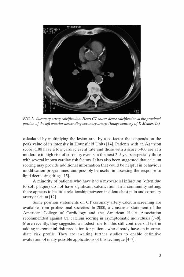
calculated by multiplying the lesion area by a co-factor that depends on the peak value of its intensity in Hounsfield Units [14]. Patients with an Agatston score <100 have a low cardiac event rate and those with a score >400 are at a moderate to high risk of coronary events in the next 2–5 years, especially those with several known cardiac risk factors. It has also been suggested that calcium scoring may provide additional information that could be helpful in behaviour modification programmes, and possibly be useful in assessing the response to lipid decreasing drugs [15].
A minority of patients who have had a myocardial infarction (often due to soft plaque) do not have significant calcification. In a community setting, there appears to be little relationship between incident chest pain and coronary artery calcium [12].
Some position statements on CT coronary artery calcium screening are available from professional societies. In 2000, a consensus statement of the American College of Cardiology and the American Heart Association recommended against CT calcium scoring in asymptomatic individuals [7–8]. More recently, they suggested a modest role for this still controversial test in adding incremental risk prediction for patients who already have an interme-diate risk profile. They are awaiting further studies to enable definitive evaluation of many possible applications of this technique [4–7].
FIG. 1. Coronary artery calcification. Heart CT shows dense calcification at the proximal portion of the left anterior descending coronary artery. (Image courtesy of F. Mettler, Jr.)
3
A 2003 position statement of the Cardiac Society of Australia and New Zealand states that while the procedure provides useful information in population studies, there is not yet sufficient evidence to provide practical value to an individual above that obtained from a thorough assessment of cardiac risk [16]. The 2004 statement of the US Preventive Services Task Force recommends against CT scanning for calcium scoring of coronary stenosis in adults at low risk for coronary events. They also found insufficient evidence to recommend for or against these procedures in adults at increased risk for coronary heart disease [17]. European guidelines issued on behalf of eight societies are somewhat more positive in tone and find that the calcium score “is an important parameter to detect asymptomatic individuals at high risk for future CVD events, independent of traditional risk factors” [18, 19].
3. COMPUTED TOMOGRAPHY AND CORONARY ANGIOGRAPHY
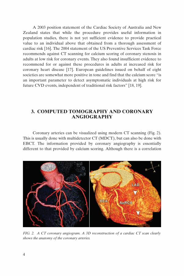
Coronary arteries can be visualized using modern CT scanning (Fig. 2). This is usually done with multidetector CT (MDCT), but can also be done with EBCT. The information provided by coronary angiography is essentially different to that provided by calcium scoring. Although there is a correlation
FIG. 2. A CT coronary angiogram. A 3D reconstruction of a cardiac CT scan clearly shows the anatomy of the coronary arteries.
4
between patterns of coronary calcification and atherosclerotic plaques, significant stenosis or narrowing may occur in areas without calcium deposits.
Thus, while CT coronary angiography (CTA) is not a screening procedure, its uses include evaluation of coronary artery anomalies, bypass-graft patency and surgical planning [20, 21]. In some patients with suspected coronary artery disease, CTA obviates the need for invasive arterial catheteri-zation and the risks associated with conventional radiographic coronary angiography [22]. The value of CTA in patients with a low to intermediate likelihood of coronary artery disease is still to be fully determined, but it has emerged as a good ‘rule-out’ test in defined clinical circumstances [23].
For many current CT angiographic applications, 16 slice multi-detector spiral scanners are the minimum level of technology needed and 64 slice scanners are needed for good visualization of lesions [24]. Current studies indicate that 64 slice CTA is highly accurate for exclusion of significant coronary artery stenosis (>50% luminal narrowing) with negative predictive values in excess of 95% unless there is heavy arterial calcification [25–28]. CTA is not normally advised when a patient has an irregular heart rhythm, a heart rate greater than 70 beats per minute and contraindications to medication for heart rate control or is likely to require revascularization surgery [29]. Standard invasive coronary angiography remains the gold standard for evaluation of coronary anatomy.
4. GENERAL ASPECTS OF RADIATION PROTECTION OF THE PATIENT
The International Commission on Radiological Protection (ICRP) has recommended a multi-step approach to protection of the patient [30]. First, a practice is identified (such as the use of CT scanning to detect and score coronary artery calcification). The second step is to justify this practice; that is, does CT coronary artery calcium scoring contribute more benefit to society than harm? This is assessed by performing large clinical population studies. If the practice is justified, then it should be optimized (i.e. can the practice be implemented at a lower radiation dose while maintaining its efficacy and accuracy?).
Two subsequent steps apply to the individual having the CT scan. There should be individual justification. This asks whether the examination will really benefit the patient about to be studied. For example, coronary artery calcium
5
scoring is not likely useful for individuals who are very young, very old or who have well known and characterized coronary artery disease. Such a decision is best made by a physician familiar with the patient and the medical history. The last step is optimization of the examination for that specific individual. This step asks whether the examination can be effectively carried out in a way that reduces dose for the particular patient (for example, can a lower dose be used because the patient is very thin or the irradiated volume is reduced?).
5. DOSES FROM CT SCANNING OF THE HEART AND POSSIBILITIES FOR DOSE REDUCTION
As with other radiology procedures, there is often a wide variation in doses reported for the same type of CT scan. The actual absorbed dose received during a cardiac CT scan varies depending on the type of scanner, protocol and the technique used [30]. However, this document concentrates on the effective doses and stochastic risk as good practice should eliminate deter-ministic risks and the possibilities of skin injuries [31].
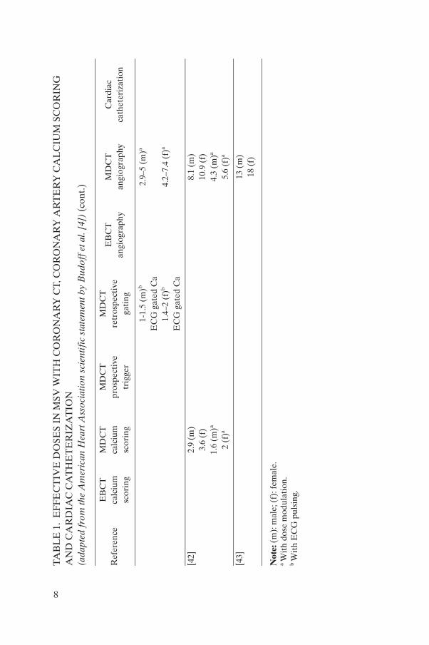
Effective dose values reported in the literature for EBCT calcium scoring are relatively low, ranging from 0.7 to 1.3 mSv, with tissue doses from 2.8 to 4.3 mGy (Table 1). However, this manifestation of CT technology is now deployed less frequently. On the other hand, with MDCT calcium scoring, effective doses range from 0.8 mSv to a high of 12 mSv and tissue doses range from 4.8 to 92 mGy [13, 30, 32–34].
Contrast enhanced MDCT for coronary angiography results in a higher effective dose (5–15 mSv) or close to the effective dose from a standard MDCT scan of the chest. These doses should be susceptible to conventional dose reduction approaches. The effective dose from EBCT coronary angiography has been reported to be about 1.5–2 mSv [20, 30–36]. These doses have recently been reviewed and are summarized in Table 1 [4].
There are clearly opportunities for dose reduction with almost any type of CT scan [5, 6]. For cardiac CT specifically, use of body weight adapted MDCT protocols has been shown to reduce the effective dose by about 12% in males and 25% in females. Careful consideration of technical factors such as kVp and mAs may also be effective. In addition, studies with mA modulation during the cardiac cycle and new prospective gating techniques offer prospects of significant extra reductions [37–51].
6

TA
BL
E 1
. E
FF
EC
TIV
E D
OSE
S IN
MSV
WIT
H C
OR
ON
AR
Y C
T, C
OR
ON
AR
Y A
RT
ER
Y C
AL
CIU
M S
CO
RIN
G
AN
D C
AR
DIA
C C
AT
HE
TE
RIZ
AT
ION
(a
dapt
ed f
rom
the
Am
eric
an H
eart
Ass
ocia
tion
scie
ntif
ic s
tate
men
t by
Bud
off
et a
l. [4
])
Ref
eren
ceE
BC
T
calc
ium
sc
orin
g
MD
CT
calc
ium
scor
ing
MD
CT
pros
pect
ive
trig
ger
MD
CT
retr
ospe
ctiv
ega
ting
EB
CT
an
giog
raph
yM
DC
T
angi
ogra
phy
Car
diac
ca
thet
eriz
atio
n
[37]
3.0
(m)
4.0
(f)
[33]
2.8–
10.3
(m
)3.
6–12
.7 (
f)
[38]
1.9
(m)
2.5
(f)
1 (m
)a
1.4
(f)a
[32]
1 (
m)
1.5–
5.2
(m)
3 (m
)1.
5 (m
)6.
7–10
.9 (
m)
2.1
(m)
1.3
(f)
1.8–
6.2
(f)
3.6
(f)
2.0
(f)
8.1–
13.0
(f)
2.5
(f)
[35]
0.7
1.0
2.6–
4.1
1.1
9.3–
11.3
[39]
5–6
(m)
6–7
(f)
[40]
3.9–
5.8
1.7
[41]
0.5
(m)
EC
G tr
ig C
a1.
9–2.
2 (m
)E
CG
gat
ed C
a5.
7–7.
1 (m
)E
CG
gat
ed0.
7–0.
8 (f
)E
CG
trig
Ca
2.8–
3.1
(f)
EC
G g
ated
Ca
8.5–
10.5
(f)
EC
G g
ated
7
1-1.
5 (m
)b
EC
G g
ated
Ca
2.9–
5 (m
)a
1.4–
2 (f
)b
EC
G g
ated
Ca
4.2–
7.4
(f)a
[42]
2.9
(m)
8.1
(m)
3.6
(f)
10.9
(f)
1.6
(m)a
4.3
(m)a
2 (f
)a 5.
6 (f
)a
[43]
13 (
m)
18 (
f)
Not
e: (
m):
mal
e; (
f): f
emal
e.a W
ith
dose
mod
ulat
ion.
b W
ith
EC
G p
ulsi
ng.
TA
BL
E 1
. E
FF
EC
TIV
E D
OSE
S IN
MSV
WIT
H C
OR
ON
AR
Y C
T, C
OR
ON
AR
Y A
RT
ER
Y C
AL
CIU
M S
CO
RIN
G
AN
D C
AR
DIA
C C
AT
HE
TE
RIZ
AT
ION
(a
dapt
ed f
rom
the
Am
eric
an H
eart
Ass
ocia
tion
scie
ntif
ic s
tate
men
t by
Bud
off
et a
l. [4
]) (
cont
.)
Ref
eren
ceE
BC
T
calc
ium
sc
orin
g
MD
CT
calc
ium
scor
ing
MD
CT
pros
pect
ive
trig
ger
MD
CT
retr
ospe
ctiv
ega
ting
EB
CT
an
giog
raph
yM
DC
T
angi
ogra
phy
Car
diac
ca
thet
eriz
atio
n
8

It is instructive to compare the dose from the various cardiac CT procedures with that from standard radiographic contrast coronary angio-graphy. While there is variation related to the difficulty of the procedure, effective doses of about 2–6 mSv have been reported for diagnostic coronary angiography [32, 52–54]. A comparison of doses is presented in Table 2. This comparison is based on radiation risks only, and does not address the other risks inherent in the procedures.
The BSS and other IAEA publications [1, 3] recommend the use of formally established reference or guidance doses for medical procedures to assist in the implementation of optimization programmes. The dose values cited here provide a valuable basis for comparison and represent what has been achieved in experienced centres. However, they are not guidance or reference levels as these remain to be established in the future.
Both ultrasound and magnetic resonance imaging (MRI) are also currently used for cardiac imaging and have the advantage of not using any ionizing radiation and do not have any known cancer risk. Currently, ultrasound is not useful as a general screening test for coronary artery disease and MRI is not useful for identifying or scoring calcium deposits. MRI can, however, visualize coronary arteries and whether MRI cardiac scanning will ultimately replace either CT coronary angiography or standard contrast coronary angiography is unknown.
TABLE 2. APPROXIMATE EFFECTIVE DOSE FROM CT CALCIUM SCORING AND CTA COMPARED TO OTHER COMMON SOURCES
Source Approximate effective dose (mSv)
CT calcium scoring 1–5 (multidetector helical CT)1 (electron beam CT)
CT coronary angiography 5–15 (multidetector helical CT)1–2 (electron beam CT)
CT scan of thorax 10
Conventional invasive coronary angiography 2–6
Chest X ray (one film) 0.02
Annual natural background 2.4
Typical annual effective dose to transatlantic pilot 4.0
9

6. RADIATION RISK FROM CT CARDIAC EXAMINATIONS
The radiation risk at the doses of interest from a cardiac CT scan is the potential for radiogenic cancer induction. Individual radiation risk from a CT examination varies significantly depending upon many factors including the absorbed dose, age and sex of the patient, and expected lifespan. Risk is generally higher in younger patients and is slightly higher in females than in males. CT scanning during pregnancy is occasionally performed for specific medical reasons but requires special consideration of the risk to the foetus.
Excess cancer risk has not been demonstrated by epidemiological studies at doses below 100 mSv. Since doses from cardiac CT scans are lower than this, the potential risk can only be estimated by assuming a dose response relationship [55, 56]. The ICRP has estimated that the radiogenic fatal cancer risk for an adult population is about 5%/Sv [57] or (by using the linear non-threshold dose–response hypothesis) 0.005%/mSv. The US National Academy of Sciences BEIR VII Committee has recently provided radiogenic cancer estimates of risk by age [58]. Potential radiation risks can be compared to the spontaneous fatal cancer risk of about 20% and the spontaneous cancer incidence of about 40% (Tables 3 and 4).
While these approximate radiation related cancer risks may seem low in terms of percentage, the situation is somewhat different if the potential risk is expressed in terms of numbers of excess cancer cases. An example of the possible effect is as follows: if 100 000 persons received a CT coronary angiogram (10 mSv effective dose) each year from age 40 to 70, there might be about 2500 excess cases of cancer and leukaemia, and about 1300 excess fatalities [59]. This would be in addition to the spontaneous cancer cases of about 40 000 and cancer fatalities of about 20 000. This level of detriment would have to be set against the lives saved/disease averted by cardiac CT applied to angiography. From the above, it is clear that in the opinion of the professional societies involved, these benefits are too tenuous to warrant use of cardiac CT in mass screening programmes. On the other hand, less frequent referrals of patients with well identified risk profiles, combined with dose reduction methodology, provides a more favourable risk–benefit profile and in the opinion of the professional bodies is justified [4–6].
10

TABLE 3. ESTIMATION OF AVERAGE DOSE AND RISK FROM SEVERAL TYPES OF CARDIAC IMAGING PROCEDURES PERFORMED ON AN ADULT POPULATION
Approximate effective dose (mSv)
Approximate risk per scan of fatal radiogenic cancer (%)a
Approximate spontaneous risk of fatal cancers (%)
1 0.005 20
2 0.01 20
3–5 0.015–0.25 20
10 0.05 20
2–50 0.01–0.25 20
a Radiogenic and spontaneous cancer incidence is approximately twice the fatal risk.
TABLE 4. POTENTIAL LIFETIME RADIOGENIC FATAL CANCER RISK AFTER AN EFFECTIVE DOSE OF 10 mSv AS A FUNCTION OF SEX AND AGE FOR CARDIAC CT SCANNING (adapted from BEIR VII Table 12 D-2 [59])
Age, sex at exposure Fatal radiogenic cancer/leukaemia risk (%)
30, male 0.038
40, male 0.038
50, male 0.036
60, male 0.032
70, male 0.025
80, male 0.015
30, female 0.054
40, female 0.051
50, female 0.047
60, female 0.041
70, female 0.032
80, female 0.019
Note: Approximate risk can be calculated for scans of varying doses by using a simple proportional relationship (for 5 mSv divide risk by two). Radiogenic and spontaneous cancer incidence is approximately twice the fatal risk.
11
7. SUMMARY
CT scanning for coronary artery calcium scoring remains a somewhat controversial screening test and is not generally recommended in asympto-matic individuals. However, European guidelines do recommend calcium scoring for identifying individuals at high risk of a coronary event.
CTA has not been recommended as a general screening examination but may be useful for certain persons with suspected coronary artery disease. Since CTA has a high negative predictive value, it may be useful in excluding significant coronary artery narrowing in individuals at intermediate risk. This test should be considered as a complementary and not a replacement modality for invasive coronary angiography.
Health authorities and professional groups should evaluate many factors before recommending adoption of these tests for screening asymptomatic persons. The factors include, but are not limited to, prevalence and severity of disease in the population, age of the proposed screening group, accuracy of the test, costs (including false positive and false negative results), effect on outcome, possibility for radiation dose reduction and evaluation of potential risks. The radiation dose from these procedures is relatively well documented and the potential risk of radiogenic cancers can be estimated. In addition, the combination of conventional dose reduction techniques and newer techno-logical developments is likely to reduce the dose levels reported at present. Outside of screening programmes, persons who are symptomatic should be individually evaluated by a physician to determine what medical care is appropriate and necessary (including justification and optimization of the radiological examination).
Finally, while the above considerations are valuable in initiating the justi-fication and optimization processes envisaged in the BSS, they are not compre-hensive. Much remains to be done in further resolving the appropriate referral patterns, optimization of technique and developing guidance or reference levels.
12
REFERENCES
[1] EUROPEAN ATOMIC ENERGY COMMUNITY, FOOD AND AGRICULTURE ORGANIZATION OF THE UNITED NATIONS, INTERNATIONAL ATOMIC ENERGY AGENCY, INTERNATIONAL LABOUR ORGANIZATION, INTERNATIONAL MARITIME ORGANIZATION, OECD NUCLEAR ENERGY AGENCY, PAN AMERICAN HEALTH ORGANIZATION, UNITED NATIONS ENVIRONMENT PROGRAMME, WORLD HEALTH ORGANIZATION, Fundamental Safety Principles, IAEA Safety Standards Series No. SF-1, IAEA, Vienna (2006).
[2] FOOD AND AGRICULTURE ORGANIZATION OF THE UNITED NATIONS, INTERNATIONAL ATOMIC ENERGY AGENCY, INTERNATIONAL LABOUR ORGANISATION, OECD NUCLEAR ENERGY AGENCY, PAN AMERICAN HEALTH ORGANIZATION, WORLD HEALTH ORGANIZATION, International Basic Safety Standards for Protection against Ionizing Radiation and for the Safety of Radiation Sources, Safety Series No. 115, IAEA, Vienna (1996).
[3] INTERNATIONAL ATOMIC ENERGY AGENCY, INTERNATIONAL LABOUR OFFICE, INTERNATIONAL ORGANIZATION FOR MEDICAL PHYSICS, INTERNATIONAL SOCIETY OF RADIOLOGY, PAN AMERICAN HEALTH ORGANIZATION, WORLD HEALTH ORGANIZATION, Applying Radiation Safety Standards in Diagnostic Radiology and Interventional Procedures Using X Rays, Safety Reports Series No. 39, IAEA, Vienna (2006).
[4] BUDOFF, M.J., et al., Assessment of coronary artery disease by cardiac computed tomography: A scientific statement from the American Heart Association Committee on Cardiovascular Imaging and Intervention, Council on Cardiovascular Radiology and Intervention, and Committee on Cardiac Imaging, Council on Clinical Cardiology, Circulation 114 (2006) 1761–1791.
[5] INTERNATIONAL COMMISSION ON RADIOLOGICAL PROTECTION, Managing Patient Dose in Multi-Detector Computed Tomography (MDCT),Publication 102, Ann. ICRP 37 1, Elsevier, Oxford (2007).
[6] INTERNATIONAL COMMISSION ON RADIOLOGICAL PROTECTION, Managing Patient Dose in Computed Tomography, Publication 87, Ann. ICRP 304, Elsevier, Oxford (2001).
[7] AMERICAN HEART ASSOCATION, Clinical expert consensus document on coronary artery calcium scoring by computed tomography in global cardiovascular risk assessment and in evaluation of patients with chest pain, ACCF/AHA 2007, Circulation 115 (2007) 402–426.
13
[8] O’ROURKE, R., et al., American College of Cardiology/American Heart Association expert consensus document on electron beam computed tomography for the diagnosis and prognosis of coronary artery disease, Circulation 102 (2000) 126–140.
[9] SCHMERMUND, A., MOHLENKAMP, S., ERBEL., R., Coronary artery calcium and its relationship to coronary artery disease, Cardiol. Clin. 21 (2003) 521–534.
[10] DUBINSKY, T.J., Coronary artery calcification scoring, Am. J. Roentgenol. 185(2005) 1540–1541.
[11] DANIELL, A.L., et al., Concordance of coronary artery calcium estimates between MDCT and electron beam tomography, Am. J. Roentgenol. 185 (2005) 1542–1545.
[12] TAYLOR, A.J., et al., Coronary calcium independently predicts incident premature coronary heart disease over measured cardiovascular risk factors: Mean three-year outcomes in the prospective Army coronary calcium (PACC) project, J. Am. Coll. Cardiol. 46 5 (2005) 807–814.
[13] NASIR, K., et al., Electron beam CT versus helical CT scans for assessing coronary calcification: Current utility and future directions, Am. Heart J. 146 6 (2003) 969–977.
[14] AGATSTON, A.S., et al., Quantification of coronary artery calcium using ultrafast computed tomography, J. Am. Coll. Cardiol. 15 4 (1990) 827–832.
[15] GREENLAND, P., et al., Coronary artery calcium score combined with Framingham score for risk prediction in asymptomatic individuals, J. Am. Med. Assoc. 291 (2004) 210–215.
[16] CARDIAC SOCIETY OF AUSTRALIA AND NEW ZEALAND, High Speed Computed Tomography to Detect Coronary Calcification, Position Statement from the Cardiac Society of Australia and New Zealand, www.csanz.edu.au/news/inthenews/highspeed_computer_tomography.pdf
[17] US PREVENTIVE SERVICES TASK FORCE, Screening for Coronary Heart Disease, http//www.ahrq.gov/clinic/uspstf/uspscad.htm
[18] DE BACKER, G., et al., European guidelines on cardiovascular disease prevention in clinical practice, Third Joint Task Force of European and other Societies on Cardiovascular Disease Prevention in Clinical Practice (constituted by representatives of eight societies and by invited experts), Eur. Heart J. 24(2003) 1601–1610.
[19] SILBER, S., RICHARTZ, B.M., Impact of both cardiac CT and cardiac MR on the assessment of cardiac risk, Z. Kardiol. 94 Suppl. 4:IV (2005) 70–80.
[20] SCHOEPF, U.J., BECKER, C.R., OHNESORGE, B.M., YUCEL, E.K., CT of coronary artery disease, Radiology 232 (2004) 18–37.
[21] PACHE, G., et al., Initial experience with 64 slice cardiac CT; non-invasive visualization of coronary artery bypass grafts, Eur. Heart J. 27 8 (2006) 976–980.
[22] HABERL, R., et al., Multislice spiral computed tomographic angiography of coronary arteries in patients with suspected coronary artery disease: An effective filter before catheter angiography, Am. Heart J. 149 (2005) 1112–1119.
14
[23] BAX, J.J., SCHUIJF, J.D., Which role for multislice computed tomography in clinical cardiology? Am. Heart J. 149 (2005) 960–961.
[24] SEIFARTH, H., et al., 64- versus 16-slice CT angiography for coronary stent assessment; in vitro experience, Invest. Radiol. 41 1 (2006) 22–27.
[25] LESCHKA, S., et al., Accuracy of MSCT coronary angiography with 64 slice technology: first experience, Eur. Heart J. 26 (2005) 1482–1487.
[26] HOFFMAN, M.H., LESSICK, J., Multidetector computed tomography for non-invasive coronary imaging, Expert Rev. Cardiovasc. Ther. 4 4 (2006) 583–594.
[27] ESCOLAR, E., et al., New imaging techniques for diagnosing coronary artery disease, CMAJ 174 4 (2006) 487–495.
[28] SUN, Z., JIANG, W., Diagnostic value of multislice computed angiography in coronary artery disease: A meta analysis, Eur. J. Radiol. 60 2 (2006) 279–286.
[29] GERBER, T.C., et al., Computed tomographic angiography of the coronary arteries: Techniques and applications, Sem. Ultrasound CT MR 27 1 (2006) 42–55.
[30] BECKER, C., et al., Assessment of the effective dose for routine protocols in conventional CT, electron beam CT and coronary angiography, Rofo. Fortschr. Geb. Rontgenstr. Neuen Bildgeh. Verfahr. 170 (1999) 99–104 (in German).
[31] INTERNATIONAL COMMISSION ON RADIOLOGICAL PROTECTION, Publication 73, Ann. ICRP 26 2, Elsevier, Oxford (1996).
[32] HUNOLD, P., , Radiation exposure during cardiac CT: Effective doses at multi-detector row CT and electron beam CT, Radiology 226 1 (2003) 145–152.
[33] COHNEN, M., POLL, L., PÜTTMANN, C., EWEN, K., MÖDDER, U., Radiation exposure in multi-slice CT of the heart, Fortschr. Röntgenstr. 173(2001) 295–299.
[34] KALENDAR, W.A., SCHMIDT, B., ZANKL, M., SCHMIDT, M., A PC program for estimating organ dose and effective dose values in computed tomography, Eur. Radiol. 9 (1999) 552–562.
[35] MORIN, R., GERBER, T., McCOLLOUGH, C., Radiation dose in computed tomography of the heart, Circulation 107 (2003) 917–922.
[36] McCOLLOUGH, C., Patient dose in cardiac computed tomography, Herz 28(2003) 1–6.
[37] OHNESORGE, B., et al., Reproducibility of coronary calcium quantification in repeat examinations with retrospectively ECG-gated multisection spiral CT, Eur. Radiol. 12 (2002) 1532–1540.
[38] JAKOBS, T.F., et al., Multi-slice helical CT of the heart with retrospective ECG gating: reduction of exposure by ECG-controlled current tube modulation, Eur. Radiol. 12 (2002) 1081–1086.
[39] KOPP, A.F., et al., Non-invasive coronary angiography with high resolution multidetector-row computed tomography: results in 102 patients, Eur. Heart J. 23(2002) 1714–1725.
[40] ACHENBACH, S., et al., Detection of coronary artery stenoses by contrast-enhanced, retrospectively electrocardiographically-gated, multi-slice spiral computed tomography, Circulation 103 (2001) 2535–2538.
15
[41] FLOHR, T.G., et al., Advances in cardiac imaging with 16-section CT systems, Acad. Radiol. 10 (2003) 386–401.
[42] TRABOLD, T., et al., Estimation of radiation exposure in 16-detector row computed tomography of the heart with retrospective ECG-gating, Rofo. Fortschr. Geb. Rontgenstr. Neuen Bildgeh. Verfahr. 175 (2003) 1051–1055.
[43] RAFF, G.L., GALLAGHER, M.J., O’NEILL, W.W., GOLDSTEIN, J.A., Diagnostic accuracy of noninvasive coronary angiography using 64-slice spiral computed tomography, J. Am. Coll. Cardiol. 46 (2005) 552–557.
[44] MAHNKEN, A.H., et al., Detection of coronary calcifications: Feasibility of dose reduction with a body-weight-adapted examination protocol, Am. J. Roentgenol. 181 (2003) 533–553.
[45] SHEMESH, J., et al., Coronary artery calcium measurement with multi-detector row CT and low radiation dose: Comparison between 55 and 165 mAs, Radiology 236 (2005) 810–814.
[46] ABADA, H.T., et al., MDCT of the coronary arteries; feasibility of low dose CT with ECG-pulsed tube current modulation to reduce radiation dose, Am. J. Roentgenol. 186 6 Suppl. 2 (2006) 387–390.
[47] JAKOBS, T.F., et al., Ultra-low dose coronary artery calcium screening using multislice CT with retrospective ECG gating, Eur. Radiol. 13 (2003) 1923–1930.
[48] POLL, L.W., COHNEN, M., BRACHTEN, S., EWEN, K., MODER, U., Dose reduction in multislice CT of the heart by using ECG-controlled tube current modulation (“ECG pulsing”); phantom measurements, Rofo. Fortschr. Geb. Rontgenstr. Neuen Bildgeh. Verfahr. 170 (1999) 99–104 (in German).
[49] WILDBERGER, J.E., et al., Individually adapted examination protocols for reduction of radiation exposure in chest CT, Invest. Radiol. 36 (2001) 604–611.
[50] THOMAS, C.K., et al., Coronary artery calcium scoring with multislice computed tomography: In vitro assessment of a low tube voltage protocol, Invest. Radiol. 41 9 (2006) 668–673.
[51] HSIEH, J., et al., Step-and-shoot data acquisition and reconstruction for cardiac X-ray computed tomography, Med. Phys. 33 11 (2006) 4236–4248.
[52] LEUNG, K.C., MARTIN, C.J., Effective doses for coronary angiography, Br. J. Radiol. 69 (1996) 426–431.
[53] HARRISON, D., RICCIARDELLO, M., COLLINS, L., Evaluation of radiation dose and risk to the patient from coronary angiography, Austr. N.Z. J. Med. 28(1998) 597–603.
[54] BETSOU, S., et. al., Patient radiation dose during cardiac catheterization procedures, Br. J. Radiol. 846 (1998) 634–639.
[55] BRENNER, D.J., Radiation risks potentially associated with low-dose CT screening of adult smokers for lung cancer, Radiology 231 (2004) 440–445.
[56] BRENNER, D.J., ELLISTON, C.D., Estimated radiation risks potentially associated with full-body CT screening, Radiology 232 (2004) 735–738.
[57] INTERNATIONAL COMMISSION ON RADIOLOGICAL PROTECTION, Recommendations of the International Commission on Radiological Protection, Publication 60, Ann. ICRP 21 1–3, Elsevier, Oxford (1990).
16

[58] NATIONAL ACADEMY OF SCIENCES, Health Risks from Exposure to Low Levels of Ionizing Radiation, Rep. BEIR VII Committee, National Academy Press, Washington, DC (2005).
[59] NATIONAL ACADEMY OF SCIENCES, Health Risks from Exposure to Low Levels of Ionizing Radiation, Rep. BEIR VII Committee, National Academy Press, Washington, DC (2005) Table 12 D-2.
17

.

CONTRIBUTORS TO DRAFTING AND REVIEW
Bernardi, G. Azienda Ospedaliero-Universitaria di Udine, Italy
Cousins, C. International Commission on Radiological Protection
Holm, L.E. International Commission on Radiological Protection
Malone, J. St. James’ Hospital and Trinity College, Ireland
Manelfe, C. International Society of Radiology
Mettler, F., Jr. New Mexico Federal Regional Medical Center, United States of America
Murphy, R. St. James’ Hospital, Ireland
Ostenson, H. World Health Organization
Rehani, M.M. International Atomic Energy Agency
Schnyder, P. University Hospital Lausanne, Switzerland
Walsh, M. St. James’ Hospital, Ireland
19
IAEA SAFETY RELATED PUBLICATIONS
IAEA SAFETY STANDARDS
Under the terms of Article III of its Statute, the IAEA is authorized to establish or adopt standards of safety for protection of health and minimization of danger to life and property, and to provide for the application of these standards.
The publications by means of which the IAEA establishes standards are issued in the IAEA Safety Standards Series. This series covers nuclear safety, radiation safety, transport safety and waste safety. The publication categories in the series are Safety Fundamentals, Safety Requirements and Safety Guides.
Information on the IAEA’s safety standards programme is available at the IAEA Internet site
http://www-ns.iaea.org/standards/
The site provides the texts in English of published and draft safety standards. The texts of safety standards issued in Arabic, Chinese, French, Russian and Spanish, the IAEA Safety Glossary and a status report for safety standards under development are also available. For further information, please contact the IAEA at P.O. Box 100, 1400 Vienna, Austria.
All users of IAEA safety standards are invited to inform the IAEA of experience in their use (e.g. as a basis for national regulations, for safety reviews and for training courses) for the purpose of ensuring that they continue to meet users’ needs. Information may be provided via the IAEA Internet site or by post, as above, or by email to [email protected].
OTHER SAFETY RELATED PUBLICATIONS
The IAEA provides for the application of the standards and, under the terms of Articles III and VIII.C of its Statute, makes available and fosters the exchange of information relating to peaceful nuclear activities and serves as an intermediary among its Member States for this purpose.
Reports on safety and protection in nuclear activities are issued as Safety Reports, which provide practical examples and detailed methods that can be used in support of the safety standards.
Other safety related IAEA publications are issued as Radiological Assessment Reports, the International Nuclear Safety Group’s INSAG Reports, Technical Reportsand TECDOCs. The IAEA also issues reports on radiological accidents, training manuals and practical manuals, and other special safety related publications. Security related publications are issued in the IAEA Nuclear Security Series.
RELATED PUBLICATIONS
www.iaea.org/books
FUNDAMENTAL SAFETY PRINCIPLESSAFETY FUNDAMENTALSIAEA Safety Standards Series No. SF-1STI/PUB/1273 (21 pp.; 2006)ISBN 92-0-110706-4 Price: €25.00
INTERNATIONAL BASIC SAFETY STANDARDS FOR PROTECTIONAGAINST IONIZING RADIATION AND FOR THE SAFETY OF RADIATION SOURCESSafety Series No. 115STI/PUB/996 (353 pp.; 1996)ISBN 92-0-104295-7 Price: €78.50
RADIATION PROTECTION IN NEWER MEDICAL IMAGING TECHNIQUES: PET/CTSafety Reports Series No. 58STI/PUB/1343 (50 pp.; 2008)ISBN 978-92-0-106808-8 Price: €28.00
RADIATION PROTECTION IN NEWER MEDICAL IMAGING TECHNIQUES: CT COLONOGRAPHYSafety Reports Series No. 61STI/PUB/1367 (37 pp.; 2008)ISBN 978-92-0-111308-5 Price: €20.00
APPLYING RADIATION SAFETY STANDARDS IN NUCLEAR MEDICINESafety Reports Series No. 40 STI/PUB/1207 (124 pp.; 2005)ISBN 92-0-111104-5 Price: €28.00
APPLYING RADIATION SAFETY STANDARDS IN DIAGNOSTICRADIOLOGY AND INTERVENTIONAL PROCEDURES USING X RAYSSafety Reports Series No. 39 STI/PUB/1206 (96 pp.; 2006)ISBN 92-0-111004-9 Price: €28.00
08-39811_P1366_covI-IV.indd 2 2009-02-02 08:59:50
S a f e t y R e p o r t s S e r i e s N o. 6 0
This pub l i ca t i on f ocuses on r ad i a t i on p ro tec t i on o f the pa t i en t when us ing computed tomography (CT ) f o r : co ronar y a r t e r y ca l c ium scor ing and v isua l i za t ion o f the coronary ar ter ies (ang iography) . I n add i t i on , i t e xp l a ins when i t i s app ropr i a te t o u s e t h e s e t e c h n i q u e s i n s y m p t o m a t i c a n d / o r asymptomat i c sc reen ing popu la t i ons . I n fo rmat i on i s a l so p rov i ded on pa t i en t dose and r i sk l e ve l s . S o m e b a c k g r o u n d i n f o r m a t i o n i s p r o v i d e d o n ca rd i ac CT, and t he concep t s o f j u s t i f i c a t i on and op t im i za t i on , wh ich a re cen t ra l t o the BSS approach to pa t i en t p ro tec t i on , a re ou t l i ned .
INTERNATIONAL ATOMIC ENERGY AGENCYVIENNA
ISBN 978–92–0–111208–8ISSN 1020–6450
R a d i a t i o n P r o t e c t i o n i n N e w e r M e d i c a l
I m a g i n g T e c h n i q u e s : C a r d i a c C T
Join t l y sponsored b y the IAEA , WHO, ISR
Atoms for Peace
Atoms for Peace
W ith con t r i bu t i ons f rom the
08-39811_P1366_covI-IV.indd 1 2009-02-02 08:59:49