radiation protection (1)
TRANSCRIPT

Dr. Pramod Kumar Gahwai

The harmful effects of exposure to ionizing radiation were largely unsuspected at the time of discovery of xray.
The acceptance by society of risks associated with radiation is conditional on the benefits to be gained from the use of radiation.

RADIATION HAZARDS
RADIATION UNITS
BIOLOGICAL EFFECTS OF RADIATION
POPULATION EXPOSURES

DEFINITIONS ABSORBED DOSE- radiation necessary to deposit energy of 1
joule in 1 kg of tissue.(SI unit- Gray)
ABSORBED DOSE EQUIVALENT- measure of biological effectiveness of radiation.
ABSORBED DOSE EQUIVALENT = absorbed dose x quality factor
Quality factor – function of particle type and energy.
X rays, β particles,ϒ rays, electrons 1
thermal neutron 5

The amount of energy deposited per unit length of travel called LINEAR ENERGY TRANSFER.
Particle size α linear energy transfer α biological damage
EFFECTIVE DOSE equivalent (hε)- purpose is to relate exposure to risk.
.

NATURAL RADIATION
External and internal sources
EXTERNAL SOURCES cosmic radiation, terrestrial gamma radiation.
Exposure varies with latitude and altitude.
INTERNAL SOURCE Radionuclide's within the body.
K40, Rb87, C14 and members of the thorium and uranium series.
Radon is largest contributor to avg annual effective dose equivalent.
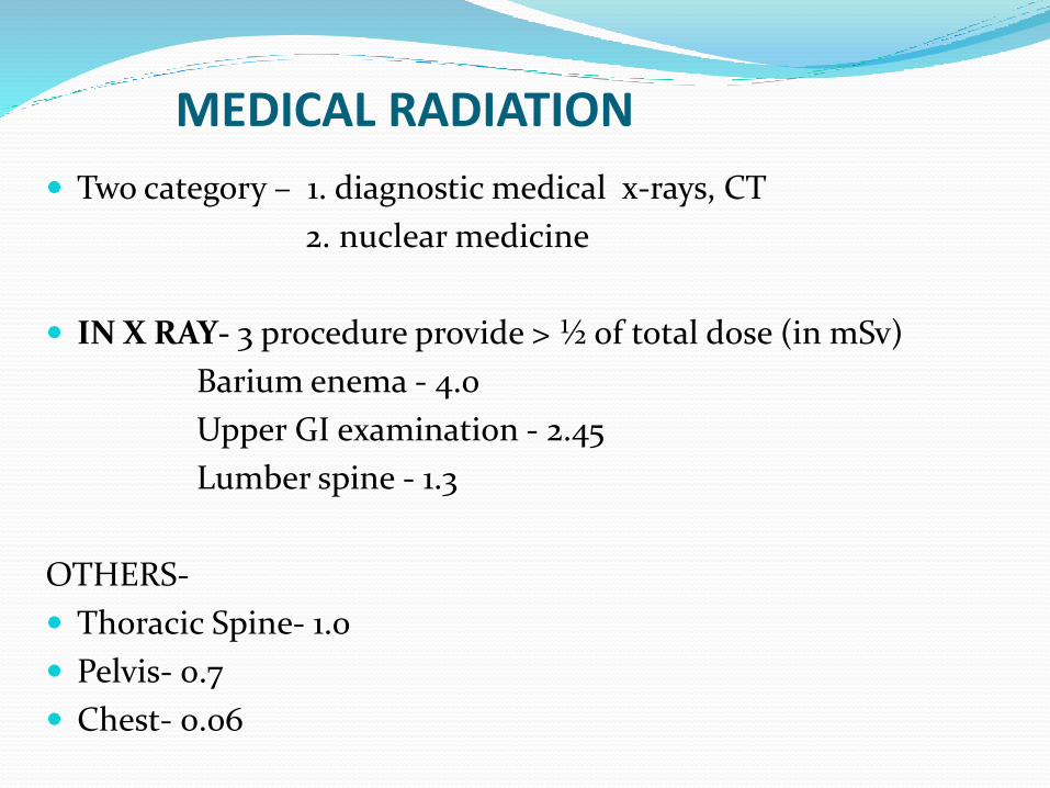
MEDICAL RADIATION
Two category – 1. diagnostic medical x-rays, CT
2. nuclear medicine
IN X RAY- 3 procedure provide > ½ of total dose (in mSv)
Barium enema - 4.0
Upper GI examination - 2.45
Lumber spine - 1.3
OTHERS-
Thoracic Spine- 1.0
Pelvis- 0.7
Chest- 0.06

In CT scan(mSv)-
- CT Head -2
- CT Chest- 7
- CT Abdomen/Pelvis -10
- Whole-Body CT Screening -10

EFFECTS
1. STOCHASTIC EFFECT
2. NONSTOCHASTIC EFFECT

STOCHASTIC EFFECT Defined as an effect in which the probability of occurrence
increases with increasing absorbed dose.
severity does not depend on the magnitude of absorbed dose.
ALL OR NONE PHENOMENON, NO DOSE THRESHOLD.
Example –cancers and genetic effects.

Radium watch dial workers – bone ca.
Uranium miners – lung ca
Early medical radiation workers - leukemia
Thymus gland treatment – thyroid ca
Atomic bomb survivors – leukemia/breast, lung and bone Ca

NONSTOCHASTIC EFFECTS Defined as somatic effect that increases in SEVERITY with
increasing absorbed dose.
degenerative effects such as organ atrophy and fibrosis.
Examples- lens opacification
blood changes
decreased sperm production

REGULATORY BODIES
ICRP – International Commission for radiation protection is the international regulatory body.
AERB – Atomic Energy Regulatory board is Indian regulatory body.
NCRP – National Commission for radiological protection is the American counterpart .
.

FUNCTIONS
• Lay down norms for protection against radiation.
• Guidelines regarding the specification of medical x–ray equipment, room layout of x– ray installation, protective devices
• Responsibilities of the radiation personal, employer and radiation safety officer
• Recommends the dose limits for radiation workers and general public.
Approval for new models of x– ray equipment.
• Registration, inspection and ISO certification

Atomic energy regulatory board (AERB) making recommendations on limits of exposure to ionizing radiation.
There are 3 classes of individuals-
-occupationally exposed individuals
-the general public
-embryo-fetus
DOSE LIMITING RECOMMENDATIONS

OCCUPATIONAL LIMITS
For stochastic effects-ANNUAL LIMIT- 50 mSv
For lifetime dose equivalent limit(mSv) to the whole body= 10 times of age (in yr) of individual
For nonstochastic effect:-- lens 150 mSv- all other organ 500 mSv
Worker exposed to x ray for their own medical diagnosis or treatment does not count as an occupational exposure.
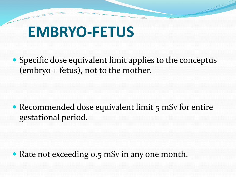
EMBRYO-FETUS
Specific dose equivalent limit applies to the conceptus (embryo + fetus), not to the mother.
Recommended dose equivalent limit 5 mSv for entire gestational period.
Rate not exceeding 0.5 mSv in any one month.

CNS – Exencephaly , microcephaly , skull malformation & hydrocephalus.
Ocular-absence of eyes, microphthalmia, absence of lens, cataract.
Skeletal malformation- stunting, cleft palate, spina bifida
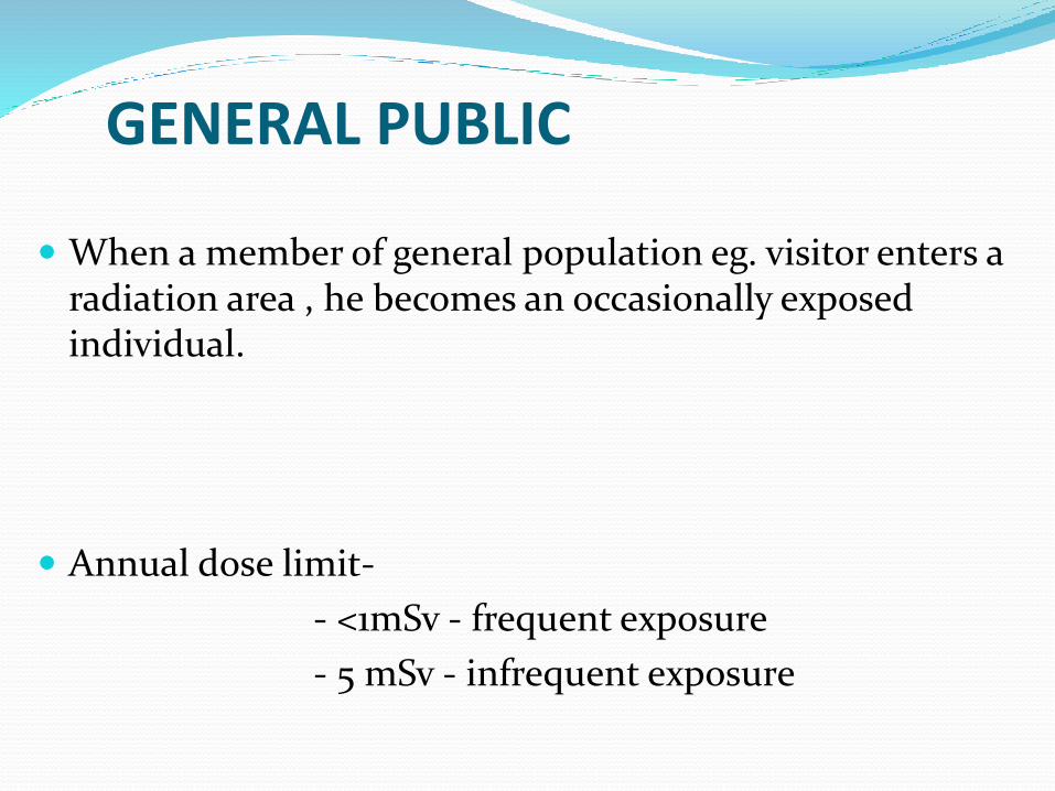
GENERAL PUBLIC
When a member of general population eg. visitor enters a radiation area , he becomes an occasionally exposed individual.
Annual dose limit-
- <1mSv - frequent exposure
- 5 mSv - infrequent exposure

ALARA CONCEPT
` As Low As Reasonably Achievable`
For any given radiation source, magnitude of individual doses, number of people exposed, and likelihood of incurring exposures should be kept to as low as reasonably achievable, taking economic
and social factors into considerations.

.
Judicious use of investigation
These include substituting non-ionizing methods of examination in place of ionizing radiation methods.
Ex – Evaluation of LN status in the abdomen by USG in place of repeated CT scan & use of colour Doppler flow imaging in place of diagnostic angiography.

PERSONNEL PROTECTION FROM EXPOSURE TO X RAYS
RADIATION MONITORING
Devices
- pocket dosimeter
- film badge
- TLD( thermo luminescent dosimeter)
TLD(principle):-depends on the ability of certain crystalline materials to store energy on exposure to ionizing radiation because of the trapping of valence electron in crystal lattice defect.

Crystal (lithium fluoride) heated under controlled condition
↓
electrons return to normal state
↓
stored energy released in the form of light
↓
photomultiplier device
↓
initial radiation exposure

PROTECTIVE MEASURES
Basic 3 principles:-
- exposure time
- distance
- lead barriers
EXPOSURE TIME :- total dose equivalent α time
DISTANCE:-
- Inverse square law applies.
- whenever possible , distance should be 2 meter from x-ray tube.
-Distance 1 m - 400 (exposure)
-Distance 2 m - 100

LEAD BARRIERS :-
- efficient absorber of x rays.
- great reduction of exposure by placing it in between source and person.
- thickness stated in HALF VALUE LAYER (HVL) for kilo voltage x rays.
(HVL – any material thickness which reduces exposure rate by one –half.)
Means if initial dose H 100 mSv and HVL is 1 mm Pbfor given kVp , then 1 mm Pb will reduces H to 50 mSv and second HVL of 1 mm Pb will reduce another one- half(25 mSv).

PROTECTIVE BARRIERS IN RADIOGRAPHY AND FLUOROSCOPY
RADIATION SOURCES :-
- tube must enclosed in metal housing that reduces leakage radiation ( is radiation which penetrates the protective housing )
WALL PROTECTION :- 4 type of radiation
- USEFUL BEAM – is radiation passing through tube aperture (aka Iry radiation)
- LEAKAGE RADIATION
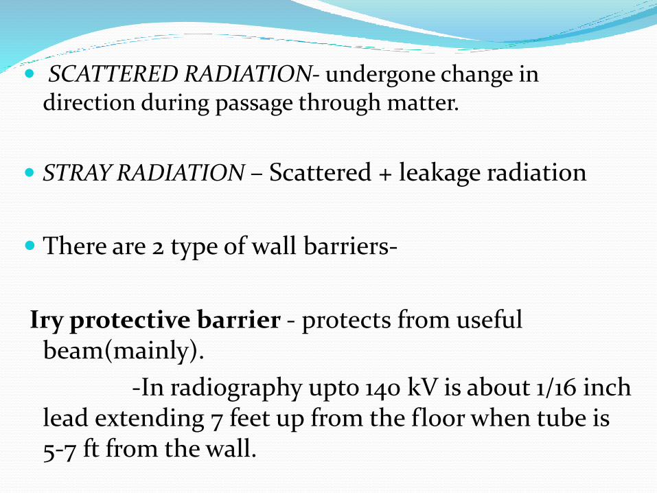
SCATTERED RADIATION- undergone change in direction during passage through matter.
STRAY RADIATION – Scattered + leakage radiation
There are 2 type of wall barriers-
Iry protective barrier - protects from useful beam(mainly).
-In radiography upto 140 kV is about 1/16 inch lead extending 7 feet up from the floor when tube is 5-7 ft from the wall.

IIry protective barrier :- is about 1/32 inch lead.
- extends from the top of the 1ry barrier to ceiling.
- ordinary plaster often suffice as a 2ry barrier without added lead.
- leaded glass observation port in the control booth should have same lead equivalent as the adjacent wall.
- leaded glass must 4 times as thick as lead sheet.

Lead apron – worn in fluoroscopy room.
- Pb equivalent 0.5 mm.
Check lead protective apron periodically for cracks by means of a radiography test.

DOSE REDUCTION IN RADIOGRAPHY
BEAM FILTRATION –
- exposure greatly reduced by ALUMINIUM filter.
- removes lower energy photons.
- recommendations:-
operating kVp Minimum HVL Al (mm)
< 50 0.3
50-70 1.2
> 70 2.3

COLLIMATION( BEAM LIMITATION) :-
- decrease in cross sectional area of the beam avoids unnecessary exposure of tissues outside the area of interest.
- also reduces amount of scattered radiation.
- modern equipment have automatic variable beam limiting device with manual override.
GONADAL ,THYROID SHIELDING :-
- beam should be so restricted that direct exposure of gonads does not occur.
- thyroid ,testes shield must have lead equivalent 0.5 mm.
- ovaries should be shielded whenever possible.

MODIFIED PROJECTION :-
- In radiography of girls for scoliosis should be use PA view.
- Reduces breast dose at least 98 % without loss of radiographic quality.

HIGH KILOVOLTAGE :-
- high kVp with low mAs delivers smaller absorbed dose to the patient.
CAREFUL TECHNIQUE :-
- to minimize repeat examination.
Radiographic examination in fertile women preferably performed during 1st 10 days following onset of menstrual period.
Ovulation and pregnancy are much less apt during this time than later menstrual cycle.

PROTECTION IN MAMMOGRAPHY
Skillful technique minimizes breast dose.
Goal achieved by molybdenum targets and filters in mammographic tubes.
Low dose screens and films, with/ without grid having ratios of 3:1 or 4:1.
Efficient breast compression device :- reduces breast thickness and make more uniform.

ADVANTAGE :-
1. decreases exposure factors with reduction of dose.
2. diminished amount of scattered thereby improving contrast.
3. improved recorded details by bringing breast closer to the image receptor.

CT SCANNING
Dose in CT scanning , by measuring absorbed dose at the centre of one “slice” with small dosimeter in water phantom.
Scanning this slice and 3 adjoining slices on both side.
Dosimeter record dose from direct beam through the centre slice, as well as scattered radiation from adjoining slices.
Collimator should also checked periodically to assure its proper function.

PATIENT PROTECTION IN FLUOROSCOPY
Intermittent fluoroscopy – decreases exposure and prolong tube life.
Restriction of field size – must be limited by suitably lead shutters placed between tube and patient.
Correct operating factors – exposure decreases as kVp increases and mA is lowered.
Recommended factors are 90-100 kVp, 2-3 mA and 2. mm aluminium filter

The source-skin distance must be at least 15 inch with stationary and 12 inch with mobile fluoroscopic equipment.
Filtration :-
– increase in hardness of x ray beam by filter.
– Filter removed relatively more soft than hard xrays.

PROTECTION IN NUCLEAR MEDICINE Medical compounds containing radionuclide's are called
radiopharmaceuticals.
Types of radiation
– alpha particles
– beta particles
– gamma rays
radiopharmaceuticals emits beta and gamma rays.

Gamma rays are electromagnetic wave and have much greater penetrability than beta particle.
Beta particles consist of high-speed electrons.
Beta particles are much less penetrating ,their effect limited to the skin (external source) immediate vicinity(internal source).

PRINCIPLES:-
- Distance
- Shielding
- Time of exposure

DISTANCE: -
- In case of gamma rays inverse square law applies for the purpose of protection in storage and handling.
- long forceps should be used.
- avoid spillage of liquids
- container should be as remote as possible.

SHIELDING:-
- shield may be incorporated in the container itself or it may be placed as a barrier around sources.
- γ emitting radiopharmaceuticals should be stored in their container and surrounded with lead bricks (5 cm) on all sides.
LEAD-SHIELDED SYRINGES – always be used for i.v. injection.
- Disposable gloves should be worn to protect against contamination by radionuclide's and by infection as well.

Low energy β particles are absorbed by container , cornified layer of skin.
Medical radionuclide P32 requires plastic such as polystyrene.
LENGTH OF EXPOSURE-
- faster procedure → less exposure

THANK YOU