radial breathing mode of single-walled carbon nanotubes: optical transition energies ... · ·...
TRANSCRIPT

Radial breathing mode of single-walled carbon nanotubes: Optical transition energiesand chiral-index assignment
J. Maultzsch,1,* H. Telg,1 S. Reich,2,† and C. Thomsen1
1Institut für Festkörperphysik, Technische Universität Berlin, Hardenbergstrasse. 36, D-10623 Berlin, Germany2Department of Engineering, University of Cambridge, Cambridge CB2 1PZ, United Kingdom
�Received 19 June 2005; revised manuscript received 19 August 2005; published 28 November 2005�
We present a comprehensive study of the chiral-index assignment of carbon nanotubes in aqueous suspen-sions by resonant Raman scattering of the radial breathing mode. We determine the energies of the first opticaltransition in metallic tubes and of the second optical transition in semiconducting tubes for more than 50 chiralindices. The assignment is unique and does not depend on empirical parameters. The systematics of theso-called branches in the Kataura plot are discussed; many properties of the tubes are similar for members ofthe same branch. We show how the radial breathing modes observed in a single Raman spectrum can be easilyassigned based on these systematics. In addition, empirical fits provide the energies and radial breathing modesfor all metallic and semiconducting nanotubes with diameters between 0.6 and 1.5 nm. We discuss the relationbetween the frequency of the radial breathing mode and tube diameter. Finally, from the Raman intensities weobtain information on the electron-phonon coupling.
DOI: 10.1103/PhysRevB.72.205438 PACS number�s�: 78.67.Ch, 73.22.�f, 78.30.Na
I. INTRODUCTION
Single-walled carbon nanotubes are tiny cylinders madeout of carbon.1–3 They have many unique, fascinatingproperties.1 They are very strong and of extremely lightweight, they are excellent conductors of heat, and transportelectrons ballistically. The properties of carbon nanotubesdepend strongly on their microscopic structure, which is de-fined by the chiral index �n1 ,n2�. The best known examplefor this is that 2 /3 of all possible nanotubes are semiconduc-tors and 1/3 are metals.1
Many applications of carbon nanotubes need one particu-lar type of tube, e.g., semiconducting tubes for transistors.Also, in fundamental studies we want to know which nano-tube is probed experimentally. The growth of carbon nano-tubes with a predefined microscopic structure remains a ma-jor challenge. Therefore the experimental determination ofthe chiral index is a current focus of carbon nanotuberesearch.4–9 In principle, the chiral index of an individualtube can be determined by direct imaging techniques likescanning tunneling microscopy. However, the experimentalerror in the measurement of diameter and chiral angle leadsto uncertainties in the assignment of the chiral index. Forexample, the �13,1� tube with diameter d=1.06 nm and chi-ral angle �=3.7° is geometrically close to the �14,1� tube�d=1.14 nm and �=3.4°�, but the �13,1� tube is metallic andthe �14,1� tube is semiconducting.
Optical spectroscopy like photoluminescence and Ramanscattering uses properties that are different for each chiralindex, i.e., electronic energies and phonon frequencies, toassign n1 and n2. These methods are suitable also for macro-scopic amounts of nanotubes. At least two pieces of informa-tion are needed for an assignment, e.g., the combination ofoptical absorption and emission energies in photolumines-cence and the phonon frequency plus one optical transitionenergy in Raman scattering. The assignment is then based onpattern recognition between experimental and theoretical
data.4,7,8 Because of the systematics in the data of manynanotubes used for pattern recognition, the assignment isstable against variations from small experimental errors.
Optical methods are nondestructive and carry a largeamount of information besides the information needed forthe assignment itself. This has been demonstrated over thepast three years by absorption and emission spectroscopy,time-resolved optical spectroscopy, and two-photonabsorption.4–16 Because of the photoluminescence-based as-signment suggested by Bachilo et al.,4 the tubes selected inthe optical studies were known. In this way, the electronicstates, optical selection rules, carrier dynamics, etc., of semi-conducting tubes were studied as a function of the tube di-ameter and chiral angle.
The advantage of Raman scattering over photolumines-cence is that it can identify both metallic and semiconductingnanotubes.7,8,17–19 Also, in semiconducting nanotubes Ramanspectroscopy can be performed in resonance with the secondoptical transition, which is in the visible energy range. Thusno infrared-sensitive spectrometers and detectors are needed.Finally, the Raman signal is more robust with respect to theenvironment of the nanotube. Photoluminescence, for ex-ample, is quenched in nanotube bundles by the presence ofmetallic tubes,14 whereas the Raman signal is still present.Raman spectroscopy holds promise of identifying nanotubesin different environments with standard equipment.
The Raman experiments for an assignment of n1 and n2reported so far were very laborious and required tunable la-sers over wide energy ranges.7,8,17,18 Now a straightforwardprocedure to perform an assignment using one or two Ramanspectra is needed. Also, we need to know by how much theenvironment of the tube affects the nanotube phonons andoptical transition energies, because these two features areessential for an assignment based on Raman scattering. Earlystudies on the environment related effects concentrated onbundled tubes versus isolated surfactant-coated nanotubes insolution.8,20 The interaction between the tubes in a bundle
PHYSICAL REVIEW B 72, 205438 �2005�
1098-0121/2005/72�20�/205438�16�/$23.00 ©2005 The American Physical Society205438-1

was found to shift the optical transition energies to the red aspredicted by ab initio calculations by our group.21 In bothexperimental studies sodium dodecyl sulfate �SDS� was usedas the surfactant for the debundled nanotubes. It will be im-portant to know whether small changes in the tube’s environ-ment, e.g., a different surfactant, affect the phonon frequen-cies and optical transition energies.
Here we present a full analysis of the �n1 ,n2� assignmentfrom resonant Raman scattering of the radial breathing mode�RBM�. We discuss the systematics of the so-called branchesin the Kataura plot, where the optical transition energies aregiven as a function of the RBM frequency. The experimentaldata are extended by empirical fits to a larger range of tubediameters. We show that our assignment is unique withoutadditional parameters. From this assignment, the coefficientsc1 and c2 for the relation between diameter and RBM fre-quency are determined. By analyzing the intensity of theRaman signal, we observe systematic dependences of theRaman cross section on the chiral angle and on the nanotubefamily index �= ±1. Our results confirm recent ab initio cal-culations of the matrix elements of the electron-phonon in-teraction for the RBM.22 Changing the nanotube environ-ment by using a different surfactant leads to variations in theRBM intensity and to small shifts in the optical transitionenergies. In metallic tubes we also observed small shifts inthe RBM frequencies. These changes do not affect ourpattern-based Raman assignment, but they show that theRBM frequency alone—discarding the information about theRaman excitation energy—will never be sufficient to identifythe chirality of a tube. Finally, we explain the procedure forusing our experimental and empirical data to assign the RBMpeaks observed in a single Raman spectrum.
This paper is organized as follows: in Sec. II we give abrief overview over the radial breathing mode and previousattempts for �n1 ,n2� assignment based merely on the RBMfrequency. The experimental methods are presented in Sec.III. In Sec. IV we explain the concept of resonant Ramanscattering to determine the optical transition energies. The�n1 ,n2� assignment is discussed in Sec. V. The experimentaldata are compiled and extended to arbitrary nanotubes usingempirical functions in Sec. VI. In Sec. VII we derive theconstants c1 and c2 of the relation between RBM frequencyand inverse diameter and discuss deviations from a linearbehavior. In Sec. VIII the RBM intensity is analyzed as afunction of the chiral angle. In Sec. IX we discuss surfactant-induced changes of the RBM spectra and transition energies.Finally, we give an instruction on how to assign the RBMpeaks in a Raman spectrum to �n1 ,n2� in Sec. X.
II. RADIAL BREATHING MODE
The radial breathing mode is the characteristic phononmode of single-walled carbon nanotubes.1 All atoms of thetube vibrate in-phase in the radial direction; see Fig. 1. Asmall nonradial component of the atomic displacement arisesfrom mixing with the fully symmetric high-energyphonons.22–24 If the nanotube is approximated by a homoge-neous cylinder, the frequency of the radial vibration is linearwith the inverse tube diameter 1 /d,1
�RBM =c1
d+ c2. �1�
The offset c2 was originally introduced to account for addi-tional external forces, e.g., from interactions with a substrateor neighboring tubes in a bundle.25,26 On the other hand,changes in the environment of the tubes probed so far lead torather small changes in the RBM frequencies.8,20 Thereforec2 should be regarded more as a fitting parameter. The geo-metrical diameter d is given by
d = a0�n1
2 + n1n2 + n22/� , �2�
where a0=2.461 Å is the in-plane lattice constant of graph-ite. Kürti et al.24 showed by ab initio calculations that devia-tions from Eq. �1� occur for small-diameter tubes, which ad-ditionally depend on the chiral angle. These deviations arecaused by first the small nonradial component of the vibra-tion. Second, the fully relaxed atomic structure of the tubehas a slightly different diameter than the ideal geometricaldiameter from Eq. �2�.24
The �RBM-diameter relation �Eq. �1�� is often used to de-termine the tube diameters and the diameter distribution in ananotube sample from Raman scattering. Comparing the di-ameter and Eq. �2�, the chiral indices are extracted, giving anassignment of RBM frequencies to particular �n1 ,n2�nanotubes.27–29 This method requires the knowledge of thecoefficients c1 and c2. However, experimental and theoret-ical values of c1 reported in the literature vary between 220and 260 cm−1 nm; c2 varies between 0 and 20 cm−1, see, forinstance, Refs. 27 and 30–32. For example, withc1=248 cm−1 nm and c2=0 cm−1, the observed RBM at164 cm−1 was assigned to the �11,11� tube.27 But if wechoose a value obtained from ab initio calculations instead,c1=223 cm−1, a different peak �148 cm−1� is assigned to the�11,11� tube. Thus the assignment is very sensitive to theprecise values of c1 and c2, and a particular RBM peak willbe correlated with different �n1 ,n2� depending on the varia-tion of c1 and c2. An assignment based on �RBM alone is inmost cases not reliable, in particular for larger-diameter tubeswith close-by RBM frequencies. It cannot be improved byhigher accuracy in the experiment. Therefore a second piece
FIG. 1. �Color online� Radial breathing mode of an �8,4� nano-tube. The arrows show the phonon eigenvector. The RBM leads toa periodic increase and decrease of the tube diameter as shown bythe wire model of the tube.
MAULTZSCH et al. PHYSICAL REVIEW B 72, 205438 �2005�
205438-2

of information must be taken into account, as we show inSec. V.
III. EXPERIMENTAL METHODS
Raman experiments were performed on HiPCO-producedcarbon nanotubes,33 suspended in D2O and wrapped by asurfactant �SDS, sodium dodecyl sulfate, and SDBS, sodiumdodecylbenzene sulfonate�.5 The samples were excited bytunable Ti:sapphire and dye lasers and by an ArKr laser withpowers of �15 mW focused into the nanotube solution. Thescattered light was collected in backscattering geometry, dis-persed by a Dilor XY800 triple monochromator and detectedby a charge-coupled device. The spectra were calibrated witha neon lamp. We normalized the Raman intensity with re-spect to the nonresonant Raman signal of CaF2 and BaF2 andto the laser power and integration time.
IV. RESONANT RAMAN SCATTERING
In Raman scattering, the signal intensity increasesstrongly when the excitation energy approaches an allowedoptical transition.19,34 If the incoming or scattered lightmatch the transition energy, this is called a resonance, andthe intensity is at maximum. Recording the Raman intensityas a function of laser energy, we can determine the transitionenergies Eii in carbon nanotubes. The method is suitable forboth semiconducting and metallic nanotubes, in contrast tophotoluminescence, which probes only semiconductingtubes. By Raman spectroscopy we can directly probe theoptical transition probability for Eii, given the electron-phonon coupling is known. In contrast to photolumines-cence, the strength of the signal is not additionally deter-mined by the efficiency of absorption into other electronicbands and of relaxation into dark and luminescent states.35,36
In Fig. 2�a� we show the RBM spectra at different exci-tation energies. We see groups of several close-by peaks hav-ing their maximum strength one after the other, starting fromthe highest frequency and resembling a laola wave.37 Eachpeak will be assigned to a different nanotube chirality�n1 ,n2�; see Sec. V. In Fig. 2�b� we show as an example theresonance profiles of four peaks belonging to the samegroup. The peak with the largest RBM frequency has itsresonance maximum at the lowest energy. The resonance en-ergy increases as the RBM frequency decreases, and only forthe last RBM peak, the resonance energy decreases slightlyagain. From the assignment �Sec. V� we find that suchgroups of RBM peaks form so-called branches in the Katauraplot. Each tube in a branch is related to its neighbor by�n1� ,n1��= �n1−1 ,n2+2�.7,19
The Raman resonance profile is a superposition of an in-coming and an outgoing resonance and can be described by34
I�El� = � Mc
��RBM�2 1
�El − Eii − i�/2�
−1
�El − ��RBM − Eii − i�/2�2
, �3�
where El is the laser energy, Eii the energy of the allowed
optical transition, and � the lifetime broadening of the inter-mediate electronic states. M contains all matrix elementsand c summarizes all remaining factors. An incoming reso-nance occurs when El=Eii, and an outgoing resonance whenEl=Eii+��RBM. If the incoming and outgoing resonances arenot resolved in the resonance profile, the resonance maxi-mum is at �Eii+0.5 ��RBM.
Equation �3� describes Raman scattering for a single reso-nant intermediate state Eii. This corresponds to an excitonictransition, where the wave vector of the optically createdexciton Q=ke+kh is fixed by the momentum of the incomingphoton ki=Q�0 �ke and kh are the wave vector of the elec-tron and hole, respectively�. Excitons have been shown todominate optical transitions at room temperature in single-walled carbon nanotubes.15,16 We therefore use resonant Ra-man scattering by excitons to describe our spectra.
We now briefly comment on the modifications of the reso-nant Raman cross section �Eq. �3�� when considering band-to-band transitions, i.e., uncorrelated electrons and holes. Afull discussion can be found in a review article by Thomsenand Reich.19 For band-to-band transitions, Eii is identifiedwith the band gap of the resonant state instead of the Q=0
FIG. 2. �Color online� �a� RBM spectra of carbon nanotubes atdifferent excitation energies. The spectra are vertically offset forclarity. From top to bottom the laser energy increases between 1.51and 1.75 eV. Each peak arises from a different �n1 ,n2� nanotube. �b�Resonance profiles for the peaks marked in �a� by vertical lines. Thedots are experimental data; the lines are fits according to Eq. �3�.
RADIAL BREATHING MODE OF SINGLE-WALLED … PHYSICAL REVIEW B 72, 205438 �2005�
205438-3
exciton energy. Additionally, in Eq. �3� the square root of theincoming and outgoing resonance term has to be taken, seealso Bussi et al.38 Despite these differences in the Ramanmatrix elements, the Raman profiles �squared matrix ele-ment� obtained for excitonic and band-to-band transitions areidentical for all practical purposes. Only the line shapes areslightly different, but they are indistinguishable in practice.Depending on the exact value of the electronic lifetime pa-rameter, the experimental linewidth can also be different forband-to-band compared to excitonic resonances when usingthe same � in both calculations. Since � is not known inde-pendently, a Raman resonance profile cannot be used to dis-criminate between the two transition models; see Ref. 19. Westress that under no circumstances is a resonant Raman pro-file with the line shape of a square-root singularity �as sug-gested in Ref. 27� expected.38
Since excitons were found to dominate the optical spectraof semiconducting nanotubes by two-photon spectro-scopy,15,16 Eq. �3� is certainly correct for semiconductingtubes. For metallic tubes, binding energies �50 meV werepredicted by first-principles calculations.39 This binding en-ergy is still more than twice the thermal energy at roomtemperature. It thus seems that metallic nanotubes also haveexcitonic resonances, because the Coulomb interaction isonly screened along the nanotube axis. We therefore use Eq.�3� to fit the resonance Raman profiles of both semiconduct-ing and metallic tubes. We stress that the experimental opti-cal transition energies are not affected by the choice of themodel.19 Eii are simply experimental values that need to beinterpreted by a theoretical model.36
By fitting the resonance profiles in Fig. 2�b� with Eq. �3�,we obtain for each RBM peak the corresponding transitionenergy Eii. For the broadening � we obtained �0.06 eV formost transitions. The experimental data presented in Refs. 8and 9 from SDS-wrapped HiPCO tubes agree with ours towithin experimental error. In Tables I and II we summarizeall measured RBM frequencies and optical transition ener-gies Eii.
V. „n1 ,n2… ASSIGNMENT OF RBM FREQUENCIESAND TRANSITION ENERGIES
Having determined the pairs of RBM frequencies andtransition energies ��RBM,Eii�, we show in this section howwe assign them to particular tube chiralities �n1 ,n2�.7 Theassignment is based on characteristic patterns in the experi-mental and theoretical data. We do not require a quantitativeagreement between theory and experiment. Neither do weuse any calculated transition energies or RBM frequenciesnor the luminescence-based assignment suggested byBachilo et al.4
The RBM frequency �RBM is proportional to the inversediameter, �RBM�1/d �Eq. �1��. We therefore obtain an ex-perimental Kataura plot by plotting Eii as a function of1/�RBM; see Fig. 3 �large open and closed circles�.7,40 We donot use any additional assumptions; in particular, the valuesof the coefficients c1 and c2 in Eq. �1� are unknown. Theassignment is found by comparing the experimental Katauraplot with a theoretical one �small gray circles in Fig. 3�. The
theoretical transition energies are calculated from a third-nearest-neighbor tight-binding approximation, fit to density-functional-theory calculations �DFT�.41 To make both Ka-taura plots match we have to shift and stretch the axes of oneKataura plot with respect to the other.
On the energy axis, the necessary shift and stretching re-flects the uncertainties in the calculation of the optical tran-sitions. The tight-binding calculation does not account forcurvature effects, electron-electron, and electron-hole inter-action. In particular, excitonic effects36,39,42–44 have beenshown to dominate the optical transitions with binding ener-gies 400 meV.15,16 Along the diameter axis, stretching ofthe Kataura plot corresponds to adjusting the unknown coef-ficient c1 in Eq. �1�; the shift leads to the offset c2.
In the following we explain the procedure and the crite-rion for a correct assignment in more detail. The transitionenergies as a function of tube diameter follow roughlya 1/d dependence �solid lines in Fig. 3�. Chirality-dependentdeviations from this behavior result in “V”-shaped branches�dashed lines in Fig. 3�. The tubes with largest chiral angle�armchair �n ,n� or near-armchair �n ,n−1� direction� are atthe inner position, i.e., closest to the 1/d line. At the outer-most positions are the tubes with smallest chiral angle, i.e.,zigzag �n ,0� or near-zigzag �n ,1� tubes. Starting there withchirality �n1 ,n2�, the neighboring tubes in the same branch�laola� are given by
�n1�,n2�� = �n1 − 1,n2 + 2� �4�
with n1�n2�. For example, the �12,1� laola contains the fol-lowing tubes: �12,1�, �11,3�, �10,5�, �9,7�; compare also Fig.2. The tubes belonging to the same branch can also be speci-fied by 2n1+n2 being constant.8 Our assignment makes useof these branches, requiring a good agreement between thepatterns of the branches in theory and experiment. In particu-lar, the number of tubes within a given branch is unambigu-ously determined by the construction of a nanotube from agraphite sheet.1
Figure 3 shows the best match between our experimentaland theoretical Kataura plot obtained by the above method.The assignment of our data points to the chiralities �n1 ,n2�directly follows from this plot and is indicated for the firsttube of each branch. We assign all of our data to the secondtransition of semiconducting tubes, E22
S , and to the first tran-sition of metallic tubes, E11
M . For semiconducting tubes, weobserve both the upper �open circles� and lower branches�full circles�, whereas only the lower branches of metallictubes are seen in the experiment. Because of the systematicsof the Kataura plot, tubes with similar diameter and chiralangle but from different branches, such as the metallic �13,1�and the semiconducting �14,1� tube, cannot be easily con-fused.
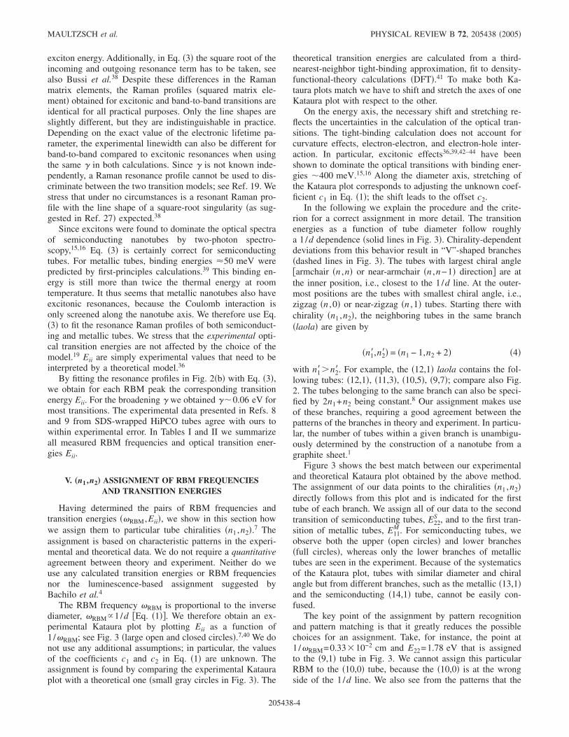
The key point of the assignment by pattern recognitionand pattern matching is that it greatly reduces the possiblechoices for an assignment. Take, for instance, the point at1 /�RBM=0.3310−2 cm and E22=1.78 eV that is assignedto the �9,1� tube in Fig. 3. We cannot assign this particularRBM to the �10,0� tube, because the �10,0� is at the wrongside of the 1/d line. We also see from the patterns that the
MAULTZSCH et al. PHYSICAL REVIEW B 72, 205438 �2005�
205438-4

�9,1� point has to be assigned to the outermost tube in abranch. This leaves us with two or three alternative assign-ments, where we shift the entire experimental and theoreticalplot with respect to each other. We show in the next para-graph that such an attempt leads to contradictions betweentheory and experiment. We also discuss the possibility of
assigning some of our data to different sets of transitions likeE11
S instead of E22S .
(Im)possible alternative assignments
We first consider assigning our ��RBM,Eii� pairs to a dif-ferent branch, keeping the overall assignment to the second
TABLE I. Summary of all observed RBM frequencies and transition energies of semiconducting tubes and their assignment. The tubesare grouped according to the branches �laola� in the Kataura plot. �RBM
emp and Eiiemp give the empirical values from Eq. �1� with c1
=215 cm−1 nm, c2=18 cm−1, and from Eq. �5�, respectively. The index �= �n1−n2�mod 3= ±1 indicates the two types of semiconductingtubes ��=−1 corresponds to �=2, as sometimes used in the literature�. The diameters are calculated with a0=2.461 Å. The nanotube samplecontained SDS as surfactant for excitation energies above 1.99 eV; below SDBS was used as surfactant. The experimental error of thetransition energies is between 3 meV �three digits� and 30 meV �two digits�. In the last column we give the Raman susceptibility in arbitraryunits, obtained by normalizing the maximum RBM peak area to integration time, laser power, relative spectrometer response, and �4. Theintensity changes due to different surfactants are taken into account; see Secs. VIII and IX. The asterisks indicate susceptibility values withlarge experimental errors due to weak signal or incomplete resonance profiles.
� laola n1 n2 d �Å� �RBM�cm−1�Eii
�eV��RBM
emp
�cm−1�E ii
emp
�eV� � Suscept.
−1 �12,1� 12 1 9.82 236.4 1.551 237 1.54 4.0 4.111 3 10.00 232.6 1.570 233 1.55 11.7 2.010 5 10.36 226.1 1.578 225 1.56 19.1 2.3
9 7 10.88 216.0 1.564 215 1.55 25.9 1.3
−1 �11,0� 11 0 8.62 266.7 1.657 267 1.68 0.0 1.710 2 8.72 264.6 1.690 264 1.69 8.9 2.3
9 4 9.03 257.5 1.72 256 1.71 17.5 2.58 6 9.53 246.4 1.73 243 1.72 25.3 1.4
−1 �9,1� 9 1 7.47 306.2 1.78 305 1.83 5.2 9.18 3 7.72 297.5 1.857 296 1.88 15.3 35.87 5 8.18 283.3 1.915 281 1.92 24.5 18.3
−1 �8,0� 8 0 6.27 352.2 1.99 361 1.97 0.0 0.17 2 6.41 353 2.03 12.26 4 6.83 333 2.15 23.4
+1 �14,1� 14 1 11.38 205.8 1.646 207 1.66 3.4 0.313 3 11.54 203.3 1.610 204 1.62 10.2 0.612 5 11.85 198.5 1.554 199 1.56 16.6 0.111 7 12.31 192 1.47 22.710 9 12.90 185 1.38 28.3
+1 �13,0� 13 0 10.18 230.8 1.84 229 1.85 0.0 0.2*
12 2 10.27 228.1 1.82 227 1.82 7.6 12.2*
11 4 10.54 221.8 1.76 222 1.74 14.9 0.1*
10 6 10.97 214 1.64 21.89 8 11.54 204.0 1.535 204 1.52 28.1 0.5
+1 �11,1� 11 1 9.03 256.0 2.031 256 2.06 4.3 9.810 3 9.24 252.1 1.945 251 1.98 12.7 3.6
9 5 9.63 241 1.84 20.68 7 10.18 230.4 1.710 229 1.69 27.8 0.4
+1 �10,0� 10 0 7.83 291.4 2.38 292 2.35 0.09 2 7.95 288 2.28 9.88 4 8.29 280.9 2.10 277 2.11 19.1 0.37 6 8.83 264.2 1.909 261 1.91 27.5 2.6
+1 �8,1� 8 1 6.69 339 2.69 5.87 3 6.96 327 2.48 17.06 5 7.47 308.6 2.20 305 2.18 27.0
RADIAL BREATHING MODE OF SINGLE-WALLED … PHYSICAL REVIEW B 72, 205438 �2005�
205438-5

transition in semiconducting and the first transition in metal-lic tubes. Figure 4�a� shows the combined experimental andtheoretical Kataura plot when shifting the experimental datato the left or “up” by one branch. The upshift results inexperimental branches where the number of RBMs is largerthan the number of tubes in the branch �circle in Fig. 4�a��.This assignment is therefore incorrect.
Figure 4�b� is the plot for shifting the experimental data tothe right or “down.” Some branches then have less RBMsthan tubes. This is quite possible if we have not detected allnanotubes in the diameter range. The assignment suggestedin Fig. 4�b�, however, strongly violates the patterns in thenanotube diameter distribution as we explain now. The diam-eter patterns are best seen for the outermost members of eachbranch in Fig. 3: because every second branch starts with azigzag tube, the distance between the two outermost pointsof the branches alternates between extremely close in1/�RBM and slightly further apart when going from onebranch to the next. This pattern is found in the experimental
data as well, compare, for instance, the �9,1� and �12,1�branches in Fig. 3 �two outermost points more separatedalong the x axis� to the �11,0� branch �two outermost pointsclose together�. The important argument here is again thepattern, not the agreement or disagreement on an absolute1 /�RBM scale. We can therefore exclude the assignment ofFig. 4�b� as well. A shift of the branches by more than onebranch up or down increases the disagreement betweentheory and experiment.
One might also think of assigning the experimental datato different transitions Eii, for example, shifting the experi-mental data down such that the measured RBM resonancescorrespond to the first transition of semiconducting tubes E11
S
instead of the second transition E22S . The data assigned to
metallic tubes in Fig. 3 then correspond either to E22S or to
E11M �as before�. Likewise, all data might be shifted up such
that the E22S data correspond to the metallic transitions E11
M
and so forth. Although these alternative assignments aremore of academic interest, since we know the E22
S transitions
TABLE II. Summary of all observed RBM frequencies and transition energies of metallic tubes and their assignment. For metallic tubes,�=0. Except for armchair tubes, each metallic tube has two close-by transition energies �Ref. 45� of which always the lower one wasobserved in the experiment.
� laola n1 n2 d �Å� �RBM �cm−1� Eii �eV� �RBMemp �cm−1� Eii
emp �eV� � Suscept.
0 �16,1� 16 1 12.94 182.0 1.81 184 1.81 3.0 0.5
15 3 13.08 179.0 1.83 182 1.81 8.9 1.8
14 5 13.36 174.5 1.83 179 1.81 14.7 1.9
0 �15,0� 15 0 11.75 200.4 1.908 201 1.93 0.0 0.7
14 2 11.83 196.3 1.934 200 1.94 6.6 3.6
13 4 12.06 193.5 1.944 196 1.94 13.0 2.5
12 6 12.44 189.4 1.948 191 1.93 19.1 1.9
11 8 12.94 183.2 1.936 184 1.92 24.8 1.9
10 10 13.57 175.7 1.889 176 1.88 30.0 0.9
0 �13,1� 13 1 10.60 220.3 2.057 221 2.07 3.7 1.5
12 3 10.77 217.4 2.075 217 2.07 10.9 2.6
11 5 11.11 212.4 2.084 211 2.08 17.8 1.7
10 7 11.59 204.0 2.067 203 2.07 24.2 0.9
9 9 12.21 195.3 2.02 194 2.04 30.0 0.5
0 �12,0� 12 0 9.40 244.9 2.18 247 2.21 0.0
11 2 9.50 244 2.22 8.2
10 4 9.78 238 2.24 16.1
9 6 10.24 228 2.25 23.4
8 8 10.85 216 2.23 30.0
0 �10,1� 10 1 8.25 276.3 2.38 278 2.35 4.7
9 3 8.47 272.7 2.43 272 2.40 13.9
8 5 8.90 262.7 2.47 259 2.45 22.4
7 7 9.50 247.8 2.45 244 2.46 30.0
0 �9,0� 9 0 7.05 323 2.47 0.0
8 2 7.18 315.5 2.52 317 2.53 10.9
7 4 7.55 305.4 2.63 302 2.65 21.1
6 6 8.14 282 2.71 30.0
MAULTZSCH et al. PHYSICAL REVIEW B 72, 205438 �2005�
205438-6

from photoluminescence,4 we want to discuss these “exotic”assignments briefly. We show that assigning our measuredenergies to other transitions than E22
S and E11M systematically
violates the Kataura-plot patterns and therefore result in in-correct assignments. It is an intriguing exercise highlightingvery nicely the systematics in the Kataura plot and the pat-tern recognition idea.
Let us consider assigning the experimental E22S data in
Fig. 3 to the metallic E11M resonance. The upper and lower
branches of metallic transitions in the Kataura plot belong tothe same chiralities. Thus the RBM frequencies in the upperand lower branches must be the same. This is in contrast withexperiment, where the upper branches �open circles in Fig. 3�begin and end at larger diameter than the correspondingbranches on the opposite side of the 1/d line.
A downshift of the E22S data to the E11
S transition is alsoimpossible. When switching from a transition Eii
S to the nexthigher or lower transition, the family dependence is reversed.For E22
S the +1 tubes are above the 1/d line �see Fig. 3�; forE11
S the �= +1 tubes are below the 1/d line, vice versa for the�=−1 tubes.1,45 The two families start and end at differentrelative diameters for a given V-like curve. Changingthe family dependence in the theoretical points by goingfrom E22
S to E11S completely disturbs the patterns. In Fig. 5 we
show such an attempt in detail. The former-assigned E22S
data are shifted down to the E11S transitions. We stretched
and displaced the Kataura plot to get the best match for theupper parts of the V-shaped curves, which now correspond to�=−1 tubes �open circles�. Obviously, the lower, �= +1,branches strongly violate the Kataura patterns. They areshifted to larger diameters with respect to the theoreticaldata. The situation gets even worse if we take the metallictubes into account as well. Assigning them, e.g., to E22
S isimpossible, because this results in nanotubes with the samechirality, but two different RBM frequencies.
We systematically considered other “exotic” assignmentsas well, e.g., the idea that certain data points in an experi-mental V-like curve are resonances coming from a differentoptical transition than the other points, say E22
S transitions aremixed with E11
S transitions in one and the same V-like curvein Fig. 3. All these ideas can be excluded in a similar way asdiscussed for some selected alternative assignments in thepreceding paragraphs. The assignment in Fig. 3 is the onlyone that matches the systematics in the Kataura plot andobeys the 1/d patterns of the RBM frequencies. We summa-rize all measured RBM frequencies together with the assign-ment in Tables I and II.
Our Raman based assignment agrees with Bachilo’s et al.4
suggestions from photoluminescence. They used a similaridea of pattern matching to correlate the theoretical transition
FIG. 3. �Color online� Experimental �large open and closedcircles, left and bottom axes� and theoretical �small gray circles,right and top axes� Kataura plot. The second transitions of semicon-ducting tubes E22
S and the first transitions E11M of metallic tubes are
shown. The solid lines give the approximate 1/d dependence of thetransition energies. The dashed lines indicate the “V”-shapedbranches, where the chirality of a tube is related to its left neighbor�n1 ,n2� by �n1� ,n2��= �n1−1,n2+2�. In the experimental data, theassignment is given for the first tube in each branch, where uprightnumbers indicate semiconducting and italic numbers indicate me-tallic tubes. The semiconducting tubes are divided into two familieswith �= �n1−n2�mod 3=−1 �full circles, lower branches� and with�= +1 �open circles, upper branches�.
FIG. 4. �Color online� Kataura plot showing two assignmentsthat violate the pattern system and are therefore incorrect. �a� Theexperimental data were shifted “up” �to the upper left corner� whencompared to Fig. 3. Only two experimental branches are shown forclarity. The circle highlights the Kataura branch where this assign-ment produces five experimental RBMs for a branch that containsonly four nanotubes. �b� same as �a�, but for the shift down. Thevertical lines highlight regions where the 1/d patterns of the RBMare violated.
RADIAL BREATHING MODE OF SINGLE-WALLED … PHYSICAL REVIEW B 72, 205438 �2005�
205438-7

energies with experiment.4,41 The luminescence-based as-signment was ambiguous; it needed an additional anchor el-ement. There are two reasons why the ��RBM,Eii� pairsfound by Raman scattering restrict the possible assignmentmore strongly than the �E11,E22� pairs from luminescence:First, Raman scattering detects more nanotubes than photo-luminescence, in particular, the metallic tubes and the tubeswith small chiral angles at the end of the Kataura branches�zigzag and close-to-zigzag tubes�. Second, �RBM is to goodapproximation independent of the chiral angle, whereas theEii depend on diameter and chiral angle. This reduces thedegrees of freedom for the ��RBM,Eii� pattern matchingwhen compared to the �E11,E22� matching and makes Ramanscattering less ambiguous than photoluminescence.
In summary, we assigned experimental RBM frequencies�together with Eii� to particular nanotubes indices �n1 ,n2�.Based on this assignment, we correlate the RBM frequencieswith tube diameters in Sec. VII. Our assignment is indepen-dent of empirical parameters. The measured optical transitionenergies are excitonic energies, as known from recentexperiments.15,16 As the assignment procedure does not relyon absolute energies when comparing with the theoreticalKataura plot, the results are not affected by the strength ofexciton binding in carbon nanotubes. In fact, our assignmentis based on two patterns: The systematics in the distribu-tions of tube diameters with n1 and n2, which comes from thec=n1a1+n2a2 construction of a tube, and the family behaviorof the nanotube transition energies.1,45 As long as these twovery general concepts in nanotube physics remain valid, ourRaman based assignment is unique.
VI. TRANSITION ENERGIES OF SEMICONDUCTINGAND METALLIC TUBES
In this section we provide empirical fits to the experimen-tal transition energies E22
S and E11M in order to apply these
expressions to chiralities not observed in the experi-ment. We start with the 1/d relation for the band gap,Eii=2i�0aC-C /d,1,46 expanding it in 1 /d. We add a chiral-angle dependent term to model the branches of the Katauraplot, which is larger for smaller diameters.45,47 For the E22
S
transitions of semiconducting tubes,
E22S = �0�4aC-C
d+ �1
aC-C2
d2 � + ��2 cos�3��aC-C
2
d2 , �5�
with the parameters �0 ,�1, and �2. aC-C is the length of thecarbon-carbon bonds �aC-C=a0 /�3�, and �= �n1−n2�mod 3is the family index taking the values ±1 in semiconductingtubes and �=0 in metallic tubes. � is the chiral angle of the�n1 ,n2� tube, being zero in zigzag tubes.1 From the fit to ourdata we obtain �0=3.53 eV, �1=−4.32, and �2=8.81 eV.Analogously, we approximate the transition energies in me-tallic tubes by
E11M = �0�6aC-C
d+ �1
aC-C2
d2 � − �2 cos�3��aC-C
2
d2 , �6�
and find �0=3.60 eV, �1=−9.65, and �2=11.69 eV. The em-pirically determined energies are shown as a function of di-ameter in Fig. 6, where d is given by �n1 ,n2� �Eq. �2��. Be-cause the electronic properties change dramatically forsmall-diameter tubes,48,49 the empirical data given here arevalid only for tubes with diameters d�6 Å. Our empiricalextrapolation fits well with the experimental E22
S data ob-tained by Doorn et al.17 for the tubes outside the range of ourexperiments. All empirical transition energies are listed inthe supplementary material, Ref. 50.
The �0 obtained from Eqs. �5� and �6� are quite close tothe corresponding parameter in graphite �3.2 eV, Ref. 51� andmuch larger than assumed for carbon nanotubes in the past�2.5–2.75 eV, Refs. 27 and 52�. From our fits it seems that �1scales with the square of i �E11=E11
S , E22=E22S , E33=E11
M ; seeRefs. 1 and 46�, whereas the chirality-dependent correctionshows only a linear scaling. These corrections reflect a mix-ture of trigonal warping of the graphene band structure,45
curvature effects,21,53 and exciton effects.42,43 Very recently,
FIG. 5. �Color online� Kataura plot showing a trial assignmentwhere the experimental data are shifted down to the E11
S transitions.This assignment is incorrect for several reasons; see text. While theexperimental upper branches �open circles� match quite well thepattern of the theoretical data, the lower branches disagree. The dataassigned to the metallic tubes �large dark circles� cannot be as-signed to the second semiconducting transitions E22
S , because theyshould be at exactly the same RBM frequencies �diameters� as theupper E11
S branches �open circles�, which they are not.
FIG. 6. �Color online� Experimental transition energies �opensymbols� and empirical values from Eqs. �5� and �6� �closed sym-bols�. For the metallic branches, the inner �armchair� tube isindicated.
MAULTZSCH et al. PHYSICAL REVIEW B 72, 205438 �2005�
205438-8

experimental results on the exciton binding energies in car-bon nanotubes were published.15,16 Although Maultzsch etal.16 reported a family dependence of the exciton bindingenergies and also a dependence on the size of the nanotubeband gap, it is difficult to quantify these effects from the dataat hand. Perebeinos et al.43 suggested a scaling of the excitonbinding energy with the effective mass of the electron andthe hole as Eb�m�−1, where �=1.4 was found from tight-binding calculations. The effective mass in turn depends onthe family of a tube and its diameter and chiral angle. Nev-ertheless, for the time being we prefer to understand Eqs. �5�and �6� as empirical functions instead of giving the param-eters and dependences a strict physical meaning in terms ofcurvature and exciton effects. The two relations fit the ex-perimental data very well; see Fig. 6. It would be interestingto obtain Raman data for the first optical transition energy tosee whether the proposed scaling for �1 and �2 applies to thistransition as well.
Strano and co-workers18,54 used a similar expression to fitthe observed transition energies in semiconducting and me-tallic tubes. Our results for semiconducting tubes are in goodagreement with the empirical values in Refs. 18 and 54. Formetallic tubes, in contrast, we find large deviations betweenour experimental data and the empirical data in Ref. 18. Inparticular, the Raman resonances of some metallic tubeswere assigned to the second metallic transitions in Ref. 18,whereas from our data it is obvious that only the first metal-lic transitions are observed. For example, the resonance ofthe �7,7� armchair tube �see Fig. 6�, i.e., at the inner positionof a “V”-shaped branch, is in Ref. 18 assigned to the secondresonance of the �12,0� zigzag tube. Therefore the empiricalexpressions given by Strano et al.18 underestimate the tran-sition energies of metallic tubes.
In Ref. 9 a detailed comparison between the experimentaltransition energies with tight-binding results can be found.The authors present diameter and chirality-dependent correc-tions involving eight fitting parameters for each set of tran-sitions.
VII. RELATION BETWEEN �RBM AND DIAMETER
In contrast to previous attempts to obtain �n1 ,n2� from theRaman spectrum of a tube27,29 we first assigned an RBMfrequency to a nanotube. We used the fact that the RBMfrequency is approximately linear with the inverse tube di-ameter. We did, however, not include Eq. �1� explicitly, inparticular, no values c1 and c2 were given. From this assign-ment we now calculate the tube diameter and fit c1 and c2from the �RBM vs diameter plot. The key difference to otherwork is that a particular Raman line is always assigned to thesame nanotube within our approach. Using, e.g., the diam-eters reported by Kürti et al.24 from first-principles calcula-tions instead of the geometrical expression in Eq. �2� we findslightly different numbers for c1 and c2. The assignment inTables I and II, however, remains the same. For an assign-ment it is usually better to work with the tables instead of Eq.�1� as we discuss in Sec. X.
In Fig. 7 we show a linear fit to our data points accordingto Eq. �1�. Using a0=2.461 Å, we obtain c1=215 cm−1 nm
and c2=18 cm−1. With aC-C=a0 /�3=1.44 Å, also used in theliterature, c1=218 cm−1 nm and c2=18 cm−1. The coeffi-cients are thus very sensitive to how the tube diameter isdetermined. Therefore they provide an estimate of the RBMfrequency for a given diameter �Eq. �1��, but should not beused to compare �or even assign� an experimental RBM fre-quency to a nanotube diameter with high accuracy.
Deviations from the linear dependence of the RBM fre-quency on the inverse diameter have been predicted forsmall-diameter tubes by Kürti et al.24 from first-principlescalculations. In Raman experiments, based on the assignmentof Ref. 4, Jorio et al.9 observed deviations of a few wavenumbers, depending slightly on the chiral angle. Here weshow explicitly that the behavior of the experimental RBMagrees very well with the calculations in Ref. 24.
Figure 8 shows the difference �RBM between the experi-mental �RBM and the empirical values calculated from Eq.�1� with c1=215 cm−1 nm and c2=18 cm−1 �full circles�. Ingeneral, the deviations from the linear fit increase for smallerdiameters, in agreement with the predictions. They vary be-tween +4 and −2 cm−1, depending on the chiral angle. Fortubes with d�10 Å, �RBM has a large and positive valuefor �near-�armchair tubes and decreases to negative valuesfor �near-�zigzag tubes.
Kürti et al.24 showed that isolated armchair tubes followthe linear relation with the smallest deviations, whereas zig-zag tubes have the largest �negative� deviation. If we assumethe line connecting the armchair tubes in Fig. 8�a� to be the �RBM=0 line,55 we observe the same trend of increasingdeviation towards zigzag tubes. This agrees with the predic-tion that zigzag tubes show the strongest rehybridizationeffects21 and the largest increase in bond length,24 both ef-fects resulting in a weakening of the RBM frequency.
To compare with the theoretical data of Ref. 24 quantita-tively, we performed a linear fit to the ab initio values �RBM
DFT
and analyzed the differences between �RBMDFT and this linear fit
�open circles in Fig. 8�. We find a very good quantitativeagreement between experiment and theory, although the cal-culations were performed for isolated tubes in vacuum. Thisconfirms that the deviations from the linear relation aremostly due to changes in the strength of the bonds �rehybrid-ization and bond lengths�.
VIII. RBM INTENSITIES
The Raman resonance profile given by Eq. �3� accountsfor the position of the resonance maximum, which we dis-
FIG. 7. �Color online� Linear fit of the observed RBM frequen-cies as a function of inverse tube diameter 1 /d. The diameter of theassigned nanotubes is calculated from Eq. �2� with a0=2.461 Å.
RADIAL BREATHING MODE OF SINGLE-WALLED … PHYSICAL REVIEW B 72, 205438 �2005�
205438-9

cussed so far. In this section, we evaluate the relativestrength of the Raman signal to obtain information on thematrix elements M. The matrix elements M consist of theelectron-photon coupling, Me-r, and the electron-phononcoupling, Me-ph, and �M�2= �Me-rMe-phMe-r�2. The con-stant c contains the remaining factors such as response of thespectrometer and �4 dependence of the Raman cross section�taken into account by normalization to the nonresonant Ra-man signal of BaF2 and CaF2�, laser power, integration time,scattering volume, and concentration of the nanotube solu-tion. We do not make any assumptions on the diameter andchirality distribution in our sample, thus showing the bareRaman intensities. The diameter distribution can be deter-mined by electron miscroscopy56 or electron diffraction.57 Aswe show below, the relative abundance of particular chirali-ties cannot be determined by the bare Raman or lumines-cence intensities.
In Fig. 9 we show part of the experimental Kataura plot,where the area of the circles indicates the strength of theRaman signal. We observe two trends: first, the Raman crosssection increases for smaller diameter; second, it is in generalmuch larger for the lower branches ��=−1 for E22
S � than for
the upper branches. The upper branches of metallic tubeswere not observed at all. Both the diameter dependence andthe �= ±1 dependence were predicted by first-principles cal-culations of the electron-phonon coupling matrix elementsMe-ph,
22 see also Ref. 58.The electron-phonon coupling for the RBM becomes
stronger for smaller-diameter tubes, because the same radialdisplacement results in a larger change of the carbon-carbonbonds in smaller tubes. The dependence on �= ±1 can beunderstood within the zone-folding picture. The index �= ±1 indicates from which side of the K point in thegraphene Brillouin zone the electronic states are derived. Ingraphene, the coupling between electrons close to the Kpoint and phonons analogous to the RBM is larger betweenthe K and M points than in the K-� direction.22 Similar re-sults from Raman scattering were reported by Doorn et al.17
and Jorio et al.;9 calculations within an empirical tight-binding description give the same dependence on �= ±1.58,59
According to the ab initio calculation by Machón et al.,22
we expect the opposite family behavior for Raman scatteringin resonance with the first transition energy of semiconduct-ing tubes. For this resonance, the Raman susceptibility of the�= +1 nanotubes should be larger than the susceptibility ofthe �=−1 nanotubes. Once again, this can be understood inthe zone-folding approach, because the first and second op-tical transition in a nanotube originate from opposite sides ofthe K point.
Within each branch, the Raman intensity depends system-atically on the chiral angle. It is small for tubes close to thearmchair direction �inner position� and first increases withdecreasing chiral angle. Close to the zigzag direction �outerpositions� the intensity is small again. This is explained bythe chiral-angle dependence of both the electron-phonon
FIG. 8. �Color online� Difference between experimental RBMfrequencies and calculated RBM frequencies from Eq. �1� with c1
=215 cm−1 nm and c2=18 cm−1 �full circles�. The lines connecttubes of the same branch, labeled by the first member according tothe Kataura plot and Tables I and II. �a�, �b�, and �c� show the tubeswith �=0, �= +1, and �=−1, respectively. The chiral angle withineach branch decreases with decreasing diameter. Open circles showthe difference between �RBM
DFT from first-principles calculations �Ref.24� and �RBM from a linear fit to these theoretical data. In �a� thearmchair tubes are indicated.
FIG. 9. �Color online� Part of the experimental Kataura plot,where the intensity of the Raman signal is given by the area of thecircles. Grey and black symbols indicate semiconducting and me-tallic tubes, respectively. The smallest circles account for all valuesbelow 0.5 in Table I. The crosses indicate tubes with large uncer-tainty in the Raman intensity; see also Table I. In additionto the normalization procedure described in the text, the intensity ofthe �11,1� tube is divided by 3.4 and the intensity of the �13,1�branch is multiplied by 3. This accounts for the changes in theRaman signal strength due to a different surfactant for the sampleused above El=1.99 eV; see Sec. IX.
MAULTZSCH et al. PHYSICAL REVIEW B 72, 205438 �2005�
205438-10

coupling and the strength of the optical transitions.7,36 Theelectron-phonon coupling is stronger for zigzag tubes thanfor armchair tubes, explaining the weaker signal for theclose-to-armchair tubes.22 The luminescence, on the otherhand, was observed to decrease for close-to-zigzag tubes,4 inparticular for the �= +1 branches.36 Although we do notknow directly the chirality dependence of absorption strengthinto the second semiconducting transitions E22
S , we can as-sume that it has an opposite dependence on chiral angle thanthe electron-phonon coupling, explaining the decrease of theresonant RBM signal towards the zigzag direction.
In the last column of Tables I and II we show the intensityof the measured RBM signal normalized to the Raman signalof CaF2 and BaF2 and divided by the Bose-Einstein occupa-tion number. These values are proportional to the Ramansusceptibility.
IX. DEPENDENCE ON THE TYPE OF SURFACTANT
In the previous sections we assumed the RBM frequencyand the optical transition energies to reflect only the intrinsicproperties of carbon nanotubes. Now we address the depen-dence of their properties on the surfactant �SDS or SDBS�,i.e., the environment of the tube. The surfactant has a smallinfluence on the position of the experimental data points inthe Kataura plot both along the frequency �diameter� and theexcitation energy axes. Besides the fundamental interest inthe interaction between a nanotube and its surrounding,environment-related effects can affect a nanotube assignmentbased on a single Raman spectrum and the RBM frequencyalone �see Sec. X�. The pattern-based assignment, however,is unaffected by the shifts, because they are small and do notfundamentally change the experimental patterns.
To analyze the surfactant-induced changes in the Ramanspectra, we recorded resonance profiles for both surfactantswith excitation energies between 1.85 and 2.2 eV; see Fig.10. In this region, the laser energies are in resonance withboth metallic and semiconducting tubes. We observe changesof �i� the transition energies, �ii� the Raman intensities, and�iii� the RBM frequencies.
For semiconducting tubes �Figs. 10�a� and 10�b��, thetransition energies shift by �5–10 meV to larger values inthe SDS sample �open dots�. In addition, the RBM signal ofsemiconducting tubes is stronger in the SDS sample than inthe SDBS sample �closed dots�. The behavior of metallictubes is opposite: their RBM intensity is larger in the SDBSsample. The shift of transition energies is, if detectable, ofsimilar magnitude as in semiconducting tubes but in oppositedirection, i.e., the transition energies of metallic tubes areslightly larger with SDBS as surfactant, see also Table III.These changes are, however, small, and in particular for sev-eral tubes of the metallic �15,0� branch within the experi-mental error.
In Figs. 11 and 12 we compare the RBM spectra for bothsurfactants at the same laser energy in the region of semicon-ducting and metallic tubes, respectively. The RBM frequencyof semiconducting tubes is the same in both surfactantswithin experimental error �gray black curves in Fig. 11�. Therelative RBM intensities, in particular of the �8,3� and the
�7,5� tube, are different in these spectra, reflecting thesmall shift of transition energy and hence of the resonancecondition. The original intensity ratio in the SDS sample atEl=1.916 eV is recovered if the SDBS sample is excited at aslightly lower energy �El=1.908 eV, dashed curve�. Thisshift in laser energy compensates for the shift in the opticaltransition energy of semiconducting tubes. In metallic tubeswe observe an upshift of the RBM frequencies in the SDSsample by about 2 cm−1; see Fig. 12.
The dependence of the RBM frequency and intensity inmetallic and semiconducting tubes on the type of surfactantagrees with the observation by Strano et al.60 of selectivefunctionalization of metallic tubes. They found a decrease ofthe absorption strength for the metallic E11
M transitions, result-ing from functionalization with tetrafluoroborate salt and for-mation of covalent bonds. Simultaneously, the RBM shiftedto larger frequency. We can thus interpret our results as dueto an interaction between the surfactant and the nanotube,which is stronger for SDS than for SDBS. Although it isunlikely that a covalent bond forms as in the case of Ref. 60,an electron transfer from the metallic tubes to the surfactantmight occur. The Raman intensity decreases in SDS as theresonant absorption becomes weaker, simultaneously the in-teraction leads to a larger RBM frequency. From our data we
FIG. 10. �Color online� Resonance profiles of the RBM for sur-factants SDS �open red dots� and SDBS �filled black dots�. Thechiral indices, the chiral angle �, and the family � are given. Semi-conducting tubes, �a� and �b�, show a small upshift in the position ofresonance maximum for SDS. In metallic tubes, �c� and �d�, theshift of the transition energies is in opposite direction. The RBMintensity of metallic tubes is stronger in the SDBS sample, while theintensity of semiconducting tubes is stronger in the SDS sample.Since the concentration of nanotubes in both solutions is not knownexactly, we cannot quantify the absolute intensities. Comparing therelative intensities between the two surfactants, they change in op-posite directions for metallic and semiconducting tubes.
RADIAL BREATHING MODE OF SINGLE-WALLED … PHYSICAL REVIEW B 72, 205438 �2005�
205438-11

cannot detect such a difference in the interaction for semi-conducting tubes, as the RBM is constant when changing thesurfactant.
The small shift in transition energies might be to firstapproximation assigned to a change in the dielectric environ-ment. It is for several tubes within the range of experimentalerror. We have no explanation yet for the shift in oppositedirection for metallic and semiconducting tubes.
Izard et al.61 studied the development of RBM spectrafrom bundled tubes to bundles wrapped by SDS and to indi-vidual tubes in SDS. They also observed an upshift of theRBM due to wrapping by SDS which they assigned to pres-sure induced by the surfactant. The metallic tubes appear tobe more sensitive to the surfactant, as the RBM shift is ingeneral larger than for semiconducting tubes, in agreementwith our results. Izard et al.61 observed changes in the rela-tive RBM intensities as well, which they ascribed to a selec-tive exfoliation process. From our data, we rather suggest asmall change in resonance condition.
Our assignment of Sec. V is not affected by thesurfactant-induced variation in the transition energies andRBM frequencies. Figure 13 shows a small section of theKataura plot with data from SDS �open dots� and fromSDBS �closed dots�. The differences in excitation energiesand RBM frequencies are minor on the scale of the Katauraplot. The most important criteria for the assignment are theRBM frequency patterns and the number of tubes within abranch. Figure 13 shows that the small variations of theRBM do not change these systematics. In particular, thechanges are too small to shift the data to a different branch.
TABLE III. Comparison of transition energies and RBM frequencies for different surfactants �SDS andSDBS�. All transition energies are obtained from resonance profiles. The experimental errors for the firstthree tubes of the �15,0� branch are larger than for the majority of our data.
Tube
SDBS SDS
�RBM �cm−1� Eii �eV� �RBM �cm−1� Eii �eV� � �cm−1� E�meV�
Metallic nanotubes
�15,0� 200 1.91 203 1.91 −3 0±20
�14,2� 196 1.93 199 1.93 −3 0±10
�13,4� 193 1.94 196 1.93 −3 10±10
�12,6� 189.4 1.948 191.1 1.938 −1.7 10±6
�11,8� 183.2 1.936 184.1 1.906 −0.9 30±8
Semiconducting nanotubes
�8,3� 297.5 1.857 297.5 1.877 0.0 −20±13
�7,5� 283.3 1.915 283.2 1.919 0.1 −4±4
�7,6� 264.2 1.909 263.6 1.917 0.6 −8±6
�10,3� 252.1 1.945 252.1 1.953 0.0 −8±6
FIG. 11. �Color online� RBM spectra of nanotubes dispersed inD2O using SDBS �black� and SDS �gray� as surfactants at excita-tion energy 1.916 eV �solid lines� and 1.908 eV �dashed lines�. Thespectra are normalized to the RBM amplitude of the �7,5� tube.
FIG. 12. �Color online� Metallic part of the RBM spectrum.Gray: SDS sample; black: SDBS sample. Thin lines show the fit ofthe RBM peaks by Lorentzians. The peaks are shifted to higherfrequencies in the SDS sample.
MAULTZSCH et al. PHYSICAL REVIEW B 72, 205438 �2005�
205438-12

Therefore the assignment is valid for both types of surfac-tant.
X. HOW TO ASSIGN „n1 ,n2… IN A RAMAN EXPERIMENT
A great need for nanotube research is to identify thechirality of a tube before performing an experiment. Ideally,the method is nondestructive, does not require special equip-ment or substrates, works for semiconducting and metallictubes, as well as individual tubes and bulk samples �nano-tubes in solution or bundled tubes�. It should also give reli-able results regardless of the tube environment. We discussedpossible ways for identifying carbon nanotubes in the intro-duction of this paper. Raman scattering meets many of therequirements for becoming one of the prime assignmentmethods for single-walled carbon nanotubes.
Most Raman-based assignments of individual andbundled tubes relied mainly on the �RBM�1/d relationshipusing one value for c1 and c2 or the other; see the review inRef. 1. In this paper we showed that the RBM frequencyalone will never be sufficient for assigning the chirality, be-cause it depends on the environment of the tubes. Althoughthe changes in the RBM frequencies are small between dif-ferent surfactants �Sec. IX� and also between bundled andsurfactant-coated tubes,8,20,61 they are large enough to changean assignment that uses the RBM frequencies as the onlyinput; see also Ref. 50. Therefore a Raman-based assignmentof an individual tube, suspended or on a substrate, nanotubesin solution, nanotube bundles, and so forth, should alwaysuse the combined information of RBM frequency and exci-tation energy.
Once the full resonance Raman experiment has been per-formed and the assignment of �RBM to the chiral index hasbeen found �Sec. V�, the chiral indices of a sample contain-ing tubes with similar diameters can be determined from onesingle Raman spectrum. Environment-related effects oftencan be taken into account by estimating the changes in thetransition energies. In this section we explain the procedureand show how the RBM peaks in a given Raman spectrumcan be assigned to �n1 ,n2�.
For simplicity, we plot the experimental data of Fig. 3�Tables I and II� now with the RBM frequency along the xaxis; see Fig. 14. The procedure of the assignment is as fol-lows:
1. Record a Raman spectrum at a given excitation energyEl and determine the RBM frequencies �RBM �Fig. 15�.
2. Identify groups of 3–5 close-by RBM peaks as indi-cated in Fig. 15 by the shaded areas. The members of thesegroups form a branch �laola� in the Kataura plot.
3. Find the excitation energy El in the experimental Ka-taura plot �horizontal line at 1.96 eV in Fig. 14�.
4. Find along the x axis of the experimental Kataura plotthe regions of observed RBM frequencies �shaded areas�.The branches closest to El within these regions most likelycontribute to the RBM spectrum.
5. Compare all RBM frequencies in detail with the ex-perimental Kataura plot and the sequence of tubes in a
FIG. 13. �Color online� Section of the Kataura plot showing thetransition energy vs inverse RBM frequency of nanotubes in twodifferent surfactants: SDS �open dots� and SDBS �closed dots�.Semiconducting tubes �gray� show a uniform shift of the transitionenergies. Metallic tubes �black� are shifted in energy and RBMfrequency.
FIG. 14. �Color online� Experimental Kataura plot: transitionenergies vs RBM frequencies, from Tables I and II. The shadedregions correspond to the RBM peaks in Fig. 15.
FIG. 15. �Color online� RBM spectrum at El=1.96 eV. Theshaded regions indicate groups of metallic and semiconductingtubes; the chiral indices at the bottom give the first element of thecorresponding laola in the Kataura plot �Fig. 14�. The chiral indicesat the top show the assignment of the strongest peaks.
RADIAL BREATHING MODE OF SINGLE-WALLED … PHYSICAL REVIEW B 72, 205438 �2005�
205438-13

branch to find the final assignment; see Fig. 15. Compare thenumber of tubes within this branch to how many of them areobserved.
In the example given in Figs. 14 and 15, we identify theleft and the right RBM groups as metallic and semiconduct-ing tubes, respectively. Since the width of the resonance pro-files is typically around 60 meV, we assume that we canobserve tubes from a window of approximately 100–200-meV width around the excitation energy. These areas areindicated in Fig. 14. For the metallic tubes, mainly the �15,0�branch contributes to the spectra. In the region of semicon-ducting tubes, the members of several branches are close tothe excitation energy. We identify the peaks as resulting fromtubes of the �11,1�, �10,0�, and �9,1� branches. In the finalstep, we assign the strongest peaks to the �11,8�, �12,6�, and�13,4� tube �metallic region� and to the �10,3�, �7,6�, �7,5�,and �8,3� tube �semiconducting region�, as indicated in Fig.15. The remaining tubes of these branches, such as the �15,0�tube, are weaker shoulders of the strongest peaks and be-come evident when changing the laser wavelength. Others,like the members of the �10,0� branch, are outside the reso-nance window. Thus not all members of each branch areexpected in the same single Raman spectrum; because of thechirality dependence of the Raman cross section some tubesmight not be observed.
The most relevant piece of information is already ob-tained in the fourth step, i.e., by identifying the correctbranches. Many properties are similar for tubes of the samebranch. In contrast, they differ strongly for tubes of differentbranches even if the diameters or transition energies are al-most the same. For example, the �7,5� and the �7,6� tube havevery similar Eii, but the �7,6� tube gives a much weakerRBM signal. This is due to the different strength of theelectron-phonon coupling for �=−1 and �= +1 tubes,22 asexplained in Sec. VIII.
In Raman measurements on a single, individual nanotube,the chiral index is found by the same method. For this typeof samples the difficulty is to obtain an observable RBMsignal, because of the narrow resonance window of theRBM. One, two or even three laser energies might be neededto find the resonance window of a particular tube. The laolagroups—typical for samples with different chiralities—thatallowed to identify branches, are absent in individual tubes.Nevertheless, following the procedure described above, thechoice of possible chiralities can be narrowed to one or twotubes. If the ambiguity concerns two nanotubes from thesame branch, a further refinement of the assignment is notnecessary. Two neighboring tubes in the same branch are toosimilar in properties to easily distinguish between them. Inturn, this implies that neither fundamental studies nor appli-cations benefit much from narrowing down the choice. If, onthe other hand, a single Raman spectrum is insufficient todistinguish between two branches, the ambiguity arose be-tween two tubes of different family—metallic, �= +1 or �−1 semiconducting. In this case the assignment should beverified by using a different excitation energy or by combin-ing Raman scattering with a second assignment technique.
In Sec. IX we showed that changing the environment ofthe nanotubes did not affect the RBM frequencies of semi-conducting tubes and only slightly those of metallic tubes.
This is consistent with the observation of only small RBMchanges in bundles vs separated tubes and in tubes in severaldifferent surfactants.8,20,61 On the other hand, the transitionenergies Eii appear to be more sensitive to the nanotubeenvironment.8,20,61,62 Therefore the empirical values of Eiigiven in Tables I and II are strictly valid only for nanotubesin SDS or SDBS and should be used with care for othertypes of samples. Ideally, one would perform the full reso-nance Raman experiment once for each tube environment,e.g., different surfactants, bundled tubes, individual tubes ona substrate, individual suspended tubes in air, etc. Some ofthese data have been reported in the literature, see, for in-stance, Refs. 8 and 20. In addition, photoluminescence datacan be used for Eii in different samples. The Raman-basedassignment procedure, however, is always the same as de-scribed in this section. In particular, if entire branches areobserved, the data presented in Tables I and II can be readilyused, taking into account changes in the Eii. As the RBMfrequencies vary only slightly, the Raman-based assignmentis much more stable against changes in the nanotube envi-ronment than an assignment based on the Eii alone.
In standard Raman setups often just a few laser lines areavailable. To facilitate an assignment, we show in Fig. 16 theRBM spectra for the most common laser lines �514, 752, and785 nm� together with the chiral indices. These spectra can
FIG. 16. �Color online� RBM spectra with chiral-index assign-ment at several standard laser lines, 514, 752, and 785 nm. Thepeaks in the middle panel consist of two RBMs each which canonly be resolved when changing the excitation energy. For the spec-trum at 633 nm �1.96 eV� see Fig. 15.
MAULTZSCH et al. PHYSICAL REVIEW B 72, 205438 �2005�
205438-14

be directly compared to Raman spectra taken on HiPCOtubes in solution with standard equipment and used for asimple assignment. Two tubes with very similar diametersare sometimes difficult to resolve from a single Raman spec-trum, see the 752-nm spectrum in Fig. 16. Apparently onlytwo tubes contribute. From the excitation-energy dependentmeasurements we know that each peak in the middle panelof Fig. 16 is, in fact, composed of two RBM lines.
XI. SUMMARY
In summary, we presented a chiral-index assignment forcarbon nanotubes from resonant Raman scattering. The as-signment is independent of any additional parameters, but itis based on pattern recognition. The two pieces of informa-tion that are required for this assignment are the frequency ofthe radial breathing mode and the energy of an optical tran-sition �here E22
S and E11M �. They constitute an experimental
Kataura plot where all chiral indices are systematicallygrouped into so-called branches with neighboring indicesgiven by �n1� ,n2��= �n1−1 ,n2+2�. Because of these systemat-ics, the assignment remains the same even if parameters inthe calculation of Eii or the diameter change or if the experi-mental values vary due to slightly different experimentalconditions. �n1 ,n2� is assigned to experimental RBM fre-quencies and transition energies, irrespective of changes inthe theoretical description. We consider all measured transi-tion energies to be excitonic energies, as excitonic effectsdominate the optical spectra in carbon nanotubes.
We derived the parameters c1=215±2 cm−1 nm and c2=18±2 cm−1 from our assignment for the RBM-diameter re-
lation. These values vary depending on the type of sampleand on the details of the diameter calculation. The RBMintensities are in general stronger for �=−1 nanotubes thanfor �= +1 tubes for the E22
S transitions. They decrease fromtheir maximum around ��10°–15° towards both the arm-chair and the zigzag direction. These results are in goodagreement with ab initio calculations of the electron-phononcoupling.22 The intensities also depend on the type of surfac-tant in our samples with different behavior for metallic andsemiconducting tubes. For metallic tubes, we observed astronger interaction with SDS, and an upshift of the RBMfrequencies.
Finally, we provided a description on how to find a chiral-index assignment from a single Raman spectrum. Forsamples with similar tube diameters in a similar environ-ment, the experimental and empirical data given in Tables Iand II and in Ref. 50 can be used for a straightforward as-signment. Changes in the tube environment usually affectmainly the optical transition energies, which can be takeninto account for an assignment. We stress that the RBM fre-quencies alone are insufficient for an assignment. It shouldalways be based on the combined information of RBM fre-quency and excitation energy. Taking into account these twopieces of information results in a robust and reliable assign-ment based on Raman spectroscopy.
ACKNOWLEDGMENTS
We thank F. Hennrich for providing us with the samples.S.R. was supported by the Oppenheimer Fund and NewnhamCollege.
*Electronic address: [email protected]†Permanent address: Department of Materials Science and Engi-
neering, Massachusetts Institute of Technology, Cambridge, MA02139, USA.
1 S. Reich, C. Thomsen, and J. Maultzsch, Carbon Nanotubes: Ba-sic Concepts and Physical Properties �Wiley-VCH, Berlin,2004�.
2 S. Iijima, Nature �London� 354, 56 �1991�.3 S. Iijima and T. Ichihasi, Nature �London� 363, 603 �1993�.4 S. M. Bachilo, M. S. Strano, C. Kittrell, R. H. Hauge, R. E.
Smalley, and R. B. Weisman, Science 298, 2361 �2002�.5 S. Lebedkin, F. Hennrich, T. Skipa, and M. M. Kappes, J. Phys.
Chem. B 107, 1949 �2003�.6 Y. Miyauchi, S. Chiashi, Y. Murakami, Y. Hayashida, and S.
Maruyama, Chem. Phys. Lett. 387, 198 �2004�.7 H. Telg, J. Maultzsch, S. Reich, F. Hennrich, and C. Thomsen,
Phys. Rev. Lett. 93, 177401 �2004�.8 C. Fantini, A. Jorio, M. Souza, M. S. Strano, M. S. Dresselhaus,
and M. A. Pimenta, Phys. Rev. Lett. 93, 147406 �2004�.9 A. Jorio, C. Fantini, M. A. Pimenta, R. B. Capaz, G. G. Sam-
sonidze, G. Dresselhaus, M. S. Dresselhaus, J. Jiang, N. Koba-yashi, A. Gruneis, and R. Saito, Phys. Rev. B 71, 075401�2005�.
10 S. M. Bachilo, L. Balzano, J. E. Herrera, F. Pompeo, D. E. Re-
sasco, and R. B. Weisman, J. Am. Chem. Soc. 125, 11186�2003�.
11 L. Huang, H. N. Pedrosa, and T. D. Krauss, Phys. Rev. Lett. 93,017403 �2004�.
12 G. N. Ostojic, S. Zaric, J. Kono, M. S. Strano, V. C. Moore, R. H.Hauge, and R. E. Smalley, Phys. Rev. Lett. 92, 117402 �2004�.
13 F. Wang, G. Dukovic, L. E. Brus, and T. F. Heinz, Phys. Rev. Lett.92, 177401 �2004�.
14 S. Reich, M. Dworzak, A. Hoffmann, C. Thomsen, and M. S.Strano, Phys. Rev. B 71, 033402 �2005�.
15 F. Wang, G. Dukovic, L. E. Brus, and T. Heinz, Science 308, 838�2005�.
16 J. Maultzsch, R. Pomraenke, S. Reich, E. Chang, D. Prezzi, A.Ruini, E. Molinari, M. S. Strano, C. Thomsen, and C. Lienau,cond-mat/0505150, Phys. Rev. B �to be published�.
17 S. K. Doorn, D. A. Heller, P. W. Barone, M. L. Usrey, and M. S.Strano, Appl. Phys. A: Mater. Sci. Process. 78, 1147 �2004�.
18 M. S. Strano, S. K. Doorn, E. H. Haroz, C. Kittrell, R. H. Hauge,and R. E. Smalley, Nano Lett. 3, 1091 �2003�.
19 C. Thomsen and S. Reich, in Light Scattering in Solids IX, Topicsin Applied Physics, edited by M. Cardona and R. Merlin�Springer, Berlin, in press�.
20 M. J. O’Connell, S. Sivaram, and S. K. Doorn, Phys. Rev. B 69,235415 �2004�.
RADIAL BREATHING MODE OF SINGLE-WALLED … PHYSICAL REVIEW B 72, 205438 �2005�
205438-15

21 S. Reich, C. Thomsen, and P. Ordejón, Phys. Rev. B 65, 155411�2002�.
22 M. Machón, S. Reich, H. Telg, J. Maultzsch, P. Ordejón, and C.Thomsen, Phys. Rev. B 71, 035416 �2005�.
23 E. Dobardžić, I. Milošević, B. Nikolić, T. Vuković, and M. Dam-njanović, Phys. Rev. B 68, 045408 �2003�.
24 J. Kürti, V. Zólyomi, M. Kertesz, and G. Sun, New J. Phys. 5,125.1 �2003�.
25 C. Thomsen, S. Reich, P. M. Rafailov, and H. Jantoljak, Phys.Status Solidi B 214, R15 �1999�.
26 U. D. Venkateswaran, A. M. Rao, E. Richter, M. Menon, A. Rin-zler, R. E. Smalley, and P. C. Eklund, Phys. Rev. B 59, 10928�1999�.
27 A. Jorio, R. Saito, J. H. Hafner, C. M. Lieber, M. Hunter, T.McClure, G. Dresselhaus, and M. S. Dresselhaus, Phys. Rev.Lett. 86, 1118 �2001�.
28 R. R. Bacsa, A. Peigney, C. Laurent, P. Puech, and W. S. Bacsa,Phys. Rev. B 65, 161404�R� �2002�.
29 C. Kramberger, R. Pfeiffer, H. Kuzmany, V. Zólyomi, and J.Kürti, Phys. Rev. B 68, 235404 �2003�.
30 A. M. Rao, E. Richter, S. Bandow, B. Chase, P. C. Eklund, K. A.Williams, S. Fang, K. R. Subbaswamy, M. Menon, A. Thess, R.E. Smalley, G. Dresselhaus, and M. S. Dresselhaus, Science275, 187 �1997�.
31 J. Kürti, G. Kresse, and H. Kuzmany, Phys. Rev. B 58, R8869�1998�.
32 L. Henrard, E. Hernandez, P. Bernier, and A. Rubio, Phys. Rev. B60, R8521 �1999�.
33 P. Nikolaev, M. J. Bronikowski, R. K. Bradley, F. Rohmund, D.T. Colbert, K. A. Smith, and R. E. Smalley, Chem. Phys. Lett.313, 91 �1999�.
34 M. Cardona, in Light Scattering in Solids II, Topics in AppliedPhysics, Vol. 50, edited by M. Cardona and G. Güntherodt�Springer, Berlin, 1982�, p. 19.
35 J. Jiang, R. Saito, A. Gruneis, S. G. Chou, G. G. Samsonidze, A.Jorio, G. Dresselhaus, and M. S. Dresselhaus, Phys. Rev. B 71,045417 �2005�.
36 S. Reich, C. Thomsen, and J. Robertson, Phys. Rev. Lett. 95,077402 �2005�.
37 I. Farkas, D. Helbing, and T. Vicsek, Nature �London� 419, 131�2002�.
38 G. Bussi, J. Menéndez, J. Ren, M. Canonico, and E. Molinari,Phys. Rev. B 71, 041404�R� �2005�.
39 C. D. Spataru, S. Ismail-Beigi, L. X. Benedict, and S. G. Louie,Phys. Rev. Lett. 92, 077402 �2004�.
40 H. Kataura, Y. Kumazawa, Y. Maniwa, I. Umezu, S. Suzuki, Y.Ohtsuka, and Y. Achiba, Synth. Met. 103, 2555 �1999�.
41 S. Reich, J. Maultzsch, C. Thomsen, and P. Ordejón, Phys. Rev. B66, 035412 �2002�.
42 C. L. Kane and E. J. Mele, Phys. Rev. Lett. 93, 197402 �2004�.43 V. Perebeinos, J. Tersoff, and P. Avouris, Phys. Rev. Lett. 92,
257402 �2004�.44 E. Chang, G. Bussi, A. Ruini, and E. Molinari, Phys. Rev. Lett.
92, 196401 �2004�.45 S. Reich and C. Thomsen, Phys. Rev. B 62, 4273 �2000�.46 J. W. Mintmire and C. T. White, Phys. Rev. Lett. 81, 2506
�1998�.47 R. Saito, G. Dresselhaus, and M. S. Dresselhaus, Phys. Rev. B
61, 2981 �2000�.48 M. Machón, S. Reich, C. Thomsen, D. Sánchez-Portal, and P.
Ordejón, Phys. Rev. B 66, 155410 �2002�.49 H. J. Liu and C. T. Chan, Phys. Rev. B 66, 115416 �2002�.50 See EPAPS Document No. E-PRBMDO-72-109544 for a table
containing the empirical transition energies. This document canbe reached via a direct link in the online article’s HTML refer-ence section or via the EPAPS home page �http://www.aip.org/pubservs/epaps.html�.
51 W. W. Toy, M. S. Dresselhaus, and G. Dresselhaus, Phys. Rev. B15, 4077 �1976�.
52 J. W. Mintmire, D. H. Robertson, and C. T. White, J. Phys. Chem.Solids 54, 1835 �1993�.
53 V. N. Popov and L. Henrard, Phys. Rev. B 70, 115407 �2004�.54 M. S. Strano, J. Am. Chem. Soc. 125, 16148 �2003�.55 The “slope” in the line connecting the armchair tubes comes from
the offset c2.56 T. de los Arcos, M. G. Garnier, P. Oelhafen, J. W. Seo, C. Dom-
ingo, J. V. Garca-Ramos, and S. Sánchez-Cortés, Phys. Rev. B71, 205416 �2005�.
57 Z. Liu, Q. Zhang, and L.-C. Qin, Phys. Rev. B 71, 245413�2005�.
58 V. N. Popov and L. H. P. Lambin, Nano Lett. 4, 1795 �2004�.59 S. V. Goupalov, Phys. Rev. B 71, 153404 �2005�.60 M. S. Strano, C. A. Dyke, M. L. Usrey, P. W. Barone, M. J. Allen,
H. Shan, C. Kittrell, R. H. Hauge, J. M. Tour, and R. E. Smalley,Science 301, 1519 �2003�.
61 N. Izard, D. Riehl, and E. Anglaret, Phys. Rev. B 71, 195417�2005�.
62 V. C. Moore, M. S. Strano, E. H. Haroz, R. H. Hauge, and R. E.Smalley, Nano Lett. 3, 1379 �2003�.
MAULTZSCH et al. PHYSICAL REVIEW B 72, 205438 �2005�
205438-16