quinolinic acid metabolism in the rat brain ... · pdf filelmmunohistochemical identification...
TRANSCRIPT
The Journal of Neuroscience, March 1988, 8(3): 975-987
Quinolinic Acid Metabolism in the Rat Brain. lmmunohistochemical Identification of 3-Hydroxyanthranilic Acid Oxygenase and Quinolinic Acid Phosphoribosyltransferase in the Hippocampal Region
Christer Ktihler,’ Lars G. Eriksson,’ Per R. Flood,* Jon A. Hardie,* Etsuo Okuno,3 and Robert Schwarcz3
Department of Neuropharmacology, Astra Alab AB, S-151 85 Sijdertglje, Sweden, *Institute of Anatomy, University of Bergen, N-5000 Bergen, Norway, and 3Maryland Psychiatric Research Center, Baltimore, Maryland 21228
Quinolinic acid (QUIN) is a potent endogenous excitotoxin, which has been shown to be present in the brain (Wolfens- berger et al., 1983). In order to study the cellular localization of QUIN metabolism in the hippocampus, specific antibodies raised against purified rat liver 3hydroxyanthranilic acid ox- ygenase (3HAO) and quinolinic acid phosphoribosyltrans- ferase (QPRT), the enzymes directly responsible for QUIN synthesis and catabolism, respectively, were used for im- munohistochemical studies in the adult male rat. Cells con- taining 3HA0 immunoreactivity (3HAO-i) were present in all subfields of the hippocampal region, including the area den- tata, Ammon’s horn, the subicular complex, and the ento- rhinal area. The highest density of 3HAO-i cells was found in the molecular layer of Ammon’s horn and in the hilus of area dentata, while the granular cell layer of area dentata and stratum pyramidale of Ammon’s horn contained the low- est number of 3HAO-stained cells. A majority of hippocampal 3HAO-i cells were also stained with monoclonal antibodies against glial fibrillary acidic protein (GFAP) or S-l 00 protein, suggesting that 3HAO-i is present primarily in astrocytes. At the ultrastructural level, 3HAO-i was found to be distributed uniformly throughout the cytoplasm, with intense immuno- staining present in the internal and the external layers of the mitochondria.
QPRT-i was detected in 3 morphologically distinct cell types present in all parts of the hippocampus. The total number of QPRT-i cells was lower than that of the SHAO-i cells. QPRT-i cells were relatively numerous in the molecular and radial layers of Ammo0 horn, while they occurred only spo- radically in stratum pyramidale of Ammon’s horn and in the granular cell layer of area dentata. Many QPRT-i cells stained with antibodies against GFAP and S-l 00, but the proportion of cells in which QPRT was colocalized with these glial mark- er proteins was lower than that for 3-HA04 cells. At the ultrastructural level, 2 types of QPRT-i glial cells were de- tected. The smaller cell type had a diffuse cytoplasmic stain- ing, while the larger cell type, which also contained glial filaments, showed diffuse cytoplasmic staining and intense staining of lysosomal structures. The observation that 3HA0 and QPRT only partially coexist in hippocampal glial cells
Received June 16, 1987; revised Sept. 22, 1987; accepted Sept. 23, 1987. This work was supported by USPHS Grants NS 16 102 and NS 20509. Correspondence should be addressed to Christer KGhler at the above address.
Copyright 0 1988 Society for Neuroscience 0270-6474/88/030975-13$02.00/O
suggests that while synthesis and catabolism of QUIN may occur in the same glial cells, catabolism of QUIN can also take place in cells lacking the synthetic enzyme. These find- ings may have implications for the hypothesis that QUIN is a potential pathogen in neurodegenerative diseases afflict- ing the hippocampal region.
The central role of the hippocampal formation in the initiation and/or propagation of seizure phenomena has been recognized for several decades (see Falconer et al., 1955, and Delgado- Escueta et al., 1986 for review) and descriptions of selective hippocampal neuronal loss (Ammon’s horn sclerosis) in tem- poral lobe epileptics reach back even to the 19th century (Som- mer, 1880). Until relatively recently, however, only sporadic attention has been paid to the neurochemical basis and corre- lates of seizures. From these studies, 2 prominent hypotheses have emerged; they propose either disinhibition (namely, a lack of y-aminobutyric acid, or GABA) or overexcitation by gluta- mate or its congeners as constituting the single most important principle of epileptogenesis (Delgado-Escueta et al., 1986; Schwartz and Ben-Ari, 1986). Supportive evidence for both views has been gathered primarily in the hippocampus and other limbic brain areas and in regions of the cerebral cortex.
The current interest in a possible pathogenic role of endog- enous excitatory amino acids in epilepsy stems from the realiza- tion that hyperphysiological synaptic concentrations of these compounds not only precipitate several physiological events that are characteristic for ictal episodes, but also cause, via excitotoxic mechanisms, neuronal loss in the hippocampus that is highly reminiscent of that observed in Ammon’s horn scle- rosis. Of the 3 established subtypes of excitatory amino acid receptors (Foster and Fagg, 1984), the N-methyl-D-aspartate (NMDA) site is most likely preferentially involved in the me- diation of seizure phenomena. Thus, specific antagonists of the NMDA receptor not only possess powerful anticonvulsant prop- erties in a spectrum of experimental models of epilepsy, but also prevent the occurrence of excitotoxic cell death induced by NMDA agonists (Croucher et al., 1982; Schwartz et al., 1982, 1984; Wong et al., 1986).
Several endogenous neuroexcitatory compounds have been shown to interact with the NMDA receptor and are therefore viable candidates as pathogens in seizure disorders. Glutamate and aspartate, present in the brain in millimolar concentrations, not only lack specificity for the NMDA site, but are also very weak convulsants and neurotoxins, probably owing to their rap-
976 Kbhler et al. - Quinolinic Acid and Rat Hippocampal Region
id removal from the synapse by transport processes and me- tabolism (Kiihler and Schwartz, 1981; Foster and Fagg, 1984). In contrast, quinolinic acid (QUIN), a quantitatively minor, yet regular, constituent of rodent and human brain (Wolfensberger et al., 1983; Moroni et al., 1984) is a potent convulsant (Lapin, 198 1; Lapin et al., 1982; Schwartz et al., 1984) likely to act exclusively via the NMDA receptor (Stone and Perkins, 198 1). Notably, there exists pronounced regional variability in both the excitatory and toxic effects of QUIN (Perkins and Stone, 1983; Schwartz and Kohler, 1983), with the hippocampal for- mation being particularly sensitive.
The investigation of QUIN neurobiology has recently been greatly facilitated by the availability of specific antibodies against the 2 enzymes directly responsible for QUIN metabolism, 3-hydroxyanthranilic acid oxygenase (3HAO) and quinolinic acid phosphoribosyltransferase (QPRT; Foster et al., 1985, 1986; Okuno and Schwartz, 1985; Okuno et al., 1987). In view of the possible role of QUIN in seizure disorders (Schwartz et al., 1986) and other neuropsychiatric disorders involving the hip- pocampus (Schwartz and Meldrum, 1985), we have now there- fore studied the morphological characteristics of the normal rat hippocampal QUIN system by immunocytochemistry using anti- 3HA0 and -QPRT antibodies.
Materials and Methods Preparation of tissue. Male Sprague-Dawley rats (Alab AB, Sollentuna, Sweden; 200 gm) were deeply anesthetized (Mebumal; ACO, 60 mg kg-‘, I.P.) and perfused through the ascending aorta with 50 ml saline (22°C) fohowed by 400 ml ofa fixative containing paraformaldehyde : lvsine : neriodate DreDared as described by MacLean and Nakane (1974). The perfused brains- were postfixed for-no more than 4 hr and then transferred to PBS containing 10% (wt/vol) sucrose. After 2 d, the brains were cut on a freezing microtome and horizontal sections (30 Nrn thick) were collected in wells containing PBS.
Antibodies. The anti-3HA0 and anti-QPRT antibodies were raised in rabbits aeainst ourified rat liver 3HA0 and OPRT (Okuno and Schwartz, 1585; Okuno et al., 1987). The antiserum against glial fi- brillary acidic protein (GFAP; Dako Patts, Copenhagen, Denmark) was produced in rabbits against GFAP purified from human astrocytoma. In addition, a monoclonal anti-GFAP antibody (BioGenex) was used in double-staining experiments. Monoclonal antibody against the S- 100 protein was kindly donated by Dr. B. Boss, Salk Institute, San Diego, CA.
Immunohistochemicalprocedure. The brain sections were thoroughly rinsed in PBS and, floating free, were incubated in anti-3HA0 (diluted 1:8000 in PBS containing 0.2% Triton X-100 and 1% normal goat serum), anti-QPRT (diluted 1:6000), anti-GFAP (both polyclonal and monoclonal antiserum, diluted 1: 12,000), or anti-S- 100 (diluted 1:6000) antibodies for 7 d.
Each antigen-antibody complex was made visible through the avidin- biotin<omplex (ABC) method of Hsu et al. (198 l), using a commer- ciallv available ABC kit (Vector Laboratories, Burlingame, CA) with 3’,3’-diaminobenzidine (DAB; Sigma Chemical Co., Si. Louis, MO) as a chromogen. While most sections were defatted and coverslipped in Permount (Histolab, Gothenburg, Sweden), others were processed for thionin staining. In some experiments, the sections were treated with 0.2% osmium tetroxide to enhance the intensity of the DAB reaction product (Johansson and Backman, 1983).
Double-staining experiments. In the double-staining experiments, which were aimed at localizing 2 antigens in the same tissue section, the incubations were performed in a cocktail containing 2 antibodies (anti-rabbit 3HA0 or QPRT antiserum together with either monoclonal GFAP or monoclonal S-100 antiserum). Localization of 3HA0 and QPRT-i in GFAP cells was made by first visualizing either 3HA0 or QPRT with DAB, which yielded a brown reaction product. The sections were then washed extensively, exposed to 4% paraformaldehyde for 1 hr, and thereafter reacted with a rabbit anti-mouse ABC kit with 4-chloronaphtol (Sigma) as the chromogen, which yielded a blue-black color. The rabbit anti-mouse IgG was preadsorbed with purified rabbit IgG. In the experiments in which colocalization with S-100 was ex-
amined, the primary antibody was made visible by either goat anti- rabbit fluorescein (for 3HA0 or QPRT) or rabbit anti-mouse rhodamine (for S-100) isothyiocyanate-conjugated IgG (Miles-Yeda, Rehovot, Is- rael).
Control experiments. Control experiments with 3HA0 and QPRT antisera included incubation of sections in preimmune serum from the same rabbits that generated the antisera or incubation in antiserum preadsorbed in liquid phase with the respective pure antigens. Neither of these procedures resulted in staining of cells in the tissue sections.
Electron microscopy. Rats were perfusion-fixed according to the pH- shift technique of Berod et al. (198 l), using 0.05% glutaraldehyde in addition to the 4% formaldehyde in the second fixative. After 10 min perfusion through the ascending aorta, the brain was isolated and post- fixed for another 4 hr in the same fixative before transfer to PBS. The next morning, 100 pm cross sections of the hippocampal formation were cut on a Lancer Vibratome. These were processed for indirect peroxidase immunostaining, as described above, with the following modifications: the primary antibodies were diluted between 1: 1000 and 1:2000 and used for 1 d only. No detergents were added. After the DAB reaction, the sections were treated with 0.01% OsO,, rinsed in PBS, incubated in 0.5% thiosemicarbazide, rinsed, postfixed for 1 hr in 1% OsO,, rinsed in distilled water, stained by 1% uranyl acetate in water, dehydrated in increasing concentrations of ethanol, and embedded in Epon. Sections, 2 Frn thick, were cut on a LKB ultramicrotome, ex- amined, and photographed through a Zeiss photomicroscope, reembed- ded in Epon, and resectioned as ultrathin sections. These were mounted on carbon and formar-coated copper grids and examined in a Philips EM300 transmission electron microscpe without further staining.
Results Light-microscopic observations 3HAO-immunoreactive cells. A large number of cells in the hippocampal region were stained with 3HA0 antiserum (Figs. 1, A-D; 2, A, c). 3HAO-immunoreactive (3HAO-i) cells ranged in size between 5 and 10 pm across their cell bodies. All cells had well-stained processes, which in some cases ramified in all directions, while in other cases showed a unipolar orientation (Fig. 3B). The light-microscopic appearance of 3HAO-i cells suggests that they are glial rather than neuronal in nature, a conclusion supported by double-labeling experiments with glial markers (see below).
3HAO-i cells were present in all subfields of the hippocampal region, and individual 3HAO-i cells were encountered in each lamina of each hippocampal subfield (Figs. 1, A-D; 2, A, C). However, a clear difference in the total number of 3HAO-i cells existed between individual laminae. Thus, cell counts per- formed in horizontally cut sections of the dorsal one-third of the hippocampus showed that, within the area dentata and Am- mon’s horn, the granular cell layer and stratum pyramidale, respectively, contained relatively few 3HAO-stained cells, while the molecular layers of both subfields were rich in 3HAO-i glial cells (Table 1). Similarly, the molecular layer of the subiculum harbored more 3HAO-i cells than did the pyramidal layer of this structure (Table 1, Fig. 1 C). In the entorhinal area, all layers, with the exception of layer 1, were rich in 3HAO-i glial cells (Fig. 1A). There existed no apparent gradient in the number of 3HAO-i cells along the longitudinal axis of the hippocampus. When all 3HAO-i cells in each region were counted, the area dentata was found to contain more cells than any of the other subfields (Table 1). Each hippocampal subfield was far richer in 3HAO-i than in QPRT-i glial cells with regard to the total number of cells (Fig. 1, A-H, Table 1).
QPRT-immunoreactive cells. Specific QPRT-i was detected within a large number of small cells scattered throughout all layers of every hippocampal subfield (area dentata, Ammon’s horn, subicular complex, and entorhinal area; Figs. 1, E-H, 2, B, D). Some QPRT-i cells were situated among the myelinated

Figu
re
1.
A-H
, Lo
w-po
wer
phot
omicr
ogra
phs
show
ing
the
dist
ribut
ion
of 3
HAO
- (A
-D)
and
QPR
T-
(I?,
F) i
mm
unor
eact
ive
glia
l ce
lls i
n th
e ou
ter
3 la
yers
of
the
med
ial
ento
rhin
al
area
(A,
E),
pres
ubicu
lum
(B
, F)
, st
ratu
m
pyra
mid
ale
of th
e su
bicu
lum
(C
, G)
and
all
lam
ina
of re
gio
supe
rior
of A
mm
on’s
horn
(D
, H
). Ab
brev
iatio
ns
in f
igur
es,
unle
ss o
ther
wise
no
ted:
gl
, gr
anul
ar
cell
laye
r; h,
hilu
s of
are
a de
ntat
a;
ml,
mol
ecul
ar
laye
r; sm
, st
ratu
m
mol
ecul
are;
sp
, st
ratu
m
pyra
mid
ale;
ST
, stra
tum
ra
diat
um.
Bar,
50 p
m.
979 Kdhler et al. * Quinolinic Acid and Rat Hippocampal Region
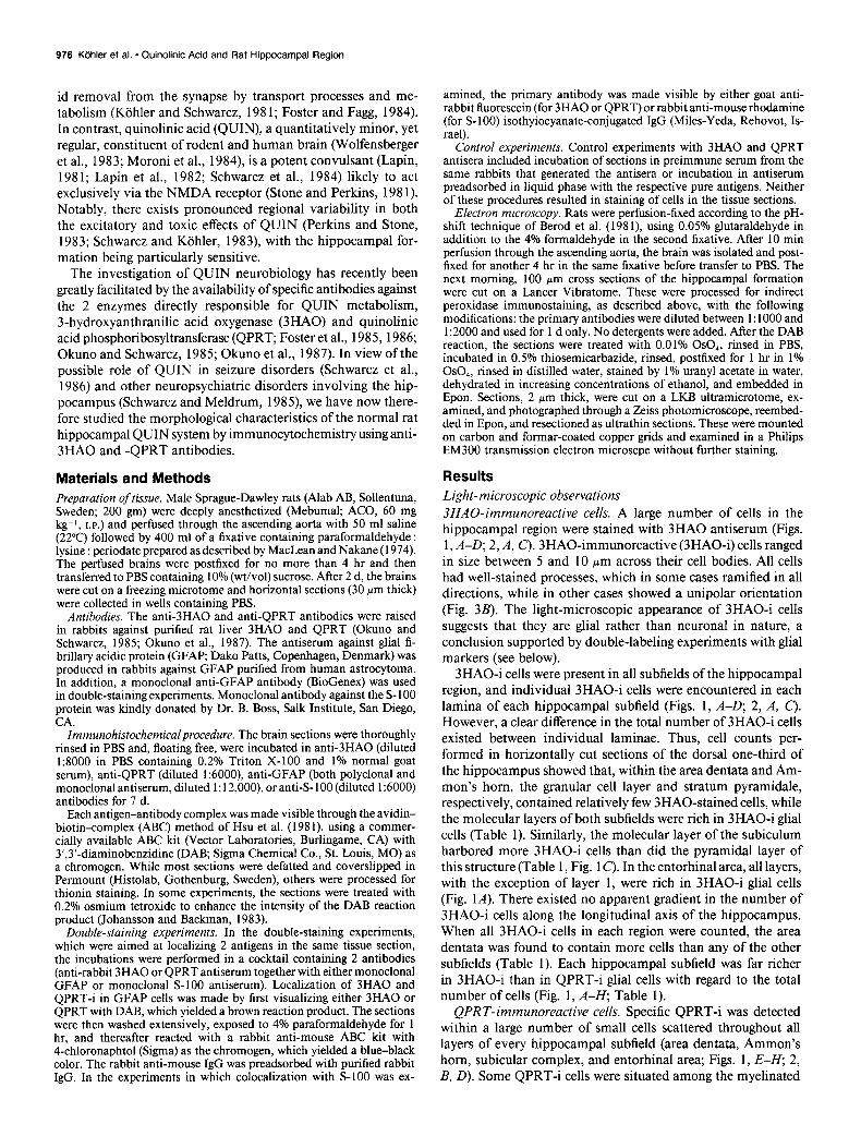
Figure 2. A-D, Low-power photo- micrographs of 3HAO- (A, C) and QPRT- (B, D) immunoreactive glial cells in the regio inferior of Ammon’s horn and area dentata (C, 0). Bar, 50 m.
fibers of the alveus, angular bundle, and fornix-fimbria. They (Fig. 3G). Both light- and electron-microscopic (see below) ob-
were small in size (cell body diameter, 5-l 2 pm) and possessed servations suggest that these QPRT structures are ghal cells.
round or oval cell bodies. Most of the QPRT-i cells had short, With one exception (the area dentata), there existed no clear
highly tortuous processes ramifying in all directions (Fig. 34, difference in the density of QPRT-i cells between the individual while some cells had few and relatively poorly developed ones hippocampal subfields (Table 1). When all cells were counted,
The Journal of Neuroscience, March 1989, 8(3) 979
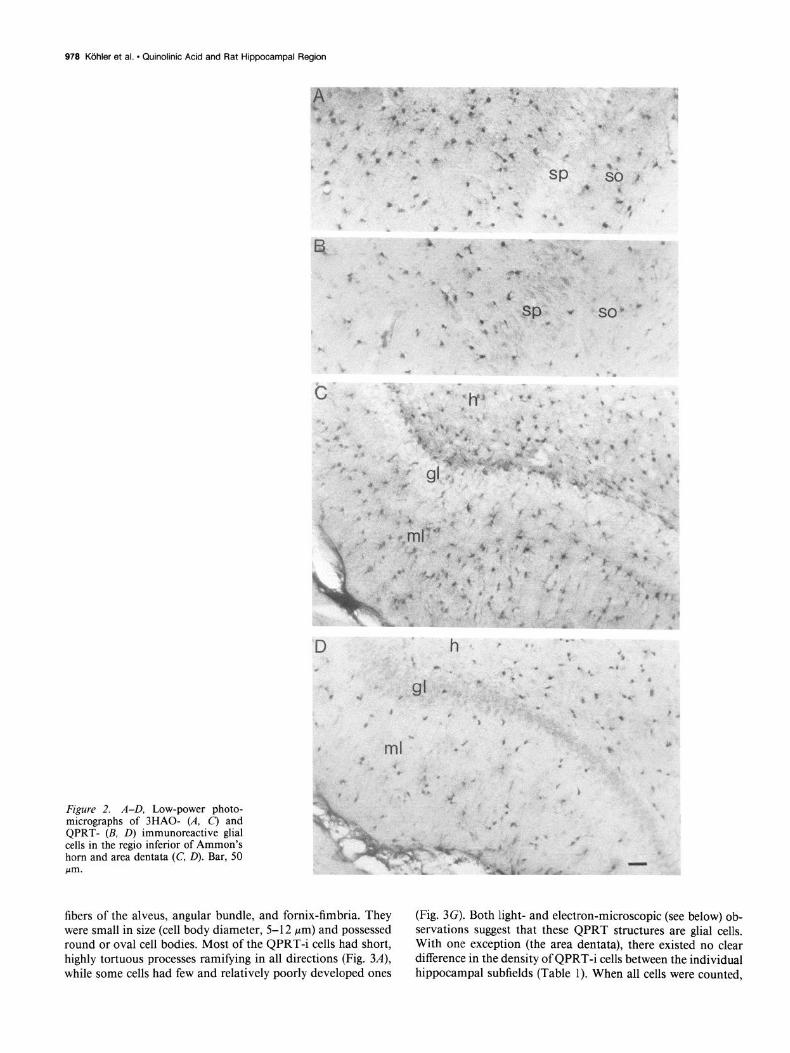
Figure 3. A-G, Photomicrographs showing QPRT-i cells of different morphological types in the molecular layer of area dentata (A, C, D), hilus (E) and granular cell layer (G). Photomicrograph in F shows QPRT-i punctate structures (small arrows) on a presumed astroglial cell in the molecular layer of area dentata. In C and E QPRT-i glial cells are shown in close association with neuronal somata (n), suggesting direct contact between QPRTi cells and neurons. Arrows in D mark large QPRT-i glial cells in the molecular layer. Bar, 10 pm.
980 Kijhler et al. * Quinolinic Acid and Rat Hippocampal Region
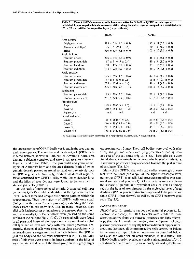
Table 1. Mean (+SEM) number of cells immunoreactive for 3HA0 or QPRT in each layer of individual hippocampal subfields, measured either along the entire layer or sampled in a restricted area (25 x 25 pm) within the respective layer (in parentheses)
3HA0 OPRT
Area dentata Molecular layer 395 t 55 (14.6 f 0.8) 182 k 11 (5.2 k 0.3) Granular cell layer 95 f 5 (9.4 k 0.5) 20 f 4 (1.2 ?c 0.2) Hilus 350 f 33 (13.8 f 0.9) 103 f 10 (9.0 f 0.5)
Regio inferior Stratum oriens 215 k 34 (13.8 k 0.9) 46 f 3 (5.8 f 0.4) Stratum pyramidale 67 k 9 (4.5 + 0.4) 40 f 3 (1.2 f 0.2) Stratum lucidum 158 k 17 (10.7 k 0.5) 35 ri 15 (6.2 f 0.6) Stratum radiatum 163 + 22 (14.7 + 0.6) 39 k 16 (3.6 XL 0.6)
Regio superior Stratum oriens 195 + 39 (11.5 + 0.6) 42 f 4 (4.7 i 0.4) Stratum pyramidale 47 f 4 (5.0 k 0.6) 19 f 5 (0.7 + 0.2) Stratum radiatum 235 2 12 (8.6 ZIZ 0.4) 99 f 11 (4.1 f 0.3) Stratum moleculare 295 k 30(19.3 +- 1.1) 101 f 15 (4.2 f 0.3)
Subiculum Stratum pyramidale 185 k 29 (12.6 i 0.6) 79 ‘I 14 (4.2 k 0.4) Stratum moleculare 231 + 32 (16.7 + 0.6) 63 k 3 (4.8 + 0.4)
Presubiculum Layer 1 89 f 30 (7.3 k 1.3) 19 f lO(3.4 * 0.3) Layer 2 540 f 65 (15.3 2 1.2) 36 f 5 (3.1 k 0.3) Layers 3-6 n.d. n.d. n.d. n.d.
Entorhinal area Layer 1 65 k 18 (5.4 f 0.4) 56 -c 6 (4.4 k 0.3) Layer 2 246 t 38 (15.3 + 1.0) 52 -t 9 (6.0 k 0.3) Layer 3 241 f 15 (16.4 + 0.6) 46 + 10 (5.9 k 0.3) Layers 4-6 168 f 14(16.0 k 1.0) 35 -+ 3 (5.4 f 0.3)
The values represent cell counts performed in 4 hippocampi of 2 rats. n.d., Not determined. -
the largest number of QPRT-i cells was found in the area dentata and regio superior. The number and the density of QPRT-i cells differed between individual laminae of Ammon’s horn, area dentata, subicular complex, and entorhinal area. As shown in Figures 1 and 2 and Table 1, the pyramidal and granular cell layers of Ammon’s horn and the area dentata (both of which contain densely packed neuronal somata) were relatively poor in QPRTi glial cells. Similarly, stratum lucidum of regio in- ferior contained few QPRT-i cells, while the molecular layer and the hilus of area dentata were found to be very rich in stained glial cells (Table 1).
On the basis of morphological criteria, 3 principal cell types containing QPRT-i could be identified at the light-microscopic level. Each of these had unique distribution patterns within the hippocampus. Thus, the majority of QPRT-i cells were small (5-7 pm), with one or 2 major process(es) extending short dis- tances from the cell body (Fig. 3G). In many instances, such glial cells had processes oriented towards a neuronal perikaryon, and occasionally QPRT-i “endfeet” were present on the soma surface of the neuron (Fig. 3, C, G). These glial cells were found in all parts and layers of the hippocampal region. Another glial cell type had an oval cell body with few, short processes. Fre- quently, these glial cells were situated in close association with neuronal somata, suggesting direct contacts between the QPRTi glial cell body and the neuronal perikaryon (Fig. 3, C, E). Glial cells of this type were present in large numbers in the hilus of area dentata. Glial cells of the third group were slightly larger
(approximately 12 Km). Their cell bodies were oval with rela- tively straight and widely ramifying processes extending from one end of the cell soma (Fig. 3, A, D). These glial cells were found almost exclusively in the molecular layer of area dentata. Their main processes always extended towards the pial surface of this layer (Fig. 30).
Many of the QPRT-i glial cells had endfeet seemingly in con- tact with neuronal perikarya. At the light-microscopic level, numerous QPRT-i glial cells had processes extending over neu- ronal somata, and punctate QPRT-i structures were visible on the surface of granule and pyramidal cells, as well as among cells in the hilus of area dentata. In the molecular layer of area dentata, QPRT-i punctate structures appeared to be present on some QPRT-i (not shown), as well as on QPRT-negative glial cells (Fig. 30.
Electron microscopy 3HAO-i cells. In semithin sections of material processed for electron microscopy, the 3HAO-i cells were similar to those described above from the material processed for light micros- copy (Fig. 4). Although the number, size, and pattern of cyto- plasmic processes varied slightly between different hippocampal areas and laminae, all immunoreactive cells seemed to belong to the same cell type. Their ultrastructure, as described below, was also the same for all areas included in this study. The 3HAO-i cells mostly revealed a weakly stained nucleus of 5-l 0 pm diameter, surrounded by an intensely stained cytoplasmic
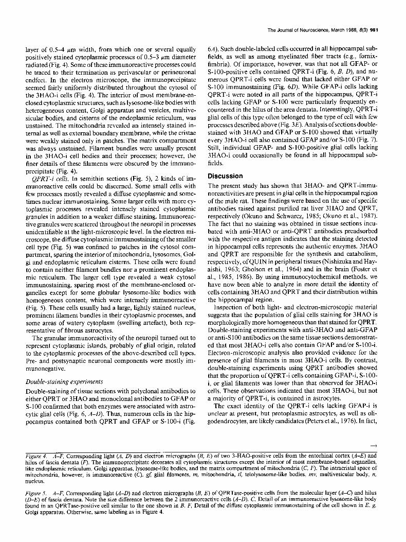
layer of OS-4 pm width, from which one or several equally positively stained cytoplasmic processes of OS-3 pm diameter radiated (Fig. 4). Some of these immunoreactive processes could be traced to their termination as perivascular or perineuronal endfeet. In the electron microscope, the immunoprecipitate seemed fairly uniformly distributed throughout the cytosol of the 3HAO-i cells (Fig. 4). The interior of most membrane-en- closed cytoplasmic structures, such as lysosome-like bodies with heterogeneous content, Golgi apparatus and vesicles, multive- sicular bodies, and cisterns of the endoplasmic reticulum, was unstained. The mitochondria revealed an intensely stained in- ternal as well as external boundary membrane, while the cristae were weakly stained only in patches. The matrix compartment was always unstained. Filament bundles were usually present in the 3HAO-i cell bodies and their processes; however, the finer details of these filaments were obscured by the immuno- precipitate (Fig. 4).
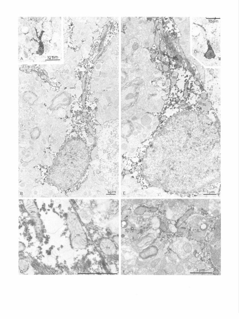
QPRT-i cells. In semithin sections (Fig. 5) 2 kinds of im- munoreactive cells could be discerned. Some small cells with few processes mostly revealed a diffuse cytoplasmic and some- times nuclear immunostaining. Some larger cells with more cy- toplasmic processes revealed intensely stained cytoplasmic granules in addition to a weaker diffuse staining. Immunoreac- tive granules were scattered throughout the neuropil in processes unidentifiable at the light-microscopic level. In the electron mi- croscope, the diffuse cytoplasmic immunostaining of the smaller cell type (Fig. 5) was confined to patches in the cytosol com- partment, sparing the interior of mitochondria, lysosomes, Gol- gi and endoplasmic reticulum cisterns. These cells were found to contain neither filament bundles nor a prominent endoplas- mic reticulum. The larger cell type revealed a weak cytosol immunostaining, sparing most of the membrane-enclosed or- ganelles except for some globular lysosome-like bodies with homogeneous content, which were intensely immunoreactive (Fig. 5). These cells usually had a large, lightly stained nucleus, prominent filament bundles in their cytoplasmic processes, and some areas of watery cytoplasm (swelling artefact), both rep- resentative of fibrous astrocytes.
The granular immunoreactivity of the neuropil turned out to represent cytoplasmic islands, probably of glial origin, related to the cytoplasmic processes of the above-described cell types. Pre- and postsynaptic neuronal components were mostly im- munonegative.
Double-staining experiments
Double-staining of tissue sections with polyclonal antibodies to either QPRT or 3HA0 and monoclonal antibodies to GFAP or S-100 confirmed that both enzymes were associated with astro- cytic glial cells (Fig. 6, A-D). Thus, numerous cells in the hip- pocampus contained both QPRT and GFAP or S-100-i (Fig.
The Journal of Neuroscience, March 1988, 8(3) 981
6A). Such double-labeled cells occurred in all hippocampal sub- fields, as well as among myelinated fiber tracts (e.g., fomix- fimbria). Of importance, however, was that not all GFAP- or S-lOO-positive cells contained QPRT-i (Fig. 6, B, D), and nu- merous QPRT-i cells were found that lacked either GFAP or S-100 immunostaining (Fig. 60). While GFAP-i cells lacking QPRT-i were noted in all parts of the hippocampus, QPRT-i cells lacking GFAP or S-100 were particularly frequently en- countered in the hilus of the area dentata. Interestingly, QPRT-i glial cells of this type often belonged to the type of cell with few processes described above (Fig. 3E). Analysis of sections double- stained with 3HA0 and GFAP or S-100 showed that virtually every 3HAO-i cell also contained GFAP and/or S- 100 (Fig. 7). Still, individual GFAP- and S-lOO-positive glial cells lacking 3HAO-i could occasionally be found in all hippocampal sub- fields.
Discussion
The present study has shown that 3HAO- and QPRT-immu- noreactivities are present in glial cells in the hippocampal region of the male rat. These findings were based on the use of specific antibodies raised against purified rat liver 3HA0 and QPRT, respectively (Okuno and Schwartz, 1985; Okuno et al., 1987). The fact that no staining was obtained in tissue sections incu- bated with anti-3HA0 or anti-QPRT antibodies preadsorbed with the respective antigen indicates that the staining detected in hippocampal cells represents the authentic enzymes. 3HA0 and QPRT are responsible for the synthesis and catabolism, respectively, of QUIN in peripheral tissues (Nishizuka and Hay- aishi, 1963; Gholson et al., 1964) and in the brain (Foster et al., 1985, 1986). By using immunocytochemical methods, we have now been able to analyze in more detail the identity of cells containing 3HA0 and QPRT and their distribution within the hippocampal region.
Inspection of both light- and electron-microscopic material suggests that the population of glial cells staining for 3HA0 is morphologically more homogeneous than that stained for QPRT. Double-staining experiments with anti-3HA0 and anti-GFAP or anti-S 100 antibodies on the same tissue sections demonstrat- ed that most 3HAO-i cells also contain GFAP and/or S-100-i. Electron-microscopic analysis also provided evidence for the presence of glial filaments in most 3HAO-i cells. By contrast, double-staining experiments using QPRT antibodies showed that the proportion of QPRT-i cells containing GFAP-i, S- 1 OO- i, or glial filaments was lower than that observed for 3HAO-i cells. These observations indicated that most 3HAO-i, but not a majority of QPRT-i, is contained in astrocytes.
The exact identity of the QPRT-i cells lacking GFAP-i is unclear at present, but protoplasmic astrocytes, as well as oli- godendrocytes, are likely candidates (Peters et al., 1976). In fact,
Figure 4. A-F, Corresponding light (A, D) and electron micrographs (B, E) of two 3-HAO-positive cells from the entorhinal cortex (A-E) and hilus of fascia dentata (F). The immunoprecipitate decorates all cytoplasmic structures except the interior of most membrane-bound organelles, like endoplasmic reticulum, Golgi apparatus, lysosome-like bodies, and the matrix compartment of mitochondria (C, I+). The intracristal space of mitochondria, however, is immunoreactive (C). gf; glial filaments, m, mitochondria, tl, telolysosome-like bodies, mv, multivesicular body, n, nucleus.
Figure 5. A-F, Corresponding light (A-D) and electron micrographs (B, E) of QPRTase-positive cells from the molecular layer (A-C) and hilus (D-E) of fascia dentata. Note the size difference between the 2 immunoreactive cells (A-D). C, Detail of an immunoreactive lysosome-like body found in an QPRTase-positive cell similar to the one shown in B. F, Detail of the diffuse cytoplasmic immunostaining of the cell shown in E. g, Golgi apparatus. Otherwise, same labeling as in Figure 4.
. . / .
E
C % ml
The Journal of Neuroscience, March 1988, 8(3) 985
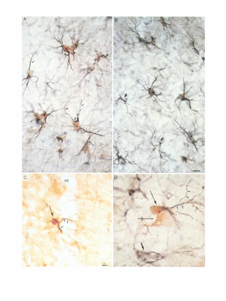
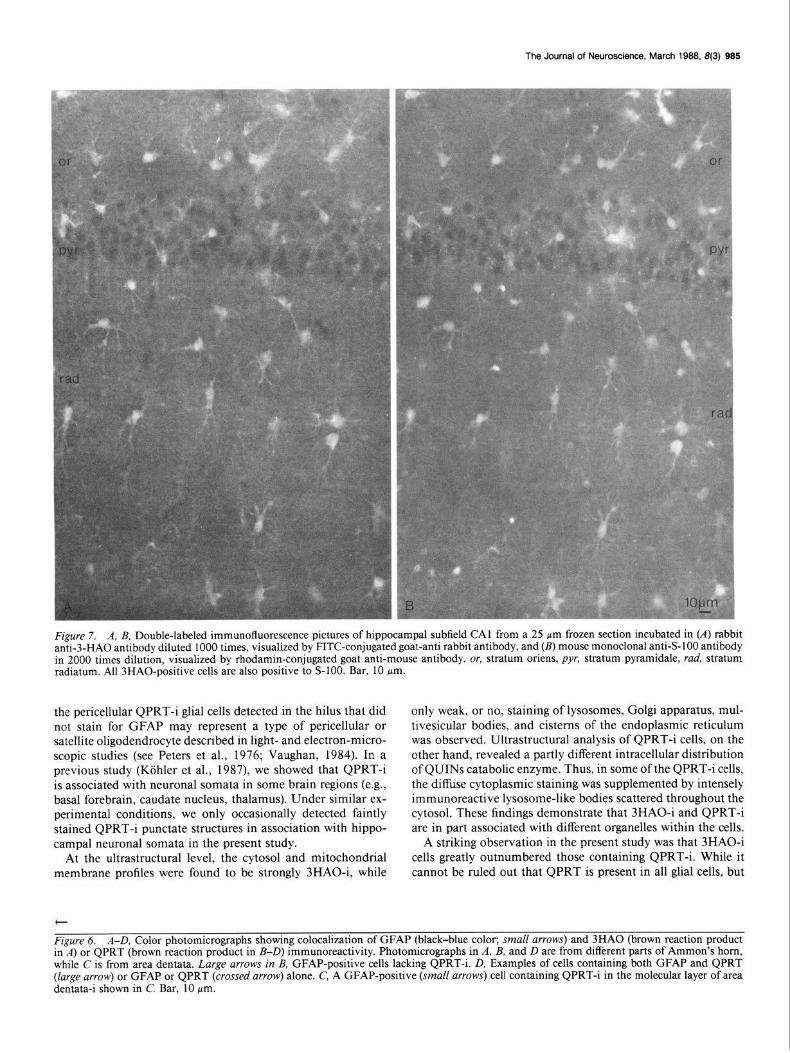
Figure 7. A, B, Double-labeled immunofluorescence pictures of hippocampal subfield CA1 from a 25 pm frozen section incubated in (A) rabbit anti-3-HA0 antibody diluted 1000 times, visualized by FITC-conjugated goat-anti rabbit antibody, and (B) mouse monoclonal anti-S- 100 antibody in 2000 times dilution, visualized by rhodamin-conjugated goat anti-mouse antibody. or, stratum oriens, pyr, stratum pyramidale, rud, stratum radiatum. All 3HAO-positive cells are also positive to S-100. Bar, 10 Km.
the pericellular QPRT-i glial cells detected in the hilus that did not stain for GFAP may represent a type of pericellular or satellite oligodendrocyte described in light- and electron-micro- scopic studies (see Peters et al., 1976; Vaughan, 1984). In a previous study (Kohler et al., 1987), we showed that QPRT-i is associated with neuronal somata in some brain regions (e.g., basal forebrain, caudate nucleus, thalamus). Under similar ex- perimental conditions, we only occasionally detected faintly stained QPRT-i punctate structures in association with hippo- campal neuronal somata in the present study.
At the ultrastructural level, the cytosol and mitochondrial membrane profiles were found to be strongly 3HAO-i, while
only weak, or no, staining of lysosomes, Golgi apparatus, mul- tivesicular bodies, and cisterns of the endoplasmic reticulum was observed. Ultrastructural analysis of QPRT-i cells, on the other hand, revealed a partly different intracellular distribution of QUINs catabolic enzyme. Thus, in some of the QPRTi cells, the diffuse cytoplasmic staining was supplemented by intensely immunoreactive lysosome-like bodies scattered throughout the cytosol. These findings demonstrate that 3HAO-i and QPRTi are in part associated with different organelles within the cells.
A striking observation in the present study was that 3HAO-i cells greatly outnumbered those containing QPRT-i. While it cannot be ruled out that QPRT is present in all glial cells, but
t
Figure 6. A-D, Color photomicrographs showing colocalization of GFAP (black-blue color; small arrows) and 3HA0 (brown reaction product in A) or QPRT (brown reaction product in B-D) immunoreactivity. Photomicrographs in A, B, and D are from different parts of Ammon’s horn, while C is from area dentata. Large arrows in B, GFAP-positive cells lacking QPRTi. D, Examples of cells containing both GFAP and QPRT (large arrow) or GFAP or QPRT (crossed arrow) alone. C, A GFAP-positive (small arrows) cell containing QPRT-i in the molecular layer of area dentata-i shown in C. Bar, 10 pm.
988 Kijhler et al. * Quinolinic Acid and Rat Hippocampal Region
in some instances escapes immunocytochemical detection be- cause of excessively low levels of enzyme protein, the presence of a relatively small number of QPRT-i cells is certainly in line with biochemical findings demonstrating low QPRT activity in the rat hippocampus (Foster et al., 1985). Moreover, the present immunohistochemical analysis of 3HAO-i cells is in agreement with the detection of high JHAO activity in rat hippocampal homogenates (Foster et al., 1986). Finally, the present obser- vations are in accordance with the results from recent studies performed in lesioned hippocampi, in which substantial in- creases in both 3HA0 and QPRT activities were found follow- ing ibotenate-induced neuronal degeneration (Speciale and Schwartz, 1987). Biochemical assessment had, therefore, pre- viously led us to assume that 3HA0 activity, in order to prevent the production of (toxic quantities of) QUIN, must be under the stringent control in vivo either of its substrate’s or other regulatory mechanisms’ bioavailability (Schwartz et al., 1987). The present finding that not all QPRT-containing cells harbor 3HA0 (and vice versa) may have further implications for hip- pocampal QUIN function. It thus seems possible that the bio- synthesis of QUIN at times takes place in cells different from those responsible for its catabolism. By inference, QUIN can be expected to be expelled into the extracellular space (where it can interact with NMDA receptors; Stone and Perkins, 198 1) and subsequently enter (actively or passively) QPRT-containing cells.
Future studies will also have to explore the precise nature of the mechanisms governing the extracellular concentration of QUIN in view of its toxic potential. QUIN is present in normal cerebrospinal fluid in low concentration (Schwartz et al., 1987) and increases may occur solely under pathological conditions. In this regard, it is noteworthy that QPRT-i cells exclusively are often seen in close association with neuronal somata, where they could serve a detoxifying role. Since an overabundance of QUIN has been hypothetically linked to the pathogenesis of a spectrum of neuropsychiatric disorders involving the hippocam- pal formation (see the introduction), it will be of importance to assess whether similar anatomical arrangements of 3HAO- and QPRT-containing cells also exist in the human hippocampus. Such studies are currently in progress.
References Berod, A., B. K. Hartman, and J. F. Pujol (198 1) Importance of fixation
in immunohistochemistry: Use of formaldehyde at variable pH for the localization of tyrosine hydroxylase. J. Histochem. Cytochem. 29: 844-850.
Croucher, M. J., J. F. Collins, and B. S. Meldrum (1982) Anticon- vulsant action ofexcitatory amino acid antagonists. Science 216: 899- 901.
Delgado-Escueta, A. V., A. A. Ward, Jr., D. M. Woodbury, and R. J. Porter (1986) Basic Mechanisms of the Epilepsies: Molecular and Cellular Approaches. Advances in Neurology, vol. 44, Raven, New York.
Falconer, M. A., D. Hill, A. Meyer, W. Mitchell, and D. A. Pond (1955) Treatment oftemporal lobe epilepsy by temporal lobectomy: A survey of findings and results. Lancet I: 827-835.
Foster, A. C., and C. E. Fagg (1984) Acidic amino acid binding sites in mammalian neuronal membranes: Their characteristics and rela- tionship to synaptic receptors. Brain Res. Rev. 7: 103-164.
Foster, A. C., B. S. Zinkand, and R. Schwartz (1985) Quinolinic acid phosphoribosyltransferase in rat brain. J. Neurochem. 44: 446-454.
Foster, A. C., R. J. White, and R. Schwartz (1986) Synthesis of quin- olinic acid by 3-hydroxyanthranilic acid oxygenase in rat brain tissue in vitro. J. Neurochem. 47: 23-30.
Gholson, R. K., I. Ueda, N. Ogasawara, and L. M. Henderson (1964)
The enzymic conversion of quinolinate to nicotinic acid mononu- cleotide in mammalian’ liver. J. Biol. Chem. 239: 1208-l 2 14.
Hsu. S. M.. L. Raine. and H. Faneer (1981) Use of avidin-biotin- peroxidase complex’(ABC) in immunoperoxidase techniques: A com- parison between ABC and unlabelled antibody (PAP) procedures. J. Histochem. Cytochem. 29: 577-580.
Johansson. 0.. and J. Backman (1983) Enhancement of immunouer- oxidase staining using osmium tetroxide. J. Neurosci. Methods 7: 185-193.
Kiihler, C., and R. Schwartz (198 1) Monosodium glutamate: Increased neurotoxicity after removal of neuronal reuptake sites. Brain Res. 221: 485-491.
Kohler, C., E. Okuno, P. R. Flood, and R. Schwartz (1987) Quinolinic acid phosphoribosyltransferase: Preferential localization in the rat brain visualized by immunocytochemistry. Proc. Natl. Acad. Sci. USA 54: 349 l-3495.
Lapin, I. P. (198 1) Kynurenines and seizures. Epilepsia 22: 257-265. Lapin, I. P., I. B. Prakhie, and I. P. Kiseleva (1982) Excitatory effects
of kynurenine and its metabolites, amino acids and convulsants ad- ministered into brain ventricles: Differences between rats and mice. J. Neural Transm. 54: 229-238.
MacLean, I., and P. Nakane (1974) Periodate-lysine paraformalde- hyde fixative: A new fixative for immunoelectronmicroscopy. J. His- tochem. Cytochem. 22: 1077-1083.
Moroni, F., G. Lombardi, V. Carola, and G. Moneti (1984) The ex- citotoxin quinolinic acid is present and unevenly distributed in the rat brain. Brain Res. 295: 352-355.
Nishizuka, Y., and 0. Hayaishi (1963) Studies on the biosynthesis of nicotinamide adenine dinucleotide. I. Enzymatic synthesis of niacin ribonucleotides from 3-hydroxy-anthranilate oxygenase from beef kidney. J. Biol. Chem. 238: 3369-3377.
Okuno, E., and R. Schwartz (1985) Purification of quinolinic acid phosphoribosyltransferase from rat liver and brain. Biochim. Bio- phys. Acta 841: 112-l 19.
Okuno, E., C. Kohler, and R. Schwartz (1987) Rat 3-hydroxyanthra- nilic acid oxygenase: Purification from the liver and immunocyto- chemical localization in the brain. J. Neurochem. 49: 77 l-780.
Perkins, M. N., and T. W. Stone (1983) Pharmacology and regional variations of quinolinic acid-evoked excitation in the rat central ner- vous svstem. J. Pharmacol. Exn. Ther. 226: 55 l-557.
Peters, A., S. L. Palay, and H. Webster (1976) The Fine Structure of the Nervous System. The Neurons and Supporting Cells, Saunders, Philadelphia, London.
Schwartz, R., and Y. Ben-Ari (1986) Excitatory Amino Acids and Epilepsy. Plenum, New York.
Schwartz. R.. and C. Kijhler (1983) Different vulnerabilitv of central neurons ofthe rat to quinolinic acid. Neurosci. Lett. 38: 85-90.
Schwartz, R., and B. Meldrum (1985) Excitatory amino acid antag- onists provide a therapeutic approach to neurological disorders. Lan- cet 2: 140-143.
Schwartz, R., J. F. Collins, and D. A. Parks (1982) oc-Amino-w-phos- phonocarboxylates block ibotenate but not kainate neurotoxicity in rat hippocampus. Neurosci. Lett. 33: 85-90.
Schwartz, R., G. S. Brush, A. C. Foster, and E. D. French (1984) Seizure activity and lesions following intrahippocampal injection of quinolinic acid. Exp. Neurol. 84: 1-17.
Schwartz, R., C. Speciale, E. Okuno, E. D. French, and C. Kiihler (1986) Quinolinic acid: A pathogen in seizure disorders. In Excitatory Amino Acids and Epilepsy, R. Schwartz and Y. Ben-Ari, eds., pp. 697-707, Plenum, New York.
Schwartz, R., E. Okuno, C. Speciale, C. Kohler, and W. D. Whetsell, Jr. (1987) Neuronal degeneration in animals and man: The quin- olinic acid connection. In Neurotoxins and their Pharmacological Implications, P. G. Jenner, N. G. Bowery, J. E. Cremer, J. 0. Dolly, and M. Sandler, eds., pp. 19-33, Raven, New York.
Sommer, W. (1880) Erkrankungen des Ammonshoms als aetiologisch- es Moment der EDikDSie. Arch. Psvchiat. Nerve&r. 10: 63 l-675.
Speciale, C., and R. Schwartz (1987) *Effect of systemic kainate admin- istration on cerebral quinolinic acid metabolism in the rat. Exp. Neu- rol. (in press).
Stone, T. W., and M. N. Perkins (1981) Quinolinic acid: A potent endogeneous excitant at amino acid receptors in CNS. Eur. J. Phar- macol. 72: 411412.
Vaughan, D. W. (1984) The structure of neuroglial cells. In Cerebral Cortex, vol. 2: Functional Properties of Cortical Cells, E. G. Jones and
The Journal of Neuroscience, March 1988, 8(3) 987
A. Peters, eds., pp. 285-325, Plenum Press, New York. Wong, E. H. F., J. A. Kemp, T. Priestley, A. R. Knight, G. N. Woodruff, Wolfensberger, M., V. Amsler, M. Cuenod, A. C. Foster, W. U. Whetsell, and L. L. Iversen (1986) The anticonvulsant MK-801 is a potent
Jr., and R. Schwartz (1983) Identification of quinolinic acid in rat N-methyl-n-aspartate antagonist. Proc. NatLAcad. Sci. USA 8.3: 7 104- and human brain tissue. Neurosci. Lett. 41: 247-252. 7108.