quantitative measurement of cerebral protein synthesis in vivo: theory and methodological...
TRANSCRIPT
ELSEVIER
JOURNAL OF NEUROSCIENCE
METNODS Journal of Neuroscience Methods 76 (1997) 35-44
Quantitative measurement of cerebral protein synthesis in vivo: theory and methodological considerations
Giinter Mies *, Bogdan Djuricic, Wulf Paschen, Konstantin-Alexander Hossmann
Max-Planck-Institute for Neurological Research, Department of Experimental Neurology, Gleueler StraJBe 50, D-50931 Kiiln (Lindenthal), Germany
Received 16 August 1996; received in revised form 11 March 1997; accepted 7 April 1997
Abstract
The true rate of cerebral protein synthesis can be calculated from the ratio of labeled proteins to integrated arterial plasma amino acid specific activity (SA) only when the fraction of amino acid precursor pool dilution is known. In the following, current experimental designs on the measurement of cerebral protein synthesis are discussed and compared to our own approach in which the determination of regional precursor pool dilution by recycled unlabeled leucine is combined with the quantitation of regional cerebral protein synthesis rates. For this purpose, a constant arterial plasma leucine SA level is maintained for 45 min by programmed intravenous infusion which is sufficient for complete equilibrium between tissue leucine pool SAs and plasma free leucine SA. In addition to the regional assessment of the precursor dilution factor, protein radioactivity can be determined in the same tissue sample or in parallel brain sections of the same animal by quantitative autoradiography. It is then possible to calculate the actual rate of protein synthesis using the correct fraction of precursor pool dilution. This renders our approach particularly suitable for the quantitative measurement of regional CPS under pathological conditions. 0 1997 Elsevier Science B.V.
Keywords: Cortical protein synthesis; Rat; Tritium-labeled leucine; Tissue free leucine specific activity; Tissue leucyl-tRNA specific activity
1. Introduction
Polypeptides are involved in an enormous variety of cell functions, e.g. enzymatic catalysis, signal transmis- sion, cell differentiation and growth (Doolittle, 1985) which suggests that proteins uniquely determine the structure and function of a cell within a biological system. Since a steady state level of all proteins is essential for the developmental and functional state of cells, in particular of those of the central nervous system, overall cerebral protein synthesis (CPS), i.e. the continuous translation of the genomic code via mR- NAs, plays a pivotal role in cell survival. The impor- tance of disturbed cerebral protein synthesis for post-injury cell damage is supported by findings on the
* Corresponding author. Tel.: +49 221 4726216; fax: +49 221 4726298; e-mail: [email protected]
01650270/97/$17.00 0 1997 Elsevier Science B.V. All rights reserved. PZISO165-0270(97)00077-O
failure of CPS recovery after pathophysiologic events such as following an ischemic (Cooper et al., 1977), hypoglycemic (Kiessling et al., 1984a) or epileptic (Kiessling et al., 1984b) insult of the brain. In these earlier experimental studies, however, findings and con- clusions on the post-injury role of CPS inhibition were examined qualitatively neglecting the implicit necessity of quantitative information on brain protein synthesis.
According to the in vitro principle for the quantita- tive measurement of a biochemical process using a radioactive compound in tracer amounts, the rate of substrate turnover can be calculated by the ratio of the amount of labeled product accumulated over time to the integrated specific activity (SA) of the precursor over the same time (Sokoloff and Smith, 1983). Under in vivo condition, however, the complete time course of precursor pool SA in brain tissue cannot be determined directly. Instead, the arterial plasma SA is measured

and used for the estimation of the regional integrated precursor SA. The assessment of rates of amino acid incorporation into brain proteins is further complicated by the fact that the actual precursor of cerebral protein synthesis is the intracellular aminoacyl-tRNA and not the free amino acid as transported from arterial plasma to brain tissue across the blood brain barrier. The appropriate estimation of integrated tissue precursor aminoacyl-tRNA SA, therefore, requires that the inte- grated arterial plasma amino acid SA is corrected not only for a delay in equilibrium with the tissue precursor SA after systemic administration of the labeled amino ;icid but also for possible differences in the distribution volume between the endogenous and labeled tissue precursor.
In the following, therefore, previous approaches on the quantitative determination of CPS in vivo will be itnalyzed and compared with our own results on the quantitation of CPS comprising correction of precursor pool dilution for the appropriate calculation of regional rates of CPS in the same experiment.
2. Theoretical considerations
The rate of amino acid incorporation R in vitro using a labeled amino acid can be calculated by the ratio,
R =: P*(T) ~ ‘Tp’p(t) 3
(1)
I ,,j c,,,\ df
with the amount of the labeled protein P*(T) formed and accumulated between zero and time T to the integral of the tissue aminoacyl-tRNA precursor SA, i.e. (C,T(r)/C,,). over the same time. @ is the isotope correction factor which is equal to 1 under most condi- tions.
When arterial plasma leucine SA is kept constant by programmed intravenous infusion of a labeled amino acid, the rate of protein synthesis can be calculated from the following equation (Smith et al., 1988; Smith, 1991: Sun et al., 1992)
R= P*(T) -
where C$(t)/Cp is the specific activity of leucine in arterial plasma at any time t. i is a variable which
(2)
represents the fraction of leucine in the precursor pool for protein synthesis derived from arterial plasma.
The denominator in Eqs. (1) and (2) represents the integrated tissue leucyl-tRNA pool SA over the experi- mental duration of tracer circulation time T which can be solved for L with CD equal to I as follows
44
(3)
J C;(t j/c’, dt 0
where i equals the ratio of integrated leucine S.4 in the tRNA-bound precursor (C,*)/C,,,,) lo arterial plasma (C,*;‘CJ. When steady state between plasma leucine and 1eucyLtRNA SA is reached, above equation can be solved for the tRNA-bound precursor at any time T
C&( T) i C,,,, = iC:C T 1; C,, (4)
where 7, represents the linear slope of the regression analysis between of leucyl-tRNA SA and plasma leucine SA at various times T after onset of cquilibra- tion.
If one assumes rapid equilibrium between the tissue free leucine and leucyl-tRNA SA, the rate of amino acid incorporation into cortex proteins K’, in analogy to Eq. (1). can also be calculated by the following ratio with an isotope correction fdctor (1, equal to 1
D*l 7’\
(5)
where CE):C’, is the tissue free leucine SA at any time (t). In analogy to Eq. (2), the protein synthesis rate R’ is then calculated from the ratio of labeled proteins to the integrated arterial plasma leucine SA multiplied by a correction factor \y
R’ = P*(T)
!
’ P(r) * (6)
Y --r_dt (1 c,,
where ‘I’ is the fraction of leucine in the tissue free leucine pool for protein synthesis derived from arterial plasma. Following equilibrium between tissue free leucine SA and plasma leucine SA ,it time T, one obtains in allalogy to Eq. (4)
cg T):C’, = !‘q( Tl c;, (7)
where Y represents the slope from linear regression analysis between both parameters.
Eq. (1) can be re-arranged to estimate the rate of amino acid incorporation into brain proteins by linear regression analysis of the relationship between labeled proteins accumulated from time 0 to 7’ and the inte- grated tissue leucyl-tRNA SA over the same time, i.e.
P*(T)=R ‘W’
JO
C .-dt. I’P
(8)
where the protein synthesis rate is represented by the regression coefficient R.
When Eq. (2) is re-arranged in the same manner. i.e.
(9)

G. A&s et al. /Journal of Neuroscience MethodF 76 (1997) 35-44 37
it follows that linear regression analysis of integrated arterial plasma amino acid SA versus labeled proteins yields a slope which is equivalent to the protein synthe- sis rate R times the tissue precursor dilution fraction R. The ratio of the slopes from Eq. (9) to Eq. (8) then provides a further estimate of the l-value, i.e. the fraction of leucine in the tissue tRNA precursor pool derived from arterial plasma.
An estimate for the equilibration time between tissue leucine pool SAs and arterial plasma leucine SA is obtained by the half-time of the tissue precursor pool, i.e. t,,z = In 2/k. For this purpose, the turnover time of the compartment (l/k) is calculated from the following ratio
1 precursor compartment size -= k rate of reaction ’ (10)
The leucyl-tRNA turnover time (l/k,,) is derived from the ratio of the charged leucyl-tRNA compart- ment size to the rate of reaction which is equivalent to leucine incorporation into brain proteins R’ from the exogenous labeled leucine source. In the case of tissue leucine turnover time l/k,, the compartment size is equivalent to the tissue pool of labeled leucine in ex- change with plasma.
3. Previous methodological approaches on the quantitation of cerebral protein synthesis
Earlier in vitro findings suggested evidence on the predominant extracellular source of amino acids for cellular protein incorporation (Gainer et al., 1975; Robertson and Wheatley, 1979). Subsequently, rates of cerebral protein synthesis in vivo were calculated by the product-precursor relationship (Garlick and Marshall, 1972; Dunlop et al., 1975) or in analogy to the kinetic model of the deoxyglucose method for the measure- ment of glucose utilization (Sokoloff et al., 1977) with the assumption that admixture of the precursor pool is ,negligible. One has to realize that the protein synthesis data then represent the minimal rather than the actual rate of amino acid incorporation into proteins. Irre- spective of the mode of tracer application, e.g. by flooding technique, by intravenous (i.v.) bolus adminis- tration, or by i.v. infusion, and the choice of the labeled amino acid, e.g. valine, leucine or methionine, protein synthesis rates in whole brain of rodents (expressed as ‘equivalent’ leucine incorporation into cerebral proteins) ranged from 3.2 to 8.0 nmol/g per min (mean + S.D. 6.0 + 2.0 nmol/g per min) whereas corti- cal rates of protein synthesis amounted only to 2.4-5.5 nmol/g per min (mean f S.D. 4.0 f 1.1 nmol/g per min), (Table 1).
Although experimental indications existed on possi- ble admixture of the tissue free amino acid pool by
protein degradation (Waterlow and Stephen, 1968; Garlick and Marshall, 1972), it was recognized only until recently that endogenous amino acids from prote- olysis significantly dilute the tissue free leucine and the tRNA precursor pool for protein biosynthesis. Smith and coworkers (1988) were the first to establish a kinetic model of cerebral protein synthesis which ac- counted for the recycling of leucine from proteolysis in vivo. The fraction of leucine pool dilution was mea- sured by the ratio of tissue free leucine specific activity (SA) to arterial plasma leucine SA, i.e. the Y-value, following steady state of plasma amino acid SA via programmed intravenous infusion of labeled leucine for 30-90 min. As indicated by the findings summarized in Table 1, the ratio of tissue free leucine SA to arterial plasma leucine SA for whole brain ranged from 0.24 to 0.59 (mean f S.D. 0.43 + 0.13). With these Y-values determined for whole brain, rates of protein synthesis for whole brain varied from 0.62-13.3 nmol/g per min (mean + S.D. 7.0 f 6.3 nmol/g per min equivalent leucine incorporation) and for cortex from 9.9-10 nmol/g per min (Smith et al., 1988; Keen et al., 1989; Hargreaves-Wall et al., 1990; Sun et al., 1992). In comparison, specific activity of tissue free leucine mea- sured in small tissue sample such as cortex revealed smaller Y-values with 0.36 and 0.46 and discretely larger cortical protein synthesis rates of 13.9 and 10.8 nmol/g per min, respectively (Widmann et al., 1992b; Mies, 1993). Tissue amino acid SA dilution in cortex was also demonstrated following administration of [i4C-methyllmethionine via programmed i.v. infusion. The resultant average Y-value was 0.5 and the protein synthesis rate for cortex using labeled methionine amounted to 10 nmol/g per min (equivalent leucine incorporation) (Lajtha and Toth, 1974; Planas et al., 1992) (Table 1). It follows that the correction for tissue amino acid SA dilution provides larger average rates of cortical protein synthesis with 11.2 + 1.9 nmol/g per min (mean + S.D., equivalent leucine incorporation), i.e. a more appropriate estimate of activity of the protein synthesis machinery, compared to protein syn- thesis rates with 4.0 + 1 .l nmol/g per min (equivalent leucine incorporation) for the calculation of which the tissue amino acid pool dilution was ignored.
One has to aware of the fact that the actual tissue precursor pool SA for protein synthesis is the tissue aminocyl-tRNA pool and not that of tissue free amino acids. Since the concentration of tissue aminoacyl- tRNA is z lo3 lower than that of tissue free amino acids, the measurement of tissue aminoacyl-tRNA SA so far was performed only in whole brains. Similar as for tissue free amino acids, dilution of the tissue precur- sor pool SA for protein synthesis could be established as a general phenomenon and did not depend on the choice of the amino acid. As shown in Table 1, the ratio of tissue leucyl-tRNA SA to arterial plasma

Iahl
e 1
Prev
ious
stu
dies
on
the
quan
titat
ion
of p
rote
in
synt
hesi
s ra
tes
in r
at b
rain
Pt1
hhca
t101
1 La
bele
d am
mo
a&
AS a
pplic
atio
n An
imal
An
esth
esia
(Dun
lop
et a
l..
1975
) (D
wye
r et
al..
198
2)
(Dw
yer
et a
l.. 1
982)
(In
gvar
et
al.,
19
85)
(Les
tage
et
al.,
1987
) (K
irika
e et
al.,
19
88)
(Kee
n et
al.,
198
9)
(Gra
nge
et a
l., 1
991)
(M
ies
et a
l.,
1993
) (G
iaum
e et
al.,
199
4)
(Gia
ume
et a
l.,
1995
) (S
mith
et
al.,
198
8)
(Kee
n et
al.,
198
9)
(Har
grea
ves-
Wal
l et
al.
1990
) (S
un e
t al
.. 19
92)
(Pla
nas
et a
l..
1992
)
(Wid
man
n et
al..
19
921
(Mie
s.
1993
) (S
mith
et
al..
19
88)
(Kee
n et
al..
198
9)
(Har
grea
ves-
Wal
l et
al.
1990
) (S
mith
. I Y
Y 1)
(Sun
et
al..
1992
) (G
rang
e et
al.,
199
2)
(Gra
nge
et a
l.. 1
994)
(S
un
et a
l..
lY95
l
(AS)
[“Cjv
alin
e (‘H
jval
ine
[‘Hlv
alin
e [‘“
Clle
ucin
e [“‘
Slm
ethi
onin
e [T
lval
ine
[‘Tlle
ucin
e [‘%
]met
hion
ine
[‘Hlle
ucin
e [‘s
S]m
ethi
onin
e [%
]met
hion
ine
[‘Hlle
ucin
e [‘T
lleuc
ine
[‘Hlle
ucin
e
[‘Hlle
ucin
e [‘T
lleuc
ine
[‘%-m
ethy
l] m
ethi
onin
e [‘Q
leuc
ine
[“C]le
ucin
e [‘H
lleuc
ine
[“C]le
ucin
e [‘H
]!euc
ine
[“C]le
ucin
e [‘H
lleuc
ine
[“S]m
ethi
onin
e [“S
lmet
hion
inc
j’H]le
ucin
e ! ‘“
Clle
ucin
c
Floo
ding
Fl
oodi
ng
Floo
ding
i.v
. bo
lus
i.v.
bolu
s i.v
. bo
lus
i.v.
bolu
s iv
. in
fusi
on
i.v.
bolu
s i.v
. bo
lus
i.v.
bolu
s Pr
ogr.
infu
sion
i.v
. bo
lus
Con
stan
t i.v
. in
fu-
sion
Pr
ogr.
infu
sion
i.v
. bo
lus
Prog
r. i.v
.infu
sion
Prog
r. in
fusi
on
Prog
r. in
fusi
on
Prog
r. in
fusi
on
i.v.
bolu
s C
onst
ant
i.v.
infu
- si
on
iv.
bolu
s Pr
ogr.
infu
sion
1.
v. in
fusi
on
i.v.
bolu
s Pr
ogr.
infu
sion
1.
1. b
olus
~.- Kat
Yo
ung
rat
Youn
g ra
t R
at
Rat
R
at
Rat
R
at
Rat
R
at
Rat
R
at
Rat
R
at
Awak
e I
Awak
e I
Awak
e I
Awak
e 1
Awak
e 1
Awak
e I
Awak
e 1
Awak
e 1
Hal
otha
ne
1 Aw
ake
1 Aw
ake
1 Aw
ake
Y=O
.59
Awak
e ‘t’
= 0
.24
Keta
min
e Y
= 0
.36
Rat
Aw
ake
Rat
Aw
ake
Rat
Aw
ake
Rat
H
alot
hane
‘f’
:- 0
.36
Rat
tfa
loth
ane
‘4’ =
0.4
6 R
at
Awak
e i
= 0.
58
Rat
Aw
ake
i =
0.48
R
at
Keta
min
e ‘P
= 0
.35
Rat
Aw
ake
Rat
Aw
ake
Rat
Aw
ake
Rat
Aw
ake
Yolm
g Ia
l Aw
ake
Youn
g ra
t Aw
ake
Y =
0.4
9
Y =
0.5
0
i =
0.58
i
.=. 0.
58
; =
n.14
;
~ n.x
x
I -,,
!I.4
3 ~~
----
-.
..~
Dilu
tion
faC
t0t
Amin
o ac
id
inco
rpol
atia
n (n
mol
ig
per
min
) _.
~~ ~
~~~
Who
le
brai
n C
orte
x
Equi
vale
nt
leuc
ine
inco
r po
ratio
n (n
mol
;g
per
min
)
Who
le
brai
n C
orte
x
4.7
5.5
5.6
3.5
f 0.
2 4.
7 *
0.2
0.8
+ 0.
0 1.
7+0.
1 3.
2 F
0.6
I.7
5.5
& 0.
8 1.
2 k
0.0
1 .o
* 0.
0
6.7 7.8
8.0
3.5
3.2
6.3
13.3
13
.3
0.62
+ 0
.06
(0.6
2)
7.2
9.9
7.7
+ 0.
9 7.
2
I ?.Y
+ 2.
2 IO
.8 It
_ 0.6
6.1
6.7
0.62
_+ 0
.06
(0.6
2)
Y. I
j: 0.
3 6.
1 iO
.1
8.1
LO.1
?X
-.
I 3
3-o.
If;
6.1
10.4
s,
3
4.7
3.0
2.4
5.5
4.4
3.7
9.9
10.0
13.9
10
.8
G. Mies et al. /Journal of Neuroscience Methods 76 (1997) 35-44 39
leucine SA, i.e. the A-value, ranged from 0.43 to 0.58 for whole brain. The resultant whole brain protein synthesis rates varied from 6.1 to 10.2 nmol/g per min (mean + SD. 7.7 + 2.2 nmol/g per min) and those for cortex. from 8.1-11.7 nmol/g per min (mean + SD. 9.6 f 1.9 nmol/g per min) (Smith et al., 1988; Keen et al., 1989; Smith, 1991; Sun et al., 1992; 1995). Tissue precursor pool SA dilution is also detectable after administration of [35S]methionine as constant or pro- grammed i.v. infusion providing A-values with 0.74 and 0.88 (Grange et al., 1992; 1994) and leucine equivalent incorporation rates into brain proteins of 10.4 and 5.3 nmol/g per min. Although ‘flooding’ of the brain amino acid compartment has been proposed to minimize dilu- tion of tissue precursor pools for protein synthesis (Dunlop et al., 1975), the l-value only reached 0.69 + 0.02 after the ‘flooding’ of the brain valine pool com- pared with a A-value of 0.4 _+ 0.01 following programmed i.v. infusion of [3H]valine (Smith et al., 1991). Taking together, inclusion of the appropriate dilution of the tissue aminoacyl-tRNA pool for the calculation of CPS, whole brain protein synthesis amounted to 7.7 + 2.4 nmol/g per min (equivalent leucine incorporation) and cortical protein synthesis to 9.6 f. 1.9 nmol/g per min (equivalent leucine incorpora- tion).
The experimental and methodological design of sev- eral studies (Ingvar et al., 1985; Lestage et al., 1987; Kirikae et al., 1988; Keen et al., 1989; Hargreaves-Wall et al., 1990; Grange et al., 1992; 1994) however, ap- pears to be less suited for the determination of the ratios of tissue free amino acid/precursor pool SA to arterial plasma free amino acid SA, i.e. the Y- or the I-value, respectively, and subsequently for the estima- tion of cerebral protein synthesis rates. In the study by Keen et al. (1989) 14C-labeled leucine was applied as intravenous bolus following which the brain precursor pool SA lags behind that of plasma SA (Sokoloff and Smith, 1983). Since the total leucyl tRNA pool of rat brain is within the nannomolar range and the specific activity of 14C-labeled leucine is rather low, inaccuracies may arise in the determination of whole brain leucyl tRNA SA and thus in the quantitation of cerebral protein synthesis rates. The rather low rate of leucine incorporation into cerebral proteins in rats of 3.2 nmol/ g per min probably also results from neglecting precur- sor pool dilution. As specified by the investigators (Keen et al., 1989), their rate applies to incorporation of leucine derived from an ‘exogenous source’, pre- sumably from plasma. In this instance, only minimal possible rates of protein synthesis are derived because the tissue precursor SA remains uncorrected for recy- cled leucine derived from proteolysis. Hargreaves-Wall et al. (1990) used an in situ perfusion technique of one hemisphere of rodent brain which permits the infusion of a constant level of perfusate leucine SA for a rapid
equilibrium between distribution volumes of the en- dogenous and the labeled precursor. However, such brain preparations lack metabolic stability as indicated by elevated levels of tissue lactate at increasing time after the beginning of 3H-labeled leucine perfusion (Hargreaves-Wall et al., 1990). The low rate of amino acids incorporation into brain proteins of 0.6 nmol/g per min probably reflects inhibition of protein synthesis due to the impairment of energy metabolism (Dwyer and Wasterlain, 1980). Inaccuracies in the calculation of protein synthesis rates may also arise when metabo- lites of the amino acid other than labeled proteins and the free amino acid are estimated from integrated plasma amino acid SA via a set of rate constants and subtracted from total tissue radioactivity (Ingvar et al., 1985; Lestage et al., 1987; Kirikae et al., 1988; Grange et al., 1992; 1994). A more straight forward approach in this context is the direct use of regional protein radioac- tivity by precipitation of labeled proteins in a brain section and the removal of the label other than from proteins after incubation in an aqueous solution of trichloroacetic acid (Yoshimine et al., 1987; Xie et al., 1989; Smith, 1991; Sun et al., 1992; Widmann et al., 1992a; Mies, 1993).
Other disadvantages of previous experimental designs are the determination of tissue leucyl-tRNA dilution in whole brains of sets of animals different from those used for the calculation of regional protein synthesis (Smith et al., 1988; Smith, 1991; Grange et al., 1992; Sun et al., 1992; Grange et al., 1994; Sun et al., 1995). This implies that no physiologic differences exist be- tween such experimental groups and between regional R-values. To account for the possibility of regional heterogeneity of the /Z-value, Sun et al. (1992) corre- lated the whole brain Y-value with the respective whole brain l-value from various measurements obtained fol- lowing programmed i.v. infusion of labeled leucine for 60 and 90 min and were able to establish a linear relationship between both parameters. Since the ratio of tissue free leucine SA to arterial plasma leucine SA can be determined in tissue of very small brain regions, the linear regression equation is then used to estimated the actual regional I-value. It still remains to be estab- lished, however, whether the relationship between the Y-and A-value measured in a whole brain preparation is the same for individual brain regions.
4. Simultaneous measurement of precursor dilution and regional cerebral protein synthesis in vivo
In our own study, shortcomings in previous experi- mental designs on the quantitation of cerebral protein synthesis rates in vivo were minimized. Tissue amino acid and precursor pool SAs were measured in a defined grey matter structure, i.e. parietal cortex, to

G. Mies rt al. ,!Journal of Neuroscience Method,\ 76 (1937) .lZ 44
5rnin 10 min 20 min
lo-
a-
30 min
-e--#l
--o--#2 . ..Q... # 3
45 min
107
6-
6-
4-
2.
,
0 I 1
10 20 30 40 50
Duration of tracer infusion (min)
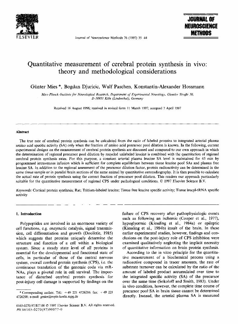
Fig. 1. Time course of arterial plasma leucine specific activity (SA) following programmed intravenous infusion of [3H]leucirle for 5. 10. 20. 30. and 45 min. Different symbols indicate the individual experiment in each group, Note that 5 min ;ifter the beginning of the infusinn. plasma leucine SA remained constant within k 9% of the mean.
allow a regional resolution of the /i-value determina- tion. Moreover, we were interested in determining the minimum time necessary for complete equilibration be- tween tissue precursor and plasma amino acid SA following programmed iv. infusion of labeled leucine to allow an estimate of regional protein synthesis rates in the same experiment. In the same experiment, protein radioactivity of cortex was assessed simulta- neously to calculate the rate of amino acid incorpora- tion in brain proteins including precursor pool dilution from protein degradation, a prerequisite for determin- ing regional quantitative CPS in pathologic conditions of the central nervous system,
4.1. Measurtwent oj’ tissue precursor pool SA to nrtcrid pksmrr irucine SA rquilihrium
344 rats under physiologic control of arterial blood gases and rectal temperature (37°C). [ZH-4,5]leucine (total delivery in 45 min: 1 mCi in 1 ml 0.9% saline; SA. 140-170 Cijmmol, Amersham, Germany) was applied by programmed i.v. infusion to achieve constant levels of arterial plasma leucine specific activity (Patlak and Pettigrew. 1976). The infusion lasted 5 min (it = 3), IO min (n = 3), 20 min (n = 3), 30 min (n = 3). and 45 min (n = 3). After beginning of the tracer infusion, arterial blood samples were taken at I .5 min (only in 5 min experiments). 3, 5. IO. 15, 20, 25. 30, 35. 40, and 4.5 min, and centrifuged immediately. Twenty microliters of plasma was deproteinized by addition of the same volume of 10% trichloroacetic acid (TCA) and analyzed for plasma leucine specific activity as described below. At the end of respective tracer infusion periods, brains were frozen with liquid nitrogen for later analyses.
In anesthetized (0.8% halothane delivered by 70% As shown in Fig. I, the time course of arterial plasma N,O, remainder 0,), immobilized (1.5 mg/kg pancuro- leucine SAs of individual animals in each of the experi- nium bromide i.v.) and mechanically ventilated CDF- mental groups revealed no systematic trend for an

G. Mies er al. /Journal of Neuroscience Merkods 76 (1497) I.5 44 ?I
increase or decrease 10 min after the beginning of programmed iv. [3H]leucine infusion. With the excep- tion of the 5 min group, the time course of arterial plasma SAs remained constant within f 9% of their means. This suggests that the applied infusion schedule of labeled amino acid is sufficient for the rapid induc- tion of a constant arterial level of plasma leucine SA.
4.2. Anctiysis of fissur jhrc~ leucinr und leucyl-tRNA Lifief prqgrammed i. 11. kucine itzfusion
Brains were removed in a cold box ( - 20”(Z), and tissue samples were taken from forebrain cortex ( z 200 mg) for tRNA extraction and adjacent cortex (60-100 mg) for the determination of label incorporation in TCA-precipitable material and the measurement of spe- cific activity of free leucine in tissue. Total brain RNA was extracted as described elsewhere (Chomczynski and Sacchi, 1987). After the last precipitation step, 50 ,~l of sodium carbonate buffer (50 mM, pH 10) was added to each pel!et which was incubated at 37°C for 1 h to ensure complete hydrolysis of leucyl-tRNA. Samples were then placed in an autosampler device and ana- lyzed for amino acid content by high performance liquid chromatography (HPLC) (see below). In parallel RNA isolations, brain tissue was examined for possible contamination by tissue free leucine. Tracer amounts of [‘Hlleucine were added to unlabeled brain tissue but no radioactivity was detected in the final extracts at the end of the extraction procedure.
For the measurements of the amount of labeled leucine incorporated into brain proteins and the specific activity of free tissue leucine. samples were homoge- nized in IO volumes 10% trichloroacetic acid (TCA). Homogenates were equilibrated for 30 min at 4°C and then centrifuged. Supernatant fractions were collected. pellets were washed with 5 volumes 5% TCA, and centrifuged again. Supernates were pooled, and used for free leucine analysis as described below. One mililiter of 1 M NaOH was added to each TCA pellet, allowed to dissolve overnight, and then warmed up to 56°C for 30 min. An aliquot of these samples was taken for liquid scintillation counting to determine incorpo- rated radioactivity into brain tissue.
Amino acid analysis and fraction collecting was per- formed on an automated Kontron HPLC system. Amino acids in the various plasma and tissue samples were converted into fluorescent 0-phthalaldehyde derivates by precolumn derivatization and fluorescence intensity was measured after chromatographic separa- tion on a Partisil ODS column (4.6 x 250 mm, particle size 5 /lrn) by a Hewlett-Packard fluorescence HPLC detector (excitation wavelength 340 nm, emission wave- length 450 nm) at column temperature kept constant at 30°C. Precolumn derivatization, injection sequence, the start and end of the data acquisition, and fraction
collecting was controlled by Kontron I)450 HPLC soft- ware. The HPLC eluent collection was started 2 min before the appearance of the leucine peak and ended 2 min after the leucine peak. The resultant fraction vol- ume was 1 ml. The amount of radioactivity in the fractions was determined by liquid scintillation count- ing. Leucine peak identification and quantification was carried out using external standards of authentic leucine. In some measurements, norvaline was added to tissue extracts as an internal standard, and it was found that intersample variation was negligible.
Separation of amino acids was achieved by mixing the mobile phase A (9% methanol dissolved in 50 mM sodium acetate buffer with 1% tetrahydrofuran at pH 5.9) and the mobile phase B (90% methanol in sodium acetate buffer) to provide a stepwise methanol gradient at a flow rate of 1 ml/min. The mobile phase A solution was provided for 2 min followed by a linear increase to 22.5% methanol over 6 min. This methanol level was kept for 4 min. Then the eluent soluGon was further elevated linearly to 54% methanol and maintained for 2 min. Thereafter. the methanol content was increased to 90% over 9 min and kept at this level for 2 min. Initial conditions were then restored and lht: column was allowed to re-equilibrate for 7 min.
Tissue free leucine SA was corrected by thz leucine content contained in the blood of cortex employing recent data for cortical blood volume (X.8 /“l/g brain wet weight) and plasma volume (6.1 ~1, g brain wet weight) (Cremer and Seville. 1983) and a value of 0.67 for the equiiibrium of free leucine between red cells and plasma (Sun et al., 1992). Since the amount of labeled proteins in plasma rises from 10% of total plasma radioactivity at 15 min to more than 80’>:, of total plasma radioactivity at 45 min after the beginning of i.v. [“Hlleucine infusion (unpublished observations; see also (Keen et al.. 1989)). tissue protein radioactivity was corrected for plasma protein radioactivity using above cortex plasma volume.
4.3. Equilihriutn hrt~cven tissue mtim
The ratio of EH]leucyl-tRNA SA to arterial plasma leucine SA at 5 min following programmed i.v. infusion was less than 0.5. After 10 min or longer, the ratio reached a steady plateau ( 10 min: 0.59 i 0.17; 20 min: 0.65 + 0.20; 30 min: 0.67 F 0.22; 45 min: 0.69 f 0.27). Similarly, the ratio of tissue free leucine SA to plasma leucine SA was lowest at 5 min with 0.6 but reached a steady level at 10 min (10 min: 0.67 t 0.15; 20 min: 0.62 F 0.15; 30 min: 0.61 & 0.18; 45 min: 0.67 + 0.12).
Precursor pool dilution of brain cortex was further evaluated by subjecting tissue [‘Hlleucine pool SA and plasma [3H]leucine SA values measured at IO 45 min after onset of programmed i.v. r3H]leucine infusion to

42 G. Mies et al. /Journal of Neuroscience Methods 76 (1997) 35-44
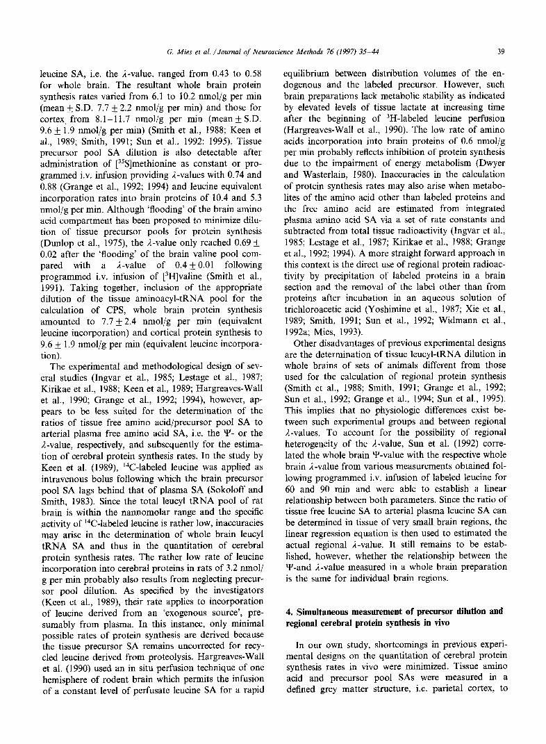
linear regression and correlation analysis. According to Eq. (4), the plot of tissue tRNAj3H]leucine SA versus arterial plasma [3H]leucine SA revealed a slope, i.e. a I-value, of 0.62 + 0.06 (r = 0.643, P < 0.05; Fig. 2, top). Using Eq. (7), the relationship between tissue free [3H]leucine SA versus plasma [3H]leucine SA yielded a linear regression coefficient, i.e. the Y-value, of 0.66 _+ 0.06 (r = 0.678; P < 0.05; Fig. 2, bottom). The statisti- cal comparison of the latter slopes did not reveal a significant difference (IJI 2,, = 0.68). Prediction of corti- cal tissue tRNA-[3H]leucine SA from measurements of cortex free [3H]leucine SA by trend analysis revealed a discrete underestimation as indicated by a linear regres- sion coefficient of 0.9147 f 0.07 (r = 0.788, P < 0.01).
4.4. Measurement of the rate of cortical protein synthesis
The rate of cortical protein synthesis estimated via the product/precursor relationship, i.e. by regression
I 4
0 2 4 6 6 10
Plasma leucine SA (nCi/nmol)
I I
0 2 4 6 8 10 Plasma leucine SA (nCiinmol)
Fig. 2. Relationship between tissue pool leucine SA to arterial plasma leucine SA. Top, linear regression analysis of cortex tRNA-leucine SA to arterial plasma leucine SA (Eq. (4)) revealed a slope, i.e. the i-value, equal to 0.62 + 0.06 (I = 0.643, P < 0.05). Bottom, regression analysis of cortex free leucine SA versus plasma leucine SA (Eq. (7)) yielded a regression coefficient, i.e. the Y-value, equal to 0.66 + 0.04 (r = 0.663, P i 0.05). Statistical comparison did not reveal a signifi- cant difference between regression coefficients, i.e. the ,I- and Y-value (F, 20 = 0.75). This indicates that leucine pool SAs of cortex are in rapid equilibrium between each other and with plasma leucine SA.
;=A12 f 0.8)x
p=&.OOl
, , 0 50 100 150 200 250 300
Integrated leucyl-tRNA SA (nCi/nmol/min)
6 5’0 lb0 Go 2&l 260 360 Integrated plasma leucine SA (nCiinmol/min)
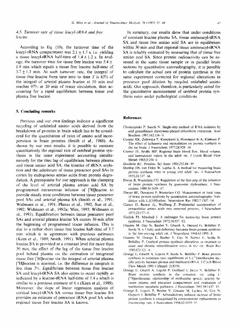
Fig. 3. Precursor-product relationship for the estimation of the corti- cal protein synthesis rate. Top, employing the appropriate precursor pool for protein biosynthesis, the slope calculated from linear regres- sion analysis of integrated leucyl-tRNA SA and labeled proteins (Eq. (8)) provided a cortical protein synthesis rate of 11.6 &- 0.8 nmol/g per min (r = 0.904; Pi 0.001). Bottom, linear regression analysis of integrated plasma leucine SA versus labeled proteins (Eq. (9)) re- vealed a slope, i.e. R x 1 (Eq. (9)) equal to 7.5 k 0.6 nmol/g per min (r = 0.852; P i 0.001). The ratio of slopes, i.e. (R x 1)/R amounted to a A-value of 0.65 which represents the fraction of leucine in the precursor derived from arterial plasma.
analysis of integrated leucyl-tRNA SA versus labeled proteins (Eq. (8)), amounted to a protein synthesis rate R of cortex of 11.6 + 0.80 nmol/g per min (r = 0.904; P < 0.001; Fig. 3, top) which is discretely larger than previously determined CPS rates in separate groups of animals (Smith, 1991; Sun et al., 1992; 1995). When precursor pool of protein synthesis is ignored, however, the cortical protein synthesis rate as estimated by the slope R x /z (Eq. (9)) from regression analysis of inte- grated plasma leucine SA versus labeled proteins turned out significantly lower with 7.5 t- 0.6 nmol/g per min (r = 0.852; P < 0.001; Fig. 3, bottom; Fr,20 = 17.6; P < 0.01). The ratio of the slope (R x 1)/R (equal to the dilution fraction R of the leucyl-tRNA pool) was 0.65 and provided a comparable estimate as determined in previous studies (Smith et al., 1988 31; Keen et al., 1989; Smith, 1991; Sun et al., 1992; 1995).
G. Mies et al. /Journal qf Neuroscience Methods 76 (IYY71 73 ?I 42
3.5. Turnover rute oj‘ tissuc~ leucyl-tRNA und free Itwinc
According to Eq. (lo), the turnover time of the 1eucyLtRNA compartment was 2.1 + 1.7 s, i.e. yielding a tissue leucyl-tRNA half-time of 1.4 i 1.2 s. In anal- ogy, the turnover time for tissue free leucine was 5.4 +_ 1.9 min which equals a tissue free leucine half-time of 3.7 & 1.3 min. At such turnover rate, the integral of tissue free leucine from time zero to time T is 83% of the integral of arterial plasma leucine at 10 min and reaches 97% at 20 min of tracer circulation, thus ac- counting for a rapid equilibrium between tissue and plasma free leucine.
5. Concluding remarks
Previous and our own findings indicate a significant recycling of unlabeled amino acids derived from the breakdown of proteins in brain which has to be consid- ered for the quantitation of rates of amino acid incor- poration in brain proteins (Smith et al., 1988). As shown by our own results, it is possible to measure quantitatively the regional rate of cerebral protein syn- thesis in the same experiment accounting simulta- neously for the time lag of equilibrium between plasma and tissue amino acid SA, the kinetics of tRNA acyla- tion and the admixture of tissue precursor pool SAs in cortex by endogenous amino acids from protein degra- dation. A prerequisite for our approach is the clamping of the level of arterial plasma amino acid SA by programmed intravenous infusion of [3H]leucine to provide steady state conditions between tissue precursor pool SAs and arterial plasma SA (Smith et al., 1991; Widmann et al., 1991; Planas et al., 1992; Sun et al., 1992; Widmann et al., 1992a; Mies, 1993; Widmann et al.. 1993). Equilibration between tissue precursor pool SAs and arterial plasma leucine SA occurs 30 min after the beginning of programmed tracer infusion. This is due to a rather short tissue free leucine half-time of 3.7 min which is in agreement with previous estimates (Keen et al., 1989; Smith, 1991). When arterial plasma leucine SA is provided at a constant level for more than 30 min. the effect of the lag of the tissue free leucine pool behind plasma on the estimation of integrated tissue free [JH]leucine via the integral of arterial plasma [3H]leucine is minimal, i.e. the difference in integrals is less than 3%. Equilibrium between tissue free leucine SA and leucyl-tRNA SA also seems to occur rapidly as indicated by a leucine-tRNA half-time of 1.4 s which is similar to a previous estimate of 6 s (Keen et al., 1989). Moreover. the slope of linear regression analysis of cortical leucyl-tRNA SA versus tissue free leucine SA provides an estimate of precursor tRNA pool SA when regional tissue free leucine SA is known.
In summary. our results show that under conditions of constant leucine plasma SA, tissue aminoacyl-tRNA SA and tissue free amino acid SA are in equilibrium within 30 min and that regional tissue aminoacyl-tRNA SA is reliably estimated by measuring that of tissue free amino acid SA. Since protein radioactivity can be as- sessed in the same tissue sample or in parallel brain sections by quantitative autoradiography, it is possible to calculate the actual rate of protein synthesis in the same experiment corrected for regional alterations in precursor pool dilution by recycled unlabeled amino acids. Our approach, therefore, is particularly suited for the quantitative measurement of cerebral protein syn- thesis rates under pathological conditions.
References
Chomczynski P. Sacchi N. Single-step method of RNA isolation by acid guanidinium thiocynate-phenol-chloroform extraction. Anal Biochem 1987~162: 156 -9.
Cooper HK, Zalewska T. Kawakami S, Hossmann K-A, Kleihues P. The effect of ischaemia and recirculation on protein synthesis in the rat brain. J Neurochem 1977;28:929 34.
Cremer JE. Seville MP. Regional brain blood flow. blood volume. and haematocrit values in the adult rat. J Ccreb Blood Flow Metab 1983;3:254-6.
Doolittle RF. Proteins. Sci Amer 1985;25?:88 -99 Dunlop DS, van Elden W. Lajtha A. A method for measuring brain
protein synthesis rates in young and adult rat\. .I Neurochem 1975;24:331 44.
Dwyer B, Wasterlain CG. Regulation of the first step of the initiation of brain protein synthesis by guanosine diphosphate. J Neu- rochem 1980:34:1639--47.
Dwyer BE, Donatoni P, Wasterlain CC?. Measurement of local rates of brain protein synthesis by quantitative autoradiography: Vaii- dation with L-[(3)H]valine. Neurochem Res 1982;?: 15Oi- 14.
Gainer H, Barker JL. Wollberg Z. Preferential incorporation of extracellular amino acids into neuronal proteins. .I Neurochem 1975;25: 177 9.
Garlick PJ, Marshall 1. A technique for measur-inp brain protein synthesis. J Neurochem 1972;19:557 -83.
Giaume M. Gay N. Baubet V. Gharib A, Durand G, Bobillier P. Sarda N. n-3 fatty acid deficiency increases brain protein synthesis in the free-moving adult rat. J Neurochem 1994:63:1995 -8.
Giaume M. Grange E. Baubet V. Ga> N. Sermt:! f3. Sarda N. Bobillier P. C’erebral protein synthesis alterations m response 10 acute and chronic immobilization stress in rbr r:!t. Brain Re* 1995575: I2 I 6.
Grange E. Gharib A. Lepetit P. Sarda Iv. Bobillier I’. Brain protem synthesis in conscious rats: equilibrium of L-( “S)methionine spc- cific activity between plasma and methionyl-tRihA. J Cerrb Blood Flow Metab 1991;l I(Suppl. 2):S356.
Grange E. Gharih A. Lepetit P, Guiliaud J. Sarda N. Bobillier P. Brain protein synthesis in the consc;iou\ rat using t- [“Slmethionine: relationship of methionine specific activity be- tween plasma and precursor compartment and evaluation of methionine metabolic pathways. J Neurochem 199X9:1437 43.
Grange E. Lepetit P. Baubet V, Giaume M. I,rc:lerc M. Gay Y. Gagnon J. Bobillier P. ,Adrenalectom>-induced Increase of brain protein synthesis is antagonized by corricosterone rcplaccments ill free-moving rats. J Neurochem 1994;67: 1079 8‘~

44 G. Mies et al. /Journal of Neuroscience Methoris 76 (1997) 35-44
Hargreaves-Wall KM, Buciak JL, Pardridge WM. Measurement of free intracellular and transfer RNA amino acid specific activity and protein synthesis in rat brain in vivo. J Cereb Blood Flow Metab 1990;10:162-9.
Ingvar MC, Maeder P, Sokoloff L, Smith CB. Effects of ageing on local rates of cerebral protein synthesis in Sprague-Dawley rats. Brain 1985;108:155-70.
Keen RE, Barrio JR, Huang SC, Hawkins RA, Phelps ME. In vivo cerebral protein synthesis rates with leucyl-transfer RNA used as a precursor pool: determination of biochemical parameters to structure tracer kinetic models for positron emission tomography. J Cereb Blood Flow Metab 19899~429-45.
Kiessling M, Xie Y, Kleihues P. Regionally selective inhibition of cerebral protein synthesis in the rat during hypoglycemia and recovery. .I Neurochem 1984a;43:1507-14.
Kiessling M, Xie Y, Kleihues P. Regional impairment of protein synthesis in the rat brain during bicuculline-induced seizures. Brain Res 1984b;296:1-13.
Kirikae M, Diksic M, Yamamoto YL. The transfer coefficients for L-valine and the rate of incorporation of L-[1-‘%]valine into proteins in normal adult rat brain. J Cereb Blood Flow Metab 1988;8:598-605.
Lajtha A, Toth J. Postmortem changes in the free amino acid pool. Brain Res 1974;76:546-74.
Lestage P, Gonon M, Lepetit P, Vitte PA, Debilly G, Rossatto C, Lecestre D, Bobillier P. An in vivo kinetic model with L-
[35S]methionine for the determination of local cerebral rates for methionine incorporation into protein in the rat. J Neurochem 1987;48:352-63.
Mies G. Inhibition of protein-synthesis during repetitive cortical spreading depression. J Neurochem 1993;60:360-3.
Mies G, Kawai K, Saito N, Nagashima G, Nowak TSJ, Ruetzler CA, Klatzo I. Cardiac arrest-induced complete cerebral ischemia in the rat: Dynamics of postischemic in vivo calcium uptake and protein synthesis. Neurol Res 1993; 15:253-63.
Patlak CS, Pettigrew KD. A method to obtain infusion schedules for prescribed blood concentration time courses. J Appl Physiol 1976;40:458-63.
Planas AM, Prenant C, Mazoyer BM, Comar D, Di Giamberardino L. Regional cerebral L-[‘4C-methyl]methionine incorporation into proteins: evidence for methionine recycling in the rat brain. J Cereb Blood Flow Metab 1992;12:603-12.
Robertson JH, Wheatley DN. Pools and protein synthesis in mam- malian cells. Biochem J 1979;178:699-709.
Smith CB. The measurement of regional rates of cerebral protein synthesis in vivo. Neurochem Res 1991;16:1037-46.
Smith CB, Deibler GE, Eng N, Schmidt K, Sokoloff L. Measurement of local cerebral protein synthesis in vivo: Influence of recycling of amino acids derived from protein degradation. Proc Nat1 Acad Sci USA 1988;85:9341-5.
Smith CB, Sun Y, Deibler GE, Sokoloff L. Effect of loading doses of L-valine on relative contributions of valine derived from protein degradation and plasma to the precursor pool for protein synthe- sis in rat brain. J Neurochem 1991;57:1540-7.
Sokoloff L, Reivich M, Kennedy C, Des Rosiers MH, Patlak CS, Pettigrew KD, Sakurada 0, Shinohara M. The [i4C]-deoxyglucose method for the measurement of local cerebral glucose utilization: Theory, procedure and normal values in the conscious and anes- thetized albino rat. J Neurochem 1977;28:897-916.
Sokoloff, L, Smith, CB. Basic principles underlying radioisotopic methods for assay of biochemical processes in vivo, In: Lam- brecht, RM, Rescigno, A, (Eds.), Tracer Kinetics and Physiologic Modeling, Berlin: Springer Verlag, 1983:202-234.
Sun Y, Deibler GE, Jehle J, Macedonia J, Dumont I, Dang T, Smith CB. Rates of local cerebral protein synthesis in the rat during normal postnatal development. Am J Physiol 1995;268:R549- 61.
Sun Y, Deibler GE, Sokoloff L, Smith CB. Determination of regional rates of cerebral protein synthesis adjusted for regional differences in recycling of leucine derived from protein degradation into the precursor pool in conscious adult rats. J Neurochem 1992;59:863-73.
Waterlow JC, Stephen JM. The effect of low protein diets on the turn-over rates of serums, liver and muscle proteins in the rat, measured by continuous infusion of L-[‘4C]lysine. Clin Sci 1968;35:287-305.
Widmann R, Kocher M, Ernestus R-I, Hossmann K-A. Biochemical and autoradiographic determination of protein synthesis in exper- imental brain tumors of rats. J Neurochem 1992a;59:18-25.
Widmann R, Weber C, Bonnekoh P, Schlenker M, Hossmann K-A. Neuronal damage after repeated 5 minutes of ischemia in the gerbil is preceded by prolonged impairment of protein metabolism. J Cereb Blood Flow Metab 1992b;12:425-33.
Widmann R, Kuroiwa T, Bonnekoh P, Hossmann K-A. [%]leucine incorporation into brain proteins in gerbils after transient is- chemia: relationship to selective vulnerability of hippocampus. J Neurochem 1991;56:789-96.
Widmann R, Miyazawa T, Hossmann K-A. Protective effect of hypothermia on hippocampal injury after 30 minutes of forebrain ischemia in rats is mediated by postischemic recovery of protein synthesis. J Neurochem 1993;61:200-9.
Xie Y, Mies G, Hossmann K-A. Ischemic threshold of brain protein synthesis after unilateral carotid occlusion in gerbils. Stroke 1989;20:620-6.
Yoshimine T, Hayakawa T, Kato A, Matsumoto K, Ushio Y, Mogami H. Autoradiographic study of regional protein synthesis in focal cerebral ischemia with TCA wash and image subtraction techniques. J Cereb Blood Flow Metab 1987;7:387-93.