quantitative evaluation of liver-specific promoters from
TRANSCRIPT

Quantitative Evaluation of Liver-Specific Promoters From Retroviral Vectors After In Vivo Transduction of Hepatocytes
By Daniel G. Hafenrichter, Xiaoyun Wu, Steven D. Rettinger, Susan C. Kennedy, M. Wayne Flye, and Katherine Parker Ponder
Hepatic gene therapy could be used to treat a number of inherited blood diseases such as hemophilia or thrombophi- lia. Although liver-directed retroviral transduction can result in long-term gene expression in vivo, the low level of protein production has limited its clinical application, We reasoned that the insertion of liver-specific promoters into retroviral vectors would increase gene expression in vivo. The 347-bp human a,-antitrypsin (hAAT), the 810-bp murine albumin (mAlb), the 490-bp rat phosphoenolpyruvate carboxykinase (rPEPCK), and the 596-bp rat liver fatty acid binding protein promoters were inserted into a Moloney murine leukemia retroviral backbone containing the hAAT reporter gene. Vec- tors that produced appropriately sized RNA and hAAT pro-
EPATIC GENE THERAPY could dramatically alter the treatment of many inherited hematologic diseases.
The liver synthesizes a myriad of proteins that play pivotal roles in hemostasis, metabolism, and protection against in- fection. Although some genetic diseases such as hemophilia can be treated by repeated infusion of the deficient protein, this is expensive, carries a risk of viral infection, and only temporarily ameliorates the disease manifestations. Liver transplantation can correct serum protein deficiencies,’ but is limited by donor organ availability and the need for immu- nosuppression. The transfer of a functional gene into the cells of a genetically deficient individual should correct the clinical manifestations by providing long-term production of the gene product.
The liver is an attractive organ for gene therapy of serum protein deficiencies because it performs the proper posttrans- lational modifications necessary for full activity for many serum proteins, such as the y-carboxylation of coagulation factors, and the rich blood supply of the liver bathing the transduced hepatocytes facilitates the secretion of protein products into the bloodstream. Both ex vivo and in vivo methods have been used to transfer genes into mammalian hepatocytes. The ex vivo approach’.’ requires hepatocyte harvest, in vitro transduction with retrovirus, and reintroduc-
H
From the Departments of Surgery, Internal Medicine, and Bio- chemistry and Molecular Biophysics, Washington University School of Medicine, St Louis, MO.
Submitted April 13, 1994; accepted July 22, 1994. Supported by grants from the National Institutes of Health (R29-
DK44593). the American Lung Association, rhe Markey Foundation, and the American Liver Foundation awarded to K.P.P. and the Surgical Infection Society awarded to D.G.H.
Address reprint requests to Katherine Parker Ponder, MD, De- partment of Biochemistry and Molecular Biophysics, Washington University School of Medicine, Box 8231, 660 S Euclid Ave, St Louis, MO 63110.
The publication costs of this article were defrayed in part by page charge payment. This article must therefore be hereby marked “advertisement” in accordance with 18 U.S.C. section 1734 solely to indicate this fact. 0 1994 by The American Society of Hematology. 0006-4971/94/84I0-0027$3.00/0
3394
tein in vitro were tested in vivo by transducing regenerating rat livers. Long-term serum expression of the hAAT reporter gene was normalized to retroviral transduction efficiency as determined by using a polymerase chain reaction-based assay of genomic DNA from transduced rat livers. The hAAT, mAlb, and rPEPCK promoters were, respectively, 35-, 8-, and 0.02-fold as strong as the previously studied constitutive Pol- II promoter. We conclude that the hAAT promoter resulted in the highest expression from a retroviral vector and may re- sult in therapeutically significant expression of other clini- cally significant blood proteins. 0 1994 by The American Society of Hematology.
tion of the transduced hepatocytes into the portal circulation. The in vivo delivery of foreign genes to hepatocytes in situ has been accomplished by injecting DNA,6-9 adenoviral vec- t o r ~ , ” ” ~ or retroviral vector^^^-'^ into the liver parenchyma, peripheral veins, or the portal vein. Of these, only retroviral vectors result in long-term expression because of their ability to integrate into chromosomal DNA.
A major limitation to achieving successful gene therapy with retroviral vectors in the liver has been the low level of expression observed. For example, hepatic gene therapy experiments using retroviral vectors containing the human factor E’” or human al-antitrypsin’8~22~24 (hAAT) genes have resulted in expression of the therapeutic gene at only 0.1 % of the normal level. One explanation for the low level of expression observed is that transcription from a retroviral vector is inadequate. To identify promoters that may function well in the liver in vivo, plasmids containing a variety of promoters have been introduced into primary hepatocytes by using optimized lipofection.25 These experiments showed that the human cytomegalovirus (CMV) immediate early promoter was much stronger than other viral, cellular, or liver-specific promoters tested. These results led Armentano et a126 to place the CMV promoter into an LNL-6-based retroviral vector, which indeed led to high-level expression of a human factor IX gene in cultured primary hepatocytes. Similarly, Wilson et al’’ determined that the viral Moloney murine leukemia virus (MO-MLV) long terminal repeat (LTR) promoter was better expressed from a retroviral vector than three other promoters, including the chicken p-actin promoter, in rabbit hepatocytes.
Although it was logical to assume that promoter function in primary hepatocytes in vitro would correlate well with promoter function in liver cells in vivo, this, unfortunately, is not the case. Although the CMV promoter was expressed well from either a plasmid or a retroviral vector in vitro,”.” it was shut off rapidly in vivo using either an ex vivo or an in vivo approach to transfer the vector into the hepatocytes of dogs or ~ n i c e . ~ . ’ ~ Similarly, Wilson et a1’ found that the retrovirus in which the LTR promoter drives the expression of the low-density lipoprotein receptor (LDL-R) gene led to only short-term improvement in the hypercholesterolemia of Watanabe rabbits, although rejection of the allogeneic
Blood, Vol 84, No 10 (November 15), 1994: pp 3394-3404

IN VIVO ANALYSIS OF LIVER-SPECIFIC PROMOTERS 3395
hepatocytes was cited as the most likely reason for the loss of expression. Similarly, we recently quantitated expression from the MO-MLV LTR promoter and determined that it was only 1% as active in hepatocytes in vivo as in cultured hepatoma cells in In contrast, although the chicken &actin gene was poorly expressed in primary hepatocytes,*’ this promoter has led to long-term expression of the LDL- R gene in Watanabe rabbits3 and is currently being used in human gene therapy trial^.^ Thus, we conclude that expres- sion of a promoter in cultured hepatocytes correlates poorly with expression in hepatocytes in vivo. This result is not totally surprising, because the constellation of transcription factors present in replicating hepatocytes in vitro is almost certainly quite different from those present in quiescent he- patocytes in vivo. Similar results have been observed in primary fibroblasts by Palmer et alZ8 and Scharfman et who determined that the CMV, SV40, and MO-MLV LTR promoters are expressed well in vitro, but are shut off upon returning the cells to animals.
Although it is clear that the relative strengths of promoters need to be compared in vivo, few studies have undertaken quantitative comparisons of promoter strength. Unfortu- nately, studies which use a single promoter with a particular reporter gene cannot be easily compared with those that study another reporter gene, because of potential differences in protein half-life, RNA stability, and vector construction. We have recently developed quantitative assays for nor- malizing gene expression to in vivo transduction efficiency and have used these techniques to compare the relative in vivo strengths of the MO-MLV LTR promoter with the pro- moter for the large subunit for murine RNA polymerase I1 ( P O ~ - I I ) . ~ ~ In this study, we use our quantitative techniques to directly compare liver-specific promoters for their ability to direct transcription from a retroviral vector in vivo. We determine that the hAAT promoter is stronger than the Pol- II, murine albumin (mAlb), or phosphoenolpyruvate carboxy- kinase (PEPCK) promoters in vivo.
MATERIALS AND METHODS
Construction of Retroviral Vectors LTR-MT. LTR-hAAT is a retroviral vector constructed from
the MO-MLV derivative LNL-6.30 It contains a 1.3-kb hAAT cDNA, the encephalomyocarditis virus internal ribosome entry site (IRES), and a mutant, methotrexate resistant (MtxR) dihydrofolate reductase (*DHFR) gene.24 The LTR-ALTR retroviral vector contains a 178- bp deletion in the 3’ LTR enhancer region from the Pvu I1 site at position 3312 to the Xba I site at position 3488 (numbers refer to the locations on LNL-6).
rPEPCK-LTR. The rat cytosolic PEPCK promote9’ was cut at the Xba I site at -490 relative to the transcription start site, blunt- ended with T4 DNA polymerase, and ligated with BamHI linkers. After BamHI and Bgl I1 digestion (cuts at +73), the 560-bp DNA fragment was cloned into the BgZ I1 site of LTR-hAAT to create rPEPCK-LTR.
rLFABP-LTR. The rat LFABP promote? was cut at -596 with Sal I, blunt-ended, and ligated with Bgl I1 linkers. After BamHI (cuts at +21) and Bgl I1 digestion, the 620-bp fragment was cloned into the Bgl I1 site of LTR-hAAT to create rLFABP-LTR.
rn4Zb-ALTR. To create the cloning intermediate, mAlb-SP72, the mouse albumin promoter present in pAT2’, was digested with
Hpa I1 (which cuts 8 nt downstream from the transcription start site), blunt-ended, and ligated with BamHI linkers. After digestion with BamHI and EcoRI (cuts at -810), the 820-bp fragment was ligated into the EcoRUBamHI site of pSP72 (Promega, Madison, WI) to create d lb -SP72 . mAlb-SP72 was cut with Bgl I1 and BamHI and the 840-bp fragment was ligated into the unique Bgl I1 site of LTR-ALTR to create mALB-ALTR.
MT-ALTR. The DNA fragment containing the hAAT pro- motelJ4 was digested with Sma I, which cuts at +56, and ligated with BcZ I linkers. After Bcl I and Bgl I1 (cuts at -347) digestion, the 403-bp fragment was cloned into the Bgl I1 site of LTR-ALTR to create hAAT-ALTR.
HNF-3-mAlb-ALTR and HNF-3-hAAT-ALTR. pAT-2eG3x con- tains 3.5 copies of the DNA sequence that binds to the HNF-3 transcription factor (5’-GCTCCAGGGAATGTTTGTTCl”AAATA CCATGCT-3’) upstream of the 810-bp mAlb promoter in ~ u C 1 8 . ~ ’ The polymerase chain reaction (PCR) was used to amplify a 198- bp DNA containing the HNF-3 binding sites as follows: an oligonu- cleotide containing an EcoRI site and identity with the top strand of the pUC18 sequence 5’ to the HNF-3 sites (5“GTCGAATTCGCA- TGCGTCGACGGAAACAGCTATGACCATGA-3‘) and an oligo- nucleotide with an EcoRI site and identity with the bottom strand at the 5’ end of the mAlb promoter (5”CAGGAATTCAGTCAA- GACTACATCCAAAT-3’) were synthesized. The oligonucleotides were used in a 25-cycle PCR reaction with pAT-2eG3x, as described previously,” with denaturation at 94°C for 2 minutes, annealing at 45°C for 2 minutes, and extension at 72°C for 2 minutes. The PCR products were digested with EcoRI and cloned into the EcoRI site of pSP72 to create (HNF-3),pSP72. A 237-bp (HNF-3)3 fragment was removed from (HNF-3),pSP72 by Bgl I1 and BamHI digestion and was cloned into the unique 5’ Bgl I1 sites of mAlb-ALTR and hAAT-ALTR to create HNF-3-mAlb-ALTR and HNF-3-hAAT- ALTR, respectively.
Creation of Retrovirul Producer Cell Lines The amphotropic G P + ~ ~ v A M I ~ ~ ’ and ecotropic GP+E-8630 mu-
rine fibroblast lines were maintained in Dulbecco’s modified Eagle’s medium (DMEM) supplemented with 10% heat-inactivated calf se- rum (Hyclone Laboratories, Logan, UT), penicillin ( IO5 UL) , and streptomycin (IO5 pgL). After calcium phosphate transfection with retroviral vector DNAs? cells were cultured for 14 days in 250 nmoVL methotrexate- (Sigma Chemical, St Louis, MO) supple- mented DMEM with dialyzed calf serum. Supernatant from 48 MtxR clones were screened for the production of retrovirus by infecting NIH 3T3 cells and measuring hAAT production 72 hours later. High- titer clones were proven to be free of replication competent helper virus by amplification and marker rescue assay.” For the rPEPCK- LTR retroviral vector, both GP+E-86 and GP+AM-I2 packaging cell lines were transfected and the best clones were subjected to “ping-pong” co~ultivation’~ for 4 weeks. Amphotropic cells were then selected by treating the cocultures with 200 pg/mL hygromycin- B (Sigma Chemical), which eradicated the ecotropic cells.
In Vitro Transduction of NIH 3T3 and Hepu AI Cells With Retrovirul Vectors Containing the Liver-Specific Promoters
Conditioned medium from confluent retroviral packaging cells was filtered (0.45 km), spiked with polybrene to a final concentration of 8 pg /mL, and added at various dilutions to 50% confluent murine NIH 3T3 fibroblasts3* or murine Hepa A1 hepatoma ~e l l s .3~ Forty- eight hours later, the transduced cells were trypsinized, diluted 1:20, and administered fresh DMEM supplemented with 250 nmol/L Mtx. After 14 days of selection, MtxR colonies were counted to determine the MtxR retroviral titer? pooled together, and cultured to confluence

3396 HAFENRICHTER ET AL
in Mtx-supplemented DMEM. The transduced cells were analyzed for hAAT production by culturing the cells for 6 hours in hormonally defined mediumZ5 (HDM). Supernatants were assayed for hAAT activity by enzyme-linked immunosorbent assay (ELBA).” After the collection of the supernatants, the transduced cell monolayers were trypsinized, counted on a hemocytometer, and used to obtain either RNA or DNA for further analysis, as described previou~ly.~~ Where indicated, cells transduced with rPEPCK-LTR were stimu- lated for 6 hours with 0.5 mmolR. dibutyryladenosine 3’:5’-cyclic monophosphate (BtzcAMP), 1 mmoVL theophylline, and 1 pmoUL dexamethasonem in 75% tyrosine-free minimum essential medium and 25% tyrosine-free Waymouth’s medium, and nonstimulated cells were incubated in the basal medium without any hormones. Southern blot was performed on DNA from cells transduced with each of the vectors by using restriction enzymes that cut within the LTRs; all vectors resulted in DNA of the expected size.
In Vivo Hepatocyte Transduction Protocol Adult male Sprague-Dawley rats (SASCO, Omaha, NE), weighing
200 to 275 g, received standard National Institutes of Health-ap- proved institutional care with rodent chow and tap water ad libitum. Rats received a 70% partial hepatectomy, as described.24 Twenty- four hours later, packaging cell conditioned medium was concen- trated by ultracentrifugation and spiked to a final concentration of 8 pg/mL polybrene, and in vivo hepatocyte transduction was accom- plished by portal vein injection of retrovirus during hepatic-inflow occ~~sion.*~
Northern Blotting of Cellular RNA Ten micrograms of total cellular RNA was electrophoresed on
a 1% formaldehyde denaturing agarose gel4’ and transferred to a nitrocellulose membrane (BA-S-NC; Schleicher and Schuell, Keene, NH). Ethidium bromide staining and UV visualization showed that the RNA isolated was of good quality and loaded in equal amounts. The 592-bp Xho UEcoFU fragment of the IRES DNA4’ was labeled with a-”P-dCTP by random primer extension‘” and hybridized with the nitrocellulose membrane at 42°C with 50% formamide and washed according to the manufacturer’s instructions.
PCR Amplification of Proviral DNA Sequences A 30-cycle multiplex PCR technique was employed using primer
sets that amplified both proviral (IRES) and rat genomic (liver fatty acid binding protein [LFABP]) DNA sequences, as described pre- v i o u ~ l y . ~ ~ Amplified DNA sequences were electrophoresed through a 2% agarose/l X Tris-ammonium acetate gel:’ transferred to BA- S-NC nitrocellulose membrane, and hybridized with a 217-bp HindIIUKpn I fragment of the IRES labeled to a specific activity of 3 X IO9 cpdpg DNA by random primer extension. After quantita- tion, the membrane was stripped and reprobed with a radiolabeled 620-bp LFABP probe. Radioactivity was quantitated on a Betascope 630 two-dimensional Beta counter (Betagen, Waltham MA).
RESULTS
Development of Packaging Cell Lines With Retroviral Vectors Containing Liver-Specific Promoters
To date, expression from retroviral vectors has been disap- pointingly low in hepatocytes in vivo. We reasoned that the insertion of strong, liver-specific promoters might increase expression and selected four promoters for evaluation based on their ability to direct expression in the livers of transgenic mice or in hepatoma cells. We also evaluated the effect of inserting three hepatocyte nuclear factor-3 (HNF-3) binding
sites upstream of the mAlb and hAAT promoters, because this increased expression from the mAlb promoter in hepa- toma cells.33 We decided to clone the promoters into a ret- roviral vector with a deletion in the enhancer region of the 3‘ LTR. Such a deletion is transferred to the 5‘ LTR after one round of retroviral infection, resulting in a -90% decrease in LTR-initiated tran~cription.~~.“ This facilitates the in vitro analysis of internal promoter function, because high levels of transcription from the LTR can interfere with the expres- sion from the internal promoter.
Retroviral vectors (Fig 1) were transfected into GP+envAMl2 amphotropic retroviral packaging cells. After Mtx selection, 48 clones for each vector were screened for their ability to program NIH 3T3 cells to express hAAT. Although adequate expression was achieved with the vectors containing the hAAT and mAlb promoters in transduced NIH 3T3 cells (Table l), both the rLFABP-ALTR and rPEPCK-ALTR vectors resulted in negligible hAAT protein production (data not shown). We therefore placed the rLFABP and rPEPCK promoters in a retroviral vector with an intact 3’ LTR to facilitate isolation of high-titer clones. The rLFABP-LTR and rPEPCK-LTR vectors were trans- fected into GP+envAM12 cells and the best clones selected as described above. In addition, the rPEPCK-LTR vector was transfected into the ecotropic GP+E-86 packaging cells and the technique of ecotropiclamphotropic “ping-pong” c o c ~ l t i v a t i o n ~ ~ was performed to generate high-titer packag- ing cell lines. The GP+envAMl2 clones remaining after hygromycin B and Mtx selection were screened for hAAT protein production after infection of NIH 3T3 cells.
M t g Retroviral Titers and Protein Expression From Liver-Specific Retroviral Vectors in Singly Transduced Tissue Culture Cells
The best amphotropic packaging cell line for each of the retroviral vectors was used to infect NIH 3T3 fibroblasts and Hepa AI hepatoma cells at various dilutions. After selection with Mtx, colonies were counted to determine the MtxR ret- roviral titers, as shown in Table 1. The rLFABP-LTR pack- aging cell lines produced insignificant numbers of MtxR colo- nies. The rPEPCK-LTR construct had a high titer, as expected for a vector that underwent “ping-pong” amplifi- cation, whereas the hAAT and mAlb promoter-containing constructs had lower titers, as is generally observed after direct transfection into amphotropic packaging cell lines without “ping-pong” amplification.
Resistant cells should each contain a single copy of inte- grated retrovirus when they are infected at a low multiplicity of infection. Such singly transduced cells were used to quan- titate hAAT protein production by performing a timed super- natant collection and normalizing to the number of cells on the plate. It should be noted that both LTR- and internal promoter-initiated transcripts can result in hAAT protein production. hAAT-ALTR produced a similar amount of hAAT protein in transduced NIH 3T3 and Hepa A1 cells, whereas HNF-3-hAAT-ALTR resulted in a threefold and fourfold increase in hAAT protein production in the NIH 3T3 and Hepa AI cells, respectively. Although mAlb-ALTR resulted in high levels of hAAT protein production in NIH

IN VIVO ANALYSIS OF LIVER-SPECIFIC PROMOTERS
3T3 cells, much lower expression was observed in the Hepa AI cells. The addition of HNF-3 binding sites to the mAlb promoter had little effect on expression in NIH 3T3 cells, but increased hAAT protein expression by 35-fold in the hepatoma cells. The rLFABP-LTR vector had no detectable
A LTR-hAAT 4.4 kb
Pol-II-hAAT
rPEPCK-LTR
rLFABP-LTR
B LTR-ALTR
mAlb-ALTR
hAAT-ALTR
I 4 1 kb .5 kb
I ~ 5 . 0 kb 5 kb
l ~ 5 . 0 kb 4 kb
4.2 kb
kb
kb
-5.2 kb HNF-3-mAlb kb -ALTR
HNF-3-hAAT -A LTR
kb kb
Fig 1. Retroviral vectors containing liver-specific promoters. (A) Retroviral vectors containing an intact 3' LTR. LTR-hAAT contains the 1.3-kb hAAT cDNA and the encephalomyocarditis virus IRES se- quence upstream of the mutant, MtxR "DHFR?' Transcription initiat- ing from the retroviral LTR promoter results in a 4.4-kb transcript, as indicated by the arrow. The 700-bp murine Pol-ll promoter.z4 the 490- bp rat PEPCK promoter?' and the 596-bp rat LFABP promoter32 were placed immediately upstream from the hAAT cDNA in LTR-hAAT to create Pol-II-hAAT (as described previouslyZ4), rPEPCK-LTR, and rLFABP-LTR, respectively; transcription can initiate from either the LTR promoter (resulting in transcripts of 5.1, 5.0, and 5.0 kb, respec- tively) or from the internal promoter (resulting in transcripts of 3.5, 3.5, and 3.4 kb, respectively), as depicted by the arrows. (B) Retroviral vectors containing a deletion in the 3' LTR (ALTR). The LTR-ALTR retroviral vector contains a 178-bp deletion in the enhancer of the 3' LTR. Transcription initiating at the LTR promoter results in a 4.2- kb RNA (arrow). The 810-bp mAlb promoter33 and the 347-bp hAAT promoter3' were each inserted upstream from the hAAT cDNA in LTR-ALTR to create mAlb-ALTR and hAAT-ALTR, respectively; tran- scription initiating from the LTR results in 5.0- and 4.6-kb RNAs, respectively, whereas transcription initiating from the internal pro- moters results in a 3.2-kb RNA (arrows). The HNF-3-mAlb-ALTR and HNF-3-hAAT-ALTR vectors were created by inserting three HNF-3 binding site sequences33 upstream of the internal promoter of mAlb- ALTR and hAAT-ALTR, respectively; transcription initiating from the retroviral LTR promoter results in 5.2- and 4.8-kb RNAs, respectively, whereas transcription initiating from the mAlb or hAAT internal pro- moters results in a 3.2-kb RNA.
3397
Table 1. The Production of hAAT Protein by the Liver-Specific Retroviral Vectors
hAAT Protein Production in hAAT Protein
MtxR Titer Transduced on NIH 3T3 NIH 3T3 Cells*
Production
Fibroblasts (ng hAAT1lO6 in Transduced Hepa A1 Cells*
Promoter (cfu/mL) cells/24 h) (ng hAAT/106 cellsi24 h)
mAlb 60,000 351.6 i 55.2 7.1 t- 0.47 HNF-3-mAlb 10,000 356.0 ? 159 247.5 i 37.0 hAAT 10,000 63.4 ? 11.7 50.3 -t 10.0 HNF-3-hAAT 10,000 192.0 2 41 221.0 2 52.0 rPEPCK 2,000,000 4.6 5 2 t 108.6 5 6.0t
6.3 5 l* 137.0 -t 19 .N
* hAAT protein production by the retroviral transduced NIH 3T3 or Hepa A I cells was measured in a 6-hour collection from confluent cell cultures in HDM and was normalized to the number of cells present.
t hAAT protein production by the transduced NIH 3T3 and Hepa A1 cells was measured in a 6-hour collection from confluent cell cultures in basal medium, without any additives, and was normalized to the number of cells present. * hAAT protein production by the retroviral transduced NIH 3T3 and Hepa A I cells was measured in a 6-hour collection from confluent cell cultures in basal medium supplemented with 0.5 mmol/L Bt2- CAMP, 1 kmol/L dexamethasone, and 1 mmol/L theophylline and was normalized to the number of cells present.
activity in either cell type (data not shown). Although the fibroblasts transduced with rPEPCK-LTR produced much less hAAT than the other promoters in NIH 3T3 cells, ex- pression was similar to the other promoters in the hepatoma cells. Because the rPEPCK promoter is induced by the addi- tion of the Bt,-CAMP and t h e ~ p h y l l i n e , ~ ' , ~ " , ~ ~ transduced ti- broblasts and hepatoma cells were tested for their hormonal responsiveness. As shown in Table 1 , stimulated fibroblasts and hepatoma cells increased hAAT protein production by 1.37- and 1.26-fold, respectively.
Analysis cf RNA From Puckaging and Transduced Cells
When constructing a retroviral vector with an internal promoter, it is important to document that full-length geno- mic transcripts are being produced and that no re- arrangements have occurred during transduction and to as- sess the level of expression from the internal promoter. RNA from packaging cell lines for each liver-specific promoter was analyzed by Northern blot. LTR-initiated transcripts are predicted to range from 5.2 to 4.2 kb in size, whereas the internal promoter-initiated transcripts are predicted to range from 3.5 to 3.2 kb, depending on the particular vector, as shown in Fig 1. The major bands for the packaging cells containing hAAT-ALTR and HNF-3-hAAT-ALTR were derived from the LTR promoter, as expected in the tibro- blast-derived packaging cells (Fig 2B, lanes 6 and 7, and Fig 2A, lane 11). The minor short band of 3.5 kb may be derived from a splice product that uses the 5' retroviral splice site'" and a putative 3' splice site (UCUUCUUCUCCCCN) at position +260 within the hAAT cDNA, which closely matches the consensus 3' splice site [(Py),NPy&].46 The presence of such a spliced product is suspected because of the fact that an identical sized band is observed for all pro-
3398 HAFENRICHTER ET AL
n n n n n - n
n
4 II"
J LTR - Spliced? 1 Internal
1 2 3 4 5 6 7 8 9 1 0 1 1
B rPEPCK h M T HNF3-hAAT mAlb HNF3-mAlb
""._""I_ " 'P N H " P N H " N H " N H"N H '
l - 0-
1 23 4 5 67 8 91011 12131415 16171819 20 2122
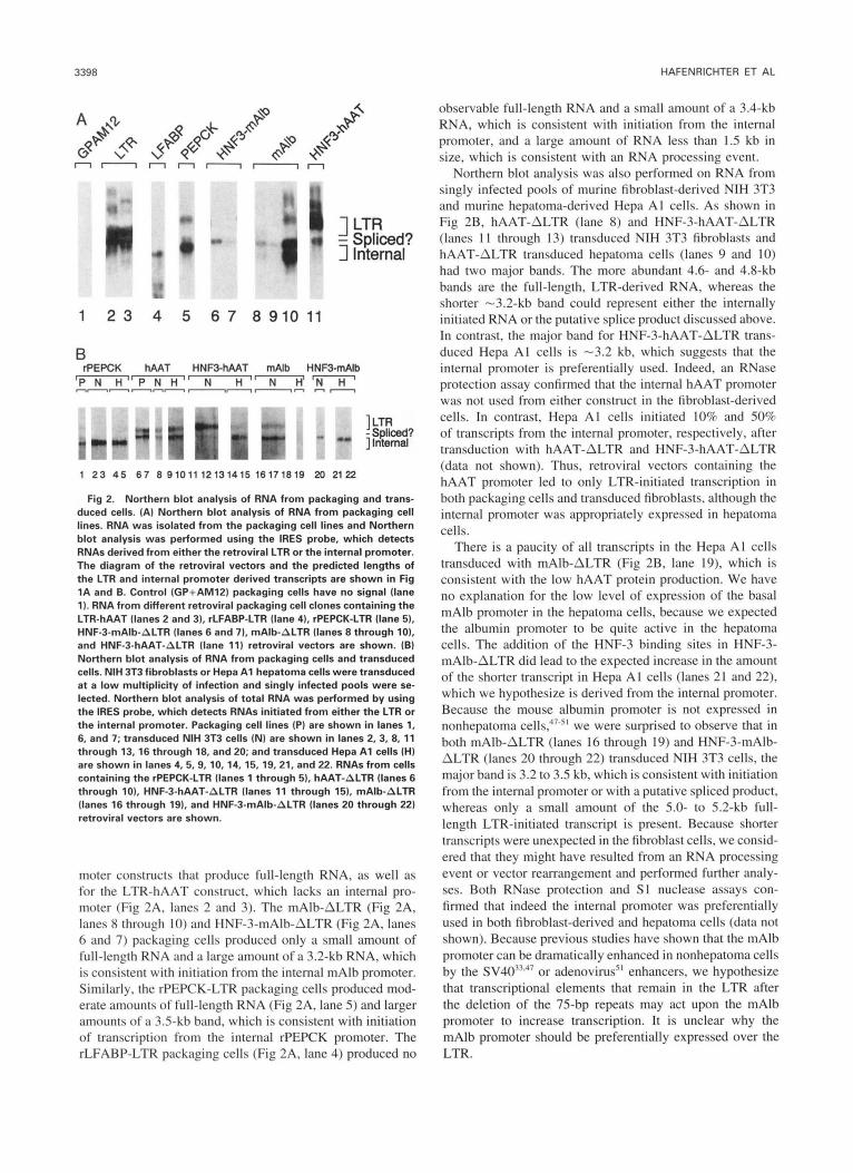
Fig 2. Northern blot analysis of RNA from packaging and trans- duced cells. (A) Northern blot analysis of RNA from packaging cell lines. RNA was isolated from the packaging cell lines and Northern blot analysis was performed using the IRES probe, which detects RNAs derived from either the retroviral LTR or the internal promoter. The diagram of the retroviral vectors and the predicted lengths of the LTR and internal promoter derived transcripts are shown in Fig 1A and B. Control (GP+AM12) packaging cells have no signal (lane 11. RNA from different retroviral packaging cell clones containing the LTR-hAAT (lanes 2 and 31, rLFABP-LTR (lane 41, rPEPCK-LTR (lane 5). HNF-3-mAlb-ALTR (lanes 6 and 71, mAlb-ALTR (lanes 6 through 10). and HNF-3-hAAT-ALTR (lane 11) retroviral vectors are shown. (B) Northern blot analysis of RNA from packaging cells and transduced cells. NIH 3T3 fibroblasts or Hepa A1 hepatoma cells were transduced at a low multiplicity of infection and singly infected pools were se- lected. Northern blot analysis of total RNA was performed by using the IRES probe, which detects RNAs initiated from either the LTR or the internal promoter. Packaging cell lines (P) are shown in lanes 1, 6, and 7; transduced NIH 3T3 cells (NI are shown in lanes 2, 3,8, 11 through 13,16 through 18, and 20; and transduced Hepa A1 cells (H) are shown in lanes 4, 5,9, 10, 14, 15, 19,21, and 22. RNAs from cells containing the rPEPCK-LTR (lanes 1 through 5). hAAT-ALTR (lanes 6 through 101, HNF-3-hAAT-ALTR (lanes l1 through 15), mAlb-ALTR (lanes 16 through 19). and HNF-3-mAlb-ALTR (lanes 20 through 22) retroviral vectors are shown.
moter constructs that produce full-length RNA, as well as for the LTR-hAAT construct, which lacks an internal pro- moter (Fig 2A, lanes 2 and 3). The mAlb-ALTR (Fig 2A, lanes 8 through 10) and HNF-3-mAlb-ALTR (Fig 2A, lanes 6 and 7) packaging cells produced only a small amount of full-length RNA and a large amount of a 3.2-kb RNA, which is consistent with initiation from the internal mAlb promoter. Similarly, the rPEPCK-LTR packaging cells produced mod- erate amounts of full-length RNA (Fig 2A, lane 5) and larger amounts of a 3.5-kb band, which is consistent with initiation of transcription from the internal rPEPCK promoter. The rLFABP-LTR packaging cells (Fig 2A, lane 4) produced no
observable full-length RNA and a small amount of a 3.4-kb RNA, which is consistent with initiation from the internal promoter, and a large amount of RNA less than 1.5 kb in size, which is consistent with an RNA processing event.
Northern blot analysis was also performed on RNA from singly infected pools of murine fibroblast-derived NIH 3T3 and murine hepatoma-derived Hepa A I cells. As shown in Fig 2B, hAAT-ALTR (lane 8) and HNF-3-hAAT-ALTR (lanes 1 1 through 13) transduced NIH 3T3 fibroblasts and hAAT-ALTR transduced hepatoma cells (lanes 9 and IO) had two major bands. The more abundant 4.6- and 4.8-kb bands are the full-length, LTR-derived RNA. whereas the shorter -3.2-kb band could represent either the internally initiated RNA or the putative splice product discussed above. In contrast, the major band for HNF-3-hAAT-ALTR trans- duced Hepa AI cells is -3.2 kb, which suggests that the internal promoter is preferentially used. Indeed, an RNase protection assay confirmed that the internal hAAT promoter was not used from either construct in the fibroblast-derived cells. In contrast, Hepa AI cells initiated 10% and SO% of transcripts from the internal promoter, respectively, after transduction with hAAT-ALTR and HNF-3-hAAT-ALTR (data not shown). Thus, retroviral vectors containing the hAAT promoter led to only LTR-initiated transcription in both packaging cells and transduced fibroblasts, although the internal promoter was appropriately expressed in hepatoma cells.
There is a paucity of all transcripts in the Hepa AI cells transduced with mAlb-ALTR (Fig 2B, lane 19). which is consistent with the low hAAT protein production. We have no explanation for the low level of expression of the basal mAlb promoter in the hepatoma cells, because we expected the albumin promoter to be quite active in the hepatoma cells. The addition of the HNF-3 binding sites in HNF-3- mAlb-ALTR did lead to the expected increase in the amount of the shorter transcript in Hepa AI cells (lanes 21 and 22), which we hypothesize is derived from the internal promoter. Because the mouse albumin promoter is not expressed in nonhepatoma c ~ I I s , ~ " ~ ' we were surprised to observe that in both mAlb-ALTR (lanes 16 through 19) and HNF-3-mAlb- ALTR (lanes 20 through 22) transduced NIH 3T3 cells, the major band is 3.2 to 3.5 kb. which is consistent with initiation from the internal promoter or with a putative spliced product, whereas only a small amount of the 5.0- to 5.2-kb full- length LTR-initiated transcript is present. Because shorter transcripts were unexpected in the fibroblast cells, we consid- ered that they might have resulted from an RNA processing event or vector rearrangement and performed further analy- ses. Both RNase protection and SI nuclease assays con- firmed that indeed the internal promoter was preferentially used in both fibroblast-derived and hepatoma cells (data not shown). Because previous studies have shown that the mAlb promoter can be dramatically enhanced in nonhepatoma cells by the SV40""47 or adenovirus5' enhancers, we hypothesize that transcriptional elements that remain in the LTR after the deletion of the 75-bp repeats may act upon the mAlb promoter to increase transcription. It is unclear why the mAlb promoter should be preferentially expressed over the LTR.

IN VIVO ANALYSIS OF LIVER-SPECIFIC PROMOTERS 3399
For the rPEPCK-LTR transduced NIH 3T3 and Hepa A1 cells (Fig 2B, lanes 2 through 5), the major 3.2-kb band observed is consistent with initiation from the internal pro- moter and there is little difference in the levels of RNA in hormonally stimulated cells as compared with the nonstimu- lated cells. It is unlikely that the shorter transcripts in trans- duced cells are caused by a rearrangement that occurred during transduction, because DNA from cells transduced with all of the vectors was analyzed by Southern blot and shown to be full length (data not shown). This finding sug- gests that the shorter RNA species are indeed derived from the internal promoter of an intact provirus. This is similar to the results of Hatzoglou et who placed the same rPEPCK promoter upstream of the neomycin phosphotrans- ferase gene in an MO-MLV retroviral vector and observed a marked predominance of rPEPCK-initiated transcripts as compared with LTR-initiated transcripts in G418 selected cells. Thus, the rPEPCK promoter is preferentially expressed over the LTR in the rPEPCK-LTR retroviral vector. The fact that the expression was quite low in the rPEPCK-ALTR retroviral vector leads us to hypothesize that the retroviral LTR may exert an enhancer effect on the internal rPEPCK promoter.
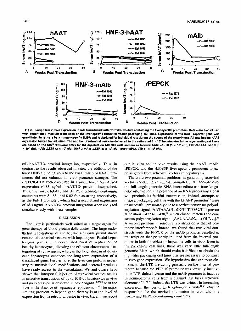
Long-Term Expression of Liver-Specijc Promoters After In Vivo Transduction
One of the principal goals of gene therapy is stable expres- sion of the transduced gene in vivo. We used conditioned medium from each of the retroviral vector packaging cell lines discussed above to transduce regenerating rat liver cells in vivo. The transduced rats were followed for serum hAAT protein expression by ELISA. hAAT was undetectable in all rats before transduction, demonstrating the specificity of the antibody for the human protein. The hAAT-ALTR construct (Fig 3A) resulted in an average expression of 43.0 ng/mL for three animals and expression was quite stable, as serum hAAT protein levels at 40 weeks posttransduction were 80% of their posttransduction week-l levels. Rats transduced with the HNF-3-hAAT-ALTR construct (Fig 3B) had an average serum level of 23.2 ng/mL for two animals and maintained 88% of their posttransduction week-l values at 40 weeks. Rats transduced with the mAlb-ALTR construct (Fig 3C) initially had mean hAAT levels of 148.5 ng/mL for two animals, but by 40 weeks posttransduction, expression had decreased to 48% of posttransduction week-l values. Al- though the HNF-3-mAlb-ALTR construct (Fig 3D) resulted in lower early levels of hAAT protein in the serum (mean 16.7 ng/mL for 2 animals) as compared with mAlb-ALTR, expression similarly decreased to 46% at 40 weeks. Despite the high titer of the rPEPCK-LTR vector (Table l), the mean serum hAAT protein level at posttransduction week 1 was 19.3 ng/mL for two animals and had decreased to 13% of this level by 20 weeks posttransduction (Fig 3E).
PCR Quantitation of Proviral DNA in the Livers of Transduced Rats
To compare in vivo promoter strengths, expression must be normalized to transduction efficiency. We isolated geno-
mic DNA from rat liver biopsy specimens from at least two animals for each promoter and used a multiplex PCR technique to amplify proviral (IRES) and rat genomic (LFABP) sequences. We minimized the potential inaccura- cies of the semiquantitative PCR technique for the estimation of transduction efficiency by (1) isolating nontransduced control rat DNA simultaneously with the transduced samples to control for contamination; (2) using primers that amplify a rat genomic sequence (LFABP) simultaneously with the retroviral (IRES) sequence to control for PCR efficiency and DNA loading; (3) analyzing the samples from all of the transduced rats, controls, and standard curve in the same PCR reaction and blots; and (4) performing the PCR reac- tions and hybridizations multiple times on the rat liver DNA samples and reporting the mean transduction efficiency of at least four independent analyses.
For semiquantitative assessment of retroviral gene fre- quency, PCR standards were created by diluting DNA iso- lated from a transduced NIH 3T3 cell clone that was shown to contain a single copy of retroviral DNA with varying amounts of nontransduced rat liver DNA (Fig 4A, lanes 1 through 6), as described previously.24 The signal for each sample was quantitated by using a Betagen two-dimensional imager and a standard curve was constructed by correcting the IRES signal for the LFABP signal. Nontransduced con- trol rat liver samples, isolated simultaneously with the trans- duced rat liver samples, showed a signal for LFABP but not for IRES, as expected (Fig 4B, lane 12).
The proviral integration frequencies of DNA from trans- duced rats were determined by comparing the ratios of the IRESLFABP signals with those of the standard curve. Based on the mean of at least four independent PCR-based compari- sons, the in vivo transduction efficiency of the mAlb-ALTR (lanes 13 and 14) vector ranged from 0.32% to 0.5% for individual rats, whereas the transduction efficiency for the HNF-3-mAlb-ALTR construct (lanes 15 and 16) was one- sixth as high at 0.055% to 0.082%. Transduction with the hAAT-ALTR construct (lanes 17 through 19) resulted in 0.034% to 0.065% of hepatocytes containing the provirus, and those transduced with the HNF-3-hAAT-ALTR con- struct (lanes 20 and 21) showed a 0.038% to 0.050% trans- duction frequency. The rPEPCK-LTR construct (lanes 10 and 11) resulted in transduction of 5.3% to 6.9% of hepato- cytes. It is noteworthy that the observed variations in retrovi- ral transduction efficiency (Table 2) are quite consistent with variations in the MtxR titer of these retroviral vectors (Table 1).
Comparison of the Relative Promoter Strengths of the Liver-Specijc Retroviral Constructs
The strengths of the liver-specific internal promoters were compared by normalizing the serum hAAT protein levels measured at the time of liver biopsy for the proviral integra- tion frequency (Table 2). As shown in Fig 5, the mean nor- malized hAAT protein expression for the mAlb-ALTR and HNF-3-mAlb-ALTR vectors was similar at 141.5 and 158.2 ng/mL hAAT/l% proviral integration, respectively. The hAAT-ALTR and HNF-3-hAAT-ALTR vectors resulted in similar levels of normalized expression at 657 and 590 ng/
3400 HAFENRICHTER ET AL
hAAT +, 100 C
7 5 3 5 0
5 2 5
* o z
-Rat 1097 -Rat 1095 -Rat IO96
0 10 20 3 0 40
A Weeks Post Transduction
-c- Rat log1 "Rat 1092 -6- Rat 1093 -0- Rat 1094
0 10 20 30 40
B Weeks Post Transduction
250
200
150
100
50
-Rat 1082
-Rat 1083
0 10 20 30 40 Weeks Post Transduction
lZ5 1 HNF-3-mAlb rPEPCK a I_ 100 5 100 m -Rat 1085
*Rat 1087 2 5o = -Rat 1086 5 50
-Rat 10
-cr. Rat 1079 I80
r " I
0 10 20 30 40 0 10 20 30
D Weeks Post Transduction E Weeks Post Transduction
40
Fig 3. Long-term in vivo expression in rats transduced with retroviral vectors containing the liver-specific promoters. Rets were t ransdud with conditioned medium from each of the liver-specific retroviral vector packaging cell lines. Expression of the hAAT reporter gene was quantitated in rat sera by a human-specific ELSA and is depicted for individual rats during the course of the experiment. All rats had no hAAT expression before transduction. The number of retroviral particles delivered to the estimated 3 x 10' hepatocytes in the regenerating rat livers are based on the W retroviral titers for the injectate on NIH 3T3 cells and are as follows: hAAT-ALTR (5 x lo5 cfu), HNF-3-hAAT-ALTR (5 x 10' du), mAlb-ALTR (3 x lo6 cfu), HNF-3-mAlb-ALTR (5 x 10' cfu), and rPEPCK-LTR (1 x 10' cfu).
mL hAAT/l% proviral integration, respectively. Thus, in contrast to the results observed in vitro, the addition of the three HNF-3 binding sites to the basal mAlb or hAAT pro- moters did not enhance in vivo promoter strength. The rPEPCK-LTR vector resulted in a much lower normalized expression (0.33 ng/mL hAAT/I% proviral integration). Thus, the mAlb, hAAT, and rPEPCK promoter containing constructs were 8-, 3 5 , and 0.02-fold as strong, respectively, as the Pol-I1 promoter, which had a normalized expression of 18.3 ng/mL hAAT/I% proviral integration when analyzed simultaneously with these samples.
DISCUSSION
The liver is particularly well suited as a target organ for gene therapy of blood protein deficiencies. The large endo- thelial fenestrations of the hepatic sinusoids permit direct contact of retroviral vectors with hepatocytes. Partial hepa- tectomy results in a coordinated burst of replication of healthy hepatocytes, allowing the efficient chromosomal in- tegration of retroviruses, whereas the long lifespan of quies- cent hepatocytes enhances the long-term expression of a transduced gene. Furthermore, the liver can perform neces- sary posttranslational modifications and secreted products have ready access to the vasculature. We and others have shown that intraportal injection of retroviral vectors results in selective transduction of up to 10% of hepatocytes in vivo and no expression is observed in other or in the liver in the absence of hepatocyte rep1i~ation.l'~~~ The major limiting problem to hepatic gene therapy is at the level of expression from a retroviral vector in vivo. Herein, we report
our in vitro and in vivo results using the hAAT, mAlb, rPEPCK, and the rLFABP liver-specific promoters to ex- press genes from retroviral vectors in hepatocytes.
There are two potential problems in generating retroviral vectors containing an internal promoter. First, because only the full-length genomic RNA intermediate can transfer ge- netic information, the presence of an RNA processing signal will preclude its faithful transmission. Indeed, attempts to make a packaging cell line with the LFABP promote?' were unsuccessful, presumably due to a perfect consensus polyad- enylation signal [AATAAA(N,,)GGlTTTGAGTT] present at position -472 to -438,32 which closely matches the con- sensus polyadenylation signal [AAUAAA(N,,,,) GU[Rich].4h A second problem in retroviral construction is that of pro- moter interferen~e.~' Indeed, we found that retroviral con- structs with the PEPCK or the mAlb promoter resulted in transcription that primarily initiated from the internal pro- moter in both fibroblast or hepatoma cells in vitro. Even in the packaging cell lines, there was very little full-length genomic RNA, which should make it difficult to obtain the high-titer packaging cell lines that are necessary to optimize in vivo gene expression. We hypothesize that enhancer ele- ments in the LTR are acting primarily on the internal pro- moter, because the PEPCK promoter was virtually inactive in an LTR-deleted vector and the mAlb promoter is inactive in nonhepatoma cells from a plasmid that lacks retroviral element^.^^.^^.^' If indeed the LTR was critical in increasing expression, the loss of LTR enhancer a~t iv i ty '~ . '~ may be responsible for the marked attenuation in vivo with the mAlb- and PEPCK-containing constructs.

IN VIVO ANALYSIS OF LIVER-SPECIFIC PROMOTERS 3401
A Standards No
'10% 3% 1 % .3% .l % .03%' Pol-ll DNA
l
L ]::nomic
1 2 3 4 5 6 7 8 9
B rPEPCK mAlb HNF3-mAlb hAAT HNF3-hAAT
1 Retrovirus
1011 121314 1516 171819 2021 Fig 4. PCR quantitation of in vivo retroviral integration frequency
in livers of the transduced rats. Liver DNA was isolated from biopsy specimens from the transduced rats at either 16 to 18 or 40 weeks posttransduction. Multiplex PCR using retroviral (IRES) and rat geno- mic (LFABPI primers was performed simultaneously on all samples. Amplified sequences were electrophoresed, transferred t o a nylon membrane, and hybridized first to an IRES probe (Chow autoradio- gram), stripped, and reprobed with a LFABP probe (6-hour autoradio- gram). Standards (lanes 1 through 61 were prepared by mixing vari- ous concentrations of DNA derived from NIH 3T3 cells containing a single copy of retroviral DNA with nontransduced rat liver DNA. Lane 9 represents a PCR mixture devoid of DNA, whereas lane 12 repre- sents DNA from a nontransduced rat liver that was simultaneously isolated with the transduced rat liver samples. Lanes 7 and 8 repre- sent Pol-II-hAAT transduced rat liver samples that had been studied previously.2' The DNA from the livers of rats transduced with the rPEPCK-LTR (lanes 10 and 11). mAlb-ALTR (lanes 13 and 141, HNF-3- mAlb-ALTR (lanes 15 and 161, hAAT-ALTR (lanes 17 through 191, and HNF-3-hAAT-ALTR (lanes 20 and 21) retroviral vectors are shown. The specific rat used in each assay is indicated above the sample.
Of the promoters that resulted in retroviral vectors which could be packaged and used to transduce new cells, the hierarchy of promoter strength is hAAT>mAlb>Pol II>>> PEPCK. The low activity of the PEPCK promoter was sur- prising, because it directs expression in transgenic and in rat hepatocytes when delivered as plasmid DNA com- plexed with galactosylated poly (L-lysine).' However, in the transgenic mouse study, the use of a heterologous reporter gene precluded quantitative comparison with the endogenous gene. In addition, Hatzoglou et al'' reported relatively low levels of expression from a retroviral vector containing a PEPCK promoter in rats, and Cheng et alx reported low expression from a plasmid construct containing the rPEPCK promoter delivered to rat livers by using a DNA ballistic
Table 2. Comparison of In Vivo Promoter Strengths of the Retroviral Constructs
hAAT Protein Proviral Integration Normalized Serum
Time of Liver hepatocytes InglmL hAAT/l% in Serum at Frequencyt I% of Expression*
Rat Biopsy. containing No. Promoter InglmL) provirus) integration)
proviral
1082 mAlb 1083 mAlb 1085 HNF-3-mAlb 1086 HNF-3-mAlb 1095 hAAT§ 1096 hAAT§ 1097 hAAT 1091 HNF-3-hAAT 1092 HNF-3-hAAT 1079 rPEPCK5 1080 rPEPCK5 13 Pol-H§
74.0 46.8 5.6
17.6 10.0 12.6 89.5 22.2 28.0 3.2 1 .o
100.0
0.540 2 .l20 0.320 f .l00 0.055 f ,016 0.082 t .023 0.034 -C .007 0.042 f .015 0.065 -c ,014 0.050 f ,030 0.038 -C ,017 6.90 t 1.04 5.30 2 1.80 5.36 t 1.98
137.0 t 30.4
101.8 f 28.8 214.8 It 60.5 294.1 t 64.7 300.0 3- 109
1,377.0 f 293 444.0 f 269 736.8 t 338
146.0 2 44.9
0.46 -c 0.07 0.19 f 0.06 18.3 f 6.9
~~
hAAT protein level in the serum of rats transduced with the liver- specific retroviral vectors at the time of liver biopsy (see Fig 3).
t Percentage of hepatocytes containing provirus as determined by at least four separate experiments performed in duplicate t the stan- dard error of the mean5' (see Fig 4 for a representative example). * Ratio of the serum hAAT protein level in the transduced rats to the proviral integration frequency.
§ Liver biopsy and serum samples were obtained at approximately 16 to 18 weeks posttransduction. All other samples were obtained approximately 40 weeks posttransduction.
device. It is possible that details of vector construction a n d or the particular cDNA used might influence expression in vivo.
Although expression from the mAlb promoter was consid- erably higher than from the PEPCK promoter, this would result in an hAAT level that is still only 1.4% of normal if
basal 1096 1095 m 1097
HNF-3 :!:l hAAT [ Pol4 13 I
l : : : : : : : (
0 200 400 600 800 lo00 1200 l400 1600 Serum hAAT (nglmL) per 1% proviral integration
Fig 5. Normalized in vivo promoter strengths of the retroviral constructs. The relative strengths of the liver-specific promoter con- structs were compared by normalizing the serum hAAT level mea- sured at the time of liver biopsy (Fig 3) for the percentage of hepato- cytes that integrated the provirus as determined by a minimum of four independent PCR-based assays of liver genomic DNA for an indi- vidual animal (Fig 4 and Table 2). Results are expressed as the mean of the serum hAAT protein level (ng/mLI/l% proviral integration 2 SEM of these determinations for an individual animal.

3402 HAFENAICHTER ET AL
100% of the cells could be transduced. This is considerably less active than the endogenous m41b gene, although a quan- titative comparison of relative promoter strength is difficult because of the use of a heterologous reporter gene. Relatively low expression is not surprising, as a 2-kb albumin enhancer increases expression 30-fold over that observed with a basal promoter in transgenic Unfortunately, inclusion of this 2-kb enhancer led to inappropriately short RNA in our hands, which precluded the generation of a retroviral vector. Indeed, Kay et all8 tested an identical promoter/enhancer for expression from a retroviral vector. A packaging cell line obtained after prolonged “ping-pong” amplication was later found to have deletion in the promoter/enhancer region that has not yet been mapped (M. Kay, personal communication, February 1994). Because the level of expression they ob- served was similar to that seen in our study,’’ we suspect that important enhancer elements were probably deleted. It is possible that a 8 3 0 - b ~ ’ ~ or 330-b~” enhancer fragment that has activity in tissue culture cells would not undergo rearrangements, but still increase expression in vivo. Alter- natively, the high level of initiation from the internal mAlb promoter in a retroviral vector might make it difficult to obtain the high-titer packaging cell lines needed for in vivo transduction. We have therefore chosen not to pursue this promoter further.
The hAAT promoter exhibited the highest level of expres- sion in vivo, with an average of 657 ngImL hAAT/l% trans- duction efficiency. Although this is stronger than the others tested, expression is still only -6.5% that of the endogenous gene. It is possible that more promoter and/or enhancer ele- ments are necessary to maximize expression. Although 2 kb of 5‘ ff anking sequence directs high-leveI expression of the genomic hAAT g e r ~ e , ~ ~ , ~ ’ shorter promoter constructs have only been tested with a heterologous reporter gene in transgenic mice, making it very difficult to quantitate the absolute level of e x p r e ~ s i o n . ~ ~ . ~ ~ Thus, although the 347-bp fragment of the hAAT promoter tested here has most of the activity of longer promoters in hepatoma cells,58 it is possible that elements important for expression in vivo were excluded because of the need to use a shorter promoter fragment to create a retroviral vector. Peng et have also shown that a 732-bp hAAT promoter functions well from a retroviral vector in hepatocytes in vitro, although this construct was never tested in vivo. We are currently adding enhancer ele- ments to attempt to further increase the level of expression. Finally, because promoters function by recruiting transcrip- tion factors to the vicinity of the start site, we reasoned that simply adding more binding sites might lead to increased levels of expression in vivo. Despite the fact that we and others?’ have observed that addition of three HNF-3 binding sites increases expression from either the mAlb or the hAAT promoter in hepatoma cells in vitro, this unfortunately had little effect on expression in vivo in this study.
Importance of the LTR Enhancer in the Retroviral Backbone
A potential criticism of our study is the fact that slightly different retroviral vectors were used to test different pro- moters. Those designated ALTR had a 178-bp deletion that
included the two 75-bp tandem repeats of the U3 enhancer region in the 3‘ LTR; all other constructs had an intact 3’ LTR. Upon reverse transcription of the retroviral RNA ge- nome, the U3 sequences from the 3’ LTR serve as a template for the synthesis of the 5’ LTR, which transfers the deletion to the 5’ LTR in the provirus and weakens the LTR promoter by -85% to 90%.“.” Such a 3‘ LTR deletion facilitates the analysis of promoter function in tissue culture C ~ I I S . ~ ~ , ~ In addition, it is possible that sequences in the LTR might exert a negative influence on expression from a retroviral vector in vivo. Richards and Huber6‘ reported that an albumin pro- motedenhancer was inactive in transgenic mice when inte- grated with retroviral sequences, although their construct was not tested in the absence of the retroviral elements. Furthermore, sequences within the LTR enhancer are respon- sible for the loss of expression from the LTR promoter in embryonic carcinoma cells6* and may be involved in the attenuation of expression from the LTR after transduction of preimplantation embryos63 or adult hepatocyte^.'^ In con- trast, others believe that the LTR enhancer does not exert a negative influence on an internal promoter. Soriano et a14.‘ tested retroviral vectors with an intact LTR or a 3’-LTR deletion identical to that used in our study and observed no significant difference in the level of expression from an inter- nal promoter in embryonic stem cells, which do not express the LTR promoter. Stewart et a163 transduced preimplantation embryos with retroviral vectors and observed that, although the LTR was inactive in vivo in all animals, the internal thymidine kinase promoter was active in the liver. Similarly, Kay et all8 tested expression from the albumin promoter/ enhancer in hepatocytes in vivo from both 3”deleted and 3’- intact LTR retroviral backbones and observed no significant difference in expression after correction for the number of retroviral particles delivered. Thus, although we believe it is unlikely that the 3’ LTR deletion will significantly effect in vivo expression, we can only formally conclude that the rPEPCK promoter i s significantly weaker than the Pol-I1 promoter in vivo, whereas the hAAT promoter is approxi- mately fourfold stronger than the mAlb promoter.
Implications for Gene Therapy
Although the stability of expression from retroviral vec- tors is quite promising for use in hepatic gene therapy, low levels of protein production per integrated provirus currently limits the level of in vivo expression. Although in vitro studies are an excellent means of developing and screening retroviral vectors, the discrepancy between in vivo and in vitro comparisons of promoter strengths underscore the im- portance of quantitative in vivo comparisons. We conclude from our studies that the hAAT promoter is more efficiently expressed in vivo than the other liver-specific, cellular, or viral promoters tested. Furthermore, the absence of initiation from the liver-specific hAAT promoter in the fibroblast-de- rived packaging cell lines has allowed us to obtain high-titer retroviral vectors by using “ping-pong” amplification that are currently being tested in vivo. We predict that a 10% transduction efficiency will result in a serum hAAT level of 6 ,ug/mL. Although this level is still insufficient for the gene therapy of hAAT deficiency (at least 150 pgmL is needed),

IN VIVO ANALYSIS OF LIVER-SPECIFIC PROMOTERS 3403
it would be in the therapeutic range for other serum protein deficiencies such as factor IX, factor X, or protein C.
ACKNOWLEDGMENT
We thank Richard Hanson and Jin Sung Liu of Case Western University (Cleveland OH), Kenneth Zaret of Brown University (Providence, RI), Jeffrey Gordon of Washington University (St Louis, MO), and Rong-Fong Shen and Savio L.C. Woo of Baylor University (Houston, TX) for supplying plasmid constructs.
REFERENCES
1. Starzl TE, Demetris AJ, Van Thiel D Liver Transplantation. N Engl J Med 321:1014, 1989
2. Wilson JM, Chowdhury NR, Grossman M, Wajsman R, Ep- stein A, Mulligan RC, Chowdhury JR: Temporary amelioration of hyperlipidemia in low density lipoprotein receptor-deficient rabbits transplanted with genetically modified hepatocytes. Proc Natl Acad Sci USA 87:8437, 1990
3. Chowdhury JR, Grossman M, Gupta S, Chowdhury NR, Baker JR, Wilson JM: Long-term improvement of hypercholesterolemia after ex vivo gene therapy in LDL-R deficient rabbits. Science 254:1802, 1991
4. Kay MA, Baley P, Rothenberg S, Leland F, Fleming L, Ponder KP, Liu TJ, Finegold M, Darlington G, Pokorny W, Woo SLC: Expression of human alpha,-antitrypsin in dogs after autologous transplantation of retroviral transduced hepatocytes. Proc Natl Acad Sci USA 89:89, 1992
5. Grossman M, Raper SE, Kozarsky K, Stein EA, Engelhardt JF, Muller D, Lupien PJ, Wilson JM. Successful ex vivo gene therapy directed to the liver in a patient with familial hypercholesteremia. Nature Genet 6:334, 1994
6. Wu CH, Wilson JM, Wu CY: Targeting genes: Delivery and persistent expression of a foreign gene driven by mammalian regula- tory elements in vivo. J Biol Chem 264:16985, 1989
7. Wilson JM, Grossman M, Cabrera JA, Wu C, Wu GY: A novel mechanism for achieving transgenic persistence in vivo after somatic gene transfer into hepatocytes. J Biol Chem 267:11483, 1992
8. Cheng L, Ziegelhoffer PR, Yang N-S: In vivo promoter activity and transgene expression in mammalian somatic tissues evaluated by using particle bombardment. Proc Natl Acad Sci USA 90:4455, 1993
9. Perales JC, Ferkol T, Beegen H, Ratnoff OD, Hanson RW: Gene transfer in vivo: Sustained expression of genes introduced into the liver by receptor-targeted uptake. Roc Natl Acad Sci USA 91:4086, 1994
10. Stratford-Pemcaudet LD, Levrero M, Chasse J-F, Pemcaudet M, Briand P: Evaluation of the transfer and expression in mice of an enzyme-encoding gene using a human adenovirus vector. Hum Gene Ther 1:241, 1990
1 1. Jaffe H, Dane1 C, Longenecker G, Metzger M, Setoguchi Y, Rosenfeld MA, Grant W , Thorgeirsson SS, Stratford-Perricaudet LD, Pemcaudet M, Pavirani A, Lecocq J-P, Crystal R: Adenovirus- mediated in vivo gene transfer and expression in normal rat liver. Nature Genetics 1:372, 1992
12. Li Q, Kay MA, Finegold M, Stratford-Pemcaudet LD, Woo SLC: Assessment of recombinant adenoviral vectors for hepatic gene therapy. Hum Gen Ther 4:403, 1993
13. Herz J, Gerard RD: Adenovirus mediated transfer of low density lipoprotein receptor acutely accelerates cholesterol clearance in normal mice. Proc Natl Acad Sci USA 90:2812, 1993
14. Kay, MA, Landen CN, Rothenberg SR, Taylor LA, Leland F, Wiehle S, Fang B, Bellinger D, Finegold M, Thompson AR, Read M, Brinkhous KM, Woo SLC: In vivo hepatic gene therapy:
Complete albeit transient correction of factor IX deficiency in hemo- philia B dogs. Proc Natl Acad Sci USA 91:2353, 1994
15. Hatzoglou M, Lamers W, Bosch F, Wynshaw-Boris A, Clapp D, Hanson RW: Hepatic gene transfer in animals using retroviruses containing the promoter from the gene for phosphoenolpyruvate carboxykinase. J Biol Chem 265:17285, 1990
16. Kaleko M, Garcia JV, Miller AD: Persistent gene expression after retroviral gene transfer into liver cells in vivo. Hum Gene Ther 2:27, 1991
17. Ferry N. Duplessis 0, Houssin D, Danos 0, Heard JM: Ret- roviral-mediated gene transfer into hepatocytes in vivo. Proc Natl Acad Sci USA 88:8377, 1991
18. Kay MA, Li Q, Liu T-J, Leland F, Toman C, Finegold M, Woo S E : Hepatic gene therapy: Persistent expression of human alpha-l-antitrypsin in mice after direct gene delivery in vivo. Hum Gene Ther 3:641, 1992
19. Rozga J, Moscioni AD, Neuzil D, Demetriou AA: A model for directed foreign gene delivery to rat liver cells in vivo. J Surg Res 52:209, 1992
20. Kay MA, Rothenberg S, Landen CN, Bellinger DA, Leland F, Toman C, Finegold M, Thompson AR, Read MS, Brinkhous KM, Woo SLC: In vivo gene therapy of hemophilia B Sustained partial correction in factor IX deficient dogs. Science 266:117, 1993
21. Cardoso JE, Branchereau S, Roy JP, Houssin D, Danos 0, Heard JM: In situ retrovirus medated gene transfer into dog liver. Hum Gene Ther 4:411, 1993
22. Kolodka TM, Finegold M, Woo SLC: Hepatic gene therapy: Efficient retroviral mediated transfer into rat hepatocytes in vivo. Som Cell Mol Genet 19:491, 1993
23. Rettinger SD, Ponder KP, Saylors RL, Kennedy SC, Hafen- richter DC, Flye MW: In vivo hepatocyte transduction with retrovirus during hepatic inflow occlusion. J Surg Res 54:418, 1993
24. Rettinger SD, Kennedy SC, Wu X, Saylors RS, Hafenrichter DG, Flye MW, Ponder Kp. Liver-directed gene therapy: Quantitative evaluation of promoter elements using in vivo retroviral transduction. Proc Natl Acad Sci USA 91:1460, 1994
25. Ponder KP, Dunbar R, Wilson D, Darlington G, Woo SLC: Evaluation of relative promoter strength in primary hepatocytes us- ing optimized lipofection. Hum Gene Ther 2:41, 1991
26. Armentano D, Thompson AR, Darlington G, Woo SLC: Ex- pression of human factor IX in rabbit hepatocytes by retrovirus mediated gene transfer: Potential for gene therapy of hemophilia B. Proc Natl Acad Sci USA 87:6141, 1990
27. Wilson JM, Jefferson DM, Chowdhury JR, Novikoff PM, Johnston DE, Mulligan RC: Retrovirus mediated transduction of adult hepatocytes. Proc Natl Acad Sci USA 85:3014, 1988
28. Palmer TD, Rosman G, Osborne W, Miller AD: Genetically modified skin fibroblasts persist long after transplantation but gradu- ally inactivate introduced genes. Proc Natl Acad Sci USA 88:1330, 1991
29. Scharfmann R, Axelrod JH, Verma IM: Long-term in vivo expression of retrovirus-mediated gene transfer in mouse fibroblast implants. Proc Natl Acad Sci USA 88:4626, 1991
30. Miller AD, Rosman GJ: Improved retroviral vectors for gene transfer and expression. Biotechniques 7:980, 1989
31. Liu J, Park M, Gurney A, Roesler W, Hanson RW: Cyclic AMP induction of phosphoenolpyruvate carboxykinase (GTP) gene transcription is mediated by multiple promoter elements. J Biol Chem 266: 19095, 199 1
32. Sweetser DA, Lowe JB, Gordon JI: The nucleotide sequence of the rat liver fatty acid binding protein gene. J Biol Chem 261:5553, 1986
33. DiPersio CM, Jackson DA, Zaret KS: The extracellular matrix coordinately modulated liver transcription factors and hepatocyte morphology. Mol Cell Biol I 1 :4405, 1991

3404 HAFENRICHTER ET AL
34. Shen RF, Clift S, DeMayo J, Sifers R, Finegold M, Woo SLC: Tissue-specific regulation of human alpha l-antitrypsin gene expression in transgenic mice. DNA 8:101, 1989
35. Markowitz D, Goff S, Bank A: Construction and use of a safe and efficient amphotropic packaging cell line. Virology 167:400, 1988
36. Markowitz D, Goff S, Bank A: A safe packaging line for gene transfer: Separating viral genes on two different plasmids. J Virol 62:1120, 1988
37. Kozak SL, Kabat D: Ping-pong amplification of a retroviral vector achieves high-level gene expression: Human growth hormone production. J Virol 64:3500, 1990
38. Jainchill JL, Aaronson SA, Todaro GJ: Murine sarcoma and leukemia viruses: Assay using clonal lines of contact inhibited mouse cells. J Virol 4549, 1969
39. Darlington G: Liver cell lines. Methods Enzymol 151:19, 1987
40. Hatzoglou M, Park E, Wynshaw-Boris A, Cheng Kaung HL, Hanson RW: Hormonal regulation of chimeric genes containing the phosphoenolpyruvate carboxykinase promoter regulatory region in hepatoma cells infected by murine retroviruses. J Biol Chem 263:17798, 1988
41. Sambrook J, Fritsch EF, Maniatis T: Molecular Cloning: A Laboratory Manual. Cold Spring Harbor, NY, Cold Spring Harbor Laboratory, 1989, p E3, 7.71, 9.16, and 10.13
42. Ghattas IR, Sanes JR, Majors JE: The encephalomyocarditis virus internal ribosome entry site allows efficient co-expression of two genes from a recombinant provirus in cultured cells and in embryos. Mol Cell Biol 11:5848, 1991
43. Soriano P, Friedrich G, Lawinger P: Promoter interactions in retrovirus vectors introduced into fibroblasts and embryonic stem cells. J Virol 65:2314, 1991
44. Graves BJ, Eisenman RN, McKnight SL: Delineation of tran- scriptional control signals within the Moloney murine sarcoma virus long terminal repeat. Mol Cell Biol 5:1948, 1985
45. Hatzoglou M, Bosch F, Park E, Hanson RW: Hormonal con- trol of interacting promoters introduced into cells by retroviruses. J Biol Chem 26623416, 1991
46. Steitz JA, Black DL, Gerke V, Parker KA, Keller W: Func- tions of abundant U-snRNPs, in Small Nuclear Ribonucleoprotein Particles: Structure and Function. New York, NY, Springer-Verlag, 1987
47. Heard JM, Herbomel P, Ott MO, Mottura-Rollier A, Weiss M, Yaniv M: Determinants of rat albumin promoter tissue specificity analyzed by an improved transient expression system. Mol Cell Biol 7:2425, 1987
48. Izban MG, Papaconstantinou J: Cell-specific expression of mouse albumin promoter. J Biol Chem 264:9171, 1989
49. Herbst RS, Friedman N, Damell JE, Babiss LE: Positive and negative regulatory elements in the mouse albumin enhancer. Proc Natl Acad Sci USA 86:1553, 1989
50. Hu J, Camper SA, Tilghman SM, Miller T, Georgoff I , Serra R, Isom HC: Functional analysis of albumin expression in a series of hepatocyte cell lines and in primary hepatocytes. Cell Growth Diff 3:577, 1992
51, Babiss LE, Friedmann JM, Damell JE: Cellular promoters incorporated into the adenovirus genome: Effects of viral regulatory elements on transcription rates and cell specificity of albumin and &globin promoters. Mol Cell Biol 6:3798, 1986
52. Daniel W: Biostastics: A Foundation For Analysis In The Health Sciences. New York, NY, Wiley, 1991
53. Sweetser DA, Birkenmeier EH, Hoppe PC, McKeel DW. Gor- don JI: Mechanisms underlying generation of gradients in gene ex- pression within the intestine: An analysis using transgenic mice containing fatty acid binding protein-human growth hormone fusion genes. Genes Dev 2: 13 18, 1988
54. McGrane MM, De Vente J, Yan J, Bloom J, Park E, Wyn- shaw-Boris A, Wagner T, Rothman F, Hanson RW: Tissue specific expression and dietary regulation of a chimeric phosphoenolpyruvate carboxykinaselbovine growth hormone gene in transgenic mice. J Biol Chem 263: 11443, 1988
55. Pinkert CA, Ornitz DM, Brinster RL, Palmiter RD: An albu- min enhancer located 10 kb upstream functions along with its pro- moter to direct efficient, liver-specific expression in transgenic mice. Genes Dev 1268, 1987
56. Ponder KP, Leland F, Gupta S, Chowdhury JR. Darlington G, Finegold M, Woo SLC: Mouse hepatocytes migrate to liver paren- chyma and function indefinitely after intrasplenic transplantation. Proc Natl Acad Sci USA 88:1217, 1991
57. Sifers RN, Carlson JA, Clift SM, DeMayo FJ, Bullock DW, Woo SLC: Tissue specific expression of the human alpha-l-antitryp- sin gene in transgenic mice. Nucleic Acids Res 15:1459, 1987
58. Shen RF, Li V, Sifers RN, Wang H, Hardick C, Tsai SY, Woo SLC: Tissue specific expression of the human alpha- l-antiuyp- sin gene is controlled by multiple cis-acting regulatory elements. Nucleic Acids Res 15:8399, 1987
59. Peng H, Armentano D, Mackenzie-Graham L, Shen RF, Dar- lington G, Ledley FD, Woo SLC: Retroviral mediated gene transfer and gene expression of human phenylalanine hydroxylase in primary mouse hepatocytes. Proc Natl Acad Sci USA 85:8146, 1988
60. Yee JK, Moores JC, Jolly DJ, Wolff JA, Respess JG, Fried- mann T: Gene expression from transcriptionally disabled retroviral vectors. Proc Natl Acad Sci USA 845197, 1987
61. Richards CA, Huber BE: Generation of a transgenic model for retrovirus-mediated gene therapy for hepatocellular carcinoma is thwarted by the lack of transgene expression. Hum Gen Ther 4:143, 1993
62. Tsukiyama T, Niwa 0 , Yokoro K: Mechanism of suppression of the long terminal repeat of moloney murine leukemia virus in mouse embryonal carcinoma cells. Mol Cell Biol 9:4670, 1989
63. Stewart CL, Schuetze S, Vanek M, Wagner EF: Expression of retroviral vectors in transgenic mice obtained by embryonic infec- tion. EMBO J 2:383, 1987