quantitative autoradiographic localization of ... · the journal of neuroscience, june 1988, c?(6):...
TRANSCRIPT
The Journal of Neuroscience, June 1988, C?(6): 2003-2010
Quantitative Autoradiographic Localization of Prostaglandin E, Binding Sites in Monkey Diencephalon
Yumiko Watanabe,’ Yasuyoshi Watanabe,‘B2 and Osamu Hayaishil
‘Hayaishi Bioinformation Transfer Project, Research Development Corporation of Japan, Kyoto 601, and *Department of Medical Chemistry, Osaka Medical College, Osaka 569, Japan
Quantitative autoradiography was performed to investigate the mapping of prostaglandin E, binding sites in the Macaca fuscata fuscafa diencephalon. Autoradiographs were pre- pared by incubation of lo-pm-thick serial frozen sections with 3H-prostaglandin E, and were processed by using a rotating drum-scanner and a computer-assisted image-pro- cessing system with 3H-microscales as standards. The lo- calization of prostaglandin E, binding sites was remarkably discrete in the diencephalon. The highest concentrations were found in the median and medial preoptic areas, supra- mammillary nucleus of the hypothalamus, and centromedian nucleus of the thalamus. High density was observed in the medial and dorsal hypothalamic areas; paraventricular, an- terior, dorsomedial, and infundibular nuclei of the hypothal- amus; and in the anteroventral, periventricular, paraventricu- lar, laterodorsal, and habenular nuclei of the thalamus. The distribution correlates well with the known effects of pros- taglandin E, and may also give us useful clues in unveiling the novel role of prostaglandin E, in a variety of brain func- tions.
Prostaglandin (PG) E series has various neurophysiological functions, such as in the central mediation of fever (Milton and Wendlandt, 1970; Potts and East, 1972; Milton, 1976) as one of the major regulators of food intake (Scaramuzzi et al., 1970) and luteinizing hormone release (Ojeda et al., 1977; Kinoshita et al., 1982) and as an endogenous anticonvulsant (Poddubiuk, 1976; Rosenkrantz, 1978). The high-affinity and specific binding protein of 3H-PGE, was found in membrane preparations from bovine pineal gland (Cardinali et al., 1979) and from rat (Malet et al., 1982) and human (Watanabe et al., 1985a) brains. From a regional study of dissected brain areas, PGE, binding was found to be high in the hypothalamus, pituitary, and amygdala of rat brain (Malet et al., 1982) and in the amygdala, hypo- thalamus, and hippocampus of postmortem human brain (Wa- tanabe et al., 1985a). The PGE, receptor protein was solubilized
Received Apr. 22, 1987; revised Sept. 15, 1987; accepted Sept. 29, 1987. We are indebted to Dr. H. Suginami of Ehime University, School of Medicine;
Dr. A. Yamashita of Kanebo Pharmaceutical Co.; and Dr. S. Ito, Mr. K. Hamada, and Dr. N. Yumoto of our project for useful discussions. We also thank Mr. T. Ueno and Mr. H. Yamada for photoprints and Dr. L. D. Frye for critical reading of the manuscript. Y.W. was supported in part by a Grant-in-Aid for Special Project Research of Plasticity of Neural Circuits from the Japanese Ministry of Education, Science and Culture, and by grants from the Japanese Foundation of Metabolism and Diseases snd .%nkyo Co., Ltd.
Correspondence should be addressed to Dr. Osamu Hayaishi, Hayaishi Bioin- formation Transfer Project, Research Development Corporation of Japan, % Osaka Medical College, 2-7 Daigaku-machi, Takatasuki, Osaka 569, Japan,
Copyright 0 1988 Society for Neuroscience 0270-6474/88/062003-08$02.00/O
in our laboratory in an active form from porcine brain using 3-[(3-cholamidopropyl) dimethylammoniol-l-propane sulfo- nate (Yumoto et al., 1986a). Furthermore, PGE, binding to the receptor is sensitive to guanine nucleotide (GTP), and, after solubilization, the receptor and GTP-regulatory protein were separated by gel filtration using high-performance liquid chro- matography (HPLC) and were reconstituted (Yumoto et al., 1986b). These results provide evidence that PGE, in the brain exerts various neurophysiological functions via specific receptor binding, and that GTP is involved in its regulation. Recently, we demonstrated the localization of PGD, binding by using in vitro autoradiography coupled with a computerized image-anal- ysis system (Watanabe et al., 1983; Yamashita et al., 1983). The specific PGD, binding was predominantly localized in the gray matter in the rat brain (Yamashita et al., 1983) and, more pre- cisely, in specific neuronal cells, such as the Purkinje cell layer in the swine cerebellum (Watanabe et al., 1983). Preliminarily, we obtained the distinct localizations of PGD,, PGE,, and PGF,, bindings in the monkey brain (Watanabe et al., 1985b, 1986). In this study, we developed a digital subtraction method for the quantitation of autoradiographs and demonstrated by detailed mapping the concentration of PGE, binding sites in the monkey diencephalon.
Materials and Methods Tissue preparation. Macaca fuscata fuscata (female, 6.5-8.0 kg) was anesthetized with Ketalar (ketamine-HCl) and perfused via the left ventricle with cold 10 mM sodium phosphate-buffered saline (pH 7.4) containing 5% sucrose. The brain was rapidly removed, dissected into 3-4 mm-thick blocks (ca. 2.0 x 2.5 cm), and frozen on dry ice. Frozen serial sections of 10 brn thickness were cut in a cryostat at - 14°C and mounted on gelatin-coated glass slides.
‘H-PGE, binding. All procedures of ‘H-PGE, binding assay were performed at 4°C as described by Yamashita et al. (1983) and Watanabe et al. (1983) with a slight modification. After preincubation in four 150 ml changes of 50 mM Tris-HCl (pH 7.4) containing 0.1 M NaCl (buffer A) for a total of 1 hr, the tissue sections were incubated with 150-200 ~1 (the volume varied depending on the size of section) of 20 nM 3H- PGE, in buffer A for 30 min with gentle shaking (60 times/min, 4 cm reciprocal). The sections were then rinsed in 4 sequential short (15 set each) dips in 50 ml of buffer A, quickly splashed with distilled water, and air-dried. Nonspecific binding was obtained with the consecutive sections by the addition of 100 PM unlabeled PGE, to the incubation mixture.
The ionic condition and the concentration of IH-PGE, for the incu- bation of sections were determined according to the results of the 3H- PGE, binding to the Pz fraction of porcine cerebral cortex (Ynmoto et al., 1986a). The Kd value of the ‘H-PGE2 binding to that fraction was a few nanomolar at 37°C. Since the PGE, binding sites showed a tem- perature-dependent rise in affinity (K,, for PGEz binding to the P2 fraction prepared from monkey diencephalon and amygdala, 1.1 nM at 37°C and

2004 Watanabe et al. * Prostaglandin E, Receptor in Monkey Diencephalon
Relative optical density
Figure 1. Typical standard curve for the tissue equivalent tritium con- tent versus relative optical density of each layer of JH-microscales. 0, High-level microscale; 0, low-level microscale. Calibration curve (dot- ted line) was generated from the log-log relationship of the tritium content with the corresponding relative optical density for each layer in a high-level ‘H-microscale: Y = up, where a = 0.00189 and b = 1.84 (rl = 0.9987), and for each layer in a low-level JH-microscale: Y = cX + d, where c = 0.0776 and d = - 1.40. The relative optical density of the film background (BG) was 18.1. The standard deviation of the average density of each layer of low and high JH-microscales was very small; for the SDS of the lower 3 layers of high-level microscale (1.3, 4.9 and 7.8 nCi/mg tissue), those most frequently used for calibration were 0.13, 0.072, and 0.070% of the average, respectively, on the basis of data from 10 separate densitometry readings.
14 nM at 4°C; Y. Watanabe, N. Yumoto, and M. Hatanaka, unpublished observations), 20 nM )H-PGE, might be close to the saturated level of all the binding sites.
The association ofthe specific ‘H-PGE? binding to the section reached a plateau within 30 min at 4°C. For the dissociation of bound ‘H-PGE, from incubated sections, we have no extensive time course, but 1 or 2 further 15 set dips of the section into the buffer following the routine 4 dips resulted in little change in the total and nonspecific bindings.
Autoradiography. After being dried in a desiccator, the slides were tightly juxtaposed with tritium-sensitive films (Ultrofilm, LKB) and stored at 4°C for 4 weeks. ‘H-Plastic standards (3H-microscales; Amer- sham) were included in each cassette. After exposure, the films were developed in Kodak D19 at 20°C for 5 min and then fixed for 5 min. The nuclei were identified by toluidine blue staining on the adjacent sections, with reference to the stereotaxic atlases of Emmers and Akert (1963) and Kusama and Mabuchi (1970).
Computer-assisted image-processing system. Films were analyzed us- ing a computerized image-analysis system, including a rotating-drum scanning densitometer (Model 2605; Kimoto, Tokyo) and a minicom- puter system (ECLIPSE S/120; Data General, Westboro, MA). The rotating-drum scanning densitometer was used to convert the photo- metric data from the autoradiographs into the digital values of 0 to 255 (relative O.D. value). The digitized data were arranged in parallel with the optical density between 0 and 1.5. Sampling pitches were 50 pm. The array of optical density readings was processed through a mini- computer. These data were converted to pseudocolor images on an image display (1024 x 1024 elements).
To produce quantitative images of the autoradiographs of )H-PGE2 binding, we transformed the original O.D. value ofthe pixels (pictures +
elements) to the binding value (BD value) that was linearly related to the quantity of ZH-PGE, bound in each pixel. This transformation was processed via the following steps: First, the mean O.D. value of each layer of ‘H-microscales measured by the rotating-drum scanner was plotted against each tritium content. These data were expressed as a power function, Y = ax’, where a and b were different depending on the exposure periods, the batches, and the developing conditions of the films. Then, according to this standard curve, the original O.D. value of each pixel in the image was converted to the BD value (fmol 3H- PGE,/mg tissue). This linearization procedure was performed for each sheet of autoradiographic film with a set of ‘H-plastic standards.
After the conversion of the O.D. value to the BD value, further pro- cessing via subtraction between the 2 images (total and nonspecific) was performed. To superimpose the image of nonspecific binding precisely on that of total binding, we selected several characteristic points in both images. Using their locations, we transferred the image of the nonspecific binding to the position of the image of the total binding by parallel and rotational transforms (alline transforms by the least-squares method). In affine transforms, the BD value of each pixel of the image was ob- tained from that of the corresponding pixels of the original image by linear interpolation. After all positions of the images were put into exactly the same position, subtraction among the BD values was per- formed to give the image of the specific binding. To determine the average concentration of ?H-PGE2 bound in various nuclei, a cursor box was positioned over each nucleus in the image ofthe specific binding on the color display. The average BD value within the cursor box was calculated by the computer.
Materials. [5,6,8,11,12,14,15-‘H (N)]PGE, (165 Ci/mmol) and Om- nifluor were purchased from New England Nuclear (Boston, MA). Un- labeled PGE, was the generous gift of Ono Pharmaceutical Co. (Osaka). Autoradiographic 3H-microscales (RPA 501, batch 3, and RPA 507, batch 1) were obtained from Amersham.
Results A typical standard curve for transformation of the O.D. value to the BD value in shown in Figure 1. Both high and low mi- croscales consisted of 8 layers. Eight points of high microscale were described as a power function, while 8 points of low mi- croscale were fitted for a linear function. The boundary points between low and high microscales were fitted for both functions. Therefore, we employed a straight line for the standard curve that was between 0 and 1.3 nCi/mg tissue of the BD value and used a power function for the curve above 1.3 nCi/mg. In all cases in this study, the goodness of fit of the standard curve with high microscale was ascertained; the relative coefficients (r2) were greater than 0.998.
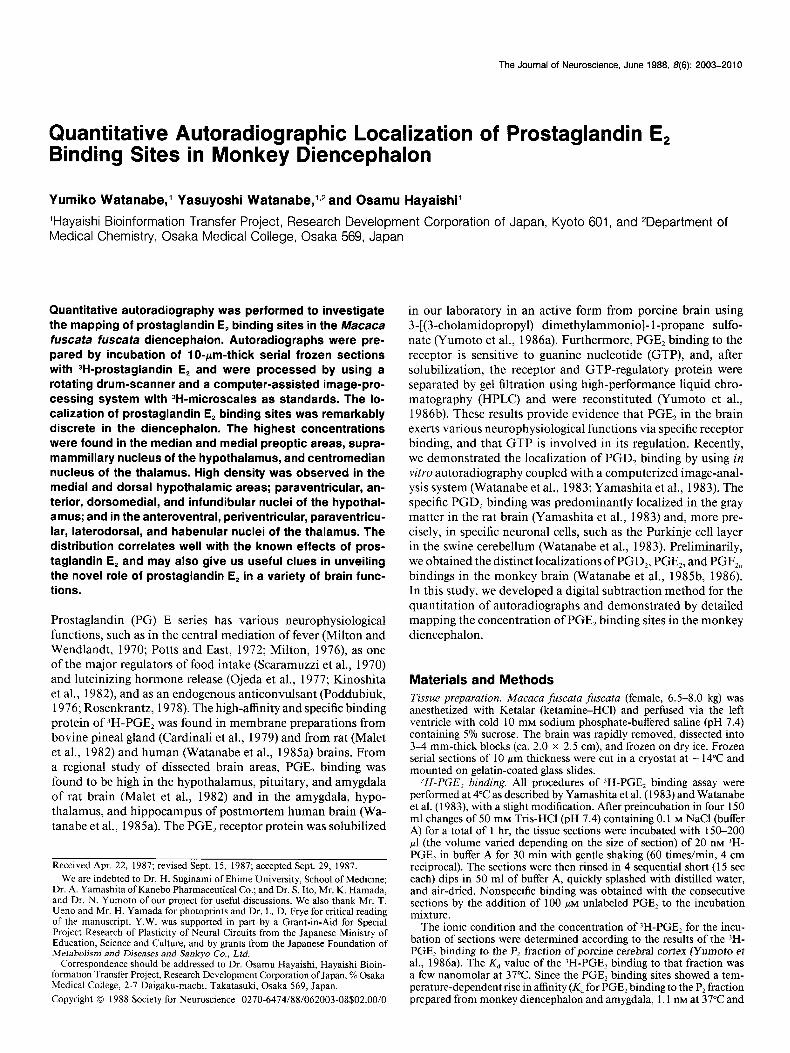
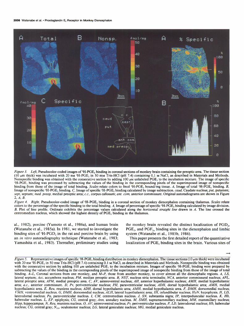
According to the above-mentioned standard curve, the ‘H- PGEz content bound in each pixel was calculated for the total and nonspecific binding. An example of this quantitation pro- cedure is shown in Figures 2 and 3. Figure 2, A, B, represents the original autoradiograms of total and nonspecific binding of IH-PGE, in coronal sections of monkey brain containing the preoptic area. Figure 3, A, B, represents the corresponding pseu- docolor-coded images. While the nonspecific binding (Fig. 3B) was low and rather homogeneously distributed, the total binding (Fig. 3A) was remarkably concentrated in the medial preoptic area. The image of the specific binding (Fig. 3C) was then pro- cessed by subtracting the binding values of the image of non- specific binding from those of total binding. The image of the specific binding exhibited a pattern very similar to that of the total binding. The proportion of specific binding to the total binding was calculated to be more than 80% in the medial preoptic area. A similar presentation of the percentage of specific binding in the specific binding image is also possible; a typical result is shown in Figure 4. The BD value of each pixel in the image of the specific binding was divided by that of the corre- sponding pixel in the image of the total binding to produce the image of the percentage of specific )H-PGE, binding (Fig. 4A).

The Journal of Neuroscience, June 1988, 8(8) 2005
Figure 2. Original autoradiograms of ‘H-PGE, binding in coronal sections of monkey brain containing the preoptic area. The tissue section (10 pm thick) was incubated with 20 nM 3H-PGE, in 50 mM Tris-HCl (pH 7.4) containing 0.1 M NaCl, as described in Materials and Methods. A, Total binding. B, Nonspecific binding, obtained with the consecutive section by adding 100 PM unlabeled PGE, to the incubation mixture.
Figure 4B shows the percentage of the specific binding value of each pixel along the horizontal straight line in Figure 4A. The highest specific binding in the centromedian nucleus reached 80% of the total binding.
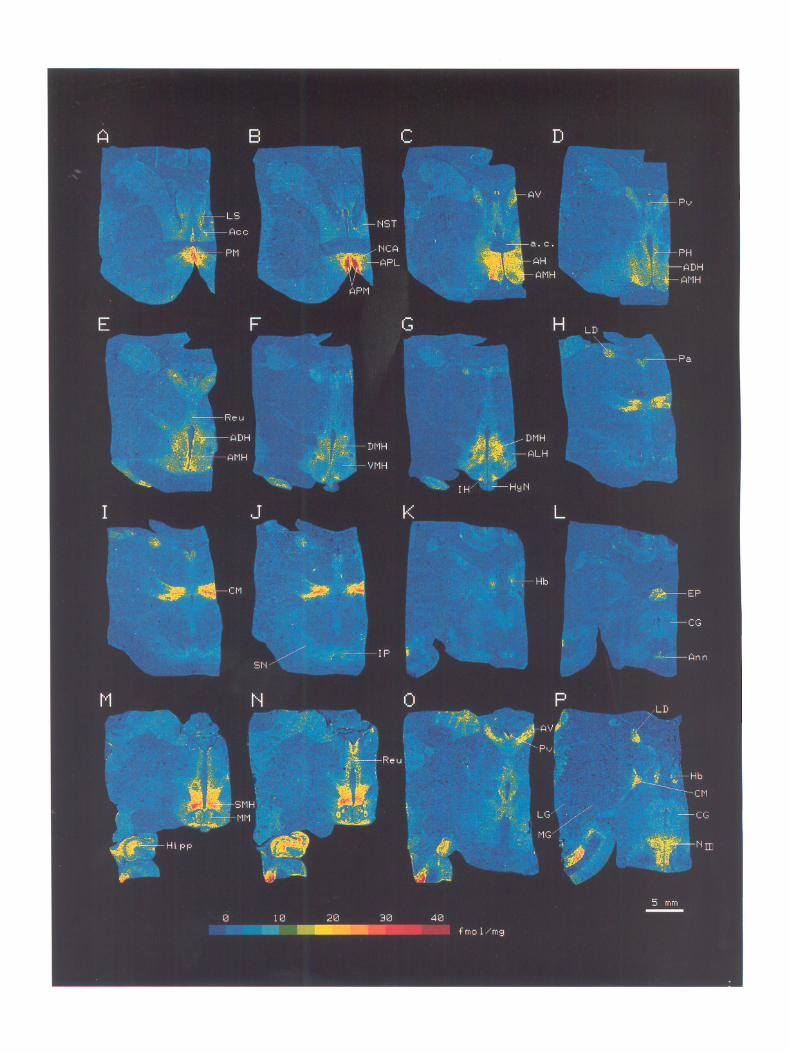
The localization of 3H-PGE, binding was characteristic and discrete throughout the diencephalon (Fig. 5). The average 3H- PGE, concentrations in various nuclei were calculated and are summarized in Table 1. For example, the anteroventral nucleus of the thalamus is seen in Figure 5, C-G, and the centromedian nucleus of the thalamus in Figure 5, H-J. Their respective lengths were at least 1.5 and 1.9 mm in the sagittal direction, as judged from the section thickness (10 pm) and the number of sections. Since the mean value of 3H-PGE, binding in each nucleus varied anteriorly to posteriorly, we selected the highest values for pre- sentation in Table 1.
The densities of 3H-PGE, binding sites were by far the highest (2 30 fmol/mg tissue) in the median and medial preoptic areas (Fig. 5, A, B), supramammillary nucleus of the hypothalamus (Fig. 5, M, iV), and centromedian nucleus of the thalamus (Fig. 5, H-J, P).
In the limbic forebrain, a moderately high density (16 fmol/ mg tissue) of PGE, binding sites was observed in the anterior commissural nucleus (Fig. 5, A, B), and a moderate concentra- tion (1 l-l 3 fmol/mg tissue) was observed in the lateral septum and accumbens nucleus (Fig. 5A).
In the hypothalamus, high densities (17-2 1 fmol/mg tissue) of PGE, binding sites were observed in the paraventricular nu- cleus, anterior nucleus, dorsal and medial hypothalamic areas,
dorsomedial nucleus, and infundibular nucleus. Moderate den- sities (12-14 fmol/mg tissue) were found in the lateral hypo- thalamic area and mammillary nucleus (Fig. 5, C-G, M, N).
In the thalamus, PGE, binding sites were the most discretely located. In addition to a very high density of binding sites in the centromedian nucleus, the nuclei surrounding the third ven- tricle, such as the anteroventral (Fig. 5, C-G, O), p&ventricular (Fig. 5, D, E, O), paraventricular (Fig. 5, H-J), and laterodorsal (Fig. 5, H-J, P) nuclei showed high densities (16-22 fmoVmg tissue), and the reuniens nucleus (Fig. 5, E, N) showed moderate density (11 fmol/mg tissue). The habenular nucleus (Fig. 5, K, P) also possessed a high concentration of PGE, binding sites.
In the midbrain, the annulary nucleus (Fig. 5L) and oculo- motor nucleus (Fig. 5P) displayed high densities (16-17 fmol/ mg tissue) of the binding sites.
We employed 2 monkey brains in the quantitative study and 4 other brains in the qualitative study. We observed little het- erogeneity among the brain specimens in the pattern of local- ization of PGE, binding sites throughout the diencephalon.
Discussion
A number of reports have depicted the central action of PGE, mainly on the basis of pharmacological studies. However, there had been no reports concerning the precise localization of PGE, receptors in the CNS nor in the peripheral tissue before our recent studies. High-affinity PGE, binding was demonstrated by the glass filter assay using membrane preparations of bovine pineal gland (Cardinali et al., 1979) and that of rat (Malet et
2006 Watanabe et al. * Prostaglandin E, Receptor in Monkey Diencephalon
-’
,i
9
3mm
f mu 1 /ma c iptlcific 40 B -1
30
20
10
I
Figure 3. Left, Pseudocolor-coded images of )H-PGE, binding in coronal sections of monkey brain containing the preoptic area. The tissue section (10 ym thick) was incubated with 20 nM )H-PGE, in 50 mM Tris-HCl (pH 7.4) containing 0.1 M NaCl, as described in Materials and Methods. Nonspecific binding was obtained with the consecutive section by adding 100 PM unlabeled PGE, to the incubation mixture. The image of specific ‘H-PGE? binding was processed by subtracting the values of the binding in the corresponding pixels of the superimposed image of nonspecific binding from those of the image of total binding. Scales relate colors to fmol 3H-PGE2 bound/mg tissue. A, Image of total IH-PGE2 binding. B, Image of nonspecific ‘H-PGE2 binding. C, Image of specific 3H-PGE, binding calculated by image subtraction. cuud, Caudate nucleus; put, putamen; sept, septum; med. preop, medial preoptic area; c.c., corpus callosum; ant. corn, anterior commissure. Original autoradiograms are shown in Figure 2, A, B. Figure 4. Right, Pseudocolor-coded image of ‘H-PGE, binding in a coronal section of monkey diencephalon containing thalamus. Scales relate colors to the percentage of the specific binding to the total binding. A, Image of percentage of specific ‘H-PGE, binding calculated by image division. B, Plot of line profile. Ordinate exhibits the percentage values calculated along the horizontal straight line drawn in A. The line crossed the centromedian nucleus, which showed the highest density of PGE, binding in the thalamus.
al., 1982), porcine (Yumoto et al., 1986a), and human brain the monkey brain revealed the distinct localization of PGD,, (Watanabe et al., 1985a). In 198 1, we started to investigate the PGE,, and PGF,, binding sites in the diencephalon and limbic binding sites of 3H-PGD, in the rat and porcine brain by using system (Watanabe et al., 1985b, 1986). an in vitro autoradiography technique (Watanabe et al., 1983; This paper presents the first detailed report of the quantitative Yamashita et al., 1983). Thereafter, preliminary studies using localization of PGE, binding sites in the brain. Various sites of
Figure 5. Representative images of specific 3H-PGE, binding distribution in monkey diencephalon. The tissue sections (10 Km thick) were incubated with 20 nM )H-PGE2 in 50 mM Tris-HCl (pH 7.4) containing 0.1 M NaCl, as described in Materials and Methods. Nonspecific binding was obtained with the consecutive section by adding 100 PM unlabeled PGE, to the incubation mixture. Images of specific 3H-PGE2 binding were prepared by subtracting the values of the binding in the corresponding pixels of the superimposed image of nonspecific binding from those of the image of total binding. A-L, Coronal sections from one monkey, and M-P, those from another monkey, to cover almost all the diencephalic regions. A, LS, lateral septum; Act, accumbens nucleus; PM, median preoptic area. B, NST, nucleus stria terminalis; NCA, anterior commissural nucleus; APL, lateral preoptic area; APM, medial preoptic area. C, AV, anteroventral nucleus; AH, anterior hypothalamic nucleus; AMH, medial hypothalamic area; u.c., anterior commissure. D, Pv, periventricular nucleus; PH, paraventricular nucleus; ADH, dorsal hypothalamic area; AMH, medial hypothalamic area. E, Reu, reuniens nucleus; ADH, dorsal hypothalamic area; AMH, medial hypothalamic area. F, DMH, dorsomedial nucleus; VMH, ventromedial nucleus. G, DMH, dorsomedial nucleus; ALH, lateral hypothalamic area; IH, infundibular nucleus; HyN, hypophysis. H, LD, laterodorsal nucleus; Pa, paraventricular nucleus. I, CM, centromedian nucleus. J, SN, substantia nigra; IP, interpeduncular nucleus. K, Hb, habenular nucleus. L, EP, epiphysis; CG, central gray; Ann, annulary nucleus. M, SMH, supramammillary nucleus; MM, mammillary nucleus; Hipp, hippocampus. N, Reu, reuniens nucleus. 0, AV, anteroventral nucleus; Pv, periventricular nucleus. P, LD, laterodorsal nucleus; Hb, habenular nucleus; CG, central gray; N,,,, oculomotor nucleus; LG, lateral geniculate nucleus; MG, medial geniculate nucleus.

2008 Watanabe et al. * Prostaglandin E, Receptor in Monkey Diencephalon
Table 1. PGE, binding levels in monkey diencephalon
Binding (fmol/mg
Brain region tissue)
Limbic forebrain LS, lateral septum 13 Act, accumbens nu. 11 NST, nu. stria terminalis 8.0
NCA, anterior commissural nu. 16
Preoptic area PM, median preoptic area 30
APM, medial preoptic area 62
APL, lateral preoptic area 9.5
Hypothalamus SOH, supraoptic area <3
SCH, suprachiasmatic nu. not identified PH, paraventricular nu. 17 AH, anterior hypothalamic nu. 18 ADH, dorsal hypothalamic area 19 AMH, medial hypothalamic area 21 ALH, lateral hypothalamic area 14 DMH, dorsomedial nu. 20
VMH, ventromedial nu. 6.6
SMH, supramammillary nu. 41
IH, infundibular nu. 19
MM, mammillary nu. 12
ME, median eminence not identified Thalamus AV, anteroventral nu. 16
Pv, periventricular nu. 22
Pa, paraventricular nu. 17
Reu, reuniens nu. 11 VA, ventroanterior nu. <3
VL, ventrolateral nu. <3
VP, ventroposterior nu. <3
LD, laterodorsal nu. 22
LP, lateroposterior nu. <3
MD, mediodorsal nu. <3
CM, centromedian nu. 49
LG, lateral geniculate nu. <3
MG, medial geniculate nu. <3
Hb, habenular nu. 24
Pu, pulvinar <3
Midbrain SN, substantia nigra 7.0
CG, central gray 5.6
RN, red nu. (3
IP, interpeduncular nu. 8.6
Ann, annulary nu. 16
N,,,, oculomotor nu. 17
Frozen IO-rm-thick sections were labeled with 20 WI ‘H-PGE,, as described in Materials and Methods, and apposed against LKB Ultrofilm for 4 weeks to generate autoradiographs. Brain sections were selected at 300-500 pm intervals for the demonstration of typical brain structures. The concentration of PGE, binding was calculated by grid sampling of the specific 3H-PGE, binding images, and the maximum values were taken from the concentrations in several sections of the same structure.
the central action of PGE, were unveiled on the basis of the results of the nucleus-level localization. Some are closely related to the known functions of PGE,, as defined by pharmacological studies discussed below, and others may be useful clues in elu- cidating the novel physiological roles of PGE, in a variety of brain functions.
In the present study, the density of binding sites was calculated quantitatively in terms of fmol/mg of tissue. The standard curves
for the calibration of the tissue tritium concentration were ob- tained from Amersham’s SH-microscales. Since the standard curve in each film was strictly interpolated between 16 points of )H-microscales, the reproducibility and reliability of the data were ascertained; further, comparisons between the independent experiments could be made in a quantitative manner. Rainbow et al. (1984) pointed out a serious problem of the autoradiog- raphy technique, that is, the difference in autoabsorption be- tween gray and white matter due to the greater density of lipids in the white matter, especially in the case of tritium-labeled ligands. Kuhar and Unnerstall (1985) and Geary et al. (1985) also reported such a difference, and Geary et al. (1985) dem- onstrated that the O.D. value of the gray matter paste containing a certain tritium concentration was ca. 2-fold higher than that of the white matter paste containing the equivalent amount of radioactivity. In the present case, i.e., the study of the total and nonspecific binding of 3H-PGE,, very little radioactivity was distributed in the white matter. Even if the radioactivity in the white matter were double the value obtained from the autora- diographic study, the pattern of localization of 3H-PGE, would not be significantly altered. Especially, in this study, we focused on the localization of PGE, binding sites in the gray matter regions (Table 1).
Since Milton and Wendlandt (1970) studied the febrile re- sponse following central administration of PGE,, and Vane (197 1) observed the inhibition of prostaglandin biosynthesis by anti- pyretic drugs, a number of studies have been done; PGE, was finally proposed to be an intermediary substance in the preoptic/ anterior hypothalamic genesis of fever (Milton, 1976; Wolfe and Coceani, 1979). However, there were no reports as to whether PGE, actually binds to the thermoregulatory neurons in the preoptic area. Recently, by use of the push-pull cannula tech- nique, Coceani et al. (1987) assessed the enhancement of actual release of PGE, at discrete preoptic/anterior hypothalamic sites by systemic injection of exogenous (endotoxin) or endogenous (interleukin-1) pyrogens. Our finding that the high density of PGE, binding sites exists in the median and medial preoptic areas offers strong evidence for the central mediator role of PGE, in the febrile response. Moreover, we ascertained that the lo- calization of PGE, binding is identical to that of PGE, in the monkey brain (Y. Watanabe, unpublished observations).
PGE, has been postulated as being involved in the modifi- cation of algesia (Poddubiuk, 1976; Horiguchi et al., 1986). Horiguchi et al. (1986) described the biphasic effect of PGE, in regulating pain responses after its intracistemal administration to mice, and suggested that the site of PGE, action might be located in the lower portion of the CNS, i.e., brain stem and spinal cord. On the other hand, a high concentration of opiate receptors was found not only in the lower part of the CNS, but also in the centromedian and parafascicular nuclei of the thal- amus in the monkey brain (Wamsley et al., 1982). The thalamus is a relay point for the fibers carrying protopathic pain. The present results show that PGE, binding is highly concentrated in the centromedian nucleus of the thalamus, although we could not distinguish the parafascicular nucleus from the centrome- dian nucleus. In addition, we observed the localization of PGE, binding in the central gray (midbrain periaqueductal gray mat- ter) of monkey brain. These observations suggest a role of PGE, in the control of pain at multiple sites in the brain.
The neuroendocrine role of PGs is most evident in the hy- pothalamohypophysial pathway (Hedge, 1977; Behrman, 1979). Intraventricular injection of PGE, and PGD, caused stimulation
The Journal of Neuroscience, June 1988, f?(6) 2009
and suppression, respectively, of pulsatile luteinizing hormone release (Kinoshita et al., 1982). These responses were due to the reduction or enhancement of the release of luteinizing hormone- releasing hormone (LHRH) from the hypothalamic nuclei. In the rat brain, a high level of 3H-PGD, binding was observed in the arcuate nucleus (Yamashita et al., 1983), one of the major sites of LHRH production in the rat brain. Ojeda et al. (1982) reported that PGE, stimulated the secretion of LHRH from incubated median eminence of rats and postulated that PGE, acts as a presynaptic mediator of norepinephrine via the a-ad- renergic receptor. In 1985, Dray et al. found episodic fluctua- tions of the hypothalamic PGE, binding capacity during the rat estrus cycle, i.e., a biphasic rise of PGE, binding activity just before and after the LH surge. In the present study, a high density of 3H-PGE, binding sites was observed in the infundibular nu- cleus, which is adjacent to the median eminence, and is one of the sites of production of anterior pituitary hormone-regulating hormone in primates (Nieuwenhuys et al., 198 1).
PGs may also be a potent regulator of autonomic function (Poddubiuk, 1976). By the use of a microinjection technique, Feuerstein et al. (1982) determined the hypothalamic sites for cardiovascular stimulation by PGE, to be in the dorsomedial and posterior hypothalamic nuclei of rats; this corresponds well with our finding that the PGE, binding concentration was dense in the dorsomedial nucleus of the monkey hypothalamus (Fig. 5, F, G).
Concerning food intake, the issues of the effect of intrahy- pothalamic injection of PGE, and of effective sites are both controversial; both are seemingly species-specific (Wolfe and Coceani, 1979). The present results indicate moderately high density of PGE, binding sites in the lateral hypothalamic area, the feeding center. However, we observed a negligible amount of PGE, binding in the ventromedial nucleus of the hypothal- amus, the satiety center.
Although we can propose the above-mentioned functions for some of the PGE, binding sites localized by quantitative au- toradiography, the significance of the binding sites in most other nuclei remains unknown. Especially the supramammillary nu- cleus of the hypothalamus and the habenular nucleus were rich in PGE, binding (Table l), but the precise functions of these nuclei have not yet been clarified. Using RIA and gas chro- matography-mass spectrometry, Ogorochi et al. (1984) inves- tigated the occurrence of PGE, in various regions of postmortem human brain. They found the PGE, level to be high in the preoptic area, hypothalamus, amygdala, nucleus accumbens, pi- neal body, and pituitary, which were also rich in PGE, binding (Watanabe et al., 1985a). However, because of the limitation of the methodology, the PGE, contents at the nuclear level were not determined. If the hypothalamic PGE, receptor fluctuates with an infradian rhythm, as does the menstrual cycle as de- scribed by Dray et al. (1985), the present quantitative autora- diography technique may be a useful tool for the analysis of the alteration of PGE, receptor density and its functional role in a particular nucleus.
cations for the pathogenesis of fever. In Advances in Prostaglandin, Thromboxane, and Leukotriene Research, vol. 7, B. Samuelsson, R. Paoletti, and P. W. Ramwell, eds., pp. 949-952, Raven, New York.
Drav. F.. A. Wisner. M. C. Bommelaer. M. Heaulme. I. Viossat, K. Gkrozissis, and C.’ A. Renard (1985) Hypothalamic prostaglandin E2 receptors: Biochemical characteristics and episodic fluctuations during rat estrus cycle. In Advances in Prostaglandin, Thromboxane, and Leukotriene Research, vol. 15, 0. Hayaishi and S. Yamamoto, eds., pp. 555-557, Raven, New York.
Emmers. R.. and K. Akert (1963) Stereotaxic Atlas of the Brain of the Squirrel Monkey, U. Wisconsin Press, Madison, WI.
Feuerstein, G., S. A. Adelberg, I. J. Kopin, and D. M. Jacobowitz (1982) Hypothalamic sites for cardiovascular and sympathetic modulation by prostaglandin El. Brain Res. 231: 335-342.
Geary II, W. A., A. W. Toga, and G. F. Wooten (1985) Quantitative film autoradiography for tritium: Methodological considerations. Brain Res. 337: 99-108.
Hedge, G. A. (1977) Roles for the prostaglandins in the regulation of anterior pituitary secretion. Life Sci. 20: 17-34.
Horiguchi, S., R. Ueno, M. Hyodo, and 0. Hayaishi (1986) Alterations in nociception after intracisternal administration of prostaglandin D,, El or F2irfo conscious mice. Eur. J. Pharmacol. 122: 173-179.
Kinoshita. F.. Y. Nakai. H. Katakami. H. Imura, T. Shimizu. and 0. Hayaishi (I 982) Suppressive effect of prostaglandin (PG)D; on pul- satile luteinizing hormone release in conscious castrated rats. Endo- crinology 110: 2207-2209.
Kuhar, M. J., and J. R. Unnerstall (1985) Quantitative receptor map- ping by autoradiography: Some current technical problems. Trends Neurosci. 8: 49-53.
Kusama, T., and M. Mabuchi (1970) Stereotaxic Atlas of the Brain of Macaca fuscata, U. Tokyo Press, Tokyo; University Park Press, Bal- timore, MD.
Malet, C., H. Scherrer, J. M. Saavedra, and F. Dray (1982) Specific binding of [3H]prostaglandin E2 to rat brain membranes and synap- tosomes. Brain Res. 236: 227-233.
Milton, A. S. (1976) Modem views on the pathogenesis of fever and the mode of action of antipyretic drugs. J. Pharm. Pharmacol. 28: 393-399.
Milton, A. S., and S. Wendlandt (1970) A possible role for prostaglan- din E, as a modulator for temperature regulation in the central nervous system of the cat. J. Physiol. (Lond.) 207: 76-77.
Nieuwenhuys, R., J. Voogd, and C. van Huijzen (1981) The Human Central Nervous System-A Synopsis andAtlas, Springer-Verlag, Ber- lin.
Ogorochi, T., S. Narumiya, N. Mizuno, K. Yamashita, H. Miyazaki, and 0. Hayaishi (1984) Regional distribution of prostaglandins D2, El, and FZcr and related enzymes in postmortem human brain. J. Neurochem. 43: 7 1-82.
Oieda, S. R., H. E. Jameson, and S. M. McCann (1977) Hypothalamic -areas involved in prostaglandin(PG)-induced gonadotropin release. I: Effects of PGE, and PGF,.. imulants on luteinizinn hormone release. Endocrinology iO0: 1585-1’594.
Ojeda, S. R., A. Negro-Vilar, and S. M. McCann (1982) Evidence for involvement of a-adrenergic receptors in norepinephrine-induced prostaglandin E2 and luteinizing hormone-releasing hormone release from the median eminence. Endocrinology I1 0: 4094 12.
Poddubiuk, Z. M. (1976) A comparison of the central actions of pros- taglandins A,, E,, Ez, F,,,, and Fz,, in the rat. I. Behavioral, antinoci- ceptive and anticonvulsant actions of intraventricular prostaglandins in the rat. Psychopharmacology 50: 89-94.
Potts, W. J., and P. F. East (1972) Effects of prostaglandin E2 on the body temperature of conscious rats and cats. Arch. Int. Pharmacodyn. 197: 31-36.
Rainbow, T. C., A. Biegon, and D. J. Berck (1984) Quantitative re- ceptor autoradiography with tritium-labeled ligands: Comparison of biochemical and densitometric measurements. J. Neurosci. Methods 11: 231-241.
Rosenkrantz, R. P. (1978) Effects of intracerebroventricular admin- istration of PGE,, E2 and Fzcr on electrically induced convulsions in mice. Prostaglandins 15: 925-942.
Scaramuzzi, 0. E., C. A. Baile, and J. Mayer (1970) Prostaglandins and food intake of rats. Experientia 27: 256-257.
Vane, J. R. (197 1) Inhibition of prostaglandin synthesis as a mecha- nism of action for aspirin-like drugs. Nature New Biol. 231: 232- 235.
References Behrman, H. R. (1979) Prostaglandins in hypothalamo-pituitary and
ovarian function. Annu. Rev. Physiol. 41: 685-700. Cardinali, D. P., M. N. Ritta, N. S. Speziale, and M. F. Gimeno (1979)
Release and specific binding of prostaglandins in bovine pineal gland. Prostaglandins 18: 577-589.
Coceani, F., I. Bishai, J. Lees, and S. Sirko (1987) Effects of pyrogens on prostaglandin Ez and thromboxane A, synthesis in brain: Impli-

2010 Watanabe et al. * Prostaglandin E, Receptor in Monkey Diencephalon
Wamsley, J. K.,, M. A. Zarbin, W. S. Young III, and M. J. Kuhar (1982) Distribution of opiate receptors in the monkey brain: An autoradiographic study. Neuroscience 7: 595-6 13.
Watanabe, Y., A. Yamashita, H. Tokumoto, and 0. Hayaishi (1983) Localization of prostaglandin D2 binding protein and NADP-linked 15hydroxyprostaglandin D, dehydrogenase in the Purkinje cells of miniature pig cerebellum. Proc. Natl. Acad. Sci. USA 80: 4542-4545.
Watanabe, Y., H. Tokumoto, A. Yamashita, S. Narumiya, N. Mizuno, and 0. Hayaishi (1985a) Specific bindings of prostaglandin D2, E2 and F, in postmortem human brain. Brain Res. 342: 110-l 16.
Watanabe, Y., Y. Watanabe, T. Kaneko, and 0. Hayaishi (1985b) Localization of prostaglandin bindings in the central nervous system. In Advances in Prostaglandin, Thromboxane, and Leukotriene Re- search, vol. 15, 0. Hayaishi and S. Yamamoto, eds., pp. 553-554 Raven, New York.
Watanabe, Y., Y. Watanabe, and 0. Hayaishi (1986) Prostaglandin
bindings in the central nervous system. In Biomedical Imaging, 0. Hayaishi and K. Torizuka, eds., pp. 227-238, Academic, Orlando, FL.
Wolfe, L. S., and F. Coceani (1979) The role of prostaglandins in the central nervous system. Annu. Rev. Physiol. 41: 669-684.
Yamashita, A., Y. Watanabe, and 0. Hayaishi (1983) Autoradio- graphic localization of a binding protein(s) specific for prostaglandin D1 in rat brain. Proc. Natl. Acad. Sci. USA 80: 6114-6118.
Yumoto, N., Y. Watanabe, K. Watanabe, Y. Watanabe, and 0. Hayaishi (1986a) Solubilization and characterization ofprostaglandin E, bind- ing protein from porcine cerebral cortex. J. Neurochem. 46: 125-l 32.
Yumoto, N., M. Hatanaka, Y. Watanabe, and 0. Hayaishi (1986b) Involvement of GTP-regulatory protein in brain prostaglandin E2 receptor and separation of the two components. Biochem. Biophys. Res. Commun. 135: 282-289.