python molecular viewer - mgltools website -...
TRANSCRIPT

1
Python Molecular
Viewer
Written by Ruth Huey, Sargis Dallakyan and Michel Sanner
The Scripps Research Ins ti tute Molecular Graphics Labora tory
10550 N. Torrey Pines Rd. La Jolla, California 92037-1000
USA 13 July 2007

2
Contents
Contents ................................................................. 2
Introduction ............................................................ 4 Before We Start… .................................................................4
FAQ – Frequently Asked Questions ............................ 5
Exercise One: Gett ing Started: PMV Basics ................ 8 Procedure: ............................................................................9 Summary: what have we learned? ........................................16 Add-On: Binding Commands to Keys ...................................16
Hemolysin: Secondary Structure colored by Chain. ..... 17 Procedure: ..........................................................................17 Summary: what have we learned? ........................................20 Add-On: Color by Secondary Structure ................................21 Add-On: Measure hemolysin beta barrel..............................21
HIV Protease: Active Site Residues and Inhibitor ........ 22 Procedure: ..........................................................................23 Summary: what have we learned? ........................................25 Add-On: Using the DejaVu GUI............................................25 Add-On: Creating an mpeg video file ...................................26
HIV Protease: Binding Site Surface Colored by Property........................................................................... 28
Procedure: ..........................................................................28 Summary: what have we learned? ........................................32 Add-On: Intermolecular Hydrogen bonds .............................33 Add-On: Displaying Interactions ..........................................34
HMG-I(Y) -DNA: Secondary Structure ........................ 36 Procedure: ..........................................................................36 Add-On: Input files for 3D Models .......................................37
Postscript: Calculating electrostatic potential using Adaptive Poisson Boltzmann Solver (APBS). .............. 39
Procedure: ..........................................................................39 Procedure: ..........................................................................41
Appendix 1: Advanced Display Control via the DejaVuGUI. .......................................................... 46
Procedure: ..........................................................................47

3
Summary: what have we learned? ........................................54
Appendix 2: Selecting a subset ............................... 55 Procedure: ..........................................................................55 Summary: what have we learned? ........................................59
Appendix 3: Working with MolKit in PMV’s Python shell........................................................................... 61
Appendix 4: PMV StartUp Options ........................... 64
Appendix 5: PMV Modules...................................... 66
Appendix 6: Mouse Modif iers and Keystrokes recognized by the DejaVu Viewer ............................ 71
Appendix 7: Customization Options and Default Values for User Preferences............................................... 72
Customization Options for PMV............................................72 user_prefs defaults: .............................................................72
Appendix 8: Definit ions of Named Residue and Atom Sets recognized by PMV ......................................... 74

4
Introduction
This tutorial will introduce you to PMV, which is short for Python-based Molecular Viewer. It is a general purpose, OpenGL-based viewer which supports interactively viewing molecules. It provides an extensible set of tools for displaying and editing molecules.
Before We Start…
Since PMV is distributed as a part of MGLTools, we need to download and install MGLTools first. Go to our website: http://mgltools.scripps.edu, click on “Downloads”, and download current release (1.4.5) of MGLTools corresponding to your operating system.
Please read “Instructions” for information on how to install MGLTools. Once MGLTools is installed, you can start PMV either by clicking on the PMV icon - - or by running the “pmv” script from the command line∗.
∗ People attending the tutorial given at TSRI can also use PMV installed on TSRI network: /mgl/prog/share/bin/pmv14

5
FAQ – Frequently Asked Questions
1. Where can I find the download site for PMV? http://mgltools.scripps.edu under the Downloads link…
2. Where should I start PMV? If you are working on a computer on the network at TSRI, you can start PMV in any directory. See instructions of page 4. If you are using a local installation of PMV, start it according to the instructions which came with it.
3. How do I customize PMV according to my personal preferences?
PMV will source a preference file, “_pmvrc” in the .mgltools/###/Pmv directory located in your home directory where ### is the number of the version you are running. The preference file can contain a list of PMV commands to be executed when the Viewer starts. For example, it is possible to set the font type and size in the _pmvrc. If you prefer to work with a ‘docked’ camera, you can set this in the _pmvrc. You can further customize PMV using a second _pmvrc file located in the directory where you start PMV. If another _pmvrc exists in the directory where you start PMV, it will be sourced after the one located in your home directory/.mgltools/###/Pmv . Also, it is possible to customize PMV on the fly. PMV uses a dictionary of user preferences which can be set interactively. It is accessed from PMV via: File ➞ Preferences➞ Modify Defaults
4. What documentation is available? http://mgltools.scripps.edu under the Documentation link has links to various forms of information such as Latest FAQs, Latest How-tos, Latest Tutorials, Latest Glossary Definitions among others. Documentation in the code itself is available in PMV under the Help button. For citations, you can use the citation command in the Help menu which gives this reference for PMV: Michel F. Sanner. Python: A Programming Language for Software Integration and Development, J. Mol. Graphics Mod. 17, 57-61 (1999).

6
5. Where can I get help finding specific commands?
Descriptions of specific modules and the commands they contain can be accessed in PMV itself via Help->Commands Documentation. Also, you can search for commands in PMV via Help->Search For Commands. This lets you find commands from strings you enter. For instance, searching on “closecontacts” reveals that a command “checkForCloseContacts” exists in the repairCommands module.
6. How do I report bugs? Bugs can be reported directly from PMV via Help->BugReport. A widget opens which lets you immediately report any problems you encounter with PMV. Bugs can also be reported via MGLBuzilla, our bugs and issues tracking system (mgldev.scripps.edu/bugs). Before entering a new bug, search MGLBugzilla to see if the bug you have encountered has already reported. In some cases the bugs have also been fixed. If so, it may be possible to obtain a corrected module from the cvs repository. (cvs stands for Concurrent Versions System)
7. How can I access the cvs repository? This is covered in the following How-to: http://mgltools.scripps.edu/documentation/how-to/ anonymous-access-to-our-cvs-repository
8. Whom do I contact with ideas/suggestions for added functionality?
This can be done using MGLBuzilla or by sending email to [email protected].
9. Is there a mailing list for Pmv?
Yes. Send messages to [email protected]. To subscribe to this list go to mgldev.scripps.edu/mailman/listinfo/pmv. Type in your email address and name (optional) and choose a password, then click “Subscribe”.
10. Can I view molecules in stereo in PMV? How can I change the stereo separation distance?
Yes. You can switch from mono to stereo by clicking on the mono/stereo checkbutton on the PMV GUI . It is also possible to adjust the stereo separation distance using a visual programming environment (Vision) network. To do

7
so, start a Vision network editor by clicking on the checkbutton labeled “Vision” on the right edge of the PMV GUI. In the network editor which opens, load the 3D Visualization library. Find StereoSep in the macros panel and drag it onto canvas. Right click on this StereoSep node to display a list of possible actions and choose Expand Node. In the expanded representation of this node, you can directly access a Dial which lets you adjust the stereo separation distance interactively. Click on the Dial and drag to adjust the separation distance.

8
Exercise One: Getting Started: PMV Basics
This exercise covers five basics of using PMV: • loading and interacting with molecules • graphical representations of molecules • executing commands • picking objects in the Viewer • starting pmv with options
When you start PMV, a docked camera and a control panel open. Place the cursor over icons in Tool bar for more info.
Menu bar Tool bar →→→→→ Tool bar
3D Viewer
Info bar
Dashboard widget

9
Various menubuttons, such as File, Edit, Display and Help, are located on the Menu bar. Icons on the Tool bar provide quick access to most commonly used functions such as:
Read Session Float Camera
Save Session DejaVu GUI
Undo Vision
Clear Selection Toggle Mono/Stereo
Picking Commands
AutoDockTools
Python Shell Cartoon Rendering
Message Box Volume Rendering
The PCOM bar, which is displayed and activated by clicking on the Picking Commands icon, lets you bind commands to picking events. A picking event occurs when you click on the left mouse button while the cursor is over a geometry in the Viewer. The Dashboard widget lists all molecules currently loaded in PMV and provides quick access to various PMV commands. The Message Box, which is displayed below the Viewer when the Message Box icon is clicked, displays the log of recently executed commands. The Info bar contains information about the current keyboard modifier to picking, the time used for the last command, the number of entities in the current selection, a menu used to set automatic movement of the objects in the Viewer, information about frame rate and the progress of the current operation.
Procedure:
1. Load a Molecule:
A. Position the cursor over PMV Molecules in the Dashboard and click with the right mouse button.
This will open a file browser listing all the files in the current directory which have extensions in the list of input types we support at present. These types include ‘cif’, ‘mol2’, ‘pdb’, ‘pqr’, ‘pdbq’ among others. B. Select hsg1.pdbqs and click on Open . This results in loading the molecule hsg1 into PMV and displaying covalent bonds between its atoms as lines. By default, bonds are built based on the distances between atoms and are represented as lines while non-bonded atoms, such as
Note: Some keys modify the picking event. That is, if you hold down one of these modifier keys while you click on the left mouse button, a different type of picking event occurs which can be linked to a different command. The keys recognized by the Viewer are listed in Appendix 6.
Note: You can submit bug reports directly from the Help menu in PMV. If you encounter any bugs today, please submit them! If you have any suggestions or feature requests, we’d like to hear about them, also…
Note: Commands available via the Dashboard can also be invoked using the menus at the top of the PMV GUI. Here you could use this menu sequence to open the file browser:
File ->Read Molecule
Note: Instead of using the mouse to click on the Open button in the GUI, you could also press the <Enter> key on the keyboard while the cursor is still in the entry. This is true for many parts of the GUI in PMV, but not for all of them….

10
metal ions and oxygen atoms of water molecules, are shown as small 3D crosses.
2. A tour of the Dashboard Widget:
The Tree Widget on the left lists all molecules currently loaded in PMV. Click on the arrows to navigate between molecules , chains , residues and atoms
. Clicking on a shape in one of the columns in the right section executes the PMV command corresponding to the label at the top of the column on the group of nodes corresponding to the row. There 16 different commands that can be executed this way - gray rectangle(Show/Hide), select/unselect (Sel.), display lines(Lines), display CPK (CPK), display sticks and balls (S&B), display secondary structure (Rib.), display molecular surface (MS), display labels (Lab.), color by atom type (Atom), color by molecule (Mol), color by chain (Chain), color by residue according Rasmol (RAS), color by residue according Shapely (SHA), color according to David Goodsell colors (DG), color by secondary structure element type (Sec.Str.) and color by instance (Inst).
To help users see the connection between molecular fragments and PMV commands, a crosshair is drawn when cursor is inside the Dashboard widget.
Right-clicking on a shape displays an input parameter panel for the command and allows the user to customize specific input parameters for the command.
The Sel: entry in the top left corner of the Dashboard can be used to select entries in the Tree using a Pmv compound selector. Nodes matching the specified string will be selected. Selected nodes are outlined with a yellow selection box. When a shape is clicked for a selected node, the corresponding command is applied to all currently
Note: You can change, add or remove commands which are applied to new molecules via: File ➞ Preferences ➞ Set Commands to be Applied on Objects Click on the button for colorByAtomType to display new molecules colored by atom type….
Select entry + Command Buttons → Tree Widget
Note: Clicking on a shape - rectangle, circle, square or diamond - under a command causes the command linked to the shape to be applied to each node in the corresponding row. If the shape is off (colored white), the command will be applied to nodes and the shape will be colored red. If the shape is on (colored red), clicking on the command button will undo the command and the shape will be colored white. Circles are used for display commands, squares for label commands and diamonds for color commands. Coloring can be replaced by a different coloring scheme but cannot be undone. The gray rectangle is used for show/hide and the white rectangle for select.
Note: A selection in the Tree is used to build a group of nodes to be the target for commands linked to shapes. It is not the same as the current selection in the Viewer. It can be selected using the appropriate rectangles….

11
selected nodes. Hovering over this entry shows samples of the required syntax.
The option menu on the top allows the user to specify whether commands should be applied to the backbone atoms only (BB), the side chain atoms only (SC), the sidechain atoms and CA atoms (SC+CA) or the full molecular fragment (ALL). This setting can be overridden for each column (CMD).
Click on the gray rectangle under Show/Hide. Notice that the molecule in the viewer disappears. Click on the same rectangle again to redisplay it. Click on the rectangle under the Sel level to select or deselect all. Experiment by clicking on each of the other buttons. These are short cuts to a basic set of commands for displaying and coloring various molecular representations which we will cover in step 5.
3. Mouse Bindings:
You can directly interact with the molecules in the PMV Viewer with your computer’s mouse or touchpad. By default, PMV is configured to work with a three-button mouse. The mouse buttons can be used alone or with a modifier key to perform different operations. To zoom the molecule (make the molecule look bigger or smaller) in the Viewer window, press and hold down the <Shift> key and then click and drag with the middle mouse button. To rotate the molecule, just click and drag with the middle mouse button. To summarize what the mouse buttons do:
Button Mod
Left
Middle
Right
None Pick Rotate Translate left/right (X) and up/down
(Y)
Shift Select Scale or Zoom
Translate in/out (Z)
Note: you can customize the mouse bindings (and other user preferences) in a _pmvrc preference file which will be sourced whenever you start PMV.
Note: as you translate a molecule out in the Z dimension, it will disappear into the fog which is used for depth-cueing.
On Apple Computers:
option : Rotate
command : Translate left/right shif t +option : Scale or Zoom
shif t +command : Translate in/out

12
You can also press the following keys in the Viewer window to change the view of the molecule:
Key Action
R Reset view
N Normalize – scale so all visible molecules fit in the Viewer
C Center on the center of gravity of all the molecules
D Toggle on/off Depth-cueing (blends molecule into background farther away)
T Toggle between transform root (ie scene) and transform the Viewer’s current object
4. Color molecular representations (i.e. geometries):
Each row in the Dashboard is linked to a single entity. The entity is either PMV Molecules which is the group comprised of all the Molecules in the Viewer, a single Molecule, a single Chain, a single Residue or a single Atom. The diamonds on the right side of the Dashboard are used to color the displayed geometries for that row’s entity by different coloring schemes.
A. In the PMV Molecules row, click on the diamond under Atom to color the lines by Atom Type. In this coloring scheme, all of the displayed objects will be colored according to the chemical element, as follows:
○ Carbons that are aliphatic (C) - white,
●Nitrogens (N) - blue,
● Oxygens (O) - red,
● Sulfurs (S) - yellow,
● Hydrogens (H) - cyan.
Note: here the menu equivalent is Color ➞ by Atom Type Click on All Geometries and then click OK . Most PMV menu commands can be undone. ColorByAtomType is one of them. Try it out by clicking on the Undo button (curved arrow icon) or this sequence: Edit->UndoColorByAtom Before going on, redo colorByAtom type.
Note: By default, the Viewer’s current object is root so you will not see any changes here if you toggle between transform root and transform current object. The Viewer GUI lets you change the current object. This is covered in Appendix 1.

13
B. Click on the other diamonds in the row to try coloring the lines by the other available coloring schemes which include by Molecule, by Chain, using Rasmol residue colors, using SHApley residue colors, using David Goodsell atom colors, by Secondary Structure element type and by Instance.
5. Display options for molecules:
The circles on the left side of a row in the Dashboard are used to display different geometries for the entity corresponding to the row.
A. In the PMV Molecules row, click on the circle under CPK to display hsg1 with a sphere for each atom whose radius depends on the element of the atom.
B. Undisplay CPK by clicking on the circle again. Positioning the cursor over a circle and clicking with the right mouse button opens an option panel for that display command. For CPK, you can adjust the radii, scaling each by a constant factor and/or adding a constant to each.
C. Try displaying hsg1 with S&B (Sticks and Balls), Rib. (secondary structure), and MS (molecular surface).
D. Right click on the MS circle to open the MS options widget. In the widget which opens, click on the square labeled Per Molecule. Click on the MS circle again. With Per Molecule turned off, the molecular surface is calculated only for the chain so the result is a closed surface. When calculated Per Molecule, calculating a molecular surface on a Chain results in a surface fragment.
6. Color a single representation:
A. Click on the other circles in the row to undisplay all geometries except CPK.
B. Color it by chain by clicking on the diamond under Chain. Note that each chain in hsg1 is colored a different color. We chose to color only the CPK geometry by Chain; the lines are still colored by Atom. To see this for yourself, use the circles to hide the CPK geometry and to display lines. Note that the lines are still colored by Atom.
Note: here the menu equivalents are: Display ➞ CPK
Display ➞ Sticks And Balls
Display ➞ Secondary Structure
Display ➞ Molecular Surface
Note: here the menu equivalents are: Color ➞ by Chain
Display ➞ CPK
click on undisplay .
In the color scheme developed by David Goodsell (mgl.scripps.edu/people/goodsell), acidic residues such as ASP and GLU are colored red while basic residues such as LYS and ARG are colored blue. The more acidic or more basic the residue the more saturated the color used to display it. Sulfur atoms are colored yellow. Neutral atoms such as carbons are white.
The MSMS Parameters Panel lets you set a Surface Name for the surface, whether to calculate the surface Per Molecule, the probe radius which determines the bumpiness of the surface and the density of the triangles used in drawing the surface where the more triangles the smoother the surface. Use the default name MSMS-MOL and default probe radius, 1.5. For a smoother surface set the density to 2.0. Then click on OK .

14
7. Load a second molecule:
A. Position the cursor over PMV Molecules in the Dashboard and click with the right mouse button. This opens the file browser again.
B. Select indinavir.pdbq and click on Open . Now a second Molecule named ‘indinavir’ has been loaded into PMV.
C. Using the circles in their respective rows, try displaying indinavir only, switch to show only hsg1, hide both molecules, redisplay both.
8. Picking commands:
PCOM stands for “picking command” (earlier versions used ICOM for “interactive command”– they are the same thing). A pick happens when you position the cursor over an object in PMV and left-click with the mouse (possibly while holding down a keyboard modifier key, such as <Shift>, <Ctrl> or <Alt>). The result of the pick is that the picking command is applied to the picked object. The type of the picked object is determined by the PCOM level. For instance, if the PCOM level is Atom, the pick will return an atom. Alternatively, picking can be done by dragging the mouse while holding down the left mouse button. In this case, all the objects in the dragged rectangle are picked. When the PCOM is printNodeNames and the PCOM level is Atom, the names of the picked atoms are printed in the Message box. By default, the PCOM is set to selecting at the Atom level.
A. Click on the Pointing Finger button to display the picking command controls:
PCOM level: is a drop-down menu which lets you set the level of object(s) to be picked. By default the level is set to Atom. Consequently picked objects are Atoms.
B. Click on Atom = to display other levels. With 4 different modifiers, None, Shift , Ctrl and Alt , up to 4 different picking commands can be used at the same time. The commands are bound to these four via the 4 drop-down menus here:
printNodeNames = bound to picking with no modifier
Note: When you click on the left mouse button with the cursor over the Viewer, there may be multiple objects in the tiny picking ‘rectangle’ next to the cursor, If there is more than one object, the closest object in the z dimension is ‘picked’.
You can see all the files you have opened with File-> Recent Files,
Note: the menu equivalent is: Display ➞ Show/Hide Molecule Click on the red circle at the top left of this widget (or X at the top right) to hide this widget.

15
Shift: None= no command bound to picking + Shift key
Ctrl.: None= no command bound to picking + Control.
Alt: None= no command bound to picking + Alt.
PMV uses a hierarchical data structure to represent molecules. There are four levels in this hierarchy, and each level can include many instances of the structures at the next lowest level. In order from highest to lowest, these levels (and their PMV color code) are:
■ Molecule - red ■ Chain - cyan ■ Residue - green ■ Atom - yellow In this section, we will use the mouse to pick on the objects in the Viewer to find their names.
C. Using the mouse, position the cursor over a line and click the left mouse button. Change the PCOM command to PrintNodeNames by clicking on the down arrow to the right of select to display the list of possible picking commands. Scroll down by dragging the rectangular box on the top right side of the list until you can see printNodeNames. Click on it to set the PCOM.
D. Open the Message Box by clicking on the Message Box button and look at the text which appears below the Viewer.
E. Change the PCOM level from Atom to Residue and click again. Try clicking somewhere else. You can pick on a region by dragging, that is: hold the left-mouse button down while you move the mouse. Try it on a small region. Look at the message which appears when you pick on hsg1 with the PCOM level set to Chain.
9. Starting PMV using command line arguments
PMV will automatically load a molecule for each filename with a recognized extension listed after ‘pmv’. Thus we could have used this shortcut instead of steps 1 and 5.
Note: PMV uses a tree data structure composed of these four levels. The nodes within a level are siblings. Each node, except for those of the lowest level-Atom, has children which is a set of nodes in the next level down. Each node, except for those of the highest level-Molecule, has a both a parent and a top. For an atom, its parent is a residue while its top is the molecule to which it belongs. Its siblings are the other atoms in the same residue. See Appendix 3 for more details.
Note: See Appendix 4 for more details on available startup options.

16
Preface pmv with the path to your local installation of MGLTOOLS:
$MGLTOOLS/pmv hsg1.pdbqs indinavir.pdbq
Summary: what have we learned?
1. PMV can load multiple molecules from a variety of file types. These molecules can be displayed and colored with different coloring schemes independently.
2. Any geometry can by colored by properties of the underlying entities.
3. Picking in the Viewer can be bound to commands acting on Atoms, Residues, Chains or Molecules using optional keyboard modifiers.
4. PMV startup options allow you to specify molecules to load and how they are to be displayed.
Add-On: Binding Commands to Keys
PMV allows you to bind executing a command on a particular set of entities to a keystroke. This provides a useful mechanism for repeated actions. To demonstrate this, we will bind displaying the first 10 atoms as CPK to the F1 key.
Open the Python Shell by clicking on the Python-Shell button which is sixth button on the Button bar and type this:
mv.bindCmdToKey(“F1”, “None”, mv.displayCPK, \ (mv.allAtoms[:10],)) mv.bindCmdToKey(“F2”, “None”, mv.displayCPK, \
(mv.allAtoms[:10],), {‘negate’:True}) Now click the F1 key. You can undisplay the CPK representation of these atoms using the F2 key. Close the Python Shell by clicking on the Python-Shell button.
Before going on, clean up!
Delete all the molecules in the Viewer. At this point we will delete all the molecules.
Edit ➞ Delete ➞ Delete All Molecules
You will be asked if you wish to continue because deleting cannot be undone. Click on Continue .
Note: Instead of typing these lines you could use File ➞ Read ➞ Python or Session scr ipts In the file browser which opens select bindCmdToKeyExample.py and click on Open.
Note: You could delete one or more molecules using Edit ->Delete ->Delete Molecule . In the widget which opens you can select a single molecule to delete or you can use the control key to select more than one molecule to delete. You will still be asked whether you wish to Continue or not.
For TSRI tutorials only:
On linux machines: /home/user##/MGLTools/share/bin/pmv On mac os X: /Library/MGLTools/share/bin/pmv

17
Hemolysin: Secondary Structure colored by Chain.
image courtesy of David Goodsell, TSRI
Procedure:
1. Load hemolysin:
A. Position the cursor over PMV Molecules in the Dashboard and click with the right mouse button.
B. In the file browser which opens, select 7ahl.pdb and click on Open .
Note: You can also use File Read PDB from RCSB.org menu to load hemolysin. Type 7ahl for the PDB ID and press OK to fetch this molecule from the web.

18
2. Construct a representation of the secondary structure of 7ahl and display it:
A. Click on the circle under Rib. in the 7ahl row of the Dashboard.
The displayed secondary structure, an example of a beta barrel, is composed of 46 sections of alpha helix, beta sheet, random coils and turns. 7 Coils, 9 Strands, 1 Helix and 6 Turns for each Chain.
3. Color the secondary structure to distinguish the chains: A. Left-click on the diamond under Chain in the 7ahl row of the dashboard to open the colorByChains widget. B. Click on secondarystructure and OK.
Notice that each of the 7 chains in 7ahl is colored a different color. We chose to color only the secondarystructure geometry by Chain; the color of lines is unchanged.
4. Clean up by removing the lines and adjust the view. A. Click on the red circle under Lines in the 7ahl row of the dashboard to undisplay the lines. B. Use the middle mouse button to position 7ahl so that you are looking down the cylinder at the center of the molecule.
In the final steps we will customize the colors of chains B, D, F and G to achieve the rainbow effect. 5. First label the chains to help keep track in this process.
A. Display ➞ Label ➞ by Propert ies
B. In the widget which opens, change the PCOM level to Chain and choose name from the list of properties.
C. Click on Label sett ings to open the ChainLabels widget. D. Set global scale to 5 and close this ChainLabels widget by clicking on the top left red button or top right x. E. Select First for Label Location.
F. Finally click on OK in the LabelAtom by properties widget.
Using the labels, notice the chains are ordered clockwise from A to G. Here are the default colorings of the 4 chains we’ll
Note: Representations of more sophisticated molecular properties are possible using the PMV module secondarystructureCommands, Secondary structure representations of a molecule show sections of alpha helix as rectangular strips [extruded circles], beta sheets as arrow-capped strips [extruded arrows] and turns and random coils as thin tubes [extruded circles].

19
change and the new custom colors we’ll apply in the following steps 6-10: Chain A is Blue Chain B is Green ➞ Magenta Chain C is Red Chain D is Cyan ➞ Orange Chain E is Yellow Chain F is Magenta ➞ Yellow-Green Chain G is Cyan ➞ Green
6. Color chain B magenta:
A. Select chain B:
Click on the gray rectangle next to B in the Dashboard. B. Color its secondary structure magenta.
Color ➞ Choose Color
Click on secondarystructure in the color widget which opens and then click OK .
In the Choose Color widget which opens next, click on the magenta button and on DISMISS .
C. Unselect chain B by clicking on its gray rectangle.
7. Color chain D orange
A. Select chain D by clicking on its gray rectangle. B. Color its secondary structure orange.
Color ➞ Choose Color
Click on secondarystructure in the color widget and then click OK
In the color chooser widget, click on the red button and Edit ➞ Edit Selected Color to open a color wheel for customizing the color. In the color wheel, move the tiny box into the orange region by clicking on it with the left mouse button and holding the mouse button down while you drag it onto the orange edge of the color wheel. Click on DISMISS
C. Unselect chain D by clicking on its gray rectangle. 8. Color chain F yellow-green.
Note:. In PMV the current selection is a particular homogeneous subset of the Atoms, Residues, Chains or Molecules currently loaded in the Viewer. It cannot contain entities of different levels such as Residues and Atoms. The current selection plays a pivotal role in PMV because most commands, such as display, color label etc, operate on the current selection. The current selection is dynamtic: it can be modified, saved, cleared or restored. See Appendix 2 for more details. Note: you can also use Direct Select to unselect chain B. In PMV there are often several ways to do things. For sanity reasons, we will not try to cover them all.

20
A. Select chain F by clicking on its gray rectangle . B. Color its secondary structure yellow-green.
Color ➞ Choose Color
Click on secondarystructure and then click OK In the color chooser widget, click on the yellow button and Edit ➞ Edit Selected Color
In the color wheel, move the tiny box into the yellow-green region and click on DISMISS C. Unselect chain F by clicking on its rectangle.
9. Color chain G green.
A. Select chain G by clicking on its gray rectangle. B. Color its secondary structure green.
Color ➞ Choose Color
Click on secondarystructure and then click OK
In the color chooser widget, click on the green button and DISMISS
In the color wheel, move the tiny box into the yellow-green region and click on DISMISS
C. Unselect chain G by clicking on its rectangle.
10. Clean up by removing the chain labels. A. Click on each red Lab. square in Dashboard.
Summary: what have we learned?
1. Pmv can display secondary structural properties of proteins using different geometries for alpha helix, beta sheet, turns and random coils.
2. In Pmn, commands are applied to the current selection. This is a homogeneous set of entities which are atoms, residues, chains or molecules.
3. Individual entities can be directly selected or deselected. 4. Geometries can be labeled according to properties of the
underlying entities. 5. Specific geometries can be colored using a custom color.
Note: You may notice that the blue and the red chains stand out (‘leap out’) from the other colors. You can modify this by changing the saturation of these two colors using the color wheel. Saturation is the amount of white mixed into the color. Saturated colors have no white. Saturation varies along the radius of the wheel. Decrease the saturation of a chosen color by dragging the tiny box toward the center of the color wheel circle.
Note: Alternatively you could select all the chains by clicking on the first one and holding down the shift key while you click on the last. Then click on a single red Lab. Square to turn them all off at once.

21
Add-On: Color by Secondary Structure
Position the cursor over the diamond under Sec. Str. and click with the left mouse button. This view of Hemolysin shows that the secondary structure of each of the seven identical chains is composed of beta strands which are yellow, an alpha helix which is pink, random coils which are white (turns would be colored blue).
Add-On: Measure hemolysin beta barrel
Set the picking command (PCOM) to measureDistance by clicking on the Picking COMmands ICON to display the PCOM Bar and then clicking on the down arrow next to the first entry to scroll down to measureDistanceGC. Click on measureDistanceGC. Now left-mouse clicks in the viewer are bound to measuring distances. Left-click somewhere on the top edge of the molecule. In PMV all geometries are mapped to underlying atoms. A yellow sphere appears at coordinates of underlying atom. Left-click somewhere on the bottom edge of the molecule. Now a dotted line appears connecting the two picked atoms labeled by the distance. (You may need to undisplay the molecule using show/hide molecule to see this line.) Measure Distance displays up to 4 distances in a row. You can change this measureDistanceSL via
File ➞ Preferences ➞ Modify Defaults
To clear the displayed distances, set the PCOM to something else and then to measureDistanceGC again. Alternatively use:
Edit ➞ Measure ➞ Distance
You may want to redisplay the lines to make it easier to pick on specific atoms. To stop measuring distances, use down arrow to the right of the PCOM entry to set the PCOM back to printNodeNames .
Before going on, clean up! At this point we will delete all the molecules.
Edit ➞ Delete ➞ Delete All Molecules
Note: PMV colors secondary structure using the color appropriate for the alpha carbon of the corresponding residue for each section of secondary structure. Thus secondary structure should not be colored by per atom schemes. ColorByAtom would result in a white geometry because that is the CA color.
Note: You may want to redisplay the lines to make it easier to pick on specific atoms.
Note: you can measure angles formed by 3 atoms using Edit ->Measure -> Angle and torsion angles formed by 4 atoms using Edit ->Measure ->Torsion .
Note: There is a list of all the user preferences and their defaults in Appendix 7. The newest one is “Startup Directory” which lets you specify where to start PMV.

22
HIV Protease: Active Site Residues and Inhibitor
image courtesy of David Goodsell, TSRI

23
Procedure:
1. Load a new command we will need for this exercise:
A. Click on File ➞ Browse Commands
B. Select Pmv package in the list of Packages.
C. Highlight traceCommands in the list of available PMV modules.
D. Click on Load Module to load it.
E. Click on DISMISS to close the widget.
2. Display a Calpha-trace representation of hiv protease. A. Load hsg1 by positioning the cursor over
PMV Molecules in the Dashboard and click with the right mouse button. In the file browser which opens, select hsg1.pdbqs and click on Open .
B. Construct a CATrace representation of the secondary structure of hsg1 and display it:
Compute ➞ Trace ➞ ComputeExtrude Trace
A CATrace is a spline, a smooth 3D curve calculated from the coordinates of the calpha atoms. It represents the backbone of the protein. The default extrudes a spline using a circle of radius 0.1. You could use compute and extrude as separate steps if you wish to change the defaults. C. Color the CATrace geometry cyan.
Color ➞ Choose Color
Click on CATrace and then click OK
In the color chooser widget, click on the cyan button and DISMISS
D. Clean up by removing the lines. Click on the red circle under Lines in the hsg1 row of the Dashboard to undisplay the lines.
3. Display the side chains of the active site residues as CPK. A. Select the carbon atoms and the oxygen atoms in the two ASP25 residues at the active site. Clicking on
Select ➞ Select From String opens this GUI:
Note: splines are smooth curves computed based on user-defined control points which result in a 3D representation of a molecule as a thin tube. They can be either interpolating or approximating splines, depending on whether the curve goes through the control points or approximates them. PMV traceCommands use approximating splines while PMV splineCommands use interpolated splines. Here we are using the smoother, Calpha-trace geometry. By default, for a protein the control points are the CA atoms of the residues and the resulting tube represents the backbone of the protein.
Note: the Browse Commands widget supports a shortcut device called quick keys. What this means is that if you first click in the list of modules, the next keystroke will be matched to the entries in this list. For example, if you first click in the list of modules then type the letter m, the first entry which starts with m will be highlighted and can be loaded using Load Module. You can match the first 2 characters if you can type fast enough.
Note: you load what you need when you need it. Observe the effects of loading the traceCommands module: A new menubuttons Compute Trace appears in the GUI. Also, in the Browse Commands widget you can view the documentation strings of individual commands in modules listed by clicking on the Show Documentation checkbutton and selecting a command. If the selected command has inline documentation, it will be displayed below the Show documentation checkbutton.

24
Select From String lets you build a selection based on strings you enter for the Molecule, Chain, Residue and/or Atom level. These strings can be names, numbers, ranges of numbers, or lambda expressions which are evaluated to build a set. A comma-separated list of items can be entered in any entry. The strings can contain regular expressions including wild cards such as * which match anything. This command also matches residue sequences in single letter format. In addition, it supports selecting user-defined sets as well as predefined residue and atom sets. (See Appendix 2 for more details) A. Type: (!Caution!: there are no spaces in C*,O* )
ASP25 in the Residue entry C*, O* in the Atom entry
B. Click on Add You will be asked if you want to set the selection level to MolKit.molecule.Atom. Click YES and Dismiss
C. Display these selected atoms using a CPK representation
Display ➞ CPK
Using the thumbwheels, set Scale Factor to 0.7, make sure display is on and then click on OK
D. Color the CPK representation of these selected atoms by Atom
Color ➞ By Atom Type
In the colorByAtomType widget, select the CPK geometry and click on OK
Note: 1. The 4 entries specify strings which are matched at the molecule, chain, residue and atom levels resulting in a new group of entities. 2. The menus which drop down from the Molecule List..., Chain List..., Residue Sets... , Atom Sets... buttons let you add or remove strings by clicking on checkbuttons. 3. The buttons Add, Remove, Xor and Intersect define how the specified subset will be combined with the current selection: currentselection op newgroup 4.When the current selection is empty, the only allowable operation is Add. Therefore, the buttons Remove, Xor and Intersect are disabled when nothing is selected.
To set a value using a thumbwheel: Position the cursor over the thumbwheel and click the left-mouse button. While continuing to hold down the left-mouse button, drag the mouse to the right to increase the value and to the left to decrease the value. Alternatively, you can right-click on the thumbwheel to open a widget which lets you type in the desired value. With the latest version, you can position the cursor over the thumbwheel and type the new value followed by clicking on enter.
Note: C* matches all the atoms in the two ASP25 residues whose names begin with C. O* matches all the atoms in these two residues whose names begin with O. SelectFromString uses regular expressions to build sets.

25
E. clear select ion before going on.
3. Display a sticks and balls representation of indinavir, an hiv protease inhibitor.
A. Load indinavir:
Position the cursor over PMV Molecules in the Dashboard and click with the right mouse button. In the file browser which opens, select indinavir.pdbq and click on Open .
B. Display indinavir as Sticks and Balls: Position the cursor over the circle under S&B in the hsg1 row in the Dashboard and click with the left mouse button. C. Color indinavir according to atom type: Position the cursor over the diamond under Atom in the hsg1 row in the Dashboard and click with the left mouse button.
Summary: what have we learned?
1. Modules can be loaded when they are needed.
2. Splines approximating the coordinates of the Calpha atoms can be used to represent a molecule.
3. The current selection can be set using strings to specify particular subsets of atoms, residues, chains or molecules.
4. Subset of atoms can be displayed using a different represention and coloring.
Add-On: Using the DejaVu GUI
PMV provides the user with direct access to its graphics engine DejaVu, which is an OpenGL-based 3D Viewer. The DejaVuGUI is a graphical user interface to the Viewer.
Click on the Sphere-Cube-Cone button, the ninth button in the row of buttons to open the DejaVuGUI. At any time, the DejaVuGUI lets you set properties of the current object, camera, clipping plane or light. The properties control panel manages which set of options is displayed at any time. For this

26
section, we will set properties of the Camera to improve the image. [see Appendix 1 for more details]
Click on Camera in the Properties Control panel to display the Camera Properties Panel. 1. Increase antialiasing. [see Appendix 1 for details]
Click on Scene Antial iasing and 15
2. Set the background color to white.
Click on Background Color and set Value slider near 1.0 by left clicking on its small triangle and dragging it to the right.
3. Save the image
Save ➞ Save Image As
In the widget which open type in “hsg1_activesite.tif” and click on OK .
Add-On: Creating an mpeg video file
DejaVu GUI also provides access to video recorder powered by PyMedia - http://www.pymedia.org. Click on Camera under “Show properties panel for:” group. Next, click on the “Video Recorder” button to open panel shown below. Click on start/stop recording button () to start the recoding. This button changes its color to red to indicate that all changes in the Viewer are recorded in out.mpg file. For demonstration purposes, rotate molecule and press stop recording button (). This finishes up recording by closing out.mpg in the current working directory. Save As button can be used to save mpg file at a different location.
Note: You can also set the camera’s background color using: 3DGraphics->SetBackGroundColor
and adjust antiliasing using: 3DGraphics->SetAntialiasing
Note: it is possible to save the image with a transparent background. You would need a transparent background if you wanted to use the image in a complicated composite. Transparent background images can only be saved in png format.
Note: in general you should save images as ‘tif’ files. When you save an image as ‘jpg’ with PMV it automatically compresses the file so you lose resolution with that format.

27
Before going on, clean up!
Delete all the molecules in the Viewer.
Edit ➞ Delete ➞ Delete All Molecules
You will be asked if you wish to continue because deleting cannot be undone. Click on Continue .
If you have changed the Camera background color, restore it to black before going on. Use the DejaVuGUI for this. Close the DejaVuGUI before going on. Set Scene Antialiasing back to 0 if you have changed it.

28
HIV Protease: Binding Site Surface Colored by Property
MSMS surface for residues within 4Angstrom of indinavir, colored by Temperature Factor
Procedure:
1. Display a molecular surface for hiv protease. A. Load hsg1:
Position the cursor over PMV Molecules in the Dashboard and click with the right mouse button.
In the file browser which opens, select hsg1.pdbqs and click on Open .

29
B. Compute the molecular surface for hsg1 and display it: Position the cursor over the circle under MS in the hsg1 row in the Dashboard and click with the left mouse button.
2. Load indinavir and select it.
A. Position the cursor over PMV Molecules in the Dashboard and click with the right mouse button. B. In the file browser which opens, select indinavi r.pdbq and click on Open .
C. Select indinavir by clicking on the grey rectangle in the indinavir row. D. Display it as Sticks and Balls colored by Atom Type by clicking on the circle under S&B and the diamond under Atom in this row.
3. Select all the atoms in all residues in hsg1 which have any atom within 4 Angstrom of an atom in indinavir:
A. Click on Select ➞ SphericalRegion
B. In the widget which opens: 1. Click on current selection to use the atoms currently selected, as the centers for the ‘spherical region.’ 2. Type 4.00 in the entry for Selection Sphere Radius. 3. To select atoms in hsg1 only, click on from list and
hsg1. 4. Finally click on Select .
Close the widget by clicking on Close .
This results in selecting 72 atoms.

30
C. Select all the Atoms in all the Residues with any atom in this group of 72:
1. Use Select ➞ Set Selection Level to change the selection level to Residue. Now the selection consists of 26 residues in hsg1 which have an atom within 4 Angstrom of an atom in indinavir.
2. Use Select ➞ Set Selection Level to change the selection level to Atom. Now the selection consists of 214 atoms.
Save these atoms as a set for use later on. Select ➞ Save current select ion as a set . In the Set Name widget which opens type in ats and click on OK .
4. Show the MSMS surface only for these atoms:
Display ➞ Molecular Surface
Click on display only, MSMS-MOL and OK .
5. Color MSMS surface according to the Temperature Factor property of the underlying atoms:
A. Click on Color ➞ by Propert ies
In the colorByProperty widget which opens, select MSMS-MOL and click on OK .
In the Color by properties widget which opens: • Change the property level: to Atom . • Scroll down to temperatureFactor and click on it. • Click on Edit under Color Map: • Click on >> to accept Property Values range 0-49.71

31
• Finally click on OK .
This results in coloring the MSMS surface according to the temperatureFactor of the underlying atoms. It also opens a widget which lets you edit the colorMap and permits you to add a colorMap Legend to the VIEWER.
6. ColorMaps map a range of values to a range of colors. The red-blue-green colorMap rgb256 maps the lowest values to blue and the greatest values to red using 256 different colors. Here we are using the range of temperatureFactors in the atoms in residues near indinavir, 0-49.71, for the range of values. Atoms with the lowest temperature factor will be colored blue and those with the highest red. Each polygon used to display the msms surface is colored according to the temperatureFactor of its underlying atom.
You can edit the rgb256 colorMap using its graphical-user-interface:

32
This widget lets you customize the Hue, Saturation, Value or Opacity range of the colorMap by clicking on the buttons in the middle of the widget. Currently, Hue is displayed and can be changed using the mouse. Click on any portion of the center rectangle to change the color for the corresponding section of the map. Clicking on Apply updates the objects colored using this colorMap in PMV. You can use Edit->Reset to restore the previous values.
You can add a color map legend to PMV by clicking on show legend. The rgb256 widget’s Edit ->Edit Legend menu lets you enter a string for the Units at the top of the legend, set the text font, scale the text font, enter Numeric custom labels or the number of automatic labels to use,, whether to display the Frame around the ligand, whether to invert the color of the labels from white to black. Clicking on the legend (especially the white square at the top when it is displayed) lets you drag it from vertical to horizontal and/or reposition it. If you have dragged it, click on show legend in the color map widget to refresh its colors.
Summary: what have we learned?
1. You can select atoms in specified molecules which are within spherical regions centered on one or more atoms.
2. Geometries can be colored according to specific properties of the underlying atoms, using color maps which assign a unique color to each value in the range of the property in the atoms to be colored.
3. The color maps can be edited interactively and saved for future use.
Note: To view the active site msms surface from the side, you need to use the DejaVuGUI to set culling for the msms surface to None. By default, the back side of all geometries is culled and the geometry is ‘invisible.’. To change the culling of a geometry, use the DejaVuGUI to make the geometry the current object in the Viewer and then set culling in the Object Panel. See Appendix 1 for more details.

33
Add-On: Intermolecular Hydrogen bonds
The hbondCommands module contains commands which allow you to build hydrogen bonds between specified atoms using adjustable parameters for distances and angles. This module also contains commands which allow you to display the resulting hydrogen bonds using various geometries.
Here we will use the default parameters to build intermolecular hydrogen bonds between hsg1 and indinavir and display them as lines.
Hydrogen Bonds ➞ Build ➞ Set Parms + Build In the widget which opens, you opt to either to use all atoms or to specify two sets. Also, you can adjust the default criteria and decide whether to remove previous hydrogen bonds or not. This command builds hydrogen bonds based pairs of interacting atoms it detects using a dictionary of angle and distance cutoffs for pairs of sp2 or sp3 atoms. You can change any of the parameters. For hsg1, if you were to use all atoms vs. all atoms, >120 hydrogen bonds would be formed. Instead we want to build hydrogen bonds between hsg1 and indinavir only.
A. Build hydrogen bonds:
Hydrogen Bonds ➞ Build ➞ Set Parms + Build
In the widget which opens:
• Click on Specify two sets .
• Click on the top Molecule List button and select hsg1 in the list which drops down.
• Click on the lower Molecule List button and select indinavir in the list which drops down.
• Click on Ok to build hydrogen bonds.
A widget should open informing you that 4 hydrogen bonds have been formed. The hydrogen bonds are not displayed until you specifically display them.
B. Display the hydrogen bonds: You can display hydrogen bonds as lines, extruded shapes, cylinders or spheres.
Hydrogen Bonds ➞ Display ➞ As Lines
Note The criteria used for building hydrogen bonds includes distance and angle values for specific pairs of atom types. Atom types in this context means sp2 donors, sp3 donors, sp2 acceptors and sp3 acceptors.
Note: You may want to redisplay the hsg1 lines to see specific atoms in hsg1 which are involved in the hydrogen bonds. To do this, use Un/Display-> lines
Note: hydrogen bonds are displayed while the DisplayHbondsAsXXX widgets are open and are undisplayed when the widgets are closed. [this is not a popular feature]

34
The hydrogen bonds are shown as green lines between atoms. By default these lines are labeled by the distance between the atoms. You can use check buttons in the widget to undisplay distance measurements. It is also possible to display the angles between the atoms involved and an energy value for the hydrogen bonds. Before the Add On, clean up the display to show only lines for each molecule:
• Click on the circle under MS to display MS for the entire molecule. Click on it again to hide the MS.
• Click on the circle under S&B to undisplay the StickAndBall geometry for indinavir.
• Click on the circles to show both molecules as lines. • Close the hydrogen bond widget to undisplay the
hydrogen bonds.
Add-On: Displaying Interactions
Interactions between two molecules can be visualized in PMV using a new command which was initially developed to show interactions between ligands and receptors. Here is a quick look at it:
1. Click on Display ➞ Interactions to open a widget which lets you Specify two groups of nodes for interaction display: 2. Select indinavir in the list which drops down when you click on the top Molecule List .
3. Select hsg1 in the list which drops down when you click on the lower Molecule List .
4. Click on Ok
As a result, the ligand is displayed with a molecular surface. In this surface, the ligand atoms which do not interact with receptor atoms are colored dark gray while the ligand atoms which are interacting with receptor atoms are colored by atom type. Atoms in the receptor which are interacting with atoms in the ligand are displayed as spheres. Secondary structure is displayed for sequences of at least 3 residues in row in the receptor which have atoms interacting with the ligand. These interactions are either close-contacts or hydrogen bonds.

35
The Interactions Display Options widget lets you customize this display by toggling on or off different representations. It is also possible to calculate and display pi-pi (and t-shaped) interactions, if they exist and/or pi-cation interactions. This calculation is slow… Clicking on the down arrow attached to the entry for output list of results in writing the list of interacting residues and atoms in the python shell, which you open by clicking on its ICON on the ICON Bar. You can use the Save Image button to write the image to a file. In principle, you can undo the display created by this command via the Revert button which also Closes the Interaction Display Options widget.
Before going on, clean up!
Delete all the molecules in the Viewer.
Edit ➞ Delete ➞ Delete All Molecules
In the widget which opens, click on Continue .
Note: In principle, this command can be used to illustrate interactions between any two groups of nodes in PMV. For example, you could choose hsg1:A for the first group and hsg1:B for the second to look at the intramolecular interface between these two chains. Please be aware, however, that this new command is under development……

36
HMG-I(Y)-DNA: Secondary Structure
MSMS surface for second dna binding domain of human HMG-I(Y) and ribbon representation of DNA dodecamer
Procedure:
1. Load the molecule:
A. Position the cursor over PMV Molecules in the Dashboard and click with the right mouse button.
B. In the file browser which opens, select 2ezd.pdb and click on Open . This results in loading the molecule 2ezd into PMV and displaying covalent bonds between its atoms as lines.
2. Compute a molecular surface for chain A and display it:

37
A. Position the cursor over the Circle under MS in the A row in the Dashboard and click with the right mouse button to open the MSMS Parameters Panel widget.
B. Click on the red square next to Per Molecule to calculate a surface for chain A only and then click on OK .
3. Color the Molecular Surface for 2ezd:A blue: A. Position the cursor over the diamond under Chain in the A row in the Dashboard and click with the left mouse button.
4. Display secondary structure representation for 2ezd:B and 2ezd:C: A. Position the cursor over the circle under Rib. in the B row and click with the left mouse button.
B. Click OK in the Set Nucleic Acids colors and scaling factor widget which opens.
C. Repeat this process for chain C. D. Clean up by undisplaying the lines by positioning the cursor over the red circle under Lines in the the 2ezd row and clicking with the left mouse button.
Add-On: Input files for 3D Models
Printing 3D models is a new service offered at TSRI by the Olson lab in conjunction with Biomolecular Graphics. PMV can be used to produce the input files required by this service. The following steps result in a file which would produce a model of this representation of 2ezd.
1. Load fileCommands.
File ➞ Browse Commands
In the Load Modules and Commands widget which opens click on Pmv in the list of packages. Scroll down the list under Select a module until you see fileCommands. Click on f i leCommands , Load Module and DISMISS
2. Save a VRML 2.0 file:

38
Save ➞ Write VRML 2.0
Type in indinavir.wrl and click on OK .
Before going on, clean up!
Delete all the molecules in the Viewer.
Edit ➞ Delete ➞ Delete All Molecules
In the widget which opens, click on Continue .

39
Postscript: Calculating electrostatic potential using Adaptive Poisson Boltzmann Solver (APBS).
So far we have introduced two main uses of PMV: as a toolset for looking at molecules and for preparing publication quality images. In closing we point to a third use of PMV: as a platform for the development of applications and we take a look at the APBS commands.
AutoDockTools (ADT), the graphical-user-interface to AutoDock, is one example of an application built on top of PMV. It extends PMV with 5 specialized modules for the preparation of AutoDock input files and for the analysis AutoDock results. Someone using ADT has access to all of the PMV functionality.
Another example is the APBSCommands module in PMV which provides a GUI for APBS (for more information on APBS, see http://apbs.sourceforge.net). APBS is a versatile program that can calculate electrostatic interactions between molecular solutes in aqueous media as well as calculate solvation and binding energies. The APBSCommands module implements setting up APBS input files, running the APBS program and analyzing the results.
Here we present a brief look at the APBS module.
Procedure:
1. Load histidine:
File ➞ Read Molecule
Select HIS.pdb and click on Open .
2. Run APBS to compute the electrostatic potential using the Linearized Poisson-Boltzmann (PB) equation with default parameters:
Compute ➞ Electrostatics ➞ Compute Potentia l using APBS
Note: We have a separate tutorial about ADT called Using AutoDock with AutoDockTools.

40
Wait until the calculation is complete and you can see this message: “Thanks for using APBS” in the stdout widget which opens. Click on OK
At this point we should have created an apbs-HIS subdirectory in the current working directory. This directory contains the input files for APBS as well as a file named HIS.potential.dx produced by APBS.
3. Click on the 3D Grid/Volume Rendering icon on the Tool bar to open the 3D Grid Rendering Control Panel widget. This widget contains a table of the 3D grids currently loaded in PMV, together with Add/Remove, Isocontour, VolRender commands. Click on the Add icon in the 3D Grid Rendering Control Panel widget, navigate to apbs-HIS directory and select HIS.potential.dx. 4. Click on the Isocontour icon to show the histogram of electrostatic potential. Shift-Click inside the widget to add an isocontour. This should create a bar inside the histogram and corresponding isocontour geometry in the PMV viewer. Move this bar to change the isovalue.
Note: HIS.potential.dx is the output electrostatic potential in OpenDX format for a 3D grid with 65 grid points in each dimension. The coordinates of the grid’s back lower left corner (origin) are -27.853, -40,546, -9.7595 and the step sizes are .2536 in x, .22746 in y and .21292 in z. The data values u(nx,ny,nz) are stored with increasing z-index, followed by increasing y-index and then by increasing z-index. (See the APBS User’s Guide for more details: apbs.sourceforge.net/doc/user-guide/index.html)
Note: Isocontours are shown in PMV as a surface or as a set of lines through points of a constant value in 3D data. The constant value is called the isovalue

41
4. Click on OrthoSlice icon to see options available for displaying orthogonal slices along X, Y and Z directions. Checkbuttons toggle visibility of orthogonal slices and the sliders lets you move slices along corresponding directions. ColorMap buttons open ColorMap GUI where user can change the color map used to display orthogonal slices.
Add-On: How-to modify defaults and use APBS Setup: case study for superoxide dismutase.
Before we finish lets have a look at different parameters needed to Setup APBS.
Procedure:
1. Delete all the molecules in the Viewer.
Edit ➞ Delete ➞ Delete All Molecules
In the widget which opens, click on Continue .

42
2. Load 2sod_O.pqr :
File ➞ Read Molecule
This will open a file browser, showing all the pdb files in the current directory. Click on ‘-‘ in the file browser to display other file extensions. Select “.pqr” to list the pqr files. Select 2sod_O.pqr (or just type 2sod_O.pqr in the entry) and click on Open .
3. Run APBS:
Compute ➞ Electrostatics ➞ Compute Potentia l using APBS
Wait until the calculation is complete and you will see this message:
“Thanks for using APBS”. Click on OK
4. Color the MSMS surface by APBS Potential.
Compute ➞ Electrostatics ➞ Map Potential to Surface

43
5. Open APBS Setup widget
Compute ➞ Electrostatics ➞ Setup
There are the following four tabs in this widget:
• Calculation – used for setting up Calculation type, Boundary conditions, selecting molecules, specifying the output, etc..
• Grid – used for setting up the grid dimensions.
• Physics - contains protein and solvent dielectric constant, etc.
• Web Services - used for running APBS remotely.
The widget opens with Calculation tab selected where one of the most important options to select is the Calculation type. By default, Calculation type is set to Electrostatic potential, which tells APBS to calculate electrostatic potential for a selected molecule. Two other options for Calculation type are Solvation energy and Binding energy.

44
When Calculate type is set to Solvation energy, APBS calculates total energy of a molecule twice, once in a solvent (Esolvent) and once in a vacuum (Evacuum). The Solvation energy is outputted as Esolvent - Evacuum.
User can also choose to calculate binding energy between two molecules by setting Calculation type to Binding energy, in which case s/he should provide Molecule1, Molecule2 and the complex between these two molecules. APBS then calculates the energy for Molecule1 (E1), Molecule2 (E2) and Complex (E3), and outputs the binding energy as E3 - E2- E1.
Before finishing, let’s have a look at the parameters in the Physics tab that can be changed as needed.

45

46
Appendix 1: Advanced Display Control via the DejaVuGUI.
In these exercises, we introduced some tools useful for a primary function of PMV which is looking at molecules. PMV is also useful in preparing publication quality representations of molecules. PMV is well-suited to this purpose in that it provides the user with direct access to its graphics engine DejaVu, which is an OpenGL-based 3D Viewer.
DejaVu GUI, the graphical user interface to DejaVu, lets you alter any displayed geometry (i.e. graphical representation) interactively.
In this exercise we introduce six ‘advanced’ uses of the DejaVuGUI: We will use it to demonstrate moving geometries independently, to access the Material Editor for customizing materials, to smooth the edges of the lines and polygons via scene-antialiasing, to change the background color, to set
Note: An early DejaVu tutorial exists: www.scripps.edu/~sanner/software/documentation/index.html
DejaVu GUI:
Transformation Target
Object Tree
Scene Control
Properties Panel Selector
CurrentObject menu
Inheritance control Material menu Line width Point width
Rendering Buttons
Zsort
Note: whereas PMV uses the concept of the current selection to which most commands are applied, DejaVu uses the concept of the CurrentObject to which specified actions are applied. The CurrentObject at any time is highlighted with a yellow stripe. Initially, the CurrentObject is root, which is the parent of all other objects in the Viewer. By clicking on the + to the left of a node, you display its children. Expanding the root node shows children of root, which include all ‘toplevel’ objects present in the Viewer. You set the CurrentObject to any geometry at any level in the tree by clicking on its name.
Object Property Panel
Note: The Properties Panel Selector buttons control which Property Panel is displayed below them. Here, the currently active Property Panel Selector button is Object , and the Object Property Panel is displayed. The other three panels - Camera Property Panel, Clip Property Panel and Light Property Panel - are shown in the following pages.
Note: The current Transformation Target is transformed by mouse events. Here the target is Object. and the CurrentObject is root. Thus if you hold down the right mouse button and drag the mouse, root, hence the whole scene, will be translated.
Note: The tiny tab above the left edge of the yellow stripe is an Easter Egg. If you click on it and drag it to the right, it enables and opens a History panel which provides shortcuts for accessing previous CurrentObject geometries. Double click on a CurrentObject to add it to this list. Click in the list to set a CurrentObject.

47
clipping planes and to set light direction and color. Finally, we will save the resulting image to a file. [Read in hsg1 and display it as SticksandBalls, cpk and compute an msms surface for it if necessary. Also read in indinavir if necessary]
Procedure:
1. Open the DejaVu GUI by clicking on the Sphere-Cube-Cone Button on the Tool bar.
2. Move one object independently of the rest:
Display the object tree by clicking on the + to the right of root in the Object Tree.
Find the object you want to move in the object tree (you may need to open its parent) and click on it. This makes it the CurrentObject in the Viewer.
In the Preferences menu at the top of DejaVu GUI turn off Tranf.Root Only. [You can also just type ‘T’ with the cursor in the camera]
Note: Reset, normalize and center operations are bound to the current object. By default this is the root, so everything is reset, normalized or centered with ‘r’, ‘n’ or ‘c’. If you are transforming a current object other than root, you can reset it. If you move one object, then switch back to transform root only and transform the scene, you may not be able to reset the object.
Note: DejaVu Quick Keys provide a convenient way to switch between transforming a geometry and transforming the scene. Here is how to use them: 1. Preferences-> Display Quick Key Panel 2. Add Quick Key #this adds button Xform root to panel 3. Preferences-> Trans. Root only #off 4. Make cpk geometry CurrentObject 5. Add Quick Key this adds button Xform root/hsg1/cpk Use the buttons to alternate between moving only the cpk geometry and moving the whole scene.

48
Use the mouse to position the CurrentObject as you wish. Be sure to restore Transf.Root Only when done.
3. Set Material properties of a Geometry:
Click on Front button in the Material menu to open the Material Editor. To edit the material of an object, it must be the DejaVu Viewer’s CurrentObject. Also it must not inheritMaterial. InheritMaterial is controlled by a checkbutton in the menu displayed under Current Geom Properties.
Experiment with setting the color (HSV) and the opacity of the msms surface. Opacity is controlled by a slider. The buttons directly below the color wheel let you save and restore a color. The left box displays the current color; the right box displays the previous color. The buttons underneath the boxes switch the two colors as indicated by their arrows. -> saves the current value; <- restores the saved value; <-> switches them.
4. Change the color of the background using a color wheel:
Note: The large sphere in the top left corner illustrates the current light settings for the 4 different kinds of light: Specular, Emisive, Diffuse and Ambient. - Specular refers to light from a light source which is reflected by an object. The sphere currently shows two round, specular reflects, one from each light source. The size of the specular reflect decreases as the shininess of the object increases. -Emissive light is light which is emitted by a glowing object. -Diffuse light is light from a light source which is not reflected back. The diffuse light is shown as the lighter-sector surrounding the two specular reflects. -Ambient is the light from the rest of the scene. In the sphere the darker sectors at the top and bottom of the sphere illustrate the ambient light. -Changes to the color, shininess and/or opacity are applied to the types of light for which the Edit button is checked when the change is made. -Ambient and Diffuse light should be set together. -If the current object is colored by part, ie an array of colors, these menus will be disabled. There are some preset ‘light’ palettes which can be applied to the current object. They are accessed and used via the buttons in the top right corner.
Note: Now you can also set the background color from the PMV Menu bar using:
3D Graphics->SetBackGroundColor
and use Edit->Edit Selected Color to customize it

49
Display Camera Property Panel by clicking on Camera in the Properties Panel Selector. Click on Background color checkbutton to display a color wheel and a Value slider. Set the slider to 0.6 to set value Click any where else in the color wheel to change the background hue. Click near the center for a light colored background
5. Smooth edges by increasing scene antialiasing:
Note: The fog used for depthcueing changes along with the background color.
Note: The Current box displays the current color. The Saved box displays the color in memory. S : copy current color into memory. R : copy memory color into current. SW : swap current color and memory color.
Note: Scene antialiasing is done by a cycle of drawing the image, accumulating data about the image in a buffer, jittering the scene by moving the image to a slightly different position which is repeated ‘n’ times. The final displayed image is the average of the n different images. The higher the value of n, the smoother the lines in the images but the longer it takes to draw (from Chapter11, OpenGL Programming Guide, Second Edition).
Note: The color wheel uses a Hue, Saturation, Value scheme. Hue is the color and changes along the edge of the wheel. Saturation is the amount of white mixed into the color. Saturated colors have no white. Saturation varies along the radius of the wheel. Value is the amount of black added to the color. It is set using the slider under the wheel. Grey is obtained with 100% white and Value <1.0.

50
Click on Scene Antial iasing to drop down menu of antialiasing choices. Choose 15. Notice the changes to the lines.
no anti-aliasing with anti-aliasing
Camera Property Panel
Note: Now you can also set scene antialiasing from the PMV Menu bar using:
3D Graphics>SetAntialiasing
and clicking on the desired Value in the SetAntialiasing widget which opens.

51
6. Activate and position a clipping plane
Display Clip Property Panel by clicking on Clip in the Properties Panel Selector.
Open the object tree by clicking on the + next to root in the object list.
Show the list of hsg1 geometries by clicking on the + next to hsg1 and select MSMS-MOL. Now the msms surface is the current object of the DejaVu Viewer.
Clicking the ‘on’ checkbutton enables a clipping plane. When you enable a clipping plane, it is bound to the Viewer’s CurrentObject. You must set the object before enabling the clipping plane.
Enable the first clipping plane and d isplay it by clicking on the checkbutton under ‘disp’.
Note: If you rotate a clipped scene, you may notice that some geometries appear to disappear. This is because Culling is set to back by default. In the PropertyPanel, set it to None instead. You can control the representation of these back faces. Experiment by setting the Back to Fill or Line
Clip Property Panel
Note: Clipping planes can be bound to any displayed geometrical object which they divide into visible or displayed and invisible or undisplayed sections. The position and orientation of the plane can be controlled by the user.

52
Toggle which side of the clipping plane is removed by clicking on checkbutton under ‘side.’
Edit its color. Changing the clipping plane color is useful when you are working with more than one clipping plane.
Set the Transform target to Clip. Use the mouse to position the clipping plane.
Now select hsg1’s secondarystructure in the Viewer’s Object Tree making it the CurrentObject. Look at the Clip Property Panel. Note that on is disabled for the current clipping plane because its associated geometry is no longer the CurrentObject in the Viewer. Make the second clipping plane current by clicking on the radiobutton at the right of the second row.
Click ‘on’ in this row. Nothing happens because secondarystructure is a geometry container which does not display any geometry.
Click on the inherit checkbutton labelled “inh’. Now all the component secondary structure geometries are clipped.
Click on ‘side’. Note that secondarystructure is clipped on 1 side and MSMS-MOL on the other side.
7. Customize the lights.
Display the Object Property Panel using the Object button in the Property Panel Selector
Note: Compute->Secondary structure->Ribbon if you have not already done so.

53
Show the current light as a line by clicking on Show Lights. Set the Transformation Target to Light and use the mouse to position the light. Notice the changes to the image. Customize the color of the ambient, specular and diffuse light by clicking on the Edit Light Color button to open a color wheel. Add a second light by clicking on the checkbutton labelled 2. Position and color it.
8. Save the image to a file.
File ➞ Save ➞ Save image as
In the SaveImage widget which opens, you can choose to have a Transparent Background but images with transparent backgrounds can only be written to png files. You can either enter a filename or click on Choose to open a filebrowser. The extension of the filename determines the filetype. PMV supports writing TIFF, JPEG, GIF, PNG, PPM, EPS, IM and PDF files.
Click on ‘-‘ in file browser for a list of supported formats.
Note: RenderLargeImage exists in PMV
Light Property Panel

54
Type in first_image.tif. Click on OK to write the file and DISMISS .
Use xv to look at the new image: xv first_image.tif
Summary: what have we learned?
1. DejaVuGUI allows you to interactively modify images.
2. The background color can be modified.
3. Properties of materials used to display geometries can be customized.
4. Any geometry in the Viewer can be clipped and clipping planes can be enabled for multiple geometries.
5. Geometries can be moved independently.
6. Light sources can be positioned and colored.
7. Images can be saved to files of a variety of types.

55
Appendix 2: Selecting a subset
In PMV the current selection is a particular homogeneous subset of the Atoms, Residues, Chains or Molecules currently loaded in the Viewer. It cannot contain entities of different levels such as Residues and Atoms. The current selection plays a pivotal role in PMV because most commands (display, undisplay, color, label, etc…) operate on the current selection. The current selection is dynamic: it can be modified, saved, cleared or restored. This appendix covers the four principal ways of constructing the current selection in PMV:
• directly selecting molecules, chains or sets • selecting by picking objects in the Viewer • specifying strings to match at specific levels • specifying center(s) and a radius for spherical
regions of selection.
In this exercise we will experiment with building selections using these methods. We will build, clear, invert and save selections and examine the effects of changing the selection level. [Read in hsg1.pdb and indinavir if necessary]
Procedure:
1. Load all the selection commands:
File ➞ Browse Commands Select Pmv package. Highlight selectionCommands in the list of available PMV modules. Click on Load Module to load it. Click on DISMISS to close the widget.
2. Construct the current selection using Direct Select:
Note: if there is no current selection, PMV expands the selection to include all atoms in the viewer. If the userpref, ‘warnOnEmptySelection’ is set to 1, PMV will ask you if it should “expand empty selection to all molecules.” The default behavior is to not ask you if you want the empty selection to be expanded to include every molecule in the viewer.
Note: Atoms in the entities which are currently selected are marked with yellow 3D crosses. Info bar shows both the current level and the current number of selected entities. You can set the current selection level directly using: Select->Set Selection Level. In previous versions of PMV, setting the PCOM level also set the selection level. The PCOM level and the selection level are now separate.
Note: you load what you need when you need it. You can observe the effects of loading the selectionCommands module if you display (by clicking on it) and tear off the Select menu before you load selectionCommands module. Also, you can view the documentation strings of individual commands in modules listed by clicking on the Show Documentation checkbutton and selecting a command. If the selected command has inline documentation, it will be displayed below the Show documentation checkbutton.

56
Select ➞ Direct Select
This opens a widget which lets you select a Molecule, Chain or user-defined Set present in PMV by clicking on its checkbutton. Clicking on an ‘active’ checkbutton deselects. This is a very simple way to set the current selection. Try these operations using this widget: Select both hsg1 and indinavir Deselect hsg1 Select chain hsg1:W You can always empty the current selection by clicking on clear selection which is located on Button bar. It is also possible to invert the selection which deselects everything in the current selection and selects everything else. This is done via Select ➞ invert select ion . The level for the inversion can be all the molecules in the Viewer or, if all the selected entities belong to a single molecule, a single molecule. Try selecting one chain in hsg1, then invert the selection using ‘all’ as the level. Clear the selection and try it again using ‘molecule’. At any time the current selection can be saved as a set. This lets you easily restore a previous selection.
Use the Select➞Save current select ion as a set to save chain W as “waters”. clear selection Restore it using Select➞Select a set or using Sets List… in the Direct Select widget.
2. Construct a subset using picking:
In this section, we will use the mouse to select subsets and investigate the effects of changing the picking level. Try these operations after setting PCOM to select: Click on the down arrow to the right of the PCOM
Note: typing Ctrl-s with the cursor in the Viewer is a short cut to setting the PCOM to select. Typing Ctrl-d is a short cut for setting it to deselect.
Note: you can use the Python shell to check the current selection at any time. After selecting both hsg1 and ind, open the Python shell and type: mv.getSelection().name The result is a list of names of the currently selected molecules: [‘hsg1’,’ind’] See Appendix 3 for more examples.

57
window to display a list of possible PCOM commands. Choose select . PCOM ➞ select Set the PCOM level to Atom. Pick on any vertex in hsg1. Change the selection level to Residue using
Select➞Set Selection Level
Notice that if any atom in a Residue was previously selected, now the selection is expanded to include all the atoms in that residue.
Try selecting two Residues in hsg1 by setting the PCOM level to Residue and holding down the left-button while you move the mouse. Save the selected residues as a set named “two_Residues”. Restore ‘two_Residues’ using Select➞Select a set . Change the selection level to Atom. Note the entities in the current selection change to 13 Atom(s) but the selection does not expand.
Bind Shift-PCOM to deselect. Try it out!
3. Use strings to specify a selection.
Select ➞ Select From String
Select From String lets you build a selection based on strings you enter for the Molecule, Chain, Residue
Note: 1. The 4 entries specify strings which are matched at the molecule, chain, residue and atom levels resulting in a new group of entities. 2. The menus which drop down from the Molecule List..., Chain List..., Residue Sets... , Atom Sets... buttons let you add or remove strings by clicking on checkbuttons. 3. The buttons Add, Remove, Xor and Intersect define how the specified subset will be combined with the current selection: currentselection op newgroup 4.When the current selection is empty, the only allowable operation is Add. Therefore, the buttons Remove, Xor and Intersect are disabled when nothing is selected.

58
and/or Atom level. These strings can be names, numbers, ranges of numbers, or lambda expressions which are evaluated to build a set. A comma-separated list of items can be entered in any entry. The strings can contain regular expressions including wild cards such as * which match anything. This command also matches residue sequences in single letter format. In addition, it supports selecting user-defined sets as well as predefined residue and atom sets.
For example, to select all atoms (*) in residues named HOH* type HOH* in the Residue entry and type * in the Atom entry. Now click Add . You get a warning asking you if you want to “change selection level to Atom”: click Yes . [127 Atoms]
Try these, clear selection and clear form after each: To select all alanine residues:
type “ALA*” in the Residue Entry [6 residues] To select residues 23 and 47 in ChainB:
type “B” for Chain, and “23, 47” for Residue. [hsg1:B:LEU24, GLY48]
To select aromatic residues: type “aromatic” for Residue. [12 residues] To select all atoms whose name starts with N: type “N*” for Atom [67 atoms] Clear the form but keep N* selection for next step: To deselect all atoms in Chain B: type “B” for Chain, ‘*’ for Residue, “*” for Atom. Now click Remove . [36 Atoms] Integers are matched to position. 0-2 selects the first 3 atoms in the Viewer. #integers are matched to relative positions. For example: Typing 0 for Atom, selects the first atom in the first Molecule in the Viewer. [1 Atom] Typing #0 for Residue, selects the first Residue in each Chain in the Viewer. [4 Residues] To select a specific sequence, type in the Residue entry: ITLW This selects 8 residues because hsg1 has 2 identical Chains. To select all the atoms with one bond, type in the Atom entry: lambda x: len(x.bonds)==1
Note: the named residue sets are similar to the ones used by Rasmol. For details on these sets, please see Appendix 2

59
[517 atoms]
4. Select within spherical regions.
Select ➞ SphericalRegion
This command lets you select atoms in specified molecules which are within a given distance of another atom or atoms. The reference atom or atoms can either be the current selection or a picked atom. The radius of selection can be dynamically adjusted. You specify the basis set of molecules from which the selection is made: either all molecules in the Viewer OR molecules which you select in a list of all molecules in the Viewer OR a user-defined saved set.
To select all the residues in hsg1 which are within 4 angstrom of any atom in indinavir: 1. Direct Select ‘indinavir’ 2. Select➞Set Selection Level Set the level to Atom.
3. Select ➞ SphericalRegion In the widget which opens: Choose “current selection” Set radius to 4. Choose “from list” and highlight ‘hsg1’ Click Select and Close .
This results in selecting 65 atoms in hsg1.
4. Select ➞ Set Selection Level Set the level to Residue. The 36 residues in hsg1 with at least one atom within 4 Angstrom of any atom in indinavir are now selected.
Summary: what have we learned?
1. The current selection is must be homogeneous: it cannot be made up of Residues and Atoms. It is dynamic: it can be modified, saved, cleared or restored.
Note: if you select using all the atoms in indinavir as centers for selection spheres vs all the molecules, ind will be selected. If instead you select using all the atoms in ind vs hsg1, the resulting selection will contain atoms in hsg1 only. That is, in this case the selected atoms in ind will be deselected.

60
2. Sets of entities in the Viewer have stringRepr attributes which are shorthand representations of the set. See Appendix3 for more details.
3. When you make a selection by choosing some entities, you can combine them with the current selection using Boolean operations: add, remove, intersect and xor. When you clear the selection, all currently selected entities are removed from the selection.
4. Changing to a higher level expands the selection to include any non-selected siblings. Changing to a lower level does not expand the selection.
5. When you invert the selection, all the currently selected entities are removed from the current selection and all the currently unselected entities are added to the selection.

61
Appendix 3: Working with MolKit in PMV’s Python shell
In the Python shell, PMV itself can be referred to as ‘mv’. All molecules loaded into PMV are added to a molecule set stored in the ‘Mols’ attribute of the mv object.
>>> mv.Mols
<MoleculeSet instance> holding 2 Protein, “hsg1, indinavir”
Molecules have a hierarchical structure with 4 levels (Molecule, Chain, Residues, Atoms).
Below are some examples of navigating and inspecting this hierarchical structure:
# sets can be indexed, here we get a handle to the first molecule >>> mol = mv.Mols [0]
# we print the name attribute of the molecule object >>> print mol.name
# print the name attribute of each molecule in the set >>> print mv.Mols.name
# find out about object attributes: # print the name of all attributes of the 1st chain of the 1st molecule >>> dir(mv.Mols[0].chains[0])
# navigate the tree
# print the names of all residues in all chains in mol >>> print mol.chains.residues.name
# build a list of all atoms in all residues or all chains of mol, # and get the subset of atoms 20 through 84 # and call the ‘full_name’ method on each of these 64 atoms >>> print mol.chains.residues.atoms[20:85].full_name()

62
# ask a molecule to report all of its atoms (as an AtomSet) >>> from MolKit.molecule import Atom >>> allAtoms = mol. findType(Atom) # select all atoms with the attribute temperatureFactor larger than 20. >>> set1 = allAtoms.get(“lambda x: x.temperatureFactor >20.0”) # new attributes can be created interactively. # Here we compute the geometric center of all atoms # and save it in the residues >>> allResidues = allAtoms.parent.uniq() >>> import Numeric >>> for r in allResidues: . . . coords = r.atoms.coords . . . r.geomCenter = Numeric.sum(coords) / len(coords) Selecting molecular fragments in PMV
Pmv’s molecules are stored in a hierarchy of 4 levels. One can select molecular fragments by selecting subsets at each level of the hierarchy. However, a selection string must always start at the molecule level.
The general syntax of a selection string is:
Mol_selector:chain_selector:residue_selector:atom_selector
- can be used to specify ranges example # are positions * matches any number of any characters , separated lists of selectors ….
Multiple selection strings have to be separated by a ‘;’ semi-colon character
The results of a selection can be added, subtracted, intersected, xor’ed using the +, -, &, ^ operators
Example:
An atom the nitrogens in hsg1 alanine residues plus all the carbons in indinavir

63
hsg1::ALA*:N/+/indinavir:::C*
The s operator allows select a subset from a previous selection
Get all the backbone atoms in the viewer
bb = mv.get(‘:::backbone’) # other set names are in Appendix 2
bb = bb + mv.get(“::CYS36:/+/::HIS*:/-/:::H*”)

64
Appendix 4: PMV StartUp Options
PMV will also automatically display molecules according to a display mode entered on the command line following -d flag and a color mode entered following –c flag. Type this at the shell prompt to display these molecules as cpk colored by residue:
pmv –d cpk –c cr hsg1.pdb indinavir.pdb
Here are the currently recognized command line arguments:
-h or –help : print this message -i : use Unix shell as the Python shell -a or –again : play back last log file --overwriteLog : overwrite log file --uniqueLog : create a log file with a unique name --noLog : turn off logging --die : do not start GUI event loop --customizer file : run the user specified file --lib packageName : add a library of commands -d or --dmode modes : specify a display mode which can be any combination of display modes ‘cpk’ : cpk ‘lines’ : lines ‘ss’ : secondary structure ribbon ‘sb’ : sticks and balls ‘ms’ : molecular surface ‘ca’ : C-alpha trace ‘bt’ : backbone trace ‘sp’ : CA-spline ‘sssb’ : secondary structure for proteins, sticks and balls for other residues with bonds lines for other residues without bonds -c or --cmode modes : specify a display mode color scheme : ‘ca’ : color by atom ‘cr’ : color by residue (RASMOL scheme)
Note: the flags for display mode and color mode must precede the list of molecule filenames.

65
‘cc’ : color by chain ‘cm’ : color by molecule ‘cdg’ : color using David Goodsell’s scheme ‘cs’ : color residues using Shapely scheme ‘css’ : color by secondary structure element
For example: to display protein as ribbon, non-protein as sticks and balls and color by secondary structure element:
pmv –d sssb –c css hsg1.pdb indinavir.pdb
Restart PMV restoring hsg1 and indinavir as lines colored by atom before going on to Exercise Two.
pmv –d lines –c ca hsg1.pdb indinavir.pdb

66
Appendix 5: PMV Modules
APBSCommands.py APBSSetup APBSRun APBSMap_Potential_to_MSMS APBSDisplay_Isocontours APBSLoad_Profile APBSSave_Profile APBSWrite_APBS_Parameter_File APBSPreferences bondsCommands.py BuildBondsByDistance AddBondsCommand RemoveBondsCommand colorCommands.py Color ColorFromPalette ColorByAtomType ColorByDG ColorByResidueType ColorShapely ColorFromRamp ColorByChain ColorByMolecule ColorByInstance ColorByProperties ColorByExpression colorPaletteCommands.py EditColorPaletteByAtomType dashboardCommands.py FloatDashboard DockDashboard DashboardSuspendRedraw ShowDashboard AddDashboardCmd deleteCommands.py DeleteMolecule DeleteAllMolecules DeleteAtomSet DeleteHydrogens
Note: Each module is listed by name and followed by a list of the commands in it. Commands in bold were used in this tutorial.

67
displayCommands.py DisplayLines UndisplayLines DisplayCPK UndisplayCPK DisplaySticksAndBalls DisplaySSSB UndisplaySticksAndBalls DisplayBackboneTrace UndisplayBackboneTrace ShowMolecules BindGeomToMolecularFragmentBase BindGeomToMolecularFragment DisplayBoundGeom UndisplayBoundGeom DisplayComplexCommand editCommands.py TypeAtomsCommand EditAtomTypeCommand TypeBondsCommand AddHydrogensCommand FixHydrogenAtomNamesCommand MergeNPHsCommand MergeLonePairsCommand ComputeGasteigerCommand AddKollmanChargesCommand AssignAtomsRadiiCommand extrusionCommands.py ComputeSheet2DCommand DisplayPath3DCommand f i leCommands.py PDBReader MMCIFReader GROReader PDBQReader PDBQSReader PDBQTReader PQRReader MOL2Reader PDBWriter PQRWriter PDBQWriter PDBQSWriter PDBQTWriter SaveMMCIF STLWriter VRML2Writer readFromWeb

68
genparserCommands.py GenPDBReader DefinePdbSpecifications grid3DCommands.py readAnyGrid Grid3DCommands AddRemove IsocontourCommand OrthoSliceCommand VolRenCommand gridCommands.py GridReader AutoGridReader DXReader AutoGridReader DeleteAutoGrid AutoGridIsoSurface SetIsovalue GetOrthoSlice hbondCommands.py GetHydrogenBondDonors GetHydrogenBondAcceptors GetHydrogenBondEnergies ShowHBDonorsAcceptors ShowHydrogenBonds BuildHydrogenBonds AddHBondHydrogens LimitHydrogenBonds DisplayHBonds DisplayHBondsAsSpheres DisplayHBondsAsCylinders ExtrudeHydrogenBonds WriteAssemblyHBonds WriteIntermolHBonds ReadIntermolHBonds AddHBondComman RemoveHBondCommand helpCommands.py mailingListsCommand BugReportCommand interactiveCommands.py MVSetInteractiveCmd MVIcomGUI MVBindCmdToKey

69
labelCommands.py LabelByProperties LabelByExpression measureCommands.py MeasureAtomCommand MeasureDistance MeasureAngle MeasureTorsion msmsCommands.py ReadMSMS SaveMSMS ComputeMSMS DisplayMSMS UndisplayMSMS ComputeMSMSApprox MsmsNPR IdentifyBuriedVertices DisplayBuriedTriangles DisplayIntermolecularBuriedTriangles AssignBuriedAreas multiresCommands.py GoCoarserFiner goCoarserFiner Multires pdb2pqrCommands.py SetupPdb2Pqr povrayCommands.py Povray repairCommands.py CheckForMissingAtomsGUICommand CheckForMissingAtoms CheckForCloseContactsGUICommand CheckForCloseContacts RepairMissingAtomsGUICommand RepairMissingAtoms EditHistHydrogensGUICommand EditHistHydrogens AddOXTGUICommand AddOXT ModifyCTerminus ModifyNTerminus ModifyTermini secondaryStructureCommands.py ComputeSecondaryStructureCommand

70
ExtrudeSecondaryStructureCommand DisplayExtrudedSSCommand ExtrudeSecondaryStructureCommand DisplayExtrudedSSCommand RibbonCommand ColorBySSElementType selectionCommands.py MVClearSelection MVSaveSetCommand MVCreateSetIfNeeded MVSelectSetCommand MVSelectFromStringCommand MVDirectSelectCommand MVSelectSphericalRegion MVInvertSelection setangleCommands.py SetRelativeTorsion SetTranslation SetQuaternion SetTorsion splineCommands.py ComputeSplineCommand ComputeSplineGUICommand ExtrudeSplineCommand DisplayExtrudedSplineCommand DisplaySplineAsLineCommand CustomSplineCommand ComputeExtrudeSplineCommand traceCommands.py ComputeTraceCommand ExtrudeTraceCommand DisplayTraceCommand ComputeExtrudeTraceCommand ExtrudeSecondaryStructureCommand DisplayExtrudedSSCommand RibbonCommand ColorBySSElementType trajectoryCommands.py OpenTrajectoryCommand PlayTrajectoryCommand updateCommands.py Update writeMsmsAsCommands.py WriteSTL WriteColoredM
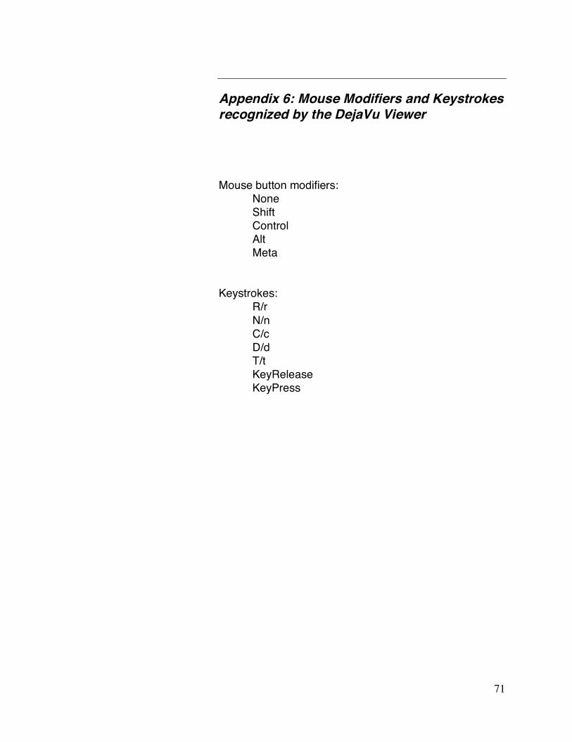
71
Appendix 6: Mouse Modifiers and Keystrokes recognized by the DejaVu Viewer
Mouse button modifiers: None Shift Control Alt Meta
Keystrokes: R/r N/n C/c D/d T/t KeyRelease KeyPress

72
Appendix 7: Customization Options and Default Values for User Preferences
Customization Options for PMV
_pmvrc user_prefs
user_prefs defaults: 'warnOnEmptySelection' no/yes ask user before expanding to all 'NumberOfUndo' 100 # of cmds that can be undone 'centerScene' firstMoleculeOnly center scene on new molecule ‘useDepthCueing’ yes/no depthcueing on by default 'showProgressBar' show/hide progress bar 'useBusyCursor' 0/1 change cursor shape while cmd running ‘changeCursor' 0/1 change cursor shape <window may flash!> 'expandNodeLogString' 1/0 use fullname of nodes in log string or not 'transformationLogging' final/continuous/no when transformations are logged 'showSelectionSpheres' 0/1 display yellow crosses on selected atoms 'visualPickingFeedBack' 0/1 display sphere on picked vertex 'fillSelectionBox' 1/0 draw box around drag select after delay 'fillSelectionBoxDelay' 200 msec w/out motion before drawing box 'raiseExceptionForMissingKey' 0/1 raise exception if key not in dict 'warningMsgFormat' pop-up/printed format for warning messages 'inputFormInitializationMode' lastValue/defaultValue 'measureDistanceSL' 4 # of labelled distances displayed simultaneously 'measureAngleSL' 4 # of labelled angles displayed simultaneously 'measureTorsionSL' 4 # of labelled torsions displayed simultaneously 'continuousUpdateDist' yes/no update distance if move objects independently 'continuousUpdateAngle' yes/no update angles if transform objects independently 'continuousUpdateTorsion' yes/no update torsions if transform objs independently 'selectStringMatchMode' caseSensitive/caseInsensitive/CIWEC ‘Startup Directory’ current working directory #can be set in _pmvrc

73
You can change the user_prefs values via:
File ➞ Preferences ➞ Modify Defaults
See also the following how-tos:
• How-to change default parameters using _pmvrc: http://mgltools.scripps.edu/documentation/how-to/changing-default-parameters
• How-to change Camera size on start-up or on-the-fly: http://mgltools.scripps.edu/documentation/how-to/ChangingCameraSize
• How-to change the default font: http://mgltools.scripps.edu/documentation/how-to/change-the-default-font

74
Appendix 8: Definitions of Named Residue and Atom Sets recognized by PMV
Named Residue Sets: 'acidic' = asp, glu 'acyclic' = ala, arg, asn, asp, cys, glu, gln, gly, ile, leu. lys, met, ser, thr, val, 'aliphatic' = ala, gly, ile, leu, val ‘aromatic' = his, phe, trp, tyr 'basic' = arg, his, lys 'buried' = ala, cys, ile, leu, met, phe, trp, val 'charged' = arg, asp, glu, his, lys 'cyclic’ = his, phe, pro, trp, tyr 'hydrophobic' = ala, gly, ile, leu, met, phe, pro, trp, tyr, val 'large' = arg, glu, gln, his, ile, leu, lys, met, phe, trp, tyr 'medium' = asn, asp, cys, pro, thr, val 'negative' = asp, glu 'neutral' = ala, asn, cys, gln, gly, his, ile, leu, met, phe, pro, ser, thr, trp, tyr, val 'polar' = arg, asn, asp, cys, glu, sln, his, lys, ser, thr 'positive' = arg, his, lys 'small' = ala, gly, ser 'surface' = arg, asn, asp, glu, gln, gly, his, lys, pro, ser, thr, tyr Named Atom Sets: 'backbone' = C, CA, N, O 'backbone+h' = C, CA, N, O, HN, HN1, HN2 'sidechain' = SG, SD, CB, CG, CD, CD1, CD2, CE, CE1, CE2, CE3, CG1, CG2, CZ, CZ2, CZ3, CH2, ND1, ND2, NE, NE1, NE2, NH1, NH2, NZ, OD1, OD2, OG, OG1, OE1, OE2, OH, HD1, HE1, HE2, HE,HE21, HE22, HH23, HH11, HH12, HG, HG1, HH21, HD22, HD21, HZ1, HZ2, HZ3, HH One Character Residue Names:

75
ALA=A, ARG=R, ASN=N, ASP=D, ASX=B, CYS=C, GLU=E, GLN=Q, GLX=Z, GLY=G, HIS=H, ILE=I, LEU=L, LYS=K, MET=M, PHE=F, PRO=P, SER=S, THR=T, TRP=W TYR=Y, VAL=V, XAA=X, SEL=U #www.ensembl.org/Docs/Pdoc/bioperllive/Bio/SeqUtils.html