pyroptosis: a new paradigm of cell death for fighting
TRANSCRIPT

REVIEW Open Access
Pyroptosis: a new paradigm of cell deathfor fighting against cancerYixin Tan1,2,3,4, Quanzhu Chen1,2,3, Xiaoling Li1,2,3, Zhaoyang Zeng1,2,3, Wei Xiong1,2,3, Guiyuan Li1,2,3, Xiayu Li3,Jianbo Yang5, Bo Xiang1,2,3* and Mei Yi1,2,6*
Abstract
Background: Unraveling the mystery of cell death is one of the most fundamental progresses of life sciencesduring the past decades. Regulated cell death (RCD) or programmed cell death (PCD) is not only essential inembryonic development, but also plays an important role in the occurrence and progression of diseases, especiallycancers. Escaping of cell death is one of hallmarks of cancer.
Main body: Pyroptosis is an inflammatory cell death usually caused by microbial infection, accompanied by activationof inflammasomes and maturation of pro-inflammatory cytokines interleukin-1β (IL-1β) and interleukin-18 (IL-18).Gasdermin family proteins are the executors of pyroptosis. Cytotoxic N-terminal of gasdermins generated fromcaspases or granzymes proteases mediated cleavage of gasdermin proteins oligomerizes and forms pore across cellmembrane, leading to release of IL-1β, IL-18. Pyroptosis exerts tumor suppression function and evokes anti-tumorimmune responses. Therapeutic regimens, including chemotherapy, radiotherapy, targeted therapy and immunetherapy, induce pyroptosis in cancer, which potentiate local and systemic anti-tumor immunity. On the other hand,pyroptosis of normal cells attributes to side effects of anti-cancer therapies.
Conclusion: In this review, we focus on the regulatory mechanisms of pyroptosis and the tumor suppressive functionof pyroptosis. We discuss the attribution of pyroptosis in reprogramming tumor microenvironments and restoration ofanti-tumor immunity and its potential application in cancer immune therapy.
Keywords: Tumor microenvironment, Gasdermin, Necroptosis, Ferroptosis, Immune checkpoint, Adaptive immunity,Immunogenic cell death
BackgroundCell death is one of the most fundamental issues of life.As a hallmark of cancer, the ability to escape cell deathnot only contributes to the origin of cancer, but also playsan essential role in acquisition of therapy-resistance, re-lapse and metastasis [1]. The ultimate goal of cancer ther-apeutics, including radiotherapy, chemotherapy, andimmunotherapy that has recently made great achieve-ments, is to maximize the destruction of tumor cells, but
minimize the damage to normal tissues. However, the in-herent genetic and epigenetic heterogeneity of tumor cells,as well as metabolic plasticity and other factors, confertumor cells a greater adaptability to the unfavorable tumorenvironments, resulting in acquisition of therapy resist-ance and metastatic potential. Cell death is generally cate-gorized as regulated cell death (RCD) or accidental celldeath (ACD). ACD is referred to a biologically uncon-trolled cell death or non-programmed cell death whichusually presents as lytic or necrotic like form, whereasRCD is a genetically controlled process. Necrotic celldeath has been considered merely as a non-programmedcell death for a long time. However, now we clearly knowthat necrotic like cell death can be executed in a finely
© The Author(s). 2021, corrected publication 2021. Open Access This article is licensed under a Creative Commons Attribution4.0 International License, which permits use, sharing, adaptation, distribution and reproduction in any medium or format, aslong as you give appropriate credit to the original author(s) and the source, provide a link to the Creative Commons licence,and indicate if changes were made. The images or other third party material in this article are included in the article's CreativeCommons licence, unless indicated otherwise in a credit line to the material. If material is not included in the article's CreativeCommons licence and your intended use is not permitted by statutory regulation or exceeds the permitted use, you will needto obtain permission directly from the copyright holder. To view a copy of this licence, visit http://creativecommons.org/licenses/by/4.0/. The Creative Commons Public Domain Dedication waiver (http://creativecommons.org/publicdomain/zero/1.0/) applies to the data made available in this article, unless otherwise stated in a credit line to the data.
* Correspondence: [email protected]; [email protected] Key Laboratory of Cancer Metabolism, Hunan Cancer Hospital andthe Affiliated Cancer Hospital of Xiangya School of Medicine, Central SouthUniversity, Tongzipo Road, Changsha 410013, Hunan, ChinaFull list of author information is available at the end of the article
Tan et al. Journal of Experimental & Clinical Cancer Research (2021) 40:153 https://doi.org/10.1186/s13046-021-01959-x
controlled manner [2]. During the past decades, character-izations of new forms of RCD and exploration of its rolesin physiological or pathological conditions have deepenedour understanding on inflammation, immunity and cancerdevelopment.The anti-tumor strategy has now switched from killing
the entire tumors barely through drugs or radiation toachieving long-term control of cancer by eliminating resi-due malignant cells through the body’s inherent immunemechanism. The death of tumor cells may be immuno-genic or non-immunogenic. Induction of immunogeniccell death (ICD) of tumor cells is prerequisite for rebuild-ing anti-tumor immunity. ICD refers to cell death thatgenerates adaptive immunity against endogenous or ex-ogenous antigens carried by dying cells [3]. The mostessential nature of ICD is the complex cell-to-cell commu-nications between immune cells and dying cells [4]. Thekey parameters that determine the immunogenicity of celldeath include antigenicity, inflammation and adjuvanticity[5]. Dying cells undergo lytic death, providing dendriticcells (DC) with antigen and inflammatory stimuli, andthen activate CD8+ T cells through a process called anti-gen cross-priming [6]. ICD was initially identified as a pro-tective mechanism against pathogen infection. Pathogen-infected cells release pathogen-related molecular patterns(PAMPs) that are conserved microbial molecules whichcould be recognized by pattern-recognition receptors(PRRs) of the innate immune system to initiate PAMP-triggered immunity [4]. Sterile ICD can be induced bychemotherapy [7]. In ICD induced by chemotherapy orradiotherapy, dying cells release damage-associated mo-lecular patterns (DAMPs), also known as alarmin, whichmay initiate and exacerbate the immune response throughcorresponding PRRs on immune cells [8, 9].The discovery of new forms of ICD and their roles in
immunity and tumorigenesis have promoted the renewalof anti-tumor treatment strategies. Pyroptosis is a newlycharacterized form of ICD and has gradually emerged asa great opportunity to improve the efficacy of cancer im-mune therapy. Pyroptosis usually occurs in macrophageupon pathogen infection. It plays an essential role inclearance of pathogens [10]. Morphologically, pyroptosisis featured by cell swelling and plasma membrane rup-ture, leading to release of pro-inflammatory cytokinesIL-1β, IL-18 and cellular contents into the extracellularspace and activating inflammatory response (Fig. 1).Mitochondria remain intact and there is no leakage ofcytochrome C during pyroptosis in macrophage [11, 12].Epithelial cells also undergo sterile pyroptosis in physio-logical or pathological conditions. For example, pyropto-tic cell death of intestinal epithelial cells mediated bycaspase-1 activation is a cause of mucosal barrier dys-function in Crohn’s disease [13]. Sterile pyroptosis alsooccurs in epithelial cells upon various death stimuli,
including anti-neoplastic drugs [14, 15]. Pyroptosis inepithelial cells could occur at downstream of the mito-chondrial apoptotic pathway [16]. As a highly-immunogenic form of cell death, pyroptosis causes localinflammation and attracts inflammatory cell infiltration,providing a great opportunity to relieve immunosuppres-sion of tumor microenvironments (TME) and induce asystemic immune response in treating solid tumors [17].
Main textTo date, there are four distinct pathways were identifiedto induce the action of pyroptosis. Basically, pyroptosiscould be executed in an inflammasome dependent or in-dependent manner. The inflammasome dependent path-ways include the canonical and non-canonical pathways,whereas the independent pyroptosis pathways includecaspase-3 mediated pathway and granzymes proteasesmediated pathway. We will discuss in detail in the fol-lowing section.
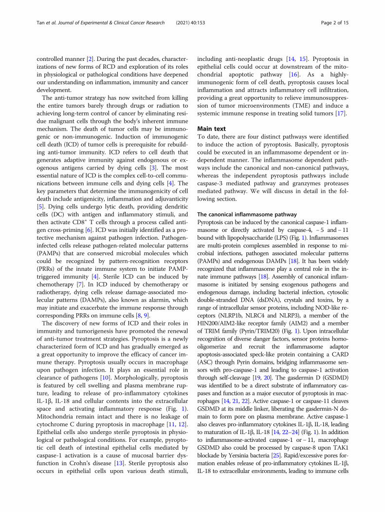
The canonical inflammasome pathwayPyroptosis can be induced by the canonical caspase-1 inflam-masome or directly activated by caspase-4, − 5 and− 11bound with lipopolysaccharide (LPS) (Fig. 1). Inflammasomesare multi-protein complexes assembled in response to mi-crobial infections, pathogen associated molecular patterns(PAMPs) and endogenous DAMPs [18]. It has been widelyrecognized that inflammasome play a central role in the in-nate immune pathways [18]. Assembly of canonical inflam-masome is initiated by sensing exogenous pathogens andendogenous damage, including bacterial infection, cytosolicdouble-stranded DNA (dsDNA), crystals and toxins, by arange of intracellular sensor proteins, including NOD-like re-ceptors (NLRP1b, NLRC4 and NLRP3), a member of theHIN200/AIM2-like receptor family (AIM2) and a memberof TRIM family (Pyrin/TRIM20) (Fig. 1). Upon intracellularrecognition of diverse danger factors, sensor proteins homo-oligomerize and recruit the inflammasome adaptorapoptosis-associated speck-like protein containing a CARD(ASC) through Pyrin domains, bridging inflammasome sen-sors with pro-caspase-1 and leading to caspase-1 activationthrough self-cleavage [19, 20]. The gasdermin D (GSDMD)was identified to be a direct substrate of inflammatory cas-pases and function as a major executor of pyroptosis in mac-rophages [14, 21, 22]. Active caspase-1 or caspase-11 cleavesGSDMD at its middle linker, liberating the gasdermin-N do-main to form pore on plasma membrane. Active caspase-1also cleaves pro-inflammatory cytokines IL-1β, IL-18, leadingto maturation of IL-1β, IL-18 [14, 22–24] (Fig. 1). In additionto inflammasome-activated caspase-1 or − 11, macrophageGSDMD also could be processed by caspase-8 upon TAK1blockade by Yersinia bacteria [25]. Rapid/excessive pores for-mation enables release of pro-inflammatory cytokines IL-1β,IL-18 to extracellular environments, leading to immune cells
Tan et al. Journal of Experimental & Clinical Cancer Research (2021) 40:153 Page 2 of 15

infiltration and establishment of an inflammatory micro-environment [26]. Pyroptosis also contributes to release ofDAMPs such as the protein high-mobility group box 1(HMGB1) and lactate dehydrogenase (LDH), resulting inamplifying inflammation and recruiting immune cells in thetissue [27–29]. However, the means by which HMGB1 re-leases to extracellular space is controversial. Though GSDMD is required for secretion of IL-1β and HMGB1 followinginflammasome activation, GSDMD pore is not a direct con-duit for HMGB1. HMGB1 release after inflammasome acti-vation only occurs when plasma membrane integrity isdisrupted [30]. A latest study revealed that ill-characterized
nerve injury-induced protein 1 (NINJ1), a transmembraneprotein localized at cell surface, is the essential mediator forplasma membrane rupture during pyroptotic cell death.Ninj1−/− macrophages are unable to release HMGB1 andLDH in response to diverse inducers of lytic cell death [31].
The non-canonical inflammasome pathwayIn the non-canonical inflammasome pathway, intracellularLPS binds directly to caspase-4/5/11 via CARD domainand initiates oligomerization of caspase-4/11, leading toactivation of the caspases [32] (Fig. 1). In non-canonicalinflammasome pathway, cleavage of GSDMD at Asp276 is
Fig. 1 The canonical inflammasome and non-canonical inflammasome pathway in pyroptosis. The canonical inflammasome is assembled inresponse to exogenous pathogens and endogenous damage by intracellular sensor proteins, including NLRP1b, NLRC4, NLRP3, AIM2 and Pyrin.The canonical inflammasomes recruit pro-caspase 1 through inflammasome adaptor protein ASC, leading self-cleavage and activation of caspase1. Active caspase 1 cleaves pro-inflammatory cytokines pro-IL-1β, pro-IL-18, leading to maturation of IL-1β, IL-18. Active caspase 1 cleaves GSDMDprotein at the middle linker, liberating the cytotoxic N-terminus to form pore on plasma membrane, which allows the release of mature IL-1β, IL-18. In non-canonical pathway, LPS directly binds to murine pro-caspase 11 or its human homologs pro-caspase 4 and 5, leading activation ofcaspase 11/4/5. In non-canonical inflammasome pathway, cleavage of GSDMD is executed by active caspase 11 or caspase 4 and 5 upon directbinding of cytosolic LPS. Chemotherapy drugs could induce pyroptosis in epithelial cells through activating mitochondrial death machinery andcaspase 3. In this case, GSDME is cleaved by active caspase 3. GSDME-N in turn activates NLRP3 inflammasome, leading to activation of caspase1/GSDMD cascade, which promotes maturation of IL-1β, IL-18. Gasdermins could be cleaved by Lymphocyte-derived granzymes proteases,unleashing the pore-formation ability to trigger pyroptosis of cancer cells
Tan et al. Journal of Experimental & Clinical Cancer Research (2021) 40:153 Page 3 of 15

executed by active murine caspase-11 or its human homo-logs caspase-4 and -5 upon direct binding of cytosolic LPS[21, 33–35] (Fig. 1). The catalytic domains of inflamma-tory caspases directly bind to GSDMD and execute cleav-age at residues FLTD [36]. A p10 product generated fromcaspase-4/11 autoprocessing is necessary and sufficient tocleave GSDMD. The p10 fragment of caspase-4/11 bindswith GSDMD-C domain, leading to dimerization-mediated caspase activation and cleavage of GSDMD [37].NLRP3 is also activated by caspase-11 mediated non-canonical inflammasome, thus leading to maturation ofIL-1β and IL-18 [38, 39]. Cytosolic LPS or cytosolicGram-negative bacteria activates non-canonical (caspase-4/11) inflammasome signaling and induces pyroptosis ofneutrophil in a GSDMD-dependent manner, leading toextrusion of neutrophil extracellular traps (NETs) [40],thus non-canonical inflammasome links pyroptosis andNETosis, which is a unique type of regulated neutrophilcell death in response to infection of pathogens [41].
Caspase-3/GSDME mediated pathwaySterile pyroptosis may occur in epithelial cells. For ex-ample, chemotherapy drugs induce pyroptosis in epithe-lial cells through caspase-3 mediated cleavage ofgasdermin E (GSDME) [16, 42] (Fig. 1). Active caspase-3after TNF-α stimulation cleaves human and mouseGSDME at position 267 or 270 amino acid residue.GSDME mutants in which aspartate at position 267 or270 was substituted by alanine lose the activity to exe-cute pyroptosis [42]. Neither the canonical nor the non-canonical inflammasome is required for caspase-3/GSDME mediated pyroptosis. However, GSDME-N gen-erated by active caspase-3 could activate the canonicalinflammasome pathway and thus promote maturationand release of IL-1β and IL-18 (Fig. 1). During the pastdecades, activation of caspases, especially caspase-3, wasthought to be one of the biochemical features of apop-tosis process. Now we know that activation of caspase-3is not specific to apoptosis. The gasdermins, rather thancaspases, is the central switch from apoptosis to pyrop-tosis upon death stimuli.
The granzymes mediated pathwayKiller cells mediated elimination of tumor cells is previ-ously considered to be noninflammatory. Recently, stud-ies indicated that natural killer cells and cytotoxic Tlymphocytes elicit pyroptosis of cancer cells throughgranzymes proteases mediated cleavage of specific gas-dermin family members [43, 44] (Fig. 1). For example,lymphocyte-derived granzyme A (GZMA) cleaves gas-dermin B (GSDMB) at the linker, which unleashes itspore-forming activity and results in pyroptotic cell deathof GSDMB-expressing cancer cells [43] (Fig. 1). Thegranzyme B (GZMB) from natural killer cell or chimeric
antigen receptor (CAR) T cell directly cleaves GSDMEafter D270 residue where the site caspase-3 also cleaves,liberating cytotoxic N-terminus to form pore in mem-brane [44, 45] (Fig. 1). Granzymes mediated pyroptosisof cancer cells may magnify inflammation signals inTME, thus recruit more immune cells and further igniteantitumor immunity.
Gasdermin family members, the executioner of pyroptosisGasdermin is a family of pore-forming proteins playing anessential role in the execution phase of pyroptotic celldeath. Human gasdermin family contains six conservedmembers, including gasdermin A, B, C, D, E (also namedas DFNA5), and DFNB59. Mice do not have gasdermin B,but there are triplicated gasdermin A (gasdermin A1–3)and quadruplicated gasdermin C (gasdermin C1–4) [46].The gasdermin family members have an autoinhibitedtwo-domain architecture that is consisted of a cytotoxicN-terminal domain and a C-terminal repressor domainconnected by a flexible linker [47]. The N domain isshared by all gasdermin family members and can bindwith acidic lipids, including phosphatidylinositol phos-phates (PIPs), phosphatidic acid (PA), phosphatidylserine(PS) and cardiolipin, which in turn form pores contained16 symmetric protomers in plasma membrane [47, 48].Intramolecular interaction between N-terminal and C-terminal fragments of gasdermins prevents activation ofpore-forming activity of N-terminal domain and executionof pyroptosis, whereas proteolytic cleavage by inflamma-tory caspases, including caspase-1 and caspase-11, at theflexible linker between these two domains liberates thecytotoxic N-terminal domain to oligomerize in membraneand form large oligomeric pores where IL-1β and IL-18are secreted [49, 50]. Structure-guided mutagenesis indi-cates that execution of pyroptosis is dependent on pore-forming activities of the gasdermin-N domain [47].GSDMD is the first executor of pyroptosis to be discov-ered. GSDMD could be cleaved by caspase-1 and caspase-11 to trigger pyroptosis in macrophages [14, 21, 22].GSDMD is the only caspase-1 substrate that induces pyr-optosis. GSDMD-deficent cells resist to pyroptosis in-duced by activation of inflammasome. However, cellslacks of GSDMD are still susceptible to caspase-1-mediated cell death [51]. In the absence of GSDMD, acti-vation of caspase-1 results apoptosis through activatingcaspase-3 and -7. During apoptosis, active caspase-3 and-7 inactivate GSDMD by cleaving GSDMD at Asp-87,thus blocking pyroptosis [52]. Murine GSDMD also couldbe cleaved at Asp-27 within an IPVD motif by caspase-7[53]. Pore formation by GSDMD N-terminus is requiredfor release of IL-1β and IL-18, but not essential for plasmamembrane rupture during lytic cell death. It has been pro-posed that cells may repair the damage to cell membranecaused by the GSDMD-N, because of that formation of
Tan et al. Journal of Experimental & Clinical Cancer Research (2021) 40:153 Page 4 of 15
Gasdermin-N pore does not definitely leads to cell death[54]. For example, hyperactive macrophages release IL-1βthrough GSDMD pore, but keep alive [55]. Recently, astudy suggests that formation of GSDMD pores is suffi-cient for inducing the maturation and release of IL-1αupon inflammasomes activation, which suggests that itmay have an important role in settings without IL-1β [56].In chemotherapy induced pyroptosis of epithelial cells,
cleavage of GSDME at the linker is executed by activecaspase-3 [16, 42]. TNF-α treatment and chemotherapyalso induce pyroptosis in GSDME-expressing cancer cellsvia activation of caspase-3 [42]. Deletion of GSDME tendsto disassemble into small apoptotic bodies upon activationof caspase-3. For example, lobaplatin treatment inducesGSDME-mediated pyroptosis in colon cancer cells. How-ever, knocking out GSDME switches cell death from pyr-optosis to apoptosis, without affecting the cytotoxicity oflobaplatin on tumor growth and tumor formation of coloncancer cells [15]. Co-treatment of a PLK1 inhibitor BI2536with cisplatin activates caspase-3/GSDME pathway andenhances the chemosensitivity of cisplatin in esophagealsquamous cell carcinoma through induction of pyroptoticcell death [57]. Thus, gasdermin proteins, rather than cas-pases, act as the central molecule that switches apoptosisto pyroptosis upon death stimuli [16, 47]. In addition tocaspase-3, killer cell or CAR T cell -derived GZMB alsocleave GSDME at D270, unleashing its pore-forming ac-tivity to trigger caspase-independent pyroptosis inGSDME-positive cancer cells [44, 45]. Thus, gasdermin-mediated pyroptosis underlies the main killing mechanismof cytotoxic lymphocyte. Interestedly, GSDME-N not onlyforms pores in the plasma membrane, but also permeabi-lizes the mitochondrial membrane, leading to release ofcytochrome c and augment caspase-3 activation and apop-tosome. GSDME-deficient cells exhibit reduced cyto-chrome c release and caspase-3 activation upon intrinsicand extrinsic apoptotic stimuli. Like GSDME-N, GSDMD-N generated by inflammasome also permeabilizes themitochondria [58]. Thus, cleavage of gasdermins linksinflammasome activation to downstream activation of theapoptosome.In addition to GSDMD and GSDME, N-terminal domain
of GSDMA, GSDMA3, GSDMB, and GSDMC have all beenproposed to form pores in membrane and execute pyroptosis[47]. GSDMB is specifically cleaved by lymphocyte-derivedGZMA, unleashing its pore-forming activity and inducingpyroptosis in GSDMB-expressing cancer cells [43]. Caspase-8 activated by TNF-α cleaves GSDMC at it linker, liberatingthe GSDMC N-terminal domain to trigger pyroptosis in can-cer cells [59].The pore-forming activity of GSDME may be regulated by
phosphorylation at a highly conserved Thr6 residue, becausewhen Thr6 residue was replaced by glutamate (phosphomi-metic), the pyroptotic activity of GSDME is significantly
inhibited. Mechanistic study revealed that phosphorylation ofThr6 prevents GSDME-N dimerization/oligomerization inmembranes but does not affect its membrane localization[58]. Similarly, phosphorylation of Thr8 of GSDMA which isequivalent to Thr6 in GSDME also causes complete abolish-ment of its pyrototic activity [58]. Recently, it has been dem-onstrated that palmitoylation on C-terminal of GSDME isrequired for pyroptosis induced by chemotherapy drugs. Pal-mitoylation of GSDME-C seems to dissociate the intramo-lecular interaction between N-terminal and C-terminal ofGSDME protein, because 2-bromopalmitate treatment in-hibits palmitoylation of GSDME-C and then promotes inter-action between GSDME-C and GSDME-N [60].
Pyroptosis exerts tumor suppressive functionResistant to cell death is one of hallmarks of human can-cers [1]. Apoptosis has been linked to tumor suppressionand recognized as the major mechanism underlying anti-tumor therapeutic approaches, like radiation therapy (RT)and chemotherapy. As a non-lytic form of cell death,apoptosis is generally immunogenically silent. In contrast,lytic cell death, including necroptosis [61–64] and pyrop-tosis, is pro-inflammatory. Chronic inflammation is awell-known cancer-fueling process during cancer initi-ation and progression. It has been proposed that chronicinflammation increases the risk of cancer. Local inflamma-tory microenvironment is favorable tumor growth, angio-genesis, invasion, and metastasis [65]. Active necroptosispromotes intestinal inflammation in children with inflam-matory bowel disease (IBD) and IBD mouse models [66],which is an inflammatory disease with enhanced risk fordevelopment of gastrointestinal malignancies. It has beenshown that in vivo necroptosis is more efficient to induceantigen cross-priming [6]. However, it is still uncertainwhether necroptosis promotes or restricts tumors initi-ation and progression [67]. Evidence showed that in vivonecrosome activation suppresses anti-tumor immunitythrough enhancing the infiltration of immune-suppressivemyeloid cellular subsets, whereas deletion RIPK3 or inhib-ition RIPL1 in vivo enhances adaptive immunogenicityand prevents pancreatic oncogenesis in mice throughrepressing chemokine attractant CXCL1 and Mincle sig-naling [68], consistent with the pro-inflammatory proper-ties of necroptosis and the cancer-promoting effects ofinflammation. Inhibition of necroptosis by a specificchemical inhibitor Nec-1 also ameliorates inflammationand prevents colitis-associated tumorigenesis in a mousemodel of inflammatory bowel disease [69]. In addition,necroptosis of endothelial cells promotes tumor cell ex-travasation and metastasis [70]. As a lytic, inflammatorytype of cell death, pyroptosis leads to inflammation, whichcould increase the risk of cancer. However, it has beenshown that Pycard(−/−), Casp1(−/−) mice and Nlrp3(−/−)
mice are prone to inflammation associated colon cancer
Tan et al. Journal of Experimental & Clinical Cancer Research (2021) 40:153 Page 5 of 15

[71, 72], suggesting that inflammasome activation or in-duction of pyroptosis restricts, rather than promotes,colon cancer development. A study indicates that geneticablation of GSDMD mitigates the development of non-alcoholic steatohepatitis (NASH) [73]. NASH-related cir-rhosis is associated with increased risk for liver cancer[74]. However, to date, there is no direct evidence suggeststhat deletion of would gasdermins family member affectspontaneous or induced cancer occurrence. Thus, the spe-cific role of pyroptosis in tumorigenesis deserves furtherstudy. Studies suggested pyroptosis may function as atumor suppression mechanism. Recent studies haveproved that induction of pyroptosis in malignant cells pro-vides an alternative approach to kill cancer cells. GSDMEis silenced in most cancer cells [42]. Overexpression ofwild type GSDME inhibits tumor growth in immunocom-petent mice, whereas overexpression cancer associatedGSDME mutants which lose the ability to execute pyrop-tosis fail to delay tumor growth [44]. Uncleavable GSDME(D270A) or pore-forming defective F2A nonfunctionalmutants also fail to inhibit tumor growth, indicatingtumor suppression by GSDME is dependent on its activityto execute pyroptosis [44]. It has been shown that
mammalian STE20-like kinase 1 (MST1) is decreased inpancreatic ductal adenocarcinoma. Restored expression ofMST1 in suppresses the proliferation, migration, invasion,and cell spheroid formation of pancreatic ductal adenocar-cinoma cells through caspase-1–induced pyroptosis [75].Thus, these studies suggest that pyroptosis represents anew way to eliminate cancer cells.
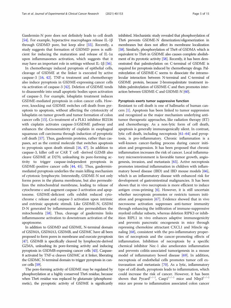
Pyroptosis reprograms tumor microenvironments andevokes anti-tumor immunityTumor microenvironments (TMEs) are composed of cel-lular components, extracellular matrix (ECM) and intersti-tial fluid. The cellular components of TME include tumorcells themselves, stromal cells (such as fibroblasts), endo-thelial cells of blood and lymphatic vessels, neuronal cellsand infiltrating immune cells [76] (Fig. 2). The balance be-tween pro-tumorigenic and anti-tumor factors in themicroenvironment regulates tumor growth [77]. Cancercells extensively communicate with stroma cells and con-vert them into allies from within, thus impacting diverseaspects of tumor biology [78, 79]. On the one hand, tumorcells recruit stromal cells, inflammatory or/and immunecells, etc. by actively secreting inflammatory factors,
Fig. 2 Immunosuppressive tumor microenvironments. Tumor microenvironments are composed of cellular components, extracellular matrix andinterstitial fluid. Chemokines secreted from cancer cells recruit a variety of immune cells infiltrating into tumor. The interactions between cancercells and the infiltrated immune cells determine the progression of cancer and therapeutic efficacy. Immune suppressive cells, including Treg,MDSC and M2 type TAM, limit function of the cytotoxic T cells and make tumor microenvironments immunosuppressive. Furthermore, tumorderived cytokines, like TGF-β, IL-6, etc., also suppress immune responses
Tan et al. Journal of Experimental & Clinical Cancer Research (2021) 40:153 Page 6 of 15

growth factors, extracellular matrix and its metabolites tostimulate angiogenesis and lymphogenesis. On the otherhand, the stromal cells and inflammatory cells residing inTME have protection and supportive effects on the cancercells. The interplay of tumor cells and non-tumor cellsshapes the TME that allows tumor cells growth, invasion,and facilitates escaping the tumor immunosurveillance[78, 79]. Failure of tumor immunosurveillance causes theclinical appearance of cancer and cancer progression. Ac-cumulating evidences indicate that TME plays a centralrole in the process of tumor occurrence, immune escape,progression and metastasis [80]. Immunotherapeutic ap-proaches, including approved immune checkpoint inhibi-tors (ICIs) anti-CTLA-4, anti-PD-1/PD-L1 antibody andchimeric antigen receptor (CAR) T-cell therapy, havegreatly improved clinical outcome in treating human can-cer [81–83]. However, only a small portion of patientsachieve durable benefit [84]. The efficacy of immune ther-apy relies on the pre-existing anti-tumor immunity,whereas is hampered by suppressive tumor immunemicroenvironment (TIME) that limits the ability of T cellsto eradicate tumor cells [85–88]. Aberrant tumor vascula-ture generates a physical barrier for T cell trafficking.Otherwise, infiltration of immune suppressive cells, in-cluding regulatory T cell (Treg), myeloid derived suppres-sive cells (MDSC) and M2 type tumor associatedmacrophages (TAM), limits function of the cytotoxic Tcells and makes solid tumor are refractory to immune-therapy [89–93] (Fig. 2). Furthermore, the TIME is char-acterized by hypoxia and low pH (pH < 4), which suppressthe activity of cytotoxic T lymphocytes [94]. In addition,glucose-deprived whereas cholesterol enriched TIME fur-ther exacerbates T cell exhaustion [95–99]. Microenviron-ment normalization is prerequisite for initiating anti-tumor immunity [100]. In addition, vascular normalizationnot only promotes the delivery anti-tumor drugs, but alsoimproves the immune cell infiltration [101]. The complex-ity of TIME has brought great obstacles to cancer treat-ment. Therefore, it is necessary to develop new strategiesto ameliorate the suppressive TIME or normalize TIME[100, 102, 103].Cell death plays a vital role in establishing adaptive im-
munity responding to microbial pathogen infection andtransformed malignant cells [104]. Induction of immuno-genic death of tumor cells is a reasonable strategy to es-tablish a more immunologically active microenvironment,thus creating an opportunity to turn “cold” tumor to “hot”[105, 106] (Fig. 3). As a pro-inflammatory cell death form,induction of pyroptotic cell death of cancer cells providesan opportunity to overcome the immune desert pheno-type of TME. It has been shown that tumors with high ex-pression of wild type GSDME exhibit increased immunecell infiltration, including CD8+ T cells and natural killer(NK) cells, whereas GSDME-deficient tumors or tumors
expressing loss-of-function GSDME mutants exhibit re-duced immune cell infiltration [44]. Single-cell RNA se-quencing revealed that pyroptosis-inducible therapyincreases infiltration of CD4+, CD8+ T cells and naturalkiller cells, whereas reduces monocyte, neutrophil andmyeloid-derived suppressor cell populations in experi-mental breast 4 T1 tumors. Furthermore, pyroptosis of 4T1 tumor cells induces macrophage M1 polarization[107]. Importantly, tumor suppression effect of GSDME isabrogated in killer cytotoxic lymphocytes depleted mice orimmune deficient mice, indicating tumor-suppressivefunction of GSDME requires pyroptosis-dependent activa-tion of antitumor immunity [44]. In addition to directlyeliminate tumor cells, induction of pyroptosis may over-come immunosuppression and reactivate a systemic anti-tumor immunity, which provides a great opportunity toachieve long-term control of cancer. As a major form ofICD, tumor cells undergoing pyroptosis generate largeamounts of neoantigens that stimulate the systemic im-mune response to significantly hamper tumor progression[108] (Fig. 3). In addition, GSDME expression enhancesanti-tumor adaptive immunity by promoting macrophage-mediated phagocytosis [44], which prevents immune eva-sion of tumor [109, 110].
Therapeutic strategies to induce pyroptosis in cancerPrecise modulation of inflammasome activation and pyr-optosis may provide great opportunity to improve the ef-ficacy of immune therapy in the near future [111].Recently, Wang et al. demonstrated that pyroptosis ofless than 15% of tumor cells was sufficient to clear theentire 4 T1 mammary tumor graft. The degree of tumorregression is correlated with augmented anti-tumor im-mune responses and is absent in immune-deficient miceor upon T cell depletion [107], suggesting induction ofpyroptosis is a powerful approach for cancer treatment.
Induction of pyroptotic cell death by targeted therapyIt has been shown that inhibitors of the serine dipeptidasesDPP8 and DPP9 (DPP8/9) activates the Nlrp1b inflamma-some and induces pro-caspase-1 dependent pyroptosis inmonocyte and macrophage [112, 113]. The effect of DPP8/9inhibition to induce pyroptosis can be exploited to treat ma-lignant cancer of myeloid origin. DPP8/9 inhibitors select-ively induce pyroptotic cell death in human acute myeloidleukemia (AML) cells and inhibit human AML progressionin mouse models, highlighting it’s a potential utility for AMLtreatment [114]. Specifically targeting KRAS, EGFR, or ALKmutants by small-molecule inhibitors elicit GSDME-mediated pyroptotic cell death in oncogenic mutations-driven lung cancer, pinpointing a previously unrecognizedrole of GSDME-dependent pyroptosis in molecular targetedtherapy [115]. A latest study demonstrated that combinationsof BRAF inhibitors and MEK inhibitors (BRAFi + MEKi)
Tan et al. Journal of Experimental & Clinical Cancer Research (2021) 40:153 Page 7 of 15

treatment, a FAD-approved approach for BRAFV600E/K-mu-tant melanoma, induce GSDME dependent pyroptosis andenhance cytotoxic T-cell infiltration. However, BRAFi +MEKi treatment loses the therapeutic effect againstGSDME-deficient melanoma, indicating BRAFi + MEKitreatment kills melanoma cells mainly through induction ofpyroptosis. More importantly, the efficacy of BRAFi + MEKitreatment on GSDME-expressing melanoma completely dis-appear in immune-deficient mice, suggesting that BRAFi +MEKi treatment eliminates melanoma cells through GSDEM-dependent anti-tumor immune responses [116].
Induction of pyroptotic cell death by chemotherapydrugsIt has long been believed that apoptosis is the main form ofchemotherapy-induced tumor cells death. However, recentprogresses suggest pyroptotic cell death is a novel killing
mechanism of conventional therapeutics including chemo-therapy. For example, taxol treatment causes GSDMD-medi-ated pyroptosis in nasopharyngeal carcinoma, whereassuppression of pyroptosis has been proposed to be associatedwith taxol resistance in nasopharyngeal carcinoma [117]. Al-though cisplatin is widely believed to kill tumors by causingapoptosis, recent evidence suggests cisplatin induces lungcancer A549 cell through caspase-3/GSDME pathway. Silen-cing GSDME significantly attenuates the cytotoxicity of cis-platin against A549 cell [118]. Lobaplatin also inducesGSDME-mediated pyroptosis by activating caspase-3 innasopharyngeal carcinoma cells [119]. Combination of lose-dose cisplatin with PLK1 inhibitor BI2536 also inducesGSDME-mediated pyroptosis in oesophageal squamous cellcarcinoma cell lines [57]. Induction of sustainable anti-tumorimmunity may improve the efficacy of cancer chemotherapy.Induction of GSDME-mediated pyroptosis evokes anti-
Fig. 3 Induction of pyroptosis by therapeutic regimens evokes anti-tumor immune responses. Therapeutic modalities, including chemotherapy, targetedtherapy, radiotherapy and CAR T cells, induce pyroptosis in cancer cells. Cancer cells undergoing pyroptotic cell death release pro-inflammatory factors (IL-1β, IL-18), alarmin (HMGB1, ATP, etc.), and causing intensive inflammation in the local environments. Pyroptosis in cancer generates abundant neoantigens, which areprocessed by antigen-presenting cells to promote the formation of antigen-specific cytotoxic T lymphocyte (CTL), thereby evoking anti-tumor immunity.Combination of pyroptosis-inducible therapeutic regimens with ICIs enhances anti-tumor immune responses and promotes tumor regression, achieving long-term control of cancer
Tan et al. Journal of Experimental & Clinical Cancer Research (2021) 40:153 Page 8 of 15

tumor immunity, which enhances the ability of cisplatin toregress non-small cell lung cancer [120]. Our group foundthat the natural product triptolide exerts tumor suppressionactivity through inducing GSDME-mediated pyroptosis inhead and neck cancer cells (unpublished data), highlightingits potential to serve as an adjuvant approach for cancer im-mune therapy. Mechanistically, displacement of hexokinase-II from mitochondria facilitates the release of cytochrome cand activation of caspase-3, which acts as up-stream eventsof GSDME-mediated pyroptosis upon triptolide treatment(unpublished data).DNMTs mediated DNA hypermethylation of pro-
moters represses transcription of gasdermins [121]. Zhaoet al. developed a tumor-homing biomimetic nanoparti-cle (BNP) loaded with indocyanine green (ICG) andDecitabine (DCT), which can be photo-activated to in-duce pyroptotic death of cancer cells. DCT activatesGSDME expression through reducing DNA methylation.Upon low-dose photo-activation, ICG in BNP causesextracellular calcium influx, leading to activation ofcaspase-3 and cleavage of GSDME. Thus, ICG and DCTin BNP synergistically promote cancer cell pyroptosis.Importantly, photo-activated pyroptosis induced by BNPnot only directly eliminates primary tumor, but also en-hances systemic antitumor immunity to suppress distantmetastatic tumors. Thus, pyroptosis-inducible BNP is anovel approach to ameliorate immunosuppressive TIMEand enhance the adaptive immunity in treating solidtumor [17]. Jin-Xuan Fan et al. developed chemothera-peutic nanocarriers combined decitabine with tumor-targeting nanoliposome loaded with cisplatin [122]. Theydemonstrated that administration of chemotherapeuticnanocarriers reactivates expression of GSDME and facil-itates GSDME mediated pyroptotic cell death of tumorcells. This pyroptosis-based chemotherapy strategy en-hances immunological effects of chemotherapy, reducingtumor growth, metastasis, and recurrence [122]. Thus,epigenetics-based tumor cell pyroptosis induced by che-motherapeutic nanocarriers provides an opportunity toenhance sensitivity to pyroptosis in cancers.
Induction of pyroptotic cell death by radiation therapyand other physics therapyRadiation therapy may release tumor antigens and maybe an endogenous tumor vaccination event to create aproimmunogenic milieu stimulating local and systemichost cancer-specific immune responses [123]. It has beenshown that radiotherapy elicits tumor-specific immuneresponses through promoting tumor infiltration of CD8+
T cells [124]. Local radiotherapy triggers ICD in cancercells in a dose-dependent manner. Furthermore, radi-ation enhances chemotherapy-induced ICD in cancercells [125]. Although there is limited literature describ-ing pyroptosis of cancer cells directly induced by
radiotherapy, it has been shown that radiation inducespyroptosis in bone marrow derived macrophages(BMDMs) [126]. Combinations of radiation regimenswith immunotherapy are rational approaches to enhanceanti-tumour immune responses and are actually used inclinical trials of a variety of human cancers [127–129].Thus, it is needed to study whether pyroptosis is themajor form of ICD caused by radiotherapy in vivo.It has been shown that local treatment with high-
frequency irreversible electroporation (H-FIRE) resultsin necrosis and pyroptosis in the mouse 4 T1 mammarytumor model, inducing a pro-inflammatory shift in theTME and enhancing cellular immunity. Local treatmentwith H-FIRE not only ablates the primary tumor, butalso reduces metastatic lesions, which is dependent onthe adaptive immune system [130]. Recently, Xiaoruiet al. reported that cold atmospheric plasma, a novelpromising anti-cancer treatment, induces GSDME-dependent pyroptotic cell death in GSDME-expressingtumor cells [131].
Induction of pyroptotic cell death by immune therapyGasdermins-dependent pyroptosis elicited by granzymesunderlies cytotoxic lymphocyte-killing mechanism [43,44]. In addition to caspase-3, GzmB also cleaves GSDMEprotein at D270 to initiate pyroptosis in GSDME-expressing tumor cells [44]. Granzyme A (GZMA) fromNK cells and cytotoxic T lymphocytes (CTLs) activatesgasdermin B (GSDMB) in target cells. Introducingcleavable-GSDMB to mouse tumor cells improves tumorcontrol by immune checkpoint therapy [43]. CAR T cellsalso induces target cell pyroptosis through release ofgranzyme B, activating caspase 3 and then resulting incleavage of GSDME in B leukemic and other target cells[45]. Notably, the quantity of perforin/granzyme B inCAR T cells, rather than in existing CD8+ T cells, deter-mines the activity of CAR T cells to induce target cellpyroptosis [45]. GSDMB expression is induced byinterferon-γ (IFN-γ), thus it is reasonable to combinedtherapeutics of IFN-γ and immune checkpoint blockadeto activate robust antitumour immunity [43]. Recently,Chengui et al. developed a tailored chimeric costimula-tory converting receptor (CCCR) that comprised of theextracellular domain of PD1, transmembrane and cyto-plasmic domains of NKG2D, and the cytoplasmic do-main of 41BB. The CCCR-modified NK92 cells exhibitaugmented activity against human lung cancer H1299cells in vitro through induction of extensive pyroptosis[132]. However, another study argued that tumor sup-pression by antigen-specific primed cytotoxic T cells isindependent of necroptosis or pyroptosis [133]. Thus,pyroptosis is an immune-stimulatory form of cell deathand can synergize with immune checkpoint agents toimprove the efficacy of immune therapy. Recently,
Tan et al. Journal of Experimental & Clinical Cancer Research (2021) 40:153 Page 9 of 15

metformin has been reported to induce pyroptosis incancer cells [134, 135]. Given that metformin promotesantitumor immunity and improves efficacy of ICIs inmalignant cancers [136–139], it is possibly that induc-tion of pyroptosis of cancer cells by metformin may re-program TIME toward “infiltrated-inflamed”.
Inflammasome activation mediates therapy inducedtissue damageConventional cancer treatments such as chemotherapyand radiotherapy tend to kill cells that are in a rapidly pro-liferating status, including rapidly growing cancer cellsand normal cells (e.g., hematopoietic cells). Therefore,these conventional therapies often cause adverse side ef-fects, including myelosuppression and reduced immunity,which may reduce the quality of life for patients and maypotentially lead to treatment failure. For example, damageof hematopoietic stem and progenitor cells induced bychemotherapy results in multi-lineage myelosuppression[140]. Understanding the mechanisms underlying normaltissue injury caused by chemotherapy and radiotherapy isthe basis for preventing these unwanted side effects. Re-cent evidence suggests that pyroptotic cell death of nor-mal cells induced by therapeutic approaches plays anessential role in therapy-induced tissue damage and in-flammation. During the past decades, apoptosis was con-sidered to be the primary death form triggered bychemotherapy drugs. However, GSDME expression allowsoccurrence of pyroptosis upon death stimuli which origin-ally induce apoptosis [47]. It has been shown that GSDMEis widely expressed in normal tissues. Chemotherapydrugs activate pyroptotic cell death in GSDME-expressingcells through caspase-3-mediated cleavage of GSDME.GSDME-dependent pyroptosis largely contributes tochemotherapy drugs-induced tissue damage, because lossof GSDME ameliorate/mitigate chemotherapy related tox-icity in mice [42]. A latest study suggested that suppres-sion of inflammasome assembly prevents pyroptotic celldeath of conventional dendritic cells (cDCs), thus makingcDCs retain the ability to prime both CD4+ and CD8+ Tcells [141]. Cisplatin is one of the most broadly usedchemotherapy drugs. Nephrotoxicity is one of severe sideeffects caused by cisplatin. A latest study revealed thatGSDMD-mediated pyroptosis in mouse kidney tissuesand renal tubular epithelial cells may contribute tocisplatin-induced acute kidney injury. Deletion of GSDMD significantly ameliorate cisplatin-induced acute kidneyinjury in mice, whereas mice with GSDMD-N fragmentoverexpression in the kidney are more vulnerable to acutekidney injury caused by cisplatin [142]. Pyroptosis of car-diomyocytes is a plausible mechanism for severe cardio-toxicity caused by anti-tumor drugs [143]. Recently,Zheng et al. demonstrated that activation of Bnip3-caspase-3-GSDME pathway upon doxorubicin (Dox)
treatment triggers GSDME-mediated pyroptosis, which isresponsible for DOX-induced cardiotoxicity in vivo [144].Dox treatment leads to hyper activation of NLRP3 inflam-masome and pyroptotic cell death of cardiomyocytes,which underlies mechanism for dilated cardiomyopathy(DCM) occurred in Dox-treated heart tissues. Loss of ei-ther NLRP3 or caspase-1 protects mice from Dox-inducedDCM [145]. These results suggest that targeting theinflammasome may help to control the adverse side effectsinduced by chemotherapy drugs.It has been demonstrated that radiation could activate
inflammasome in various immune cells, including macro-phages, dendritic cells, NK cells, T cells, and B cells, in adose-dependent manner. Knocking out caspase-1 signifi-cantly alleviate hematopoietic cell lose induced by radi-ation [146]. Radiation induced caspase-1 activation inimmune cells is NLRP3-independent, but could be pre-vented by allopurinol treatment [146]. It has been shownthat radiation induces pyroptotic cell death of BMDMsin vitro and in vivo through activating NLRP3 inflamma-some [126]. Deletion of NLRP3 remarkably suppressespyroptosis of BMDMs as well as IL-1β level. Additionally,knocking out NLRP3 protects mice from radiation in-duced death. Thus, inhibition of NLRP3 inflammasomemediated pyroptosis may provide an effective strategy todiminish radiation caused tissue injury. It has been shownthat 5-androstenediol treatment significantly suppressedthe radiation-induced activation of inflammasome-mediated pyroptosis by disrupting the interaction betweenAIM2 and ASC, leading to amelioration of myeloid sup-pression and radiation injury in mice [147]. Another studyalso revealed that radiation activates AIM2 inflammasomeand pyroptosis in BMDMs, attributing to radiation-induced lung inflammation and fibrosis [148]. Clinicallyrelevant high dose of radiation activates NLRP3 and AIM2inflammasomes but not the NLRC4 inflammasome, caus-ing GSDMD-dependent pyroptosis in bones and thespleen [149]. These studies suggest that inhibitinginflammasome-pyroptosis signaling has the potential toprevent the tissue damage by intense radiation regimens.In a latest study, Jun et al. reported that disulfiram, a drugfor treating alcohol addiction, selectively inhibits GSDMD-dependent pyroptosis and IL-1β release upon LPS stim-uli and protects mice from LPS-induced septic death.Mechanistically, disulfiram at nanomolar concentration al-lows IL-1β and GSDMD processing but abrogates poreformation of GSDMD-N through covalently modifyinghuman/mouse Cys191/Cys192 in GSDMD, making it anattractive new therapeutic indication for repositioning thissafe drug to counteract inflammation [150].
Conclusion and future perspectivesAnti-tumor immunity not only prevents tumorigenesis, butalso is prerequisite for the success of cancer immunotherapy.
Tan et al. Journal of Experimental & Clinical Cancer Research (2021) 40:153 Page 10 of 15

Growing evidences consistently suggest that pyroptosis-based therapeutic strategies could be combined with im-munotherapy to improve the systemic control of cancer.Established tumors are typified with immunosuppressiveTME, which reduces infiltration of T cells and restricts theanti-tumor activity of cytotoxic T cells. Philosophy of cancerbiology has now been shifting from the tumor-cell-centricview to a microenvironment-centric paradigm. In order toachieve long-term success of cancer control, it is undoubt-edly necessary to establish anti-tumor immunity by thera-peutic approaches. Induction of pyroptosis of tumor cells hasbeen proved to enhance immunogenicity of tumor and turn“cold” tumor to “hot” by attracting more anti-tumor lympho-cytes. Recent studies have shown that conventional treat-ments, including chemotherapy and targeted therapy, reverseimmunosuppressive microenvironments surrounding tumorcells and re-establish local or systemic anti-tumor immunityby inducing pyroptotic cell death of tumor cells [116]. Inaddition to directly kill tumor cells, chemotherapy and tar-geted therapy have the potential to induce local and systemicimmune responses through inducing ICD. Induction of pyr-optosis in tumor cells generates an antigen source for therestoration of antitumor immunity. It is deduced that the“restoration” of host antitumor immunity by chemotherapy,radiotherapy or targeted therapy is essential for completetumor regression after administering treatment modalities[151–156]. On the other hand, both chemotherapy andradiotherapy may cause pyroptosis in immune cells orhematopoietic cells, resulting in impairment of anti-tumorimmunity. In that case, it is vital to reduce side effects of con-ventional therapies by preventing pyroptotic cell death of im-mune cells induced by anti-cancer treatments. Intensivestudies are needed to develop novel strategies that could spe-cifically activate the pyroptosis in tumor cells but withoutdamaging the immune system. To this end, specifically indu-cing pyroptosis in cancer cells by activating GSDME mayprovide a promising strategy to evoke anti-tumor immunity.Hypermethylation of promoter DNA silences the transcrip-tion of GSDME in human cancers. Future studies are neededto restore the expression of GSDME in tumor cells and de-velop specific GSDME agonists. Current clinical testing doesnot allow measurement of specific cell death types in vivo.Non-invasive molecular imaging methods that accurately de-termine forms of cell death should be developed.
AbbreviationsRCD: Regulated cell death; PCD: Programmed cell death; ACD: Accidental celldeath; ICD: Immunogenic cell death; IL-1β: Interleukin-1β; IL-18: Interleukin-18; DC: Dendritic cells; PAMPs: Pathogen-related molecular patterns;PRRs: Pattern-recognition receptors; LPS: Lipopolysaccharide;DAMPs: Damage-associated molecular patterns; NETs: Neutrophil extracellulartraps; NINJ1: Ill-characterized nerve injury-induced protein 1; RT: Radiationtherapy; IBD: Inflammatory bowel disease; MST1: Mammalian STE20-like kin-ase 1; TMEs: Tumor microenvironments; TIME: Tumor immunemicroenvironment; ECM: Extracellular matrix; Treg: Regulatory T cell;MDSC: Myeloid derived suppressive cells; TAM: Tumor associatedmacrophages; NK: Natural killer; BMDMs: Bone marrow derived macrophages;
AML: Acute myeloid leukemia; H-FIRE: High-frequency irreversibleelectroporation
AcknowledgementsWe apologize to those researchers whose work in the field could not becited in the present review due to space constrains.
Authors’ contributionsTXT, MY, and BX wrote the manuscript. QZC, XLL, ZZY, WX, GYL and XYLrevised and corrected the manuscript. The authors read and approved thefinal manuscript.
FundingThis study was supported in part by grants from The National Natural ScienceFoundation of China (81772902, 81872278, 81703131, 82072596), the National“111” Project (Project #111–2-12), the Natural Science Foundation of HunanProvince, China (2018JJ1040, 2020JJ4920, 2020JJ4838, 2020JJ4766, 2020JJ3055),the Hunan Provincial Key Research and Development Program (2018SK2130,2018SK2131), and the Scientific Research Project of Hunan Provincial HealthCommission (20201067, 20201040).
Availability of data and materialsNot applicable.
Declarations
Ethics approval and consent to participateNot applicable.
Consent for publicationAll authors read and approved the final manuscript.
Competing interestsThe authors declare that they have no competing interests.
Author details1Hunan Key Laboratory of Cancer Metabolism, Hunan Cancer Hospital andthe Affiliated Cancer Hospital of Xiangya School of Medicine, Central SouthUniversity, Tongzipo Road, Changsha 410013, Hunan, China. 2NHC KeyLaboratory of Carcinogenesis, The Key Laboratory of Carcinogenesis andCancer Invasion of the Chinese Ministry of Education, Cancer ResearchInstitute and School of Basic Medical Sciences, Central South University,Changsha 410078, Hunan, China. 3Hunan Key Laboratory of NonresolvingInflammation and Cancer, The Third Xiangya Hospital, Central SouthUniversity, Changsha 410013, Hunan, China. 4Department of Dermatology,The Second Xiangya Hospital, The Central South University, Changsha410011, Hunan, China. 5Department of Laboratory Medicine and Pathology,University of Minnesota, Minneapolis, MN 55455, USA. 6Department ofDermatology, Xiangya Hospital, The Central South University, Changsha410008, Hunan, China.
Received: 3 February 2021 Accepted: 21 April 2021
References1. Hanahan D, Weinberg RA. Hallmarks of cancer: the next generation. Cell.
2011;144(5):646–74.2. Vanden Berghe T, Linkermann A, Jouan-Lanhouet S, Walczak H,
Vandenabeele P. Regulated necrosis: the expanding network of non-apoptotic cell death pathways. Nat Rev Mol Cell Biol. 2014;15(2):135–47.
3. Kepp O, Senovilla L, Vitale I, Vacchelli E, Adjemian S, Agostinis P, et al.Consensus guidelines for the detection of immunogenic cell death.Oncoimmunology. 2014;3(9):e955691. https://doi.org/10.4161/21624011.2014.955691.
4. Yatim N, Cullen S, Albert ML. Dying cells actively regulate adaptive immuneresponses. Nat Rev Immunol. 2017;17(4):262–75.
5. Legrand AJ, Konstantinou M, Goode EF, Meier P. The diversification of celldeath and immunity: memento Mori. Mol Cell. 2019;76(2):232–42.
6. Yatim N, Jusforgues-Saklani H, Orozco S, Schulz O, da Silva RB, Reise SousaC, et al. RIPK1 and NF-kappaB signaling in dying cells determines cross-priming of CD8(+) T cells. Science. 2015;350(6258):328–34.
Tan et al. Journal of Experimental & Clinical Cancer Research (2021) 40:153 Page 11 of 15

7. Galluzzi L, Buque A, Kepp O, Zitvogel L, Kroemer G. Immunogenic cell deathin cancer and infectious disease. Nat Rev Immunol. 2017;17(2):97–111.
8. Hernandez C, Huebener P, Schwabe RF. Damage-associated molecularpatterns in cancer: a double-edged sword. Oncogene. 2016;35(46):5931–41.
9. Ma Y, Pitt JM, Li Q, Yang H. The renaissance of anti-neoplastic immunityfrom tumor cell demise. Immunol Rev. 2017;280(1):194–206. https://doi.org/10.1111/imr.12586.
10. Wang M, Jiang S, Zhang Y, Li P, Wang K. The Multifaceted Roles ofPyroptotic Cell Death Pathways in Cancer. Cancers (Basel). 2019;11(9):1313.
11. Jesenberger V, Procyk KJ, Yuan J, Reipert S, Baccarini M. Salmonella-inducedcaspase-2 activation in macrophages: a novel mechanism in pathogen-mediated apoptosis. J Exp Med. 2000;192(7):1035–46.
12. Cervantes J, Nagata T, Uchijima M, Shibata K, Koide Y. Intracytosolic listeriamonocytogenes induces cell death through caspase-1 activation in murinemacrophages. Cell Microbiol. 2008;10(1):41–52.
13. Osterman MT, Gordon IO, Davis EM, Ciorba M, Glover SC, Abraham B, et al.Mucosal Biomarker of Innate Immune Activation Predicts Response toVedolizumab in Crohn's Disease. Inflamm Bowel Dis. 2020;26(10):1554–61.
14. Shi J, Zhao Y, Wang K, Shi X, Wang Y, Huang H, et al. Cleavage of GSDMDby inflammatory caspases determines pyroptotic cell death. Nature. 2015;526(7575):660–5.
15. Yu J, Li S, Qi J, Chen Z, Wu Y, Guo J, et al. Cleavage of GSDME by caspase-3determines lobaplatin-induced pyroptosis in colon cancer cells. Cell DeathDis. 2019;10(3):193.
16. Rogers C, Fernandes-Alnemri T, Mayes L, Alnemri D, Cingolani G, Alnemri ES.Cleavage of DFNA5 by caspase-3 during apoptosis mediates progression tosecondary necrotic/pyroptotic cell death. Nat Commun. 2017;8:14128.
17. Zhao P, Wang M, Chen M, Chen Z, Peng X, Zhou F, et al. Programming cellpyroptosis with biomimetic nanoparticles for solid tumor immunotherapy.Biomaterials. 2020;254:120142.
18. Lamkanfi M, Dixit VM. In retrospect: the inflammasome turns 15. Nature.2017;548(7669):534–5.
19. He Y, Hara H, Nunez G. Mechanism and regulation of NLRP3 Inflammasomeactivation. Trends Biochem Sci. 2016;41(12):1012–21.
20. Guo H, Callaway JB, Ting JP. Inflammasomes: mechanism of action, role indisease, and therapeutics. Nat Med. 2015;21(7):677–87.
21. Kayagaki N, Stowe IB, Lee BL, O'Rourke K, Anderson K, Warming S, et al.Caspase-11 cleaves gasdermin D for non-canonical inflammasomesignalling. Nature. 2015;526(7575):666–71.
22. He WT, Wan H, Hu L, Chen P, Wang X, Huang Z, et al. Gasdermin D is anexecutor of pyroptosis and required for interleukin-1beta secretion. Cell Res.2015;25(12):1285–98.
23. Van Gorp H, Lamkanfi M. The emerging roles of inflammasome-dependentcytokines in cancer development. EMBO Rep. 2019;20(6):e47575.
24. Martinon F, Burns K, Tschopp J. The inflammasome: a molecular platformtriggering activation of inflammatory caspases and processing of proIL-beta.Mol Cell. 2002;10(2):417–26.
25. Orning P, Weng D, Starheim K, Ratner D, Best Z, Lee B, et al. Pathogenblockade of TAK1 triggers caspase-8-dependent cleavage of gasdermin Dand cell death. Science. 2018;362(6418):1064–9.
26. Ruan J. Structural insight of Gasdermin family driving Pyroptotic cell death.Adv Exp Med Biol. 2019;1172:189–205.
27. Hou L, Yang Z, Wang Z, Zhang X, Zhao Y, Yang H, et al. NLRP3/ASC-mediatedalveolar macrophage pyroptosis enhances HMGB1 secretion in acute lunginjury induced by cardiopulmonary bypass. Lab Investig. 2018;98(8):1052–64.
28. Lamkanfi M, Sarkar A, Vande Walle L, Vitari AC, Amer AO, Wewers MD, et al.Inflammasome-dependent release of the alarmin HMGB1 in endotoxemia. JImmunol. 2010;185(7):4385–92.
29. Murao A, Aziz M, Wang H, Brenner M, Wang P. Release mechanisms ofmajor DAMPs. Apoptosis. 2021;26(3–4):152–62.
30. Volchuk A, Ye A, Chi L, Steinberg BE, Goldenberg NM. Indirect regulation ofHMGB1 release by gasdermin D. Nat Commun. 2020;11(1):4561.
31. Kayagaki N, Kornfeld OS, Lee BL, Stowe IB, O'Rourke K, Li Q, et al. NINJ1mediates plasma membrane rupture during lytic cell death. Nature. 2021;591(7848):131–6.
32. Shi J, Zhao Y, Wang Y, Gao W, Ding J, Li P, et al. Inflammatory caspases areinnate immune receptors for intracellular LPS. Nature. 2014;514(7521):187–92.
33. Kayagaki N, Warming S, Lamkanfi M, Vande Walle L, Louie S, Dong J, et al.Non-canonical inflammasome activation targets caspase-11. Nature. 2011;479(7371):117–21.
34. Vigano E, Diamond CE, Spreafico R, Balachander A, Sobota RM, Mortellaro A.Human caspase-4 and caspase-5 regulate the one-step non-canonicalinflammasome activation in monocytes. Nat Commun. 2015;6(1):8761.https://doi.org/10.1038/ncomms9761.
35. Knodler LA, Crowley SM, Sham HP, Yang H, Wrande M, Ma C, et al.Noncanonical inflammasome activation of caspase-4/caspase-11 mediatesepithelial defenses against enteric bacterial pathogens. Cell Host Microbe.2014;16(2):249–56. https://doi.org/10.1016/j.chom.2014.07.002.
36. Yang J, Liu Z, Wang C, Yang R, Rathkey JK, Pinkard OW, et al. Mechanism ofgasdermin D recognition by inflammatory caspases and their inhibition bya gasdermin D-derived peptide inhibitor. Proc Natl Acad Sci U S A. 2018;115(26):6792–7.
37. Wang K, Sun Q, Zhong X, Zeng M, Zeng H, Shi X, et al. Structuralmechanism for GSDMD targeting by autoprocessed Caspases in Pyroptosis.Cell. 2020;180(5):941–55 e20.
38. Zanoni I, Tan Y, Di Gioia M, Broggi A, Ruan J, Shi J, et al. An endogenouscaspase-11 ligand elicits interleukin-1 release from living dendritic cells.Science. 2016;352(6290):1232–6.
39. Lee BL, Stowe IB, Gupta A, Kornfeld OS, Roose-Girma M, Anderson K, et al.Caspase-11 auto-proteolysis is crucial for noncanonical inflammasomeactivation. J Exp Med. 2018;215(9):2279–88.
40. Torii S, Shintoku R, Kubota C, Yaegashi M, Torii R, Sasaki M, et al. Anessential role for functional lysosomes in ferroptosis of cancer cells.Biochem J. 2016;473(6):769–77.
41. Burgener SS, Schroder K. Neutrophil Extracellular Traps in Host Defense.Cold Spring Harb Perspect Biol. 2020;12(7):a037028.
42. Wang YP, Gao WQ, Shi XY, Ding JJ, Liu W, He HB, et al. Chemotherapydrugs induce pyroptosis through caspase-3 cleavage of a gasdermin.Nature. 2017;547(7661):99.
43. Zhou Z, He H, Wang K, Shi X, Wang Y, Su Y, et al. Granzyme A fromcytotoxic lymphocytes cleaves GSDMB to trigger pyroptosis in target cells.Science. 2020;368(6494):eaaz7548.
44. Zhang Z, Zhang Y, Xia S, Kong Q, Li S, Liu X, et al. Gasdermin E suppressestumour growth by activating anti-tumour immunity. Nature. 2020;579(7799):415–20.
45. Liu Y, Fang Y, Chen X, Wang Z, Liang X, Zhang T, et al. Gasdermin E-mediated target cell pyroptosis by CAR T cells triggers cytokine releasesyndrome. Sci Immunol. 2020;5(43):eaax7969.
46. Kovacs SB, Miao EA. Gasdermins: effectors of Pyroptosis. Trends Cell Biol.2017;27(9):673–84.
47. Ding J, Wang K, Liu W, She Y, Sun Q, Shi J, et al. Pore-forming activity andstructural autoinhibition of the gasdermin family. Nature. 2016;535(7610):111–6.
48. Liu X, Zhang Z, Ruan J, Pan Y, Magupalli VG, Wu H, et al. Inflammasome-activated gasdermin D causes pyroptosis by forming membrane pores.Nature. 2016;535(7610):153–8.
49. Broz P, Pelegrin P, Shao F. The gasdermins, a protein family executing celldeath and inflammation. Nat Rev Immunol. 2020;20(3):143–57.
50. Sborgi L, Ruhl S, Mulvihill E, Pipercevic J, Heilig R, Stahlberg H, et al. GSDMDmembrane pore formation constitutes the mechanism of pyroptotic celldeath. EMBO J. 2016;35(16):1766–78.
51. Tsuchiya K, Nakajima S, Hosojima S, Thi Nguyen D, Hattori T, Manh LeT, et al. Caspase-1 initiates apoptosis in the absence of gasdermin D.Nat Commun. 2019;10(1):2091. https://doi.org/10.1038/s41467-019-09753-2.
52. Taabazuing CY, Okondo MC, Bachovchin DA. Pyroptosis and apoptosispathways engage in bidirectional crosstalk in monocytes and macrophages.Cell Chem Biol. 2017;24(4):507–14 e4.
53. Karmakar M, Minns M, Greenberg EN, Diaz-Aponte J, Pestonjamasp K,Johnson JL, et al. N-GSDMD trafficking to neutrophil organelles facilitates IL-1beta release independently of plasma membrane pores and pyroptosis.Nat Commun. 2020;11(1):2212.
54. Ruan J, Xia S, Liu X, Lieberman J, Wu H. Cryo-EM structure of the gasderminA3 membrane pore. Nature. 2018;557(7703):62–7.
55. Evavold CL, Ruan J, Tan Y, Xia S, Wu H, Kagan JC. The pore-forming proteinGasdermin D regulates Interleukin-1 secretion from living macrophages.Immunity. 2018;48(1):35–44 e6.
56. Tsuchiya K, Hosojima S, Hara H, Kushiyama H, Mahib MR, Kinoshita T, et al.Gasdermin D mediates the maturation and release of IL-1alpha downstreamof inflammasomes. Cell Rep. 2021;34(12):108887.
Tan et al. Journal of Experimental & Clinical Cancer Research (2021) 40:153 Page 12 of 15

57. Wu M, Wang Y, Yang D, Gong Y, Rao F, Liu R, et al. A PLK1 kinase inhibitorenhances the chemosensitivity of cisplatin by inducing pyroptosis inoesophageal squamous cell carcinoma. EBioMedicine. 2019;41:244–55.
58. Rogers C, Erkes DA, Nardone A, Aplin AE, Fernandes-Alnemri T, Alnemri ES.Gasdermin pores permeabilize mitochondria to augment caspase-3activation during apoptosis and inflammasome activation. Nat Commun.2019;10(1):1689.
59. Hou J, Zhao R, Xia W, Chang CW, You Y, Hsu JM, et al. PD-L1-mediatedgasdermin C expression switches apoptosis to pyroptosis in cancer cellsand facilitates tumour necrosis. Nat Cell Biol. 2020;22(10):1264–75.
60. Hu L, Chen M, Chen X, Zhao C, Fang Z, Wang H, et al. Chemotherapy-induced pyroptosis is mediated by BAK/BAX-caspase-3-GSDME pathway andinhibited by 2-bromopalmitate. Cell Death Dis. 2020;11(4):281.
61. Silke J, Rickard JA, Gerlic M. The diverse role of RIP kinases in necroptosisand inflammation. Nat Immunol. 2015;16(7):689–97.
62. Degterev A, Hitomi J, Germscheid M, Ch'en IL, Korkina O, Teng X, et al.Identification of RIP1 kinase as a specific cellular target of necrostatins. NatChem Biol. 2008;4(5):313–21.
63. He S, Wang L, Miao L, Wang T, Du F, Zhao L, et al. Receptor interactingprotein kinase-3 determines cellular necrotic response to TNF-alpha. Cell.2009;137(6):1100–11.
64. Zhang DW, Shao J, Lin J, Zhang N, Lu BJ, Lin SC, et al. RIP3, an energymetabolism regulator that switches TNF-induced cell death from apoptosisto necrosis. Science. 2009;325(5938):332–6.
65. Multhoff G, Molls M, Radons J. Chronic inflammation in cancerdevelopment. Front Immunol. 2011;2:98.
66. Pierdomenico M, Negroni A, Stronati L, Vitali R, Prete E, Bertin J, et al.Necroptosis is active in children with inflammatory bowel disease andcontributes to heighten intestinal inflammation. Am J Gastroenterol. 2014;109(2):279–87.
67. Najafov A, Chen H, Yuan J. Necroptosis and Cancer. Trends Cancer. 2017;3(4):294–301.
68. Seifert L, Werba G, Tiwari S, Giao Ly NN, Alothman S, Alqunaibit D, et al. Thenecrosome promotes pancreatic oncogenesis via CXCL1 and Mincle-induced immune suppression. Nature. 2016;532(7598):245–9. https://doi.org/10.1038/nature17403.
69. Liu ZY, Wu B, Guo YS, Zhou YH, Fu ZG, Xu BQ, et al. Necrostatin-1 reducesintestinal inflammation and colitis-associated tumorigenesis in mice. Am JCancer Res. 2015;5(10):3174–85.
70. Strilic B, Yang L, Albarran-Juarez J, Wachsmuth L, Han K, Muller UC, et al.Tumour-cell-induced endothelial cell necroptosis via death receptor 6promotes metastasis. Nature. 2016;536(7615):215–8.
71. Allen IC, TeKippe EM, Woodford RMT, Uronis JM, Holl EK, Rogers AB, et al. TheNLRP3 inflammasome functions as a negative regulator of tumorigenesisduring colitis-associated cancer. J Exp Med. 2010;207(5):1045–56.
72. Hu B, Elinav E, Huber S, Booth CJ, Strowig T, Jin CC, et al. Inflammation-induced tumorigenesis in the colon is regulated by caspase-1 and NLRC4. PNatl Acad Sci USA. 2010;107(50):21635–40.
73. Xu B, Jiang M, Chu Y, Wang W, Chen D, Li X, et al. Gasdermin D plays a keyrole as a pyroptosis executor of non-alcoholic steatohepatitis in humansand mice. J Hepatol. 2018;68(4):773–82.
74. Ascha MS, Hanouneh IA, Lopez R, Tamimi TA, Feldstein AF, Zein NN. Theincidence and risk factors of hepatocellular carcinoma in patients withnonalcoholic steatohepatitis. Hepatology. 2010;51(6):1972–8.
75. Cui J, Zhou Z, Yang H, Jiao F, Li N, Gao Y, et al. MST1 suppresses pancreaticCancer progression via ROS-induced Pyroptosis. Mol Cancer Res. 2019;17(6):1316–25.
76. Kozlova N, Grossman JE, Iwanicki MP, Muranen T. The interplay of theextracellular matrix and stromal cells as a drug target in Stroma-richcancers. Trends Pharmacol Sci. 2020;41(3):183–98.
77. Wu AA, Drake V, Huang HS, Chiu S, Zheng L. Reprogramming the tumormicroenvironment: tumor-induced immunosuppressive factors paralyze Tcells. Oncoimmunology. 2015;4(7):e1016700.
78. Wellenstein MD, de Visser KE. Cancer-cell-intrinsic mechanisms shaping thetumor immune landscape. Immunity. 2018;48(3):399–416.
79. Mundry CS, Eberle KC, Singh PK, Hollingsworth MA, Mehla K. Local andsystemic immunosuppression in pancreatic cancer: targeting the stalwartsin tumor's arsenal. Biochim Biophys Acta Rev Cancer. 1874;2020(1):188387.
80. Hinshaw DC, Shevde LA. The tumor microenvironment innately modulatesCancer progression. Cancer Res. 2019;79(18):4557–66.
81. Hahn AW, Gill DM, Pal SK, Agarwal N. The future of immune checkpointcancer therapy after PD-1 and CTLA-4. Immunotherapy. 2017;9(8):681–92.
82. Sharma P, Allison JP. The future of immune checkpoint therapy. Science.2015;348(6230):56–61. https://doi.org/10.1126/science.aaa8172.
83. Kalos M, Levine BL, Porter DL, Katz S, Grupp SA, Bagg A, et al. T cells withchimeric antigen receptors have potent antitumor effects and can establishmemory in patients with advanced leukemia. Sci Transl Med. 2011;3(95):95ra73.
84. Thommen DS, Schumacher TN. T cell dysfunction in Cancer. Cancer Cell.2018;33(4):547–62. https://doi.org/10.1016/j.ccell.2018.03.012.
85. Jenkins RW, Barbie DA, Flaherty KT. Mechanisms of resistance to immunecheckpoint inhibitors. Br J Cancer. 2018;118(1):9–16.
86. Schoenfeld AJ, Hellmann MD. Acquired resistance to immune checkpointinhibitors. Cancer Cell. 2020;37(4):443–55.
87. Lin A, Wei T, Meng H, Luo P, Zhang J. Role of the dynamic tumormicroenvironment in controversies regarding immune checkpoint inhibitorsfor the treatment of non-small cell lung cancer (NSCLC) with EGFR mutations.Mol Cancer. 2019;18(1):139. https://doi.org/10.1186/s12943-019-1062-7.
88. Binnewies M, Roberts EW, Kersten K, Chan V, Fearon DF, Merad M, et al.Understanding the tumor immune microenvironment (TIME) for effectivetherapy. Nat Med. 2018;24(5):541–50.
89. Munn DH, Bronte V. Immune suppressive mechanisms in the tumormicroenvironment. Curr Opin Immunol. 2016;39:1–6. https://doi.org/10.1016/j.coi.2015.10.009.
90. Ghirelli C, Hagemann T. Targeting immunosuppression for cancer therapy. JClin Invest. 2013;123(6):2355–7.
91. Saleh R, Elkord E. Treg-mediated acquired resistance to immune checkpointinhibitors. Cancer Lett. 2019;457:168–79.
92. Eil R, Vodnala SK, Clever D, Klebanoff CA, Sukumar M, Pan JH, et al. Ionicimmune suppression within the tumour microenvironment limits T celleffector function. Nature. 2016;537(7621):539.
93. Lei X, Lei Y, Li JK, Du WX, Li RG, Yang J, et al. Immune cells within thetumor microenvironment: biological functions and roles in cancerimmunotherapy. Cancer Lett. 2020;470:126–33.
94. Nakagawa Y, Negishi Y, Shimizu M, Takahashi M, Ichikawa M, Takahashi H.Effects of extracellular pH and hypoxia on the function and development ofantigen-specific cytotoxic T lymphocytes. Immunol Lett. 2015;167(2):72–86.
95. MacIver NJ, Michalek RD, Rathmell JC. Metabolic regulation of Tlymphocytes. Annu Rev Immunol. 2013;31:259–83.
96. Jiang Y, Li Y, Zhu B. T-cell exhaustion in the tumor microenvironment. CellDeath Dis. 2015;6:e1792.
97. Ma X, Bi E, Lu Y, Su P, Huang C, Liu L, et al. Cholesterol induces CD8(+) T cellexhaustion in the tumor microenvironment. Cell Metab. 2019;30(1):143–56 e5.
98. Blank CU, Haining WN, Held W, Hogan PG, Kallies A, Lugli E, et al. Defining'T cell exhaustion'. Nat Rev Immunol. 2019;19(11):665–74.
99. Im SJ, Ha SJ. Re-defining T-cell exhaustion: subset, function, and regulation.Immune Netw. 2020;20(1):e2.
100. Mpekris F, Voutouri C, Baish JW, Duda DG, Munn LL, Stylianopoulos T, et al.Combining microenvironment normalization strategies to improve cancerimmunotherapy. Proc Natl Acad Sci U S A. 2020;117(7):3728–37.
101. Huang Y, Goel S, Duda DG, Fukumura D, Jain RK. Vascular normalization asan emerging strategy to enhance cancer immunotherapy. Cancer Res. 2013;73(10):2943–8.
102. van den Ende T, van den Boorn HG, Hoonhout NM, van Etten-Jamaludin FS,Meijer SL, Derks S, et al. Priming the tumor immune microenvironment withchemo (radio)therapy: a systematic review across tumor types. BiochimBiophys Acta Rev Cancer. 1874;2020(1):188386.
103. Sanmamed MF, Chen L. A paradigm shift in Cancer immunotherapy: fromenhancement to normalization. Cell. 2019;176(3):677.
104. Lu JV, Chen HC, Walsh CM. Necroptotic signaling in adaptive and innateimmunity. Semin Cell Dev Biol. 2014;35:33–9.
105. Duan Q, Zhang H, Zheng J, Zhang L. Turning cold into hot: firing up thetumor microenvironment. Trends Cancer. 2020;6(7):605–18.
106. Kroemer G, Galluzzi L, Kepp O, Zitvogel L. Immunogenic cell death incancer therapy. Annu Rev Immunol. 2013;31:51–72.
107. Wang Q, Wang Y, Ding J, Wang C, Zhou X, Gao W, et al. A bioorthogonalsystem reveals antitumour immune function of pyroptosis. Nature. 2020;579(7799):421–6.
108. Tang R, Xu J, Zhang B, Liu J, Liang C, Hua J, et al. Ferroptosis, necroptosis,and pyroptosis in anticancer immunity. J Hematol Oncol. 2020;13(1):110.
Tan et al. Journal of Experimental & Clinical Cancer Research (2021) 40:153 Page 13 of 15

109. Werfel TA, Cook RS. Efferocytosis in the tumor microenvironment. SeminImmunopathol. 2018;40(6):545–54.
110. Kumar S, Calianese D, Birge RB. Efferocytosis of dying cells differentiallymodulate immunological outcomes in tumor microenvironment. ImmunolRev. 2017;280(1):149–64. https://doi.org/10.1111/imr.12587.
111. Zhou CB, Fang JY. The role of pyroptosis in gastrointestinal cancer andimmune responses to intestinal microbial infection. Biochim Biophys ActaRev Cancer. 2019;1872(1):1–10.
112. Okondo MC, Johnson DC, Sridharan R, Go EB, Chui AJ, Wang MS, et al. DPP8and DPP9 inhibition induces pro-caspase-1-dependent monocyte andmacrophage pyroptosis. Nat Chem Biol. 2017;13(1):46–53.
113. Okondo MC, Rao SD, Taabazuing CY, Chui AJ, Poplawski SE, Johnson DC,et al. Inhibition of Dpp8/9 activates the Nlrp1b Inflammasome. Cell ChemBiol. 2018;25(3):262–7 e5.
114. Johnson DC, Taabazuing CY, Okondo MC, Chui AJ, Rao SD, Brown FC, et al.DPP8/DPP9 inhibitor-induced pyroptosis for treatment of acute myeloidleukemia. Nat Med. 2018;24(8):1151–6.
115. Lu H, Zhang S, Wu J, Chen M, Cai MC, Fu Y, et al. Molecular targetedtherapies elicit concurrent apoptotic and GSDME-dependent Pyroptotictumor cell death. Clin Cancer Res. 2018;24(23):6066–77.
116. Erkes DA, Cai W, Sanchez IM, Purwin TJ, Rogers C, Field CO, et al. MutantBRAF and MEK inhibitors regulate the tumor immune microenvironment viaPyroptosis. Cancer Discov. 2020;10(2):254–69. https://doi.org/10.1158/2159-8290.CD-19-0672.
117. Wang X, Li H, Li W, Xie J, Wang F, Peng X, et al. The role of Caspase-1/GSDMD-mediated pyroptosis in Taxol-induced cell death and a Taxol-resistant phenotype in nasopharyngeal carcinoma regulated by autophagy.Cell Biol Toxicol. 2020;36(5):437–57.
118. Zhang CC, Li CG, Wang YF, Xu LH, He XH, Zeng QZ, et al.Chemotherapeutic paclitaxel and cisplatin differentially induce pyroptosis inA549 lung cancer cells via caspase-3/GSDME activation. Apoptosis. 2019;24(3–4):312–25.
119. Chen Z, Xu G, Wu D, Wu S, Gong L, Li Z, et al. Lobaplatin inducespyroptosis through regulating cIAP1/2, Ripoptosome and ROS innasopharyngeal carcinoma. Biochem Pharmacol. 2020;177:114023.
120. Peng Z, Wang P, Song W, Yao Q, Li Y, Liu L, et al. GSDME enhancesCisplatin sensitivity to regress non-small cell lung carcinoma by mediatingpyroptosis to trigger antitumor immunocyte infiltration. Signal TransductTarget Ther. 2020;5(1):159.
121. Muhammad JS, Jayakumar MN, Elemam NM, Venkatachalam T, Raju TK,Hamoudi RA, et al. Gasdermin D Hypermethylation inhibits Pyroptosis and LPS-induced IL-1beta release from NK92 cells. Immunotargets Ther. 2019;8:29–41.
122. Fan JX, Deng RH, Wang H, Liu XH, Wang XN, Qin R, et al. Epigenetics-basedtumor cells Pyroptosis for enhancing the immunological effect ofchemotherapeutic Nanocarriers. Nano Lett. 2019;19(11):8049–58.
123. Crittenden MR, Zebertavage L, Kramer G, Bambina S, Friedman D, Troesch V,et al. Tumor cure by radiation therapy and checkpoint inhibitors dependson pre-existing immunity. Sci Rep. 2018;8(1):7012.
124. Zhao X, Shao C. Radiotherapy-Mediated Immunomodulation and Anti-Tumor Abscopal Effect Combining Immune Checkpoint Blockade. Cancers(Basel). 2020;12(10):2762.
125. Golden EB, Frances D, Pellicciotta I, Demaria S, Helen Barcellos-Hoff M,Formenti SC. Radiation fosters dose-dependent and chemotherapy-inducedimmunogenic cell death. Oncoimmunology. 2014;3:e28518.
126. Liu YG, Chen JK, Zhang ZT, Ma XJ, Chen YC, Du XM, et al. NLRP3inflammasome activation mediates radiation-induced pyroptosis in bonemarrow-derived macrophages. Cell Death Dis. 2017;8(2):e2579.
127. Kosinsky Y, Dovedi SJ, Peskov K, Voronova V, Chu L, Tomkinson H, et al.Radiation and PD-(L)1 treatment combinations: immune response and doseoptimization via a predictive systems model. J Immunother Cancer. 2018;6(1):17. https://doi.org/10.1186/s40425-018-0327-9.
128. Jagodinsky JC, Harari PM, Morris ZS. The promise of combining radiationtherapy with immunotherapy. Int J Radiat Oncol Biol Phys. 2020;108(1):6–16.
129. Johnson CB, Jagsi R. The promise of the Abscopal effect and the future oftrials combining immunotherapy and radiation therapy. Int J Radiat OncolBiol Phys. 2016;95(4):1254–6.
130. Ringel-Scaia VM, Beitel-White N, Lorenzo MF, Brock RM, Huie KE,Coutermarsh-Ott S, et al. High-frequency irreversible electroporation isan effective tumor ablation strategy that induces immunologic celldeath and promotes systemic anti-tumor immunity. EBioMedicine. 2019;44:112–25.
131. Yang X, Chen G, Yu KN, Yang M, Peng S, Ma J, et al. Cold atmosphericplasma induces GSDME-dependent pyroptotic signaling pathway via ROSgeneration in tumor cells. Cell Death Dis. 2020;11(4):295.
132. Lu C, Guo C, Chen H, Zhang H, Zhi L, Lv T, et al. A novel chimeric PD1-NKG2D-41BB receptor enhances antitumor activity of NK92 cells againsthuman lung cancer H1299 cells by triggering pyroptosis. Mol Immunol.2020;122:200–6.
133. Jaime-Sanchez P, Catalan E, Uranga-Murillo I, Aguilo N, Santiago L. P ML,et al. antigen-specific primed cytotoxic T cells eliminate tumour cells in vivoand prevent tumour development, regardless of the presence of anti-apoptotic mutations conferring drug resistance. Cell Death Differ. 2018;25(9):1536–48.
134. Zheng Z, Bian Y, Zhang Y, Ren G, Li G. Metformin activates AMPK/SIRT1/NF-kappaB pathway and induces mitochondrial dysfunction to drive caspase3/GSDME-mediated cancer cell pyroptosis. Cell Cycle. 2020;19(10):1089–104.
135. Wang L, Li K, Lin X, Yao Z, Wang S, Xiong X, et al. Metformin induceshuman esophageal carcinoma cell pyroptosis by targeting the miR-497/PELP1 axis. Cancer Lett. 2019;450:22–31.
136. Afzal MZ, Mercado RR, Shirai K. Efficacy of metformin in combination withimmune checkpoint inhibitors (anti-PD-1/anti-CTLA-4) in metastaticmalignant melanoma. J Immunother Cancer. 2018;6(1):64. https://doi.org/10.1186/s40425-018-0375-1.
137. Wang S, Lin Y, Xiong X, Wang L, Guo Y, Chen Y, et al. Low-dose metforminreprograms the tumor immune microenvironment in human esophagealCancer: results of a phase II clinical trial. Clin Cancer Res. 2020;26(18):4921–32.
138. Cha JH, Yang WH, Xia W, Wei Y, Chan LC, Lim SO, et al. Metforminpromotes antitumor immunity via endoplasmic-reticulum-associateddegradation of PD-L1. Mol Cell. 2018;71(4):606–20 e7.
139. Scharping NE, Menk AV, Whetstone RD, Zeng X, Delgoffe GM. Efficacy ofPD-1 blockade is potentiated by metformin-induced reduction of tumorhypoxia. Cancer Immunol Res. 2017;5(1):9–16.
140. Weiss JM, Csoszi T, Maglakelidze M, Hoyer RJ, Beck JT, Domine Gomez M,et al. Myelopreservation with the CDK4/6 inhibitor trilaciclib in patients withsmall-cell lung cancer receiving first-line chemotherapy: a phase Ib/randomized phase II trial. Ann Oncol. 2019;30(10):1613–21. https://doi.org/10.1093/annonc/mdz278.
141. McDaniel MM, Kottyan LC, Singh H, Pasare C. Suppression of Inflammasomeactivation by IRF8 and IRF4 in cDCs is critical for T cell priming. Cell Rep.2020;31(5):107604.
142. Li Y, Xia W, Wu M, Yin J, Wang Q, Li S, et al. Activation of GSDMDcontributes to acute kidney injury induced by cisplatin. Am J Physiol RenalPhysiol. 2020;318(1):F96–F106.
143. Ma W, Wei S, Zhang B, Li W. Molecular mechanisms of Cardiomyocytedeath in drug-induced Cardiotoxicity. Front Cell Dev Biol. 2020;8:434.
144. Zheng X, Zhong T, Ma Y, Wan X, Qin A, Yao B, et al. Bnip3 mediatesdoxorubicin-induced cardiomyocyte pyroptosis via caspase-3/GSDME. LifeSci. 2020;242:117186.
145. Zeng C, Duan F, Hu J, Luo B, Huang B, Lou X, et al. NLRP3 inflammasome-mediated pyroptosis contributes to the pathogenesis of non-ischemicdilated cardiomyopathy. Redox Biol. 2020;34:101523.
146. Stoecklein VM, Osuka A, Ishikawa S, Lederer MR, Wanke-Jellinek L, LedererJA. Radiation exposure induces inflammasome pathway activation inimmune cells. J Immunol. 2015;194(3):1178–89.
147. Wu T, Liu W, Fan T, Zhong H, Zhou H, Guo W, et al. 5-Androstenediolprevents radiation injury in mice by promoting NF-kappaB signaling andinhibiting AIM2 inflammasome activation. Biomed Pharmacother. 2020;121:109597.
148. Gao J, Peng S, Shan X, Deng G, Shen L, Sun J, et al. Inhibition of AIM2inflammasome-mediated pyroptosis by Andrographolide contributes toamelioration of radiation-induced lung inflammation and fibrosis. Cell DeathDis. 2019;10(12):957.
149. Xiao J, Wang C, Yao JC, Alippe Y, Yang T, Kress D, et al. Radiation causestissue damage by dysregulating inflammasome-gasdermin D signaling inboth host and transplanted cells. PLoS Biol. 2020;18(8):e3000807.
150. Hu JJ, Liu X, Xia S, Zhang Z, Zhang Y, Zhao J, et al. FDA-approved disulfiraminhibits pyroptosis by blocking gasdermin D pore formation. Nat Immunol.2020;21(7):736–45.
151. van der Most RG, Currie A, Robinson BW, Lake RA. Cranking theimmunologic engine with chemotherapy: using context to drive tumorantigen cross-presentation towards useful antitumor immunity. Cancer Res.2006;66(2):601–4.
Tan et al. Journal of Experimental & Clinical Cancer Research (2021) 40:153 Page 14 of 15

152. Zhang L, Feng D, Yu LX, Tsung K, Norton JA. Preexisting antitumorimmunity augments the antitumor effects of chemotherapy. CancerImmunol Immunother. 2013;62(6):1061–71.
153. Tsung K, Norton JA. An immunological view of chemotherapy.Immunotherapy. 2015;7(9):941–3.
154. Golden EB, Chhabra A, Chachoua A, Adams S, Donach M, Fenton-KerimianM, et al. Local radiotherapy and granulocyte-macrophage colony-stimulating factor to generate abscopal responses in patients withmetastatic solid tumours: a proof-of-principle trial. Lancet Oncol. 2015;16(7):795–803.
155. Zerbini A, Pilli M, Laccabue D, Pelosi G, Molinari A, Negri E, et al.Radiofrequency thermal ablation for hepatocellular carcinoma stimulatesautologous NK-cell response. Gastroenterology. 2010;138(5):1931–42.
156. Zerbini A, Pilli M, Penna A, Pelosi G, Schianchi C, Molinari A, et al.Radiofrequency thermal ablation of hepatocellular carcinoma liver nodulescan activate and enhance tumor-specific T-cell responses. Cancer Res. 2006;66(2):1139–46.
Publisher’s NoteSpringer Nature remains neutral with regard to jurisdictional claims inpublished maps and institutional affiliations.
Tan et al. Journal of Experimental & Clinical Cancer Research (2021) 40:153 Page 15 of 15