push-in head restraining apparatus for intracranial self ... · intracranial self stimulation under...
TRANSCRIPT
103
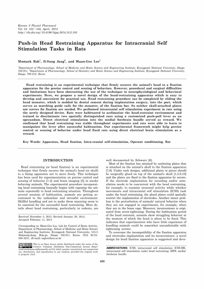
Korean J Physiol PharmacolVol 18: 103-108, April, 2014http://dx.doi.org/10.4196/kjpp.2014.18.2.103
ABBREVIATIONS: ICSS, intracranial self stimulation; ICSS-HR, intracranial self stimulation under head restraining; MFB, medial forebrain bundle.
Received November 3, 2013, Revised January 20, 2014, Accepted February 11, 2014
Corresponding to: Maan-Gee Lee, Lab for Control of Brain Activity, Department of Pharmacology, School of Medicine and Brain Science and Engineering Institute, Kyungpook National University, 515-3 Hakjeong-dong, Buk-gu, Daegu 702-911, Korea. (Tel) 82-53- 950-4231, (E-mail) [email protected]
This is an Open Access article distributed under the terms of the Creative Commons Attribution Non-Commercial License (http://
creativecommons.org/licenses/by-nc/3.0) which permits unrestricted non-commercial use, distribution, and reproduction in any medium, provided the original work is properly cited.
Push-in Head Restraining Apparatus for Intracranial Self Stimulation Tasks in Rats
Mootaek Roh1, Il-Sung Jang2, and Maan-Gee Lee1
1Department of Pharmacology, School of Medicine and Brain Science and Engineering Institute, Kyungpook National University, Daegu 702-911, 2Department of Pharmacology, School of Dentistry and Brain Science and Engineering Institute, Kyungpook National University, Daegu 700-412, Korea
Head restraining is an experimental technique that firmly secures the animal’s head to a fixation apparatus for the precise control and sensing of behaviors. However, procedural and surgical difficulties and limitations have been obstructing the use of the technique in neurophysiological and behavioral experiments. Here, we propose a novel design of the head-restraining apparatus which is easy to develop and convenient for practical use. Head restraining procedure can be completed by sliding the head mounter, which is molded by dental cement during implantation surgery, into the port, which serves as matching guide rails for the mounter, of the fixation bar. So neither skull-attached plates nor screws for fixation are needed. We performed intracranial self stimulation experiment in rats using the newly designed device. Rats were habituated to acclimatize the head-restraint environment and trained to discriminate two spatially distinguished cues using a customized push-pull lever as an operandum. Direct electrical stimulation into the medial forebrain bundle served as reward. We confirmed that head restraining was stable throughout experiments and rats were able to learn to manipulate the lever after successful habituation. Our experimental framework might help precise control or sensing of behavior under head fixed rats using direct electrical brain stimulation as a reward.
Key Words: Apparatus, Head fixation, Intra-cranial self-stimulation, Operant conditioning, Rat
INTRODUCTION
Head restraining (or head fixation) is an experimental technique that firmly secures the animal’s head (or skull) to a fixing apparatus not to move freely. This technique has been used for experimentation on precise control and sensing of behavior [1-4] and brain imaging [5] in awake behaving animals. The experimental procedure incorporat-ing head restraining basically begins with exposing the ani-mals repeatedly to head restraining situation. Throughout several sessions of habituation, animals are getting ac-customed to the unfamiliar and stressful environment. Skillful handling and not to make them annoying seem to be essential for the successful head restraining. More de-tails about head restraining, particularly in rodents, are
well documented by Schwarz [6]. Most of the fixation has attained by anchoring plates that is attached on the animal’s skull to the fixation apparatus [7,8]. Under such designs, additional plates or pieces should be surgically glued on top of the animal’s skull [1,5,9,10] and the plates are fixed to the fixation apparatus by screws. If the electrode implantation for recording and/or stim-ulation needs to be concurrent with the head restraining, for example, to examine neuronal activity while whisker movements and intracranial self stimulation (ICSS) task under the head restraining, the glued plates could spatially restrict the implantation of electrodes. Another minor prob-lem is the perturbation of animals’ natural behavior when they are not engaged in experiments, for example, when they are in the home cage. Moreover, inconvenience is origi-nated from screw-tightening. During the habituation period of the head restraint, animals show struggling behavior at the moment of which the head is about to be fixed. This indicates that experimenters who have little experience of handling animals could be somewhat uncomfortable with tightening screws. To overcome the incompatibility of the fixation apparatus and electrodes implantation and its inconvenience, a novel design for head fixation apparatus is suggested and deve-
104 M Roh, et al
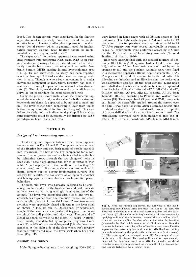
Fig. 1. Head restraining apparatus. (A) Drawing of the head- restraining bar. Shaded area indicates the rim of the port. (B) Practical implementation of the head-restraining bar and the push- pull lever. (C) The mounter is implementated during surgery by applying additional dental cement between the bar and rat skull. a: dental cement applied for electrode implantation, b: Parafilm (surrounded by black solid-lines), c: rat skull, d: mounter. After the mounter is harden, it can be pulled off (white arrow) since Parafile separates the restraining bar and mounter. (D) Head restraining is simply achieved by the guide rails in the mounter (white arrow). (E) The drawing of the push-pull lever. (F) An example of a rat performing ICSS tasks. The push-pull lever is ergonomically designed for head-restrained rats. (G) The molded overhead mounter is inserted into the port, at the middle of the fixation bar and simply tightened by a rubber band.
loped. Two design criteria were considered for the fixation apparatus used in this study. First, there should be no glu-ed attachment of metal and/or plastic plates on the skull except dental cement which is generally used for implan-tation surgery. Second, head fixation should be imple-mented without any screw-like stuff. The capacity of the devised apparatus was tested using head restraint rats performing ICSS tasks. ICSS is an oper-ant conditioning using electrical stimulation delivered di-rectly into the brain reward circuitry such as medial fore-brain bundle (MFB) passing lateral hypothalamus area [11,12]. To our knowledge, no study has been reported about performing ICSS tasks under head restraining condi-tion in rats. Though a whole-body movement is a major movement component of rats, there, recently, has been a success in training forepaw movements in head-restrained rats [6]. Therefore, we decided to make a small lever to serve as an operandum for head-restrained rats. Using the general levers installed on the commercial op-erant chambers is virtually undesirable for both its size and ergonomic problems. It appeared to be natural to push and pull the lever rather than depressing a lever from top to bottom using a unilateral forelimb (or forepaw). This idea led to the design of the bi-directional push-pull lever. Ope-rant behaviors could be successfully conditioned by ICSS paradigm in head restrained rats.
METHODS
Design of head restraining apparatus
The drawing and implementation of the fixation appara-tus are shown in Fig. 1A and B. The apparatus is composed of the fixation bar and box, both made of acrylic panel (6 mm thickness). The bar is the key component for a con-venient fixation procedure and can be installed into the box by tightening screws through the two elongated holes at each side. Those holes allowed the bar to be installed with a tilt. A port is prepared in the middle of the bar (Fig. 1A, shaded area) and it fits the overhead mounter molded in dental cement applied during implantation surgery (See surgery for details). The box serves as an operant chamber which is equipped with modules, such as levers, for operant behaviors. The push-pull lever was basically designed to be small enough to be installed in the fixation box and could indicate at least two states using a single arm operation of the animal. The lever was assembled with a stick and two mi-cro-switches disjoined from the computer mouse, and cased with acrylic plate of 1 mm thickness. Those two micro-switches were oppositely placed adjacent to the lever stick as shown in Fig. 1B and E. Operational principles are simple. If the lever stick was pushed, it triggered the micro-switch of the pull position and vice versa. The on and off signal was then delivered to the digital IO device (National Instruments) and detected by computer programs. Since about 70 to 80% of rats are right-pawed [7,8], the lever was attached at the right side of the floor where rat’s forepaw was naturally placed upon the lever stick when head was fixed (Fig. 1F).
Animals and surgery
Male Sprague-Dawley rats (n=4) weighing 300∼350 g
were housed in home cages with ad libitum access to food and water. The light cycle begins 7 AM and lasts for 12 hours and room temperature was maintained as 20 to 25 oC. After surgery, rats were housed individually in separate cages. All experiments were performed according to Guide for the Care and Use of Laboratory Animals (National Institute of Health, 1996). Rats were anesthetized with the cocktail mixture of ket-amine 10 ml (50 mg/ml), xylazine hydrochloride 1.5 ml (mg/ ml), and saline 2.5 ml. Anesthesia was confirmed by no re-sponses to tail and toe pinches. Animals were then fixed in a stereotaxic apparatus (David Kopf Instruments, USA). The position of rat skull was set to be flatted. After 2% lidocaine s.c. injection and midline incision, the periosteum was completely scraped off the skull surface. Eight holes were drilled and screws (1mm diameter) were implanted into the holes of the skull (frontal AP5.0, ML±2.0 and AP0, ML±5.0, parietal AP-9.0, ML±5.0, occipital AP-3.0 from Lambda, ML±2.0) according to Paxinos and Watson coor-dinates [13]. Then super bond (Super-Bond C&B, Sun medi-cal, Japan) was carefully applied around the screws over the skull. Two holes for stimulation electrodes (insect pins - stainless steel, No. 00 - coated with formvar except the tip) were drilled after the super bond became hard. The stimulation electrodes were then implanted into the bi-lateral MFB area of coordinate AP-2.3 mm, ML1.8 mm,

A Novel Head-Restraining Apparatus 105
Fig. 2. Procedures of habituation and experiment. (A) Habituation procedures for desensitizing rats to head-restraint constitutes repetitive and longer exposure to head-restraining condition. HR: head-restraining habituation only HR+ICSS: ICSS was allowed (B) The basic structure of the rat conditioning.
Fig. 3. Behavioral data for the initial ICSS shaping phases. The number of lever-pulls (triangle) and consequent rewards (circle) are depicted for the rat RS007 (A), RS008 (B), and RS009 (C) for consecutive three sessions (Day1 to 3). Numbers in the parenthesis indicates predetermined inter-trial intervals and stimulation current intensity. When the parameters are modified, it is denoted by the dotted vertical line. The number of rewards that is larger than the number of lever-pulls indicates the delivery of free-reward by the experimenter.
DV-8.2 (from the dura). Dental cement was applied to fix the electrodes firmly. After the implantation surgery, dental cement is applied around the edge of the port of the fixation bar (Fig. 1A) which is wrapped with ParafilmⓇ (Pechiney Plastic Packag-ing, Chicago Il, USA) (Fig. 1C). Care was taken not to cover the bare acrylic part of the fixation bar with dental cement. When it hardened enough, the bar was fixed on the stereo-taxic frame by aligning the port of the bar with the center of implantation cement (Fig. 1C, left). Additional applica-tion of dental cement into the gap between the port and cement applied during surgery adjoins dental cement of both permanently (Fig. 1C, center). When cement hard-ened, the mounter and fixation bar was separated by pull-ing the overhead mounting out of the port and this natu-rally formed the overhead mounter (Fig. 1C, right). Para-filmⓇ covering the port enables this procedure to be simply achieved. After surgery, it is natural that the head mounter serves guide rails for head restraint as shown in Fig. 1D.
Behavioral tasks
After recovery, rats were first tested whether they re-spond to the brain stimulation reward by introducing them to a general operant chamber containing a lever. Whenever rats press the lever, they could receive an electrical stim-ulation directly into their MFB. During the initial shaping stage, stimulation intensity was gradually increased. Rea-ched at a specific intensity, rats kept on pressing the lever and this was regarded as a successful demonstration of ICSS. For that case, it deemed that stimulation electrode was precisely located at the MFB area. Next, rats showing operant behavior were introduced to initial head restraining training using the devised head fix-ation apparatus. The first step of head fixation was simply pushing the overhead mounter into the port. Then the over-head mounter was tightly secured to the bar using a gen-eral rubber band (the wrist rings of latex gloves were pre-
ferred) (Fig. 1G). No more cumbersome procedure was needed. Whenever rats were removed from the fixation ap-paratus and back into their cages, a sweet-flavored food was given for a reward. Previously suggested protocols [1,9] were selectively mo-dified and combined for the habituation of head restraints. The detailed protocol for head restraining used in this study is described in Fig. 2A. Basic idea is that rats became ex-posed to head restraining condition with short time dura-tion and this duration is getting longer across sessions (from 5 min to 60 min across 5 sessions). Rats that did not show any struggling and/or awkward behavior at least 1 hour of head restraining at the final session proceeded to the ICSS tasks under the head restraining (ICSS-HR) condition. For ICSS-HR training, the push-pull lever was installed on the box floor. In general, reward stimulation into the MFB elicits hy-peractivity including exploratory and sniffing actions in the freely moving condition. In the head restraining condition, such hyperactivity could result in the separation of the overhead mounter from the rat’s skull. To prevent this trag-ic accident, stimulation intensity was lowered about 10 to 50 μA from the previous phase at the initial stage of ICSS- HR phase.

106 M Roh, et al
Fig. 4. Behavioral performance session. Given light cues (rectangular), the rats pulled the lever to get stimulation reward (circle). Lever-pulls during the break period (error-pulls, x-mark) increase the duration of the break period, indicating a longer inter-trial interval. All event occurrences and their performance (reward/cue) are depicted for each rat at the top and bottom, respectively. While the rat RS007 and RS008 shows an improve-ment within the session, the rat RS009 shows no improvement.
The experimental design of the initial ICSS-HR training was simple as shown in Fig. 2B. In a dark room, the house light and white noise were turned on, indicating the begin-ning of training. After three sec of delay, the light cue was turned on, indicating that rats could be rewarded when they pull the lever. If rats pull the lever by their right fore-limb, the light cue immediately turned off, and after 100 msec delays, contingent reward stimulation (biphasic, train duration 300 msec, pulse duration 0.2 msec, pulse width 4 msec) was delivered. The light cue was turned on again following a break period (0.2 to 9.6 sec, pre-defined) and next trial was begun. This procedure lasts for about an hour for each of the individual rat. Next, the design was slightly modified to see whether rats could learn the association between the cue and lev-er-pull. The duration of the light cue was fixed to 3 sec, and the break period after reward was randomly set for each trial (1.6 to 4.6 sec). Correct lever-pulls resulted in immediate cue-off and reward delivery. If rats pull the lever during the break, the period was reset from that moment indicating a longer break period. Lastly, rats were introduced to spatial cue discrimination tasks after several sessions of left cue and lever- push training. In this task, rats were rewarded by pushing or pulling the lever when the left or right cue had been pre-sented, respectively. The left or right cue was randomly pre-sented at each trial. The duration of both cues was fixed to 5 sec but correct lever manipulation shorten the duration (that is, cue-off). The break between trials was set to 2.6 sec. All training and task was under control of customized software implemented by Labview 8.5 (National Instru-ments) and behavioral data was collected using data acquis-ition device (National Instruments). Data was analyzed us-ing Matlab software (Mathworks) off-line. The number of lever pull or push was converted “events per min” on a 5 min bin basis. The performance of lever-pull task was de-fined as the number of reward divided by the number of cue presented. In the spatial cue discrimination task, per-formance of the left and right cue was individually analyzed and response latency (time difference from cue on to lever push or pull) was computed. The performance of correct re-sponse was finally computed and analyzed. For example, if the left cue was on then the rat could push the lever to get reward. This reward was given by a correct response. However, when the left cue was on and the rat quickly pulled and then pushed the lever, the rat could receive re-ward but it was regarded as an error response.
RESULTS
During the initial habituation in the head restraining ap-paratus, rats showed struggling behavior, usually running in place and squeaked when touched their back or trunk by an experimenter accidentally. However, those stressful responses were diminishing across the habituation session. The overhead mounter inserted into the port was firmly secured by general rubber band and rats could never fall off by their muscle force. The rats sit in the rear part of the box but nestled down somewhat forward when they were fully habituated. Respiration rate slowed down across sessions was observed by visual inspection. Food reward which was given to the rats after daily habituation was con-sumed by both rat RS007 and RS008 but the rat RS009
was not. In the ICSS check phase, three rats showed repeated lev-er presses at the stimulation intensity of 90 to 150 μA, thus those rats underwent subsequent ICSS-HR training. Rats readily pulled the lever even though their head was fixed. Their locomotion activity was increased by stim-ulation reward at first but decreased across session. Sha-ping of two rats (lever-pull task only) was successful in the ICSS-HR paradigm as shown in Fig. 3. In the first session (Day0), no rat manipulated the lever. In the second session (Day1), the rat (RS007) rarely pulled the lever until 50 min (Fig. 3A). However, the rat began to pull the lever from 45 min and the frequency tended to increase until the end of the session. The more number of rewards than lever-pulls in the first bin indicates that free reward was delivered by the experimenter for shaping purposes. At Day2, the rat pulled the lever from the beginning of the session. The high-er number of lever-pulls than rewards implies that the rat pulled the lever during the break period. The slight reduc-tion in stimulation intensity (100 to 90 μA; separated by a dotted bar, Day2 in Fig. 3A) resulted in less lever-pulls (30 to 55 min). The longer inter-trial-interval was asso-ciated with the decrease in the number of lever-pulls in the later part (2 to 10 sec; separated by a dotted bar, Day3 in Fig. 3A). Another rat (RS008) showed an abrupt increase in lever pulls around 30 min of Day1 (Fig. 3B). The number of lever-pulls increased in Day2. Increased inter-trial inter-val resulted in the decreased number of rewards (Day3). The other rat (RS009) showed no interest in pulling the lever even though extensive free reward in Day1 (Fig. 3C). The intermittent lever-pulls were highly associated with struggling behavior. The performance of lever-pull training is shown in Fig. 4. The rat RS007 showed overall 87.94% of success rate and the rat RS008 showed overall 74.94% of success rate. The performance of rats during the final 10 to 15 min of training was nearly perfect in both of the rats, indicating that rats learned how to behave (pull the lever) to receive reward. The rat RS009 did not show any capacity of task learning,
A Novel Head-Restraining Apparatus 107
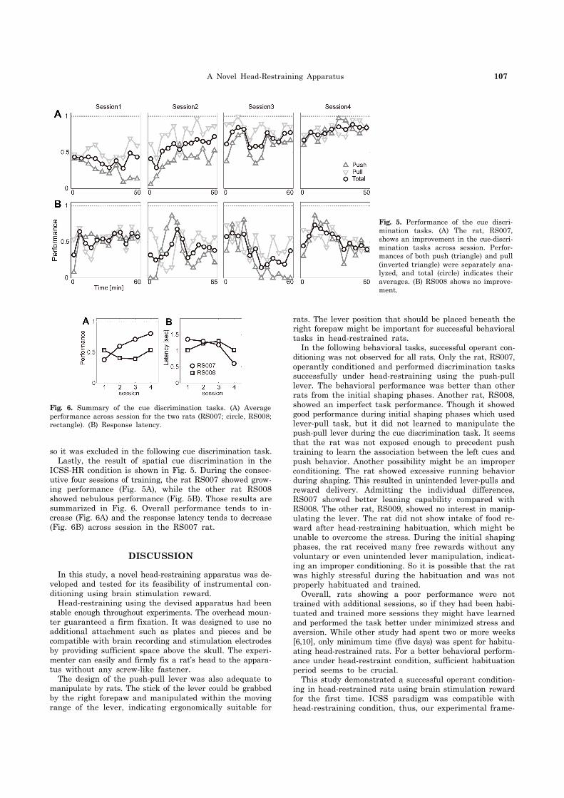
Fig. 5. Performance of the cue discri-mination tasks. (A) The rat, RS007, shows an improvement in the cue-discri-mination tasks across session. Perfor-mances of both push (triangle) and pull (inverted triangle) were separately ana-lyzed, and total (circle) indicates their averages. (B) RS008 shows no improve-ment.
Fig. 6. Summary of the cue discrimination tasks. (A) Average performance across session for the two rats (RS007; circle, RS008; rectangle). (B) Response latency.
so it was excluded in the following cue discrimination task. Lastly, the result of spatial cue discrimination in the ICSS-HR condition is shown in Fig. 5. During the consec-utive four sessions of training, the rat RS007 showed grow-ing performance (Fig. 5A), while the other rat RS008 showed nebulous performance (Fig. 5B). Those results are summarized in Fig. 6. Overall performance tends to in-crease (Fig. 6A) and the response latency tends to decrease (Fig. 6B) across session in the RS007 rat.
DISCUSSION
In this study, a novel head-restraining apparatus was de-veloped and tested for its feasibility of instrumental con-ditioning using brain stimulation reward. Head-restraining using the devised apparatus had been stable enough throughout experiments. The overhead moun-ter guaranteed a firm fixation. It was designed to use no additional attachment such as plates and pieces and be compatible with brain recording and stimulation electrodes by providing sufficient space above the skull. The experi-menter can easily and firmly fix a rat’s head to the appara-tus without any screw-like fastener. The design of the push-pull lever was also adequate to manipulate by rats. The stick of the lever could be grabbed by the right forepaw and manipulated within the moving range of the lever, indicating ergonomically suitable for
rats. The lever position that should be placed beneath the right forepaw might be important for successful behavioral tasks in head-restrained rats. In the following behavioral tasks, successful operant con-ditioning was not observed for all rats. Only the rat, RS007, operantly conditioned and performed discrimination tasks successfully under head-restraining using the push-pull lever. The behavioral performance was better than other rats from the initial shaping phases. Another rat, RS008, showed an imperfect task performance. Though it showed good performance during initial shaping phases which used lever-pull task, but it did not learned to manipulate the push-pull lever during the cue discrimination task. It seems that the rat was not exposed enough to precedent push training to learn the association between the left cues and push behavior. Another possibility might be an improper conditioning. The rat showed excessive running behavior during shaping. This resulted in unintended lever-pulls and reward delivery. Admitting the individual differences, RS007 showed better leaning capability compared with RS008. The other rat, RS009, showed no interest in manip-ulating the lever. The rat did not show intake of food re-ward after head-restraining habituation, which might be unable to overcome the stress. During the initial shaping phases, the rat received many free rewards without any voluntary or even unintended lever manipulation, indicat-ing an improper conditioning. So it is possible that the rat was highly stressful during the habituation and was not properly habituated and trained. Overall, rats showing a poor performance were not trained with additional sessions, so if they had been habi-tuated and trained more sessions they might have learned and performed the task better under minimized stress and aversion. While other study had spent two or more weeks [6,10], only minimum time (five days) was spent for habitu-ating head-restrained rats. For a better behavioral perform-ance under head-restraint condition, sufficient habituation period seems to be crucial. This study demonstrated a successful operant condition-ing in head-restrained rats using brain stimulation reward for the first time. ICSS paradigm was compatible with head-restraining condition, thus, our experimental frame-

108 M Roh, et al
work might contribute to examine not only the precise con-trol or sensing of behavior but also behavioral operant con-ditioning in head fixed rats. Though this study focused on the feasibility of operant conditioning using brain stim-ulation reward (ICSS) under head restraining, the appara-tus can be used in combination with electrophysiological techniques that are extensively employed such as single or multi unit recording and in vivo patch clamping.
ACKNOWLEDGEMENTS
This research was supported by Basic Science Research Program through the NRF funded by the Ministry of Education, Science and Technology (2010-0022362) and by Kyungpook National University Research Fund 2012.
REFERENCES
1. Isomura Y, Harukuni R, Takekawa T, Aizawa H, Fukai T. Microcircuitry coordination of cortical motor information in self-initiation of voluntary movements. Nat Neurosci. 2009;12: 1586-1593.
2. Topchiy IA, Wood RM, Peterson B, Navas JA, Rojas MJ, Rector DM. Conditioned lick behavior and evoked responses using whisker twitches in head restrained rats. Behav Brain Res. 2009;197:16-23.
3. O'Connor DH, Clack NG, Huber D, Komiyama T, Myers EW, Svoboda K. Vibrissa-based object localization in head-fixed
mice. J Neurosci. 2010;30:1947-1967.4. Mayrhofer JM, Skreb V, von der Behrens W, Musall S, Weber
B, Haiss F. Novel two-alternative forced choice paradigm for bilateral vibrotactile whisker frequency discrimination in head-fixed mice and rats. J Neurophysiol. 2013;109:273-284.
5. Dombeck DA, Khabbaz AN, Collman F, Adelman TL, Tank DW. Imaging large-scale neural activity with cellular reso-lution in awake, mobile mice. Neuron. 2007;56:43-57.
6. Schwarz C, Hentschke H, Butovas S, Haiss F, Stüttgen MC, Gerdjikov TV, Bergner CG, Waiblinger C. The head-fixed behaving rat--procedures and pitfalls. Somatosens Mot Res. 2010;27:131-148.
7. Lee MG, Manns ID, Alonso A, Jones BE. Sleep-wake related discharge properties of basal forebrain neurons recorded with micropipettes in head-fixed rats. J Neurophysiol. 2004;92:1182- 1198.
8. Parry TJ, McElligott JG. A method for restraining awake rats using head immobilization. Physiol Behav. 1993;53:1011-1015.
9. Bryant JL, Roy S, Heck DH. A technique for stereotaxic recordings of neuronal activity in awake, head-restrained mice. J Neurosci Methods. 2009;178:75-79.
10. Haiss F, Butovas S, Schwarz C. A miniaturized chronic microelectrode drive for awake behaving head restrained mice and rats. J Neurosci Methods. 2010;187:67-72.
11. Wise RA. Addictive drugs and brain stimulation reward. Annu Rev Neurosci. 1996;19:319-340.
12. Carlezon WA Jr, Chartoff EH. Intracranial self-stimulation (ICSS) in rodents to study the neurobiology of motivation. Nat Protoc. 2007;2:2987-2995.
13. Paxinos G, Watson C. The rat brain: in stereotaxic coordinates. 4th ed. New York: Academic Press; 1998.