purification and preliminary characterization of insect spherulocytes
TRANSCRIPT

Insect Biochem. Vol. 15, No. 3, pp. 419-426, 1985 0020-1790/85 $3.00 +0.00 Printed in Great Britain. All rights reserved Copyright © 1985 Pergamon Press Ltd
PURIFICATION AND PRELIMINARY CHARACTERIZATION OF INSECT SPHERULOCYTES
DOUG COOK, DONALD B. STOLTZ* and CELYNN PAULEY Department of Microbiology, Dalhousie University, Halifax, Nova Scotia, Canada B3H 4H7
(Received 6 March 1984; rev&ed 4 June 1984)
Abstract--Spherulocytes were purified from the haemolymph of two lepidopteran species, Heliothis virescens and Malacosoma disstria. Analysis by polyacrylamide gel electrophoresis revealed the presence of two to four major polypeptides of relatively low molecular weight in purified M. disstria cells and one or two in H. virescens spherulocytes. A sulphated glycosaminoglycan-like substance was extracted from the purified cells.
Key Word Index: Spherulocytes, haemocyte purification, polypeptides, glycosaminoglycans, Heliothis virescens, Malacosoma disstria
INTRODUCTION
Despite considerable progress in the last decade, insect haematology remains a profoundly inexact science. For the most part, entomologists are at present not even able to clearly identify different cell types, let alone specify their possible functions. In- deed, only recently, and for only one insect Order, has a tentative consensus been reached as to the number of different cell types present in the hae- molymph. Considering only the Lepidoptera, these are prohemocytes (PRs), forming a putative stem cell population; plasmatocytes (PLs), regarded as the chief effector cells in such defensive reactions as nodulat ion and encapsulation, and in wound healing; granulocytes (GRs), thought to be involved in blood clotting and the recognition and/or initial reaction to foreign objects; spherulocytes (SPs), function un- known; and oenocytoids (OEs), which have been tentatively linked to the metabolism of phenoloxidase (for recent reviews, the reader is directed to papers by Ratcliffe and Rowley, 1979, 1981). Many important questions remain to be answered: for example, are there several kinds of PLs; do all haemocytes develop from a common precursor, or are there several distinct cell lineages; what kinds of functional inter- relationships, if any, exist between different classes of haemocytes?
Purification of different cell subpopulations would presumably open up new and useful avenues for the study of insect haemocytes. Nevertheless, to our knowledge, this has been attempted on only one previous occasion (Peake, 1979). We describe here a simple protocol which has been applied to the purification of insect spherulocytes; in addition, some preliminary observations relating to the biochemical composit ion of these cells are presented. Finally, we briefly comment on alternative views of spherulocyte function which have appeared in the literature.
*Author to whom correspondence should be addressed.
MATERIALS AND METHODS
Experimental animals
Larvae of Malacosoma disstria and Heliothis virescens were maintained on a general purpose diet (BioServ no. 9000) at 22-25°C, under a 16hr light-8hr dark photo- period. These species were chosen because they have a relatively high proportion of spherulocytes (30-35% of the total haemocyte count; unpublished observations). In most experiments, fifth-instar larvae were used.
Purification of spherulocytes
Spherulocytes were routinely purified on Percoll (Phar- macia) density gradients. Stock Percoll was made fresh daily by dissolving solid NaC1 in Percoll to a final concentration of 0.15 M. From this, two-step gradients were prepared using equal volumes (1 ml each) of 84 and 55% Percoll (prepared v/v with 0.85% NaCI). Typically, haemolymph from 15 to 20 fifth-instar larvae (approx. 0.5ml) was collected from abdominal prolegs directly into a tube con- taining an equal volume of ice-cold SSCE/PTU (SSCE is 0.15 M NaC1, 0.015 M sodium citrate, 0.01 M EDTA; to this is added 1% v/v of saturated phenylthiourea). Samples were mixed by gentle inversion, layered onto Percoll gra- dients, and centrifuged at 350g for 20min at room tem- perature (20°C) in a Sorvall Model GLC-2B centrifuge. Following centrifugation, spherulocyte bands were removed to 1.5 ml microcentrifuge tubes, diluted with approx. 2 vol of phosphate saline (5 mM KPO4, 0.15 mM NaC1, pH 6.5), and pelleted (10,000g, 10 min at 4°C).
Polyacrylamide gel electrophoresis
Pellets of purified cells were resuspended in 50#1 of phosphate saline, mixed with 3 to 5 vol of SDS-disruption buffer and boiled for 5 min. Samples were run on either 7.5-15 or 5-15% glycerol-stabilized gradient gels, using an adaptation of the discontinuous SDS system described by Laemmli et al. (1970). Gels were stained either with coomassie blue as described by Summers and Smith (1978), or by the silver reaction (BioRad technical bulletin no. 1089). Molecular weight standards were either from Phar- macia (14,400-94,000) or Serva (3400-29,000).
Extraction of glycosaminoglycan-like material
Cell pellets from approx. 10 gradients (150-200 fifth-instar larvae) were resuspended in distilled water,
419
420 DOUG COOK et al.
pooled, and mixed with 2 vol of phenol saturated with distilled water; this was then sonicated for 2-3 min, and briefly centrifuged to separate the phases, after which the phenol was re-extracted with an equal volume of water. The aqueous phases were pooled, extracted three times with diethyl ether (approx. 10vol/extraction), dialysed against 500 vol of distilled water, and finally lyophilized. The re- sulting powder was resuspended in a minimal volume of distilled water and treated with alkali as described by Mourao et al. (1983). Following neutralization, the solution was centrifuged at 3500g for 10min at 20C to remove insoluble materials, and putative glycosaminoglycans (GAGs) were isolated by adding cetyl trimethyl ammonium bromide to a final concentration of 1')o; this was allowed to stand for a minimum of 12hr at 34C, whereupon the flocculent precipitate was collected by centrifugation at 15,000 g for 10 rain at 20~C. The pellet was resuspended in water and dialysed for 24 hr at 20C against water using a minimum volume of 31. Electrophoresis was essentially as described by Jaques et al. (1968). In preliminary experi- ments, partially purified GAGs were digested with a number of different proteases, including chymotrypsin A and papain (Sigma), and pronase and proteinase K (Boehringer- Mannheim); as these caused no apparent change in electro- phoretic mobility (data not shown), proteases were there- after not used in the extraction procedure.
M disstria GAGs were isotopically labelled in t, ivo by injecting fifth-instar larvae with 20,ul of 35SO4 (New En- gland Nuclear) at 0.2 m Ci/ml in sodium succinate buffer, pH 6.3. After injection, larvae were placed on fresh diet and maintained for 24hr to permit incorporation of sulphur; subsequent purification of SPs and GAG was as described above. Incorporation of isotope into cells was monitored by liquid scintillation spectrometry in Bray's cocktail (New England Nuclear); specific incorporation into GAG was detected by autoradiography after agarose gel electro- phoresis.
RESULTS AND DISCUSSION
Spheru locy te morpho logy
Insect spherulocytes are usually described as being spherical to ovoid cells, having small nuclei, and a cytoplasm almost completely filled with large gran- ules (Ratcliffe and Rowley, 1979; Akai and Sato, 1979; Gupta, 1979); when viewed by phase-contrast microscopy, SP granules are highly refractile. This morphology is sufficiently distinctive that SPs cannot normally be confused with any other cell type. Nev- ertheless, in preliminary work, we wished to establish that accepted criteria for the identification of such cells obtained in the present context. By light micros- copy, typical SPs could be unambiguously identified in both M . disstria and H. virescens larvae. As others have shown, SP granules can be readily stained with the cationic dye alcian blue (Neuwirth, 1973; Arnold and Hinks, 1983), although we found that astra blue (Blaies and Williams, 1981) usually produced a more intense reaction (Fig. 1). We confirm the observations of Arnold and Hinks (1983) to the effect that SPs are actively mitotic (Fig. l b), and concur with their view (Arnold and Hinks, 1976) that SPs probably represent a cell lineage distinct from that of the granulocyte population. By electron microscopy, SP granules usually exhibit a characteristic substructure consisting in large part of concentric layers of short, apparently tubular, structures (Akai and Sato, 1979; Harpaz et al., 1969); a typical example, from H. virescens, is shown in Fig. l d.
Puri f icat ion
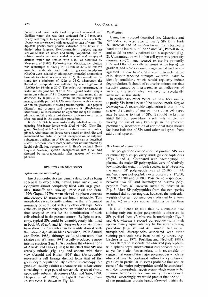
Using the protocol described (see Materials and Methods), we were able to purify SPs from both H. virescens and M. disstria larvae. Cells formed a band at the interface of the 55 and 84"~i Percoll steps. and could be readily pelleted and resuspended (Fig. 2). Contaminat ion with other cell types was general b minimal (1 5°;), and seemed to involve primarily PRs and OEs; other cells remained at the top of the gradient and were extensively aggregated and/or co- agulated. In our hands, SPs were extremely stable cells; despite repeated attempts, we were unable to identify conditions which would regularly induce degranulation. It should of course be pointed out thal stability cannot be interpreted as an indication of viability, a question which we have not specifically addressed in this study.
In preliminary experiments, we have been unable to purify SPs from larvae of the tussock moth, Orgyia leucost igma. A reasonable explanation is that in this species the density of one or more other cell types may be similar to that of SPs. It should be kept in mind that our procedure is relatively simple, in- volving the use of only two steps in the gradient: presumably, incorporation of additional steps should facilitate isolation of SPs (and other cell types) from additional species.
Biochemica l composi t ion
The polypeptide composition of purified SPs was examined by SDS polyacrylamide gel electrophoresis (Figs 3 and 4). Compared with haemolymph or plasma, the major SP polypeptides were of relatively low molecular weight in both species: in H. t:irescem, the major SP polypeptide was at 26,500; in M. disstria, major polypeptides were observed at 17,800, 27,500, 29,500 and 32,000. Possible correspondence between two SP and haemolymph plasma poty- peptides from H. virescens larvae is indicated in Fig. 3. Most SP polypeptides from the two species examined did not co-migrate; however, the molecular weights of certain polypeptides (indicated by arrows in Fig. 4c) were very similar, differing by less than 1000.
It is of interest to note that by coomassie blue staining only one major polypeptide is observed in SPs purified from H. Hrescens haemolymph (Figs 3 and 4a), whereas a second polypeptide stains with approximately equal intensity by the silver staining procedure (Figs 4b and 4c): similar, but as yet unexplained, discrepancies associated with silver staining protocols have been noted by others (e.g. Lischwe et al., 1976; Poehling and Neuhoff, 1981).
An attempt to associate the observed polypeptidcs with spherulocyte substructural components cannot as yet be made. Nevertheless, it is reasonable to suggest that some of the major polypeptides which we observed must be contained within the cytoplasmic granules; in particular, it seems probable that one or more of the major polypeptides would be associated with the microtubular substructure which seems to be common to SP granules from many different insect species. Accordingly, we would predict that one or all of the prominent protein bands observed within the

w
a b
Fig. 1. (a) Heliothis virescens haemocytes. S = spherulocyte, stained with astra blue; the other haernocytes are unidentified. Nuclei are counterstained with haernatoxylin. Bar = 10#rn. (b) Malacosoma disstria. Examples of spherulocytes in metaphase of mitosis. (c) Mouse mast cell (arrow) stained with astra blue and reproduced at the same magnification as the spherulocyte illustrated in (a). (d) Electron micrograph
of a typical H. virescens spherulocyte. Bar = 5 prn.
421

Fig. 2. Phase contrast micrograph of purified spherulocytes (H. virescens). Bar = 20/ tm.
- _
- - 9 4
I I - m 67
~ z.,.- ~ O B ~ ''4
q , . ~ ~ . ~ , a , e ~ - " 1 1 ~ 4 3
~ , ' 3 3
6
, - " 2 0
, ~ , 14.4
1 8 15 Fig. 3. Coomassie blue-stained SDS polyacrylamide gel. M. disstria: haemolymph (lanes 2 and 3); plasma (lanes 4 and 5): spherulocytes (lane 6). H. l:irescens: spherulocytes (lane 10); haemolymph (lanes I 1 and 12); plasma (lanes 13 and 14). Haemolymph and plasma were applied at volumes of 2~1 per lane: SPs were purified in each case from 60/11 of haemolymph. The two samples run in lanes I1 and 12 were from somewhat older larvae: the high molecular weight band is a storage prote in The arrowheads indicale polypeptides of similar molecular weights in H. ~'ire~'cen,~ SPs and plasma. Molecular weight s tandards
shown range from 14,400 to 94,000 (lanes I, 7, 9 and 15), or from 3400 to 29,00fl (la~ae 8).

~ ~ ~ i~ ̧ ~il;
~i ̧ i i i i
~!~ ? i!ii ~ii(~ ~ i~iii!iii
~ ~ i~i~ii~ ~ ~i~!i~
~,~ ~,ii~ ~,,~
1 7 1 6
Fig. 4. SDS polyacrylamide gel of polypeptides from purified spherulocytes. (a) H. virescens. Coomassie blue staining; (b) H. virescens. Molecular weight markers (14,000-94,000) are shown in lanes 1 avd 7, silver staining; (c) H. virescens (lanes 1-3) and M. disstria (lanes 4-6), silver staining. The positions of three examples of H. virescens and M. disstria spherulocyte polypeptides that have very similar molecular weights are indicated. In this Figure, each lane of SP polypeptides is from cells collected from a different
gradient.
423

i
Fig. 5. (A) Agarose gel of 35S-labelled glycosaminoglycan extracted from purified M. disstria spherulocytes: stained with toluidine blue. Lane 1: heparin; lane 2: chondroitin sulphate (CS) B; lane 3: CSC; lane 4: M. disstria extract (amount applied is roughly equivalent to the amount of GAG that can be extracted from 0.1 ml of haemolymph); lane 5: hyaluronic acid; lane 6: CSA. GAG standards were at 5 fig per gel lane, except for hyaluronic acid (10/lg). (B) Autoradiogram of (A). Exposure was for one week at - 80'C,
with Kodak X-OMAT AR film.
424
Purification
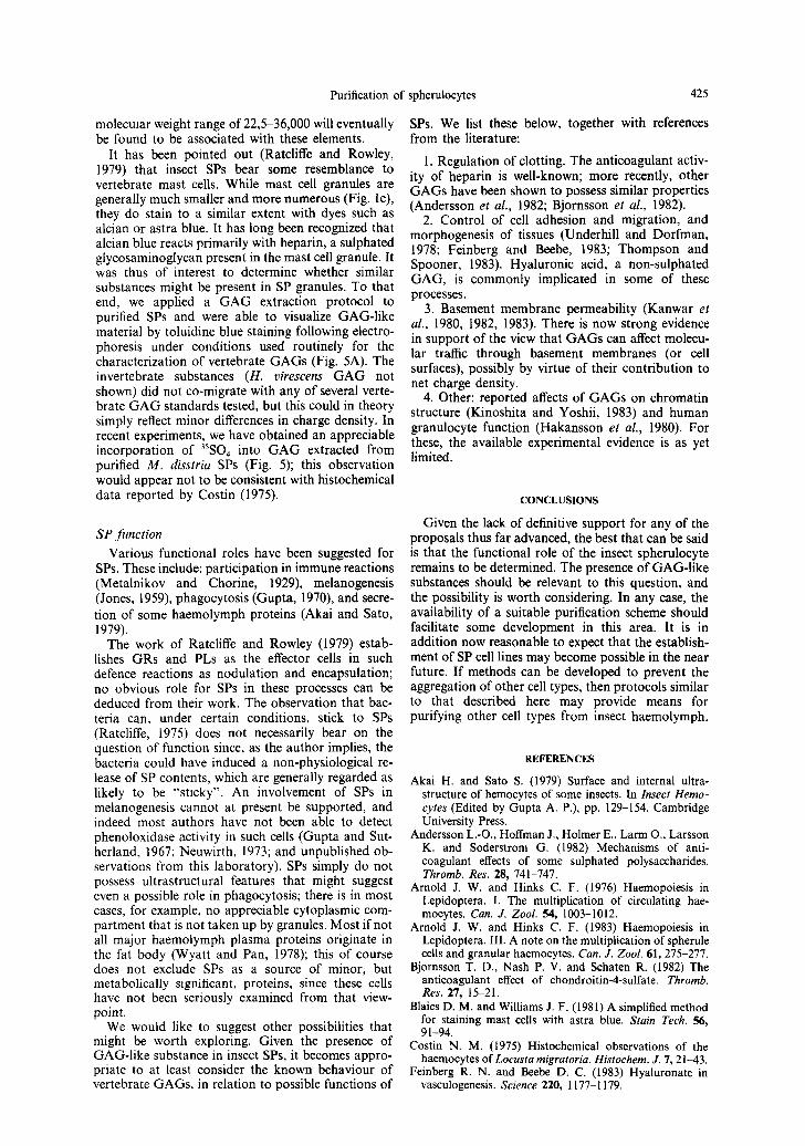
molecular weight range of 22,5-36,000 will eventually be found to be associated with these elements.
It has been pointed out (Ratcliffe and Rowley, 1979) that insect SPs bear some resemblance to vertebrate mast cells. While mast cell granules are generally much smaller and more numerous (Fig. lc), they do stain to a similar extent with dyes such as alcian or astra blue. It has long been recognized that alcian blue reacts primarily with heparin, a sulphated glycosaminoglycan present in the mast cell granule. It was thus of interest to determine whether similar substances might be present in SP granules. To that end, we applied a G A G extraction protocol to purified SPs and were able to visualize GAG-like material by toluidine blue staining following electro- phoresis under conditions used routinely for the characterization of vertebrate GAGs (Fig. 5A). The invertebrate substances (H. virescens GAG not shown) did not co-migrate with any of several verte- brate G A G standards tested, but this could in theory simply reflect minor differences in charge density. In recent experiments, we have obtained an appreciable incorporation of 35SO4 into G A G extracted from purified M. disstria SPs (Fig. 5); this observation would appear not to be consistent with histochemical data reported by Costin (1975).
SP function
Various functional roles have been suggested for SPs. These include: participation in immune reactions (Metalnikov and Chorine, 1929), melanogenesis (Jones, 1959), phagocytosis (Gupta, 1970), and secre- tion of some haemolymph proteins (Akai and Sato, 1979).
The work of Ratcliffe and Rowley (1979) estab- lishes GRs and PLs as the effector cells in such defence reactions as nodulation and encapsulation; no obvious role for SPs in these processes can be deduced from their work. The observation that bac- teria can, under certain conditions, stick to SPs (Ratcliffe, 1975) does not necessarily bear on the question of function since, as the author implies, the bacteria could have induced a non-physiological re- lease of SP contents, which are generally regarded as likely to be "sticky". An involvement of SPs in melanogenesis cannot at present be supported, and indeed most authors have not been able to detect phenoloxidase activity in such cells (Gupta and Sut- herland, 1967; Neuwirth, 1973; and unpublished ob- servations from this laboratory). SPs simply do not possess ultrastructural features that might suggest even a possible role in phagocytosis; there is in most cases, for example, no appreciable cytoplasmic com- partment that is not taken up by granules. Most if not all major haemolymph plasma proteins originate in the fat body (Wyatt and Pan, 1978); this of course does not exclude SPs as a source of minor, but metabolically significant, proteins, since these cells have not been seriously examined from that view- point.
We would like to suggest other possibilities that might be worth exploring. Given the presence of GAG-like substance in insect SPs, it becomes appro- priate to at least consider the known behaviour of vertebrate GAGs, in relation to possible functions of
of spherulocytes 425
SPs. We list these below, together with references from the literature:
1. Regulation of clotting. The anticoagulant activ- ity of heparin is well-known; more recently, other GAGs have been shown to possess similar properties (Andersson et al., 1982; Bjornsson et al., 1982).
2. Control of cell adhesion and migration, and morphogenesis of tissues (Underhill and Dorfman, 1978; Feinberg and Beebe, 1983; Thompson and Spooner, 1983). Hyaluronic acid, a non-sulphated GAG, is commonly implicated in some of these processes.
3. Basement membrane permeability (Kanwar et al., 1980, 1982, 1983). There is now strong evidence in support of the view that GAGs can affect molecu- lar traffic through basement membranes (or cell surfaces), possibly by virtue of their contribution to net charge density.
4. Other: reported affects of GAGs on chromatin structure (Kinoshita and Yoshii, 1983) and human granulocyte function (Hakansson et al., 1980). For these, the available experimental evidence is as yet limited.
CONCLUSIONS
Given the lack of definitive support for any of the proposals thus far advanced, the best that can be said is that the functional role of the insect spherulocyte remains to be determined. The presence of GAG-like substances should be relevant to this question, and the possibility is worth considering. In any case, the availability of a suitable purification scheme should facilitate some development in this area. It is in addition now reasonable to expect that the establish- ment of SP cell lines may become possible in the near future. If methods can be developed to prevent the aggregation of other cell types, then protocols similar to that described here may provide means for purifying other cell types from insect haemolymph.
REFERENCES
Akai H. and Sato S. (1979) Surface and internal ultra- structure of hemocytes of some insects. In Insect Hemo- cytes (Edited by Gupta A. P.), pp. 129-154. Cambridge University Press.
Andersson L.-O., Hoffman J., Holmer E., Larm O., Larsson K. and Soderstrom (3. (1982) Mechanisms of anti- coagulant effects of some sulphated polysaccharides. Thromb. Res. 28, 741-747.
Arnold J. W. and Hinks C. F. (1976) Haemopoiesis in Lepidoptera. I. The multiplication of circulating hae- mocytes. Can. J. Zool. 54, 1003-1012.
Arnold J. W. and Hinks C. F. (1983) Haemopoiesis in Lepidoptera. III. A note on the multiplication of spherule cells and granular haemocytes. Can. J. Zool. 61, 275-277.
Bjornsson T. D., Nash P. V. and Schaten R. (1982) The anticoagulant effect of chondroitin-4-sulfate. Thromb. Res. 27, 15-21.
Blaies D. M. and Williams J. F. (1981) A simplified method for staining mast cells with astra blue. Stain Tech. 56, 91-94.
Costin N. M. (1975) Histochemical observations of the haemocytes of Locusta migratoria. Histochem. J. 7, 21-43.
Feinberg R. N. and Beebe D. C. (1983) Hyaluronate in vasculogenesis. Science 220, 1177-1179.

426 DouG COOK et al.
Gupta A. P. (1970) Midgut lesions in Epicauta einerea (Coleoptera: Meloidae). Ann. ent. Soc. Am. 63, 1786-1788.
Gupta A. P. (1979) Hemocyte types: their structures, syn- onymies, interrelationships, and taxonomic significance. In Insect Hemocytes (Edited by Gupta A. P.), pp. 85 t27. Cambridge University Press.
Gupta A. P. and Sutherland D. J. (1967) Phase contrast and histochemical studies of spherule cells in cockroaches. Ann. ent. Soc. Am. 60, 557-565.
Hakansson L., Hallgren R. and Venge P. (1980) Regulation of granulocyte function by hyaluronic acid. J. clin. Invest. 66, 298-305.
Harpaz I., Kislev N. and Zelcher A. (1969) Electron- microscopic studies on hemocytes of the Egyptian cotton- worm, Spodoptera linoralis (Boisduval) infected with a nuclear-polyhedrosis virus, as compared to noninfected hemocytes. I. Noninfected hemocytes. J. Invertebr. Path. 14, 175 185.
Jaques L. B., Ballieux R. E., Dietrich C. P. and Kavanagh L. W. (1968) A microelectrophoresis method for heparin. Can. J. Physiol. Pharmac. 46, 351-360.
Jones J. C. (1959) A phase contrast study of the blood cells in Prodenia larvae (Order Lepidoptera). Q. J. microsc. Sci. 100, 17-23.
Kanwar Y. S., Linker A. and Farquhar M. G. (1980) Increased permeability of the glomerular basement mem- brane to ferritin after removal of glycosaminoglycans (heparan sulfate) by enzyme digestion. J. Cell Biol. 86, 688-693.
Kanwar Y. S. and Rosenzweig L. J. (1982) Clogging of the glomerular basement membrane. J. Cell Biol. 93, 489-494.
Kanwar Y. S., Rosenzweig L. J., Linker A. and Jakubowski M. L. (1983) Decreased de novo synthesis of glomerular proteoglycans in diabetes: biochemical and auto- radiographic evidence. Proc. natn. Acad. Sci. U.S.A. 80, 2272-2275.
Kinoshita Y. and Yoshii (1983) Permeable embryonic cells of sea urchins as a model for studying nucleus-cytoplasm interactions. Experientia 39, 189-190.
Laemmli U. K. (1970) Cleavage of structural proteins during the assembly of the head of bacteriophage T~. Nature 227, 680-685.
Lischwe M. A., Smetana K., Olson M. O. J. and Busch H. (1976) Proteins C23 and B23 are the major nucleolar silver staining proteins. L(/e Sci. 25, 701-708.
Metalnikov S. and Chorine V. (1929) on the natural and acquired immunity of Pyrausta nubilalis Hb. lnt. Corn Borer Invest. Sci. Rep. 2, 22-38.
Mourao P. A. S., Pillai S. and Donnelly P. V. (1983) Sulfated glycosaminoglycans synthesized by fibroblast, smooth muscle and endothelium-like cells grown in cul- ture. Cell Different. 12, 99 108.
Neuwirth M. (1973) The structure of the hemocytes of Galleria mellonella (Lepidoptera). J. Morph. 139, 105-124.
Peake P. W. (1979) Isolation and characterization of" the haemocytes of Calliphora vieina on density gradients of Ficoll. J. Insect Physiol. 25, 795 803.
Poehling H.-M. and Neuhoff V. (1981) Visualization o1 proteins with a silver "stain": A critical analysis. Electr,- phoresis 2, 141-147.
Ratcliffe N. A. (1975) Spherule cell-test particle interactions in monolayer cultures of Pieris brassieae hemocytes. J. lnvertehr. Path. 26, 217--223.
Ratcliffe N. A. and Rowley A. F. (1979) Role of hemocytes in defense against biological agents. In bisect Hemocyte.~ (Edited by Gupta A. P.), pp. 331-414. Cambridge Univer- sity Press.
Rowley A. F. and Ratcliffe N. A. (1981). Insects. In lnvertehrate Blood Cells, Vol. 2. Arthropod.~ to l, ro- chordates, Invertebrates and Vertebrate.~ Compared (Edi- ted by Ratcliffe N. A. and Rowley A. F.), pp. 421 48{). Academic Press, New York.
Summers M. D. and Smith O. E. (1978) Baculovirus structural polypeptides. Virology 84, 390 402.
Thompson H. A. and Spooner B. S. (1983) Proteo- glycan and glycosaminoglycan synthesis in embryonic mouse salivary glands: effects of fi'-D-xylosidc, an in- hibitor of branching morphogenesis. J. Cell Biol. 96, 1443-1450.
Underhill C. and Dorfman, A. (1978) The role ofhyaluronic acid in intercellular adhesion of cultured mouse cells. Lvp/ Cell Res. 117, 155-164.
Wyatt G. R. and Pan M. L. (1978) Insect plasma proteins. .4, Rev. Biochem. 47, 779-817.