pulmonary hypertension in cardiac surgery - semantic … · pulmonary hypertension in cardiac...
TRANSCRIPT

Current Cardiology Reviews, 2010, 6, 1-14 1
1573-403X/10 $55.00+.00 © 2010 Bentham Science Publishers Ltd.
Pulmonary Hypertension in Cardiac Surgery
André Denault*, Alain Deschamps, Jean-Claude Tardif, Jean Lambert and Louis Perrault
Montreal Heart Institute and Université de Montréal, Montreal, Quebec, Canada
Abstract: Pulmonary hypertension is an important prognostic factor in cardiac surgery associated with increased
morbidity and mortality. With the aging population and the associated increase severity of illness, the prevalence of pul-
monary hypertension in cardiac surgical patients will increase. In this review, the definition of pulmonary hypertension,
the mechanisms and its relationship to right ventricular dysfunction will be presented. Finally, pharmacological and non-
pharmacological therapeutic and preventive approaches will be presented.
Keywords: Cardiac surgery, pulmonary hypertension, pharmacological therapy.
1. DEFINITION OF PULMONARY HYPERTENSION
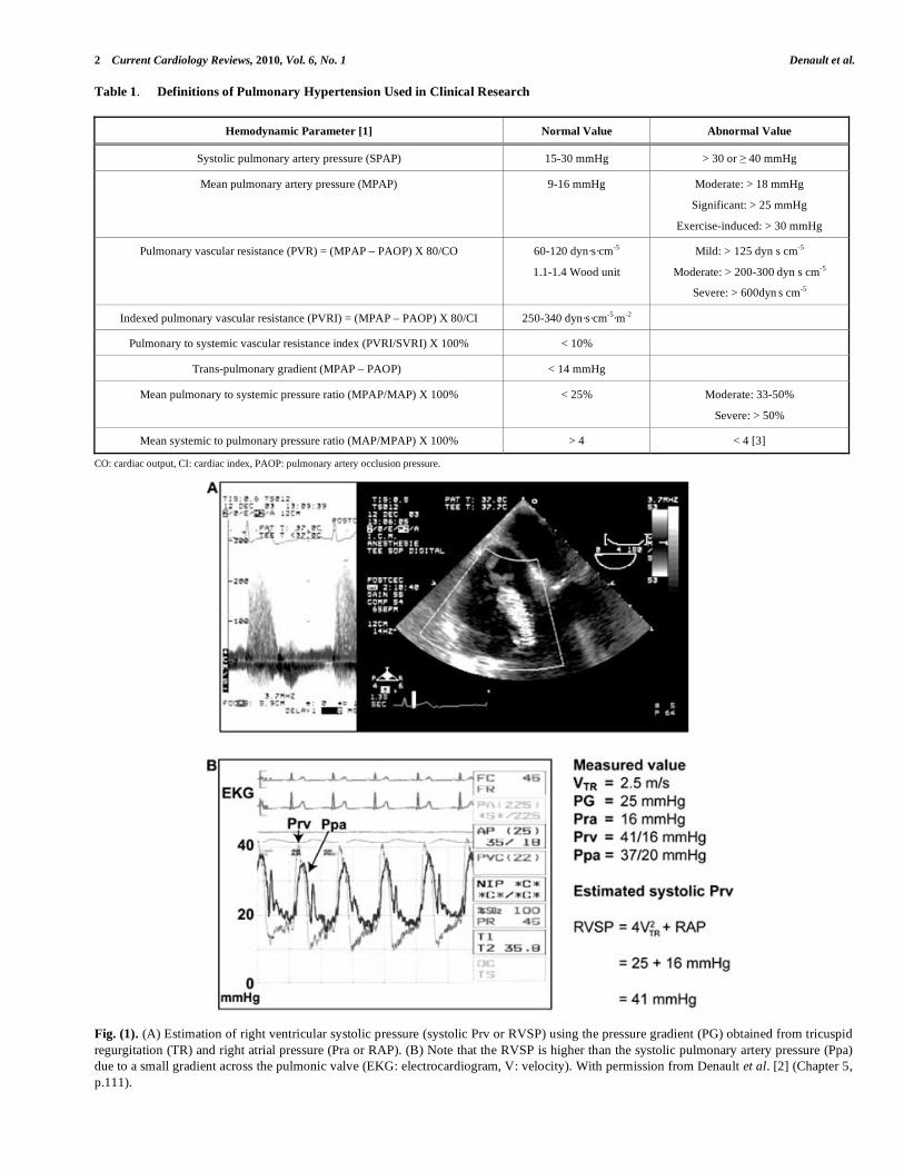
There are several hemodynamic parameters that are used in defining pulmonary hypertension (PHT) (Table 1) [1]. Several of these definitions have been used in various studies. In cardiac surgery, we obtain information on PHT before the procedure and this is usually from an awake patient. This preoperative information is either acquired through preoperative catheterization or, more frequently, estimated via transthoracic echocardiography by using the Bernoulli’s equation. In the presence of tricuspid regur-gitation, as shown in Fig. (1), [2] the simplified Bernoulli’s equation will give an estimation of the pressure gradient across the tricuspid valve. This pressure gradient is equal to the difference between the systolic pressure of the right ventricle (RV) and the right atrium (RA). Therefore, knowledge (or estimation) of the right atrial pressure allows the estimation of the right ventricular systolic pressure. In the absence of right ventricular outflow tract obstruction (RVOTO) or pulmonic valve stenosis, this value will be an estimation of the systolic pulmonary artery pressure (SPAP).
Following the induction of general anesthesia, a redu-ction of both the systemic and the pulmonary artery pressures will be observed. Consequently, absolute values of SPAP used in defining PHT will tend to underestimate its severity. In 2006, we addressed this issue and published a study involving 1557 patients who underwent cardiac surgery [3]. We first demonstrated that the induction of general anesthesia in 32 patients was associated with a significant reduction in mean arterial pressure (MAP), mean pulmonary artery pressure (MPAP) but the MAP/MPAP ratio did not change (Fig. 2). Therefore, this ratio (normal value > 4) seems to be a very robust estimator of the severity of PHT. To demonstrate the utility of the MAP/MPAP ratio, we compared it to the other hemodynamic parameters listed in Table 1 in 1439 patients undergoing cardiac surgery after the induction of general anesthesia but before cardio-
*Address correspondence to this author at the Institut de Cardiologie de
Montréal, 5000 rue Bélanger, Montréal, Québec H1T 1C8, Canada;
Tel: (514) 376-3330; Ext: 3732; Fax: (514) 376-8784;
E-mail: [email protected]
pulmonary bypass (CPB). We observed that the MAP/MPAP ratio behaved similarly to the other hemodynamic parameters (Fig. 3), and had the highest receiver operating curve value to predict hemodynamic complications after cardiac surgery. The hemodynamic complications were defined as posto-perative death or requirement for an intra-aortic balloon pump, cardiac arrest and vasoactive support for more than 24 hours. Finally, using transesophageal echocardiography (TEE), we can confirm that the presence of an abnormal MAP/MPAP ratio is almost invariably associated with abnormal systolic or diastolic cardiac function (Fig. 4) [3]. This concept of using the relative instead of absolute value of PHT indices is currently used in congenital cardiology [4, 5].
Finally, PHT is typically classified as capillary, pre-capillary or post-capillary, depending on the site where the cause of PHT is present. The 2003 World Symposium on PHT proposed a classification based on 5 groups: 1-Pulmo-nary arterial hypertension, 2-PHT secondary to left heart disease, 3-PHT secondary to lung disease and/or hypoxia, 4-PHT secondary to thrombotic and/or embolic disease and 5-A miscellaneous category [6]. In cardiac surgery, it is typi-cally post-capillary or group 2 because the cause of PHT is of cardiac origin and consequently localized after the pulmo-nary capillary. This is confirmed using pulmonary artery catheterization during which the diastolic pulmonary artery pressure is equal to the pulmonary artery occlusion pressure (PAOP). In a situation where the diastolic pulmonary artery pressure (DPAP) is significantly higher than the PAOP in the absence of tachycardia, a capillary or pre-capillary cause could be sought [1].
In summary, PHT in cardiac surgery should be carefully defined. It is generally a post-capillary PHT. In awake patients, the absolute values have been used and correlated with outcome. However, in patients under general anes-thesia, the relative value seems to be more appropriate.
2. PULMONARY HYPERTENSION IN CARDIAC SURGERY: MECHANISM AND ETIOLOGY
The mechanism of PHT in cardiac surgery is complex and can result from several mechanisms acting alone or in
2 Current Cardiology Reviews, 2010, Vol. 6, No. 1 Denault et al.
Table 1. Definitions of Pulmonary Hypertension Used in Clinical Research
Hemodynamic Parameter [1] Normal Value Abnormal Value
Systolic pulmonary artery pressure (SPAP) 15-30 mmHg > 30 or 40 mmHg
Mean pulmonary artery pressure (MPAP) 9-16 mmHg Moderate: > 18 mmHg
Significant: > 25 mmHg
Exercise-induced: > 30 mmHg
Pulmonary vascular resistance (PVR) = (MPAP – PAOP) X 80/CO 60-120 dyn·s·cm-5
1.1-1.4 Wood unit
Mild: > 125 dyn s cm-5
Moderate: > 200-300 dyn s cm-5
Severe: > 600dyn s cm-5
Indexed pulmonary vascular resistance (PVRI) = (MPAP – PAOP) X 80/CI 250-340 dyn·s·cm-5·m-2
Pulmonary to systemic vascular resistance index (PVRI/SVRI) X 100% < 10%
Trans-pulmonary gradient (MPAP – PAOP) < 14 mmHg
Mean pulmonary to systemic pressure ratio (MPAP/MAP) X 100% < 25% Moderate: 33-50%
Severe: > 50%
Mean systemic to pulmonary pressure ratio (MAP/MPAP) X 100% > 4 < 4 [3]
CO: cardiac output, CI: cardiac index, PAOP: pulmonary artery occlusion pressure.
Fig. (1). (A) Estimation of right ventricular systolic pressure (systolic Prv or RVSP) using the pressure gradient (PG) obtained from tricuspid
regurgitation (TR) and right atrial pressure (Pra or RAP). (B) Note that the RVSP is higher than the systolic pulmonary artery pressure (Ppa)
due to a small gradient across the pulmonic valve (EKG: electrocardiogram, V: velocity). With permission from Denault et al. [2] (Chapter 5,
p.111).

Pulmonary Hypertension in Cardiac Surgery Current Cardiology Reviews, 2010, Vol. 6, No. 1 3
Fig. (2). Change in MAP, MPAP, and the MAP/MPAP ratio after the induction of anesthesia in 32 patients with preoperative pulmonary
hypertension. No significant change in the MAP/MPAP ratio was observed (*p < 0.05). (MAP: mean arterial pressure, MPAP: mean
pulmonary artery pressure) [3].
combination. These mechanisms can be present before the operation, secondary for instance from valvular heart disease. The cause of PHT can appear after CPB from mechanical failure or from the pulmonary reperfusion syndrome. Finally, PHT can be present or persist posto-peratively secondary for instance from/to a mitral or aortic patient-prosthesis-mismatch (PPM). Fig. (5) focuses on the role of PHT in cardiac surgery [7] based on current literature, our research findings and our experience of this population.
2.1. Review of the Factors Involved in Pulmonary Hypertension in Cardiac Surgery
The 6 most important causes of PHT in cardiac surgery factors are illustrated in Fig. (5).
1) Left ventricular systolic or diastolic dysfunction and mitral or valvular disease either pre- or postoperative are the most common causes of PHT in cardiac surgery. Aortic PPM through a reduction in coronary reserve could also contribute to postoperative PHT [8].
2) During cardiac surgery, the extent of the systemic inflam-matory response, the pulmonary reperfusion syndrome and the need for blood transfusions may exacerbate PHT (Fig. 6) [9, 10]. The mechanism of pulmonary damage during extracorporeal circulation is thought to be mainly triggered by 1) release of cytokines [11] through endotoxin production, 2) complement activation and 3) ischemia reperfusion injury [12, 13]. This leads to the production of free radicals, endothelin and prostacyclin derivatives with nitric oxide inhibition [12].
3) The administration of protamine can induce catastrophic pulmonary vasoconstriction in up to 1.8% of patients [14]. Protamine can also activate complement and, when
given at the end of CPB, can induce PHT associated with adverse hemodynamic responses ranging from minor perturbations to cardiovascular collapse. Three types have been described: systemic hypotension, anaphy-lactoid reaction and catastrophic PHT [15]. The mecha-nism of PHT with protamine is thought to occur through an imbalance of vasoconstrictors and vasodilators which leads a reduction in the release of nitric oxide (NO) from the pulmonary vasculature [15].
4) Mitral PPM is another recently described cause of residual postoperative PHT. Magne et al.[16] studied 929 patients who underwent mitral valve replacement (MVR) and followed them up for 15 years. Mitral valve PPM was defined as not clinically significant if > 1.2 cm /m , as moderate if > 0.9 and 1.2 cm /m , and as severe if 0.9 cm /m . The prevalence of moderate PPM was 69% and that of severe PPM was 9%. Severe PPM was found to be associated with residual PHT and a 3-fold increase in postoperative mortality after adjustment for other risk factors. This new and relevant information is currently absent from the majority of the studies dealing with predictors of survival in mitral valvular surgery.
5) Hypoxia, hypercarbia and pulmonary embolism are other causes of PHT. They can appear before, during or after CPB. For instance, PHT can cause RV dysfunction, which will lead to an increase in right atrial pressure. This can lead to the opening of a patent foramen ovale (PFO), which is present in 20-30% of the general population [17]. The consequence of the opening of a PFO would be a right-to-left shunt. This would increase the severity of hypoxia and lead to an exacerbation of PHT. Pulmonary vessels constrict with hypoxia (Euler-Liljestrand reflex) and relax in the presence of hyperoxia

4 Current Cardiology Reviews, 2010, Vol. 6, No. 1 Denault et al.
Fig. (3). Relationship between the estimated probability of hemodynamic complications and variables used in the evaluation of pulmonary
hypertension: (A) systolic pulmonary artery pressure (SPAP), (B) MPAP, (C) PVRI, (D) the ratio of SVRI to PVRI, (E) the MAP/MPAP
ratio, and (F) the transpulmonary gradient defined as MPAP minus pulmonary artery occlusion pressure (PAOP). For easier comparison, the
scale of the x axis of the SVRI/PVRI and the MAP/MPAP are inverted. (n = number of patients). (MAP: mean arterial pressure, MPAP: mean
pulmonary artery pressure, PVRI: indexed pulmonary vascular resistance, SVRI: indexed systemic vascular resistance) [3].

Pulmonary Hypertension in Cardiac Surgery Current Cardiology Reviews, 2010, Vol. 6, No. 1 5
Fig. (4). Hemodynamic and transesophageal echocardiographic evaluation of a 46-year-old woman scheduled for aortic valve surgery.
Despite a normal pulmonary artery pressure of 34/16 mmHg and PVRI at 286 dyn·s·cm-5·m-2, this patient had an abnormal right ventricular
diastolic filling pressure waveform characterized by a rapid upstroke (A) and reduced systolic (S) to diastolic (D) pulmonary (B) and hepatic
(C) venous flow consistent with left and right ventricular diastolic dysfunction. In addition, a dilated right atrium and ventricle were present
without significant tricuspid regurgitation in a mid-esophageal right ventricular view (D). The MAP/MPAP ratio was 65/23 or 2.8. (CI:
cardiac index, Pa: arterial pressure, PCWP: pulmonary capillary wedge pressure, Ppa: pulmonary arterial pressure, Pra: right atrial pressure,
Prv: right ventricular pressure, PVRI: pulmonary vascular resistance index, RA: right atrium, RV: right ventricle, SVRI: systemic vascular
resistance index) [3].
Fig. (5). The most common mechanisms that could induce pulmonary hypertension in cardiac surgery. (See Section 2.1 for details) (PFO:
patent foramen ovale).

6 Current Cardiology Reviews, 2010, Vol. 6, No. 1 Denault et al.
Fig. (6). Unexpected pulmonary hypertension upon weaning from cardiopulmonary bypass (CPB) in a 76-year-old woman after aortic valve
replacement (AVR). The CPB duration was 71 minutes. A significant increase in pulmonary arterial pressure in relation to the systemic
arterial pressure was observed as the patient was weaned from CPB. No mechanical causes were found.
[18]. Hypercarbia can occur particularly if acute lung injury occurs during or after the procedure. The increase in PCO2 will increase PHT through vasoconstriction. Finally, although pulmonary embolism is rare in the immediate postoperative period, they can occur particularly in patients with predisposing factors (Fig. 7).
6) Lung volumes have a differential effect on intra- and extra-alveolar vessels, which accounts for the unique U-shaped relationship between lung volume and pulmonary vascular resistance (PVR). PVR is minimal at functional residual capacity and increased at large and small lung volumes (Fig. 8). Clinically, this may be observed when hyperinflation of the lungs greatly increases PVR [18]. Application of high levels of positive end-expiratory pressure (PEEP) may narrow the capillaries in the well ventilated lung areas and divert flow to less well ventilated or non-ventilated areas, potentially leading to hypoxia. An increase in cardiac output distends open vessels and may recruit previously closed vessels, decreasing PVR. Regional blood flow to lung is also influenced by gravity; pulmonary blood flow is greater in the dependant areas of the lung. In addition, increase in intrathoracic pressure will be transmitted to the surrounding cardiac pressure and contribute to elevate pulmonary artery pressure. Mechanical compression of pulmonary vessels can be caused by hemothoraces or tension pneumothoraces.
Finally, multiple molecular pathways are important for the regulation of PVR. These include the nitric oxide, prosta-cyclin, endothelin-1 and serotonin pathways [19]. Nitric oxide and prostacyclin are endogenous vasodilators pro-duced in the pulmonary vascular endothelium. Endothelin-1 is an endogenous vasoconstrictor peptide secreted by the vascular endothelium and plays a role in pulmonary vasoconstriction and vascular smooth muscle proliferation [20]. The neurotransmitter serotonin and the serotonin recep-tor transporter have also been implicated in the regulation of pulmonary vascular tone. An imbalance in these pathways may lead to vasoconstriction and vascular remodelling, potentially leading to progressive pulmonary vascular disease.
The most dreadful consequence of PHT is the increase in RV afterload and RV dysfunction; this issue will be addressed here.
2.2 Right Ventricular Dysfunction
There is growing evidence that morbidity and mortality associated with PHT are dependent on RV adaptation to disease rather than on the absolute value of pulmonary arterial pressure [21-25]. In studies addressing hemodynamic variables and survival in idiopathic pulmonary arterial hypertension, high mean right atrial pressures and low cardiac output (CO) were consistently associated with poorer survival when contrasted with pulmonary arterial pressure

Pulmonary Hypertension in Cardiac Surgery Current Cardiology Reviews, 2010, Vol. 6, No. 1 7
Fig. (7). Pulmonary embolism immediately after coronary revascularization. This patient was hospitalized and waiting for more than a week
before the procedure could take place. At the end of the procedure while she was transferred in her bed, she became hemodynamically
unstable. Immediate transesophageal echocardiographic exam was performed and showed the appearance of a clot in the right pulmonary
artery (A-B). She was brought back to the operating room for urgent embolectomy and a clot was removed (C). She was discharged from the
hospital in good condition. (Ao: aorta, RPA: right pulmonar artery, SCV: subclavian vein, SVC: superior vena cava) (Courtesy of Dr. David
Braco and Dr. Nicolas Noiseux).
Fig. (8). Relationship between lung volume and pulmonary vascular resistance (PVR). PVR is minimal at functional residual capacity (FRC)
and increased at large or total lung capacity (TLC) and small lung volumes residual volume (RV) decreases. The differential effect on intra-
and extra-alveolar vessels accounts for the U-shaped relationship of PVR and lung volume. (Adapted from Fischer et al. [18]).

8 Current Cardiology Reviews, 2010, Vol. 6, No. 1 Denault et al.
alone, which was only moderately related to outcome [21, 26].
The importance of RV function in cardiac surgery has been demonstrated in a variety of clinical settings such as high risk coronary or valvular heart disease, congenital heart disease, heart transplantation, in patients requiring mecha-nical assist devices and in the unstable postoperative patient (Table 2) [25]. However, most of the evidence that supports the importance of RV function is based on retrospective or small prospective studies. To date, parameters of RV func-tion have not been included in large scale risk stratification models and therefore their incremental value to the Parson-net Score or the EuroSCORE have not been well established [27-30]. A recent panel from the National Institute of Health (NIH) has stressed the importance of research in the understanding of RV failure [24].
2.2.1. Before the Procedure
In patients presenting with severe aortic stenosis, Boldt et al. have demonstrated that preoperative RV dysfunction was associated with a greater requirement of postoperative inotropic support [31]. In a retrospective study of patients undergoing mitral and mitral-aortic valvular surgery, Pinzani et al. demonstrated that preoperative RV failure was asso-ciated with perioperative mortality. In this same study, postoperative RV failure was the most important inde-pendent predictor of late survival [32]. In a small prospective study of 14 patients with severe non-ischemic mitral regurgitation and high risk descriptors (LV ejection (LVEF)
45% or RV ejection fraction (RVEF) 20%), Wencker et al. found that preoperative RVEF 20% predicted late postoperative deaths [33]. In patients under-going coronary artery surgery, Maslow et al. [34] showed than RV dysfunc-tion defined by a RV fractional area change (RVFAC) less than 35% in the context of severe LV systolic dysfunction (LVEF 25%) and non-emergent coronary artery bypass surgery was associated with an increased risk of posto-perative morbidity and mortality. In their retrospective study (n=41), patients with RV dysfunction had a higher preva-lence of diabetes mellitus and renal disease as well as a higher incidence of postoperative inotropic or mechanical support, longer intensive care unit and hospital stay and a decreased short term and long term survival.
To further assess the value of RV function relative/-compared to other validated risk factors in open valvular heart surgery, we recently published our experience related to 50 patients undergoing valvular surgery [35]. We confirmed that, in patients with a RV myocardial perfor-mance index (RVMPI) above 50% (n=20), the number of patients with DSB (difficult separation from bypass) (16/20 (80%) vs. 6/30 (20%), p<0.0001) and the end-point of mortality of postoperative heart failure (14/20 (74%) vs. 3/30 (10%), p<0.0001) were significantly higher. On a multi-variate analysis, among all other demographic, hemo-dynamic and echocardiographic variables, the RVMPI was the only independent predictor of heart failure and mortality (OR: 25.20, 95%; CI 5.24-121.15, p<0.0001).
2.2.2. After the Procedure
The presence of RV failure after CPB is associated with a mortality rate ranging from 44% to 86% [36]. The incidence
of post-cardiotomy acute refractory RV failure ranges from 0.04 to 0.1%. Acute refractory RV failure has also been reported in 2-3% patients after a heart transplant and in almost 20-30% patients who receive a left ventricular assist device support with a reported initial salvage rate is only 25-30% [10].
3. IMPORTANCE AND IMPACT OF PULMONARY HYPERTENSION IN CARDIAC SURGERY
PHT present before any cardiac procedure is associated with increased morbidity and mortality [27,37-40]. However, the presence of PHT is not routinely reported to the surgeon. This can be explained by the use of preoperative risk stratification models in cardiac surgery, in which only 4/19 models used PHT as a risk factor [41]. Interestingly, the EuroSCORE model, which had the highest discriminatory, is one of the models in which PHT is included. In a study that included 4351 CABG patients operated in Sweden, the receiver operating characteristics (ROC) of EuroSCORE model was 0.86 and 0.75 for the 30-day and one year mortality, respectively.
Using our database in 1999, the mean preoperative systolic pulmonary artery pressure (SPAP) was 31±10 mmHg. Elevated SPAP above 30 mmHg were present mostly in mitral valve replacement (n=80, 40±14 mmHg), followed by combined CABG and valve (n=126, 36±13 mmHg), multiple valves (n=60, 36±16 mmHg) and heart transplantation (n=6, 36±14 mmHg). A total of 605 patients (42%) presented with elevated SPAP defined as above 30 mmHg. If we select patients with more severe PHT using a MAP/MPAP ratio < 2, there were only 16 patients, all experienced difficult separation from CPB, 3 died (18.7% mortality) and half required vasoactive support for more than 24 hours after the procedure.
For this reason, it is relatively clear that the presence of PHT before the operation or appearing during or after it will have an impact on survival and mostly through its effect on right ventricular function. The next question is: how can we prevent or treat PHT and its consequence, RV failure?
4. PHARMACOLOGIC AND NON-PHARMACO-LOGIC APPROACHES IN THE TREATMENT AND
PREVENTION OF PULMONARY HYPERTENSION
IN CARDIAC SURGERY
The choice of the appropriate therapy should be based on evidence-based medicine. A MEDLINE search was perfor-med using the key words ‘randomized controlled trial’ (RCT), ‘humans’, ‘adults’, ‘English’ and ‘PHT’. Articles related to cardiac surgery were then selected and classified according to the levels of evidence proposed by Sackett [42] for evidence-based medical practice. Using this strategy, a total of 10 articles were retrieved. In addition, the Consort statement group has developed guidelines to assess the quality of randomized controlled clinical trials [43]. These studies are summarized in Table 3.
4.1. Treatment of Pulmonary Hypertension
4.1.1.Pharmacological
The agents studied were: inhaled prostacyclin (PGI2), nitric oxide (NO) and intravenous vasodilators such as

Pulmonary Hypertension in Cardiac Surgery Current Cardiology Reviews, 2010, Vol. 6, No. 1 9
prostacyclin E1 (PGE1), nitroglycerin (NTG), nitroprusside (NTP), milrinone, enoximone and dobutamine. One large RCT compared heparinise to protamine and explored as a secondary end-point the prevention of PHT from protamine administration [44]. Most of the studies reviewed included a small number of patients and their primary end-points were hemodynamic changes.
In the most recent trial, Fattouch et al. [45] studied patients with PHT (n=58) undergoing MVR for mitral stenosis. Inhaled PGI2 (iPGI2) and iNO were compared to conventional intravenous vasodilators. The inhaled drugs
were given just before the end of CPB. Significant reductions in PHT indices as well as increase in cardiac output (CO) and in RV ejection fraction were observed in both inhaled groups compared to conventional treatment. In addition, in both inhaled groups, separation from CPB was easier, the amount of vasoactive drugs administered was smaller and the duration of stay in the ICU and hospital was shorter. The same group also compared the same three strategies in the treatment of PHT after MVR upon arrival in the intensive care unit (ICU) [46]. Inhalation of PGI2 was associated with a reduction in PVR and an increase in stroke volume. Inhaled NO reduced PVR but did not increase
Table 2. Prognostic Value of Right Ventricular Function in Cardiac Surgery (Selected Studies)
Study Population Study Design RV Dysfunction Results
Reitchert et al.
[65]
Unstable post-operative
patients
Prospective
n=60
RVFAC < 35% RV dysfunction associated with high mortality
rates
Pinzani et al. [32] Mitral and combined
mitro-aortic surgery
Retrospective
n=382
Clinical definition
Post-operative RV failure is the strongest predictor
of postoperative mortality
Cullen et al. [66] Tetralogy of Fallot Prospective
n=35
Restrictive RV
physiology
Restrictive physiology predicts longer intensive
care unit stay post repair and lower cardiac output
Gatzoulis et al.
[67]
Tetralogy of Fallot Prospective
n=41
Restrictive RV
physiology
Restrictive physiology predicts smaller RV and
better exercise tolerance
Kromos et al. [68] LVAD and RV failure Retrospective
n=31
Clinical mean RVEF
= 11.8%
Preoperative clinical factors such as fever,
pulmonary edema, and intraoperative blood
transfusions were associated with RVAD need
Hosenpud et al.
[69]
Heart Transplantation Retrospective
International Society for
Heart & Lung
transplantation n=69,205
RV failure
associated with
circulatory failure
RV failure accounts for up to 20% of early deaths
Oehiai et al. [70] LVAD Retrospective
n=245
RV failure requiring
RVAD
23 patients (9%) required RVAD. The need for
circulatory support, female gender, and non-
ischemic etiology were predictors of RVAD need.
Maslow et al. [34] CAD undergoing
coronary bypass surgery
with LVEF < 25%
Retrospective
n=41
RVFAC < 35% RV dysfunction is associated with decreased long
term survival
Therrien et al.
[71]
Tetralogy of Fallot Prospective
n=17
RV remodeling Severe RV dilatation (RVEDV >170 ml/m2 or
RVESV >85 ml/m2) associated with incomplete
RV remodeling
Webb et al. [72,
73]
Atrial septal defect Retrospective series RV remodeling Older age at repair and abnormal RV myocardial
relaxation were associated with incomplete RV
remodeling
Denault et al. [74] Patients undergoing
bypass surgery
Retrospective and
prospective
n=800
Dynamic
obstruction of
RVOT
(Gd > 25 mmHg)
Incidence: 4%, dynamic obstruction of RVOT was
associated with a higher incidence of difficult
weaning from bypass
Haddad et al.[35] High risk valvular
surgery
Prospective
n=50
RVFAC < 32% or
RVMPI > 0.50
Preoperative RV dysfunction was associated with
a higher incidence of post-operative circulatory
failure
CAD: coronary artery disease, Gd: gradient, LV: left ventricular, LVAD: left ventricular assist device, RV: right ventricular, RVAD: right ventricular assist device, RVES: right
ventricular end-systolic volume, RVED: right ventricular end-diastolic volume, RVEF: right ventricular ejection fraction, RVFAC: right ventricular fractional area change, RVMPI:
right ventricular myocardial performance index, RVOT: right ventricular outflow tract obstruction. Based on [25].

10 Current Cardiology Reviews, 2010, Vol. 6, No. 1 Denault et al.
stroke volume, and NTP was associated with a reduction in systemic arterial pressure and systemic vascular resistance.
The administration of protamine can be associated with severe PHT followed by RV failure. This condition requires immediate treatment. In a study of CABG patients (n=3800), Ocal et al. [14] compared two therapeutic approaches in the treatment of the protamine reaction observed in 68 patients (1.8%). One group received iPGI2 and the other intravenous NTG in addition to standard vasoactive agents. The iPGI2 group showed improved hemodynamics and only 14 patients (39%) had to return on CPB compared to all 30 patients (100%) in the NTG group. A tendency for shorter length of stay in the ICU and reduced mortality was observed in the iPGI2 group, but the numbers were too small to be statistically significant.
To avoid protamine reaction, heparinise I, a heparin degrading enzyme, was compared in a multicentered randomized controlled trial [44]. However, the results of the trial were negative and heparinise I was not associated with any reduction in the intervention to treat PHT or any reduction in bleeding.
Solina et al. explore the dose-responsiveness of iNO given on termination of CPB at 10, 20, 30 and 40 ppm compared to intravenous milrinone [47]. Nitric oxide was associated with a reduction in PVR with a maximum dose of 10 ppm. No significant difference in reduction of PVR or inotropic requirement was observed compared to milrinone. The same authors compared NO 20 ppm and 40 ppm to milrinone in patients with PVR above 125 after cardiac surgery [48]. The drugs were started after CPB and for 24
Table 3. Randomized Controlled Trial in the Treatment of Pulmonary Hypertension in Adult Cardiac Surgery
Author Country Date Agents Used Design N Inclusion criteria Primary End-
Point
Level of
Evidence
1 Fattouch et
al. [45]
Italy 2006 iPGI2 vs iNO vs
intravenous
vasodilators
RCT
Unicenter
58 MVR + PHT
before the end of
CPB
Hemodynamic A1b
2 Ocal et al.
[14]
Turkey 2005 iPGI2 vs NTG RCT
Multicenter
68 CABG with
protamine
reaction after CPB
Hemodynamic A1b
3 Stafford et al.
[44]
USA NC 2005 Heparinase vs
protamine
Non-
inferiority
clinical trial
design
Multicenter
167 CABG on + off
pump after CPB
Bleeding A1b
4 Fattouch et
al. [46]
Italy 2005 iPGI2 vs iNO vs
intravenous
vasodilators
RCT
Unicenter
58 MVR + PHT in
the intensive care
unit
Hemodynamic A1b
5 Hache et al.
[53]
Canada 2003 iPGI2 vs
placebo
RCT
Unicenter
20 PHT before CPB Hemodynamic A1b
6 Solina et al.
[47]
USA 2001 iNO vs
milrinone
RCT
Unicenter
62 PHT after surgery Hemodynamic B
7 Feneck et al.
[49]
UK 2001 Milrinone vs
dobutamine
RCT
Multicenter
120 CO < 2 L/min/m
et PAOP > 10
mmHg after
cardiac surgery
Hemodynamic A1b
8 Solina et al.
[48]
USA 2000 iNO vs
milrinone
RCT
Unicenter
45 PHT after surgery Hemodynamic A1b
9 Schmid et al.
[50]
Switzerland 1999 iNO vs NTG vs
PGE1
Crossover
Unicenter
14 PHT after surgery Hemodynamic B
10 Hachenberg
et al. [51]
Germany 1997 Enoximone vs
dobutamine+N
TG
RCT
Unicenter
20 HTP in MVR
before and after
surgery
Hemodynamic A1b
CABG: coronary artery bypass graft, CO: cardiac output, CPB: cardiopulmonary bypass, iNO: inhaled nitric oxide, iPGI2: inhaled prostacyclin, MVR: mitral valve replacement, NO:
nitric oxide, NTG: nitroglycerin, OR: operating room, PAOP: pulmonary artery occlusion pressure, PGE1: prostaglandin E1, PGI2: prostacyclin, PHT: pulmonary hypertension,
RCT: randomized controlled trial, UK: United Kingdom, USA: United States of America.

Pulmonary Hypertension in Cardiac Surgery Current Cardiology Reviews, 2010, Vol. 6, No. 1 11
hours in the intensive care unit. Higher systemic arterial pressure was observed in the 20 ppm group and higher RVEF were obtained in the 40 ppm NO group. The milri-none group required significantly more phenylephrine and tended to have higher heart rate than either of the NO groups in the ICU.
In patients with CO below 2 L/min/m and PAOP > 10 mmHg, Feneck compared milrinone to dobutamine in 120 patients [49]. In a subset of patients with PHT defined as (PVR >200 dyne sec cm5; MPAP > 25 mmHg), milrinone had a similar effect to dobutamine on the reduction of PVR and increase in cardiac index (CI). The PAOP and systemic vascular resistance (SVR) were more reduced by milrinone.
Schmid et al. [50] compared three approaches (iNO vs PGE1 vs NTG) in a crossover study; these were used to treat PHT after cardiac surgery in 14 patients. Only stable patients were included in the study, which limits the application of the results. Inhaled NO decreased PVR without reducing SVR, did not change coronary perfusion pressure of the right coronary pressure and increased oxygen transport.
Finally, Hachenberger et al. [51] explored the role of enoximone compared to NTG and dobutamine, given after induction of anesthesia and then restarted before the end of CPB. Only enoximone was associated with a decrease in MPAP and PVR.
In our practice at the Montreal Heart Institute, we regularly use iPGI2 [52, 53] and inhaled milrinone [54, 55] in the presence of pulmonary hypertension and RV dysfunction before and after cardiac surgery. Inhaled NO and oral sildenafil are used in refractory cases in the ICU.
4.1.2. Non-Pharmacological Approach
The non-pharmacological approach to the treatment of PHT will be directed to the cause or the consequence of PHT, as illustrated in Fig. (5). In the presence of PHT secon-dary to LV failure, intra-aortic balloon counterpulsation will facilitate recovery of LV dysfunction. If prosthetic valve dysfunction is present after CPB, then return on CPB and correction of the problem will be the treatment of choice. The correction of hypoxia, hypercapnia and surgical thrombo-embolectomy (when surgically indicated) can help control PHT. In patients with elevated intrathoracic pressure from accumulated air or blood, chest drainage will be the solution. However, in some patients undergoing long procedures and long CPB duration, chest closure can be associated with hemodynamic instability. This is a sort of “thoracic compartment” syndrome. In these situations, the chest temporarily can be left open to reduce the surrounding pressures. Finally, pulmonary artery balloon pump, RV assist device (RVAD) or cavopulmonary diversion have been described as potential treatments for severe RV dysfunction [10].
4.2. Treatment of Right Ventricular Failure
We summarize our approach to the treatment of RV failure in Fig. (9). Right ventricular function is evaluated visually, using the RV pressure waveform and TEE. Once RVOTO is ruled out, the etiology of RV systolic dysfunction is divided in two categories. If ischemia is suspected to
contribute to RV failure, then both the medical and the surgical treatment will be oriented toward the promotion of RV perfusion. In a non-ischemic etiology is suspected, the medical and surgical treatment will be oriented toward an increase in contractility (inotropes) and a reduction in RV afterload (iNO, iPGI2, inhaled milrinone).
5. PREVENTION OF PULMONARY HYPERTENSION
5.1. Pharmacological Approach
The prevention of PHT and its consequences could repre-sent a promising strategy to prevent RV failure. However, very few studies have addressed this issue. One of the potential targets could be the prevention of the pulmonary reperfusion syndrome. In that regard, our group has demonstrated in an animal model that iPGI2 [56] and inhaled milrinone [54] could prevent endothelial dysfunction induced by CPB. Hache et al. [53] conducted a pilot RCT in patients with preoperative PHT and demonstrated that iPGI2 was superior to placebo in reducing PHT. Furthermore, in patients who received iPGI2, the amount of vasoactive support was reduced.
We have completed a randomized controlled trial on the use of inhaled milrinone administered before CPB in 21 patients. There were 8 males and 13 females of a mean age of 70±6.3 years and a mean Parsonnet Score of 32±9. All procedures were valvular surgeries, 14 of which were complex surgeries and 5 reoperations. Mean systolic pulmo-nary artery pressures (SPAP) were reduced in the inhaled milrinone group from 66±20 mmHg (pre-CPB) to 46±20 mmHg (after CPB) (p<0.001). No changes in SPAP were observed in the control group and no differences in systemic arterial pressure between the groups were observed. We also published our preliminary experience in the use of inhaled milrinone involving 70 high risk patients (Parsonnet Score of 27±14) [27, 55]. Compared with a control group with similar baseline characteristics, we observed that the administration of inhaled milrinone prior to CPB (n=30) was associated with a lesser rate of re-initiation of CPB (9 vs 1; p=0.021) in this very high risk group. We also observed postoperatively lower pulmonary artery pressures in the pre-CPB group. Further studies will be required to determine the efficacy of this approach.
5.2. Non-Pharmacological Approach
The selection of the type and size of an aortic prosthetic valve could be a very important strategy, because it has been shown that, if the effective orifice area (EOA) of the aortic valve is too small in relation to body size, the so-called PPM, the intraoperative and long-term mortality, is increased [57-64]. Hence, anticipatory strategies aiming at the prevention of PPM, such as the implantation of a better performing prosthesis (i.e. stentless bioprosthesis, new generation bileaflet mechanical valve, new generation supra-annular stented bioprosthetic valve) or the enlargement of the aortic root to accommodate a larger prosthesis could contribute to reduce PHT after cardiac surgery and facilitate the separation from CPB. On the other hand, some of the alternative options that can be used to prevent PPM are complex and may increase the risk of DSB by prolonging the duration of

12 Current Cardiology Reviews, 2010, Vol. 6, No. 1 Denault et al.
the surgical procedure and thus CPB time. As a consequence, in some cases, the drawbacks of using alternative procedures may overcome the benefits of avoiding PPM. It is therefore essential to establish accurate criteria to better assess the risk-benefit ratio with respect to the prevention of PPM. For mitral valve PPM, the best way would be to repair rather than replace the mitral valve. However, mitral valve repair cannot be provided to a significant number of patients and the options are more limited than for aortic valve replace-ment [16].
CONCLUSION
In summary, PHT in cardiac surgery is an important variable in cardiac surgery. It should be diagnosed before cardiac surgery and the impact of PHT on RV function determined. Future trials should address the role of pre-emptive reduction in the severity of PHT before cardiac surgery and their impact on post-operative outcome.
ACKNOWLEDGEMENTS
The authors want to thank Antoinette Paolitto and Denis Babin for the help with the manuscript.
REFERENCES
[1] Gomez CM, Palazzo MG. Pulmonary artery catheterization in anaesthesia and intensive care. Br J Anaesth 1998; 81(6): 945-56.
[2] Denault AY, Couture P, Tardif JC, Buithieu J. Transesophageal
echocardiography multimedia manual: a perioperative transdis-ciplinary approach. Marcel Dekker; 2005.
[3] Robitaille A, Denault AY, Couture P, et al. Importance of relative pulmonary hypertension in cardiac surgery: the mean systemic-to-
pulmonary artery pressure ratio. J Cardiothorac Vasc Anesth 2006; 20(3): 331-9.
[4] Therrien J, Dore A, Gersony W, et al. CCS Consensus Conference 2001 update: recommendations for the management of adults with
congenital heart disease. Part I. Can J Cardiol 2001; 17(9): 940-59. [5] Therrien J, Gatzoulis M, Graham T, et al. Canadian Cardiovascular
Society Consensus Conference 2001 update: Recommendations for the Management of Adults with Congenital Heart Disease--Part II.
Can J Cardiol 2001; 17(10): 1029-50. [6] Simonneau G, Galie N, Rubin LJ, et al. Clinical classification of
pulmonary hypertension. J Am Coll Cardiol 2004; 43(12 Suppl S): 5S-12S.
[7] Denault AY, Ferraro P, Couture P, et al. Transesophageal echocardiography monitoring in the intensive care department: the
management of hemodynamic instability secondary to thoracic tamponade after single lung transplantation. J Am Soc
Echocardiogr 2003; 16(6): 688-92. [8] Bakhtiary F, Schiemann M, Dzemali O, et al. Impact of patient-
prosthesis mismatch and aortic valve design on coronary flow reserve after aortic valve replacement. J Am Coll Cardiol 2007;
49(7): 790-6. [9] Lesage AM, Tsuchioka H, Young WG, Jr., Sealy WC. Patho-
genesis of pulmonary damage during extracorporeal perfusion. Arch Surg 1966; 93(6): 1002-8.
[10] Kaul TK, Fields BL. Postoperative acute refractory right ventricular failure: incidence, pathogenesis, management and
prognosis. Cardiovasc Surg 2000; 8(1): 1-9.
Fig. (9). Proposed approach in the treatment of right ventricular (RV) dysfunction. (RCA: right coronary artery, RVOT: right ventricular
outflow tract, TEE: transesophageal echocardiography) (Presented by Dr. Y. Lamarche at the 2006 CCS meeting in Vancouver).

Pulmonary Hypertension in Cardiac Surgery Current Cardiology Reviews, 2010, Vol. 6, No. 1 13
[11] Downing SW, Edmunds LH, Jr. Release of vasoactive substances
during cardiopulmonary bypass. Ann Thorac Surg 1992; 54(6): 1236-43.
[12] Wan S, LeClerc JL, Vincent JL. Inflammatory response to cardiopulmonary bypass: mechanisms involved and possible
therapeutic strategies. Chest 1997; 112(3): 676-92. [13] Asimakopoulos G, Smith PL, Ratnatunga CP, Taylor KM. Lung
injury and acute respiratory distress syndrome after cardio-pulmonary bypass. Ann Thorac Surg 1999; 68(3): 1107-15.
[14] Ocal A, Kiris I, Erdinc M, Peker O, Yavuz T, Ibrisim E. Efficiency of prostacyclin in the treatment of protamine-mediated right
ventricular failure and acute pulmonary hypertension. Tohoku J Exp Med 2005; 207(1): 51-8.
[15] Viaro F, Dalio MB, Evora PR. Catastrophic cardiovascular adverse reactions to protamine are nitric oxide/cyclic guanosine
monophosphate dependent and endothelium mediated: should methylene blue be the treatment of choice? Chest 2002; 122(3):
1061-6. [16] Magne J, Mathieu P, Dumesnil JG, et al. Impact of prosthesis-
patient mismatch on survival after mitral valve replacement. Circulation 2007; 115(11): 1417-25.
[17] Sukernik MR, Mets B, Bennett-Guerrero E. Patent foramen ovale and its significance in the perioperative period. Anesth Analg 2001;
93(5): 1137-46. [18] Fischer LG, Van AH, Burkle H. Management of pulmonary
hypertension: physiological and pharmacological considerations for anesthesiologists. Anesth Analg 2003; 96(6): 1603-16.
[19] Humbert M, Sitbon O, Simonneau G. Treatment of pulmonary arterial hypertension. N Engl J Med 2004; 351(14): 1425-36.
[20] McLaughlin VV, McGoon MD. Pulmonary arterial hypertension. Circulation 2006; 114(13): 1417-31.
[21] D'Alonzo GE, Barst RJ, Ayres SM, et al. Survival in patients with primary pulmonary hypertension. Results from a national
prospective registry. Ann Intern Med 1991; 115(5): 343-9. [22] Yeo TC, Dujardin KS, Tei C, Mahoney DW, McGoon MD, Seward
JB. Value of a Doppler-derived index combining systolic and diastolic time intervals in predicting outcome in primary pulmonary
hypertension. Am J Cardiol 1998; 81(9): 1157-61. [23] Ramakrishna G, Sprung J, Ravi BS, Chandrasekaran K, McGoon
MD. Impact of pulmonary hypertension on the outcomes of noncardiac surgery: predictors of perioperative morbidity and
mortality. J Am Coll Cardiol 2005; 45(10): 1691-9. [24] Voelkel NF, Quaife RA, Leinwand LA, et al. Right ventricular
function and failure: report of a National Heart, Lung, and Blood Institute working group on cellular and molecular mechanisms of
right heart failure. Circulation 2006; 114(17): 1883-91. [25] Haddad F, Couture P, Tousignant C, Denault AY. The right
ventricle in cardiac surgery, a perioperative perspective: II. Pathophysiology, clinical importance, and management. Anesth
Analg 2009; 108(2): 422-33. [26] Chin KM, Kim NH, Rubin LJ. The right ventricle in pulmonary
hypertension. Coron Artery Dis 2005; 16(1): 13-8. [27] Bernstein AD, Parsonnet V. Bedside estimation of risk as an aid for
decision-making in cardiac surgery. Ann Thorac Surg 2000; 69(3): 823-8.
[28] Nashef SA, Roques F, Hammill BG, et al. Validation of European System for Cardiac Operative Risk Evaluation (EuroSCORE) in
North American cardiac surgery. Eur J Cardiothorac Surg 2002; 22(1): 101-5.
[29] Shroyer AL, Coombs LP, Peterson ED, et al. The Society of Thoracic Surgeons: 30-day operative mortality and morbidity risk
models. Ann Thorac Surg 2003; 75(6): 1856-64. [30] Ambler G, Omar RZ, Royston P, Kinsman R, Keogh BE, Taylor
KM. Generic, simple risk stratification model for heart valve surgery. Circulation 2005; 112(2): 224-31.
[31] Boldt J, Zickmann B, Ballesteros M, Dapper F, Hempelmann G. Right ventricular function in patients with aortic stenosis
undergoing aortic valve replacement. J Cardiothorac Vasc Anesth 1992; 6(3): 287-91.
[32] Pinzani A, de Gevigney G, Pinzani V, Ninet J, Milon H, Delahaye JP. Pre- and postoperative right cardiac insufficiency in patients
with mitral or mitral-aortic valve diseases. Arch Mal Coeur Vaiss 1993; 86(1): 27-34.
[33] Wencker D, Borer JS, Hochreiter C, et al. Preoperative predictors of late postoperative outcome among patients with nonischemic
mitral regurgitation with 'high risk' descriptors and comparison
with unoperated patients. Cardiology 2000; 93(1-2): 37-42. [34] Maslow AD, Regan MM, Panzica P, Heindel S, Mashikian J,
Comunale ME. Precardiopulmonary bypass right ventricular function is associated with poor outcome after coronary artery
bypass grafting in patients with severe left ventricular systolic dysfunction. Anesth Analg 2002; 95(6): 1507-18.
[35] Haddad F, Denault AY, Couture P, et al. Right ventricular myocardial performance index predicts perioperative mortality or
circulatory failure in high-risk valvular surgery. J Am Soc Echocardiogr 2007; 20(9): 1065-72.
[36] Davila-Roman VG, Waggoner AD, Hopkins WE, Barzilai B. Right ventricular dysfunction in low output syndrome after cardiac
operations: assessment by transesophageal echocardiography. Ann Thorac Surg 1995; 60(4): 1081-6.
[37] Tuman KJ, McCarthy RJ, March RJ, Najafi H, Ivankovich AD. Morbidity and duration of ICU stay after cardiac surgery. A model
for preoperative risk assessment. Chest 1992; 102(1): 36-44. [38] Tremblay NA, Hardy JF, Perrault J, Carrier M. A simple
classification of the risk in cardiac surgery: the first decade. Can J Anaesth 1993; 40(2): 103-11.
[39] Reich DL, Bodian CA, Krol M, Kuroda M, Osinski T, Thys DM. Intraoperative hemodynamic predictors of mortality, stroke, and
myocardial infarction after coronary artery bypass surgery. Anesth Analg 1999; 89(4): 814-22.
[40] Malouf JF, Enriquez-Sarano M, Pellikka PA, et al. Severe pulmonary hypertension in patients with severe aortic valve
stenosis: clinical profile and prognostic implications. J Am Coll Cardiol 2002; 40(4): 789-95.
[41] Nilsson J, Algotsson L, Hoglund P, Luhrs C, Brandt J. Comparison of 19 pre-operative risk stratification models in open-heart surgery.
Eur Heart J 2006; 27(7): 867-74. [42] Sackett DL. Rules of evidence and clinical recommendations on the
use of antithrombotic agents. Chest 1989; 95(2 Suppl): 2S-4S. [43] Moher D, Schulz KF, Altman DG. The CONSORT statement:
revised recommendations for improving the quality of reports of parallel-group randomised trials. Lancet 2001; 357(9263): 1191-4.
[44] Stafford-Smith M, Lefrak EA, Qazi AG, et al. Efficacy and safety of heparinase I versus protamine in patients undergoing coronary
artery bypass grafting with and without cardiopulmonary bypass. Anesthesiology 2005; 103(2): 229-40.
[45] Fattouch K, Sbraga F, Bianco G, et al. Inhaled prostacyclin, nitric oxide, and nitroprusside in pulmonary hypertension after mitral
valve replacement. J Card Surg 2005; 20(2): 171-6. [46] Fattouch K, Sbraga F, Sampognaro R, et al. Treatment of
pulmonary hypertension in patients undergoing cardiac surgery with cardiopulmonary bypass: a randomized, prospective, double-
blind study. J Cardiovasc Med (Hagerstown ) 2006; 7(2): 119-23. [47] Solina AR, Ginsberg SH, Papp D, et al. Dose response to nitric
oxide in adult cardiac surgery patients. J Clin Anesth 2001; 13(4): 281-6.
[48] Solina A, Papp D, Ginsberg S, et al. A comparison of inhaled nitric oxide and milrinone for the treatment of pulmonary hypertension in
adult cardiac surgery patients. J Cardiothorac Vasc Anesth 2000; 14(1): 12-7.
[49] Feneck RO, Sherry KM, Withington PS, Oduro-Dominah A. Comparison of the hemodynamic effects of milrinone with
dobutamine in patients after cardiac surgery. J Cardiothorac Vasc Anesth 2001; 15(3): 306-15.
[50] Schmid ER, Burki C, Engel MH, Schmidlin D, Tornic M, Seifert B. Inhaled nitric oxide versus intravenous vasodilators in severe
pulmonary hypertension after cardiac surgery. Anesth Analg 1999; 89(5): 1108-15.
[51] Hachenberg T, Mollhoff T, Holst D, Hammel D, Brussel T. Cardiopulmonary effects of enoximone or dobutamine and
nitroglycerin on mitral valve regurgitation and pulmonary venous hypertension. J Cardiothorac Vasc Anesth 1997; 11(4): 453-7.
[52] Hache M, Denault AY, Belisle S, et al. Inhaled prostacyclin (PGI2) is an effective addition to the treatment of pulmonary hypertension
and hypoxia in the operating room and intensive care unit. Can J Anaesth 2001; 48(9): 924-9.
[53] Hache M, Denault AY, Belisle S, et al. Inhaled epoprostenol (prostacyclin) and pulmonary hypertension before cardiac surgery.
J Thorac Cardiovasc Surg 2003; 125(3): 642-9. [54] Lamarche Y, Malo O, Thorin E, et al. Inhaled but not intravenous
milrinone prevents pulmonary endothelial dysfunction after

14 Current Cardiology Reviews, 2010, Vol. 6, No. 1 Denault et al.
cardiopulmonary bypass. J Thorac Cardiovasc Surg 2005; 130(1):
83-92. [55] Lamarche Y, Perrault LP, Maltais S, Tetreault K, Lambert J,
Denault AY. Preliminary experience with inhaled milrinone in cardiac surgery. Eur J Cardiothorac Surg 2007; 31(6): 1081-7.
[56] Fortier S, DeMaria RG, Lamarche Y, et al. Inhaled prostacyclin reduces cardiopulmonary bypass-induced pulmonary endothelial
dysfunction via increased cyclic adenosine monophosphate levels. J Thorac Cardiovasc Surg 2004; 128(1): 109-16.
[57] Milano AD, Blanzola C, Mecozzi G, et al. Hemodynamic performance of stented and stentless aortic bioprostheses. Ann
Thorac Surg 2001; 72(1): 33-8. [58] Rao V, Jamieson WR, Ivanov J, Armstrong S, David TE.
Prosthesis-patient mismatch affects survival after aortic valve replacement. Circulation 2000; 102(19 Suppl 3): III5-III9.
[59] Pibarot P, Dumesnil JG. Hemodynamic and clinical impact of prosthesis-patient mismatch in the aortic valve position and its
prevention. J Am Coll Cardiol 2000; 36(4): 1131-41. [60] Blais C, Dumesnil JG, Baillot R, Simard S, Doyle D, Pibarot P.
Impact of valve prosthesis-patient mismatch on short-term mortality after aortic valve replacement. Circulation 2003; 108(8):
983-8. [61] Ruel M, Rubens FD, Masters RG, et al. Late incidence and
predictors of persistent or recurrent heart failure in patients with aortic prosthetic valves. J Thorac Cardiovasc Surg 2004; 127(1):
149-59. [62] Pibarot P, Dumesnil JG. Prosthesis-patient mismatch: definition,
clinical impact, and prevention. Heart 2006; 92(8): 1022-9. [63] Tasca G, Mhagna Z, Perotti S, et al. Impact of prosthesis-patient
mismatch on cardiac events and midterm mortality after aortic valve replacement in patients with pure aortic stenosis. Circulation
2006; 113(4): 570-6. [64] Kulik A, Burwash IG, Kapila V, Mesana TG, Ruel M. Long-term
outcomes after valve replacement for low-gradient aortic stenosis: impact of prosthesis-patient mismatch. Circulation 2006; 114(1
Suppl): I553-I558.
[65] Reichert CL, Visser CA, van den Brink RB, et al. Prognostic value
of biventricular function in hypotensive patients after cardiac surgery as assessed by transesophageal echocardiography. J
Cardiothorac Vasc Anesth 1992; 6(4): 429-32. [66] Cullen S, Shore D, Redington A. Characterization of right
ventricular diastolic performance after complete repair of tetralogy of Fallot. Restrictive physiology predicts slow postoperative
recovery. Circulation 1995; 91(6): 1782-9. [67] Gatzoulis MA, Till JA, Somerville J, Redington AN. Mechano-
electrical interaction in tetralogy of Fallot. QRS prolongation relates to right ventricular size and predicts malignant ventricular
arrhythmias and sudden death. Circulation 1995; 92(2): 231-7. [68] Kormos RL, Gasior TA, Kawai A, et al. Transplant candidate's
clinical status rather than right ventricular function defines need for univentricular versus biventricular support. J Thorac Cardiovasc
Surg 1996; 111(4): 773-82. [69] Hosenpud JD, Bennett LE, Keck BM, Boucek MM, Novick RJ.
The Registry of the International Society for Heart and Lung Transplantation: seventeenth official report-2000. J Heart Lung
Transplant 2000; 19(10): 909-31. [70] Ochiai Y, McCarthy PM, Smedira NG, et al. Predictors of severe
right ventricular failure after implantable left ventricular assist device insertion: analysis of 245 patients. Circulation 2002; 106(12
Suppl 1): I198-I202. [71] Therrien J, Provost Y, Merchant N, Williams W, Colman J, Webb
G. Optimal timing for pulmonary valve replacement in adults after tetralogy of Fallot repair. Am J Cardiol 2005; 95(6): 779-82.
[72] Davlouros PA, Niwa K, Webb G, Gatzoulis MA. The right ventricle in congenital heart disease. Heart 2006; 92 (Suppl 1): i27-
i38. [73] Webb JG, Chandavimol M, Thompson CR, et al. Percutaneous
aortic valve implantation retrograde from the femoral artery. Circulation 2006; 113(6): 842-50.
[74] Denault AY, Chaput M, Couture P, Hebert Y, Haddad F, Tardif JC. Dynamic right ventricular outflow tract obstruction in cardiac
surgery. J Thorac Cardiovasc Surg 2006; 132(1): 43-9.
Received: December 7, 2008 Revised: March 20, 2009 Accepted: March 31, 2009