published quarterly - msu librariesarchive.lib.msu.edu/dmc/euglena/pdfs/hall0388.pdf · published...
TRANSCRIPT

ORGANIZED 1878 INCORPORATED 1891
PUBLISHED QUARTERLY
BY THE SOCIETY
EDITED BY THE SECRETARY
H. J. VAN CLEAVE
URBANA, ILLINOIS
VOLUME XLVIII
Eotered as Second-class Matter August 13, 1918, at the Post-office at Menasha.Wisconsin under Act of March, 3 1879. Acceptance for mailing at the
special rate of postage provided for in Section 1103 of theAct of October 3, 1917, authorized Oct. 21, 1918
ml!~QlnlulJlal~'r~asGEORGE BANTA PUBLISHING COMPANY
MENASHA. WISCONSIN
1929

lJraparnaldiopsis, a new member of the algal family Chaetophor-aceae. Four text figures and Plate XXV. Gilbert Morgan Smithand Frederick Detlev Klyver 196
Determining the osmotic value at incipient plasmolysis. William A.Beck 204
On Didesmis spiralis sp. nov., a new ciliate from the large intestine ofthe horse. Plate XXVI. Ta-Shih Hsiung 209
Department of Methods and Reviews.Methods for cultivating and fixing clones of arcellas. Robert
Hegner 214A new fixation of general value. Seven text figures. Joseph B.
Goldsmith 216A convenient host record card for helminthologists. George W.
Hunter, III 218Book Reviews 219
Number 3. July, 1929
(Distributed October 4, 1929)Observations on some freshwater ciliates (Protozoa) I. Teuthophrys tri-
sulca Chat ton and de Beauchamp and Stokesia vernalis, n. g., n. sp.Plates XXVII and XXVIII. D. H. Wenrich 221
Taxonomic studies on the Hydras of North America, I. General re-marks and descriptions of Hydra americana, new species. PlatesXXIX and XXX. Libbie H. Hyman 242
Life history studies on the trematode family Bucephalidae. PlateXXXI. Arthur E. Woodhead 256
Some observations on the rate of mitosis in root tip meristems ofGladiolus. Plate XXXII. John M. Winter 276
A study of the intestinal glands of some urodeles. Plates XXXIIIand XXXIV. Joseph B. Goldsmith and H. W. Beams 292
New species and varieties of Michigan Algae. Plates XXXV andXXXVI. Alma B. Ackley 302
A large-tailed echinostome cercaria from North America. Harry M.Miller, Jr 310
Some abnormalities in the earthworm, Lumbricus terrestris. L. B. R.Coonfield 314
Book Reviews 318
Number 4. October, 1929
(Distributed December 4, 1929)Studies of parajulid diplopods. No. II. The micro-anatomy of the
alimentary canal of Parajulus impressus Say. Plates XXXVII toXLII. R. A. Hefner " 321
IV
Observations on some freshwater ciliates. (Protozoa) II. Paradileptus,N. Gen. Plates XLIII and XLIV. D. H. Wenrich 352
On the cytology and life-history of Trypanosoma diemyctyli and thepolynuclear count of infected newts (Triturus viridescens). PlatesXL V and XL VI. Ross F. Nigrelli 366
On the comparative cytology of certain euglenoid flagellates and thesystematic position of the families Euglenidae Stein and AstasiidaeBtitschli. Plates XLVII to XLIX. Richard P. Hall and Theo-dore L. J ahn 388
Studies on the morphology, taxonomy, and distribution of northamerican triclad Turbellaria. II. On the distinctions betweenPlanaria agilis and Planaria dorotocephala with notes on the dis-tribution of agilis in the western United States. Plate L.Libbie H. Hyman 406
Genera of pterogasterine Oribatidae (acarina). Arthur Paul Jacot. .. 416Observations on rhabdocoeles of Albemarle County, Virginia. J. S.
Carter 431Department of Methods and Reviews. A micromanipulator for bio-
logical investigation. M. J. Kopac 438Modifications of technique for demonstration of Golgi apparatus in
free-living protozoa. Richard P. Hall 443An inexpensive micro-movie apparatus. Arthur E. Woodhead 445Book Reviews 447Index to Volume XL VIII 449
v

CONTENTS OF VOLUME XLVIII
Number 1. January, 1929
(Distributed February 23, 1929)Ciliary arrangement in different species of Paramecium. Plates I and
II. Paul R. Lieberman 1Studies on the trematode family Strigeidae (Holostomidae) No.
XVIII. Tetracotyle serpentis, sp. novo Plate III. R. ChesterHughes 12
The osmiophilic bodies of the protozoans, Stentor and Leucophyrs.Plates IV and V. Orlando Park 20
Concerning the genus Neoliodes (Oribatoidea-Acarina). Plates VIand VII. Arthur Paul Jacot. 30
Certain anatomical features of the fresh water mollusk, Helisoma cor-pulenta Say. Plate VIII. Frank Collins Baker 44
The Phyllodistomes of North America. PlateIX. Fred J. Holl 48ProteocePhalus pugetensis, a new tapeworm from a stickleback. Plate
X. Ebbe C. Hoff and Hebbel E. Hoff 54The egg and first-stage (Rhabditiform) larva of the nematode Spiro-
cerca sanguinolenta. Plate XI. Ernst Carroll Faust 62The retinula cell of the turbellarian Prorkynchuf applanatt~s Kennel.
W. C. Barrett 66The male reproductive system of the turbellarian Prorhynchus aPPla-
natus Kennel. Plate XII. E. Ruffin Jones, Jr. 70A new lung fluke from Rana clamitans Latreille. Plate XIII. Marion
S. Irwin 74On the anatomy of the nematode Passalurus ambiguus (Rudolphi).
Plate XIV. Bertha L. Danheim and James E. Ackert 80The algal family Vaucheriaceae. Plates XV to XX. Helen Jean
Brown 86Book Reviews 118Proceedings of the American Microscopical Society Minutes of the
Forty-seventh annual meeting 120List of members and subscribers 126
Number 2. April, 1929
(Distributed May 2, 1929)Variations in the Nyctotherus (Protozoa, Ciliata) found in frog and
toad tadpoles and adults. Plates XXI to XXIV. Helen T.Higgins 141
The biology of sewage disposal-a preliminary study. H. P. K.Agersborg 158
Nematodes from the summit of Long's Peak, Colorado. Eighteentext figures. Gerald Thorne 181
III
1.45233

TRANSACTIONSOF
American Microscopical Society{Published in Quarterly Installments}
Vol. XLVIII OCTOBER, 1929 No.4
STUDIES OF PARAJULID DIPLOPODSlNO. II. THE MICRO-ANATOMY OF THE ALIMENTARY CANAL
OF PARAJULUS IMPRESS US SAY2
R. A. HEFNER
Miami University, Oxford, Ohio
In a series of brief papers it is proposed to present the histology ofrepresentative members of the diplopod genus Parajulus preliminary to astudy of histogenesis in the group. The selection of this genus for the pro-posed studies was influenced by the following considerations:
(a) This genus appears to be limited to North America.(b) Few studies have been directed toward the members ofthis partic-
ular group.(c) The location of eight species in the state of Ohio offers material for
comparative studies.(d) The extended life history (eleven ins tars over a period of three years
for P. impressus) offers an apt occasion for the study of the embryonic ori-gin of adult structures.
(e) Laboratory culture of the animals for observation of critical periodshas proven feasible.
(f) The chitinous exoskeleton is flexible and can be sectioned, especiallyjust before or after the ecdyses.- (g) The evidences of primitive arthropod organization in general anato-
my give reasons to believe that some trying problems of the relation of thediplopods to other arthropoda and possibly to pro-arthropod stock may berevealed by an extended study.
It is obvious that the studies proposed will invol~e much technicallabor and of necessity extend over a considerable period of time. It is theplan of the writer that other papers follow this study in definite serial order.
J The first paper in this series appeared in the Journal of Morphology and Physiology,vol. 48, no. 1.
2 Published with the aid of a grant from the Spencer-Tolles Fund.
321

ON THE COMPARATIVE CYTOLOGY OF CERTAIN EUGLE-NOID FLAGELLATES AND THE SYSTEMATIC POSITION
OF THE FAMILIES EUGLENIDAE STEIN ANDASTASIIDAE BUTSCHLI
RICHARD P. HALL AND THEODORE L. JAHN
Biological Laboratory, University College, New York University
There has been a tendency on the part of some protozoologists, Ternetz(1912) for example, to regard Astasia as merely a colorless, or non-chloro-phyll-bearing stage of Euglena, and this belief seems to have influencedReichenow (1928) in his recent revision of the families of the Euglenida.This author recognizes only two families: (1) Euglenidae, including variousgenera of the families Euglenidae Stein and Astasiidae Biitschli; and (2)Peranemidae.
During the past few years the writers have noted that in a number ofspecies of Euglena the v.egetative stages (fig. A, 1) are characterized by abasal bifurcation of the flagellum and by a 'flagellar swelling' near the levelof the stigma. Such structural peculiarities of the flagellum have not beenseen in any species of Astasia or Menoidium. These observations have beenextended to other genera of the Euglenida in order to determine whether ornot such flagellar structures are characteristic of uniflagellate, chlorophyll-bearing euglenoids, and hence whether or not they might serve as taxo-nomic characters with reference to the families Euglenidae and Astasiidae.On the basis of the observations recorded below, the writers believe thatReichenow is not justified in combining the families Euglenidae Stein andAstasiidae Biitschli into a single 'family Euglenidae.'
Material and methods. The following euglenoids have been examined:(1) Family Euglenidae: Euglena acus, E. agilis, E. deses, E. gracilis, E.granula/a, E. sp,irogyra, E. terricola, E. tripteris, Lepocinclis ovum, L. ovumvar. palatina, Phacus caudata, P. costata, P. pleuronectes, P. Pleuronectes(?) large variety, Trachelomonas sp., T. abrupta (?), T. scabra, T. volvocina;(2) Family Astasiidae: Astasia dangeardii, M enoidium falcatum, M. incur-'/Jum.
Material was fixed by the following methods: Schaudinn, Zenkerformic-osmic, Champy, Grasse's (1926a) osmic-chromic-acetic mixture, Mann-Kopsch, Altmann, Bouin and Flemming. The following stains were em-ployed: Bordeaux red followed by iron-hematoxylin, iron-hematoxylinand eosin, Regaud's hematoxylin, and neutral-gentian (after the method ofCharipper, 1928). For our purposes, Scaudinn's fixative, followed byBordeaux red and iron-hematoxylin, has been most useful. The centrifugemethod of concentrating material was used in most cases.
388

COMPARATIVE CYTOLOGY OF FLAGELLATES 389
2
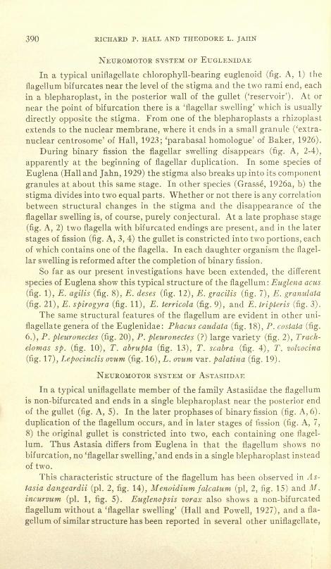
Figure A. Diagrammatic sketches of Euglena (1-4) and Astasia (5-8). 1. Euglena, vege-tative stage showing bifurcated flagellum with flagellar swelling, and rhizoplast extendingfrom one of the blepharoplasts to a granule (H extranuclear centrosome") on the nuclear mem-brane. 2. Late prophase or metaphase, with two bifurcated flagella but no flagellar swellings3. Anaphase. 4. Telophase. s. Astasia, vegetative stage showing non-bifurcated flagellumwithout flagellar swelling, and rhizoplast extending to nuclear membrane. 6. Late prophase.7. Anaphase. 8. Telophase.
390 RICHARD P. HALL AND THEODORE L. JAHN
NEUROMOTOR SYSTEM OF EUGLENIDAE
In a typical uniflagellate chlorophyll-bearing euglenoid (fig. A, 1) theflagellum bifurcates near the level of the stigma and the two rami end, eachin a blepharoplast, in the posterior wall of the gullet ('reservoir'). At ornear the point of bifurcation there is a 'flagellar swelling' which is usuallydirectly opposite the stigma. From one of the blepharoplasts a rhizoplastextends to the nuclear membrane, where it ends in a small granule ('extra-nuclear centrosome' of Hall, 1923; 'parabasal homologue' of Baker, 1926).
During binary fission the flagellar swelling disappears (fig. A, 2-4),apparently at the beginning of flagellar duplication. In some species ofEuglena (Hall and Jahn, 1929) the stigma also breaks up into its componentgranules at about this same stage. In other species (Grasse, 1926a, b) thestigma divides into two equal parts. Whether or not there is any correlationbetween structural changes in the stigma and the disappearance of theflagellar swelling is, of course, purely conjectural. At a late prophase stage(fig. A, 2) two flagella with bifurcated endings are present, and in the laterstages of fission (fig. A, 3, 4) the gullet is constricted into two portions, eachof which contains one of the flagella. In each daughter organism the flagel-lar swelling is reformed after the completion of binary fission.
So far as our present investigations have been extended, the differentspecies of Euglena show this typical structure of the flagellum: Euglena acus(fig. 1), E. agilis (fig. 8), E. deses (fig. 12), E. gracilis (fig. 7), E. granulata(fig. 21), E. spirogyra (fig. 11), E. terricola (fig. 9), and E. triPteris (fig. 3).
The same structural features of the flagellum are evident in other uni-flagellate gene~a of the Euglenidae: Phacus caudata (fig. 18), P. costata (fig.6.), P. pleuronectes (fig. 20), P. Pleuronectes (?) large variety (fig. 2), Trach-elomas sp. (fig. 10), T. abrupta (fig. 13), T. scabra (fig. 4), T. volvocina(fig. 17), Lepocinclis ovum (fig. 16), L. ovum var. palatina (fig. 19).
NEUROMOTOR SYSTEM OF ASTASHDAE
In a typical uniflagellate member of the family Astasiidae the flagellumis non-bifurcated and ends in a single blepharoplast near the posterior endof the gullet (fig. A, 5). In the later prophases of binary fission (fig. A, 6).duplication of the flagellum occurs, and in later stages of fission (fig. A, 7,8) the original gullet is constricted into two, each containing one flagel-lum. Thus Astasia differs from Euglena in that the flagellum shows nobifurcation, no 'flagellar swelling,' and ends in a single blepharoplast insteadof two.
This characteristic structure of the flagellum has been observed in As-tasia dangeardii (pl. 2, fig. 14), M enoidium falcatum (pi, 2, fig. 15) and M.incurvum (pl. 1, fig. 5). Euglenopsis vorax also shows a non-bifurcatedflagellum without a 'flagellar swelling' (Hall and Powell, 1927), and a fla-gellum of similar structure has been reported in several other uniflagellate,

COMPARATIVE CYTOLOGY OF FLAGELLATES 391
non-chlorophyll-bearing species of Euglenida-Copromonas subtilis (Dobell,1908), Copromonas major (Berliner, 1099), Astasia levis (Belar, 1916),Scytomonas pus ill a (SchUssler, 1917), J enningsia diatomoPhaga (Schaeffer,1918) and Peranema tricltopltorum (Hall and Powell, 1928).
CONSTANCY IN STRUCTURE OF NEUROMOTOR SYSTEM
It has been shown that a bifurcation of the flagellum is characteristic ofthe different species of uniflagellate Euglenidae examined and that the fla-gellum in such species always shows a 'flagellar swelling' at the level of thestigma in vegetative stages. These findings are in accord with the observa-tions of Wager (1899) on Euglena viridis, Jollos (1925) on Trachelomonasreticula/a, Baker (1926) on Euglena agilis, Grasse (1926a) on Euglena proxi-ma, Ratcliffe (1927) on Euglena sPirogyra, and Mitchell (1928) on Euglenacaudata and E. viridis.
Such structural features of the flagellum have not, however, beenobserved in vegetative stages of any of the non-chlorophyll-bearing speciesexamined by us or by other workers (Dobell, Berliner, Belar, SchUssler andSchaeffer, cited above). The statement of Jollos (1927), that uniflagellateeuglenoids "besitzt dabei ein doppelte Geisselwurzel, ein Umstead, derwohl auf ihnen zusammenhang mit zweigeisseligen Arten hinweist," isinaccurate in that it is applicable only to the Euglenidae and not to theAstasiidae.
The existence of such structural differences between the Astasiidae andthe Euglenidae suggests that these features may serve as taxonomic charac-ters. The extent of their application depends, of course, upon whether or notsuch structural features of the flagellum are constant for each species.Certain changes do occur in the Euglenidae during binary fission. Rat-cliffe (1927) has already shown that the flagellar swelling in Euglenaspirogyra is resorbed in early division stages, but reappears on the fla-gellum of each daughter individual. Each of the two flagena found in lateprophases and succeeding stages, however, shows the typical bifurcation.These observations have been confirmed in our own preparations of Eu- .glena agilis, E. gracilis and other Euglenidae. Hence, the structure of theflagellum in the Euglenidae seems to be constant enough to serve as a taxo-nomic character.
The observations of several workers, however, are not in completeagreement with those of the writers. Haase (1910), for example, has de-scribed an unusual type of flagellar insertion in Euglena sanguinea, a formwhich we have not examined. In this species the flagellum bifurcates, as inother Euglenidae, shortly after it enters the gullet, but the two rami insteadof ending in the wall of the gullet pass posteriorly into the cytoplasm, wherethey converge in a single 'basal granule' posterior to the nucleus (fig. B, 1).

392 RICHARD P. HALL AND THEODORE L. JAHN
1
5
2 3
6
4
Figure B. 1. E1tglena sanguinea (after Haase, 1910). 2. Euglenamorpha hegneri, varietypellucida with six flagella (after Wenrich, 1924). 3. E11glenamorPha hegneri, green variety withthree flagella (after Wenrich). 4. Euglenamorpha hegneri, variety pellu(;ida with four flagella(after Wenrich). 5. Euglena. gracilis, sketch of living material (after Tannreuther, 1923).6. Etlglena gracilis, from stained preparations (after Tannreuther).
COMPARATIVE CYTOLOGY OF FLAGELLATES 393
This condition is so entirely different from the flagellar insertion in otherspecies of Euglena that no explanation has occurred to the writers.
Bretschneider (1925), in addition, has figured Phacus costata with nobifurcation of the flagellum and without the usual flagellar swelling. Inour own preparations of a species believed to be Phacus costata a bifurcationof the flagellum and a flagellar swelling are both distinctly evident, and weare unable to account for Bretschneider's failure to observe them.
Tannreuther (1923), in some of his figures of living Euglena gracilis, hasshown the bifurcated flagellum ending in the cytoplasm outside the gullet(fig. B, 5), while in figures based on stained preparations (fig. B, 6) the fla-gellum ends in the posterior wall of the gullet as in other species of Euglena.In our own preparations of this species the flagellum always ends in the gul-let, and it seems probable that Tannreuther misinterpreted what he saw inthe living flagellates. There is, furthermore, no reliable evidence from anysource that such changes in position of the flagellum occur in any species ofthe Euglenidae.
In Euglenidae with two or more flagella the flagellar bifurcation islacking. In Eutreptia viridis (Steuer, 1904) there are two, and in Eugle-namorpha hegneri (Wenrich, 1924) three non-bifurcated flagella which endin the wall of the gullet; in both species, however, each flagellum in vegeta-tive stages shows a flagellar swelling similar to that seen at the bifurcationin uniflagellate Euglenidae.
Within the limits of our present observations on euglenoids and those ofcertain earlier workers (Steuer, 1904; Berliner, 1909; Belar, 1916; Schussler,1917; Hall, 1923; Baker, 1926; Ratcliffe, 1927; Hall and Powell, 1928),the number of flagella shows no change within the species except for theincrease in binary fission and the resorption which precedes encystment anddevelopment of non-swimming stages. Wenrich (1924), however, in his de-scription of Euglenamorpha H egneri and its colorless variety pellttcida, hasdescribed an increase in number of flagella and other changes which he con-siders 'evolutionary':
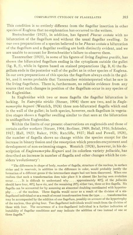
"The differences in shape of body, number of flagella, structure of the nucleu,s, in surfacestriae and in reservoir, in addition to the difference in color, would probably justify theformation of a different genus if the intermediate stages had not been discovered. When onerealizes that such a transformation does take place it is almost like having seen evolutionoccur. .... It is difficult to understand why .... approximately 40% of the pellucidashould have four, 40% have six, and the remaining 20% either two, three or five flagella. Sixflagella can be accounted for by assuming an abnormal doubling coordinated with hypertro-phy .... of the nucleus. Three flagella would occur as a result of the division of a six-flagellated individual or as a result of recent transformation from the type ..... transformationmay be accompanied by the addition of one flagellum, possibly on account of the hypertrophyof the nucleus, thus giving four. Two flagellated individuals would result from the division ofthose with four. The occurrence of a five-flagellated individual is a further indication ofinstability of flagellar conditions and may indicate the addition of two instead of one orthree flagella."

394 RICHARD P. HALL AND THEODORE L. JAHN
Reichenow (1928) even seems inclined to regard Wenrich's green andcolorless forms of Euglenamorpha as separate species: " .... zwei Varia-
. Ui.ten, die vielleicht verschiedene Arten sind, einer dreigeisseligen mitChromatophoren und Stigma und einer farblosen mit vier bis sechs Geis-seln." As a matter of fact, the points of insertion and the variations innumber of flagella in pellucida are surprisingly suggestive of behavior of theflagellum in binary fission of euglenoids (Compare figure A with figure B,2-4). Individuals are found with three, four, five and six flagella, and thisfact in itself might indicate that flagellar duplication is taking place.Furthermore, in eleven of Wenrich's figures of pellucida, four of which areapparently considered typical vegetative stages, the flagella are inserted intwo groups, the blepharoplasts of which lie near opposite sides of an en-larged gullet (fig. B, 2,4). This type of insertion of the flagella is strikinglysimilar to that observed in divison stages of other Euglenida (fig. A, 2, 3, 6,7). Since stained preparations of pellucida seem to have been made only"from the rectal contents" (of the tadpole) in which division was "rapid inthe host," and since "stages of mitosis in the green variety .... seem to beaccompanied by transformation into the pellucid variety," it would seemthat Wenrich has not eliminated the possibility that the apparent increasein number of flagella accompanying the transformation into pellucida ismerely that which occurs normally in binary fission of euglenoids. Given amature vegetative stage possessing three flagella, it follows that in theprogressive duplication of flagella in binary fission, individuals with three.four, five and six flagella would be expected. In a similar case, binary fissionof the biflagellate Eutreptia viridis (Steuer, 1904), stages with two, three andfour flagella are observed previous to constriction of the gullet. Biflagellateforms of Wenrich's variety pellucida might be accounted for by assumingthat fission was completed before the end of flagellar duplication.
It has already been pointed out that resorption of the flagellar swellingaccompanies binary fission in Euglena, just as described by Wenrich in thegreen variety of Euglenamorpha hegneri. Hence, the resorption which nor-mally accompanies fission in Euglenidae may account for the absence of theflagellar swellings in the pellucid variety.
Furthermore, Wenrich's figures of division in pellucida show no stagesearlier than a very late prophase, and in the experience of the writers dupli-cation of the flagella usually begins before that stage in binary fission ofeuglenoids. Hence, some of Wenrich's supposed vegetative stages of thepellucid variety (fig. B, 2,4) with four to six flagella might perhaps be con-sidered early prophases instead. This interpretation is suggested also by thestructure of the nuclei in some of these supposed vegetative stages; the ap-pearance is more like that of prophase than of interphase nuclei of eugle-noids.
Wenrich found also that "in the pellucid variety there is a marked ten-

COMPARATIVE CYTOLOGY OF FLAGELLATES 395
dency for the nucleus to hypertrophy." Other workers have shown that anappreciable increase in size of the nucleus occurs during prophase stages inthe division of various euglenoids. Wenrich observed, in addition, that"the reservoir is regularly of larger size in pellucida;" this was interpretedas a "probably abnormal enlargement." In other euglenoids, however, en-largement of the reservoir (gullet) is a normal process which begins in pro-phases of binary fission.
The only valid difference between the two varieties seems to be theabsence of chlorophyll in the pellucid variety. Yet it is a well known fact thatvarious species of Euglena lose their chlorophyll when grown in the dark incultures containing abundant organic food material in solution. These twoconditions-lack of sunlight and abundance of organic food material-aresupplied by the gut of the tadpole, the environment of EuglenamorPhahegneri. It seems somewhat probable, therefore, that Wenrich's varietypellucida merely represents prophase and later division stages of Euglen-amorPha hegneri without chlorophyll, and that there is no necessity forestablishing a separate variety pellucida-at least, with the implication thatmutation, or "evolution," has occurred.
STATUS OF THE FAMILIES EUGLENIDAE AND ASTASIIDAE
So far as the present evidence extends it would seem that, aside fromchanges occurring in binary fission, the flagellum in Euglenidae and Asta-siidae is constant in structure, in point of insertion and in number for anygiven species, and that the two families show certain characteristic differ-ences in structure of the flagellum. These conclusions lead us to questionthe classification which Reichenow (1928) has proposed for the Euglenida.This author divides the order into two families: (1) Euglenidae, and (2)Peranemidae. The older families Euglenidae Stein and Astasiidae Btit-schili have been combined as Reichenow's 'family Euglenidae.' The groupis characterized as follows: radially symmetrical forms with or withoutchromatophores; and it includes the following genera: Astasia, Euglena,Eutreptia, Colacium, Menoidium, Phacus and Trachelomonas. The Peran-emidae, on the other hand, are said to be bilaterally symmetrical, to possessa mouth opening and to lack chromatophores. As a matter of fact, the flagel-lates in both groups possess a well defined cytostome ('mouth') and gullet,so that this feature is obviously not limited to the family Peranemidae.Furthermore, the criterion of symmetry, which appears to be the only fea-ture distinguishing between the two groups,is of rather questionable signifi-cance. The Euglenida are, for the most part neither truly bilateral nortruly radial in symmetry, and there is no established basis for character-izing any particular genus by one type of symmetry or the other. Hence,the common possession of a hypothetical 'radial symmetry' is hardly avalid reason for combining the non-chlorophyll-bearing Astasiidae (Astasia

396 RICHARD P. HALL AND THEODORE L. JAHN
Menoidium, etc.) with the chlorophyll-bearing Euglena and its relatives.Aside from the presence of paramylum bodies, acytostome and gullet, and anucleus of the euglenoid type--features common to the Peranemidae as well-the various genera of Reichenow's 'family Euglenidae' show no diagnos-tic characteristic which is common to all of them.
In addition to Reichenow's lack of a logical basis for his 'family Eugleni-dae,' the writers have shown that there are seemingly sound morphdlogicalgrounds for separating Euglena and related genera from Astasia and Men-oidium. Various species of Euglena, Phacus, Trachelomonas and Lepocin-clis show a bifurcation of the single flagellum and also a flagellar swellingnear the level of the stigma. Such flageller swellings have also been de-scribed in the biflagellate Eutreptia 'lJiridis (Steuer, 1904) and the triflagel-late Euglenamorpha hegneri (Wenrich, 1924), although the flagella in thesespecies are not bifurcated. In none of the species of Astasia or Menoidiumexamined does the flagellum show either of these peculiarities. This is truealso for Copromonas subtilis (Dobell, 1908), Copromonas major (Berliner,1909), Astasia le'lJis(Belar, 1916) and Scytomonas pusilla (Schussler, 1917).
Furthermore, there is no stigma in Astasia levis, Copromonas, Scyto-monas, or in the species of Astasia and Menoidium examined by the writers.While such an organelle has been described in several species of 'Astasia,'some parasitic and some free-living, it is not impossible that such descrip-tions have been based upon observations of chlorophyll-free euglenas.Belar (1916), for example, concluded on the basis of cytological character-istics that Astasia captiva Beauchamp is merely a colorless form of somespecies of Euglena. Furthermore, the existence of such a form as EuglenaqtwrtanaMoroff,a species with a stigma but no chlorophyll under conditionsof saprozoic nutrition, suggests the need for caution in placing such formsin the genus Astasia. On the basis of the evidence available the writerswould be inclined to transfer the stigma-bearing species-'A stasi a capti'/JaBeauchamp, Astasia mobilis Alexeieff (if a stigma is actually present),Astasia ocellata Khawkine Astasia chaetogastris Codreanu and Codreanu(1928)-to the genus Euglena until it can be shown that the cytologicalstructure of these forms justifies a position in the genus Astasia.
Although it seems evident that the Astasiidae should remain recognizedas a family separate from the Euglenidae, there are still differences of opin-ion as to the limits comprised by the family Astasiidae. Calkins (1926), forexample, has attempted to establish the Astasiidae as a family of uniflagel-late forms including the following genera: Astasia, Clautriavia, Euglenop-sis, Jenningsia, Menoidium, Peranema, Petalomonas, Scytomonas andUrceolus. The biflagellate forms without chromatophores are placed in thenew family Heteronemidae.
The system of Calkins has the possible advantage of simplicity in diag-nosis, yet both his families (Astasiidae and Heteronemidae) are heterogen-

COMPARATIVE CYTOLOGY OF FLAGELLATES 397
eous aggregates of holozoic and saprozoic forms with and without a pharyn-geal-rod apparatus (Calkins' "parabasal body"), the members of each fam-ily showing only one diagnostic characteristic-the number of flagella.Rhodes (1926) has raised the objection that "this departure seems basedupon misinterpretation and is unjustified." From Rhodes' (1926) descrip-tion ofHeteronema, Hall and Powell (1927) were led to believe that "Rhodes,in demonstrating that the staborgan of Heteronema acus serves as amouth in the ingestion of food, has shown that there is an even more strik-ing difference between Peranema and Heteronema than the mere number offlagella. Hence, .... that Rhodes, instead of weakening the status of Cal-kins' family Heteronemidae, has really presented some very good evidencethat Peranema and Heteronema should be separated and that the familyHeteronemidae should stand" (p. 162).
Recent investigations carried on in our laboratory have shown that, inspite of Rhodes' statements in regard to Heteronema, the pharyngeal-rodapparatus is essentially similar in structure and probable function in thesetwo flagellates; and furthermore, that this organelle apparently does notfunction as a "true cytostome" in ingestion of food, but seems to serve rath-er as a supporting system for the cytostome and gullet which are character-istic of the euglenoids as a group. This similarity of the pharyngeal organ-elles in the biflagellate Heteronema and the uniflagellate Heteronema ob-viously contradicts the assumption (Hall and Powell, 1927) that the twogenera should be placed in separate families. In view of this situation,which involves Jenningsia as well as Peranema and Heteronema, Calkins'system is not entirely satisfactory.
Lemmermann (1913), on the other hand, characterizes the Astasi-idae as saprozoic, radially symmetrical forms with one or two flagella. Ithas already been pointed out that the criterion of symmetry, in the Euglen-ida, is anything but a satisfactory basis of classification; and furthermore,the lack of adequate knowledge of the feeding habits of some genera wouldseem to be a handicap in classifying the colorless euglenoids on the basis ofmethod of nutrition. Hence, neither the system of Calkins nor that ofLemmermann is at present entirely free from objection, and for this reasonit seems to the writers that an adequate classification of the euglenoidswithout chromatophores must await further investigation of the cytologyand feeding habits of these flagellates ..
SUMMARY
It has been shown that, in various species of Phacus, Euglena, Trachelo-monas and Lepocinclis, vegetative stages are characterized by a basalbifurcation of the flagellum into rami which end in separate blepharoplasts,and by a 'flagellar swelling' at the level of the stigma. Such structural fea-

398 RICHARD P. HALL AND THEODORE L. JAHN
tures of the flagellum were not observed in non-chlorophyll-bearing eugle-noids (Astasiidae). On the basis of such structural differences it is concludedthat Reichenow (1928) is not justified in combining the families EuglenidaeStein and Astasiidae Blitschli into a single 'family Euglenidae.'
LITERATURE CITED
Baker, W. B. 1926. Studies in the life-history of Euglena. BioI. Bull. 51: 321-362,2 pIs., 2text figs.
Belar, K. 1916. Protozoenstudien. 1. Amoeba diplogena, Astasia levis n. sp., Rhynchomonasnasuta Klebs. Arch. Protistenk. 36: 13-51, pIs. 2---4,3 text figs.
Berliner, E. 1909. Flagellatenstudien. Arch. Protistenk. 15: 297-326, pIs. 28-29.Bretschneider, L. H. 1925. Ueber den feineren Bau von Phacus costata Conrad. Arch. Pro-
tistenk. 53: 131-134,6 figs.Calkins, G. N. 1926. The biology of the Protozoa (Philadelphia, Lea and Febiger), 623 pp.,
238 figs.Charipper, H. A. 1928. A method for staining fibrillae in the epithelia of vertebrates and
invertebrates, as well as fibrillar structures in Protozoa. Anat. Rec. 38: 401---404.Codreanu, M. and R. Codreanu. 1928. Un nouvel Euglenien (Astasia chaetogastris n. sp.),
parasite coelomique d'un ologichHe (Chaetogaster diastrophus Gruith). C. R. Soc.BioI. 99: 1368-1370, 2 figs.
Dobell, C. C. 1908. The structure and life-history of Copromonas subtilis n. g., n. so.: a con-tribution to our knowledge of the Flagellata. Quart. J. Micr. Sci. 52: 75-120, pIs. 4-5,3 text figs.
Grasse, P. P. 1926a. Contribution a I'etude des flagelles parasites. Arch. Zool. Exp. et Gen.65: 345-602, pIs. 8-19, 76 text figs.
1926b. Sur Ie stigma ou appareil parabasal des Euglenes. C. R. Soc. BioI. 94: 1012-1014.Haase, G. 1910. Studien tiber Euglena sanguinea. Arch. Protistenk. 20: 47-59, pIs. 4-6.Hall, R. P. 1923. Morphology and binary fission of Menoidium incurvltm (Fres.) Klebs.
Univ. Calif. Publ. Zool. 20: 447---476,pIs. 40---41,2 text figs.Hall, R. P. and T. L. Jahn. 1929. Dispersed stages of the stigma in Euglena. Science 69: 522.Hall, R. P. and W. N. Powell. 1927. A note on the morphology and taxonomic position of
Peranema trichophorum. Tr. Am. Micr. Soc. 46: 155-165, pI. 1, 2 text figs.1928. Morphology and binary fission of Peranema trichophorum (Ehrbg.) Stein. BioI.
Bull. 54: 36-65,2 pIs., 3 text figs.Jollos, V. 1925. "Flagellata" in Handbuch der Zoologie (Berlin and Leipzig, Gruyter),
115-185, figs. 107-171.Le=ermann, E. 1913. "Eugleninae" in Die Stisswasserflora DeutschIands, <1sterreichsund
der Schweiz, H. 2 (Jena, Fischer), pp. 115-174, figs. 181-377.Mitchell, J. B. 1928. Studies on the life-history of a parasite of the Euglenidae. Tr. Am.
Micr. Soc. 47: 29---41,pIs. 4-6.Ratcliffe, H. 1927. Mitosis and cell division in Euglena sPirogyra. BioI. Bull. 53: 109-122,
3 pIs.Reichenow, E. 1928. Doflein's Lehrbuch der Protozoenkunde, Teil II (Jena, Fischer),
pp.436-864.Rhodes, R. C. 1926. Mouth and feeding habits of Heterotlema acUs. Anal. Rec. 34: 152-153.Schaeffer, A. A. 1918. A new and remarkable diatom-eating flagellate, Jenningsia diatomo-
phaga n.g., n.sp. Tr. Am. Micr. Soc. 37: 177-182, pI. 13.Schtissler, H. 1917. Cytologische und entwicklungsgeschichtliche Protozoenstudien. 1.
Ueber die Teilung von Scytomonas pusilla Stein. Arch. Protistenk. 38: 117-125,pI. 5, 1 text fig.

COMPARATIVE CYTOLOGY OF FLAGELLATES 399
Steuer, A. 1904. Ueber eine Euglenoide (Eutreptia) aus dem Canale grande yon Trieste.Arch. Protistenk. 3: 126-137,13 figs.
Tannreuther, G. W. 1923. Nutrition and reproduction in Euglena. Arch. Entwick.-mech.52: 367-383, 52 figs.
Ternetz, C. 1912. Beitrage zur Morphologie und Physiologie Yon Euglena gracilis. Jahrb. f.wiss. Bot. 51: 435-514, 1 pI.
Wager, H. 1899. On the eyespot and flagellum in Euglena viridis. Jour. Linn. Soc. London,Botany, 27: 463--481, pI. 32.
Wenrich, D. H. 1924. Studies on Euglenamorplta hegneri n.g., n.sp., a euglenoid flagellatefound in tadpoles. BioI. Bull. 47: 149-174, 4 pIs.

400 RICHARD P. HALL AHD THEODORE L. JAHN
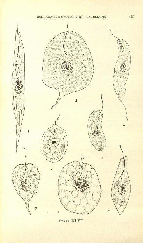
PLATE XLVII
FIG. 1. Euglena aCt/s, showing bifurcated flagellum with flagellar swelling, flagellar rhizoplastextending to nucleus, and bacilliform paramylum bodies; S-BR-IH; X750 ca.
FIG. 2. Phacus pleuronectes (?) large variety, showing bifurcated flagellum with relativelylarge flagellar swelling, numerous chromatophores; surface striations-approximatelyparallel to the rows of chromatophores-are not indicatedj S-BR-IH; X 1000.
FIG. 3. Euglena tripteris, flagellar structure and chromatophoresj S-BR-IH; X2025.FIG. 4. Trdchelomonas scabra, flagellar structure, two chromatophores; S-BR-IH; X2025.FIG. 5. Menoidium incurlJ1lm, non-bifurcated flagellum, flagellar rhiwplast extending tonucleus, surface striations indicated; S-Br-IH; X 1550 ca.FIG. 6. Phacus costata, flagellar structure and chromatophoresj surface striations indicated
diagrammatically; S-BR-IH; X 1570.FIG. 7. Euglena gracilis, late prophase stage with two bifurcated flagella; gullet partly con-
stricted; S-BR-IH; X2025.FIG. 8. Euglena agilis, flagellar structure, chromatophores with pyrenoids; S-BR-IH; X 1750.
1
COMPARATIVE CYTOLOGY OF FLAGELLATES
PLATE XLVII
8
3
401

402 RICHARD P. HALL AND THEODORE L. JAHN
PLATE XLVIII
FIG. 9. Ettglena terricola, flagellar structure and chromatophores; from a preparation byR. F. Nigrelli; S-IH-E; X2025.
FIG. 10. Trachelomonas sp., flagellar structure; S-BR-IH; X 1570.FIG. 11. Ettglena spirogyra, flagellar structure and chromatophores; S-BR-IH; X715.FIG. 12. Euglena deses, flagellar structure and chromatophores; S-BR-IH; X2025.FIG. 13. Trachelomonas abmpta (?), flagellar structure and chromatophores; S-BR-IHj
X2025.FIG. 14. Astasia dangeardii, contracted organism; flagellum is non-bifurcated; S-IH-E;
X2025.FIG. 15. Menoidiumfalcatum, optical section showing non-bifurcated flagellum and paramy-
lum bodies; surface striations not indicated; S-IH-BR; X 1010 ca.

COMPARATIVE CYTOLOGY OF FLAGELLATES 403
9
10
14
PLATE XLVIII

404 RICHARD P. HALL AND THEODORE L. JAHN
PLATE XLIX
FIG. 16. Lepocimlis ovum, flagellar structure and chromatophores; S-BR-IH; X 3040.FIG. 17. Trachelomollas volvocina, flagellar structure; S-BR-IH; X 2355.FIG. 18. Phacus caudata (?), flagellar structure; S-BR-IH; X2355.FIG. 19. Lepocinclis ovum var. palatina, flagellar structure; S-BR-IH; X 1800.FIG. 20. Phacus pleuronectes, flagellar structure and chromatophoresj oblique optical section;
S-IH; XI800.FIG. 21. Eflglena gra/lIIlata (?), flagellar structure, chromatophores and four pyrenoids shown
in optical section; organism partly contracted; S-BR-IH; X 1420.

COMPARATIVE CYTOLOGY OF FLAGELLATES 405
17
19
18
PLATE XLIX