psoriasis types, causes and medication - intech -...
TRANSCRIPT
Chapter 1
Psoriasis — Types, Causes and Medication
F.Z. Zangeneh and F.S. Shooshtary
Additional information is available at the end of the chapter
http://dx.doi.org/10.5772/54728
1. Introduction
Although the skin disease psoriasis was first recognized as a distinct disease as early as 1808[1], its pathogenic mechanisms have eluded investigators for decades, its definition byFerdinand von Hebra as a distinct entity dates back only to the year 1841 and estimates of itsprevalence around 2-3% of the general population, and is characterized by an exaggeratedproliferation of keratinocytes secondary to an activated immune system. The incidence ishighest at the age of 20–39 years in males and 40–59 years in females, with an equal male-to-female ratio [2]. Psoriasis clinically manifests as raised, well defined erythematous plaqueswith irregular borders and silvery scales, affecting the upper and lower extremities equally,but with a predilection for the elbows, knees, scalp, and trunk. Psoriasis vulgaris or plaquepsoriasis accounts for almost 90% of the dermatological presentation of the disease, but severalother forms, including guttate, inverse, erythrodermal, pustular, and palmoplantar psoriasismay occur, as well as nail involvement. Psoriasis may have significant systemic involvement,which is underscored by the coexistence of various clinical disorders, including eye, cardio‐vascular, and intestinal problems, metabolic syndrome, and joint inflammation. It has a veryhigh negative impact on quality of life, requires long-term treatment which usually has a highsocial and economic impact and is also associated with a decreased life span [3] [4].
2. Psoriasis types
Psoriasis classification
No one classification of psoriasis satisfies all the mentioned requirements. Usually, criteria areintermingled (Table 1), and subclasses are nonexclusive. Similar problems exist with theclinical classification of psoriatic arthropathy [5].
© 2013 Zangeneh and Shooshtary; licensee InTech. This is an open access article distributed under the termsof the Creative Commons Attribution License (http://creativecommons.org/licenses/by/3.0), which permitsunrestricted use, distribution, and reproduction in any medium, provided the original work is properly cited.
Morphologic aspects of
elementary lesions
Pustular, non-pustular but also plaque, nummular, guttate, gyrate, rupioid,
elephantine, ostraceous, etc.
Degree of inflammation Mainly inflammatory vs mainly hyperkeratotic
Pattern distribution Extensory, inverse, seborrhoeic, widespread
Extent One site (scalp, nail, etc.), many sites, generalized
Time of first onset Early vs late onset
Velocity of propagation Stable, unstable, eruptive
Table 1. Features which have been considered in different classifications of psoriasis [6]
Classification criteria based on purported etiology rank higher in formalization compared withpurely morphological ones.
2.1. Classifying psoriasis: The spectrum of clinical varieties
Psoriasis, a papulosquamous skin disease, has several different types, including: psoriasisvulgaris (common type), guttate psoriasis (small, drop like spots), inverse psoriasis (in thefolds like of the underarms, navel, and buttocks), and pustular psoriasis (pus-filled, yellowish,small blisters). When the palms and the soles are involved, this is known as palmoplantarpsoriasis.
2.2. Psoriasis vulgaris (chronic stationary psoriasis, plaque-like psoriasis)
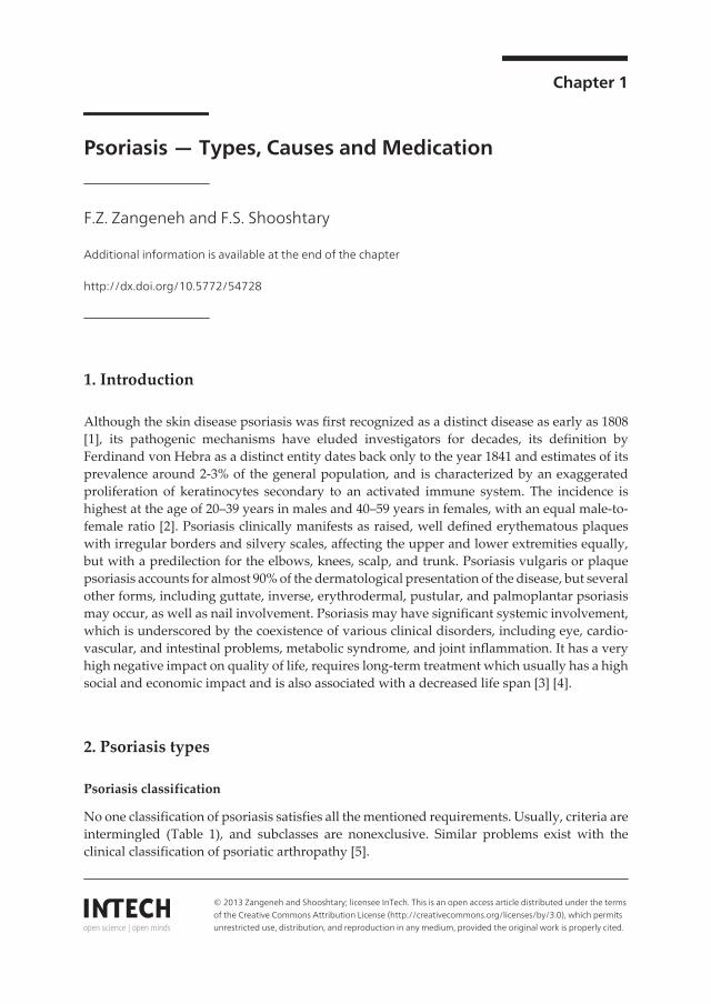
The commonest type of psoriasis, accounting for 90% of all cases, is psoriasis vulgaris, in whichpapulosquamous plaques are well-delineated from surrounding normal skin. The plaques arered or salmon pink in color, covered by white or silvery scales and may be thick, thin, large orsmall (Figure 1). They are most active at the edge: rapidly progressing lesions may be annular,with normal skin in the centre. Plaques are usually distributed symmetrically, and occur mostcommonly on the extensor aspects of elbows and knees; scalp (where they rarely encroachbeyond the hairline), lumbosacral region, and umbilicus. Active inflammatory psoriasis ischaracterized by the Koebner phenomenon, in which new lesions develop at sites of traumaor pressure [7].
2.2.1. Classification of psoriasis vulgaris according to phenotype: plaque-type psoriasis
There is also variation of features of psoriasis dependent on anatomical sites. Until the reasonsfor this variation are fully understood, they are proposed to be recorded as a phenotypic entity,although subsequently they may be shown to be part of a common pathogenetic mechanism.A further distinction arises according to the age of onset of plaque psoriasis [8]. Henseler andChristophers are credited with identifying two ages of onset: type I occurring at or before theage of 40 years—this accounts for approximately 75% of patients; and typeII presenting afterthe age of 40 years, with a distinct peak at 55–60 years [9].
Psoriasis - Types, Causes and Medication4
2.2.2. Plaque-type psoriasis: Chronic plaque psoriasis
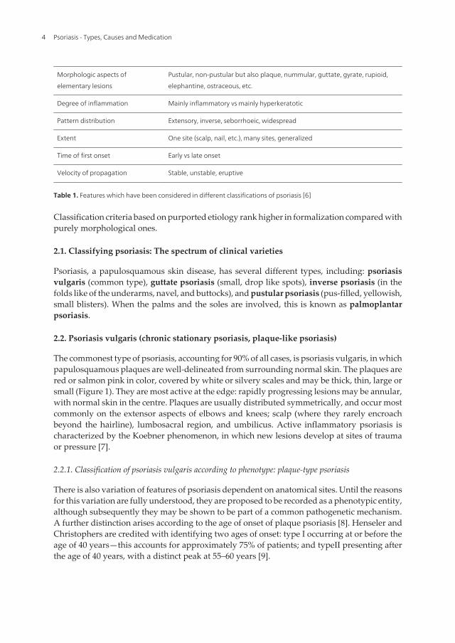
As a consequence, chronic plaque psoriasis is the form of the disease entered into clinical trialsand the object of the majority of investigations of genetics and pathogenesis of psoriasis. It ischaracterized by red, scaly, discoid lesions varying in size from 0.5 cm in diameter to large con‐fluent areas on the trunk and limbs (Figure 1). There is a sharp line of demarcation between aplaque and clinically normal, uninvolved skin. Longitudinal studies of individual plaqueshave demonstrated that plaques are dynamic [10] with an active and expanding edge, some‐times to the extent that the advancing edge may become annular (Figure. 2) leaving clinicallynormal skin in the centre of the original plaque. The variety of plaque is characterized by well-demarcated plaques with a loosely adherent silvery-white scale, which preferentially affect theelbows, knees, lumbosacral area, intergluteal cleft, and scalp. Occasionally, pustular lesionsmay appear in the plaque (so-called psoriasis with pustules). Chronic plaque psoriasis is themost common variety of psoriasis, representing about 70% to 80% of psoriatic patients [11].
Figure 1. Typical plaque of Psoriasis Vulgaris.
Figure 2. Annular psoriasis showing clearance in centre of plaque.
Psoriasis — Types, Causes and Medicationhttp://dx.doi.org/10.5772/54728
5
Under the heading of plaque psoriasis, it is proposed to include, as subdivisions, a new, morelogical nomenclature of phenotypes associated with specific anatomical sites, distribution, sizeand thickness of plaques [8].
2.2.3. Site-specific variants of Psoriasis Vulgaris (PV)
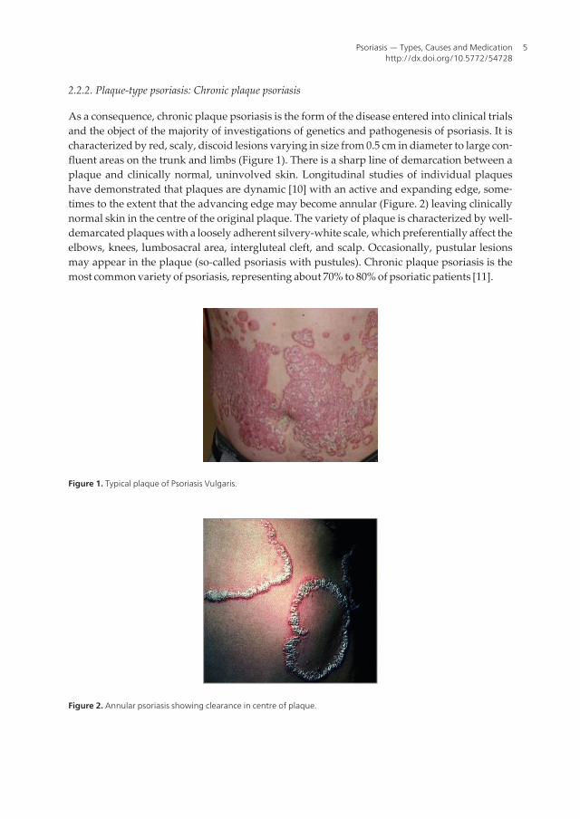
Site-specific variants of psoriasis vulgaris exist. Flexural (inverse) psoriasis in intertriginoussites is shiny, red, and typically devoid of scales (figure 3); sebopsoriasis, which can beconfused with seborrhoeic dermatitis, has greasy scales and occurs in eyebrows, nasolabialfolds, and postauricular and presternal sites. Psoriasis vulgaris will probably prove to beseveral closely related but phenotypically and genotypically distinct conditions [8].
Flexural/intertriginous: Inverse psoriasis (Flexural Psoriasis or Psoriasis of the Skin Folds) isusually located in the skin folds: i.e. armpits, under the breasts, skin folds around the groin andbetween the buttocks. It is particularly subject to irritation from rubbing and sweating becauseof its location in skin folds and tender areas (Figure 3). Plaques are thin, have minimal scale anda shiny (nonscaly) surface commonly accompanied by secondary fissuring and/or maceration.The major clinical manifestation of inverse psoriasis is sharply demarcated erythematous pla‐ques, with varying degrees of infiltration, which often tend to itch and burn [12]. The most com‐mon lesions are found in inguinal, submammary, interglutaeal, umbilicus and genital folds,whereas the popliteus and axillae are rarely involved. The humidity and heat typical of thesesites, together with the combination of local traumatic factors often associated with infectionscaused by dermatophytes and Candida albicans, together contribute to the development ofpsoriasis in accordance with the Koebner phenomenon. The Koebner phenomenon is an indi‐cator of disease activity, may have a prognostic value, and is associated with early onset ofpsoriasis [13]. The Koebner phenomenon was first described by Heinrich Koebner (1838–1904)and refers to the fact that in people with certain skin diseases, especially psoriasis, trauma is fol‐lowed by new lesions in the traumatized but otherwise normal skin, and these new lesions areclinically and histopathologically identical to those in the diseased skin [14].
Figure 3. Flexural psoriasis, notes the relative lack of scale.
Psoriasis - Types, Causes and Medication6
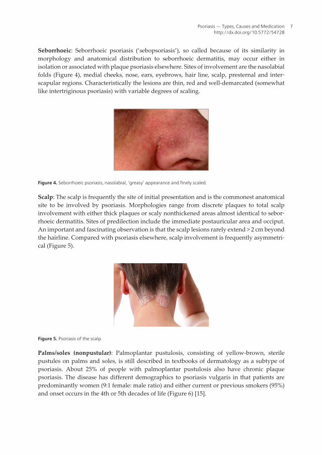
Seborrhoeic: Seborrhoeic psoriasis (‘sebopsoriasis’), so called because of its similarity inmorphology and anatomical distribution to seborrhoeic dermatitis, may occur either inisolation or associated with plaque psoriasis elsewhere. Sites of involvement are the nasolabialfolds (Figure 4), medial cheeks, nose, ears, eyebrows, hair line, scalp, presternal and inter‐scapular regions. Characteristically the lesions are thin, red and well-demarcated (somewhatlike intertriginous psoriasis) with variable degrees of scaling.
Figure 4. Seborrhoeic psoriasis, nasolabial, ‘greasy’ appearance and finely scaled.
Scalp: The scalp is frequently the site of initial presentation and is the commonest anatomicalsite to be involved by psoriasis. Morphologies range from discrete plaques to total scalpinvolvement with either thick plaques or scaly nonthickened areas almost identical to sebor‐rhoeic dermatitis. Sites of predilection include the immediate postauricular area and occiput.An important and fascinating observation is that the scalp lesions rarely extend > 2 cm beyondthe hairline. Compared with psoriasis elsewhere, scalp involvement is frequently asymmetri‐cal (Figure 5).
Figure 5. Psoriasis of the scalp.
Palms/soles (nonpustular): Palmoplantar pustulosis, consisting of yellow-brown, sterilepustules on palms and soles, is still described in textbooks of dermatology as a subtype ofpsoriasis. About 25% of people with palmoplantar pustulosis also have chronic plaquepsoriasis. The disease has different demographics to psoriasis vulgaris in that patients arepredominantly women (9:1 female: male ratio) and either current or previous smokers (95%)and onset occurs in the 4th or 5th decades of life (Figure 6) [15].
Psoriasis — Types, Causes and Medicationhttp://dx.doi.org/10.5772/54728
7
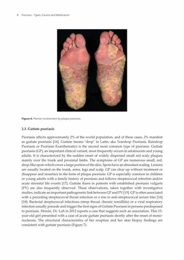
Figure 6. Plantar involvement by plaque psoriasis.
2.3. Guttate psoriasis
Psoriasis affects approximately 2% of the world population, and of these cases, 2% manifestas guttate psoriasis [16]. Guttate means "drop" in Latin; aka Teardrop Psoriasis, RaindropPsoriasis or Psoriasis Exanthematic) is the second most common type of psoriasis. Guttatepsoriasis (GP), an important clinical variant, most frequently occurs in adolescents and youngadults. It is characterized by the sudden onset of widely dispersed small red scaly plaquesmainly over the trunk and proximal limbs. The symptoms of GP are numerous small, red,drop-like spots which cover a large portion of the skin. Spots have an abundant scaling. Lesionsare usually located on the trunk, arms, legs and scalp. GP can clear up without treatment ordisappear and resurface in the form of plaque psoriasis. GP is especially common in childrenor young adults with a family history of psoriasis and follows streptococcal infection and/oracute stressful life events [17]. Guttate flares in patients with established psoriasis vulgaris(PV) are also frequently observed. These observations, taken together with investigativestudies, indicate an important pathogenetic link between GP and PV [15]. GP is often associatedwith a preceding streptococcal throat infection or a rise in anti-streptococcal serum titer [16][18]. Bacterial streptococcal infections (strep throat, chronic tonsillitis) or a viral respiratoryinfection usually precede and trigger the first signs of Guttate Psoriasis in persons predisposedto psoriasis. Herein, Dr. Loh in 2012 reports a case that suggests such an association. This 15-year-old girl presented with a case of acute guttate psoriasis shortly after the onset of mono‐nucleosis. The structural characteristics of her eruption and her skin biopsy findings areconsistent with guttate psoriasis (Figure 7).
Psoriasis - Types, Causes and Medication8
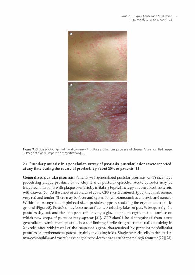
Figure 7. Clinical photographs of the abdomen with guttate psoriasiform papules and plaques. A,Unmagnified image.B, Image at higher unspecified magnification [19].
2.4. Pustular psoriasis: In a population survey of psoriasis, pustular lesions were reportedat any time during the course of psoriasis by about 20% of patients [11]
Generalized pustular psoriasis: Patients with generalized pustular psoriasis (GPP) may havepreexisting plaque psoriasis or develop it after pustular episodes. Acute episodes may betriggered in patients with plaque psoriasis by irritating topical therapy or abrupt corticosteroidwithdrawal [20]. At the onset of an attack of acute GPP (von Zumbusch type) the skin becomesvery red and tender. There may be fever and systemic symptoms such as anorexia and nausea.Within hours, myriads of pinhead-sized pustules appear, studding the erythematous back‐ground (Figure 8). Pustules may become confluent, producing lakes of pus. Subsequently, thepustules dry out, and the skin peels off, leaving a glazed, smooth erythematous surface onwhich new crops of pustules may appear [21]. GPP should be distinguished from acutegeneralized exanthematic pustulosis, a self-limiting febrile drug reaction usually resolving in2 weeks after withdrawal of the suspected agent, characterized by pinpoint nonfollicularpustules on erythematous patches mainly involving folds. Single necrotic cells in the epider‐mis, eosinophils, and vasculitic changes in the dermis are peculiar pathologic features [22] [23].
Psoriasis — Types, Causes and Medicationhttp://dx.doi.org/10.5772/54728
9
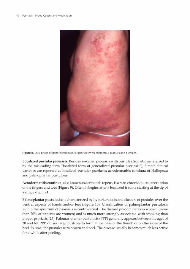
Figure 8. Early phase of generalized pustular psoriasis with edematous plaques and pustules.
Localized pustular psoriasis: Besides so-called psoriasis with pustules (sometimes referred toby the misleading term “localized form of generalized pustular psoriasis”), 2 main clinicalvarieties are reported as localized pustular psoriasis: acrodermatitis continua of Hallopeauand palmoplantar pustulosis.
Acrodermatitis continua, also known as dermatitis repens, is a rare, chronic, pustular eruptionof the fingers and toes (Figure 9). Often, it begins after a localized trauma starting at the tip ofa single digit [24].
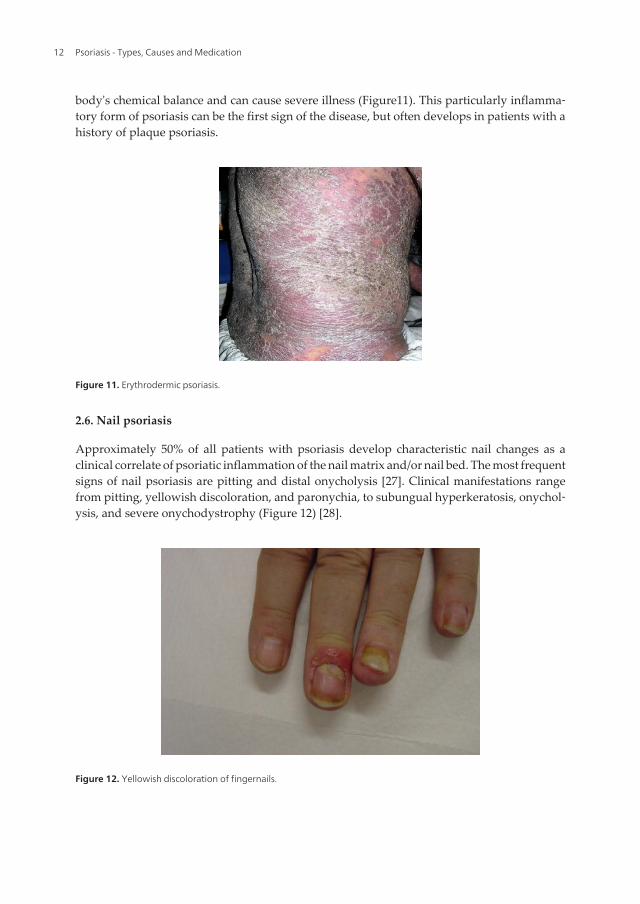
Palmoplantar pustulosis: is characterized by hyperkeratosis and clusters of pustules over theventral aspects of hands and/or feet (Figure 10). Classification of palmoplantar pustulosiswithin the spectrum of psoriasis is controversial. The disease predominates in women (morethan 70% of patients are women) and is much more strongly associated with smoking thanplaque psoriasis [25]. Palomar-plantar pustulosis (PPP) generally appears between the ages of20 and 60. PPP causes large pustules to form at the base of the thumb or on the sides of theheel. In time, the pustules turn brown and peel. The disease usually becomes much less activefor a while after peeling.
Psoriasis - Types, Causes and Medication10
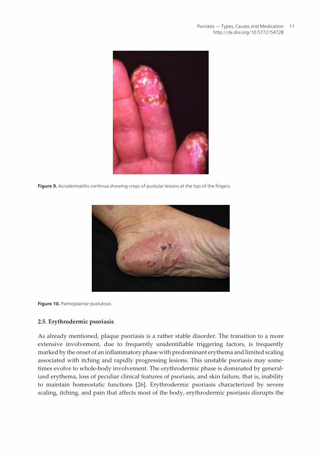
Figure 9. Acrodermatitis continua showing crops of pustular lesions at the tips of the fingers.
Figure 10. Palmoplantar pustulosis.
2.5. Erythrodermic psoriasis
As already mentioned, plaque psoriasis is a rather stable disorder. The transition to a moreextensive involvement, due to frequently unidentifiable triggering factors, is frequentlymarked by the onset of an inflammatory phase with predominant erythema and limited scalingassociated with itching and rapidly progressing lesions. This unstable psoriasis may some‐times evolve to whole-body involvement. The erythrodermic phase is dominated by general‐ized erythema, loss of peculiar clinical features of psoriasis, and skin failure, that is, inabilityto maintain homeostatic functions [26]. Erythrodermic psoriasis characterized by severescaling, itching, and pain that affects most of the body, erythrodermic psoriasis disrupts the
Psoriasis — Types, Causes and Medicationhttp://dx.doi.org/10.5772/54728
11
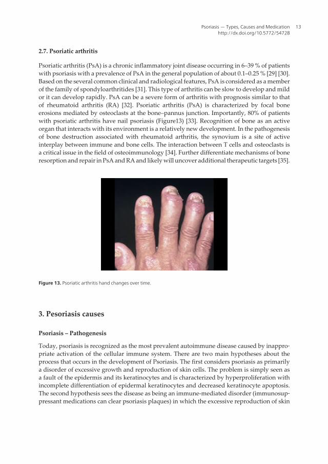
body's chemical balance and can cause severe illness (Figure11). This particularly inflamma‐tory form of psoriasis can be the first sign of the disease, but often develops in patients with ahistory of plaque psoriasis.
Figure 11. Erythrodermic psoriasis.
2.6. Nail psoriasis
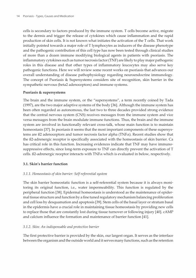
Approximately 50% of all patients with psoriasis develop characteristic nail changes as aclinical correlate of psoriatic inflammation of the nail matrix and/or nail bed. The most frequentsigns of nail psoriasis are pitting and distal onycholysis [27]. Clinical manifestations rangefrom pitting, yellowish discoloration, and paronychia, to subungual hyperkeratosis, onychol‐ysis, and severe onychodystrophy (Figure 12) [28].
Figure 12. Yellowish discoloration of fingernails.
Psoriasis - Types, Causes and Medication12
2.7. Psoriatic arthritis
Psoriatic arthritis (PsA) is a chronic inflammatory joint disease occurring in 6–39 % of patientswith psoriasis with a prevalence of PsA in the general population of about 0.1–0.25 % [29] [30].Based on the several common clinical and radiological features, PsA is considered as a memberof the family of spondyloarthritides [31]. This type of arthritis can be slow to develop and mildor it can develop rapidly. PsA can be a severe form of arthritis with prognosis similar to thatof rheumatoid arthritis (RA) [32]. Psoriatic arthritis (PsA) is characterized by focal boneerosions mediated by osteoclasts at the bone–pannus junction. Importantly, 80% of patientswith psoriatic arthritis have nail psoriasis (Figure13) [33]. Recognition of bone as an activeorgan that interacts with its environment is a relatively new development. In the pathogenesisof bone destruction associated with rheumatoid arthritis, the synovium is a site of activeinterplay between immune and bone cells. The interaction between T cells and osteoclasts isa critical issue in the field of osteoimmunology [34]. Further differentiate mechanisms of boneresorption and repair in PsA and RA and likely will uncover additional therapeutic targets [35].
Figure 13. Psoriatic arthritis hand changes over time.
3. Pesoriasis causes
Psoriasis – Pathogenesis
Today, psoriasis is recognized as the most prevalent autoimmune disease caused by inappro‐priate activation of the cellular immune system. There are two main hypotheses about theprocess that occurs in the development of Psoriasis. The first considers psoriasis as primarilya disorder of excessive growth and reproduction of skin cells. The problem is simply seen asa fault of the epidermis and its keratinocytes and is characterized by hyperproliferation withincomplete differentiation of epidermal keratinocytes and decreased keratinocyte apoptosis.The second hypothesis sees the disease as being an immune-mediated disorder (immunosup‐pressant medications can clear psoriasis plaques) in which the excessive reproduction of skin
Psoriasis — Types, Causes and Medicationhttp://dx.doi.org/10.5772/54728
13
cells is secondary to factors produced by the immune system. T cells become active, migrateto the dermis and trigger the release of cytokines which cause inflammation and the rapidproduction of skin cells. It is not known what initiates the activation of the T cells. That workinitially pointed towards a major role of T lymphocytes as inducers of the disease phenotypeand the pathogenic contribution of this cell type has now been tested through clinical studiesof more than a dozen immune modifying biological agents in patients with psoriasis. Theinflammatory cytokines such as tumor necrosis factor (TNF) are likely to play major pathogenicroles in this disease and that other types of inflammatory leucocytes may also serve keypathogenic functions. Here we will review some recent works on psoriasis that advances ouroverall understanding of disease pathophysiology regarding neuroendocrine immunology.The concept of Psoriasis & Supersystems considers site of recognition, skin barrier in thesympathetic nervous (beta2 adenoceptors) and immune systems.
Psoriasis & supersystems
The brain and the immune system, or the “supersystems”, a term recently coined by Tada(1997), are the two major adaptive systems of the body [36]. Although the immune system hasbeen often regarded as autonomous, the last two to three decades provided strong evidencethat the central nervous system (CNS) receives messages from the immune system and viceversa messages from the brain modulate immune functions. Thus, the brain and the immunesystem are involved in functionally relevant cross-talk, whose main function is to maintainhomeostasis [37]. In psoriasis it seems that the most important components of these supersys‐tems are ß2 adenoceptors and tumor necrosis factor alpha (TNFα). Recent studies show thatthe ß2-adrenergic receptor is specifically associated with the homeostasis of skin barrier. Cahas critical role in this function. Increasing evidences indicate that TNF may have immuno‐suppressive effects, since long-term exposure to TNF can directly prevent the activation of Tcells. ß2-adrenergic receptor interacts with TNFα which is evaluated in below, respectively.
3.1. Skin’s barrier function
3.1.1. Homeostasis of skin barrier: Self-referential system
The skin barrier homeostatic function is a self-referential system because it is always moni‐toring its original function, i.e., water impermeability. This function is regulated by theperipheral function [38]. Epidermal homeostasis is understood as the maintenance of epider‐mal tissue structure and function by a fine tuned regulatory mechanism balancing proliferationand cell loss by desquamation and apoptosis [39]. Stem cells of the basal layer or stratum basalin the epidermis have a crucial role in maintaining tissue homeostasis by providing new cellsto replace those that are constantly lost during tissue turnover or following injury [40]. cAMPand calcium influence the formation and maintenance of barrier function [41].
3.1.2. Skin: An indispensable and protective barrier
The first protective barrier is provided by the skin, our largest organ. It serves as the interfacebetween the organism and the outside world and it serves many functions, such as the retention
Psoriasis - Types, Causes and Medication14
of body fluids, maintenance of body temperature, and protection against UV-light, chemicalinfluxes, wounds, and the invasion of micro-organisms. The protective barrier function isperformed by the keratinocytes of the epidermis, which are continuously produced byproliferating stem cells of the basal layer or stratum basal and differentiate during a 14 dayjourney towards the surface [42].
3.1.3. Skin: Epidermal barrier capacity (lipid/protein polymer structure)
Stratum corneum (SC) & Ceramides (family of lipid molecules)
Epidermal barrier capacity is controlled by lipids that fill the extracellular space of the skin'ssurface layer-the stratum corneum. Lipid synthesis for skin barrier function takes place withinthe keratinocytes in all nucleated epidermal layers. Lipids are stored within the epidermallamellar bodies (secretory organells) or keratinosomes, which are ultrastructurally visible atthe level of the upper spinous layer and in the granular layer. In the outermost granular layer,the contents of lamellar bodies are secreted into the intercellular domains of the stratumgranulosom–stratum corneum interface. Lamellar bodies mainly contain phospholipids,glucosylceramides and cholesterol as well as hydrolytic enzymes, which convert phospholi‐pids, glucosylceramides and sphingomyelinase to free fatty acids and ceramides. Then,lamellar bodies cause in the formation of an impermeable, lipid-containing membrane thatserves as a water barrier and is required for correct skin barrier function. The Stratum Corneom(SC) contains three types of lipids -- ceramides, cholesterol and free fatty acids. These lipidshave different chemical compositions and different functions throughout the body. There arenine different types of ceramides in the Stratum Corneom, conveniently named ceramide 1through ceramide 9, and they account for 40-50% of the lipids in this outermost layer. Aceramide is composed of sphingosine and a fatty acid. Ceramides are found in high concen‐trations within the cell membrane of cells. They are one of the component lipids that make upsphingomyelin, one of the major lipids in the lipid bilayer. Ceramide can actually act as asignaling molecule. The most well-known functions of ceramides as cellular signals includeregulating the differentiation, proliferation, programmed cell death (PCD), and apoptosis(Type I PCD) of cells [43].The proliferation rate of keratinocytes to corneocytes is matched bythe shedding of old corneocytes at the SC [44] and skin tissue maintains a steady number ofSC layers regardless of age [45].
Desquamation, the process of cell shedding from the surface of the stratum corneum, balancesproliferating keratinocytes that form in the stratum basale. These cells migrate through theepidermis towards the surface in a journey that takes approximately fourteen days. Duringcornification, the process whereby living keratinocytes are transformed into non-livingcorneocytes, the cell membrane is replaced by a layer of ceramides which become covalentlylinked to an envelope of structural proteins (the cornified envelope). This complex surroundscells in the stratum corneum and contributes to the skin's barrier function [41]. SC serves asthe principal barrier against the percutaneous penetration of chemicals and microbes and iscapable of withstanding mechanical forces [46].
Stratum corneum (SC) & Proteases (kallikrein family of serine proteases)
Psoriasis — Types, Causes and Medicationhttp://dx.doi.org/10.5772/54728
15
Interestingly, two major proteases of stratum corneum SCCE/KLK7/hK7 and SCTE/KLK5/hK5together can destroy three major components of the corneodesmosomes: DSC1, DSG1 andCDSN [47]. These enzymes belong to kallikrein family of serine proteases. Their expressionstarts in suprabasal keratinocytes where their inactive precursors undergo a processing by anunidentified trypsin-like protease [48]. In stratum corneum, these enzymes appear in theintercellular spaces suggesting their involvement in the desquamation [49]. Recent discoverieshave highlighted the importance of various proteases, protease-inhibitors, and protease targetsas key players in epidermal barrier function [50]. It has become clear in recent years that serineproteases have an important role in epidermal homeostasis, and the signaling cascades aregradually being identified [41].
3.1.4. Skin: Epidermal proteases
The specific differentiation program in stratified skin requires a specialized proteolytic systemto detach the corneocytes from each other without causing a barrier defect. A number ofdifferent proteases have been reported to be involved in the desquamation process and tocontribute to the barrier function of the skin. Based on their proteolytic domain, proteases areclassified into serine, threonine, cysteine, aspertate, metallo, and glutamate proteases. Espe‐cially serine proteases (SPs) seem to be involved in epidermal permeability barrier homeostasisas it was reported that SP activity was increased after acute barrier disruption and thatblockade by topical SP inhibitors accelerated barrier recovery after acute abrogation [51].
3.1.5. Skin: Adherent junction proteins (Epidermal junction)
The Epidermal junction (EJ) plays a crucial role in the formation and maintenance of epithelialand endothelial barriers. The EJ is a complex basement membrane synthesised by basalkeratinocytes and dermal fibroblasts. It plays a fundamental role as a mechanical support forthe adhesion of the epidermis to the dermis and regulates the exchanges of metabolic productsbetween these two compartments; besides, it serves as a support for keratinocytes migrationduring wound healing, and is traversed by various cell types (LC, lymphocytes...) duringimmunologic and inflammatory processes [52]. Basal keratinocytes are connected to adjacentcells by several types of intercellular junctions (including gap and adherens junctions), themost characteristic of which are the desmosomes. Formation of adherens junctions anddesmosomes requires extracellular calcium [53].
Summary 1: Psoriasis & skin’s barrier function
Although the Psoriasis is a multifactorial disease, the studies show that disruption thehomeostasis in skin’s barrier is the main factor. Several factors interfere of hemostatic estab‐lishment in skin. 1) Heterogeneous Structure (lipid/protein) of this barrier that is the maincause of hemostasis. This two compartment structures is renewed continuously and when thebarrier function is damaged, it is repaired immediately. 2) Several proteases important fordesquamation (skin shedding). 3) The Epidermal junction (EJ) plays a crucial role in theformation and maintenance of epithelial and endothelial barriers. Formation of adherensjunctions and desmosomes requires extracellular calcium. Raising the calcium concentration
Psoriasis - Types, Causes and Medication16
in the cell culture medium from 0.05 to 1.2mM [53] stimulates keratinocytes to form strongcell-cell adhesions in vitro. 4) In epidermal keratinocytes, both extracellular and intracellularCa++ is reported to be important to cell differentiation and proliferation.
3.2. Skin’s sympathetic fibers: Neuroendocrin regulation
The skin is a complex organ containing afferent and efferent neural networks, glands, bloodvessels, smooth muscle elements, connective tissues and immune cells, many of which aremodulated by catecholamines and glucocorticoid hormones. Glucocorticoids and catechola‐mines reach skin tissues as circulating hormones and catecholamines are released in skin byprojections of the sympathetic nervous system. The sympathetic division of the autonomicnervous system within the skin is supplied by postganglionic fibers of the paravertebral chainganglia. Catecholamines also are produced locally by keratinocytes [54] [55].
3.2.1. Skin’s Beta2 adrenergic receptors (β-ARs)
Beta2 adrenergic receptors were identified in keratinocytes more than 30 years ago, but theirfunction in the epidermis continues to be elucidated [56]. The β-adrenergic (β-ARs) agonistsare capable of modulating the two distinct components of keratinocyte directional migrationvia divergent signaling pathways: 1) migration rate via a cAMP-independent, mitogen-activated-protein-kinase-dependent pathway [57] and 2) galvanotaxis by a cAMP-dependentone. Previous data have shown that both endogenous and exogenous catecholamines act toattenuate the permeability response to various inflammatory mediators via β1- [58] and β2-adrenoceptors [59] [60] [61] [62]. Additionally, because β-adrenergic agonists and antagonistsmodulate both keratinocyte migration and galvanotaxis, they could be valuable tools forcontrolling reepithelialization and restoration of barrier function, an essential component ofthe wound healing process.
3.2.2. β-ARs signaling cascade
In skin, it has been proposed that epinephrine activates keratinocyte beta2AR to modulatecalcium influx and begin the differentiation cascade crucial to the native architecture of theepidermis [54]. The beta2AR desensitizes upon repeated activation through several mecha‐nisms, including downregulation of the number of beta2AR receptors [63] [64]. Indeed,beta2AR expression is more highly expressed at the basal layers of the epidermis and decreasesin expression toward the stratum corneum [54], suggesting that epinephrine may be activatingthe receptor to increase intracellular calcium levels and induce differentiation.
3.2.3. β2 adrenergic receptor (β-ARs): Phosphodiesterase
The cyclic nucleotide phosphodiesterases comprise a group of enzymes that degrade the phos‐phodiester bond in the second messenger molecules cAMP and cGMP. They regulate the local‐ization, duration, and amplitude of cyclic nucleotide signaling within subcellular domains.The PDE superfamily of enzymes is classified into 11 families, namely PDE1-PDE11, in mam‐mals. PDEs have different substrate specificities. Some are cAMP-selective hydrolases (PDE4, 7
Psoriasis — Types, Causes and Medicationhttp://dx.doi.org/10.5772/54728
17
and 8); others are cGMP-selective (PDE5, 6, and 9). A phosphodiesterase type 4 inhibitor, com‐monly referred to as a PDE4 inhibitor, is a drug used to block the degradative action of phos‐phodiesterase 4 (PDE4) on cyclic adenosine monophosphate (cAMP). It is a member of thelarger family of PDE inhibitors. The PDE4 family of enzymes is the most prevalent PDE in im‐mune cells. They are predominantly responsible for hydrolyzing cAMP within both immunecells and cells in the central nervous system [65]. Since the late 1980s, PDE4 inhibitors have beenunder investigation as anti-inflammatory therapies against asthma and chronic obstructivepulmonary disease. Due to the broad anti-inflammatory activity of PDE4 inhibitors, their pos‐sible use in the treatment of atopic dermatitis and psoriasis was examined.
3.2.4. β2 adrenergic receptor (β-ARs): cAMP & Calcium
In psoriasis, keratinocytes within the psoriatic lesions demonstrate a low cAMP response toß2-AR activation [66]. These findings point to a role for the cutaneous ß2-AR network inmaintaining epidermal function and integrity. Moreover, it has also been shown that ß2-ARdensity in the human epidermis depends on the calcium concentration [67] [54], whereundifferentiated keratinocytes express approximately 7500 AR per cell and differentiatedkeratinocytes express only 2500 receptors underlining an important function for the 2-AR inthe differentiation process in human skin [68]. Stimulation of the beta2-AR leads to a transientincrease in the keratinocyte intracellular calcium concentration [69] [70] and this likely occursthrough several signaling cascades. The mean increase in intracellular calcium of psoriatickeratinocytes was significantly reduced compared with control keratinocytes when intracel‐lular calcium stores were mobilized from endoplasmic reticulum with thapsigargin (aninhibitor of the endoplasmic reticulum Ca2+ ATPase was used to empty the Ca2+ stores fromendoplasmic reticulum) [71].
Summary 2: Psoriasis & β2 adrenergic receptor (β-ARs)
It has already been established that the skin is an important peripheral neuro-endocrine-immune organ that is tightly networked to central regulatory systems. These capabilitiescontribute to the maintenance of peripheral homeostasis. Skin cells and skin as an organcoordinate and/or regulate not only peripheral but also global homeostasis. Activation of thesympathetic system is the most common studied in literature, but other possibilities have tobe considered, like impairment of epidermal barrier function, which is already described. ß2-AR density in the human epidermis depends on the calcium concentration and calcium playsan important part in the regulation of proliferation and differentiation of keratinocytes.
3.3. Skin’s immunity function: Keratinocytes as immune sentinels
Keratinocytes can sense pathogens and mediate immune responses to discriminate betweenharmless commensal organisms and harmful pathogens. Keratinocytes are continuously incontact with external stimuli and have the capacity to produce several soluble mediators.Pathogen-associated molecular patterns (PAMPs) are recognized, among others, by Toll-likereceptors (TLRs). Epidermal keratinocytes express several TLRs, located either on the cellsurface (TLR1, TLR2, TLR4, TLR5 and TLR6) or in endosomes (TLR3 and TLR9) [72]. Kerati‐
Psoriasis - Types, Causes and Medication18
nocytes are also an important source of chemokines and express chemokine receptors, andtherefore can modulate an immune response by attracting different cell types into the skin.
3.3.1. Keratinocytes as a secretory organ of cytokines
Keratinocytes produce a wide array of cytokines, including tumor necrosis factor and inter‐leukin 1α (IL-1α), IL-1β, and IL-6. Disruption of the permeability barrier increases the expres‐sion of these cytokines [73] [74]. Studies in mice deficient in these cytokines or their receptorshave shown delays in permeability barrier recovery after acute disruption, suggesting that theincreased cytokine production facilitates barrier repair [75] [76]. Cytokines are well known tostimulate lipid synthesis and metabolism, and one could anticipate that an increase inepidermal lipids induced by cytokines could facilitate lamellar body formation and permea‐bility barrier recovery [75] [77] [78].
3.3.2. Sympathetic regulation of innate immunity
Activation of the sympathetic nervous system (noradrenergic nerves and adrenal medulla)exerts a potent anti-inflammatory action upon the innate immune system. Adaptive immunecells are known to express primarily the β2AR, while innate immune cells appear to expressthe β2AR, a1AR, and a2AR. In the case of adaptive immune responses, however, signals fromthe brain are transmitted back to the periphery, primarily via activation of the HPA and theSNS [79]. The magnitude of an adaptive immune response appears to be regulated by therelease of norepinephrine within the direct vicinity of activated CD4+ T cells and B cells locatedwithin lymphoid tissue. The released norepinephrine stimulates the β2AR expressed on theimmune cells to regulate the level of gene activity. The immune cell self-regulated immuneresponse develops and progresses normally with the participation of norepinephrine toregulate the level of the response in an attempt to maintain immune homeostasis [80]. Theimportance of sympathetic nervous system has been studied in skin disorders. In vitiligo, thereis a dysregulation of catecholamine biosynthesis with increased plasma and epidermalnoradrenaline levels associated with high numbers of β2-ARs in differentiating keratinocytesand with a defective calcium uptake in both keratinocytes and melanocytes. In atopic eczema,a point mutation in the β-AR gene could alter the structure and function of the receptor, therebyleading to a low density of receptors on both keratinocytes and peripheral blood lymphocytes[81]. In psoriasis, β-ARs are downregulated, because the increased circulating levels ofcatecholamines have been observed in psoriatic patients [82] [83] [84] and a 10-fold increasein the expression of the Phenylethanolamine N-methyltransferase (PNMT), the epinephrinesythetic enzyme is also found in basal keratinocytes in involved psoriatic epidermis [85]. It istempting to propose that long-term exposure to increased levels of catecholamines, in thecirculation or locally derived by the keratinocytes themselves, in combination with increaseddesensitization of beta 2AR in individuals, may predispose to psoriasis. Cathecolaminesregulate the immune system at regional, local and systemic levels via adrenergic receptorsexpressed on immune cells [86] and interestingly, β-AR blockers may cause this inflammatoryautoimmune skin disease [87] [88].
Psoriasis — Types, Causes and Medicationhttp://dx.doi.org/10.5772/54728
19
3.3.3. Psoriasis & immune system
Psoriasis is a chronic inflammatory, immune-mediated skin disease, which affects 2%-3% ofthe population worldwide [89]. Psoriasis was until recently regarded as a T-cell-driven diseasewith presumed (auto) immune mechanisms as its primary cause [90] [91].
3.3.4. Psoriasis & the innate immune system
The innate immune system provides the first line of defense against infection by detecting thepresence of invading pathogens in a non-specific manner. Cells of the innate immune systeminclude macrophages, dendritic cell (DC), monocytes, neutrophils, mast cells, natural killer(NK), NKT cells and γδ T cells. Innate immune cells recruit additional leukocytes to the site ofinflammation by releasing cytokines and chemokines. Many innate immune cells can alsodirectly kill invading pathogens. In addition, the innate immune system plays a crucial role inthe initiation and direction of the adaptive immune response. Mechanisms regulating barrierintegrity and innate immune responses in the epidermis are important for the maintenance ofskin immune homeostasis and the pathogenesis of inflammatory skin diseases [92].
3.3.5. Is psoriasis a result of the bidirectional communication between the nervous and immune systems?
The existence of an association between the brain and immunity has been documented. Datashow that the nervous and immune systems communicate with one another to maintainimmune homeostasis. Activated immune cells secrete cytokines that influence central nervoussystem activity, which in turn, activates output through the peripheral nervous system toregulate the level of immune cell activity and the subsequent magnitude of an immuneresponse. One key mechanism responsible for such coordination involves the autonomicnervous system (norepinephrine), which serves as the messenger from the mind to the bodyfor all organ systems, including the immune system [93]. The antigen-activated immunesystem regulates CNS activity through the release of cytokines that bind to receptors locatedperipherally on the vagus nerve or sympathetic nerve terminals or centrally within the CNSor at the blood-brain barrier. Subsequently, the CNS communicates back to the immune systemby activating the SNS or the HPA to release the neurotransmitter norepinephrine or a corti‐costeroid hormone, respectively. Lymphocytes express receptors that bind norepinephrineand corticosteroids, providing a mechanism for these ligands to activate intracellular signalingpathways, which regulate the level of immune cell activity. A bidirectional communicationbetween the nervous and immune systems is to maintain homeostasis, whether this requiresan increase or decrease in immune cell activity. Also, skin-brain axis fMRI studies on patientswith psoriasis have revealed that the processing of facial expressions of disgust is significantlyimpaired in subjects with psoriasis as compared with normal controls in that blood flow in theanterior insular cortex is reduced. This appears to be a coping mechanism [94].
Summary 3: Psoriasis & neural immunoregulation
The brain and the immune system are the two major adaptive systems of the body. During animmune response the brain and the immune system “talk to each other” and this process is es‐sential for maintaining homeostasis. Two major pathway systems are involved in this cross-
Psoriasis - Types, Causes and Medication20
talk: the hypothalamic-pituitary-adrenal (HPA) axis and the sympathetic nervous system(SNS). This overview focuses on the role of SNS in neuroimmune interactions, an area that hasreceived much less attention than the role of HPA axis. Evidence accumulated over the last 20years suggests that norepinephrine (NE) fulfills the criteria for neurotransmitter/neuromodu‐lator in lymphoid organs. The immune cell self-regulated immune response develops and pro‐gresses normally with the participation of norepinephrine to regulate the level of the responsein an attempt to maintain immune homeostasis. Cathecolamines regulate the immune systemat regional, local and systemic levels via adrenergic receptors expressed on immune cells.
3.4. Psoriasis comorbidities: Overactivity of sympathetic nervous system
The more common comorbidities include psoriatic arthritis and anxiety/depression disorder[95] [96]. More recently, psoriasis has also been reported to be associated with metabolicdisorders including obesity, dyslipidaemia and diabetes [97] [98]. Moreover, an increasedmortality from cardiovascular disease in patients with severe psoriasis has been documented,and psoriasis may confer an independent risk of myocardial infarction especially in youngpatients [99].
3.4.1. Psoriasis & metabolic syndrome
Recent studies of epinephrine stimulation at the β2 adrenergic receptor reveal importantpotential long-term beneficial effects in the metabolic syndrome [100]. The association betweenpsoriasis and metabolic disorders such as obesity, dyslipidemia, and type 2 diabetes has shownthat severe psoriasis might be associated with increased mortality rate due to cardiovasculardisorders [97] [98] [101].
3.4.2. Psoriasis & cardiovascular disease
The study by Gelfand et al. in 2006 indicated that patients with psoriasis are more likely thanthe general population to have diabetes, high cholesterol, and other “traditional” risk factorsfor heart disease [99] [102]. Recent studies suggest that psoriasis, particularly if severe, may bean independent risk factor for atherosclerosis, myocardial infarction (MI), and stroke. Mehta etal. in 2010 conducted a cohort study using the General Practice Research Database to deter‐mine if severe psoriasis patients have an increased risk of cardiovascular (CV) mortality [103].
3.4.3. Shared risk factors
The existence of shared risk factors between psoriasis and both CV and metabolic conditionshas been shown in several epidemiological studies which demonstrate that the same co-morbidities are present in psoriasis patients, regardless of age or ethnicity [104] [105].
Summary 4: Conclusive remarks
This review shows that the overactivity of sympathetic nervous system occurs in Psoriasisdisease. Abnormalities of β-ARs in their expression, signaling pathway, or in the generationof endogenous catecholamine agonists by keratinocytes have been implicated in the patho‐
Psoriasis — Types, Causes and Medicationhttp://dx.doi.org/10.5772/54728
21
genesis of cutaneous diseases such as atopic dermatitis, vitiligo and psoriasis. These studiessuggest that mainly the localization of Beta2-adrenergic receptors in the epidermis and playan important part in the calcium dynamics and barrier homeostasis of epidermal keratinocytes[106].The decrease expression of beta2 adrenergic receptor mRNA in involved psoriaticepidermis shown by RT-PCR [107]. Together, these findings suggest that the downregulationof the number of beta adrenergic receptors, rather than an inherent defect in the receptor itself,is the mechanism that is responsible for the reduced beta-adrenergic responsiveness seen inpsoriatic epidermis. This decreased response to endogenous agonists then results in a decreasein intracellular cAMP and thus an increase in keratinocyte proliferation. This downregulationcan be about overactivity of sympathetic nervous system. Polimorphism studie show thatinactivity of Beta2 adrenoceptor is the main cause in this disorder. Beta2 antagonists wreckthis condition and reduction of cAMP could cause disruption in skin barrier hemostasis.Freund et al. in 2012 have used boron-based molecules to create novel, competitive, reversibleinhibitors of phosphodiesterase 4 (PDE4). The co-crystal structure reveals a binding configu‐ration which is unique compared to classical catechol PDE4 inhibitors, with boron binding tothe activated water in the bimetal center. These phenoxybenzoxaboroles can be optimized togenerate submicromolar potency enzyme inhibitors, which inhibit TNF-α, IL-2, IFN-γ, IL-5and IL-10 activities in vitro and show safety and efficacy for topical treatment of humanpsoriasis [108]. However, it may be that currently utilized therapies also work by modifyingthis signaling pathway. For example, vitamin D, currently used as a topical treatment ofpsoriasis, has been shown to increase the generation of cAMP in response to betaAR agonists[109] Glucocorticoids, the mainstay of topical therapy for psoriasis, increase both the expres‐sion of beta2AR in keratinocytes, and the generation of cAMP in response to agonists [110].UVB irradiation, another mainstay in the treatment of psoriasis, has been shown to increasebeta2AR-mediated cAMP accumulation [111].
4. Psoriasis — Medication
Psoriasis is skin disease with unknown etiology. There is no cure for psoriasis, but there aremany treatments that can decrease the symptoms and appearance of the disease.
Treatment options
In general, there are three treatment options for patients with psoriasis: Phototherapy, topicaland systemic. A combination of therapies is often recommended. Combining various topical,systemic and light treatments often allows lower doses of each and can result in increasedeffectiveness.
4.1. Topical treatment: Topical drugs
First line management of adult mild-to-moderate adult plaque psoriasis is with topicaltreatment, including vitamin D analogues and topical corticosteroids. Topical therapies areindicated for patients whose affected area is < 10% of the body surface area (BSA). Topicalvitamin D analogues (VD) and topical steroids (TS) are both widely used topical treatments
Psoriasis - Types, Causes and Medication22
for psoriasis. Calcipotriol is a vitamin D analogue that regulates epidermal cell proliferationand differentiation, as well as production and release of pro-inflammatory cytokines. TSpresent a wide range of biological effects such as inhibition of the recruitment and migrationof inflammatory cells, modulation of cytokine synthesis, chemokines release and regulationof DNA synthesis [112].Topical corticosteroids are available in different potencies andformulations but despite more than 40 years of experience, their use remains mostly based onindividual experience. Published guidelines often specify the place of topical steroids withinpsoriasis treatment strategies [113] [114] [115] but not the efficacy and practical modalities ofuse. It should be noted that the majority of adverse events seen with topical therapies arecutaneous rather than systemic in nature and that the risk–benefit ratio for these patients isbetter with topical therapies than with biological [116].
4.2. Light therapy (phototherapy)
Solar ultraviolet (UV) radiation has been used since ancient times to treat various diseases.This has a scientific background in the fact that a large number of molecules (chromophores)in different layers of the skin interact with and absorb UV. These interactions may have bothpositive and negative biological implications. Most of the positive effects of solar radiation aremediated via ultraviolet-B (UVB) induced production of vitamin D in skin [117]. In our day’sphototherapy is a valuable option in the treatment of many psoriatic and nonpsoriaticconditions, including atopic dermatitis, sclerosing skin conditions such as morphea, sclero‐derma, vitiligo, and mycosis fungoides [118]. UVB radiation reaches the epidermis and theupper dermis where it is absorbed by DNA, trans-urocanic acid (trans-UCA), and cellmembranes [119]. Absorption of UVB by nucleotides leads to the formation of DNA photo‐products, primarily pyrimidine dimers. UVB exposure reduces the rate of DNA synthesis. Inaddition, UVB radiation causes photoisomerization of trans-UCA to cis-UCA which hasimmunosuppressive effects. Furthermore, UV radiation can affect extranuclear moleculartargets (cell surface receptors, kinases, phosphatases, and transcription factors) located in thecytoplasm and in the cell membanes [119]. Keratinocytes, circulating and cutaneous Tlymphocytes, monocytes, Langerhans cell, mast cells and fibroblasts are all targeted bynarrowband UVB [119]. Narrowband UVB induces also local and systemic immunosuppres‐sive effects which may particularly contribute to the beneficial effects of this light source. UVAradiation penetrates more deeply into the skin than UVB, and reaches not only epidermis, butalso dermis with blood vessels affecting dermal dendritic cells, dermal fibroblasts, endothelialcells, mast cells, and granulocytes [120]. UVA radiation is absorbed by pyridine nucleotides(NAD and NADP), riboflavins, porphyrins, pteridines, cobalamins and bilirubin [120]Porphyrins and riboflavins are photosensitizers. UVA effects are dominated by indirect DNAdamage caused by reactive oxygen species such as singlet oxygen. The ability of UVA radiationto cause skin erythema is approximately 103 to 104 times lower than that of UVB. As UVA-1is even less erythematogenic than broadband UVA much higher doses of UVA-1 can betolerated by the patients. UVA-1 phototherapy works mainly through induction of apoptosisof skin infiltrating T cells, T-cell depletion and induction of collagenase-1 expression in humandermal fibroblast [121] [122].
Psoriasis — Types, Causes and Medicationhttp://dx.doi.org/10.5772/54728
23
Sunlight: Already several thousands of years ago sunlight (heliotherapy) was used to treat avariety of skin conditions in Egypt, Greece and Rome [123]. Ultraviolet (UV) light is a wave‐length of light in a range too short for the human eye to see.
UVB phototherapy: Controlled doses of UVB light from an artificial light source may improvemild to moderate psoriasis symptoms. UVB phototherapy, also called broadband UVB, can beused to treat single patches, widespread psoriasis and psoriasis that resist topical treatments.
Narrowband UVB therapy: A newer type of psoriasis treatment, narrowband UVB therapymay be more effective than broadband UVB treatment. It's usually administered two or threetimes a week until the skin improves, then maintenance may require only weekly sessions.
Goeckerman therapy: The combination of UVB treatment and coal tar treatment is known asGoeckerman treatment. The two therapies together are more effective than either alonebecause coal tar makes skin more receptive to UVB light.
Photochemotherapy: Photochemotherapy involves taking a light-sensitizing medication(psoralen) before exposure to UVA light. UVA light penetrates deeper into the skin than doesUVB light and psoralen makes the skin more responsive to UVA exposure.
Excimer laser: This form of light therapy, used for mild to moderate psoriasis, treats only theinvolved skin. A controlled beam of UVB light of a specific wavelength is directed to thepsoriasis plaques to control scaling and inflammation. Healthy skin surrounding the patchesisn't harmed.
Pulsed dye laser: Similar to the excimer laser, the pulsed dye laser uses a different form oflight to destroy the tiny blood vessels that contribute to psoriasis plaques.
Systemic treatment: Oral or injected medications
Patients with moderate to severe disease generally require systemic agents (e.g. cyclosporin,methotrexate, oral retinoids, fumaric acid esters) to control their disease adequately. Theseverity of psoriasis traditionally has been evaluated by objective measurement of the extentof the body surface affected and consideration of the subtype of psoriasis, degree of disability,and feasibility of topical therapy [124].
Retinoids: Several systemic retinoids (derivatives of vitamin A) have been developed forthe treatment of psoriasis. Systemic retinoids are known to have immunosuppressive andanti-inflammatory activity and to modulate epidermal proliferation and differentiation[125]. As mentioned previously, clinical data suggest that combination retinoid–PUVAtherapy may be more effective than either treatment alone, and may minimize the toxici‐ties associated with each modality through dose-sparing or independent chemopreventiveeffects [126] [127].
Methotrexate (MTX): It was introduced as a therapy for psoriasis in 1958 (Edmomudsonet al., 1958). Taken orally, methotrexate helps psoriasis by decreasing the production ofskin cells and suppressing inflammation. It may also slow the progression of psoriatic ar‐thritis in some people. Methotrexate is generally well tolerated in low doses. Hepatic fib‐rosis typically occurs after total cumulative MTX doses of at least1.5 g. [128]. The risk of
Psoriasis - Types, Causes and Medication24
hepatotoxicity may decrease if MTX is given in short courses and rapidly discontinued af‐ter clinical improvement [129].
Cyclosporine: It was first used (inadvertently) for the treatment of psoriasis in 1979 [130].Cyclosporine suppresses the immune system and is similar to methotrexate in effectiveness.Major toxicities associated with cyclosporin therapy include nephrotoxicity, hypertension andimmunosuppression
Fumaric acid esters (FAE): Oral FAE therapy for psoriasis was first reported in 1959. Dime‐thylfumarate, and its metabolite monomethylfumarate, appear to be the principal activecomponents of Fumaderm®. Treatment with dimethylfumarate and/or monomethylfumarateproduces a beneficial shift towards Th2-like cytokine secretion associated with a reduction inperipheral lymphocytes (primarily T cells) [131] and inhibits the proliferation of epidermalkeratinocytes in patients with psoriasis. Haematological changes, notably leucopenia, lym‐phopenia and eosinophilia, are frequently observed during FAE therapy [132].
Tumour necrosis factor alpha (TNFα) inhibitors: It is known that TNF alpha is elevated inboth the skin and synovium of psoriatic patients and the effectiveness of its blockade by thesetwo agents in psoriasis and Psoriatic arthritis (PsA) confirms its role in their pathogenesis.TNFi (infliximab, etanercept and adalimumab) revolutionised the treatment of autoimmunediseases such as rheumatoid arthritis (RA), ankylosing spondylitis (AS), Crohn's disease (CD)and plaque psoriasis. Anti-TNF alpha therapy has proved to have disease-reducing activity inPsA and psoriasis and appears to be well tolerated [133]. The widespread use of TNFalphaantagonists in recent years has led to the recognition of paradoxical adverse effects, definedas the onset or exacerbation of disorders that are usually improved by TNFalpha antagonists[134]. During these treatments, cutaneous adverse effects may occur like eczema, lupus,alopecia areata or psoriasis, which represents a paradoxical adverse effect.Then, therapy withTNF α inhibitors can be associated with paradoxical reactions. They are considered a classeffect of these drugs, and their incidence ranges from 1 to 5%, with paradoxical psoriasis(psoriasis vulgaris, palmoplantar pustulosis, scalp psoriasis and their combinations) beingmost frequently reported [135].
Phosphodiestrase inhibitors: Phosphodiesterases play a pivotal role in degrading cyclicnucleotides (cGMP, cAMP), key second messengers in all cells. Particularly cAMP playsan important regulatory role in virtually all the cell types involved in the pathophysiolo‐gy of allergic and inflammatory diseases including asthma and chronic obstructive pul‐monary disease, but also skin diseases including atopic dermatitis and psoriasis. Of thecAMP-degrading PDEs, PDE4 is the one that has been studied most extensively in recentyears. PDE4 is abundant, and is the major cAMP-degrading isoenzyme in almost all in‐flammatory and immune cells. In spite of varied structurallasses, highly selective PDE4inhibitors have the same quality in suppressing several pro-inflammatory mechanismslikecytokine generation and secretion, superoxide generation, degranulation, IgE produc‐tion, proliferation, histamine generation and chemotaxis [136] [137]. The PDE4 familycomprises four genetically distinct subtypes (PDE4 A-D). These subtypes differ with re‐spect to their regulatory behaviour and tissue expression patterns. The search for selec‐
Psoriasis — Types, Causes and Medicationhttp://dx.doi.org/10.5772/54728
25
tive inhibitors of PDE4 as novel anti-inflammatory drugs has continued for more than 30years and almost two decades have passed since targeting PDE4 became a focus in thedevelopment of novel therapeutics for pulmonary inflammatory diseases. The develop‐ment of PDE4 inhibitors with PDE4B selectivity has been considered a promising ap‐proach because much evidence demonstrates that ablation or inhibition of PDE4Bproduces a broad spectrum of anti-inflammatory effects while minimizing unwanted sideeffects [138] [139]. Nazarian et al.’s studies in 2009 showed that AN-2728 (PDE4) is welltolerated and demonstrates significant effects on markers of efficacy, with results thatwere comparable to positive controls. AN-2728 appears to have good therapeutic poten‐tial, although further and larger trials are required to assess the long-term safety andcharacterize the broad utility of this drug [140]. Nevertheless, the impact of PDE4B-selec‐tive inhibitors on inflammatory diseases awaits further clinical trials. Several PDE4B andPDE4D selective inhibitors have been designed and synthesized, and their effects on in‐flammation are under investigation. Although several compounds have demonstratedtherapeutic effects in diseases such as asthma, COPD, atopic dermatitis and psoriasis,none have reached the market. A persistent challenge in the development of PDE4 inhibi‐tors has been drug-induced gastrointestinal adverse effects, such as nausea. Despite thechallenges and complications that have been encountered during the development ofPDE4 inhibitors, these drugs may provide a genuinely novel class of anti-inflammatoryagents, and there are several compounds in development that could fulfill that promise[141]. McCann et al., in 2012 showed oral Apremilast targets PDE4 inhibitor, modulates awide array of inflammatory mediators involved in psoriasis and psoriatic arthritis, includ‐ing decreases in the expression of inducible nitric oxide synthase, TNF-α, and interleukin(IL)-23 and increases IL-10. In phase II studies of subjects with psoriasis and psoriatic ar‐thritis, apremilast reversed features of the inflammatory pathophysiology in skin andjoints and significantly reduces clinical symptoms. The use of an oral targeted PDE4 in‐hibitor for chronic inflammatory diseases, like psoriasis and psoriatic arthritis, representsa novel treatment approach that does not target any single mediator, but rather focuseson restoring a balance of pro-inflammatory and anti-inflammatory signals [142]. Now,several PDE4B and PDE4D selective inhibitors have been designed and synthesized, andtheir effects on inflammation are under investigation.
In summary: Managing psoriasis
Currently, there is no universal standard of care for patients with moderate to severepsoriasis, and the benefits and risks of systemic therapy must be weighed carefully foreach patient to ensure optimal management of psoriasis symptoms and minimization ofacute and cumulative toxicities [143]. Whether the symptoms are mild, moderate, or se‐vere, the optimal treatment plan is the one the patient is most likely to follow. For thosewith localized disease, topical therapy is a suitable first choice. Phototherapy is generallythe first-line treatment for patients with extensive psoriasis or disabling symptoms. Whenphototherapy is not feasible or is ineffective, systemic treatments with conventional oralagents or biologics are indicated [144]. Psoriasis is a common skin disorder that needs
Psoriasis - Types, Causes and Medication26

long-term management, not only because of its prevalence but also because of the pro‐found impact it can have on quality of life.
Author details
F.Z. Zangeneh* and F.S. Shooshtary
*Address all correspondence to: [email protected]
Farideh Zafari Zangeneh, Vali-e-Asr, Reproductive Health Research Center, Imam Khomai‐ni Hospital, Tehran University of Medical Sciences, Tehran, Iran
References
[1] Willan R.On Cutaneous Diseases. London: Johnson; 1808.
[2] Nestle FO, Kaplan DH, Barker J. Psoriasis. N. Engl. J. Med. 2009; 361: 496–509.
[3] Lebwohl M, Menter A, Koo J, Feldman S.Case studies in severe psoriasis: A clinicalstrategy. J Dermatolog Treat. 2003; 14 Suppl 2: 26-46.
[4] Naldi L, Mercuri SR. Smoking and psoriasis: from epidemiology to pathomechan‐isms. J Invest Dermatol. 2009; 129: 2741-3.
[5] Silman AJ, Hochberg MC. Psoriatic arthropathy. Epidemiology of the rheumatic dis‐eases, Oxford University Press, New York (1993), pp. 86–104.
[6] aNaldi L. Epidemiology of psoriasis. Curr Drug Targets Inflamm Allergy. 2004;3:121–128.
[7] aGriffiths CE, Barker JN. Pathogenesis and clinical features of psoriasis. Lancet. 2007;370 (9583): 263-71.
[8] bGriffiths CE, Christophers E, Barker JN, Chalmers RJ, Chimenti S, Krueger GG, Leo‐nardi C, Menter A, Ortonne JP, Fry L. A classification of psoriasis vulgaris accordingto phenotype. Br J Dermatol. 2007; 156: 258-62.
[9] Henseler T, Christophers E. Psoriasis of early and late onset: characterization of twotypes of psoriasis vulgaris. J Am Acad Dermatol 1985; 13:450–6.
[10] Goodfield M, Hull SM, Holland D, Roberts G, Wood E, Reid S, Cunliffe W. Investiga‐tions of the ‘active’ edge of plaque psoriasis: vascular proliferation precedes changesin epidermal keratin. Br J Dermatol 1994; 131: 808–13.
[11] bNaldi L, Colombo P, Benedetti EP, Piccitto R, Chatenoud L, La Vecchia C. Study de‐sign and preliminary results from the pilot phase of the PraKtis study: self-reported
Psoriasis — Types, Causes and Medicationhttp://dx.doi.org/10.5772/54728
27
diagnoses of selected skin diseases in a representative sample of the Italian popula‐tion. Dermatology. 2004; 208: 38–42.
[12] Beylot C. Clinical aspects of psoriasis. Rev Prat 2004; 54: 19–27.
[13] Melski JW, Bernhard JD, Stern RS. The Koebner (isomorphic) response in psoriasis:associations with early age of onset and multiple previous therapies. Arch Dermatol.1983; 119: 655–659.
[14] Zanchi M, Favot F, Bizzarini M, Piai M, Donini M, Sedona P. Botulinum toxin type-Afor the treatment of inverse psoriasis. J Eur Acad Dermatol Venereol. 2008; 22: 431-6.
[15] Asumalahti K, Ameen, Suomela S, Hagforsen E, Michaëlsson G, Evans J, Munro M,Veal C, Allen M, Leman J, Burden AD, Kirby B, Connolly M, Griffiths CEM,Trem‐bath RC, Kere G, Kere SU, Barker J NWN. Genetic analysis of PSORS1 distinguishesguttate psoriasis and palmoplantar pustulosis. J Invest Dermatol. 2003; 120: 627–632.
[16] Telfer NR, Chalmers RJ, Whale K, Colman G. The role of streptococcal infection inthe initiation of guttate psoriasis. Arch Dermatol. 1992; 128: 39-42.
[17] Naldi L, Peli L, Parazzini F, Carrel CF. Family history of psoriasis, stressful lifeevents, and recent infectious disease are risk factors for a first episode of acute gut‐tate psoriasis: results of a case-control study. J Am Acad Dermatol. 2001; 44: 433–438.
[18] Mallon E, Bunce M, Savoie H, Rowe A, Newson R, Gotch F, Bunker CB. HLA-C andguttate psoriasis. Br J Dermatol. 2000; 143: 1177-82.
[19] Loh (No authors listed, Dr. Loh Correspondence): Acute Guttate Psoriasis in a 15-Year-Old Girl With Epstein-Barr Virus infection. Arch Dermatol. 2012; 148: 658-659.
[20] Zelickson BD, Muller SA. Generalised pustular psoriasis. A review of 63 cases. ArchDermatol. 1991; 127: 1339–1345.
[21] Viguier M, Allez M, ZagdanskiAM High frequency of cholestasis in generalisedpustular psoriasis. Evidence for neutrophilic involvement of the biliary tract. Hepa‐tology. 2004; 40: 452–458.
[22] Sidoroff A, Halevy S, Bavinck JN, Vaillant L, Roujeau JC. Acute generalized exan‐thematous pustulosis (AGEP) a clinical reaction pattern. J Cutan Pathol. 2001; 28:113–119.
[23] Saissi EH, Beau-Salinas F, Jonville-Bera AP, Lorette G, Autret-Leca E. Drugs associat‐ed with acute generalized exanthematic pustulosis. Ann Dermatol Venereol. 2003;130: 612–618.
[24] Roelandts R. The history of phototherapy: something new under the sun? J Am AcadDermatol. 2002; 46: 926-30.
[25] O'Doherty CJ, MacIntyre C. Palmoplantar pustulosis and smoking. 1985; 291: 861–864.
Psoriasis - Types, Causes and Medication28
[26] Balasubramaniam P, Berth-Jones J. Erythroderma: 90% skin failure. Hosp Med. 2004;65: 100–102.
[27] Kahl C, Hansen B, Reich K. Nail psoriasis--an ignored disorder. Pathogenesis, diag‐nosis and therapy. Hautarzt. 2012; 63: 184-91.
[28] Salomon J, Szepietowski JC, Proniewicz A. Psoriatic nails: a prospective clinicalstudy. J Cutan Med Surg. 2003; 7: 317–321.
[29] Gelfand JM, Gladman DD, Mease PJ, Smith N, Margolis DJ, Nijsten T, Stern RS, Feld‐man SR, Rolstad T. Epidemiology of psoriatic arthritis in the population of the Unit‐ed States. J Am Acad Dermatol. 2005; 53: 573.
[30] Wilson FC, Icen M, Crowson CS, McEvoy MT, Gabriel SE, Kremers HM. Incidenceand clinical predictors of psoriatic arthritis in patients with psoriasis: a population-based study. Arthritis Rheum. 2009; 6: 233–239.
[31] Gaydukova I, Rebrov A, Nikitina N, Poddubnyy D. Decreased heart rate variabilityin patients with psoriatic arthritis. Clin Rheumatol. 2012 Jun 7. (Epub ahead of print)
[32] Kane D, Stafford L, Bresnihan B, FitzGerald O. A prospective, clinical and radiologi‐cal study of early psoriatic arthritis: an early synovitis clinic experience. Rheumatolo‐gy (Oxford). 2003; 42: 1460-8.
[33] Baran R. The burden of nail psoriasis: an introduction. Dermatology. 2010; 221 Suppl1:1-5.
[34] Takayanagi H. Osteoimmunology and the effects of the immune system on bone. NatRev Rheumatol. 2009; 5: 667-76.
[35] Rahimi H, Ritchlin CT. Altered bone biology in psoriatic arthritis. Curr RheumatolRep. 2012; 14: 349-57.
[36] Tada T. The immune system as a supersystem. Annu Rev Immunol. 1997; 15:1–13.
[37] Elenkov IJ, Webster EL, Torpy DJ, Chrousos GP. Stress, corticotropin-releasing hor‐mone, glucocorticoids, and the immune/inflammatory response: acute and chroniceffects. Ann NY Acad Sci. 1999; 876; 1– 11 (discussion 11-3).
[38] Grubauer G, Feingold KR, Elias PM.Relationship of epidermal lipogenesis to cutane‐ous barrier function. J Lipid Res. 1987 ; 28: 746-52.
[39] Stark HJ, Boehnke K, Mirancea N, Willhauck MJ, Pavesio A, Fusenig NE, BoukampP.Epidermal homeostasis in long-term scaffold-enforced skin equivalents. J InvestigDermatol Symp Proc. 2006; 11: 93-105.
[40] Blanpain, C. Fuchs, E. Epidermal homeostasis: a balancing act of stem cells in theskin. Molecular Cell Biology. 2009; 10, 207-217.
[41] Ovaere P, Lippens S, Vandenabeele P, Declercq W. The emerging roles of serine pro‐tease cascades in the epidermis. Trends in Biochemical Sciences. 2009; 34: 453–463.
Psoriasis — Types, Causes and Medicationhttp://dx.doi.org/10.5772/54728
29
[42] Candi E, Schmidt R, Melino G. The cornified envelope: a model of cell death in theskin Nat. Rev. Mol. Cell Biol. 2005; 6: 328–340.
[43] Hannun, YA. Obeid, LM. Principles of bioactive lipid signaling: lessons from sphin‐golipids. Nature Reviews: Molecular Cell Biology. 2008; 9: 139–150.
[44] Harding CR, Watkinson A, Rawlings AV et al. Dry skin, moisturization and corneo‐desmolysis. Int J Cosmet Sci 2000; 22: 21–52.
[45] Ya-Xian Z., Suetake T., Tagami H. Number of cell layers of the stratum corneum innormal skin—relationship to the anatomical location on the body, age, sex and physi‐cal parameters. Arch Dermatol Res. 1999; 291: 555–9.
[46] Madison K C. Barrier function of the skin: “la raison d’etre” of the epidermis. J InvestDermatol 2003: 121: 231–242.
[47] Caubet C, Jonca N, Brattsand M, Guerrin M, Bernard D, Schmidt R, Egelrud T, SimonM, Serre G. Degradation of corneodesmosome proteins by two serine proteases of thekallikrein family, SCTE/KLK5/hK5 and SCCE/KLK7/hK7. J Invest Dermatol. 2004;122: 1235-44.
[48] Brattsand M, Egelrud T. Purification, molecular cloning, and expression of a humanstratum corneum trypsin-like serine protease with possible function in desquama‐tion. J Biol Chem. 1999; 274: 30033-40.
[49] Ekholm IE, Brattsand M, Egelrud T. Stratum corneum tryptic enzyme in normal epi‐dermis: a missing link in the desquamation process? J Invest Dermatol. 2000; 114: 56–63.
[50] Meyer-Hoffert U.Reddish, scaly, and itchy: how proteases and their inhibitors con‐tribute to inflammatory skin diseases. Arch Immunol Ther Exp (Warsz). 2009; 57:345-54.
[51] Hachem JP, Wagberg F, Schmuth M, Crumrine D, Lissens W, Jayakumar A, HoubenE, Mauro TM, Leonardsson G, Brattsand M, Egelrud T, Roseeuw D, Clayman GL,Feingold KR, Williams ML, Elias PM. Serine protease signaling of epidermal permea‐bility barrier homeostasis. J Invest Dermatol. 2006; 126: 2074–2086.
[52] Kanitakis J. Anatomy, histology and immunohistochemistry of normal human skin.European Journal of Dermatology. 2002; 12: 390-401.
[53] Hennings H, Michael D, Cheng C, Steinert P, Holbrook K, Yuspa SH. Calcium regu‐lation of growth and differentiation of mouse epidermal cells in culture. Cell. 1980;19: 245-54.
[54] Schallreuter KU, Wood JM, Pittelkow MR, Swanson NN, Steinkraus V. Increased invitro expression of beta 2-adrenoceptors in differentiating lesional keratinocytes ofvitiligo patients. Arch Dermatol Res. 1993; 285: 216-20.
Psoriasis - Types, Causes and Medication30
[55] Tausk F, Christian E, Johansson O, Milgram S. Neurobiology of the skin. T. B. Fitzpa‐trick, and A. Z. Eisen, and K. Wolff, and I. M. Freedberg, and K. F. Austen, eds. Der‐matology in General Medicine 1993, 396 McGraw-Hill, New York.
[56] Tseraidis GS, Bavykina E A. Adrenergic innervation of normal human skin Vestn.Dermatol. Venerol.1972; 46: 40-45.
[57] Pullar, C. E., Isseroff, R. R. and Nuccitelli, R. Cyclic AMP-dependent protein kinase Aplays a role in the directed migration of human keratinocytes in a DC electric field.Cell Motil. Cytoskeleton. 2001; 50: 207-217.
[58] Strigl, R. Pfeiffer, U. Aschenbrenner, G. Blumel, G. Influence of the beta-1 selectiveblocker, metoprolol, on the development of pulmonary edema in tocolytic therapy.Obstet Gynecol. 1986; 67: 537–544.
[59] Inagaki, N. Miura, T. Daikoku, M. Nagai, H. Koda, A. Inhibitory effects of beta-adre‐nergic stimulants on increased vascular permeability caused by passive cutaneousanaphylaxis, allergic mediators, and mediator releasers in rats. Pharmacolog.. 1989;39: 19–27.
[60] Paul, W. Douglas, GJ. Lawrence, L. Khawaja, M. Perez, AC. Schacter M. Cutaneouspermeability responses to bradykinin and histamine in the guinea-pig: possible dif‐ferences in their mechanism of action. Br J Pharmacol. 1994; 111: 159–164.
[61] Ding, Z. Jiang, M. Li, S. Zhang,Y. Vascular barrier-enhancing effect of an endogenousbeta-adrenergic agonist. Inflammation. 1995; 19: 1–8.
[62] Dyess, DL. Hunter, JL. Lakey, JR. Moyer, D. Dougherty, FC. Townsley, MI. Attenua‐tion of histamine-induced endothelial permeability responses after pacing-inducedheart failure: role for endogenous catecholamines. Microcirculation. 2000; 7: 307–315.
[63] Broadley KJ. Review of mechanisms involved in the apparent differential desensiti‐zation of beta1- and beta2-adrenoceptor-mediated functional responses. J AutonPharmacol. 1999; 19: 335-45.
[64] Johnson M. Beta2-adrenoceptors: mechanisms of action of beta2-agonists. PaediatrRespir Rev. 2001; 2: 57-62.
[65] Spina D. "PDE4 inhibitors: current status". British Journal of Pharmacology. 2008;155: 308–315.
[66] Eedy DJ, Canavan JP, Shaw C, Trimble ER. Beta-adrenergic stimulation of cyclicAMP is defective in cultured dermal fibroblasts of psoriatic subjects. Br. J. Dermatol.1990; 122: 477-483.
[67] Gazith J, Cavey MT, Cavey D, Shroot B, Reichert U. Characterization of the beta-adrenergic receptors of cultured human epidermal keratinocytes. Biochem Pharma‐col. 1983; 32: 3397-403.
Psoriasis — Types, Causes and Medicationhttp://dx.doi.org/10.5772/54728
31
[68] Schallreuter KU, Lemke KR, Pittelkow MR, Wood JM, Körner C & Malik R. Catechol‐amines in human keratinocyte differentiation. J Invest Dermatol. 1995; 104: 953–957.
[69] Koizumi H, Yasui C, Fukaya T, Ohkawara A, Ueda T. Beta-adrenergic stimulation in‐duces intracellular Ca++ increase in human epidermal keratinocytes. J Invest Derma‐tol. 1991; 96: 234-7.
[70] Koizumi H, Tanaka H, Ohkawara A. beta-Adrenergic stimulation induces activationof protein kinase C and inositol 1,4,5-trisphosphate increase in epidermis. Exp Der‐matol. 1997; 6(3): 128-32.
[71] Karvonen SL, Korkiamäki T, Ylä-Outinen H, Nissinen M, Teerikangas H, Pummi K,Karvonen J, Peltonen J. Psoriasis and altered calcium metabolism: downregulated ca‐pacitative calcium influx and defective calcium-mediated cell signaling in culturedpsoriatic keratinocytes. J Invest Dermatol. 2000; 114: 693-700.
[72] Lebre MC, van der Aar AM, van Baarsen L, van Capel TM, Schuitemaker JH, Kap‐senberg ML, de Jong EC.Human keratinocytes express functional Toll-like receptor 3,4, 5, and 9. J Invest Dermatol. 2007; 127: 331-41.
[73] Wood, LC. Jackson, SM. Elias, PM. Grunfeld, C. Feingold, KR. Cutaneous barrier per‐turbation stimulates cytokine production in the epidermis of mice. J Clin Invest.1992; 90: 482–487.
[74] Wood, LC. Stalder, AK. Liou, A. Barrier disruption increases gene expression of cyto‐kines and the 55 kD TNF receptor in murine skin. Exp Dermatol. 1997; 6: 98–104.
[75] Jensen JM, Schütze S, Förl M, Krönke M, Proksch E. Roles for tumor necrosis factorreceptor p55 and sphingomyelinase in repairing the cutaneous permeability barrier. JClin Invest. 1999; 104: 1761-70.
[76] Wang XP, Schunck M, Kallen KJ, Neumann C, Trautwein C, Rose-John S, Proksch E.The interleukin-6 cytokine system regulates epidermal permeability barrier homeo‐stasis. J Invest Dermatol. 2004; 123: 124-31.
[77] Hardardottir, I. Kunitake, St. Moser, AH. Endotoxin and cytokines increase hepaticmessenger RNA levels and serum concentrations of apolipoprotein J (clusterin) inSyrian hamsters. J Clin Invest. 1994; 94: 1304–1309.
[78] Barland, CO. Zettersten, E. Brown, BS. Ye, J. Elias, PM. Ghadially, R. Imiquimod-in‐duced interleukin-1 alpha stimulation improves barrier homeostasis in aged murineepidermis. J Invest Dermatol. 2004; 122: 330–336.
[79] Besedovsky, H. O., Del Rey, A. E., Sorkin, E. Immune-neuroendocrine interactions J.Immunol. 1985; 135: 750s-754s.
[80] Kin NW, Sanders VM. It takes nerve to tell T and B cells what to do. J Leukoc Biol.2006; 79: 1093-104.
Psoriasis - Types, Causes and Medication32
[81] Schallreuter KU. Epidermal adrenergic signal transduction as part of the neuronalnetwork in the human epidermis. J. Investig. Dermatol. Symp. Proc 1997; 2: 37–40.
[82] Ionescu G, Kiehl R. Increased plasma norepinephrine in psoriasis. Acta Derm Vene‐reol. 1991; 71: 169-170.
[83] Schmid-Ott G, Jacobs R, Jäger B, Klages S, Wolf J, Werfel T, Kapp A, Schürmeyer T,Lamprecht F, Schmidt RE, Schedlowski M. Stress-induced endocrine and immuno‐logical changes in psoriasis patients and healthy controls. A preliminary study. Psy‐chother Psychosom. 1998; 67: 37-42.
[84] Zangeneh FZ, Fazeli A.The significance of stress hormones in Psoriasis. Acta MedicaIranica. 2008; 46: 485-488.
[85] Johansson O, Olsson A, Enhamre A, Hammar H, Goldstein M. PhenylethanolamineN-methyltransferase-like immunoreactivity in psoriasis. An immunohistochemicalstudy on catecholamine synthesizing enzymes and neuropeptides of the skin. ActaDerm Venereol. 1987; 67: 1-7.
[86] Padgett DA, Glaser R. How stress influences the immune response. Trends Immunol.2003; 24: 444–448.
[87] Halevy S, Livni E. Beta-adrenergic blocking drugs and psoriasis: the role of an immu‐nologic mechanism. J. Am. Acad. Dermatol. 1993; 29: 504–505.
[88] Yilmaz MB, Turhan H, Akin Y, Kisacik HL, Korkmaz S. Beta-blocker-induced psoria‐sis: a rare side effect—a case report. Angiology. 2002; 53: 737–739.
[89] Gisondi P, Ferrazzi A, Girolomoni G.Metabolic comorbidities and psoriasis. ActaDermatovenerol Croat. 2010;18: 297-304.
[90] Bergboer JG, Zeeuwen PL, Schalkwijk J.Genetics of Psoriasis: Evidence for EpistaticInteraction between Skin Barrier Abnormalities and Immune Deviation. J Invest Der‐matol. 2012 May 24. doi: 10.1038/jid.2012.167. (Epub ahead of print)
[91] Wu JJ, Nguyen TU, Poon KY, Herrinton LJ.The association of psoriasis with autoim‐mune diseases. J Am Acad Dermatol. 2012 Jun 2. (Epub ahead of print)
[92] Bonnet MC, Preukschat D, Welz PS, van Loo G, Ermolaeva MA, Bloch W, Haase I,Pasparakis M.The adaptor protein FADD protects epidermal keratinocytes from nec‐roptosis in vivo and prevents skin inflammation. Immunity. 2011; 35: 572-82.
[93] Nance DM, Sanders VM. Autonomic innervation and regulation of the immune sys‐tem. Brain Behav Immun. 2007; 21: 736-45.
[94] Kleyn CE, McKie S, Ross AR, Montaldi D, Gregory LJ, Elliott R, Isaacs CL, AndersonIM, Richards HL, Deakin JF, Fortune DG, Griffiths CE. Diminished neural and cogni‐tive responses to facial expressions of disgust in patients with psoriasis: a functionalmagnetic resonance imaging study. J Invest Dermatol. 2009; 129: 2613-9.
Psoriasis — Types, Causes and Medicationhttp://dx.doi.org/10.5772/54728
33
[95] Gisondi P, Girolomoni G, Sampogna F, Tabolli S, Abeni D. Prevalence of psoriatic ar‐thritis and joint complaints in a large population of Italian patients hospitalized forpsoriasis. Eur J Dermatol. 2005; 15:279–83.
[96] Kimball AB, Jacobson C, Weiss S, Vreeland MG, Wu Y. The psychosocial burden ofpsoriasis. Am J Clin Dermatol 2005; 6:383–92.
[97] Henseler T, Christophers E. Disease concomitance in psoriasis. J Am Acad Dermatol1995; 32:982–6.
[98] Mallbris L, Ritchlin CT, Ståhle M. Metabolic disorders in patients with psoriasis andpsoriatic arthritis. Current Rheumatology Reports, 2006; 8: 355–363.
[99] Gelfand JM, Neimann AL, Shin DB et al. Risk of myocardial infarction in patientswith psoriasis. JAMA. 2006; 296:1735–41.
[100] Ziegler MG, Elayan H, Milic M, Sun P, Gharaibeh M. Epinephrine and the metabolicsyndrome. Curr Hypertens Rep. 2012; 14: 1-7.
[101] Gelfand JM, Yeung H. Metabolic syndrome in patients with psoriatic disease. J Rheu‐matol Suppl. 2012; 89: 24-8.
[102] Neimann et al. “Prevalence of Cardiovascular Risk Factors in Patients with Psoria‐sis.” J Am Acad Dermatol. 2006; 55: 829-35.
[103] Mehta NN, Azfar RS, Shin DB, Neimann AL, Troxel AB, Gelfand JM.Patients with se‐vere psoriasis are at increased risk of cardiovascular mortality: cohort study usingthe General Practice Research Database. Eur Heart J. 2010; 31: 1000-6.
[104] aAugustin M, Reich K, Glaeske G, Schaefer I, Radtke M. Co-morbidity and age-relat‐ed prevalence of psoriasis: analysis of health insurance data in Germany. Acta DermVenereol 2010; 90: 147–151.
[105] bAugustin M, Glaeske G, Radtke MA, Christophers E, Reich K, Schafer I. Epidemiol‐ogy and comorbidity of psoriasis in children. Br J Dermatol 2010; 162: 633–636.
[106] Denda M, Fuziwara S, Inoue K. ß2-Adrenergic Receptor Antagonist Accelerates SkinBarrier Recovery and Reduces Epidermal Hyperplasia Induced by Barrier Disrup‐tion. Journal of Investigative Dermatology. 2003; 121: 142–148.
[107] aTakahashi H, Kinouchi M, Tamura T, Iizuka H. Decreased beta 2-adrenergic recep‐tor-mRNA and loricrin-mRNA, and increased involucrin-mRNA transcripts in psori‐atic epidermis: analysis by reverse transcription-polymerase chain reaction. Br JDermatol. 1996; 134: 1065-9.
[108] Freund YR, Akama T, Alley MR, Antunes J, Dong C, Jarnagin K, Kimura R, NiemanJA, Maples KR, Plattner JJ, Rock F, Sharma R, Singh R, Sanders V, Zhou Y. Boron-based phosphodiesterase inhibitors show novel binding of boron to PDE4 bimetalcenter. FEBS Lett. 2012 Jul 25. [Epub ahead of print]
Psoriasis - Types, Causes and Medication34
[109] bTakahashi H, Tamura T, Iizuka H. 1,25-Dihydroxyvitamin D3 increased beta-adre‐nergic adenylate cyclase response of fetal rat keratinizing epidermal cells (FRSKcells) J Dermatol Sci. 1996;11: 121–128.
[110] Takahashi H, Iizuka H. Regulation of beta 2-adrenergic receptors in keratinocytes:glucocorticoids increase steady-state levels of receptor mRNA in foetal rat keratiniz‐ing epidermal cells (FRSK cells). Br J Dermatol. 1991; 124: 341-7.
[111] Iizuka H, Kajita S, Ohkawara A. Ultraviolet radiation augments epidermal beta-adre‐nergic adenylate cyclase response. J Invest Dermatol. 1985; 84: 401–403.
[112] Cather J, Menter A. Novel therapies for psoriasis. Am J Clin Dermatol 2002; 3: 159–173.
[113] Nast A, Kopp I, Augustin M et al. German evidence-based guidelines for the treat‐ment of Psoriasis vulgaris (short version). Arch Dermatol Res 2007; 299: 111–138.
[114] Menter A, Korman NJ, Elmets CA et al. Guidelines of care for the management ofpsoriasis and psoriatic arthritis. Section 3. Guidelines of care for the managementand treatment of psoriasis with topical therapies. J Am Acad Dermatol 2009; 60: 643–659.
[115] Murphy G, Reich K. In touch with psoriasis: topical treatments and current guide‐lines. J Eur Acad Dermatol Venereol 2011; 25: 3–8.
[116] Mason AR, Mason J, Cork M, Dooley G, Edwards G. Topical treatments for chronicplaque psoriasis. Cochrane Database Syst Rev. 2009; 15: CD005028.
[117] Juzeniene A, Moan J. Beneficial effects of UV radiation other than via vitamin D pro‐duction. Dermatoendocrinol. 2012; 4: 109-17.
[118] Walker D, Jacobe H. Phototherapy in the age of biologics. Semin Cutan Med Surg.2011; 30: 190-8.
[119] Bulat V, Situm M, Dediol I, Ljubicić I, Bradić L. The mechanisms of action of photo‐therapy in the treatment of the most common dermatoses. Coll Antropol. 2011;35:147-51.
[120] Pavel S. Light therapy (with UVA-1) for SLE patients: is it a good or bad idea? Rheu‐matology (Oxford). 2006; 45: 653-5.
[121] Mang R, Krutmann J. UVA-1 Phototherapy. Photodermatol Photoimmunol Pho‐tomed. 2005; 21: 103-8.
[122] Xiang Y, Liu G, Yang L, Zhong JL. UVA-induced protection of skin through the in‐duction of heme oxygenase-1. Biosci Trends. 2011; 5: 239-44.
[123] Roelandts R. The history of phototherapy: something new under the sun? J Am AcadDermatol. 2002; 46: 926-30.
[124] Gottlieb AB. Psoriasis. Dis Manag Clin Outcomes 1998; 1: 195–202.
Psoriasis — Types, Causes and Medicationhttp://dx.doi.org/10.5772/54728
35
[125] Tristani-Firouzi P, Krueger GG. Efficacy and safety of treatment modalities for psor‐iasis. Cutis 1998; 61 (Suppl.): 11–21.
[126] Griffiths CEM, Clark CM, Chalmers RJG et al. A systematic review of treatments forsevere psoriasis. Health Technol Assess 2000; 4: 1–125.
[127] Lebwohl M, Drake L, Menter A et al. Consensus conference: acitretin in combinationwith UVB or PUVA in the treatment of psoriasis. J Am Acad Dermatol 2001; 45: 544–53.
[128] Tolman KG, Clegg DD, Lee RG, Ward JR. Methotrexate and liver. J Rheumatol Suppl1985; 12: 29–34.
[129] Kumar B, Saraswat A, Kaur I. Short-term methotrexate therapy in psoriasis: a studyof 197 patients. Int J Dermatol 2002; 41: 444–448.
[130] Mueller W, Herrmann B. Cyclosporin A for psoriasis. N Engl J Med 1979; 301: 555.
[131] Treumer F, Zhu K, Glaser R et al. Dimethylfumarate is a potent inducer of apoptosisin human T cells. J Invest Dermatol 2003; 121: 1383–8.
[132] Mrowietz U, Christophers E, Altmeyer P. Treatment of severe psoriasis with fumaricacid esters: scientific background and guidelines for therapeutic use. The German Fu‐maric Acid Ester Consensus Conference. Br J Dermatol 1999; 141: 424–9.
[133] Tobin AM, Kirby B. TNF alpha inhibitors in the treatment of psoriasis and psoriaticarthritis. BioDrugs. 2005; 19: 47-57.
[134] Wendling D, Balblanc JC, Briançon D, Brousse A, Lohse A, Deprez P, Humbert P,Aubin F. Onset or exacerbation of cutaneous psoriasis during TNFalpha antagonisttherapy. Joint Bone Spine. 2008; 75: 315-8.
[135] Puig L, Morales-Múnera CE, López-Ferrer A, Geli C. Ustekinumab Treatment of TNFAntagonist-Induced Paradoxical Psoriasis Flare in a Patient with Psoriatic Arthritis:Case Report and Review. Dermatology. 2012 Aug 10. (Epub ahead of print).
[136] Torphy TJ. Phosphodiesterase isozymes: molecular targets for novel antiasthmaagents. Am J Respir Crit Care Med. 1998; 157: 351-70.
[137] Houslay MD, Schafer P, Zhang KY. Keynote review: phosphodiesterase-4 as a thera‐peutic target. Drug Discov Today. 2005; 10: 1503-19.
[138] Jin SLC, Richter W, Conti M. Insights into the physiological functions of PDE4 fromknockout mice. In: Beavo JA, Francis SH, Houslay MD, eds. Cyclic Nucleotide Phos‐phodiesterases in Health and Disease Boca Raton, FL: CRC Press. 2007:323-46.
[139] Naganuma K, Omura A, Maekawara N, Saitoh M, Ohkawa N, Kubota T, Nagumo H, Ko‐dama T, Takemura M, Ohtsuka Y, Nakamura J, Tsujita R, Kawasaki K, Yokoi H, KawanishiM. Discovery of selective PDE4B inhibitors. Bioorg Med Chem Lett 2009; 19: 3174-6.
Psoriasis - Types, Causes and Medication36
[140] Nazarian R, Weinberg JM. AN-2728, a PDE4 inhibitor for the potential topical treat‐ment of psoriasis and atopic dermatitis. Curr Opin Investig Drugs. 2009; 10: 1236-42.
[141] Higgs G. Is PDE4 too difficult a drug target? Curr Opin Investig Drugs. 2010; 11:495-8.
[142] McCann FE, Palfreeman AC, Andrews M, Perocheau DP, Inglis JJ, Schafer P, Feld‐mann M, Williams RO, Brennan FM. Apremilast, a novel PDE4 inhibitor, inhibitsspontaneous production of tumour necrosis factor-alpha from human rheumatoidsynovial cells and ameliorates experimental arthritis. Arthritis Res Ther. 2010; 12:R107.
[143] Naldi L, Griffiths CE. Traditional therapies in the management of moderate to severechronic plaque psoriasis: an assessment of the benefits and risks. Br J Dermatol. 2005;152: 597-615.
[144] Uhlenhake EE, Mehregan DA. Managing psoriasis: what's best for your patient? JFam Pract. 2012; 61: 402-9, 451.
Psoriasis — Types, Causes and Medicationhttp://dx.doi.org/10.5772/54728
37
