pseudo-nitzschia kodamae sp. nov. … pseudo-nitzschia h. peragallo is a group of pennate...
TRANSCRIPT

Harmful Algae 34 (2014) 17–28
Pseudo-nitzschia kodamae sp. nov. (Bacillariophyceae), a toxigenicspecies from the Strait of Malacca, Malaysia
Sing Tung Teng a, Hong Chang Lim a, Po Teen Lim b, Viet Ha Dao c, Stephen S. Bates d,Chui Pin Leaw b,*a Faculty of Resource Science and Technology, Universiti Malaysia Sarawak, Kota Samarahan, 94300 Sarawak, Malaysiab Bachok Marine Research Station, Institute of Ocean and Earth Sciences, University of Malaya, Bachok, 16310 Kelantan, Malaysiac Institute of Oceanography, VAST, 01 Cau Da, Nha Trang, Vietnamd Fisheries and Oceans Canada, Gulf Fisheries Centre, P.O. Box 5030, Moncton, NB, Canada E1C 9B6
A R T I C L E I N F O
Article history:
Received 17 January 2014
Received in revised form 12 February 2014
Accepted 12 February 2014
Available online
Keywords:
Domoic acid
Malacca Strait
Malaysia
Pseudo-nitzschia
P. kodamae
A B S T R A C T
A recent field survey of Pseudo-nitzschia species from coastal waters of Malaysia demonstrated the
presence of a novel morphotype, P. sp. Port Dickson, in the Strait of Malacca. In this study, we revisited
the site and established five clonal cultures of this morphotype, assessed the strains’ morphology and
genetics, and delineated it as a novel species. As observed by electron microscopy, these strains showed
morphological features identical to those of the previous field specimens designated as P. sp. Port
Dickson. The cells differ from other Pseudo-nitzschia species in the P. pseudodelicatissima complex sensu
lato by their lower densities of fibulae, striae and band striae in 10 mm. Molecular data of the nuclear
encoded large subunit ribosomal rRNA gene and the internal transcript spacer region (ITS) further
supported the delineation of this novel lineage. Based on both morphological and molecular data, P. sp.
Port Dickson is considered to represent a new species, for which we propose the name Pseudo-nitzschia
kodamae sp. nov. Production of domoic acid (DA) in the strains was examined by FMOC–LC–FLD. Only
strains of P. kodamae were observed with a peak corresponding to DA, giving a concentration of 1.2–
42.5 pg DA cell�1. Screening of Pseudo-nitzschia caciantha, Pseudo-nitzschia batesiana, Pseudo-nitzschia
fukuyoi and Pseudo-nitzschia lundholmiae cultures from the same waters, however, showed no detectable
DA. This is the first report of a potentially toxigenic Pseudo-nitzschia species from the region.
� 2014 Elsevier B.V. All rights reserved.
Contents lists available at ScienceDirect
Harmful Algae
jo u rn al h om epag e: ww w.els evier .c o m/lo cat e/ha l
1. Introduction
The genus Pseudo-nitzschia H. Peragallo is a group of pennatechain-forming diatoms, of which at least 15 species produce thetoxin domoic acid (DA), responsible for amnesic shellfish poisoning(ASP) (reviewed in Lelong et al., 2012; Fernandes et al., 2014). Thespecies are found in coastal and oceanic waters across the world’soceans, including the Southern Ocean of Antarctica (Lelong et al.,2012). The tropics are considered to be a hotspot of biodiversity,but interestingly, the few available studies of the occurrence anddiversity of Pseudo-nitzschia species in the region show that theseoccur less frequently and at a lower diversity compared totemperate and cold waters (reviewed in Trainer et al., 2012). The
Abbreviations: DA, domoic acid; ITS2, the second internal transcript spacer; LSU
rDNA, large subunit ribosomal DNA; CBC, compensatory base change; HCBC, hemi-
compensatory base change; FMOC–LC–FLD, 9-fluorenylmethylchorofromate, with
fluorescence detection; nt, nucleotides.
* Corresponding author. Tel.: +60 97785001; fax: +60 97785006.
E-mail addresses: [email protected], [email protected] (C.P. Leaw).
http://dx.doi.org/10.1016/j.hal.2014.02.005
1568-9883/� 2014 Elsevier B.V. All rights reserved.
lack of research regarding the species in this region may explain inpart this discrepancy. However, recently, the number of speciesreported in the Southeast Asian region has increased dramaticallycompared to the last report in 2010 (Fukuyo et al., 2011), with fournew species described from Malaysia: Pseudo-nitzschia batesiana,Pseudo-nitzschia circumpora, Pseudo-nitzschia fukuyoi and Pseudo-
nitzschia lundholmiae (Lim et al., 2012a, 2013). Several studies wereundertaken along the coasts of Malaysian Borneo (Lim et al., 2010,2012a,b,c, 2013) and Peninsular Malaysia (Lim et al., 2012c,d,2013; Teng et al., 2013). A checklist of Pseudo-nitzschia speciesalong the coasts of Malaysia presented 22 Pseudo-nitzschia spp.,including a new morphotype, P. sp. Port Dickson (Teng et al., 2013).These studies revealed high species richness and diversity in thetropics, indicating a wide distribution of Pseudo-nitzschia species,and suggesting more novel species to be uncovered from theregion.
In Malaysia, particularly in the Straits of Malacca, harmful algalbloom (HAB) research and monitoring have been undertakenintensively owing to the recent increased frequency of bloomevents in the Strait (Lim et al., 2012d). Several HAB species were

S.T. Teng et al. / Harmful Algae 34 (2014) 17–2818
reported from the Strait, including toxic Alexandrium species (Usupet al., 2002; Lim et al., 2004), Pyrodinium bahamense (Lim and Leaw,2012), and the red tide formers Cochlodinium sp., Ceratium sp. andNoctiluca scintilans (Lim et al., 2012d). Several bloom events havecaused massive fish kills at aquaculture sites in the northern part ofthe Strait, leading to millions of dollars in losses (Lim et al., 2012d).
Research has also recently been directed at species of Pseudo-
nitzschia in the Strait, as a high species diversity was found (16species reported thus far) (Teng et al., 2013). Potentially toxicspecies were recorded; however, DA was not analyzed to confirmthe occurrence of toxigenic species. Even though blooms of Pseudo-
nitzschia have never been reported from the Southeast Asianregion, DA has been detected in shellfish (Bajarias et al., 2006; Daoet al., 2006, 2009a,b, 2012; Takata et al., 2009; Romero et al., 2011;Thoha et al., 2012). This presents not only a health threat to theregion, but may also cause economic losses to the aquacultureindustry.
In Teng et al. (2013), we reported a new morphotype, P. sp. PortDickson, from Port Dickson, at the south-middle part of the Strait ofMalacca (Fig. 1). The site was revisited and strains of Pseudo-nitzschia
were isolated into culture. The strains were examined molecularlyand by electron microscopy for species identification, and resultsshowed morphological features identical to those of the fieldspecimens of P. sp. Port Dickson. Based on both molecular andmorphological evidence, the cells are considered to represent a newspecies, for which Pseudo-nitzschia kodamae sp. nov. is proposedherein. Further, the production of DA in the strains was examined byanalysis using 9-fluorenylmethylchorofromate, with fluorescencedetection (FMOC–LC–FLD). This is the first time that a noveltoxigenic Pseudo-nitzschia species is reported from the region.
2. Materials and methods
2.1. Algal cultures
Plankton samples were collected from Port Dickson (2.52088 N,101.79138 E), located in the Negeri Sembilan State, PeninsularMalaysia; Santubong (1.71658 N, 110.32818 E) and Sibu Laut(1.67658 N, 110.21018 E), in the Sarawak State, Malaysian Borneo(Teng et al., 2013) (Fig. 1). Samples were collected with a 20-mmmesh plankton net by vertical haul at 2–3 m depth. Live sampleswere brought back to the laboratory for single-cell isolation.
Single chains of cells were isolated using a finely drawn pasturepipette, viewed by an Olympus IX51 inverted light microscope(Olympus, Tokyo, Japan). Cultures were established and grown insterile f/2 medium (Guillard, 1975) with a pH of 7.8–8.0 at a
Fig. 1. Map of Malaysia, show
salinity of 30. Clonal cultures were maintained in f/2 medium, at25 � 0.5 8C, under a 12:12-h L:D photoperiod in a temperature-controlled incubator (SHEL LAB, Oregon, USA) illuminated by cool-white fluorescent bulbs with a light intensity of 100 mmol photonsm�2 s�1. Clonal, non-axenic cultures of Pseudo-nitzschia used in thisstudy are listed in Table 1.
2.2. Species identification
Cultures at mid-exponential phase were harvested by centrifu-gation (4000 � g, 5 min). In addition, saline ethanol-preserved cellsfrom field samples were isolated by micropipette and transferredinto a microfuge tube prior to centrifugation. Cell pellets wereacid-washed with KMnO4 and HCl as described in Teng et al.(2013).
Acid-washed samples were mounted onto Formvar1-coatedsquare mesh Veco copper grids (Stork Veco B.V., Eerbeek,Netherlands) and dried overnight at 60 8C. Samples were examinedunder a JEOL JEM-1230 transmission electron microscope (TEM)(JEOL, Tokyo, Japan). TEM micrographs were taken using GatanDigital Micrograph (DM) software with an Erlangshen ES500 Wcamera (Gatan, Inc., CA, USA). Morphological features such as valveshape, apices, central valve and details of cingular bands werephotographed. Morphometric measurements of valve length andwidth, rows of poroids, number of poroids in 1 mm, of fibulae andstriae in 10 mm, band striae of valvocopula in 10 mm, and hymensectors were obtained from the TEM micrographs.
2.3. Morphometric data analysis
Morphometric data of the above frustule morphologicalcharacters were obtained from this study and the literature(Lundholm et al., 2002, 2012; Amato and Montresor, 2008). Thesedata were analyzed using principal component analysis (PCA) inPAST (Hammer et al., 2001) to detect morphological variationamong taxa. Similarity matrices using normalized variance–covariance (correlation) were generated, and eigenvalue andeigenvector matrices were calculated from the correlation matrix.The Jolliffe cut-off value was fixed to 0.7, with a singular valuedecomposition (SVD) algorithm implied, and 1000 row-wisebootstrappings were performed.
2.4. DNA isolation, rDNA amplification and sequencing
Exponential-phase cultures were spun at 4000 � g for 20 minGenomic DNA was isolated as described in Lim et al. (2012a) or
ing the sampling sites.

Table 1Strain designation, localities and date when samples were collected for cultures of Pseudo-nitzschia established in the present study. Strains of P. kodamae were isolated by
Hong Chang Lim, while P. caciantha was isolated by Sing Tung Teng (* represents type specimen).
Species Strain designation Origin of strain
Locality Date of collection
P. kodamae PnPd26 Port Dickson, Negeri Sembilan, Malaysia 16 March 2012
PnPd31 Port Dickson, Negeri Sembilan, Malaysia 16 March 2012
PnPd33 Port Dickson, Negeri Sembilan, Malaysia 16 March 2012
PnPd36* Port Dickson, Negeri Sembilan, Malaysia 16 March 2012
PnPd39 Port Dickson, Negeri Sembilan, Malaysia 16 March 2012
P. caciantha PnSb68 Santubong, Kuching, Sarawak, Malaysia February 2011
PnSL03 Sibu Laut, Kuching, Sarawak, Malaysia March 2011
PnSL04 Sibu Laut, Kuching, Sarawak, Malaysia March 2011
PnSL05 Sibu Laut, Kuching, Sarawak, Malaysia March 2011
S.T. Teng et al. / Harmful Algae 34 (2014) 17–28 19
using the DNeasy1 Plant Mini Kits (Qiagen, Hilden, Germany),following the manufacturer’s protocol. The internal transcribedspacers (ITS) region was amplified by using primer pairs ITS1 andITS4 (White et al., 1990). The large subunit ribosomal DNA (LSUrDNA) in the domain D1–D3 was amplified by using D1R and D3Ca(Scholin et al., 1994). DNA sequencing was performed on bothstrands.
2.5. Sequence analyses and phylogenetic reconstruction
For the LSU rDNA data set, 70 nucleotide sequences of Pseudo-
nitzschia obtained in this study, plus those selected by taxonsampling in NCBI GenBank, were multiple-aligned using T-coffee(Notredame et al., 2000). Five outgroup taxa, Amphora coffeae-
formis, Bacillaria paxillifer, Cylindrotheca closterium, Nitzschia navis-
varingica, and Phaeodactylum tricornutum, were selected for theanalyses (Supplementary Material S1).
Phylogenetic analyses of maximum parsimony (MP) andmaximum likelihood (ML) were performed using PAUP* ver.4.0b.10 (Swofford, 2001). MP was performed using heuristicsearches with 1000 random-addition replications, and branch-swapping with tree-bisection reconnection (TBR). Bootstrapanalysis was performed with 1000 bootstrap replicates and 100random-addition of sequences run per bootstrap replicate. For ML,the best evolutionary model was calculated by Akaike informationcriterion (AIC) in jModelTest 2.1.3 (Darriba et al., 2012). ML wasperformed using the best fit model, with 100 random-additionreplications; heuristic searches with branch-swapping, and TBR.Bayesian analysis (BI) was performed using MrBayes 3.2.2(Ronquist et al., 2012) based on the best fit model calculated byBayesian information criterion (BIC) using jModelTest 2.1.3. A four-chain run for 1,500,000 generations was used and trees weresampled every 100 generations; posterior probabilities (PP) wereestimated with 3750 generations burn-in.
For the ITS dataset, the ITS2 region was identified andsecondary structures of ITS2 RNA transcripts were optimizedusing free energy minimization and modeled based on homolo-gous modeling as previously described in Lim et al. (2012a, 2013).A total of 81 ITS2 secondary structures of newly obtainedsequences and of those retrieved from GenBank were foldedand modeled (Supplementary Material S1). The ITS2 secondarystructure of Fragilariopsis kerguelensis was used as the outgroup.Orthologous alignment was guided by secondary structures of theITS2 RNA transcripts using 4SALE v1.5 sequences-structurealignment (Seibel et al., 2008).
A sequences-structure informative phylogenetic tree of ITS2RNA transcript was reconstructed using ProfDistS v0.9.9 (Qtversion) (Wolf et al., 2008). Profile Neighbor Joining (PNJ) wasperformed using General Time Reversible (GTR) evolutionarymodel, followed by 1000 bootstrap replications.
Compensatory base changes (CBCs) were visualized pheneti-cally and phylogenetically. For the phenetic approach, a CBC matrixtable based on pairwise comparison between taxa was generatedby 4SALE, and hemi-CBCs (HCBCs) were observed manually. Forthe phylogenetic approach, the CBCs and HCBCs were furthermapped onto the phylogenetic tree and compared to thepleisiomorphic pairs (Caisova et al., 2011).
2.6. Toxin analysis
Cells were grown in 2-l batch cultures, under the aboveconditions. Cultures at late-stationary phase (21 days afterinoculation) were used for toxin analysis. The cells with medium(50 ml) were boiled in water for 5–10 min to extract and stabilizethe DA. Subsamples for cell counts were taken and preserved inLugol’s solution. Cells were enumerated on a Sedgwick-Raftercounting chamber under an Olympus BX51 microscope (Olympus,Tokyo, Japan).
DA concentration was determined by HPLC pre-columnderivatization with 9-fluorenylmethylchorofromate (FMOC) andfluorescence detection (FMOC–LC–FLD), as described in Kodamaand Kotaki (2005). The limit of detection for DA for a S/N = 3 was9 ng ml�1. DA standard DACS-1C (National Research Council ofCanada, Halifax, NS, Canada) was used as the reference toxin.
3. Results
3.1. Morphology
In this study, two species of Pseudo-nitzschia were describedbased on electron microscopy observation. Morphological diag-nostic characteristics of both species are presented and compared,below.
3.1.1. Pseudo-nitzschia kodamae sp. nov. S.T. Teng, H.C. Lim, C.P.
Leaw and P.T. Lim
The cells are linear and symmetrical in valve view, with atransapical axis of 2.6 � 0.3 mm and an apical axis of71.2 � 11.2 mm (Fig. 2A and B, Table 2). Apices are short and round(Fig. 2C). A large central interspace is present and occupies 3–5striae in length (Fig. 2D–F). There are 12–16 fibulae and 22–29 striaein 10 mm (Fig. 2D–F, Table 2). Each stria contains one row of poroids(Fig. 2D, E, F), some with an abnormal incomplete second row ofporoids (Fig. 2E). Poroids are divided into 2–4 sectors, with rarelyfive sectors (Fig. 2F–H, Table 2). Small central sectors are present insome poroids (Fig. 2H). The number of poroids is 3–5 in 1 mm. Theproximal mantle is 2–4 poroids high (Fig. 2I and J). The cingulumcomprises three girdle bands, i.e. the valvocopula, the second bandand the third band (Fig. 2I and J). The valvocopula consists of 33–36striae in 10 mm (Fig. 2I and J). Each band stria in the valvocopula is

Fig. 2. Pseudo-nitzschia kodamae sp. nov. (A and B) Cell valve view. (C) Apex (D and E) Striae, poroid structure of valve and mantle. Note central interspace (arrowhead). (E)
Abnormal two rows of the poroid per stria (arrow). (F) Detail of poroids. Note the proximal mantle (PM). (G and H) Poroid hymen divided into 2–4 sectors. Note irregularly
spaced fibulae. (Inset in H) Small central sector (arrow). (I) Detail of cingular bands, valvocopula (VC), second band (II) and third band (III). (J) Close-up of valvocopula (VC)
2 � 2–3, second band 1–2 � 1–2 (II), and third band (III); small poroids rarely present. Scale bars: 10 mm (A and B); 2 mm (C–E); 1 mm (F and I); 0.5 mm (J); 0.2 mm (G and H);
0.1 mm (inset in H).
S.T. Teng et al. / Harmful Algae 34 (2014) 17–2820
1–2 poroids wide and 2–3 poroids high; the second band is alsobiseriate (sometimes one poroid wide) with two poroids high, andporoids are rarely present in the third band (Fig. 2I and J). Theperforations of poroids in the striae and cingular bands are arrangedin hexagonal patterns.
Table 2Valve outline and morphometric data for strains of Pseudo-nitzschia kodamae in compar
sectors; n: number of cells examined. A central interspace was present in all species e
Taxon Valve
shape
Transapical
axis (mm)
Apical
axis (mm)
Fibulae
in 10 mm
Striae
in 10 mm
Rows
poroid
P. kodamae Linear 2.1–3.3 50.8–86.2 12–16 22–29 1 (2)
(n = 25) (n = 28) (n = 30) (n = 30)
P. sp. Port Dickson Linear 2.0–2.6 64–114 11–15 21–29 1 (2)
P. caciantha Lanceolate 3.7–4.0 72.1–85.8 15–19 29–32 1
(n = 24) (n = 23) (n = 13) (n = 13)
P. calliantha Linear 1.3–1.8 41–98 15–22 34–39 1
P. hasleana Lanceolate 1.5–2.8 37–79 13–20 31–40 1
P. mannii Linear 1.7–2.6 33–130 17–25 30–40 1
Etymology: The species is named in honor of Dr. MasaakiKodama, Japan, for his contribution in Pseudo-nitzschia and DAresearch in the Western Pacific region.
Type locality: Port Dickson, Negeri Sembilan State, Malaysia(2.52088 N, 101.79138 E).
ison with its close relatives. Poroid structure: numbers denote numbers of hymen
xamined.
of
s
Poroid
structure
Poroids
in 1 mm
Band striae
in 10 mm
Valvocopula
pattern
Reference
2–4 (5) 3–5 33–36 2 � 2–3 This study
(n = 51) (n = 33) (n = 5) (n = 5)
2–4 3–5 35 1–2 � 3 Teng et al. (2013)
2–7 3.5–4.5 34–37 3 � 2–3 This study
(n = 31) (n = 13) (n = 4) (n = 4)
7–10 4–6 42–48 2–3 � 4–5 Lundholm et al. (2003)
2–6 5–6 37–46(47) 2 � 3–6 Lundholm et al. (2012)
2–7 4–6 46–47 2 � 3–4 Amato and Montresor (2008)

S.T. Teng et al. / Harmful Algae 34 (2014) 17–28 21
Holotype: Acid-washed material and fixed material of strainPnPd36 are deposited at the Microalgae Culture Collection ofUniversiti Malaysia Sarawak as permanent slide, F4-83-9.
Epitype: Nucleotide sequences of LSU rDNA and ITS regions ofstrain PnPd36 are deposited in GenBank, with accession numbersKF482045 and KF482053.
Distribution: Pseudo-nitzschia kodamae is a common speciesfound in the coastal waters of Malaysia. The species was discoveredfor the first time in coastal samples collected from Port Dickson,Malacca Strait. The species was also found along the coasts ofMalaysia Borneo (Bintulu, Pulau Banggi and Semporna) andPeninsular Malaysia (Johore Bharu and Teluk Batik) (Teng et al.,2013).
Taxonomic remarks: The morphotype, P. sp. Port Dickson,previously reported in Teng et al. (2013), is now designated asPseudo-nitzschia kodamae.
3.1.2. Pseudo-nitzschia caciantha Lundholm, Moestrup and HasleThe cells are lanceolate and slightly asymmetrical (Fig. 3A and
B), with tapered and pointed apices (Fig. 3C). The cells are2.6 � 0.3 mm wide and 71.2 � 11.2 mm long (Table 2). A centralinterspace is present (Fig. 3D). Each stria consists of one row ofporoids, with 3.5–4.5 poroids in 1 mm (Fig. 3C–E). The poroids areround and divided into 3–7 sectors, mainly 4–6 sectors (Fig. 3E). Someporoids have central sectors. The cingulum comprises three girdlebands. The valvocopula consists of 34–37 striae in 10 mm. Each bandstria in the valvocopula is 2–3 poroids wide and three poroids high(Fig. 3F); the second band is 1–2 poroids wide and 2–3 poroids high,and poroids are rarely present in the third band (Fig. 3G).
Distribution: The species was first reported from Malaysia insamples collected from Santubong, Bintulu, Kudat and PulauBanggi (Malaysian Borneo) and Johore Bharu, in the Tebrau Strait(Teng et al., 2013). The species was previously reported fromThailand (Lundholm et al., 2003), Gulf of Mexico (Amato et al.,2007), Gulf of Naples (Lundholm et al., 2003; Amato and
Fig. 3. Pseudo-nitzschia caciantha. (A and B) Cell valve view. (C) Apex. (D) Striae, poroid
poroids. Note poroid hymen divided into 2–7 sectors. (F) Detail of valvocopula (VC). (G) D
F); 0.5 mm (E and G).
Montresor, 2008), Spain (Mediterranean) (Quijano-Scheggiaet al., 2010), New Zealand (L. Rhodes, per comm. in Lelong et al.,2012), Russia (Sea of Okhotsk) (Stonik et al., 2011) and Japan(Tokyo Bay) (Yap-Dejeto et al., 2010).
3.2. Morphometric data
A scatter plot of the morphometric data for the first (PC1) andsecond (PC2) principal components is shown in Fig. 3. PC1 accountsfor 61.9% of the variation, PC2 for 20.5% and PC3 for 6.9%, for a totalof 88.3% (see Supplementary Material S2). The characters loadedheavily on the first component, with positive coefficients(sequence in decreasing order of loadings) of: density of bandstriae of valvocopula in 10 mm, density of striae in 10 mm, densityof poroids in 1 mm and density of fibulae in 10 mm; whilst valvewidth was with a negative coefficient. The character of hymensectors was the only one with a high score loaded on the secondcomponent (positive coefficient) (see Supplementary Material S2).
Specimens of Pseudo-nitzschia kodamae and Pseudo-nitzschia
caciantha were separated from Pseudo-nitzschia hasleana, Pseudo-
nitzschia calliantha, and other species in Pseudo-nitzschia pseudo-
delicatissima group, on the PC1 component. The specimens werecharacterized by higher densities of striae, band striae, and fibulaein 10 mm, and poroids in 1 mm when moving toward the positivevalues of PC1. The PC2 component distributes the specimenstoward positive values for those with a higher number of hymensectors, separating P. kodamae from P. caciantha (Fig. 4).
3.3. Genetics
3.3.1. LSU and ITS2 phylogeny
The LSU rDNA multiple alignments yielded 675 nucleotides (nt)of maximum sequence length. The alignment comprised 720characters, of which 140 are parsimony-informative, 79 parsimo-ny-uninformative and 483 are constant. For the ITS2 dataset, the
structure of valve and mantle. Note central interspace (arrowhead). (E) Detail of
etail of second band (II) and third band (III). Scale bars: 20 mm (A and B); 2.0 mm (C–

Fibulae
Striae
Poroids
Hymen sectors
Valve width
Band striae
Component 1
Com
pone
nt 2
P. caciantha
P. kodamae
-4 -3 -2 -1 1 2 3 4
-4
-3
-2
-1
1
2
3
4
P. kodamae
P. caciantha
P. calliantha P. pseudodelicatissima
P. cuspidata
P. mannii
P. lundholmiae
P. batesiana
P. hasleana
P. fukuyoi
P. fryxelliana
P. hasleana
P. calliantha
Fig. 4. Principal component analysis (PCA) generated from six frustule
morphometric characters obtained for 11 species of the Pseudo-nitzschia
pseudodelicatissima group sensu Teng et al., (2013). Plot showing positions of
specimens in the space defined by the first and second principal components. Biplot
showing the correlations of PCA axes with the six characters: density of fibulae in
10 mm; density of striae in 10 mm; density of poroids in 1 mm; valve width (mm);
number of hymen sectors in poroids; and density of band striae in 10 mm.
Table 3Pairwise structural comparison of ITS2 RNA transcripts based on phenetic and
phylogenetic inferences of species delimitation sensu Caisova et al. (2011).
Numbering of the nucleotide position of base pairs refers to ITS2 alignment.
Nucleotide position in ITS2 transcript Phenetic Phylogenetic
P. kodamae vs. P. hasleana
59/110 helix I 1 CBC 0
529/593 helix IV 1 HCBC 1 HCBC
155/213 helix II 1 HCBC 1 HCBC
157/211 helix II 1 HCBC 1 CBC + 1 CBC
263/502 helix III 1 HCBC 1 HCBC
285/480 helix III 1 HCBC 0
294/475 helix III 1 HCBC 0
315/458 helix III 1 HCBC 1 HCBC
527/595 helix IV 1 CBC 1 CBC
P. kodamae vs. P. calliantha
55/123 helix I 1 HCBC 1 HCBC
263/502 helix III 1 HCBC 1 HCBC
315/458 helix III 1 HCBC 1 HCBC
513/605 helix IV 1 HCBC 1 HCBC
P. kodamae vs. P. mannii
55/123 helix I 1 HCBC 1 HCBC
263/502 helix III 1 HCBC 1 HCBC
315/458 helix III 1 HCBC 1 HCBC
513/605 helix IV 1 HCBC 1 HCBC
S.T. Teng et al. / Harmful Algae 34 (2014) 17–2822
sequence-structure alignment yielded a maximum sequencelength of 329 nt, with 648 columns. Both the phylogeneticinferences from LSU rDNA (Fig. 5) and ITS2 RNA transcripts (Fig. 6)are shown.
The trees showed a consistent grouping of Pseudo-nitzschia
kodamae with its sister species, Pseudo-nitzschia hasleana, Pseudo-
nitzschia calliantha and Pseudo-nitzschia mannii. In the LSU rDNAtree, Pseudo-nitzschia kodamae formed a sister clade with P.
hasleana (Fig. 5). In the ITS PNJ topology, P. kodamae made up abasal node to (P. hasleana (P. calliantha + P. mannii)), with strongnode support (PP, 0.98) (Fig. 6).
Pseudo-nitzschia caciantha was consistently placed with itssister species, Pseudo-nitzschia circumpora, Pseudo-nitzschia sub-
pacifica and Pseudo-nitzschia batesiana. The LSU rDNA tree revealedits sister relationship with P. subpacifica, whereas in the PNJ tree, itclustered with P. circumpora.
3.3.2. ITS2 RNA transcript information
The ITS2 secondary structure of Pseudo-nitzschia kodamae wasconstructed based on the homologous modeling and optimizedusing the free energy minimization method (Fig. 7). The secondarystructure of ITS2 was confirmed with the presence of universalconserved motif U–U mismatch at helix II, AAA motif located inbetween Helix II and IIa, and UGGU along the apex of helix III(Fig. 7). The presence of the 5.8S–28S proximal stem furtherconfirmed the secondary structure of the ITS2 RNA transcript of P.
kodamae.The structural comparison of ITS2 RNA transcripts between
Pseudo-nitzschia kodamae and its closely related species (Pseudo-
nitzschia hasleana, Pseudo-nitzschia calliantha and Pseudo-nitzschia
mannii) is based on the phenetic approach (e.g. Amato et al., 2007;Lim et al., 2012a). A pairwise comparison, without considering thepleisiomorphic status of the base pair, thus revealed two CBCs (inhelix I and helix IV), and seven hemi-CBCs (HCBCs) (in helix II, IIIand IV) for P. hasleana; and four HCBCs for P. calliantha and P.
mannii (in Helix I, III and IV) (Table 3). A phylogenetic approachsensu Caisova et al. (2011), taking the ancestral status of therespective base pair into consideration, resulted in a difference ofone CBC and four HCBCs between P. kodamae and P. hasleana
(Table 3).Pairwise strain-comparison of Pseudo-nitzschia caciantha
revealed no CBCs among strains PnSL03, PnSL03 (Malaysia) andMex20 (Mexico). However, comparison between these strains andstrain AL-56 from the Gulf of Naples revealed extensively highnumbers of CBCs (3) and HCBCs (15).
3.3.3. Sequence divergence
The pairwise uncorrected p-distances for Pseudo-nitzschia
kodamae, compared to its close relatives, ranged from 1.6–2.8%for the LSU rDNA dataset and 13.5–14.7% for the ITS2 dataset.These distances between species were supported by a split treestructure with a reticulated branch, showing the genetic diver-gences among the species (Supplementary Material S3).
3.4. Toxicity
Based on identical retention times with a DA standard, FMOC–LC–FLD detected DA in all five culture strains of Pseudo-nitzschia
kodamae (Table 4). For cultures of Pseudo-nitzschia caciantha,Pseudo-nitzschia batesiana, Pseudo-nitzschia fukuyoi and Pseudo-
nitzschia lundholmiae, DA was below the limit of detection.
4. Discussion
4.1. Species identity and difference among closely related species
Frustule morphology of Pseudo-nitzschia is the key characteris-tic in species identification (Hasle, 1994; Hasle et al., 1996). Therecent taxonomic grouping of Pseudo-nitzschia species usingmorphology-based cladograms sensu Teng et al., (2013) can assist
20.0
PnTb10
NWFSC040
PnPd36
NWFSC241 P. fryxelliana
RdA8 P. subpacifica
Cv19 P. multiseries
PnPd39
NWFSC194 P. cuspidata
VSP9741 Nitzschia navis-varingica
rensubfrau P. subfraudulenta
P-11
AL-59 P. dolorosa
PnSb48 P. pungens
SZN-B27 P. multistriata
PnTb72
Om1 P. australis
124C P. turgiduloides
PnSL05
NWFSC188 P. lineola
PnTb48
Limens1 P. fraudulenta
AL-22 P. delicatissima
no7 P. inflatula
SZN-B56 P. galaxiae
PnKd12 P. pungens
Tenerife7 Bacillaria paxillifer
Nd52 Phaeodactylum tricornutum
Cv3 P. delicatissima
PnSb57
PnTb38
PnTb25
PnSm20 P. brasiliana
AL-75 P. fraudulenta
PnPd31
Mex13 P. decipiens
CCAP0405 Cylindrotheca closterium
P-28
AL-56
PnKk08 P. dolorosa
PnTb61
PnTb28
PnTb45
PnTb26
CV2 P. americana
PnSb59
Zhenbo7B
NWFSC047
AL-15
PnSb58 P. circumpora
VPB-B3 P. micropora
NWFSC011 P. multiseries
PnSb68
PnPd33
PnSL03
Cv18 P. australis
PLYSt52B P. seriata
Mex23 P. galaxiae
AL-17 P. cuspidata
PnPm11 P. brasiliana
PnKk36
Nissum3 P. seriata
PnKk14 P. micropora
PnTb39
PnPd26
NWFSC242
PnTb21
KoreaA P. multistriata
Amphora Amphora coffeaeformis
MP/ML>90%, BI>0.90MP/ML<90%, BI>0.90MP/ML>90%, BI<0.90
P. kodamae
NWFSC252
AL-101 P. mannii
AL-112 P. callianthaB4 P. calliantha
P4 P. mannii
NWFSC186
PnTb19 P. batesiana
P. caciantha
P. fukuyoi
P. lundholmiae
P. hasleana
P. pseudodelicatissima
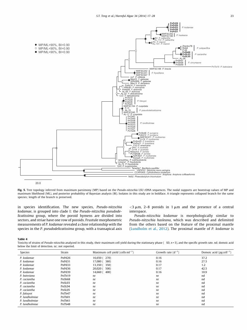
Fig. 5. Tree topology inferred from maximum parsimony (MP) based on the Pseudo-nitzschia LSU rDNA sequences. The nodal supports are bootstrap values of MP and
maximum likelihood (ML), and posterior probability of Bayesian analysis (BI). Isolates in this study are in boldface. A triangle represents collapsed branch for the same
species; length of the branch is preserved.
S.T. Teng et al. / Harmful Algae 34 (2014) 17–28 23
in species identification. The new species, Pseudo-nitzschia
kodamae, is grouped into clade I: the Pseudo-nitzschia pseudode-
licatissima group, where the poroid hymens are divided intosectors, and striae have one row of poroids. Frustule morphometricmeasurements of P. kodamae revealed a close relationship with thespecies in the P. pseudodelicatissima group, with a transapical axis
Table 4Toxicity of strains of Pseudo-nitzschia analyzed in this study, their maximum cell yield d
below the limit of detection. nr; not reported.
Species Strain Maximum cell yield (cells m
P. kodamae PnPd26 16,650 (�270)
P. kodamae PnPd31 17,080 (�380)
P. kodamae PnPd33 13,350 (�350)
P. kodamae PnPd36 20,020 (�500)
P. kodamae PnPd39 14,660 (�400)
P. batesiana PnTb19 nr
P. caciantha PnSb68 nr
P. caciantha PnSL03 nr
P. caciantha PnSL04 nr
P. caciantha PnSL05 nr
P. fukuyoi PnTb47 nr
P. lundholmiae PnTb01 nr
P. lundholmiae PnTb61 nr
P. lundholmiae PnTb48 nr
<3 mm, 2–8 poroids in 1 mm and the presence of a centralinterspace.
Pseudo-nitzschia kodamae is morphologically similar toPseudo-nitzschia hasleana, which was described and delimitedfrom the others based on the feature of the proximal mantle(Lundholm et al., 2012). The proximal mantle of P. kodamae is
uring the stationary phase (�SD, n = 3), and the specific growth rate. nd; domoic acid
l�1) Growth rate (d�1) Domoic acid (pg cell�1)
0.16 37.2
0.16 27.5
0.17 1.2
0.17 42.5
0.16 10.9
nr nd
nr nd
nr nd
nr nd
nr nd
nr nd
nr nd
nr nd
nr nd

0.06
DS2 P. calliantha
(09)12A2 P. brasiliana
Tenerife8
PnPd26
PnTb25
PnTb21 P. lundholmiae
(08)10A2
AL-19AL-29
PnSL05
8A9
PnKk36
(10)4A2
AL-60
PnPd36PnPd33
AL-15
PnTb31
PnPd31
(09)9A5
(08)8A3 P. subfraudulenta
(09)4A1
PnPd39
10A3
PnTb10 P. lundholmiaeAL-101
(07)12A2 P. multistriata
Sydney1AL-17
NWFSC252 P. hasleana
PnSL03
(07)10A2
P-11
C-AL-1
(08)9B2 P. hasleana
PnTb72
AL-112 P. calliantha
(08)5A3 P. galaxiae
(07)E-2
NerD5
PnLk02
Mex20
(09)2A3 P. fraudulenta
P. pseudodelicatissima
P. cuspidata
P. fukuyoi
P. mannii
P. kodamae
3-19 P. turgiduloides UBC100 P. granii
1-F P. subcurvata No7 P. inflatula
NWFSC188 P. lineolaLimens1 P. fraudulenta
P. cacianthaPnSb58 P. circumpora
AL-56 P. caciantha
RdA8 P. subpacifica Nezen P. subpacifica
PnTb19 P. batesianaNWFSC241 P. fryxelliana
Ner-D1 P. arenysensisAL-59 P. dolorosa
PnKK08 P. dolorosaTasm 10
Laeso2
Castell1 P. arenysensis
no16 P. micropora VPB-B3 P. micropora PnKK14 P. micropora
Mex23 P. galaxiae
Mex13 P. decipiens GranCan4-1 P. decipiens
Kervel P. americanaPnSm07 P. brasiliana
PnKK31 P. brasiliana
B5 P. multistriata
KoreaA P. multistriata PLTSt52B P. seriataNissum3 P. seriata
PLYSt19A P. australis T5 P. obtusa
PnSb44PnMt45
mu3 P. multiseries4-20 Fragilariopsis kerguelensis
(07)10A3
P. delicatissima
P. cf. delicatissima
P. pungens
Fig. 6. Profile neighbor joining tree based on the Pseudo-nitzschia ITS2 RNA transcripts. Posterior probabilities of >95% are marked with thick lines. Isolates in this study are in
boldface. A triangle represents collapsed branch for the same species; length of the branch is preserved.
S.T. Teng et al. / Harmful Algae 34 (2014) 17–2824
similar, but it differs by having a proximal mantle height of 2–4poroids, whilst in P. hasleana it is 2–3 poroids high. Our PCAanalysis further supports the separation of the two species. Theyare also readily distinguishable by having differences in thedensities of fibulae and striae in 10 mm, and valve shape. Whencompared to Pseudo-nitzschia batesiana, Pseudo-nitzschia fukuyoi
and Pseudo-nitzschia caciantha, the species is delimited from theothers by its linear valve shape, lower densities of fibulae andstriae in 10 mm, and lower density of band striae in 10 mm. It iseasily differentiated from Pseudo-nitzschia mannii and Pseudo-
nitzschia calliantha based on the numbers of hymen sectors (2–4sectors in P. kodamae; 2–7, but mainly four sectors, in P. mannii;7–10 in P. calliantha).
The significance of using molecular data to identify species inthe genus Pseudo-nitzschia cannot be underestimated. Molecularevidence has revealed several cryptic species, e.g. Pseudo-nitzschia
delicatissima–Pseudo-nitzschia arenysensis (Quijano-Scheggia et al.,2009). Recently, four new species of Pseudo-nitzschia (Pseudo-
nitzschia batesiana, Pseudo-nitzschia circumpora, Pseudo-nitzschia
fukuyoi, Pseudo-nitzschia lundholmiae) in the Pseudo-nitzschia
pseudodelicatissima complex sensu lato were described fromMalaysia, with the support from molecular data (Lim et al.,
2012a, 2013). The phylogenetic position of Pseudo-nitzschia
kodamae is strongly supported by the phylogenetic analysesperformed. The high level of genetic divergences further supportsthe split of this species lineage.
The importance of CBC information within the ITS2 RNAtranscripts for species delimitation is undeniable; this has alsobeen demonstrated in other fields of study (e.g. Ahvenniemi et al.,2009; Buchheim et al., 2011), as well as for Pseudo-nitzschia (e.g.Amato et al., 2007; Lim et al., 2012a, 2013). Our studydemonstrates that ITS2 CBC information, based on the conven-tional pairwise comparison among pairs of taxa (pheneticapproach), as well as that used in the phylogenetic approach ofCaisova et al. (2011), could be an informative molecularcharacteristic in delineating species.
Strains of Pseudo-nitzschia caciantha established in this studyare molecularly similar to the type specimen, Mex20 (Lundholmet al., 2003), with respect to both the LSU rDNA (D1–D3) and ITS2regions. The strains formed a monophyletic group in both treetopologies (Figs. 6 and 7). However, a strain from Gulf of Naples(AL-56) (Amato and Montresor, 2008), remained unresolved. Ofinterest is the occurrence of three CBCs in the ITS2 RNA transcriptof strain AL-56 compared to other P. caciantha strains; this

Fig. 7. ITS2 RNA transcript of Pseudo-nitzschia kodamae. Shaded boxes indicate compensatory base changes (CBCs) and empty boxes represent hemi-compensatory base
changes (HCBCs).
S.T. Teng et al. / Harmful Algae 34 (2014) 17–28 25
warrants a further investigation on the species identity of strainAL-56.
4.2. Toxic Pseudo-nitzschia in Southeast Asia
Even though blooms of Pseudo-nitzschia had never beenreported from the Southeast Asian region, the occurrence of DAin the region was supported by several previous findings of DAcontamination in shellfish (Bajarias et al., 2006; Dao et al., 2006,2009a,b, 2012; Takata et al., 2009; Romero et al., 2011; Thohaet al., 2012). None of the species of Pseudo-nitzschia in the region
has been confirmed to be responsible for the shellfish contami-nation thus far, although Dao et al. (2009b) showed that planktonsamples in the size fraction of <10 mm, collected from the sitewhere contaminated shellfish were found, contained DA. Thissuggested that a DA-producing organism(s) in the planktonsamples was likely to be the source of DA contamination. Pseudo-
nitzschia species become the first suspect for the DA in shellfish.Another possible culprit, Nitzschia navis-varingica, however,cannot be ruled out, as this species has been confirmed toproduce DA in the region (Kotaki et al., 2000, 2004, 2008; Romeroet al., 2012).
Environmental toxic group (8)
Culture toxic group (16)
Culture non-toxic group (32) Not tested (9) P.
gra
nii
P. brasilianaP. callianthaP. fraudulentaP. galaxiaeP. multistriataP. subpacifica P. obtusa
P. micropora
P. heimiiP. inflatulaP. lineola
P. subcurvataP. subfraudulentaP. turgiduloides
P. lundholmiae
P. americanaP. arenysensis
P. caciantha
P. decipiensP. dolorosa
P. hasleana
P. circumpora
P. batesiana
P. fukuyoi
P. australis
P. pungens
P. multiseries
P. seriata
P. cuspidataP. delicatissima
P. pseudodelicatissma
P. turgidula
P. kodamaeP. plurisecta P. antarctica
P. fryxellianaP. lineaP. manniiP. prolongatoidesP. pungiformisP. roundiiP. sinica
P. abrensis
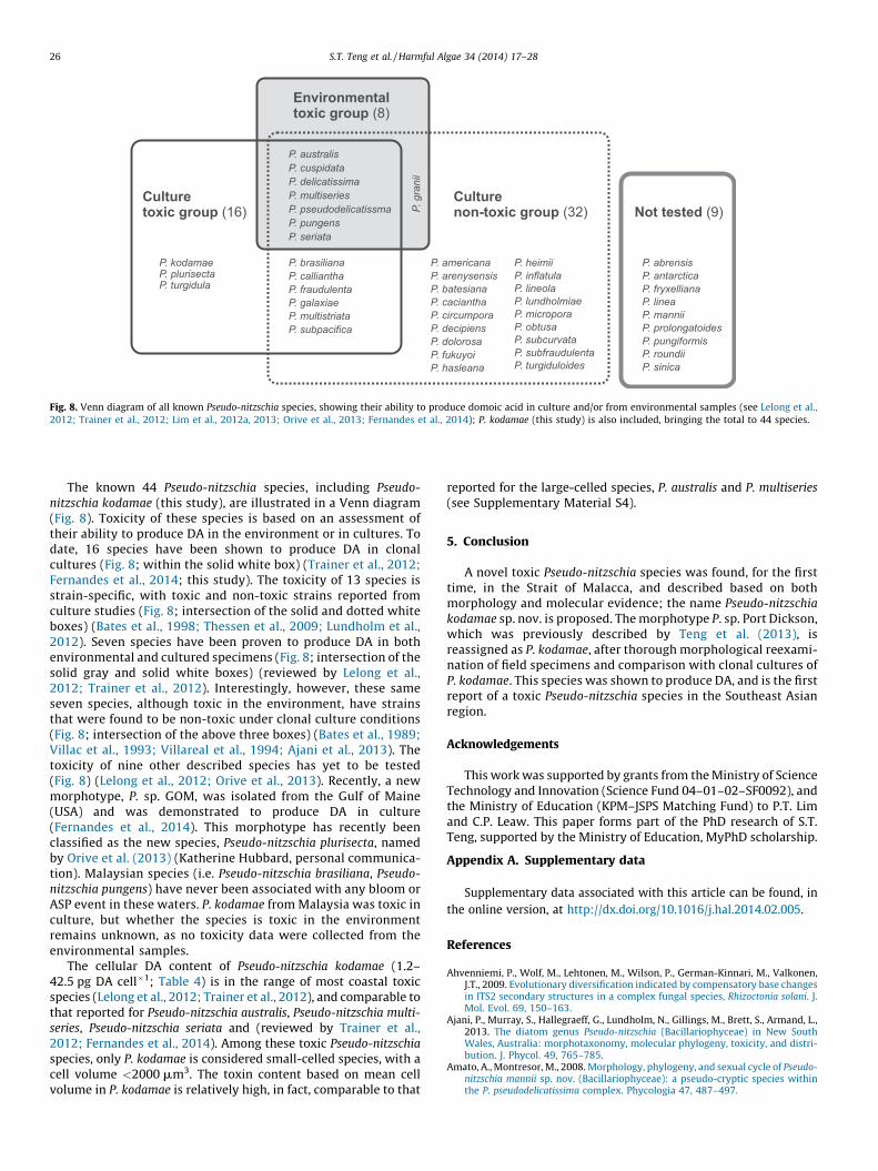
Fig. 8. Venn diagram of all known Pseudo-nitzschia species, showing their ability to produce domoic acid in culture and/or from environmental samples (see Lelong et al.,
2012; Trainer et al., 2012; Lim et al., 2012a, 2013; Orive et al., 2013; Fernandes et al., 2014); P. kodamae (this study) is also included, bringing the total to 44 species.
S.T. Teng et al. / Harmful Algae 34 (2014) 17–2826
The known 44 Pseudo-nitzschia species, including Pseudo-
nitzschia kodamae (this study), are illustrated in a Venn diagram(Fig. 8). Toxicity of these species is based on an assessment oftheir ability to produce DA in the environment or in cultures. Todate, 16 species have been shown to produce DA in clonalcultures (Fig. 8; within the solid white box) (Trainer et al., 2012;Fernandes et al., 2014; this study). The toxicity of 13 species isstrain-specific, with toxic and non-toxic strains reported fromculture studies (Fig. 8; intersection of the solid and dotted whiteboxes) (Bates et al., 1998; Thessen et al., 2009; Lundholm et al.,2012). Seven species have been proven to produce DA in bothenvironmental and cultured specimens (Fig. 8; intersection of thesolid gray and solid white boxes) (reviewed by Lelong et al.,2012; Trainer et al., 2012). Interestingly, however, these sameseven species, although toxic in the environment, have strainsthat were found to be non-toxic under clonal culture conditions(Fig. 8; intersection of the above three boxes) (Bates et al., 1989;Villac et al., 1993; Villareal et al., 1994; Ajani et al., 2013). Thetoxicity of nine other described species has yet to be tested(Fig. 8) (Lelong et al., 2012; Orive et al., 2013). Recently, a newmorphotype, P. sp. GOM, was isolated from the Gulf of Maine(USA) and was demonstrated to produce DA in culture(Fernandes et al., 2014). This morphotype has recently beenclassified as the new species, Pseudo-nitzschia plurisecta, namedby Orive et al. (2013) (Katherine Hubbard, personal communica-tion). Malaysian species (i.e. Pseudo-nitzschia brasiliana, Pseudo-
nitzschia pungens) have never been associated with any bloom orASP event in these waters. P. kodamae from Malaysia was toxic inculture, but whether the species is toxic in the environmentremains unknown, as no toxicity data were collected from theenvironmental samples.
The cellular DA content of Pseudo-nitzschia kodamae (1.2–42.5 pg DA cell�1; Table 4) is in the range of most coastal toxicspecies (Lelong et al., 2012; Trainer et al., 2012), and comparable tothat reported for Pseudo-nitzschia australis, Pseudo-nitzschia multi-
series, Pseudo-nitzschia seriata and (reviewed by Trainer et al.,2012; Fernandes et al., 2014). Among these toxic Pseudo-nitzschia
species, only P. kodamae is considered small-celled species, with acell volume <2000 mm3. The toxin content based on mean cellvolume in P. kodamae is relatively high, in fact, comparable to that
reported for the large-celled species, P. australis and P. multiseries
(see Supplementary Material S4).
5. Conclusion
A novel toxic Pseudo-nitzschia species was found, for the firsttime, in the Strait of Malacca, and described based on bothmorphology and molecular evidence; the name Pseudo-nitzschia
kodamae sp. nov. is proposed. The morphotype P. sp. Port Dickson,which was previously described by Teng et al. (2013), isreassigned as P. kodamae, after thorough morphological reexami-nation of field specimens and comparison with clonal cultures ofP. kodamae. This species was shown to produce DA, and is the firstreport of a toxic Pseudo-nitzschia species in the Southeast Asianregion.
Acknowledgements
This work was supported by grants from the Ministry of ScienceTechnology and Innovation (Science Fund 04–01–02–SF0092), andthe Ministry of Education (KPM–JSPS Matching Fund) to P.T. Limand C.P. Leaw. This paper forms part of the PhD research of S.T.Teng, supported by the Ministry of Education, MyPhD scholarship.
Appendix A. Supplementary data
Supplementary data associated with this article can be found, in
the online version, at http://dx.doi.org/10.1016/j.hal.2014.02.005.
References
Ahvenniemi, P., Wolf, M., Lehtonen, M., Wilson, P., German-Kinnari, M., Valkonen,J.T., 2009. Evolutionary diversification indicated by compensatory base changesin ITS2 secondary structures in a complex fungal species, Rhizoctonia solani. J.Mol. Evol. 69, 150–163.
Ajani, P., Murray, S., Hallegraeff, G., Lundholm, N., Gillings, M., Brett, S., Armand, L.,2013. The diatom genus Pseudo-nitzschia (Bacillariophyceae) in New SouthWales, Australia: morphotaxonomy, molecular phylogeny, toxicity, and distri-bution. J. Phycol. 49, 765–785.
Amato, A., Montresor, M., 2008. Morphology, phylogeny, and sexual cycle of Pseudo-nitzschia mannii sp. nov. (Bacillariophyceae): a pseudo-cryptic species withinthe P. pseudodelicatissima complex. Phycologia 47, 487–497.

S.T. Teng et al. / Harmful Algae 34 (2014) 17–28 27
Amato, A., Kooistra, W.H.C.F., Ghiron, J.H.L., Mann, D.G., Proschold, T., Montresor, M.,2007. Reproductive isolation among sympatric cryptic species in marine dia-toms. Protist 158, 193–207.
Bajarias, F.F.A., Kotaki, Y., Relox Jr., J.R., Romero, M.L.J., Furio, E.F., Lundholm, N.,Koike, K., Fukuyo, Y., Kodama, M., 2006. Screening of diatoms producing domoicacid and its derivatives in the Philippines. Coast. Mar. Sci. 30, 121–129.
Bates, S.S., Bird, C.J., de Freitas, A.S.W., Foxall, R., Gilgan, M., Hanic, L.A., Johnson, G.R.,McCulloch, A.W., Odense, P., Pocklington, R., Quilliam, M.A., Sim, P.G., Smith, J.C.,Subba Rao, D.V., Todd, E.C.D., Walter, J.A., Wright, J.L.C., 1989. Pennate diatomNitzschia pungens as the primary source of domoic acid, a toxin in shellfish fromeastern Prince Edward Island, Canada. Can. J. Fish. Aquat. Sci. 46, 1203–1215.
Bates, S.S., Garrison, D.L., Horner, R.A., 1998. Bloom dynamics and physiology ofdomoic-acid-producing Pseudo-nitzschia species. In: Anderson, D.M., Cembella,A.D., Hallegraeff, G.M. (Eds.), Physiological Ecology of Harmful Algal Blooms.Springer-Verlag, Heidelberg, pp. 267–292.
Buchheim, M.A., Keller, A., Koetschan, C., Forster, F., Merget, B., Wolf, M., 2011.Internal transcribed spacer 2 (nu ITS2 rRNA) sequence-structure phylogenetics:towards an automated reconstruction of the green algal tree of life. PLoS ONE 6,e16931.
Caisova, L., Marin, B., Melkonian, M., 2011. A close-up view on ITS2 evolution andspeciation – a case study in the Ulvophyceae (Chlorophyta, Viridiplantae). BMCEvol. Biol. 11, 262.
Dao, V.H., Takata, Y., Sato, S., Fukuyo, Y., Kodama, M., 2006. Domoic acid in a bivalveSpondylus cruentus in Nha Trang Bay, Khanh Hoa Province, Vietnam. Coast. Mar.Sci. 30, 130–132.
Dao, V.H., Takata, Y., Omura, T., Sato, S., Fukuyo, Y., Kodama, M., 2009a. Seasonalvariation of domoic acid in a bivalve Spondylus versicolor in association with thatin plankton samples in Nha Phu Bay, Khanh Hoa, Vietnam. Fish. Sci. 75, 507–512.
Dao, V.H., Takata, Y., Omura, T., Nyuyen, T.D., Nyuyen, T.H., Sato, S., Fukuyo, Y.,Kodama, M., 2009b. Domoic acid in small-sized plankton in Nha Phu Bay, KhanhHoa Province, Vietnam. La Mer 46, 117–120.
Dao, V.H., Omura, T., Takata, Y., Ky, P.X., Fukuyo, Y., Kodama, M., 2012. Pseudo-nitzschia species, a possible causative organism of domoic acid in Spondylusversicolor collected from Nha Phu Bay, Khanh Hoa Province, Vietnam. Coast.Mar. Sci. 35, 7–10.
Darriba, D., Taboada, G.L., Doallo, R., Posada, D., 2012. jModelTest 2: more models,new heuristics and parallel computing. Nat. Methods 9, 772.
Fernandes, L.F., Hubbard, K.A., Richlen, M., Smith, J., Bates, S.S., Ehrman, J., Leger, C.,Mafra Jr., L.L., Kulis, D., Quilliam, M., Erdner, D., Libera, K., McCauley, L.,Anderson, D.M., 2014. Diversity and toxicity of the diatom Pseudo-nitzschiaPeragallo in the Gulf of Maine, Northwestern Atlantic Ocean. Deep-Sea Res. II,http://dx.doi.org/10.1016/j.dsr2.2013.06.022 (in press).
Fukuyo, Y., Kodama, M., Omura, T., Furuya, K., Furio, E.F., Cayme, M., Lim, P.T., Dao,V.H., Kotaki, Y., Matsuoka, K., Iwataki, M., Sriwoon, R., Lirdwitayaprasit, T., 2011.Ecology and oceanography of harmful marine microalgae. In: Nishida, S., Fortes,M.D., Miyazaki, N. (Eds.), Coastal Marine Science in Southeast Asia – SynthesisReport of the Core University Program of the Japan Society for the Promotion ofScience: Coastal Marine Science (2000–2010). Terrapub, Tokyo, pp. 23–48.
Guillard, R.R.L., 1975. Culture of Phytoplankton for Feeding Marine Invertebrates.Plenum Press, New York, USA.
Hammer, Ø., Harper, D.A.T., Ryan, P.D., 2001. Past: paleontological statistics soft-ware package for education and data analysis. Palaeontol. Electr. 4, 9., http://palaeo-electronica.org/2001_1/past/issue1_01.htm.
Hasle, G.R., 1994. Pseudo-nitzschia as a genus distinct from Nitzschia (Bacillario-phyceae). J. Phycol. 30, 1036–1039.
Hasle, G.R., Lange, C.B., Syvertsen, E.E., 1996. A review of Pseudo-nitzschia, withspecial reference to the Skagerrak, North Atlantic, and adjacent waters. Helgo-ander Meeresunters 50, 131–175.
Kodama, M., Kotaki, Y., 2005. Domoic acid. In: The Manual for the Method of FoodSanitation Test. Ministry of Health, Labour and Welfare. Japan Food HygienicAssociation, Tokyo, pp. 666–673 (in Japanese).
Kotaki, Y., Koike, K., Yoshida, M., Thuoc, C.V., Huyen, N.T.M., Hoi, N.C., Fukuyo, Y.,Kodama, M., 2000. Domoic acid production in Nitzschia sp. (Bacillariophyceae)isolated from a shrimp-culture pond in Do Son, Vietnam. J. Phycol. 36, 1057–1060.
Kotaki, Y., Lundholm, N., Onodera, H., Kobayashi, K., Bajarias, F.F.A., Furio, E.F.,Iwataki, M., Fukuyo, Y., Kodama, M., 2004. Wide distribution of Nitzschia navis-varingica, a new domoic acid-producing benthic diatom found in Vietnam. Fish.Sci. 70, 28–32.
Kotaki, Y., Lundholm, N., Katayama, T., Furio, E.F., Romero, M.L.J., Relox, J.R.,Yasumoto, T., Naoki, H., Hirose, M.Y., Thann, T.D., Thuoc, C.V., Huyen, N.T.M.,Thu, P.T., Takata, Y., Kodama, M., Fukuyo, Y., 2008. ASP toxins of pennatediatoms and bacterial effects on the variation in toxin composition. In:Moestrup, Ø. (Ed.), Harmful Algae, Proceedings of the 12th International Con-ference on Harmful Algae. Intergovernmental Oceanographic Commission ofUNESCO, Copenhagen, Denmark, pp. 300–302.
Lelong, A., Hegaret, H., Soudant, P., Bates, S.S., 2012. Pseudo-nitzschia (Bacillario-phyceae) species, domoic acid and amnesic shellfish poisoning: revisitingprevious paradigms. Phycologia 51, 168–216.
Lim, P.T., Leaw, C.P., 2012. First report of Pyrodinium bahamense in the Strait ofMalacca. Harmful Algae News (No. 45) 5.
Lim, P.T., Leaw, C.P., Usup, G., 2004. First incidence of paralytic shellfish poisoningon the east coast of Peninsular Malaysia. In: Phang, S.M., Chong, V.C., Ho, S.S.,Mokhtar, N., Ooi, J.L.S. (Eds.), Marine Science into the New Millennium: New
Perspectives and Challenges. University of Malaya Maritime Research Centre,Kuala Lumpur, Malaysia, pp. 661–667.
Lim, H.C., Su, S.N.P., Ali, H.M., Kotaki, Y., Leaw, C.P., Lim, P.T., 2010. Toxicity of diatomPseudo-nitzschia (Bacillariophyceae) analyzed using high performance liquidchromatography (HPLC). J. Sci. Technol. Tropics 6, S116–S119.
Lim, H.C., Leaw, C.P., Su, S.N.P., Teng, S.T., Usup, G., Noor, N.M., Lundholm, N., Kotaki,Y., Lim, P.T., 2012a. Morphology and molecular characterization of Pseudo-nitzschia (Bacillariophyceae) from Malaysian Borneo, including the new speciesPseudo-nitzschia circumpora sp. nov. J. Phycol. 48, 1232–1247.
Lim, H.C., Lim, P.T., Su, S.N.P., Kotaki, Y., Leaw, C.P., 2012b. Morphological observa-tion of two species of Pseudo-nitzschia (Bacillariophyceae). Coast. Mar. Sci. 35,52–57.
Lim, H.C., Lim, P.T., Su, S.N.P., Teng, S.T., Leaw, C.P., 2012c. Genetic diversity ofPseudo-nitzschia brasiliana (Bacillariophyceae) from Malaysia. J. Appl. Phycol.24, 1465–1475.
Lim, P.T., Usup, G., Leaw, C.P., 2012d. Harmful algal blooms in Malaysian waters.Sains Malaysiana 41, 1509–1515.
Lim, H.C., Teng, S.T., Leaw, C.P., Lim, P.T., 2013. Three novel species in the Pseudo-nitzschia pseudodelicatissima complex: P. batesiana sp. nov., P. lundholmiae sp.nov. and P. fukuyoi sp. nov. (Bacillariophyceae), from the Strait of Malacca,Malaysia. J. Phycol. 49, 902–916.
Lundholm, N., Daugbjerg, N., Moestrup, Ø., 2002. Phylogeny of the Bacillariaceaewith emphasis on the genus Pseudo-nitzschia (Bacillariophyceae) based onpartial LSU rDNA. Eur. J. Phycol. 37, 115–134.
Lundholm, N., Moestrup, Ø., Hasle, G.R., Hoef-Emden, K., 2003. A study of thePseudo-nitzschia pseudodelicatissima/cuspidata complex (Bacillariophyceae):what is P. pseudodelicatissima? J. Phycol. 39, 797–813.
Lundholm, N., Bates, S.S., Baugh, K.A., Bill, B.D., Connell, L.B., Leger, C., Trainer, V.L.,2012. Cryptic and pseudo-cryptic diversity in diatoms – with descriptions ofPseudo-nitzschia hasleana sp. nov. and P. fryxelliana sp. nov. J. Phycol. 48, 436–454.
Notredame, C., Higgins, D.G., Heringa, J., 2000. T-Coffee: a novel method for fast andaccurate multiple sequence alignment. J. Mol. Biol. 302, 205–217.
Orive, E., Perez-Aicua, L., David, H., Garcıa-Etxebarria, K., Laza-Martınez, A., Seoane,S., Miguel, I., 2013. The genus Pseudo-nitzschia (Bacillariophyceae) in a temper-ate estuary with description of two new species: Pseudo-nitzschia plurisecta sp.nov. and Pseudo-nitzschia abrensis sp. nov. J. Phycol. 49, 1192–1206.
Quijano-Scheggia, S., Garces, E., Lundholm, N., Moestrup, Ø., Andree, K., Camp, J.,2009. Morphology, physiology, molecular phylogeny and sexual compatibilityof the cryptic Pseudo-nitzschia delicatissima complex (Bacillariophyta), includ-ing the description of P. arenysensis sp. nov. Phycologia 48, 492–509.
Quijano-Scheggia, S., Garces, E., Andree, K.B., de la Iglesia, P., Diogene, J., Fortuno,J.M., Camp, J., 2010. Pseudo-nitzschia species on the Catalan coast: characteri-zation and contribution to the current knowledge of the distribution of thisgenus in the Mediterranean Sea. Sci. Mar. 74, 395–410.
Romero, M.L.J., Kotaki, Y., Lundholm, N., Thoha, H., Ogawa, H., Relox, J.R., Terada, R.,Takeda, S., Takata, Y., Haraguchi, K., Endo, T., Lim, P.T., Kodama, M., Fukuyo, Y.,2011. Unique amnesic shellfish toxin composition found in the Southeast Asiandiatom Nitzschia navis-varingica. Harmful Algae 10, 456–462.
Romero, M.L.J., Kotaki, Y., Relox, J.J., Lundholm, N., Takata, Y., Kodama, M., Fukuyo,Y., 2012. Two new ASP toxin production types in strains of Nitzschia navis-varingica from the Philippines. Coast. Mar. Sci. 35, 67–69.
Ronquist, F., Teslenko, M., van der Mark, P., Ayres, D., Darling, A., Hohna, S., Larget, B.,Liu, L., Suchard, M.A., Huelsenbeck, J.P., 2012. MrBayes 3.2: efficient Bayesianphylogenetic inference and model choice across a large model space. Syst. Biol.61, 539–542.
Scholin, C.A., Herzog, M., Sogin, M., Anderson, D.M., 1994. Identification of group-and strain-specific genetic markers for globally distributed Alexandrium (Dino-phyceae). II. Sequence analysis of a fragment of the LSU rRNA gene. J. Phycol. 30,999–1011.
Seibel, P.N., Muller, T., Dandekar, T., Matthias, W., 2008. Synchronous visual analysisand editing of RNA sequence and secondary structure alignments using 4SALE.BMC Res. Notes 1, 91.
Stonik, I.V., Orlova, T.Y., Lundholm, N., 2011. Diversity of Pseudo-nitzschia H.Peragallo from the western North Pacific. Diatom Res. 26, 121–134.
Swofford, D.L., 2001. PAUP* Phylogenetic Analysis Using Parsimony (*and OtherMethods) Version 4. 04beta Sinauer Associates, Sunderland, MA.
Takata, Y., Sato, S., Ha, D.V., Montojo, U.M., Lirdwitayaprasit, T., Kamolsiripichai-porn, S., Kotaki, Y., Fukuyo, Y., Kodama, M., 2009. Occurrence of domoic acid intropical bivalves. Fish. Sci. 75, 473–480.
Teng, S.T., Leaw, C.P., Lim, H.C., Lim, P.T., 2013. The genus Pseudo-nitzschia (Bacillar-iophyceae) in Malaysia, including new records and a key to species inferredfrom morphology-based phylogeny. Bot. Mar. 56, 375–398.
Thessen, A.E., Bowers, H.A., Stoecker, D.K., 2009. Intra- and interspecies differencesin growth and toxicity of Pseudo-nitzschia while using different nitrogensources. Harmful Algae 8, 792–810.
Thoha, H., Kotaki, Y., Panggabean, L., Lundholm, N., Ogawa, H., Lim, P.T., Takata, Y.,Kodama, M., Fukuyo, Y., 2012. Screening of diatoms that produce ASP toxins inSouthernmost Asian waters. Coast. Mar. Sci. 35, 34–38.
Trainer, V.L., Bates, S.S., Lundholm, N., Thessen, A.E., Cochlan, W.P., Adams, N.G.,Trick, C.G., 2012. Pseudo-nitzschia physiological ecology, phylogeny, toxicity,monitoring and impacts on ecosystem health. Harmful Algae 14, 271–300.
Usup, G., Leaw, C.P., Ahmad, A., Lim, P.T., 2002. Alexandrium (Dinophyceae) speciesin Malaysian waters. Harmful Algae 1, 265–275.
S.T. Teng et al. / Harmful Algae 34 (2014) 17–2828
Villac, M.C., Roelke, D.L., Chavez, F.P., Cifuentes, L.A., Fryxell, G.A., 1993. Pseudo-nitzschia australis Frenguelli and related species from the west coast of theU.S.A.: occurrence and domoic acid production. J. Shellfish Res. 12, 457–465.
Villareal, T.A., Roelke, D.L., Fryxell, G.A., 1994. Occurrence of the toxic diatomNitzschia pungens f. multiseries in Massachusetts Bay, Massachusetts, USA.Mar. Environ. Res. 37, 417–423.
White, T.J., Bruns, T., Lee, S., Taylor, J.W., 1990. Amplification and direct sequencingof fungal ribosomal RNA genes for phylogenetics. In: Innis, M.A., Gelfand, D.H.,
Sninsky, J.J., White, T.J. (Eds.), PCR Protocols: A Guide to Methods and Applica-tions. Academic Press Inc., New York, pp. 315–322.
Wolf, M., Ruderisch, B., Dandekar, T., Schultz, J., Muller, T., 2008. ProfDistS: (profile–)distance based phylogeny on sequence–structure alignments. Bioinformatics24, 2401–2402.
Yap-Dejeto, L.G., Omura, T., Nagahama, Y., Fukuyo, Y., 2010. Observations of elevenPseudo-nitzschia species in Tokyo Bay, Japan. La Mer 48, 1–16.