documentps
TRANSCRIPT

Section 1THEPERIPHERALSMEAR
40413 Tkachuk PM Gordon(SAUNM) INTERACTIVE RIGHT
top of RHbase of RH
SN base
shortstandardlong

40413 Tkachuk PM Gordon(SAUNM) INTERACTIVE LEFT
top of RHbase of RH
top of textbase of text
shortstandardlong

Evaluation of Red Cells
In evaluating red cells the examiner looks for abnormalities in size andshape, hemoglobin content, inclusions, aggregation, and immature forms.
The normal red cell is about 8 �m in diameter, slightly less than the nucleusof a small lymphocyte, which is approximately 8.5 �m. When the erythro-cyte’s size is normal, it is normocytic; when larger, macrocytic; and when smaller,microcytic. The automated cell counters measure size as mean cell volume(MCV); microcytosis corresponds to an MCV �80 fl, macrocytosis to an MCV�100 fl. A substantial variation in size is called anisocytosis and registers onthe automated counters as an increased red cell distribution width (RDW).
The biconcave erythrocyte is thinner in the middle, creating a central palloron blood smears that is ordinarily less than of the cell’s diameter. Such a1⁄3cell, possessing the normal amount of hemoglobin, is called normochromic.When the central pallor is greater than normal, indicating decreased hemo-globin content, the erythrocyte is hypochromic. Severe hypochromia corre-sponds to a decreased mean cell hemoglobin concentration (MCHC) asmeasured by the automated analyzers. When central pallor is absent fromcells that are separated from contiguous ones, the usual reason is a decreasein cell membrane surface, making the cell denser, less concave in the center,and more spherical, hence a spherocyte. Some macrocytic cells are thicker thannormal and also lack central pallor.
The examiner should look for abnormalities in red cell shape (a significantincrease in the number of abnormally shaped cells is called poikilocytosis) andscrutinize the interior of the erythrocytes to detect inclusions and the pres-ence of nuclei or a bluish-purple color, indicating immature cells. Finally, theexaminer looks for abnormalities in red cell aggregation—agglutination, orthe clumping of red cells into a rosette configuration, or rouleaux, erythro-cytes aligned in a row like a stack of coins.
40413 Tkachuk PM Gordon(SAUNM) INTERACTIVE RIGHT
top of RHbase of RH
top of textbase of text
shortstandardlong

4 The Peripheral Smear
ABNORMALITIES IN RED CELL SIZE AND HEMOGLOBINCONTENT
Slide 1. Normal Red Cells
The red cells are fairly uniform in size and shape.Central pallor is present but constitutes less than 1⁄3of the erythrocyte’s diameter (about 8 �m), which isslightly less than that of the nucleus of the smalllymphocyte (about 8.5 �m) (also see slide 34). Thered cells are thus normal in hemoglobin content(normochromic) and size (normocytic).
40413 Tkachuk PM Gordon(SAUNM) INTERACTIVE LEFT
top of RHbase of RH
top of textbase of text
shortstandardlong
NORMAL RED CELLS
Slide 2. Microcytic, Hypochromic Red Cells
Many of the erythrocytes are smaller than the nucleusof the small lymphocyte and have markedly increasedcentral pallor, exceeding the diameter of the red1⁄3cell. The erythrocytes, therefore, are microcytic(�7.0 �m in diameter) and hypochromic. These fea-tures, which usually coexist, indicate abnormal hemo-globin synthesis. The major causes are iron deficiency,thalassemias, hemoglobinopathies, some sideroblasticanemias, and the anemia of chronic disease, in whichmicrocytosis occurs in about 20% of cases. In additionto microcytosis and hypochromia, the erythrocytes inthis slide exhibit variation in size (anisocytosis) andshape (poikilocytosis). This patient had iron deficiencyanemia, which accounts for the increased number ofplatelets.

The Peripheral Smear 5
Slide 3. Macrocytic Red Cells
Most of the red cells are larger than the nucleus ofthe small lymphocyte. The erythrocytes are thereforemacrocytic (�8.5 �m in diameter). The major causes
of macrocytic red cells are alcoholism, liver disease,vitamin B12 or folate deficiency, myelodysplastic syn-dromes, hypothyroidism, drugs that impair folate orDNA synthesis, and blood loss or hemolysis. The in-creased erythrocyte size in blood loss or hemolysisarises from the presence of large, immature formsreleased early from the bone marrow in response tointense erythropoietin stimulus. Some of these youngerythrocytes differ from mature red cells on Roma-nowsky stains only by their size; others are identifia-ble by their bluish–gray color, called polychromato-philia or polychromasia (see slide 19).
The presence of oval macrocytes (macro–ovalo-cytes) in this slide strongly suggests myelodysplasia ora deficiency in folate or vitamin B12; in other disor-ders causing macrocytosis, the large erythrocytes areuniformly round. Note the variation in red cell sizeand shape. This patient had vitamin B12 deficiency.
40413 Tkachuk PM Gordon(SAUNM) INTERACTIVE RIGHT
top of RHbase of RH
top of textbase of text
shortstandardlong
Slide 4. Spherocyte
Spherocytes form because an inherited or acquireddefect in the erythrocyte membrane decreases thesurface area to volume ratio. Because a sphere’s di-ameter is less than that of a disk-shaped object con-taining the same volume, the spherocytes are typicallysmaller than mature red cells. Hereditary spherocyto-sis is most often an autosomal dominant disease, butabout 25% of cases arise from de novo mutations.Transfused erythrocytes are often spherocytic, andtransfusion is the most common cause of spherocytesin clinical practice. The other major cause of numer-
ous spherocytes is warm-antibody hemolytic anemia,in which the spleen removes small portions of thered cell membrane from cells coated with IgG anti-bodies. The erythrocyte then emerges from thespleen as a smaller cell with a decreased surface areato volume ratio (microcytic and lacking central pal-lor). Since the normal amount of hemoglobin is con-centrated into a smaller cell, the erythrocyte appearshyperchromic. This change is reflected in an in-creased mean cell hemoglobin content (MCHC) onautomated counters, and many cases of autoimmunehemolytic anemia are initially suspected from the ele-vated MCHC. Unusual causes of spherocytes includesevere burns, in which very small cells (microsphero-cytes) are typically present, or toxin damage frombacterial sepsis caused by Clostridium perfringens. Theexaminer should look for spherocytes in areas of theblood smear where the cells do not overlap, becausenormal erythrocytes may resemble spherocytes at theedges of slides, although in that location they areusually broad, pale, and somewhat square.
This slide, from a patient with warm-antibody auto-immune hemolytic anemia, shows several spherocytes,with an especially prominent one in the middle.They are small cells lacking central pallor. Many nor-mocytic cells with central pallor are also present, as istypical in this disorder.

6 The Peripheral Smear
Slide 5. Target Cells
In target cells the erythrocyte’s center contains a cir-cle of hemoglobin pigment surrounded by a ring of
pallor. This configuration occurs from an increase inthe red cell membrane compared to the hemoglobincontent (an increased surface area to volume ratio),the opposite of what happens with spherocytes. Whenthe erythrocytes dry on the slide, the excess mem-brane pools in the middle of the cells. The cause oftarget cells may be diminished hemoglobin contentfrom iron deficiency, hemoglobinopathies, and thal-assemias; in these disorders the cells are typically mi-crocytic. Target cells can also occur from obstructivejaundice or primary hepatocellular diseases such ashepatitis, in which the cell membrane enlarges byabnormally incorporating lipids, creating target cellsthat are usually normocytic or macrocytic. Normo-cytic target cells may also be present in hyposplenismand some hemoglobinopathies. This slide, from a pa-tient with combined �-thalassemia and hemoglobin Cdisease, contains prominent target cells, numerousspherocytes in the center of the slide, and two bitecells (see slide 11) in the lower center.
40413 Tkachuk PM Gordon(SAUNM) INTERACTIVE LEFT
top of RHbase of RH
top of textbase of text
shortstandardlong
ABNORMALITIES IN RED CELL SHAPE
Slide 6. Elliptocyte
Red cells may be elliptical in various anemias, espe-cially macrocytic ones. In a group of hereditary, usu-ally autosomal dominant, disorders, elliptocytes con-stitute at least 25% of the erythrocytes, hemolysis isusually mild, and anemia is present in only about 5–20% of cases. This slide, from a patient with heredi-tary elliptocytosis, discloses red cells that are darkand lack central pallor. In iron deficiency anemiaelongated erythrocytes (“pencil cells”) may form, but,unlike elliptocytes, they are thin and pale.
Slide 7. Teardrop Cell (Dacryocyte)
These pear-shaped cells, seen most prominently inthalassemias and diseases involving bone marrow in-filtration by fibrosis or malignancy, apparently formfrom distortion of the erythrocytes as they travelthrough the vasculature of an abnormal bone mar-row or spleen. Good examples, present in the centerof this slide, illustrate that the pointed ends may besharp or blunt.

The Peripheral Smear 7
Slide 8. Burr Cells (Echinocytes)
Burr cells usually have 10–30 blunt and fairly sym-metrical projections. They are prominent in renalfailure from any cause and may also occur with liverdiseases, especially when uremia coexists. Burr cellscan also develop as a storage artifact if blood is keptin a tube for several hours before preparation of thesmear. The mechanism of echinocyte formation isunknown; perhaps an increase in fatty acids alters thecell membrane.
40413 Tkachuk PM Gordon(SAUNM) INTERACTIVE RIGHT
top of RHbase of RH
top of textbase of text
shortstandardlong
Slide 9. Spur Cells (Acanthocytes)
Because of changes in membrane lipid content, thesered cells have several irregularly distributed, sharpprojections of unequal length. Most of the affectederythrocytes are also small and lack central pallor.Acanthocytes are prominent in spur-cell anemia,where liver disease, usually alcoholic cirrhosis, causesan increase in the cholesterol : phospholipid ratio inthe red cell membrane, leading to hemolysis, whichis usually severe. Acanthocytosis also occurs in abeta-lipoproteinemia, a rare autosomal recessive disorderof lipid metabolism characterized by fat malabsorp-tion, retinitis pigmentosa, anemia, and ataxia. Spurcells are a diagnostic feature of neuroacanthocytosis,a disease that may be hereditary or nonfamilial; itsmajor features are personality change, progressive in-tellectual deterioration, and a movement disorder,most commonly chorea.
Slide 10. Stomatocytes
On wet preparations the stomatocyte, rather than be-ing biconcave like normal erythrocytes, is concave ononly one side, resembling a cup or bowl. When ex-amined on dry smears, it has a central slit or stoma(mouth). A few stomatocytes may be present in nor-mal people, occasionally constituting �5% of theerythrocytes. Conditions associated with this abnor-mality of the red cell membrane include alcoholism,liver disease, malignancies, various cardiovascular andpulmonary disorders, treatment with certain drugs,and an autosomal dominant disorder in which stoma-tocytes constitute 10–50% of the circulating erythro-cytes, hemolysis is mild, and anemia is slight or ab-sent. This slide is from a patient with alcoholic liverdisease.

8 The Peripheral Smear
Slide 11. Bite Cell (Degmacyte)
Bite cells have a semicircular defect in their edgethat resembles a bite mark. These defects occur whencertain drugs cause oxidative destruction of hemoglo-bin, often in patients with a deficiency of the enzyme
glucose-6-phosphate dehydrogenase (G6PD). Heinzbodies, which are erythrocyte inclusions of denaturedhemoglobin, result, but they are not visible on Roma-nowsky stains. They are detectable when dyes such asmethyl or cresyl violet, new methylene blue, or bril-liant cresyl blue are mixed with unfixed cells beforemicroscopic examination (see slide 104). Because theerythrocytes are still alive when the dye is added, thepreparations are called “supravital” stains. The bitecells apparently occur when the spleen removes theHeinz bodies from the erythrocytes. Other features ofsevere oxidative injury visible on Romanowsky stainsinclude “ghost cells,” in which the erythrocyte isnearly devoid of pigment, and hemi-ghost cells, inwhich hemoglobin is present in only one half of thecell. A bite cell lies in the center of this slide from apatient who developed hemolytic anemia after receiv-ing phenazopyridine (Pyridium) for a urinary tractinfection. Basophilic stippling is also present (seeslide 15).
40413 Tkachuk PM Gordon(SAUNM) INTERACTIVE LEFT
top of RHbase of RH
top of textbase of text
shortstandardlong
Slide 12. Sickle Cells
Crescent-shaped sickle cells develop in people homo-zygous for the hemoglobin S (HbS) gene (see slide97) and in those heterozygous for HbS and either athalassemia or another abnormal hemoglobin such asHbC (see slide 99). Patients with sickle cell trait—heterozygous for HbS and HbA—do not have sicklecells. In addition to the sickle cells, this slide demon-strates target cells and polychromatophilia (bluish-purple color) in the large erythrocyte at the bottomof the slide (see slide 19).
Slide 13. Schistocytes
Physical trauma to erythrocytes within the blood-stream can create fragments called schistocytes, whichinclude such strange forms as helmet cells, triangles,
crescents, and microspherocytes. These malformedcells may occur as a complication of prosthetic car-diac valves, especially the aortic valve, in which regur-gitation is usually present; the red cells are probablysheared by turbulent blood flow. Schistocytes also de-velop in microangiopathic hemolytic anemia, a termfor a group of disorders in which injury to red cellsoccurs as they traverse strands of intravascular fibrinor travel across an irregular, damaged endothelialsurface. Causes of microangiopathic hemolytic ane-mia include the hemolytic-uremic syndrome, throm-botic thrombocytopenic purpura, disseminated carci-noma (especially gastric), chemotherapy (especiallywith mitomycin C), malignant hypertension, and dis-seminated intravascular coagulation of any cause.This slide, from a patient with disseminated intravas-cular coagulation, demonstrates numerous fragmentsand irregularly shaped erythrocytes. Platelets are ab-sent.

The Peripheral Smear 9
Slide 14. Howell-Jolly Bodies
Howell-Jolly bodies are round, purple inclusions inerythrocytes that represent DNA fragments that wereonce part of the nucleus of immature red cells. Usu-ally only one inclusion is present per cell, and, be-cause the spleen normally removes them, Howell-Jollybodies are not typically visible on peripheral bloodsmears. They are universally present in patients withabsent or hypofunctioning spleens, although thenumber of cells with these inclusions varies fromsparse to abundant. They occasionally occur with in-creased red cell production, such as in macrocytic orhemolytic anemias. Single Howell-Jolly bodies arepresent in several erythrocytes throughout this slidefrom a patient who had undergone a splenectomy.
Slide 15. Basophilic Stippling
Basophilic stippling is the presence of numeroussmall, purplish inclusions within erythrocytes thatrepresent aggregates of ribosomal RNA. It occurswith defective hemoglobin synthesis, as in lead poi-soning, thalassemias, hemoglobinopathies, and mac-rocytic anemias. It is commonly present in polychro-matophilic cells (see slide 19). Its presence arguesagainst iron deficiency. Basophilic stippling is appar-ent in two erythrocytes in this slide from a patientwith lead poisoning.
40413 Tkachuk PM Gordon(SAUNM) INTERACTIVE RIGHT
top of RHbase of RH
top of textbase of text
shortstandardlong
RED CELL INCLUSIONS
Slide 16. Pappenheimer Bodies
Pappenheimer bodies are dark blue granules, usuallyirregular in size and shape, that tend to occur insmall clusters, predominantly in the cell periphery.These iron-containing inclusions are visible on Roma-nowsky stains because they are partially composed ofribosomal RNA. Their iron constitution is demonstra-ble on Perls’ or Prussian blue stains. Erythrocyteswith Pappenheimer bodies are called siderocytes, andthey are present following splenectomy and in hemo-lytic anemias, myelodysplastic syndromes, lead poison-ing, and sideroblastic anemias. Their presence is astrong argument against iron deficiency.

10 The Peripheral Smear
Slide 17. Rouleaux
Rouleaux occur when red cells adhere in a patternresembling a stack of coins. Erythrocytes, being nega-
tively charged, ordinarily repel one another. With ab-normal or excessive amounts of positively chargedproteins, the red cells stick together and form rou-leaux. Important causes include high levels of mono-clonal immunoglobulins, as in multiple myeloma orWaldenstrom’s macroglobulinemia; pregnancy (be-cause of increased fibrinogen); and inflammatorydisorders, in which polyclonal immunoglobulins,�2-macroglobulins, and fibrinogen are elevated or ab-normal. Rouleaux may also occur with erythrocytosis.Examination for rouleaux formation, best done atlow magnification, must be in a thin portion of theslide where the cells do not ordinarily overlap. Evenin healthy people, rouleaux are common in thickportions of a blood smear where erythrocytes becomesuperimposed. This slide, photographed at low powerand from a patient with multiple myeloma, revealsthe typical appearance of rouleaux—red cells ar-rayed in rows of adherent cells.
40413 Tkachuk PM Gordon(SAUNM) INTERACTIVE LEFT
top of RHbase of RH
top of textbase of text
shortstandardlong
ABNORMALITIES IN RED CELL AGGREGATION
Slide 18. Red Cell Agglutination
When coated with antibodies, erythrocytes can formirregular clumps. A small amount of such agglutina-tion may occur in warm-antibody (IgG) hemolyticanemia. More extensive clumping typically developswith high levels of IgM, which are present in Walden-strom’s macroglobulinemia or in cold-agglutinin he-molytic anemia from such causes as infectious mono-nucleosis and Mycoplasma pneumoniae infection. Thisslide, from a patient with chronic idiopathic cold-agglutinin disease, demonstrates large aggregates ofclumped red cells.

The Peripheral Smear 11
Slide 19. Polychromatophilia (Polychromasia)
Most immature, non-nucleated red cells are indistin-guishable by size or color from mature erythrocyteson Romanowsky stains. When dyes such as new meth-ylene blue or brilliant cresyl blue are mixed withunfixed cells when the cells are still alive, the prepa-rations are called supravital stains. With these prepa-rations, the ribosomes of immature erythrocytes be-come visible in a reticular pattern. Such cells arecalled reticulocytes (see slide 91). Some very early
reticulocytes, which are larger than mature erythro-cytes, are detectable on Romanowsky preparations be-cause the substantial residual RNA in the cells stainsa bluish-gray or purple. This phenomenon is calledpolychromatophilia or polychromasia (more thanone color), because the cells’ hue derives from thecombination of blue from the RNA and red from thehemoglobin. A few polychromatophilic cells arepresent on normal smears. When diseases such asmetastatic malignancy disrupt the normal bone mar-row architecture or when high levels of erythropoie-tin circulate as a response to the presence of ananemia, increased numbers of these erythrocytes, of-ten called “shift cells,” may appear in the peripheralcirculation. The absence of polychromatophilic cellsin a normocytic, normochromic anemia suggests ahypoproliferative disorder in which either erythropoi-etin is diminished (e.g., chronic renal failure) or thebone marrow is unresponsive to it (e.g., pure red cellaplasia). Polychromatophilic cells are also commonwith asplenia. They have no central pallor, and baso-philic stippling is often present. In this slide twopolychromatophilic cells appear as purplish, largecells lacking central pallor. Numerous target cells arepresent, as are some microcytes and an elliptocyte.
40413 Tkachuk PM Gordon(SAUNM) INTERACTIVE RIGHT
top of RHbase of RH
top of textbase of text
shortstandardlong
Slide 20. Nucleated Red Cells
Except in newborns, nucleated red cells are abnor-mal in the peripheral blood. They may appear inresponse to marked stimulation of the bone marrowby erythropoietin in patients with severe anemias orfrom disruption of the bone marrow by infiltratingdisorders such as myelofibrosis or metastatic malig-nancy. The nucleated erythrocyte has a dark, densenucleus in the center of a bluish (polychromato-philic) or red (orthochromatic) cell. Two nucleatedred cells are visible in this slide from a patient whohad alcoholic liver disease, as evidenced by the nu-merous acanthocytes (see slides 9 and 103), and amyeloproliferative syndrome. There are three largegranulated cells—a neutrophil, a myelocyte (in thecenter of the slide), and a band (see slides 21, 22,and 65).
IMMATURE RED CELLS

Evaluation of White Cells
An important part of examining white cells is to assess the frequency ofthe various kinds of granulocytic, lymphoid, and monocytic cells. Other
elements to scrutinize include the appearance of the nucleus, the nature ofany granules or inclusions in the cytoplasm, and the presence of immatureforms.
NORMAL NEUTROPHILS
40413 Tkachuk PM Gordon(SAUNM) INTERACTIVE LEFT
top of RHbase of RH
top of textbase of text
shortstandardlong
Slide 21. Mature Neutrophil
The mature neutrophil or polymorphonuclear leuko-cyte (“poly”) is about 12–15 �m in diameter. Thenucleus normally contains condensed, dark-stainingmaterial arranged in two to five lobes joined togetherby thick threads of chromatin. Fine violet-pink gran-ules appear throughout the pink cytoplasm. In thisslide a mature neutrophil with four lobes abuts anormal lymphocyte.
Slide 22. Band (Juvenile or Stab Forms)
Normally, bands constitute �5–10% of the whitecells. They differ from mature neutrophils primarilyin having a curved or coiled nucleus of consistentwidth, which, although sometimes indented, does notsegment into lobes. Distinguishing between matureneutrophils and bands is often difficult because thelobes of neutrophils may overlap and obscure theirconnecting filaments. Consequently, interobservervariability in enumerating bands is common. An in-crease in the number of bands and other immatureneutrophils is called a “shift to the left” and canoccur in many situations, including infections, nonin-fectious inflammatory diseases, and uncomplicatedpregnancy.

The Peripheral Smear 13
Slide 24. Pelger-Huet Anomaly
Reduced neutrophil segmentation can occur as anautosomal dominant disorder, the Pelger-Huet anom-aly, in which 70–90% of the neutrophils have hypo-lobulated, rounded nuclei with condensed chroma-
Slide 23. Hypersegmented Neutrophil
The normal number of lobes in neutrophils is two tofive, the average being about three. Hypersegmenta-tion exists when �5% of neutrophils have five lobesor when any have six or more lobes. Such hyperseg-mentation is common in folate and vitamin B12 defi-ciency and can be present in myelodysplastic andmyeloproliferative disorders. When enlarged, neutro-phils with increased segmentation are called macro-polycytes. The largest neutrophil in this slide, from apatient with pernicious anemia (vitamin B12 defi-ciency), has seven lobes.
40413 Tkachuk PM Gordon(SAUNM) INTERACTIVE RIGHT
top of RHbase of RH
top of textbase of text
shortstandardlong
ABNORMAL NUCLEI IN NEUTROPHILS
tin. A thin strand of chromatin may connect thelobes, creating a pince-nez (spectacle) shape, or alarger bridge can give the nucleus a peanut appear-ance. A few neutrophils have only one nucleus, butthey differ from immature forms (myelocytes) (seeslide 65) by the presence of small nuclei, condensedchromatin, and mature cytoplasm. This hereditary hy-polobulation has no clinical significance, and noother hematologic abnormalities coexist.
An acquired (pseudo-Pelger-Huet) anomaly, com-mon in myelodysplastic and myeloproliferative syn-dromes (see later discussions of these conditions), isdistinguishable from the inherited disorder in thatthe percentage of affected neutrophils is smaller, thecytoplasm is often hypogranular, neutropenia is fre-quent, and Dohle bodies (see slide 28) may bepresent. The neutrophil in this slide, from a patientwith myelodysplasia, is bilobed, with a pince-nez ap-pearance.
Slide 25. Hypolobulated Neutrophil
As indicated in the previous slide, hypolobulation ofneutrophils occurs in the Pelger-Huet anomaly or inmyelodysplasia, where hypersegmented neutrophilsmay also appear. Other conditions in which it devel-ops include acute or chronic granulocytic leukemia,idiopathic myelofibrosis, therapy with certain drugs,and a few other disorders. In this slide, from a pa-tient with myelodysplasia, a neutrophil with a singlenucleus is on the right of the slide; on the left is aneutrophil containing five lobes. Near the hypolobu-lated neutrophil is a scratch artifact coursing throughfive erythrocytes.

14 The Peripheral Smear
ABNORMALITIES OF NEUTROPHIL GRANULES
Slide 26. Toxic Granulation
Toxic granulation indicates the presence of increasednumbers of granules that are larger and more baso-philic than normal. It may occur from treatment withcolony-stimulating factors, during pregnancy, or as anaccompaniment of several disorders, including severeinfections (especially bacterial), burns, malignancies,drug reactions, aplastic anemia, and the hypereosino-philic syndrome. Both neutrophils in this slide dem-onstrate toxic granulation, which almost obscures thenucleus.
40413 Tkachuk PM Gordon(SAUNM) INTERACTIVE LEFT
top of RHbase of RH
top of textbase of text
shortstandardlong
Slide 27. Hypogranular Neutrophil
A decrease in granules within the neutrophils’ cyto-plasm is most common in the myelodysplastic andmyeloproliferative syndromes, reflecting the abnormalcell maturation that is a hallmark of those disorders.Basophils and eosinophils may also be hypogranular.This slide, from a patient with myelodysplasia, demon-strates two neutrophils. The one on the left has cyto-plasmic granules; the other is hypogranular.
CYTOPLASMIC INCLUSIONS IN THE NEUTROPHIL
Slide 28. Dohle Bodies
Composed of rough endoplasmic reticulum and gly-cogen granules, Dohle bodies are single or multiple,small blue-gray inclusions in the cytoplasm of neutro-phils, often at the periphery. They may occur in sev-eral settings, including uncomplicated pregnancy,infections, inflammatory disorders, burns, myelopro-liferative disorders, myelodysplastic syndromes, perni-cious anemia, and cancer chemotherapy. The neutro-phil in this slide demonstrates a pale, bluish-grayDohle body in the 5 o’clock position at the edge ofthe cytoplasm.

The Peripheral Smear 15
IMMATURE NEUTROPHILS AND RED CELLS: LEUKOERYTHROBLASTOSIS
Slide 29. Leukoerythroblastosis
Leukoerythroblastosis is the appearance of nucleatedred cells and immature granulocytes on a peripheralsmear. The most common cause is the presence inthe bone marrow of abnormal cells, which may beexcessive hematopoietic stem cells, as in the myelo-dysplastic and myeloproliferative disorders, or malig-nant cells, as in leukemia or metastatic carcinoma.Other causes include intense bone marrow stimula-tion because of severe infection, hemolytic anemia,hemorrhage, and megaloblastic anemia. This slide,from a patient with bone marrow metastases, has onenucleated red cell, an early white cell (a myelocyte;see slide 65) near the corner, and two bands.
40413 Tkachuk PM Gordon(SAUNM) INTERACTIVE RIGHT
top of RHbase of RH
top of textbase of text
shortstandardlong
OTHER GRANULOCYTES
Slide 30. Eosinophil
The eosinophil usually has a bilobed nucleus andheavily condensed chromatin; occasionally a round,single nucleus or one with three lobes is present.The granules, considerably larger than those of neu-trophils, are spherical, red-orange, and refractile.They pack the cytoplasm and often overlie the nu-clei. Two eosinophils are visible in this slide; one hastwo lobes, the other has three. The differential diag-nosis of eosinophilia is large; the most commoncauses are allergic diseases, such as asthma and ec-zema; drug reactions; and parasitic infestations inwhich the organism, typically a worm, has a stage oftissue invasion. In the center of this slide is a clumpof aggregated platelets, an artifact from the anticoag-ulant present in the tube of collected blood (seediscussion of thrombocytopenia on p. 23).
Slide 31. Basophil
Throughout its cytoplasm, the basophil has numer-ous dark purple granules that typically overlie andobscure the nucleus, which usually has three clover-leaf lobes. In this slide the prominent violaceousgranules cover the nucleus, making it impossible toenumerate the lobes. Basophils are characteristicallyincreased in the myeloproliferative disorders, espe-cially in chronic granulocytic leukemia.

16 The Peripheral Smear
MACROPHAGES
Slide 32. Monocyte
Monocytes, the largest circulating cells normallyfound in the peripheral blood, measure about 12–20 �m in diameter. They are round, but some haveblunt projections (pseudopods) that may indentneighboring cells. The large nucleus is notched,folded, or lobulated and has loose, lacy chromatinstrands. The cytoplasm is dull gray-blue and containsnumerous, diffuse, usually small and light-coloredgranules that range from light gray to pink. Theymay give the cytoplasm a dustlike or ground-glassappearance. Cytoplasmic vacuoles are common. Thisslide allows comparison of the large, vacuolatedmonocyte with the surrounding red cells and thesmall lymphocyte. Increased numbers of monocytesmay occur in several conditions, including chronicinfections, such as tuberculosis; chronic inflammatoryprocesses, such as rheumatoid arthritis; neoplasms;and certain leukemias.
40413 Tkachuk PM Gordon(SAUNM) INTERACTIVE LEFT
top of RHbase of RH
top of textbase of text
shortstandardlong
Slide 33. Activated Monocytes
With certain inflammatory conditions, such as bacte-remia, the granules of the “activated” monocytes be-come more prominent. All three monocytes in thisslide demonstrate increased granulation. The patienthad Clostridium perfringens bacteremia complicated byhemolysis, which explains the presence of numerousspherocytes.

The Peripheral Smear 17
LYMPHOCYTES
Slide 34. Small Lymphocyte
About 90% of the circulating lymphocytes are smallcells (9–12 �m in diameter) with a purplish nucleusapproximately 8.5 �m in diameter that containsdense chromatin clumps. A thin rim of cytoplasmdevoid of granules surrounds it. About two-thirds ofthese cells are T-lymphocytes and most of the re-mainder are B-lymphocytes, but these two types areindistinguishable on routine smears. The smaller cellin this slide is a normal lymphocyte with the typicalhigh nuclear : cytoplasm ratio, closed chromatin, anda very slightly irregular, ovoid nucleus. The largercell on the left is a monocyte. The causes of in-creased numbers of lymphocytes are diverse; amongthe most common are viral infections, adverse drugreactions, and chronic lymphocytic leukemia.
40413 Tkachuk PM Gordon(SAUNM) INTERACTIVE RIGHT
top of RHbase of RH
top of textbase of text
shortstandardlong
Slide 35. Large Lymphocyte
About 10% of the circulating lymphocytes are larger(12–16 �m in diameter) than the small lymphocytes,have a more abundant cytoplasm, possess a less con-densed nuclear chromatin, and often have a moreirregular, less rounded shape. In this slide the lym-phocyte is much larger than the adjacent normalerythrocytes, the nucleus is large and slightly in-dented, and the cytoplasm is pale and more abun-dant than in small lymphocytes.
Slide 36. Large Granular Lymphocyte
Some large lymphocytes have several sizable reddish-purple granules in the cytoplasm. Large granularlymphocytes normally constitute about 5% of circulat-ing white cells. Abnormal elevations in large granularlymphocytes may occur in patients with viral infec-tious, neutropenia, pure red cell aplasia, and rheu-matoid arthritis. Often the increase is unexplained,but a leukemia with this cell type can develop. Thelarge granular lymphocyte in this slide harbors sev-eral granules in the cytoplasm. Large granular lym-phocytes are usually either T cytotoxic/suppressor ornatural killer (CNK) cells.

18 The Peripheral Smear
PLASMA CELLS
Slide 37. Plasma Cell: Bone Marrow Aspirate
Plasma cells, although ordinarily absent on periph-eral smears, may appear with bacterial or viral infec-tions, drug and other allergies, immunizations, andsystemic lupus erythematosus. In some patients withmultiple myeloma, plasma cells are detectable andinitially cytologically normal. They have a diameter ofabout 14–18 �m, are oval, and possess eccentric nu-clei with purple chromatin clumps. The deep bluecytoplasm commonly has vacuoles and is pale nearthe nucleus (perinuclear clear zone) where the Golgiapparatus is present and immunoglobulins are pro-cessed. This slide, taken from a bone marrow aspirate,shows five plasma cells as well as several neutrophilprecursors (see later discussion of granulopoiesis).
40413 Tkachuk PM Gordon(SAUNM) INTERACTIVE LEFT
top of RHbase of RH
top of textbase of text
shortstandardlong
ABNORMAL MONONUCLEAR CELLS
Slide 38. Atypical Lymphocyte
“Atypical” lymphocytes, although diverse in appear-ance, are usually larger than small lymphocytes andhave an oval, kidney-shaped, or lobulated nucleus,which may appear folded. Nucleoli are sometimesprominent, and the chromatin is coarse, reticular, or
clumped. The abundant cytoplasm, which often has adeep blue or gray color, lacks granules and is com-monly vacuolated or foamy. Where atypical lympho-cytes contact red cells, the cytoplasm is frequentlyindented and the margin more darkly stained. Atypi-cal lymphocytes are most common in viral diseases,especially with Epstein-Barr virus (infectious mononu-cleosis), but also with cytomegalovirus infection innormal hosts, and occasionally in acute human im-munodeficiency virus (HIV) infection. They some-times occur in drug reactions. In this slide, from apatient with infectious mononucleosis, two large atyp-ical lymphocytes with irregularly shaped nuclei arevisible. Both nuclei appear immature in that theyhave “open” (or reticular) chromatin and one con-tains a nucleolus. The cytoplasm is basophilic; wherethe cell borders abut adjacent erythrocytes, the colorof the cytoplasm darkens. Taken out of clinical con-text, these cells could be misinterpreted as circulat-ing blasts in acute leukemia or circulating lymphomacells, emphasizing the importance of interpreting thehematologic findings in conjunction with the clinicalfindings.

The Peripheral Smear 19
Slide 39. Plasmacytoid Lymphocyte
The plasmacytoid lymphocyte resembles a plasma cellin its dark blue cytoplasm, but the nucleus is lesseccentric, the cell is round, and the perinuclear clearspace is absent or small. These cells may form in viralinfections, lymphomas, multiple myeloma, and Wal-denstrom’s macroglobulinemia, the cause of the plas-macytoid lymphocyte, the larger of the two cells, inthis slide. It has a dark blue cytoplasm and a smallperinuclear clear zone. For comparison, a normalsmall lymphocyte is present that has less cytoplasmand more condensed nuclear chromatin.
Slide 40. Smudge Cell
Fragile lymphocytes may rupture during the prepara-tion of blood smears, creating smudge cells, in whichthe nucleus appears spread out, its border hazy, andthe cytoplasm meager or absent. This slide revealstwo smudge cells, one at top left and one only par-tially seen at lower left. The patient had chronic lym-phocytic leukemia, a condition in which smudge cellssometimes appear in large numbers. The remaininglymphocytes are typical of CLL, except for the largecell with prominent nucleoli, which is a prolympho-cyte (see slide 138).
40413 Tkachuk PM Gordon(SAUNM) INTERACTIVE RIGHT
top of RHbase of RH
top of textbase of text
shortstandardlong
CYTOPLASMIC INCLUSIONS IN MONONUCLEAR CELLS
Slide 41. Erythrophagocytosis
Erythrophagocytosis is the presence of intact erythro-cytes within the cytoplasm of phagocytic cells. Thisprocess may occur in leukemia and with red celldamage from complement-fixing antibody, infectiousagents, or chemical poisons. In this slide, from apatient with autoimmune hemolytic anemia, a vacuo-lated monocyte, hardly recognizable as such, engulfsfour erythrocytes. A hypersegmented neutrophil liesabove it.

Evaluation of Platelets
Platelet characteristics to evaluate on a blood smear are their number andsize, and the presence of granules. Normally the ratio of red cells to
platelets is about 10–40 : 1; approximately 7–20 are present in each oil im-mersion field when viewed through a 100� objective. Multiplying the numberper oil immersion field by 20,000 gives an approximate platelet count permm3 (�L), or multiplying by 20 gives the count as ��� � 109/L.
Slide 42. Normal Platelets
Normal platelets are about 1–3 �m in diameter,blue-gray, and contain fine, purple to pink granulesthat may be diffuse or concentrated in the center ofthe cells, where they can resemble nuclei. Severalplatelets are visible in this slide. In the center, plate-lets overlie two red cells, a situation in which theycan mimic erythrocyte inclusions, such as Howell-Jollybodies or parasites.
40413 Tkachuk PM Gordon(SAUNM) INTERACTIVE LEFT
top of RHbase of RH
top of textbase of text
shortstandardlong
Slide 43. Giant Platelets
Platelets are considered large when about 4–8 �m indiameter and giant when wider (i.e., equal to orlarger than a normal erythrocyte). Mean platelet vol-ume (MPV) as measured with an automatic bloodcell counter increases when many large platelets arepresent. Young platelets are usually big. Other causesof large platelets include hereditary conditions, suchas Bernard-Soulier syndrome, or acquired disorders,especially immune thrombocytopenia purpura, mye-loproliferative disorders, myelodysplasia, disseminatedintravascular coagulation, and thrombotic thrombocy-topenic purpura. Several giant platelets, replete withgranules, are visible in this slide from a patient withchronic granulocytic leukemia. Numerous adjacentnormal platelets and red cells permit comparison oftheir respective sizes.

The Peripheral Smear 21
Slide 44. Megakaryocytes
Megakaryocytes are rare in the peripheral blood ofnormal adults. They usually have little or no cyto-plasm. The purplish-red to dark blue nucleus con-tains dense chromatin and is large, irregularly lobed,and ring- or doughnut-shaped. Megakaryocytes in theperipheral blood are increased in neonates, duringpregnancy, in the postpartum period, and after sur-gery, chest injury, or cardiac massage. They are alsoseen in myeloproliferative syndromes, infection, andmalignancies. The cell in this slide, from a patientwith a myeloproliferative disorder, has a large, irregu-lar, violet nucleus with no cytoplasm, characteristic ofa circulating megakaryocyte nucleus.
40413 Tkachuk PM Gordon(SAUNM) INTERACTIVE RIGHT
top of RHbase of RH
top of textbase of text
shortstandardlong
THROMBOCYTOSIS
The normal platelet count in adults is about 150–400 � 109/L (150,000–400,000/mm3), corresponding to about 7–20 per oil immersion field (100�
objective). Several conditions may cause thrombocytosis. Movement of plate-lets from extravascular platelet pools into the intravascular space occurs withepinephrine administration, parturition, and vigorous exercise. Followingsplenectomy, platelets ordinarily present in the spleen are released into thecirculating blood. The resulting thrombocytosis typically resolves, but some-times only after months to years.
The remaining forms of thrombocytosis arise from increased platelet produc-tion rather than from altered distribution. Elevated platelet counts—often togreater than 1 million/mm3—occur in the myeloproliferative diseases, which
Table 1. Causes of Thrombocytosis
Movement from extravascular pools into circulationSplenectomyExerciseEpinephrineParturition
Myeloproliferative disordersChronic granulocytic leukemiaEssential thrombocytosisIdiopathic myelofibrosisPolycythemia vera
Secondary causesIron deficiency anemiaMalignancyInfectionsNoninfectious inflammationAcute blood lossHemolysisRecovery from thrombocytopenia
22 The Peripheral Smear
are clonal disorders of multipotent stem cells characterized by increasedproduction of one or more of the hematopoietic cell lines. All the myelopro-liferative disorders can cause thrombocytosis. These include chronic granulo-cytic leukemia, polycythemia vera, agnogenic myeloid metaplasia (idiopathicmyelofibrosis), and, of course, essential thrombocythemia. The thrombocyto-sis in these conditions is called “primary.”
“Secondary” or “reactive” thrombocytosis develops from other diseases, inmany cases probably from cytokines stimulating platelet overproduction.These may arise from infections or from other conditions, such as rheuma-toid arthritis, in which inflammation is prominent. Thrombocytosis is com-mon in malignancies, especially lymphomas and cancers of the lung, breast,stomach, colon, and ovary. Acute blood loss, hemolytic anemia, and recoveryfrom thrombocytopenia (“rebound” thrombocytosis) are sometimes associatedwith increased platelet counts. About 50–75% of patients with iron deficiencyanemia have thrombocytosis, especially those with active bleeding. In all ofthese diseases causing secondary thrombocytosis, the risk of thromboemboliccomplications is low.
The number of platelets present tends to distinguish among the possiblecauses of thrombocytosis. With marked thrombocytosis (�1,000 � 109/L[�1,000,000/mm3]), the main causes are splenectomy, inflammation or infec-tion, malignancy, and myeloproliferative or other hematologic disorders.When the platelet count exceeds 2 million/mm3 (2,000 � 109/L), the diag-nosis is almost certainly a myeloproliferative disease.
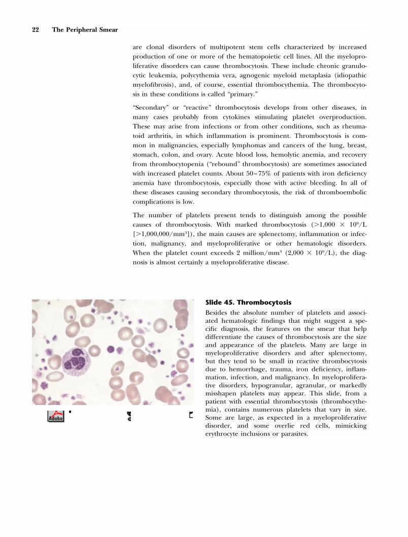
Slide 45. Thrombocytosis
Besides the absolute number of platelets and associ-ated hematologic findings that might suggest a spe-cific diagnosis, the features on the smear that helpdifferentiate the causes of thrombocytosis are the sizeand appearance of the platelets. Many are large inmyeloproliferative disorders and after splenectomy,but they tend to be small in reactive thrombocytosisdue to hemorrhage, trauma, iron deficiency, inflam-mation, infection, and malignancy. In myeloprolifera-tive disorders, hypogranular, agranular, or markedlymisshapen platelets may appear. This slide, from apatient with essential thrombocytosis (thrombocythe-mia), contains numerous platelets that vary in size.Some are large, as expected in a myeloproliferativedisorder, and some overlie red cells, mimickingerythrocyte inclusions or parasites.
40413 Tkachuk PM Gordon(SAUNM) INTERACTIVE LEFT
top of RHbase of RH
top of textbase of text
shortstandardlong

The Peripheral Smear 23
THROMBOCYTOPENIA
Thrombocytopenia is the presence of fewer than 150,000 platelets/mm3. Clin-ical signs related to thrombocytopenia rarely occur until the platelet count is�50,000/mm3 and become prominent primarily when it is �20,000/mm3.The findings relate to bleeding into the skin and from mucous membranes.Hemorrhage into the joints and soft tissue, common in hereditary disordersof coagulation such as hemophilia, is rare in thrombocytopenia. The usualcutaneous signs are nonpalpable purpura and petechiae, especially in depen-dent areas and areas subject to pressure or constriction, such as from theelastic bands of underwear. Bleeding may also be mucosal, including epistaxisand hemorrhage from the genitourinary and gastrointestinal tracts. Petechiaemay develop on the dorsum of the tongue, and small hemorrhagic cysts mayappear on its ventral surface.
A pseudothrombocytopenia occurs rarely (1 in 1,000 to 1 in 10,000 patients)with blood collected in tubes containing the calcium-chelating anticoagulantEDTA (ethylenediaminetetraacetate). In the presence of this substance, cer-tain antibodies cause platelets to form clumps or to aggregate around neutro-phils (satellitism), findings visible on a blood smear. With these physicalchanges, the automated platelet counters become inaccurate. Employing adifferent anticoagulant or using nonanticoagulated blood obtained from afingerstick will usually yield an accurate platelet count.
The causes of genuine thrombocytopenia can be classified into three generalgroups: (1) inadequate production, (2) increased destruction, and (3) abnor-mal distribution. Inadequate platelet production can occur from (a) bonemarrow hypoplasia caused by such factors as radiation, chemotherapy, ortoxins; (b) marrow infiltration by fibrosis, malignancy, or granulomas; (c)selective impairment of platelet production by drugs, infections, or ethanol;(d) ineffective thrombopoiesis because of myelodysplasia or deficiencies infolate or vitamin B12; and (e) hereditary diseases such as May-Hegglin anom-aly and Wiskott-Aldrich syndrome.
Increased destruction can occur from immune or nonimmune mechanisms.Immune-related thrombocytopenia occurs with (a) systemic lupus erythemato-sus; (b) lymphoproliferative disorders, such as chronic lymphocytic leukemia;(c) drug reactions, such as to quinidine; (d) infections, such as HIV andinfectious mononucleosis; (e) reactions following transfusions; and (f) idio-pathic immune thrombocytopenic purpura. Nonimmune causes include (a)severe bleeding; (b) disseminated intravascular coagulation; (c) abnormalitiesin the small vessels caused by such disorders as vasculitis, thrombotic throm-bocytopenic purpura, and hemolytic-uremic syndrome; and (d) infections.
Causes of abnormal distribution include (a) dilutional thrombocytopenia,seen following massive transfusion, and (b) hypersplenism, a condition in
40413 Tkachuk PM Gordon(SAUNM) INTERACTIVE RIGHT
top of RHbase of RH
top of textbase of text
shortstandardlong

24 The Peripheral Smear
which cytopenias—anemia, leukopenia, and thrombocytopenia, alone or inany combination—occur as a consequence of splenomegaly. Splenic size isthe major determinant of whether hypersplenism occurs; usually the spleen ispalpable on examination. The etiology of the splenomegaly is less important,and hypersplenism can develop with spleens enlarged by infections; inflam-matory diseases such as systemic lupus erythematosus; congestion from portalhypertension (as with hepatic cirrhosis); neoplasias, benign or malignant; andinfiltrative disorders such as sarcoidosis, amyloidosis, and Gaucher’s disease.In hypersplenism, the platelet count is usually �50,000/mm3.
Table 2. Causes of Thrombocytopenia
ArtifactualPlatelet clumping with EDTA anticoagulant
Inadequate productionBone marrow hypoplasia
RadiationChemotherapyToxins
Bone marrow infiltrationFibrosisMalignancyGranulomas
Selective impairment of platelet productionDrugsInfectionsEthanol
Ineffective thrombopoiesisFolate or B12 deficiency
Hereditary disordersMay-Hegglin anomalyWiskott-Aldrich syndrome
Increased destructionImmune related
Systemic lupus erythematosusLymphoproliferative disordersDrugsInfectionsTransfusionsIdiopathic immune thrombocytopenia
Nonimmune mechanismsSevere bleedingDisseminated intravascular coagulationAbnormalities in small vesselsVasculitisThrombotic thrombocytopenic purpuraHemolytic-uremic syndrome
Abnormal distributionDilutional, from massive transfusionHypersplenism
40413 Tkachuk PM Gordon(SAUNM) INTERACTIVE LEFT
top of RHbase of RH
top of textbase of text
shortstandardlong
Slide 46. Thrombocytopenia
With thrombocytopenia, fewer than about 7 plateletsare visible per oil immersion field with a 100� objec-tive. Associated abnormalities on the blood smear,such as red and white cell aberrations with myelodys-plastic syndromes, may suggest the cause, but other-wise the only helpful characteristic is platelet size. Ingeneral, platelets are large when thrombocytopeniaresults from increased destruction and small with dis-eases of diminished production. In this slide onlyone platelet is present. The schistocytes indicate thatthe cause is a microangiopathic process; in this case,the underlying problem was disseminated intravascu-lar coagulation associated with bacterial sepsis.

Artifacts on PeripheralSmears
Slide 47. Prolonged Storage
If a substantial delay occurs between the blood col-lection and smear preparation, the cell morphologymay change as the specimen remains in the tube.Excessive heat, dilution of the blood by intravenousfluids, or shaking the specimen also produces arti-facts. The subsequent smear can show erythrocytecrenation—the development of spikelike projectionson the red cell membrane—and degeneration of theneutrophils’ nuclei. In this slide, a band and a neu-trophil are present. The neutrophil is shrunken, cyto-plasmic granules have disappeared, and its nucleushas broken into numerous condensed fragments.Sometimes these degenerated neutrophils have singleor bilobed nuclei and resemble dysplastic cells ornucleated red cells.
40413 Tkachuk PM Gordon(SAUNM) INTERACTIVE RIGHT
top of RHbase of RH
top of textbase of text
shortstandardlong
Slide 48. Water Artifact
Water in the methanol solution used in the Wrightstain or moisture on the surface of the slide beforefixation shrinks and distorts the red cells, makingthem hypochromic, microcytic, and misshapen, oftenwith crenated borders. Several erythrocytes in thisslide resemble target cells. The presence of refractilematerial in them is the clue to a water artifact, whichis present in nearly all the erythrocytes in this slide.

Section 2BONEMARROWASPIRATE
40413 Tkachuk PM Gordon(SAUNM) INTERACTIVE RIGHT
top of RHbase of RH
SN base
shortstandardlong

40413 Tkachuk PM Gordon(SAUNM) INTERACTIVE LEFT
top of RHbase of RH
top of textbase of text
shortstandardlong

Examination of the BoneMarrow Aspirate
Examination of a bone marrow aspirate should begin at low power, usingthe 10� objective, to determine whether the specimen contains repre-
sentative cellular marrow particles that are adequately preserved and stained.The assessment should include a rough estimate of cellularity and megakaryo-cyte numbers. Using the 20� objective, the examiner should scrutinize thecellular composition, including the myeloid to erythroid (M :E) ratio, therelative numbers of mononuclear versus segmented cells, and the presence ofabnormal megakaryocyte forms. At this medium power, the normal aspiratesmear should reveal heterogeneous populations of cells, including prominentnumbers of maturing members of the myeloid series. A monotonous appear-ance is definitely abnormal and usually indicates neoplasia. Examinationshould also include a careful search for metastatic neoplastic cell populations,which may occur in clumps. The cytological detail of individual hematopoi-etic elements is best evaluated at high power (40� or 50� objective) in areasnear stromal fragments where the cells barely touch each other. The observershould scrutinize several hundred cells under high power and determine adifferential count to assess pathological increases in subpopulations such asblasts, plasma cells, lymphocytes, eosinophil and basophil precursors, mastcells, and nonhematopoietic cells. In addition, evaluation of erythroid andmyeloid precursors should include a search for megaloblastic and dysplasticfeatures. Smears stained for iron require examination under low and highmagnification, including oil, to assess storage iron within macrophages and todetect iron granules in erythroid precursors.
40413 Tkachuk PM Gordon(SAUNM) INTERACTIVE RIGHT
top of RHbase of RH
top of textbase of text
shortstandardlong
Evaluation of the Aspirate
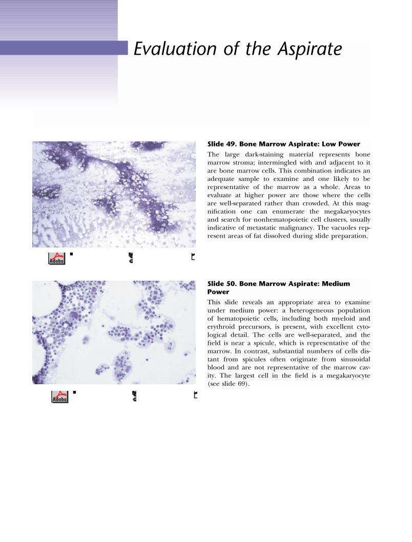
Slide 49. Bone Marrow Aspirate: Low Power
The large dark-staining material represents bonemarrow stroma; intermingled with and adjacent to itare bone marrow cells. This combination indicates anadequate sample to examine and one likely to berepresentative of the marrow as a whole. Areas toevaluate at higher power are those where the cellsare well-separated rather than crowded. At this mag-nification one can enumerate the megakaryocytesand search for nonhematopoietic cell clusters, usuallyindicative of metastatic malignancy. The vacuoles rep-resent areas of fat dissolved during slide preparation.
40413 Tkachuk PM Gordon(SAUNM) INTERACTIVE LEFT
top of RHbase of RH
top of textbase of text
shortstandardlong
Slide 50. Bone Marrow Aspirate: MediumPower
This slide reveals an appropriate area to examineunder medium power: a heterogeneous populationof hematopoietic cells, including both myeloid anderythroid precursors, is present, with excellent cyto-logical detail. The cells are well-separated, and thefield is near a spicule, which is representative of themarrow. In contrast, substantial numbers of cells dis-tant from spicules often originate from sinusoidalblood and are not representative of the marrow cav-ity. The largest cell in the field is a megakaryocyte(see slide 69).

Bone Marrow Aspirate 31
Slide 51. Bone Marrow Aspirate: Artifact
The process of slide preparation may “strip” the cyto-plasm from cells, leaving bare nuclei behind. In thisslide, the cytoplasm is absent from the cells in thecenter of the field. This artifact often gives the ap-pearance of increased numbers of monotonous cells,erroneously suggesting malignancy. Because accu-rately identifying cells is difficult, examination ofsuch areas is misleading.
Slide 52. Normal M:: E Ratio
Normally, the proportion of myeloid to erythroidprecursors (M :E ratio) is about 3 : 1. In this slide themarrow sample is appropriately cellular, lies near aspicule, has a normal M :E ratio, and demonstratesnormal myeloid maturation. The erythroid precursorsare small, round cells with closed chromatin, whereasthose in the myeloid line are larger, with more cyto-plasm. At one edge of the slide is a group of pig-ment-laden macrophages presumably containing iron,a normal finding (see slide 56).
40413 Tkachuk PM Gordon(SAUNM) INTERACTIVE RIGHT
top of RHbase of RH
top of textbase of text
shortstandardlong
Slide 53. Decreased M:: E Ratio
In this slide, the proportion of myeloid precursors issubstantially diminished relative to the erythroid se-ries, and the M :E ratio is about 1 : 1 because of in-creased erythroid cells. Erythroid hyperplasia can oc-cur as a response to anemia and, less frequently,from neoplastic disorders, such as polycythemia vera.

Iron in the Bone Marrow
Slide 54. Normal Iron Stores: Prussian BlueStain
A Prussian blue or Perls’ stain delineates hemosid-erin in erythroblasts and macrophages as blue-blackmaterial. This low-power view of an aspirate disclosesa moderate number of stained areas, indicating nor-mal iron in histiocytes, which tend to accumulate inthe center of spicules.
40413 Tkachuk PM Gordon(SAUNM) INTERACTIVE LEFT
top of RHbase of RH
top of textbase of text
shortstandardlong
Slide 55. Increased Iron Stores
This sample demonstrates increased amounts of blue-black-staining hemosiderin in bone marrow macro-phages. Increased iron stores are common followingmultiple transfusions and in the anemia of chronicdisease, many hemolytic anemias, hemochromatosis,alcoholism, and myelodysplastic disorders. In thiscase, the cause was iron overload from repeatedtransfusions.
Slide 56. Iron-laden Macrophage
The large cell in this Wright-stained bone marrowaspirate specimen contains numerous dark granules.This appearance suggests iron, an impression that
an
iron stain can confirm. These cells are normallypresent in the bone marrow, but a large number
of
them indicates increased bone marrow iron.

Erythropoiesis
surrounded by a rim of basophilic cytoplasm, with acharacteristic, single perinuclear “hof,” an area ofclearing that represents the Golgi apparatus. A singleprominent nucleolus is typically present, and the nu-clear chromatin is red-purple and finely granular or“open.” As the erythrocytes mature, the cell size di-minishes, the nucleoli disappear, the chromatin con-denses, and the color of the cytoplasm changes asred hemoglobin replaces blue-staining RNA. Thisslide shows the large size of the pronormoblast (longarrow), its dark blue cytoplasm, and the fine, diffusechromatin pattern. Nucleoli are visible in the nu-cleus. This specimen demonstrates the maturation ofred cell precursors, described more completely insubsequent slides. The smaller cell with blue cyto-plasm (medium arrow) is the basophilic erythroblast. Apolychromatophilic erythroblast (small arrow) and thetwo remaining nucleated cells (tiny arrows), which areorthochromatic erythroblasts, are also seen. The in-creasing maturation of the red cells, therefore, is in-dicated by the decreasing size of the arrows.
40413 Tkachuk PM Gordon(SAUNM) INTERACTIVE RIGHT
top of RHbase of RH
top of textbase of text
shortstandardlong
Slide 57. Proerythroblast (Pronormoblast)
The earliest recognizable erythrocyte precursor is aproerythroblast, a round or oval cell about 14–19 �m in diameter. Its large nucleus, which has aregular border, occupies about 80% of the cell and is
Slide 58. Basophilic Erythroblast
The proerythroblast differentiates into the basophilicerythroblast, a cell about 12–16 �m in diameter, inwhich nucleoli are absent and the chromatin appearscoarse and granular. In this slide the two cells arebasophilic erythroblasts. A small perinuclear clearspace (hof) lies beneath both nuclei.

34 Bone Marrow Aspirate
Slide 59. Polychromatophilic Erythroblast
The polychromatophilic erythroblast is 12–15 �m indiameter, and, compared to its predecessor, the baso-philic erythroblast, the nucleus is smaller, the chro-matin is condensed into irregular clumps, and thecytoplasm is polychromatophilic, that is, blue-gray.Pink areas from hemoglobin may be visible near thenucleus. In this example, a cluster of polychromato-philic erythroblasts appears in the center of the field.The presence of two adjacent polychromatophilicerythrocytes (the large purplish cells) and several ma-ture red cells allows a comparison of their respectivediameters.
Slide 60. Orthochromatic Erythroblast
The most mature form of the nucleated red cell, theorthochromatic erythroblast, is about 8–12 �m indiameter, contains pink cytoplasm, and has a smallnucleus with very condensed chromatin. Comparethe size of this orthochromatic erythroblast with thesurrounding normal mature red cells. This cell showssome mild nuclear : cytoplasmic dyssynchrony sugges-tive of megaloblastic changes. The nucleus should bepyknotic (dense and shrunken) at this stage.
40413 Tkachuk PM Gordon(SAUNM) INTERACTIVE LEFT
top of RHbase of RH
top of textbase of text
shortstandardlong
Bone Marrow Aspirate 35
ABNORMALITIES IN RED CELL MATURATION
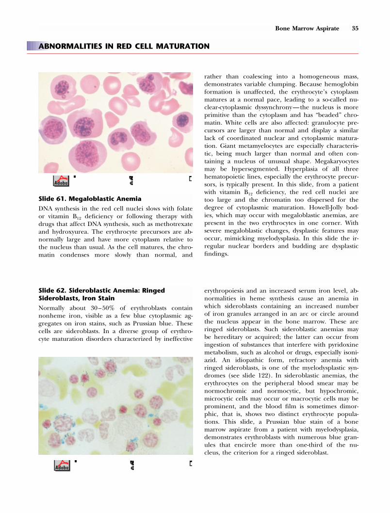
Slide 61. Megaloblastic Anemia
DNA synthesis in the red cell nuclei slows with folateor vitamin B12 deficiency or following therapy withdrugs that affect DNA synthesis, such as methotrexateand hydroxyurea. The erythrocyte precursors are ab-normally large and have more cytoplasm relative tothe nucleus than usual. As the cell matures, the chro-matin condenses more slowly than normal, and
40413 Tkachuk PM Gordon(SAUNM) INTERACTIVE RIGHT
top of RHbase of RH
top of textbase of text
shortstandardlong
rather than coalescing into a homogeneous mass,demonstrates variable clumping. Because hemoglobinformation is unaffected, the erythrocyte’s cytoplasmmatures at a normal pace, leading to a so-called nu-clear-cytoplasmic dyssynchrony—the nucleus is moreprimitive than the cytoplasm and has “beaded” chro-matin. White cells are also affected: granulocyte pre-cursors are larger than normal and display a similarlack of coordinated nuclear and cytoplasmic matura-tion. Giant metamyelocytes are especially characteris-tic, being much larger than normal and often con-taining a nucleus of unusual shape. Megakaryocytesmay be hypersegmented. Hyperplasia of all threehematopoietic lines, especially the erythrocyte precur-sors, is typically present. In this slide, from a patientwith vitamin B12 deficiency, the red cell nuclei aretoo large and the chromatin too dispersed for thedegree of cytoplasmic maturation. Howell-Jolly bod-ies, which may occur with megaloblastic anemias, arepresent in the two erythrocytes in one corner. Withsevere megaloblastic changes, dysplastic features mayoccur, mimicking myelodysplasia. In this slide the ir-regular nuclear borders and budding are dysplasticfindings.
Slide 62. Sideroblastic Anemia: RingedSideroblasts, Iron Stain
Normally about 30–50% of erythroblasts containnonheme iron, visible as a few blue cytoplasmic ag-gregates on iron stains, such as Prussian blue. Thesecells are sideroblasts. In a diverse group of erythro-cyte maturation disorders characterized by ineffective
erythropoiesis and an increased serum iron level, ab-normalities in heme synthesis cause an anemia inwhich sideroblasts containing an increased numberof iron granules arranged in an arc or circle aroundthe nucleus appear in the bone marrow. These areringed sideroblasts. Such sideroblastic anemias maybe hereditary or acquired; the latter can occur fromingestion of substances that interfere with pyridoxinemetabolism, such as alcohol or drugs, especially isoni-azid. An idiopathic form, refractory anemia withringed sideroblasts, is one of the myelodysplastic syn-dromes (see slide 122). In sideroblastic anemias, theerythrocytes on the peripheral blood smear may benormochromic and normocytic, but hypochromic,microcytic cells may occur or macrocytic cells may beprominent, and the blood film is sometimes dimor-phic, that is, shows two distinct erythrocyte popula-tions. This slide, a Prussian blue stain of a bonemarrow aspirate from a patient with myelodysplasia,demonstrates erythroblasts with numerous blue gran-ules that encircle more than one-third of the nu-cleus, the criterion for a ringed sideroblast.

Granulopoiesis
The earliest stages of the neutrophil line, the myeloblast and the promye-locyte, are also the precursors of the eosinophils and basophils. The
specific granules allowing differentiation among these three white cell typesbecome distinguishable with the next stage, the myelocyte. The neutrophilicmyelocyte matures into the metamyelocyte, which is the immediate predeces-sor of the band neutrophil. Neutrophils less mature than the band areusually confined to the bone marrow, but they may be present in the periph-eral blood in leukemias, in other disorders that disrupt the normal bonemarrow architecture, such as myelofibrosis or metastatic malignancy, andsometimes in severe infections.
Slide 63. Myeloblast
The most immature cell identifiable as a granulocyteprecursor is a myeloblast. About 10–20 �m in diame-ter, it has a large, reddish-purple, oval or round nu-cleus with finely dispersed or “open” chromatin andtwo to five prominent nucleoli. The narrow rim ofblue cytoplasm contains no granules. In this slide,three large nucleoli are readily visible. The adjacentred cells, about 8 �m in diameter, permit an esti-mate of the myeloblast’s size.
40413 Tkachuk PM Gordon(SAUNM) INTERACTIVE LEFT
top of RHbase of RH
top of textbase of text
shortstandardlong

Bone Marrow Aspirate 37
Slide 64. Promyelocyte
The promyelocyte is slightly larger than its predeces-sor, the myeloblast, and has a somewhat smaller nu-
cleus, typically located eccentrically in the cell andstill with two to five nucleoli, a darker blue cyto-plasm, and a Golgi zone, a clear area adjacent to thenucleus. The most striking difference from the my-eloblast is the presence of reddish-purple, “primary”granules in the cytoplasm. These large granules dis-appear during maturation, to be replaced by the spe-cific granules that allow distinction among the mye-loid cells—neutrophils, basophils, and eosinophils.Aside from the megakaryocyte, the promyelocyte isthe largest normal hematopoietic cell in the marrow.This slide demonstrates two promyelocytes, one show-ing especially well the nucleoli and purplish cytoplas-mic granules. The other promyelocyte is more ma-ture, characterized by fewer nucleoli, absent primarygranules, and a more central nucleus exhibitinggreater clumping of the chromatin.
40413 Tkachuk PM Gordon(SAUNM) INTERACTIVE RIGHT
top of RHbase of RH
top of textbase of text
shortstandardlong
Slide 65. Myelocyte
The myelocyte, smaller than the promyelocyte, oftenhas no nucleoli, possesses a less prominent Golgizone, and contains granules within its cytoplasm thatallow identification of the cell as belonging to theneutrophil, basophil, or eosinophil series. The nu-cleus is round to slightly oval, and the chromatinshows some coarse clumping. This slide demonstratesthree myelocytes of varying maturation, one retainingprimary granules, but all having specific granules thatare gray and finely dispersed.
Slide 66. Metamyelocyte
The metamyelocyte, about 10–12 �m in diameter,has an elliptical to horseshoe-shaped nucleus withmarkedly clumped chromatin and a plentiful, grayish-pink cytoplasm. A metamyelocyte with a U-shapednucleus differs from the next stage, a band form,whose nucleus is thinner and uniform in width. Thethree metamyelocytes in this slide reveal the indenta-tion of the nuclei, which show much more chromatinclumping than the myelocytes in the previous slide.

38 Bone Marrow Aspirate
ABNORMALITIES IN WHITE CELL MATURATION
Slide 67. Giant Band
In megaloblastic anemias (see slide 61) the whitecells, especially the metamyelocyte, may be largerthan normal. Bands can be enlarged as well, as indi-cated in this slide from a patient with folate defi-ciency. The normal erythrocyte (measuring about8 �m in diameter) near the two white cells allows asize comparison. The normal band is approximately12–15 �m in diameter, the normal metamyelocyteabout 10–12 �m.
40413 Tkachuk PM Gordon(SAUNM) INTERACTIVE LEFT
top of RHbase of RH
top of textbase of text
shortstandardlong
Slide 68. Maturation Arrest
In some situations, including severe congenital neu-tropenia and agranulocytosis from a chemical ormedication, the early white cell precursors arepresent but the more mature granulocytes are absent.This situation is called “maturation arrest,” but it ac-tually represents a left-shifted granulocytic responseto injury in the myeloid lineage. In this slide nomyelocytes, bands, or mature neutrophils are present.Most of the white cell precursors are promyelocytes.

Thrombopoiesis
Slide 69. Megakaryocyte
The biggest hematopoietic cell in the bone marrow(30–160 �m), the megakaryocyte has a large nucleuswith multiple but contiguous lobes and a basophilic,finely granular cytoplasm ranging from sparse toabundant, depending on the cell’s maturity. Themargins are irregular, often showing platelets bud-ding off the cytoplasm, which may contain azuro-philic granules. This slide demonstrates a megakaryo-cyte with a multilobulated nucleus and a finelygranular cytoplasm with knobby borders, represent-ing young platelets emerging from the megakaryo-cyte.
40413 Tkachuk PM Gordon(SAUNM) INTERACTIVE RIGHT
top of RHbase of RH
top of textbase of text
shortstandardlong
ABNORMALITIES IN PLATELET MATURATION
Slide 70. Small Megakaryocyte
Frequently in myelodysplastic syndromes and occa-sionally in myeloproliferative disorders, megakaryo-cytes are small, with a decrease in both the size ofthe nucleus and the cytoplasm, which has poor gran-ulation. This slide, from a patient with chronic gran-ulocytic leukemia, demonstrates two small megakaryo-cytes in opposite corners. Granulocytic hyperplasia ispresent, but maturation is normal, as evidenced bynumerous mature neutrophils.
Slide 71. Dysplastic Megakaryocytes
In myelodysplastic disorders, several abnormalities inthe megakaryocytes may occur. They may be small(see slide 70), and their nuclei may demonstrate de-creased lobulation, bizarre shapes, and the presenceof several separate, rather than contiguous, lobules.This slide, from a patient with myelodysplasia, revealsthree abnormal megakaryocytes, one with a singlenucleus and two containing clearly separated nuclearlobules. The smallest megakaryocyte is a micromega-karyocyte, characteristically equal in size to or smallerthan a promyelocyte.

Other Cells in the BoneMarrow Aspirate
Slide 72. Lymphocyte
In normal adults, lymphocytes constitute up to 20%of the nucleated cells in the bone marrow. They ap-pear as small cells with a rounded or slightly in-dented nuclei, surrounded by a thin rim of light bluecytoplasm. In this slide, short horizontal arrows identifyfour lymphocytes. Three late erythroid precursors inthis smear allow comparison between these cells andthe lymphocytes. The erythroid precursors (long hori-zontal arrows) have more abundant cytoplasm, andtheir nuclei are rounder, with more regular borders,and a more clumped chromatin. This slide also dem-onstrates some abnormal cells: three atypical plasmacells with prominent nucleoli (one with two nuclei),and a plasmacytoid lymphocyte (vertical arrow). Thepatient had Waldenstrom’s macroglobulinemia (seeslide 159).
40413 Tkachuk PM Gordon(SAUNM) INTERACTIVE LEFT
top of RHbase of RH
top of textbase of text
shortstandardlong
Slide 73. Mast Cell
Mast cells, normally present in the marrow, rangefrom about 5 to 25 �m in diameter. They containnumerous purple granules that do not totally obscurethe nucleus, as they often do in basophils. Further-more, the nucleus is not lobulated, as it is in baso-phils, and the chromatin is not so clumped. Bonemarrow mast cells are increased in lymphoprolifera-tive disorders, such as lymphomas and Waldenstrom’smacroglobulinemia, in myeloproliferative and mye-lodysplastic syndromes, in chronic liver and renal dis-eases, and in systemic mast cell disease, a neoplasticprocess that may range from an indolent disorder toa leukemic form. This slide demonstrates two mastcells. See also slides 187 and 188.

Bone Marrow Aspirate 41
Slide 74. Osteoclasts
This composite slide compares an osteoclast on theright with a megakaryocyte on the left. The osteoclastis involved in bone resorption. It is a large cell, rang-ing in diameter from 20 to 150 �m, with separate,multiple, round to oval nuclei of uniform size. Red-dish granules are present throughout its abundantbluish cytoplasm. In contrast to osteoclasts, normalmegakaryocytes have single nuclei that are lobulatedand contiguous, not multiple separate nuclei. Theircytoplasm does not contain reddish granules.
Slide 75. Osteoblasts
Osteoblasts form new bone. They are uncommon inadult bone marrow specimens, where they may ap-pear in small clusters. They occur in areas of boneremodeling following injury, such as fractures ormetastatic malignancy. They may mimic metastaticfoci or plasma cells because they resemble epithelialcells and tend to form cohesive clumps. They haveoval, eccentric nuclei with clumped chromatin. Thenuclei may appear to extend beyond the border ofthe abundant blue-gray cytoplasm, which has a cleararea (Golgi region) that is not directly adjacent tothe nucleus, as it is in plasma cells. This slide depictsa cohesive group of osteoblasts.
40413 Tkachuk PM Gordon(SAUNM) INTERACTIVE RIGHT
top of RHbase of RH
top of textbase of text
shortstandardlong

Abnormalities in Cell Type orNumber
Slide 76. Sea-Blue Histiocyte
Sea-blue histiocytes are bone marrow macrophageswith small nuclei and an enormous, often coarse,blue-green cytoplasm. They occur in diseases withhigh turnover of bone marrow cells because the mac-rophages ingest the lipids released during cell death.Examples include the myeloproliferative states, espe-cially chronic granulocytic leukemia; myelodysplasia;thalassemia; sickle cell anemia; and immune throm-bocytopenia purpura. They are also present inNiemann-Pick disease, in which an enzyme defectcauses excessive accumulation of phospholipids, pre-dominantly sphingomyelin, which bone marrow histi-ocytes consume. The large cell in the right centralarea of this slide demonstrates the size of the sea-blue histiocyte and its coarse aquamarine cytoplasm.The patient had chronic granulocytic leukemia, andgranulocytic hyperplasia is present.
40413 Tkachuk PM Gordon(SAUNM) INTERACTIVE LEFT
top of RHbase of RH
top of textbase of text
shortstandardlong
Slide 77. Granulocytic Hyperplasia
Ordinarily, the ratio of myeloid to erythroid precur-sors is about 3–5 : 1. In some conditions, includingsustained infection, chronic inflammation, leukemia,and response to certain drugs, the number of granu-locytes increases. In this slide, from a patient withchronic granulocytic leukemia, virtually all the cellsare myeloid, but maturation is normal.

Bone Marrow Aspirate 43
Slide 78. Erythroid Hyperplasia
In several situations, including hemolytic anemia,hemorrhage, and chronic hypoxemia, the number oferythroid precursors increases, but cellular matura-tion and morphology remain normal. In this slide,from a patient who had a severe hemorrhage, theusual myeloid-to-erythroid ratio is reversed, there be-ing about three times as many erythrocytes as whitecells.
Slide 79. Hemophagocytosis
Various disorders can provoke an increase in histio-cytes that ingest numerous bone marrow cells. Themost common causes have been infections, especiallywith the herpesviruses, including Epstein-Barr virus,Herpes simplex, and cytomegalovirus. It may occur inpatients with AIDS. Other infectious causes includebacteria, especially gram-negative bacilli; fungi; myco-bacteria; and rickettsiae. Usually, pancytopenia ispresent in the peripheral blood, and the bone mar-row shows increased benign-appearing macrophagesreplete with various cells. A familial form also occurs.In addition, hemophagocytosis may develop in cer-tain cancers, especially lymphomas, in which eitherthe ingesting cells or neighboring ones have malig-nant characteristics. In this slide, from a patient withinfection-induced hemophagocytosis, both red cellsand platelets stuff the macrophage’s cytoplasm.
40413 Tkachuk PM Gordon(SAUNM) INTERACTIVE RIGHT
top of RHbase of RH
top of textbase of text
shortstandardlong
Slide 80. Emperipolesis
Sometimes a cell enters another cell without beingdestroyed. This event can occur in the marrow whenvarious cells, including platelets, leukocytes, anderythrocytes, move into megakaryocytes. This phe-nomenon is most common in blood loss, hemolyticanemias, and several neoplasms. In this slide, a mega-karyocyte contains a granulocyte.

40413 Tkachuk PM Gordon(SAUNM) INTERACTIVE LEFT
top of RHbase of RH
top of textbase of text
shortstandardlong

Section 3BONEMARROWBIOPSY
40413 Tkachuk PM Gordon(SAUNM) INTERACTIVE RIGHT
top of RHbase of RH
SN base
shortstandardlong

40413 Tkachuk PM Gordon(SAUNM) INTERACTIVE LEFT
top of RHbase of RH
top of textbase of text
shortstandardlong
Examination of Bone MarrowBiopsy Sections
Whereas the aspirate smear reveals fine cytological details and thus pro-vides information about the maturation and lineage of individual hem-
atopoietic cells, biopsy specimens demonstrate overall cellularity and marrowarchitecture. The examiner should view the entire section at low power,preferably with a 4� objective, to evaluate changes in the bony trabeculaeand marrow elements. Structural abnormalities detectable at low power in-clude metastatic foci, granulomas, lymphomatous aggregates, necrosis, amy-loid deposition, and fibrosis. At this magnification, one can also determinethe location of immature myeloid precursors relative to the bony trabeculaeand the degree of maturation. Medium-power examination of the specimenpermits enumeration of megakaryocytes and an assessment of the relativeproportions of myeloid and erythroid precursors.
Special histochemical or immunochemical studies can be performed on bi-opsy sections. Histochemical stains, for example, may demonstrate infectiousorganisms such as fungi and acid-fast bacilli or help quantify the degree ofreticulin fibrosis present. Immunohistochemical stains help to determine phe-notypes of abnormal cells in the marrow cavity and are indispensable indiscerning whether an abnormal cellular infiltrate derives from marrow orrepresents metastatic malignancy. In addition, immunohistochemical studiesassist in elucidating whether abnormal B-cell lymphoid cell populations, in-cluding plasma cells, are clonal.
40413 Tkachuk PM Gordon(SAUNM) INTERACTIVE RIGHT
top of RHbase of RH
top of textbase of text
shortstandardlong

48 Bone Marrow Biopsy
Slide 81. Bone Marrow Biopsy: Low Power
This slide shows several bony trabeculae (the homo-geneous, pink-staining sections), which are normallythin, as here. The cellular portions of the marrowcontain the hematopoietic cells, and the clear spacesrepresent areas where fat has dissolved during slidepreparation. At birth, bone marrow specimens nor-mally show nearly complete cellularity, with very fewfat cells. With age, the cellularity diminishes, and theamount of fat increases. This low-power view allowsan estimate of marrow cellularity, which can be ex-pressed as the percentage of the space between tra-beculae occupied by cells. In this case, the estimatedcellularity is 30%, the expected amount for an olderadult.
Slide 82. Bone Marrow Biopsy: Medium Power
This slide of normal bone marrow shows a trabeculaat medium power, disclosing the presence of clearspaces (lacunae) in which osteocytes reside. The cel-lularity is normal and comprises a heterogeneouspopulation of myeloid and erythroid precursors. Theimmature myeloid precursors are characteristicallypresent next to the trabeculae (“paratrabecular”).
40413 Tkachuk PM Gordon(SAUNM) INTERACTIVE LEFT
top of RHbase of RH
top of textbase of text
shortstandardlong
Slide 83. Bone Marrow Biopsy: High Power
This specimen discloses a trabecula bordering theright edge of the slide. Adjacent to it are spindle-shaped cells, osteoblasts, and a large multinucleatedcell, likely an osteoclast. Ordinarily, as here, the nextzone contains immature myeloid precursors, whichare large cells with eosinophilic cytoplasm and a me-dium nuclear : cytoplasm ratio. Normally, a furtherstep away from the trabecula lie the erythroid precur-sors, characterized by round, regular, and central nu-clei containing closed chromatin and meager or nocytoplasm. This normal arrangement of cells is re-versed in myelodysplasia, where myeloid precursorsare isolated in islands away from their normal para-trabecular location, a situation known as abnormallocalization of immature precursors (ALIP).
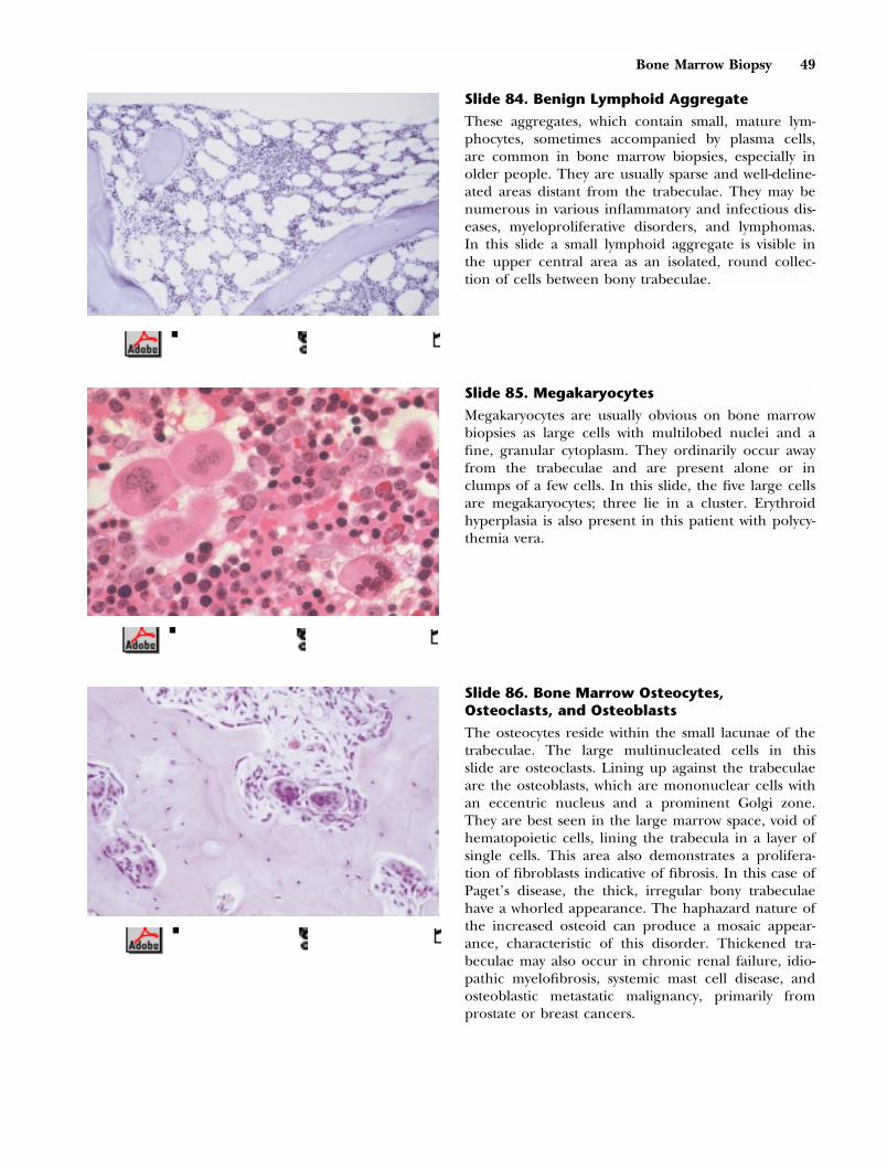
Bone Marrow Biopsy 49
Slide 84. Benign Lymphoid Aggregate
These aggregates, which contain small, mature lym-phocytes, sometimes accompanied by plasma cells,are common in bone marrow biopsies, especially inolder people. They are usually sparse and well-deline-ated areas distant from the trabeculae. They may benumerous in various inflammatory and infectious dis-eases, myeloproliferative disorders, and lymphomas.In this slide a small lymphoid aggregate is visible inthe upper central area as an isolated, round collec-tion of cells between bony trabeculae.
Slide 85. Megakaryocytes
Megakaryocytes are usually obvious on bone marrowbiopsies as large cells with multilobed nuclei and afine, granular cytoplasm. They ordinarily occur awayfrom the trabeculae and are present alone or inclumps of a few cells. In this slide, the five large cellsare megakaryocytes; three lie in a cluster. Erythroidhyperplasia is also present in this patient with polycy-themia vera.
40413 Tkachuk PM Gordon(SAUNM) INTERACTIVE RIGHT
top of RHbase of RH
top of textbase of text
shortstandardlong
Slide 86. Bone Marrow Osteocytes,Osteoclasts, and Osteoblasts
The osteocytes reside within the small lacunae of thetrabeculae. The large multinucleated cells in thisslide are osteoclasts. Lining up against the trabeculaeare the osteoblasts, which are mononuclear cells withan eccentric nucleus and a prominent Golgi zone.They are best seen in the large marrow space, void ofhematopoietic cells, lining the trabecula in a layer ofsingle cells. This area also demonstrates a prolifera-tion of fibroblasts indicative of fibrosis. In this case ofPaget’s disease, the thick, irregular bony trabeculaehave a whorled appearance. The haphazard nature ofthe increased osteoid can produce a mosaic appear-ance, characteristic of this disorder. Thickened tra-beculae may also occur in chronic renal failure, idio-pathic myelofibrosis, systemic mast cell disease, andosteoblastic metastatic malignancy, primarily fromprostate or breast cancers.

50 Bone Marrow Biopsy
Slide 87. Fibrosis
Bone marrow fibrosis refers to an increase in reticu-lin, collagen, or both, produced by fibroblasts, whichare normally present in the marrow. Reticulin is best
identified on silver stains (see next slide), while tri-chrome stains delineate collagen. A fine pattern ofreticulin is normal in the bone marrow, but collagenis not. Fibrosis is a common reaction in many neo-plasms, including chronic myeloproliferative disor-ders, various leukemias, mast cell disease, and meta-static malignancies. It may also develop fromradiation treatment, chemotherapy, and exposure tocertain toxins. It appears as amorphous material, inwhich the fibroblasts, with their elongated purplishnuclei, may be numerous or relatively sparse. Theorientation of these nuclei sometimes creates a pat-tern of whorls. This slide, from a patient with idio-pathic myelofibrosis, demonstrates a large number offibroblasts, which are the spindle-shaped cells; in theupper portion they create a swirling appearance. Onthe left, single cells line up in a row (“Indian file”),another sign of fibrosis.
40413 Tkachuk PM Gordon(SAUNM) INTERACTIVE LEFT
top of RHbase of RH
top of textbase of text
shortstandardlong
Slide 88. Reticulin Fibrosis
On silver stains of normal bone marrow, a fine,sparse network of reticulin surrounds blood vessels.Manifestations of increased reticulin include thicken-ing of the existing reticulin, more diffuse involve-ment of the bone marrow, and encirclement of indi-vidual hematopoietic cells. Increased reticulin fibrosisoften portends a poor prognosis in myelodysplasticand myeloproliferative disorders. This slide, a silverstained–specimen from a patient with polycythemiavera, demonstrates a central megakaryocyte and apattern of reticulin that is both coarser and moreextensive than normal.

Bone Marrow Biopsy 51
Slide 89. Bone Marrow Necrosis
Extensive death of bone marrow cells may occurfrom ischemia or increased levels of cytokines, such
as tumor necrosis factor. Many patients have feverand bone pain. With extensive marrow involvement,pancytopenia may develop and leukoerythroblastosisappear on the peripheral blood smear. Causes in-clude sickle cell anemia, leukemias, lymphomas,metastatic carcinomas, disseminated intravascular co-agulation, and various infections, such as gram-nega-tive bacteremia, miliary tuberculosis, and systemicfungal disease. Early on, the cell borders becomeindistinct (“ghost cells”), and the nuclei show con-densation and irregular margins. Later, the outlinesbetween cells disappear, replaced by an amorphousgranular eosinophilic debris, with nuclei either mark-edly abnormal or absent. In this slide, much of thespecimen exhibits amorphous pink granular material;where nuclei are visible, the cell outlines are blurred.Several empty lacunae in the trabeculae indicate thatnecrosis involved both the bone and the marrow.
40413 Tkachuk PM Gordon(SAUNM) INTERACTIVE RIGHT
top of RHbase of RH
top of textbase of text
shortstandardlong
Slide 90. Bone Marrow Granuloma
Granulomas are aggregates of altered macrophagescalled epithelioid cells, accompanied by various com-binations of other cells, including fibroblasts, lym-phocytes, plasma cells, neutrophils, and eosinophils.The epithelioid histiocytes have abundant pink cyto-plasm and large nuclei with dispersed chromatin.When several of these cells coalesce, they form giant
cells, which may be of two kinds: the Langhans type,with the nuclei positioned in the periphery of thecell, or the Touton type, with the nuclei dispersedthroughout the cell. The most common cause ofgranulomas is an infection, especially mycobacterialor fungal, but many other organisms can producethe same tissue reaction, including rickettsiae, gram-negative bacilli (e.g., typhoid fever and tularemia),and herpesviruses. The most common noninfectiouscauses are sarcoidosis, lymphomas, nonhematologicmalignancies, and various medications. Sometimes ca-seation necrosis, defined as the presence of pinkgranular material in the center of the granuloma,occurs. Usually this pattern indicates an infectiouscause, primarily mycobacteria or fungi, and its pres-ence warrants special stains to detect microorganisms.In this slide, from a patient with HIV infection andtuberculosis, an aggregate of epithelioid histiocytes,fibroblasts, and scattered lymphocytes lies in the cen-ter. In immunocompromised patients, the granulo-mas are often poorly delineated, and infections withorganisms like mycobacteria may elicit only an in-creased number of histiocytes that are diffusely dis-tributed or aggregated into loose collections.

Section 4SPECIFICDISORDERS
40413 Tkachuk PM Gordon(SAUNM) INTERACTIVE RIGHT
top of RHbase of RH
SN base
shortstandardlong

40413 Tkachuk PM Gordon(SAUNM) INTERACTIVE LEFT
top of RHbase of RH
top of textbase of text
shortstandardlong
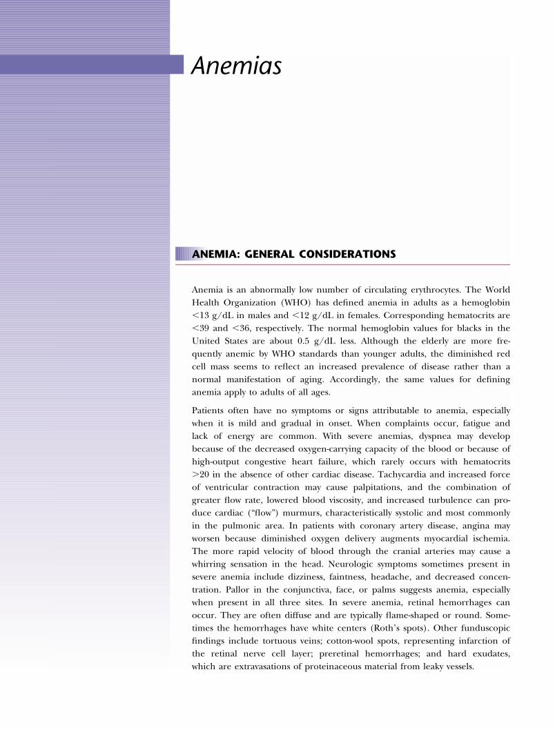
Anemias
ANEMIA: GENERAL CONSIDERATIONS
Anemia is an abnormally low number of circulating erythrocytes. The WorldHealth Organization (WHO) has defined anemia in adults as a hemoglobin�13 g/dL in males and �12 g/dL in females. Corresponding hematocrits are�39 and �36, respectively. The normal hemoglobin values for blacks in theUnited States are about 0.5 g/dL less. Although the elderly are more fre-quently anemic by WHO standards than younger adults, the diminished redcell mass seems to reflect an increased prevalence of disease rather than anormal manifestation of aging. Accordingly, the same values for defininganemia apply to adults of all ages.
Patients often have no symptoms or signs attributable to anemia, especiallywhen it is mild and gradual in onset. When complaints occur, fatigue andlack of energy are common. With severe anemias, dyspnea may developbecause of the decreased oxygen-carrying capacity of the blood or because ofhigh-output congestive heart failure, which rarely occurs with hematocrits�20 in the absence of other cardiac disease. Tachycardia and increased forceof ventricular contraction may cause palpitations, and the combination ofgreater flow rate, lowered blood viscosity, and increased turbulence can pro-duce cardiac (“flow”) murmurs, characteristically systolic and most commonlyin the pulmonic area. In patients with coronary artery disease, angina mayworsen because diminished oxygen delivery augments myocardial ischemia.The more rapid velocity of blood through the cranial arteries may cause awhirring sensation in the head. Neurologic symptoms sometimes present insevere anemia include dizziness, faintness, headache, and decreased concen-tration. Pallor in the conjunctiva, face, or palms suggests anemia, especiallywhen present in all three sites. In severe anemia, retinal hemorrhages canoccur. They are often diffuse and are typically flame-shaped or round. Some-times the hemorrhages have white centers (Roth’s spots). Other funduscopicfindings include tortuous veins; cotton-wool spots, representing infarction ofthe retinal nerve cell layer; preretinal hemorrhages; and hard exudates,which are extravasations of proteinaceous material from leaky vessels.
40413 Tkachuk PM Gordon(SAUNM) INTERACTIVE RIGHT
top of RHbase of RH
top of textbase of text
shortstandardlong

56 Specific Disorders
Anemia is not a diagnosis itself but a manifestation of an underlying disease,and its physiologic effects vary from individual to individual. The numerouscauses of anemia are diverse; generally, two different systems have been usedto classify them. One is by red cell morphology, the other by pathophysiology(or red cell kinetics). Each classification has three major subsets. The mor-phologic system divides anemias into (1) microcytic (MCV � 80), hypo-chromic (MCH � 30); (2) macrocytic (MCV � 100), normochromic (MCH30–34); and (3) normocytic (MCV 80–100), normochromic. With some dis-orders, the size of the red cell is quite variable, and therefore the samedisease may be classified in more than one category, depending on theindividual patient. For example, in the anemia of chronic disease the redcells may be either normocytic or microcytic, and in hypothyroidism they aresometimes macrocytic and sometimes normocytic.
Microcytic, hypochromic anemias are disorders of hemoglobin synthesis withimpairments resulting from inadequate iron, abnormal globin formation, orthe deficiencies in porphyrin and heme synthesis that occur in sideroblasticanemia. The most common cause of microcytic anemias is iron deficiency,usually from blood loss. Microcytosis occurs in about 20% of patients withanemia of chronic disease, in which part of the pathophysiology is impairedtransfer of iron to the plasma from its site of storage in macrophages. Exam-ples of impaired globin synthesis leading to microcytosis are the thalassemiasand certain hemoglobinopathies, such as hemoglobin C and E. Sideroblasticanemias causing microcytosis include those resulting from lead poisoning andpyridoxine deficiency.
Macrocytic anemias may arise from abnormal DNA synthesis, producing meg-aloblastic changes in the bone marrow. The major causes include deficienciesin vitamin B12 and folate. Macrocytic anemias may also occur from othermechanisms. With recent hemorrhage and with hemolytic anemias, red cellsleave the bone marrow at an earlier stage of development than normal, whenthey are larger than the usual circulating erythrocytes. These cells may stillcontain a nucleus or, more commonly, have some residual RNA, which stainsblue, along with the hemoglobin, which stains red. The result is a purple cellthat is larger than normal and is called a polychromatophilic (“lover of manycolors”) or polychromatic (“many-colored”) erythrocyte (see slide 19). Whena large number of these cells are present, the MCV can exceed 100. Anothercommon cause of macrocytosis is alcoholism, even in the absence of folate orvitamin B12 deficiency, apparently from a direct effect of ethanol on the bonemarrow. Usually, however, anemia is absent. Hypothyroidism, through un-known mechanisms, can cause a mild macrocytic anemia. In myelodysplasia, adisease primarily of the elderly, abnormal maturation of red cell precursorsoften produces macrocytosis.
Normocytic, normochromic anemias arise from several heterogeneous disor-ders. With hemorrhage or hemolysis the bone marrow may respond appropri-ately by increasing the number of early red cells. In the other forms ofnormocytic, normochromic anemia an inadequate bone marrow responseoccurs because of intrinsic bone marrow disease, diminished erythropoietin,
40413 Tkachuk PM Gordon(SAUNM) INTERACTIVE LEFT
top of RHbase of RH
top of textbase of text
shortstandardlong

Specific Disorders 57
or insufficient iron. Intrinsic bone marrow disease includes (1) hypoplasia, suchas in aplastic anemia or pure red cell aplasia; (2) infiltration by abnormaltissue, as in multiple myeloma, leukemia, fibrosis, or metastatic malignancy;and (3) myelodysplastic disorders, in which abnormal maturation occurs.Decreased erythropoietin can occur from (1) an impaired source, namely renalfailure; (2) reduced stimulation, thought to be the cause of anemia in variousendocrine disorders, such as hypothyroidism and testosterone deficiency; and(3) interference with its production by inflammatory cytokines, probably asignificant component in the pathogenesis of the anemia of chronic disease.In that disorder, other mechanisms include a slight decrease in red cell lifespan and diminished proliferation of erythroid progenitor cells, also attrib-uted to the effect of inflammatory cytokines. Insufficient iron may be a minorelement in the pathogenesis of the anemia of chronic disease as well, sincethe serum iron level is low despite adequate stores in the bone marrow. Earlyiron deficiency anemia is often normocytic, but as its severity increases, thered cell size diminishes.
A useful laboratory test in evaluating anemias is the reticulocyte count, whichenumerates the number of immature red cells that still contain residual RNA.The tests for reticulocyte count involve mixing red cells with a dye, such asbrilliant cresyl blue, which will stain the precipitated RNA, and counting thenumber of erythrocytes with blue granules or reticulum among 1,000 redcells. Automated methods utilize a chemical, such as thiazole orange, whichbinds to RNA and fluoresces, identifying these young cells. The count isexpressed as a percentage—the number of reticulocytes per 100 erythro-cytes—or in absolute numbers per volume. The use of the reticulocyte per-centage requires a correction for the severity of anemia, which is done bymultiplying the percentage by the patient’s hemoglobin (or hematocrit) di-vided by the normal hemoglobin (hematocrit). For example, if the initialreticulocyte count is 4% and the patient’s hematocrit is 25, the correctedvalue is 4% � (25/45) � 2.2%. A further correction is necessary if theanemia is severe and polychromasia is present on the peripheral bloodsmear. Ordinarily, the polychromatophilia of immature red cells lasts 4 days,3 of which are spent in the bone marrow. When these cells are releasedearly, the blue color may last for 2 or even 3 days in the circulating blood.For the reticulocyte percentage to reflect actual red cell production, it shouldbe divided by 2 if the hematocrit is 25 or lower when polychromatophilia isprominent. The reticulocyte percentage after these corrections is called thereticulocyte index. The absolute reticulocyte count or the reticulocyte indexis especially useful in distinguishing among the normocytic, normochromicanemias. Anemias with low reticulocyte counts occur from either decreasederythropoietin levels or impaired bone marrow response, and those withelevated reticulocyte counts have increased erythrocyte production in re-sponse to hemorrhage or hemolysis.
The reticulocyte count is an important element in the classification of ane-mias by red cell kinetics. In this scheme, anemias are (1) hypoproliferative, inwhich the bone marrow cannot increase its red cell production; (2) matura-
40413 Tkachuk PM Gordon(SAUNM) INTERACTIVE RIGHT
top of RHbase of RH
top of textbase of text
shortstandardlong

58 Specific Disorders
tion disorders, in which bone marrow erythroid hyperplasia occurs, but be-cause of defects in cytoplasmic or nuclear maturation, many red cells die inthe bone marrow, a situation labeled ineffective erythropoiesis; or (3) hemol-ysis, in which erythroid hyperplasia occurs and intact red cells leave the bonemarrow but are prematurely destroyed in the peripheral circulation. To cate-gorize an anemia, red cell production is determined by the reticulocyte indexand a bone marrow sample. In the absence of anemia, the reticulocyte indexis normally 1. With moderately severe anemia and a normal bone marrowresponse, the reticulocyte index should exceed 3. In a bone marrow sampleof a patient without anemia the normal erythroid :myeloid (E:M) ratio is 1:3.The expected response to moderately severe anemia is an erythroid hyperpla-sia that increases that ratio to �1:1. Measuring serum levels of indirectbilirubin and LDH help determine the presence of red cell destruction.These levels should increase whether the erythrocytes are destroyed in thebone marrow or in the peripheral circulation. As with the classification byred cell morphology, some anemias may have characteristics of more thanone category. In the anemia of renal disease, for example, the mechanismsresponsible include both hypoproliferation, because of decreased erythropoie-tin levels, and hemolysis, because red cell life span diminishes in a uremicenvironment. When more than one pathogenic element is present, the ane-mia is categorized by the predominant mechanism.
In hypoproliferative anemias the red cells are usually normocytic, normo-chromic, the reticulocyte index is �2, the E:M ratio is �1:2, and the indirectbilirubin is normal or low. Early iron deficiency anemia and the anemia ofchronic disease are hypoproliferative. Other causes include decreased eryth-ropoietin secretion (renal failure) or response (endocrine disorders, such ashypothyroidism), and bone marrow damage involving the stem cells (e.g.,chemotherapy, aplastic anemia) or marrow structure (metastatic malignan-cies, myelofibrosis), or through autoimmune or unknown mechanisms (e.g.,pure red cell aplasia, systemic lupus erythematosus). Polychromasia, reflectingerythropoietin stimulus, is present in marrow damage and iron deficiency,but absent or diminished with renal failure and inflammation.
With anemias from nuclear or cytoplasmic maturation disorders, the reticulo-cyte index is �2, the E:M ratio is �1:1 when the anemia is severe, and theindirect bilirubin and LDH levels are increased, except in iron deficiencyanemia. Polychromasia, representing erythropoietin stimulation, is present. Incytoplasmic disorders, defective hemoglobin production results in microcyticred cells. Examples are severe iron deficiency, the thalassemias, sideroblasticanemias, and some hemoglobinopathies. In nuclear maturation disorders thered cells are macrocytic, and the primary diseases are deficiencies in vitaminB12 and folic acid.
The other major cause of anemia is increased loss or destruction; blood lossusually accounts for the former and hemolysis the latter. In hemolytic ane-mias the red cells are normocytic or macrocytic (depending on the numberof large, immature erythrocytes present), polychromasia is prominent, thereticulocyte index is �3, the E:M ratio is �1:1, and serum LDH and indirect
40413 Tkachuk PM Gordon(SAUNM) INTERACTIVE LEFT
top of RHbase of RH
top of textbase of text
shortstandardlong

Specific Disorders 59
bilirubin levels are usually increased. With acute hemorrhage, the full re-sponse of the bone marrow does not appear for about 7–10 days. In the firstfew days the hematologic picture resembles a hypoproliferative process, witha reticulocyte index of 1 and an E:M ratio of 1:3. Polychromasia is present,but not prominent. Red cell progenitors increase shortly after the blood loss,but it takes time for the red cells to reach sufficient maturity to leave themarrow as young reticulocytes or even as nucleated red cells in the form ofpolychromatophilic or orthochromatic normoblasts. During this stage theresponse resembles ineffective erythropoiesis, with a reticulocyte index of 1–2and an E:M ratio, though increased, still �1:1. Polychromasia is more promi-nent than earlier. Finally, after 7–10 days the hematologic findings resemblea hemolytic anemia, with a reticulocyte index of �3 and an E:M ratio of 1:1or greater. Polychromasia is impressive at this stage. Because red cells are notundergoing increased destruction during this recovery period, however, theindirect bilirubin and LDH levels are normal.
These two ways of classifying anemia—by morphology or red cell kinetics—are not mutually exclusive. One useful approach is to use the red cell size inthe initial categorization. For macrocytic or microcytic anemias the differen-tial diagnosis is small. For the normochromic, normocytic anemias, however,further analyzing the hematologic information in terms of hypoproliferativeversus hemolytic or hemorrhagic is helpful in distinguishing among the manycauses.
Slide 91: Reticulocyte: New Methylene BlueStain
Because the erythrocyte’s life span is approximately120 days, about 0.8% of the red cells need to bereplaced daily by young cells released from the bonemarrow. These immature cells still contain ribosomalRNA that can be precipitated and stained by supravi-tal dyes, such as new methylene blue or brilliant cre-syl blue. The blood and stain are mixed together andallowed to sit for about 15 minutes before the filmsare prepared and air-dried. The cells containing bluegranules or reticulum are counted as a percentage oftotal red cells or as an absolute number per volume(mm3, L, or �L). Automated methods employ a sub-stance, such as thiazole orange, that binds to RNAand fluoresces. In this slide the four erythrocytesdemonstrating the finely dispersed, netlike aggregatesof precipitated blue RNA are reticulocytes.
40413 Tkachuk PM Gordon(SAUNM) INTERACTIVE RIGHT
top of RHbase of RH
top of textbase of text
shortstandardlong

60 Specific Disorders
Table 3. Causes of Microcytic Anemia
Iron deficiencyAnemia of chronic diseaseThalassemiasAbnormal hemoglobins (C, E)Sideroblastic anemias
Iron Deficiency Anemia
The most frequent cause of iron deficiency is chronic blood loss. In pre-menopausal women the source is usually menstruation. In men and in post-menopausal women, the usual cause is gastrointestinal hemorrhage. Lesscommon sources include hemoptysis, epistaxis, hematuria, and intrapulmon-ary hemorrhage from such disorders as Goodpasture’s syndrome or idio-pathic hemosiderosis. A rare cause is intravascular hemolysis with iron loss inthe urine in the form of ferritin, hemosiderin, and hemoglobin. This processmay occur in such disorders as paroxysmal nocturnal hemoglobinuria or redcell fragmentation from a prosthetic cardiac valve.
Iron deficiency anemia occasionally results from inadequate dietary intake.Most foods, including the majority of fruits and vegetables, provide littleabsorbable iron. Exceptions are meat, fish, poultry, beans, and peas. Sinceadult males have sparse iron losses, primarily a small amount in the stool,they need little dietary iron, and deficiency from an inadequate intake israre. When there is increased iron utilization, as in infants or during growth,or when there is concurrent blood loss, particularly in menstruating women,ordinary dietary intake may be inadequate, especially since women and youngchildren often consume less than the recommended minimum daily require-ment. An additional contributing factor in women is that during pregnancyiron is diverted to the fetus for hematopoiesis, and after delivery iron is lostthrough lactation.
Iron absorption can occur anywhere in the intestine, but it is most efficientin the duodenum. Occasionally, iron deficiency results from malabsorptiondue to disease of the small intestine or following gastric resection, which maycontribute to inadequate absorption by causing intestinal contents to travelmore rapidly through the duodenum or to bypass it by entering surgicallycreated anastomoses.
Mild iron deficiency anemia is commonly asymptomatic, but the clinical fea-tures of severe anemia may emerge as the hematocrit decreases. Two unusualfindings are pica—the bizarre craving to ingest certain unusual substances,such as clay, ice, dirt, or cardboard—and spooning of the nails (koil-onychia).
40413 Tkachuk PM Gordon(SAUNM) INTERACTIVE LEFT
top of RHbase of RH
top of textbase of text
shortstandardlong
MICROCYTIC ANEMIAS

Specific Disorders 61
In iron deficiency anemia, the serum iron level is decreased, the total iron-binding capacity increased, and the percent saturation diminished to lessthan 20%. In addition, the serum ferritin level is usually low, but, since thepresence of inflammation can elevate it, the concurrence of iron deficiencyanemia and an inflammatory process may result in a normal or nearly normalserum ferritin level. An examination of the blood smear and characteristicfindings in these serum studies usually allow a confident diagnosis of irondeficiency anemia to be made, but if these are confusing, a definitive diagno-sis can be made on iron stains of a bone marrow aspirate or on the basis of ahematologic response to a therapeutic trial of iron replacement.
flected by increased red cell distribution width(RDW) on automated counters. With increased sever-ity—hematocrits typically less than 36 in men, 30 inwomen—the MCV and MCHC diminish, and the ma-jor changes on the smear are anisocytosis, microcyto-sis, hypochromia, and poikilocytosis, including hy-pochromic, elongated, elliptical erythrocytes (“pencilcells”). Tiny microcytes and occasional target cellsmay be present, as may polychromatophilia. Baso-philic stippling is rarely found, and Pappenheimerbodies, which contain iron granules, are absent.Some normal-appearing red cells are common. Occa-sionally, mild leukopenia is present. In many casesthrombocytosis occurs, especially with active bleeding.In this slide, the red cells vary widely in shape andsize. Comparison with the lymphocyte reveals that theerythrocytes are microcytic. Their increased centralpallor indicates hypochromia. Several long, thin pen-cil cells and tiny microcytes are present. Thrombocy-tosis is apparent.
40413 Tkachuk PM Gordon(SAUNM) INTERACTIVE RIGHT
top of RHbase of RH
top of textbase of text
shortstandardlong
Slide 92. Iron Deficiency Anemia
When iron deficiency anemia is recent and mild, thesmear and erythrocyte indices (MCV, MCH, MCHC)may be normal. An early change is anisocytosis, re-
Thalassemias
In normal adults, over 90% of hemoglobin is Hb A, which consists of fourpolypeptide chains, two � and two � (�2�2). A small fraction is Hb A2,composed of two �-chains and two �-chains (�2�2). In fetal life the majorhemoglobin is Hb F (�2�2), the production of which decreases shortly beforebirth as formation of �-chains begins. At birth about 75% of the infant’shemoglobin is Hb F, but by 6 months it decreases to below 5%, and in adultlife it constitutes less than 1% of the total hemoglobin. The thalassemiasconsist of a group of inherited disorders with impaired synthesis of one ormore of the globin chains, leading to production of a defective hemoglobin,which causes microcytic hypochromic red cells. In addition, when one type of

62 Specific Disorders
polypeptide chain is absent or decreased, the other, unaffected chain is notbound and in its free form can damage red cells or their precursors. Thethalassemias are labeled according to the chain whose production is im-paired: �-thalassemia when �-chains are either absent or decreased, �-thalas-semia when the �-chains are affected. These are the two most importanttypes, although other forms exist (e.g., �-, �-, and ��-thalassemias).
40413 Tkachuk PM Gordon(SAUNM) INTERACTIVE LEFT
top of RHbase of RH
top of textbase of text
shortstandardlong
The �-thalassemias are common in the Mediterranean area, the Middle East,Southeast Asia, China, and parts of India and Pakistan. The clinical conse-quences of this disorder vary considerably, ranging from severe (thalassemiamajor) to moderate (thalassemia intermedia) to mild (thalassemia minor),depending on whether the inheritance is homozygous or heterozygous andwhether the abnormal gene leads to no �-chain production (�o) or a de-creased amount (��).
In homozygous disease, with little or no �-chain production and normal �-chain formation, an excess of �-chains develops. These are unable to com-bine to form a viable hemoglobin, and consequently precipitate in red cellprecursors, destroying many in the bone marrow (ineffective erythropoiesis).Red cells with these inclusions that do reach the circulation have a shortenedlife span, being prematurely destroyed by macrophages in the liver, spleen,and bone marrow. Because substantial amounts of Hb F are present at birth,affected infants are well until �-chain synthesis decreases and anemia beginsto appear. With sufficient transfusions to maintain relatively normal hemoglo-bin levels, children grow and develop normally until the iron overload prob-lems of repetitive transfusions emerge. Untreated or inadequately transfusedpatients have stunted growth. Increased erythropoiesis can produce systemicfeatures of hypermetabolism, such as fever, wasting, and hyperuricemia. Inaddition, expanded marrow cavities can cause fractures in long bones andcan enlarge the diameters of the skull and maxillary region, producing dis-torted head and facial appearances. Continued phagocytosis of abnormalerythrocytes causes splenomegaly, which can result in secondary thrombocyto-penia and leukopenia. In thalassemia major, the anemia is severe, and eryth-rocytes demonstrate anisocytosis and poikilocytosis, including elliptocytes,teardrop cells, and erythrocytes with other bizarre shapes. Hypochromia isvery impressive, and microcytosis is typically apparent, but the pallid cells areflat and spread out on drying, causing them to appear larger than their MCVwould suggest. Target cells are common, nucleated red cells are usuallynumerous, and basophilic stippling is prominent. Red cell inclusions havingthe same color as hemoglobin and representing excess �-chains may appearon Wright’s stain, especially after splenectomy, but are more obvious onmethyl violet stains. The bone marrow demonstrates erythroid hyperplasiawith basophilic stippling and decreased hemoglobin content of erythroblasts.Red cell inclusions are present in erythroid cells, and iron content is in-creased.
In thalassemia intermedia the disease is less severe, with a later onset, milderclinical features, and lower transfusion requirements. This disorder usually

Specific Disorders 63
develops from the inheritance of two �-thalassemia mutations, both mild orone mild and one severe. The blood film resembles that of thalassemiamajor, but the hematocrit is usually in the 20s.
The heterozygous state for �-thalassemia (thalassemia minor or thalassemiatrait) causes no clinical problems, and anemia is mild or absent, but theblood smear is abnormal. The cells are microcytic and hypochromic, with anMCV of about 50–70 and an MCH of 20–22. Target cells, poikilocytosis, andbasophilic stippling are typical. The marrow exhibits mild erythroid hyperpla-sia and rare red cell inclusions. Hemoglobin electrophoresis demonstrates HbA2 levels that are about twice normal (5%) and an Hb A2/Hb A ratio of 1:20,rather than the normal 1:40. Hb F is increased (�2%) in about one-half ofpatients.
Four gene loci govern �-globin chain production. The four �-thalassemias are�-thalassemia-2, in which one focus fails to function; �-thalassemia-1, with twoabnormal genes; Hb H disease, with three; and Hb Bart’s, with four. Nohematologic abnormalities occur with �-thalassemia-2, a silent, asymptomaticcarrier state. With �-thalassemia-1, a benign disorder found primarily in peo-ple of Asian, Mediterranean, and African descent, the hematologic findingsconsist of microcytosis, hypochromia, and slight anisocytosis and poikilocyto-sis. Anemia is absent or mild.
When three gene foci are defective, �-chain production diminishes markedly,and the excess �-chains form tetramers of Hb H, which, being soluble, doesnot precipitate in the marrow to damage erythroid precursors and causeineffective erythropoiesis. Instead, it remains in the mature circulating eryth-rocytes, but precipitates as they age, with the formation of inclusion bodies.These cells are prematurely destroyed in the spleen, causing hemolytic ane-mia. This disorder occurs predominantly in Asians, and occasionally in indi-viduals of Mediterranean or African descent. Splenomegaly and osseouschanges similar to those in �-thalassemia intermedia may occur; the anemia isusually moderate, with hematocrits of 20–30. The blood smear shows markedhypochromia, microcytosis, basophilic stippling, and polychromasia. Poikilocy-tosis is typical and may include target cells, teardrop cells, and fragmentederythrocytes, as well as nucleated red cells. Heinz body preparations showmultiple small erythrocyte inclusions, which are precipitated Hb H. On he-moglobin electrophoresis, about 3–30% of the total is Hb H.
With four genes defective, �-chains are absent, and �-chain tetramers, HbBart’s, form in the fetus. These carry oxygen poorly, causing tissue hypoxia,and are somewhat unstable, leading to hemolysis and anemia. This combina-tion causes heart and liver failure, resulting in massive edema (hydrops fe-talis) and intrauterine death. This disorder occurs almost exclusively inSoutheast Asians. Its importance in adult hematology is that a history ofhydrops fetalis in an Asian family suggests a form of �-thalassemia in theparents.
40413 Tkachuk PM Gordon(SAUNM) INTERACTIVE RIGHT
top of RHbase of RH
top of textbase of text
shortstandardlong
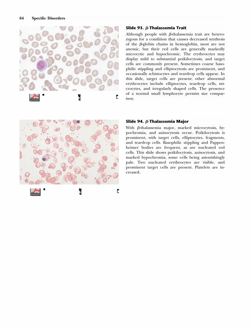
64 Specific Disorders
Slide 93. �-Thalassemia Trait
Although people with �-thalassemia trait are hetero-zygous for a condition that causes decreased synthesisof the �-globin chains in hemoglobin, most are notanemic, but their red cells are generally markedlymicrocytic and hypochromic. The erythrocytes maydisplay mild to substantial poikilocytosis, and targetcells are commonly present. Sometimes coarse baso-philic stippling and elliptocytosis are prominent, andoccasionally schistocytes and teardrop cells appear. Inthis slide, target cells are present; other abnormalerythrocytes include elliptocytes, teardrop cells, mi-crocytes, and irregularly shaped cells. The presenceof a normal small lymphocyte permits size compar-ison.
Slide 94. �-Thalassemia Major
With �-thalassemia major, marked microcytosis, hy-pochromia, and anisocytosis occur. Poikilocytosis isprominent, with target cells, elliptocytes, fragments,and teardrop cells. Basophilic stippling and Pappen-heimer bodies are frequent, as are nucleated redcells. This slide shows poikilocytosis, anisocytosis, andmarked hypochromia, some cells being astonishinglypale. Two nucleated erythrocytes are visible, andprominent target cells are present. Platelets are in-creased.
40413 Tkachuk PM Gordon(SAUNM) INTERACTIVE LEFT
top of RHbase of RH
top of textbase of text
shortstandardlong

Specific Disorders 65
MACROCYTIC ANEMIAS
Table 4. Causes of Macrocytic Anemias
Folate deficiencyB12 deficiencyHypothyroidismMyelodysplasiaMedications that impair DNA synthesisHemolysis/hemorrhage causing early bone marrow
release of young, large cellsLiver diseaseAlcoholism (macrocytosis without anemia)
40413 Tkachuk PM Gordon(SAUNM) INTERACTIVE RIGHT
top of RHbase of RH
top of textbase of text
shortstandardlong
Megaloblastic Anemias
In megaloblastic anemias, impaired DNA synthesis causes defective nuclearmaturation of hematopoietic cells in the bone marrow. In erythrocyte precur-sors, division of nuclei diminishes, while cytoplasmic growth, regulated byRNA, remains unimpeded, resulting in large cells with greater cytoplasm thannormal (megaloblasts). As these cells differentiate, the nuclear chromatincondenses more slowly than usual, giving a lacy appearance whose immaturitycontrasts with the increasing amount of hemoglobin in the cytoplasm, afeature described as nuclear-cytoplasmic dyssynchrony. In megaloblastic ane-mias, the cytoplasm is usually more mature than the nucleus, but sometimesa few cells demonstrate the reverse. Granulocyte precursors are also largerthan normal, and especially characteristic cells are the giant metamyelocytesand bands, which have a size two to three times normal, a shape that is oftenunusual, a nucleus with poorly condensed chromatin, and a vacuolated cyto-plasm. Megakaryocytes are hypersegmented and have a “megaloblastic” nu-clear chromatin. The bone marrow shows hypercellularity, but the commonpresence of anemia and a decreased reticulocyte count in the peripheralblood indicates that many of the erythrocytes, rather than reaching maturityand entering the systemic circulation, are destroyed in the bone marrow, acondition called ineffective erythropoiesis. This intramedullary hemolysiscauses increases in serum iron, unconjugated bilirubin, and LDH levels,which are further elevated by extramedullary hemolysis, since the red cell lifespan in the circulation is decreased by 30–50%.
Early in the course of disease, the only finding in the peripheral blood maybe red cell macrocytosis, usually between 110 and 130 fl, but sometimeshigher, especially as anemia develops and worsens. When the red cell countdiminishes, other abnormalities emerge, including anisocytosis, poikilocytosis,teardrop cells, schistocytes, basophilic stippling, and sometimes Howell-Jollybodies. Even in the presence of significant anemia, polychromatophilic cells
66 Specific Disorders
are absent or sparse, and the reticulocyte count is low. Leukopenia andthrombocytopenia may occur, particularly when anemia is severe. Two find-ings on a blood smear strongly suggest a megaloblastic process: some of themacrocytes are oval, a feature absent in nonmegaloblastic causes of macrocy-tosis, and polymorphonuclear leukocytes usually demonstrate hypersegmenta-tion, defined as �5% of neutrophils having five segments or any having sixor more. Sometimes circulating megaloblasts and giant platelets are visible.
The most common causes of megaloblastic anemia are deficiencies in vitaminB12 (cobalamin) and folate, whose hematologic features are indistinguishable.Some drugs can cause megaloblastic anemias by impairing DNA synthesis,including various cytotoxic agents used in cancer chemotherapy (such ascytosine arabinoside and cyclophosphamide), certain anticonvulsants (such asphenytoin), and medications interfering with folate metabolism (such asmethotrexate or trimethoprim). Rare causes are various inborn errors ofmetabolism.
Cobalamins are molecules with a central cobalt atom that are important inDNA synthesis. Strictly speaking, vitamin B12 is cyanocobalamin, a stable prod-uct used for therapy but not normally found in the human body. Often theterm vitamin B12, however, is loosely employed for all cobalamins. Microbespresent in the soil, water, or the intestinal tract of animals synthesize thesesubstances. They are not found in plants unless contaminated by microbes,and the dietary source for humans is thus meat, poultry, seafood, and dairyproducts. The average diet in Western countries contains 5–30 �g of cobala-min, the recommended daily allowance being 5 �g. The total body content isabout 2–5 mg in adults, about 1 mg of which is in the liver. Daily losses arevery small, and even with no intake, depletion sufficient to cause cobalamindeficiency takes years. Only strict vegetarians are likely to develop cobalamindeficiency on the basis of inadequate ingestion alone. Instead, cobalamindeficiency in adults almost always develops from impaired absorption. Nor-mally, cobalamin binds with substances in the gastric juice called R proteins.When this complex reaches the duodenum, pancreatic enzymes release thecobalamin, which then binds with intrinsic factor, a glycoprotein produced bythe parietal cells of the stomach. Intrinsic factor receptors are present on themucosa of the ileum, especially in its terminal portion, where cobalamin isnormally absorbed.
Diminished cobalamin absorption may occur in the ileum because of ilealresection or disease, such as radiation enteritis, lymphoma, or Crohn’s dis-ease. Cobalamin can be removed from the small intestine by a fish tapeworm,Diphyllobothrium latum, found primarily in Canada, Alaska, and the Baltic Seaand acquired by eating undercooked fish or fish roe. In small intestinaldisorders with markedly impaired motility, such as systemic sclerosis, or withanatomic lesions associated with intestinal stasis, such as diverticula and surgi-cal blind loops, bacteria can proliferate and consume cobalamin. Malabsorp-tion of cobalamin in the small intestine can also occur from pancreaticdisease when insufficient pancreatic enzymes are present to release cobalaminfrom the R protein binding it. The major cause of cobalamin deficiency,
40413 Tkachuk PM Gordon(SAUNM) INTERACTIVE LEFT
top of RHbase of RH
top of textbase of text
shortstandardlong

Specific Disorders 67
however, is an inadequate supply of intrinsic factor, either from surgicalresection of gastric parietal cells or, most commonly, from a disease causingchronic inflammation and mucosal atrophy in the fundus and body of thestomach, probably through immune mechanisms.
This disorder, pernicious anemia, occurs predominantly in older adults, witha median age at diagnosis of about 60. About 20% have a familial history ofthe disease, many are of northern European descent, and some have otherautoimmune conditions such as vitiligo, Hashimoto’s thyroiditis, and Graves’disease. About 90% of patients have serum antibodies to parietal cells, com-pared to about 5% in the general population, and approximately 60% haveserum antibodies to intrinsic factor, which is rare in healthy people. Histo-logic examination of gastric specimens demonstrates mucosal atrophy, mark-edly diminished parietal cells, and an infiltration of the submucosa andlamina propria by lymphocytes and plasma cells. As a result, gastric secretionis reduced, causing achlorhydria and decreased intrinsic factor production.The risk of gastric carcinoma in pernicious anemia is about 2–3 times that ofthe normal population.
The clinical features of cobalamin deficiency are diverse. Those due to ane-mia itself include pallor, fatigue, and dyspnea. Nonspecific gastrointestinalcomplaints are diarrhea, anorexia, and weight loss. Many patients have at-tacks of glossitis, with soreness and beefy redness, especially of the anteriorhalf of the tongue; when the episode resolves, papillae may be absent, caus-ing a smooth, glazed surface. Cobalamin deficiency causes impaired myelina-tion of nerves, leading to degeneration of white matter in the spinal cord(“subacute combined degeneration”) and cerebrum. Involvement of the dor-sal column of the spinal cord causes loss of vibratory sensation and proprio-ception, with patients complaining of numbness and tingling, initially in thefeet but later in the hands as well (stocking-glove distribution). Diminishedproprioception can cause difficulty with gait and a positive Romberg’s sign.Lateral column damage leads to limb weakness, spasticity, hyperactive re-flexes, and a positive Babinski’s sign. Depression and impaired memory arethe most common cerebral manifestations, but confusion, delusions, andhallucinations can also occur.
Serum cobalamin levels are low in most patients with pernicious anemia, butfalse positives and negatives occasionally occur. In those circumstances, otherstudies may help. One cobalamin-dependent enzyme, methylmalonyl CoAmutase, participates in the disposal of propionate generated during thebreakdown of valine and isoleucine. When inadequate cobalamin leads todiminished amounts of this enzyme, methylmalonic acid accumulates, andmeasurements of its serum level or urinary excretion are highly accurate indiagnosing cobalamin deficiency. A useful approach for the occasional pa-tient with confusing findings is a therapeutic trial of parenteral cobalamin. Abrisk reticulocytosis occurs 5–7 days later if cobalamin deficiency is present.
A Schilling test can determine the pathogenesis of cobalamin deficiency. Inhealthy people, orally administered radiolabeled cobalamin is absorbed from
40413 Tkachuk PM Gordon(SAUNM) INTERACTIVE RIGHT
top of RHbase of RH
top of textbase of text
shortstandardlong

68 Specific Disorders
the gastrointestinal tract and later excreted in the urine. In patients withpernicious anemia, the cobalamin is not absorbed normally, and therefore itsurinary excretion is decreased. When ingested with concurrent oral intrinsicfactor, however, the cobalamin is absorbed and excreted normally. Withother forms of intestinal malabsorption, urinary excretion remains low de-spite intrinsic factor. When bacterial overgrowth is the cause, the Schillingtest may become normal after a course of antibiotic therapy active againstintestinal bacteria.
Unlike cobalamin, folate is not present in high quantities in the body. Itsstore is estimated to be about 5–10 mg. Since the normal daily requirementis 50–100 �g, only a few weeks to months of decreased intake is necessarybefore deficiency develops and hematologic abnormalities emerge. The rich-est food sources are vegetables, liver, and fruit. Inadequate dietary ingestion,a common cause of folate deficiency, may occur because of poverty, igno-rance, food fads, and alcoholism. With excessive ethanol, further mechanismslead to folate deficiency: alcohol itself can increase urinary folate excretion,impede its storage in the liver, and diminish folate absorption, which occursprimarily in the duodenum and jejunum. Malabsorption of folate may alsodevelop in extensive disorders of the small bowel, such as sprue, Crohn’sdisease, lymphoma, amyloidosis, Whipple’s disease, and substantial intestinalresection. Deficiency is frequent when requirements for folate increase, as inpregnancy and conditions causing accelerated cell turnover, such as leuke-mia, acute exacerbations of chronic hemolytic anemia, multiple myeloma,and exfoliative dermatitis. Some medications, including anticonvulsants andsulfasalazine, can cause folate deficiency by altering its metabolism.
The symptoms of folate deficiency relate primarily to anemia. In contrast tocobalamin deficiency it rarely, if ever, causes neurologic damage. The mostuseful diagnostic test is determination of the serum folate level, which reflectsfolate intake. When folate ingestion first diminishes, however, the serum levelwill decrease before tissue stores are depleted, and when folate ingestionincreases, the serum level will rise before tissue stores are replaced. The redcell folate level, which remains relatively constant during the erythrocyte’s lifespan, is a better indicator of tissue folate status, but it is often normal whenthe folate deficiency is acute. Moreover, it is decreased in many patients withcobalamin deficiency and so cannot be used to accurately distinguish betweenthese two causes of megaloblastic anemia. Usually, concurrent serum levels ofcobalamin and folate will allow a reliable discrimination. Occasionally, how-ever, both will be decreased. The possible causes of this situation include (1)simultaneous deficiencies in both substances, a very unusual event; (2) cobal-amin deficiency with a recent decrease in folate intake but still adequatetissue stores; and (3) cobalamin deficiency with an unexplained decrease inserum folate levels, which occurs in some patients with cobalamin deficiencyand apparently normal folate intake. These possibilities can usually be sortedout by measuring serum or urine methylmalonic acid or by a therapeutic trialmonitoring a reticulocyte response following replacement with physiologicdoses of either cobalamin (100 �g daily) or folate (100–200 �g) given
40413 Tkachuk PM Gordon(SAUNM) INTERACTIVE LEFT
top of RHbase of RH
top of textbase of text
shortstandardlong

Specific Disorders 69
parenterally. Higher doses are misleading, since the hematologic findings ofcobalamin deficiency correct with large amounts of folate and those of folatedeficiency improve with higher doses of cobalamin. Misdiagnosis of cobala-min deficiency because of a hematologic improvement with folate therapy isa very serious error, since the neurologic abnormalities are unaffected andwill progress without cobalamin replacement.
Slide 95. Folate or Vitamin B12 Deficiency
These two deficiencies demonstrate identical appear-ances on the blood smear. The characteristic redcells are macrocytic, some of these are oval (macro-ovalocytes), and central pallor may be absent. Aniso-cytosis is always present, not all cells being macro-cytic; rather, a spectrum of size occurs, ranging fromvery large to microcytic. Poikilocytosis is prominentas well, with teardrops and fragments (schistocytes)common, especially with severe anemia. Basophilicstippling can occur, and nucleated red cells withlarge nuclei (megaloblasts) may be present. Neutro-philic hypersegmentation and macropolycytes (largehypersegmented neutrophils) are typically prominent.In this slide, a comparison of the erythrocytes withthe lymphocyte reveals that many of the red cells arelarger than normal, and some are oval. Anisocytosisand poikilocytosis are apparent, and two microcyticcells are located close to the lymphocyte.
40413 Tkachuk PM Gordon(SAUNM) INTERACTIVE RIGHT
top of RHbase of RH
top of textbase of text
shortstandardlong
Slide 96. Folate or Vitamin B12 Deficiency:Bone Marrow Aspirate
The bone marrow in these two deficiencies showshyperplasia and the characteristic pattern of megalo-blastic anemia (see slide 61). The erythroid cell situ-ated in the lower central area of this slide exhibitsboth dysplastic (irregular nucleus contours giving thenucleus a “pinched” appearance) and megaloblastic(fully matured cytoplasm with pink hemoglobin butstill retaining a nucleus—the so-called “nuclear-cyto-plasmic maturation dyssynchrony”) features. The bi-nucleated erythroid precursor in the right lower cor-ner, however, has a somewhat mature-appearingnucleus with clumped, closed chromatin, but the cy-toplasm displays immature basophilic staining. Thisfeature demonstrates that in some cells in megalo-blastic anemias, the nucleus matures more rapidlythan the cytoplasm. The three large upper white cellsare all giant granulocytes (two metamyelocytes and acentral band), and an erythrocyte in the center con-tains a Howell-Jolly body, sometimes present in mega-loblastic anemias.

70 Specific Disorders
HEMOLYTIC ANEMIAS
In hemolytic anemias the erythrocytes’ life span in the peripheral blood,normally about 120 days, substantially shortens because of red cell destruc-tion. Hemolytic anemias are generally categorized by two systems—the site ofhemolysis or the location of the abnormality responsible for the hemolysis,characterized as being either intrinsic or extrinsic to the red cell.
The site of hemolysis may be intravascular, in which case the erythrocyte isdestroyed in the circulation, or extravascular, in which case the red celldestruction occurs within macrophages in the spleen, liver, or bone marrow.Intravascular hemolysis is typically severe and results from (1) mechanicaldamage to the red cell due to prosthetic valves, the presence of fibrin withinthe vasculature (microangiopathic hemolytic anemia), or thermal injury tothe erythrocytes from serious burns; (2) infections or toxins, such as Clostrid-ium perfringens bacteremia, severe falciparum malaria, or certain snake venoms;or (3) complement-mediated damage to red cells, as with paroxysmal noctur-nal hemoglobinuria, ABO-incompatible blood transfusions, and cold aggluti-nins. Intravascular hemolysis liberates hemoglobin into the bloodstream,where it binds to haptoglobin, this complex being removed by the liver. Areduced serum haptoglobin level, therefore, is one finding in intravascularhemolysis, but it also occurs in extravascular hemolysis. When the amount offree hemoglobin in the circulation exceeds the binding capacity of haptoglo-bin, it makes the plasma pink and is filtered through the kidneys. The urinemay become red and the dipstick testing for blood turns positive in theabsence of erythrocytes on urine microscopy. The renal tubular cells, whichreabsorb some of the hemoglobin and convert it to hemosiderin, are shedinto the urine. Iron stains of urinary sediment demonstrate the hemosiderinwithin these renal tubular cells and confirm ongoing or recent intravascularhemolysis, even when hemoglobin is undetectable in the plasma or urine.
In the remaining hemolytic anemias, accounting for most cases, red celldestruction is extravascular. The causes are (1) an abnormal environment inthe circulation produced by infections, drugs, or immunologic processes; (2)red cell membrane defects, such as hereditary spherocytosis; (3) erythrocytemetabolic defects, such as deficiencies in pyruvate kinase or G6PD; and (4)hemoglobin structural defects, such as sickle cell anemia or hemoglobin C.
The other classification of hemolytic anemias distinguishes between disordersintrinsic to the red cell, generally hereditary, and those extrinsic to the redcell, generally acquired. The intrinsic disorders include (1) abnormal hemo-globins, such as HbS or HbC; (2) enzyme defects, such as deficiencies inG6PD; and (3) membrane abnormalities, such as hereditary spherocytosis orelliptocytosis. The extrinsic abnormalities are (1) immunologic—alloantibod-ies, such as those associated with ABO incompatibility; autoantibodies, as inwarm (IgG) or cold (IgM) antibody hemolytic anemias; or drug-induced anti-bodies; (2) mechanical factors, such as trauma from prosthetic valves or fibrin
40413 Tkachuk PM Gordon(SAUNM) INTERACTIVE LEFT
top of RHbase of RH
top of textbase of text
shortstandardlong

Specific Disorders 71
deposition in small vessels in microangiopathic hemolytic anemias; (3) infec-tions and toxins, such as falciparum malaria or certain snake venoms; and (4)severe hypophosphatemia.
In hemolytic anemias the reticulocyte index is �3 and the absolute reticulo-cyte count is �100,000/mm3. The MCV may be normal or increased, depend-ing on how many large, immature erythrocytes have prematurely left thebone marrow in response to the anemia. The indirect bilirubin is elevatedand accounts for �80% of the total bilirubin. The serum LDH level isincreased, and the serum haptoglobin is commonly diminished. A bloodsmear may be very helpful in demonstrating (1) polychromatophilia, confirm-ing the early departure of red cells from the bone marrow; (2) abnormalitiesin red cell shape, such as fragments, sickle cells, spherocytes, or bite cells,which may be diagnostic or at least highly suggestive of the cause; (3) redcell agglutination, indicating IgM-mediated disease; (4) organisms, such asPlasmodium falciparum or Babesia; and (5) erythrophagocytosis, seen especiallywith red cell damage from immune mechanisms, but also with certain infec-tion or toxins.
Tests useful in suspected intravascular hemolysis include evaluation of theplasma and urine for hemoglobin and an iron stain of the urine sediment todetect hemosiderin. For immune-related hemolytic anemia, Coombs’ tests todemonstrate IgG and complement on the red cell or in the serum and cold-agglutinin tests looking for IgM are indicated. With suspected hemoglobinop-athies, a hemoglobin electrophoresis is appropriate. Other laboratory evalua-tions depend on the likely abnormality and may include searching for rareenzyme defects.
Table 5. Causes of Hemolysis (by Site of Abnormality)
Intrinsic to the red cellAbnormal hemoglobins
Sickle cell anemiaHemoglobin C, E, etc.
Enzyme defectsGlucose-6-phosphate dehydrogenase deficiencyPyruvate kinase deficiency, etc.
Membrane abnormalitiesHereditary spherocytosis, elliptocytosisAcanthocytosisParoxysmal nocturnal hemoglobinuria
Extrinsic to the red cellImmunologic
Warm antibodyCold antibodyDrugs
MechanicalMarch hemoglobinuriaTraumatic cardiac hemolytic anemiaMicroangiopathic hemolytic anemia
InfectiousMalariaClostridium perfringens infection
ChemicalsHypersplenism
40413 Tkachuk PM Gordon(SAUNM) INTERACTIVE RIGHT
top of RHbase of RH
top of textbase of text
shortstandardlong

72 Specific Disorders
Table 6. Causes of Hemolysis (by Site of Destruction)
Intravascular hemolysisMechanical
March hemoglobinuriaCardiac hemolytic anemiaVasculitis
OsmoticDistilled water
Chemical or thermal damageAlpha toxin of Clostridium perfringensBurnsSnake venomsDrugs in patients with G6PD deficiency
Complement damageCold agglutininsParoxysmal nocturnal hemoglobinuriaIsoantibody plus complement
Extravascular hemolysisEnvironmental disorders
InfectionsDrug-inducedImmune-mediatedHemolytic-uremic syndrome
Membrane defectsHereditary spherocytosisHereditary elliptocytosisAcanthocytosisParoxysmal nocturnal hemoglobinuria
Metabolic defectsPhosphogluconate pathway (e.g., G6PD
deficiency)Embden-Meyerhof pathway (e.g., pyruvate kinase
deficiency)Abnormal hemoglobins
HemoglobinopathiesUnstable hemoglobins
Sickle Cell Anemia
Sickle cell anemia occurs with the inheritance of a �s gene from each parent.This gene, which encodes the �-globin subunit of hemoglobin, is most preva-lent in the populations of tropical Africa, especially in the eastern part, but italso occurs in people from Mediterranean countries, Saudi Arabia, and por-tions of India. In the United States, it is present in about 8% of blacks and,accordingly, sickle cell anemia (the homozygous state) occurs in about 1 of625 black infants.
Because of a single substitution of thymine for adenine in this gene, thehemoglobin produced differs from hemoglobin A in having valine, ratherthan glutamic acid, present in the sixth position from the N terminal of the�-chain. When deoxygenated, this abnormal hemoglobin aggregates intolarge fibers (polymers) that make the red cell rigid and can change its shapeinto the classic sickle cell. In addition, damage to the erythrocyte membraneoccurs, producing dehydrated, shrunken cells that may permanently sickle.These factors lead to chronic hemolysis. When the deoxygenated abnormalcells adhere to the vascular endothelium, they can obstruct the circulation,leading to ischemia or necrosis of areas that these vessels supply. Such vascu-lar occlusion accounts for most of the clinical features of this disease.
One manifestation is painful crises, which commonly last 4–5 days and mayinvolve any tissue but especially the skeleton, abdomen, and chest. Involve-ment of vessels to the bones causes a gnawing, progressive discomfort, espe-cially in the humerus, tibia, and femur, but also the ribs, causing thoracic
40413 Tkachuk PM Gordon(SAUNM) INTERACTIVE LEFT
top of RHbase of RH
top of textbase of text
shortstandardlong

Specific Disorders 73
pain. Sometimes vascular occlusion causes marrow necrosis and the release offat particles (emboli) that travel to the lung or other areas. Abdominal crisesprobably arise from infarcts to the mesentery and abdominal organs, includ-ing the spleen.
Some acute events can cause sudden worsening of anemia. In aplastic crisesfever occurs and red cell production markedly diminishes, resulting in arapid decrease in circulating erythrocytes as the chronic hemolysis continues.The most common cause is infection with parvovirus B19, which directlydamages the erythroid precursors. Patients typically recover in 5–10 days. Amegaloblastic crisis can also develop, in which folate deficiency from inade-quate nutrition or concurrent alcoholism decreases erythropoiesis.
Early in life, the dorsa of the hands and feet can swell (dactylitis), oftenaccompanied by fever and leukocytosis; the mechanism is probably avascularnecrosis of the bones. Young children may also experience episodes of acutesplenic sequestration, in which red cells abruptly accumulate in the spleen,causing pain, splenic enlargement, and increased anemia. As patients age, thespleen becomes shrunken, scarred, and nonfunctional because of repeatedinfarcts, and sequestration no longer occurs.
Another sudden event is the acute chest syndrome, characterized by fever,dyspnea, chest pain, leukocytosis, and pulmonary infiltrates. Several entitiescan cause this condition, including infections, pulmonary vascular occlusion,and fat emboli. In some cases the chest pain arises from rib infarcts.
Strokes may also occur, especially in children, and they tend to recur. Thepredominant cause is occlusion of the major cerebral vessels, but sometimesthey arise from subarachnoid or intracerebral hemorrhage.
Priapism can develop in both prepubertal and postpubertal males. Episodesare usually short-lived but often recurrent, and occur from stagnation ofblood in the corpora cavernosa. Repeated episodes may lead to fibrosis andimpotence.
With repetitive episodes or chronic ischemia, permanent tissue damage caneventuate in many other locations. Avascular necrosis of the femoral headcauses persistent pain and abnormal gait, and involvement of the humeralhead typically produces protracted shoulder discomfort. Ischemic or necroticbone predisposes to osteomyelitis, especially from Salmonella species. Damageto the kidneys can produce an inability to concentrate the urine normally(hyposthenuria) and impair excretion of hydrogen ion (renal tubular acido-sis). Hematuria may occur, sometimes associated with renal papillary necrosis,which also predisposes to urinary infections. Sloughing of the papillae canlead to urinary tract obstruction. Eye complications include proliferative reti-nopathy because of chronic ischemia, a cause of visual loss and vitreoushemorrhage. The abnormal vessels may bleed into the anterior chamber(hyphema). Persistent or recurrent painful ulcers sometimes form over thedistal leg, especially above the malleoli, usually in adulthood. They are more
40413 Tkachuk PM Gordon(SAUNM) INTERACTIVE RIGHT
top of RHbase of RH
top of textbase of text
shortstandardlong

74 Specific Disorders
common in males than in females and typically have a deep base with raisedmargins. Another common finding in older patients is bilirubin gallstones, acomplication of chronic hemolysis.
Sickle cell anemia predisposes to infections, not only because of the presenceof ischemic or necrotic tissues but also because of decreased splenic function,which impairs antibody production and splenic phagocytosis of organisms.The alternate complement pathway may also be faulty. In childhood the mostcommon serious infections are from Streptococcus pneumoniae.
The anemia is normochromic, normocytic, with a hematocrit typically about15–33. Mild leukocytosis (12–15,000), with increased neutrophils, andthrombocytosis (about 450,000) are common. The reticulocyte count is usu-ally 10–20%.
Slide 97. Sickle Cell Anemia
The blood film characteristically demonstrates thecrescent-shaped sickle cells, target cells, polychroma-sia, basophilic stippling, macrocytosis (from large im-mature erythrocytes released from the bone marrowin response to the anemia), and nucleated red cells.Except in very young children, Howell-Jolly and Pap-penheimer bodies, indicating splenic hypofunctionfrom recurrent infarctions and splenic fibrosis, areusually seen. Often visible are “boat-shaped cells,”noncrescentic erythrocytes with points at both ends.In this slide, two sickle cells are present. Comparisonof the red cells with the small lymphocyte indicatesthat many erythrocytes are enlarged, and some ofthese lack central pallor. The presence of macrocytes,polychromasia in several erythrocytes, and the nucle-ated red cells indicate early release of erythrocytesfrom the bone marrow secondary to a severe anemia.Two target cells lie below one of the sickle cells.
40413 Tkachuk PM Gordon(SAUNM) INTERACTIVE LEFT
top of RHbase of RH
top of textbase of text
shortstandardlong

Specific Disorders 75
Hemoglobin C Disease
Hemoglobin C occurs commonly in West Africans and in about 2–3% ofAfrican Americans. Patients may be homozygous (CC), heterozygous withnormal hemoglobin A (hemoglobin C trait), heterozygous with sickle cells(SC disease), or heterozygous with �-thalassemia. Red cells containing hemo-globin C are abnormally rigid, and their fragmentation may lead to micros-pherocytes. Their life span is shortened to about 30–35 days.
With hemoglobin C trait, anemia is absent. Blood smears show increasedtarget cells and sometimes hypochromic, microcytic erythrocytes. Electropho-resis typically demonstrates that 30–40% of the hemoglobin is C and 50–60% is A.
In homozygous (CC) disease, splenomegaly is usually present. Both aplasticcrises and cholelithiasis may occur. Mild to moderate hemolytic anemia ispresent, with hematocrits in the mid-20s to 30s and reticulocyte counts typi-cally 2–6%. The erythrocytes may be microcytic and are usually more densethan usual, as reflected in an increased MCHC. Target cells are abundant,and numerous small, dense, and irregularly shaped red cells are present.Crystals of oxygenated hemoglobin C are sometimes visible on peripheralsmears as brick-shaped objects, usually within an erythrocyte otherwise devoidof hemoglobin. Polychromatophilia and nucleated red cells, consistent withhemolysis, may be apparent. On electrophoresis, hemoglobin C predomi-nates, A is absent, and F is slightly increased.
With hemoglobin C/�-thalassemia, mild to moderate hemolytic anemia istypical. The blood smear shows microcytosis, hypochromia, target cells, andthe small, irregular erythrocytes described above. Hemoglobin C crystals maybe visible as well. Electrophoresis typically demonstrates 65–80% Hb C, 20–30% Hb A, and 2–5% Hb F.
Slide 98. Hemoglobin C Disease
In this specimen from a patient with hemoglobin Cdisease (homozygous for Hb C), the smear showsnumerous target cells and one erythrocyte containinga Hb C crystal, a densely red, blocklike rod.
40413 Tkachuk PM Gordon(SAUNM) INTERACTIVE RIGHT
top of RHbase of RH
top of textbase of text
shortstandardlong

76 Specific Disorders
Hemoglobin SC Disease
Although the red cells in SC disease sickle, causing vascular occlusion, thisdisorder is milder than sickle cell disease, and life expectancy is nearly nor-mal. It occurs in about 1 in 800 black infants in the United States. Growthand development are usually unaffected. Acute painful episodes (crises) areless frequent and typically shorter than in sickle cell disease. Aseptic necrosisof the humeral and femoral heads, however, may occur. The spleen is usuallyenlarged and, although susceptible to infarcts, does not become obliteratedas in sickle cell disease. Patients with SC disease may suddenly develop theacute splenic sequestration syndrome, especially when at high altitudes: redcells rapidly accumulate in the spleen, causing abdominal pain, progressivesplenic enlargement with tenderness, and abrupt decreases in hematocrit,sometimes producing shock.
Patients have a mildly enhanced susceptibility to infections, primarily withStreptococcus pneumoniae and Hemophilus influenzae. Spontaneous abortions areincreased, but the leg ulcerations and neurologic complications seen in sicklecell disease are uncommon. Proliferative retinopathy, however, is more fre-quent in SC than SS disease.
The hematocrit typically exceeds 27, and the red cells are usually normocytic,but denser than normal, with an increased MCHC characteristic of erythro-cytes containing Hb C. Target cells are numerous. “Billiard ball” cells, inwhich a round mass of hemoglobin appears in the erythrocyte with a clearspace separating the mass from the red cell membrane, are a characteristicfinding. The erythrocytes are often dense and disfigured, sometimes withbranching projections, and they may contain clusters of Hb C crystals ofvarious sizes and shapes, which create straight edges and blunt angles. Scan-ning electron microscopy demonstrates that these red cells have folds inthem. Sickle cells, boat-shaped cells, and nucleated red cells are other find-ings. Polychromasia may be present, and in those with functional asplenia,Howell-Jolly bodies are visible. Hemoglobin electrophoresis reveals aboutequal amounts of Hb C and Hb S.
Slide 99. Hemoglobin SC Disease
This slide from a patient with SC disease disclosesnumerous target cells, a few misshapen cells, andmoderate poikilocytosis. The nucleus of the smalllymphocyte in the corner allows comparison of theerythrocytes’ size.
40413 Tkachuk PM Gordon(SAUNM) INTERACTIVE LEFT
top of RHbase of RH
top of textbase of text
shortstandardlong

Specific Disorders 77
Red Cell Fragmentation Syndromes
Shear forces within the cardiovascular system can slice off portions of thecirculating red cells. The erythrocytes then re-seal their membranes to formfragmented cells (schistocytes). They may appear as triangles, helmet cells,microspherocytes, crescents, or other irregularly shaped erythrocytes thatphagocytes remove prematurely from the circulation. The amount of red celldestruction may be sufficient to cause intravascular hemolysis, with hemoglo-binemia, hemoglobinuria, and the presence of hemosiderin in the urine. Theerythrocyte damage can occur in the heart and great vessels or in the micro-circulation.
The most common cardiac etiology is the presence of a prosthetic valve,usually a mechanical device in the aortic position, often associated withregurgitation. Sometimes, however, the prosthetic valve is in the mitral posi-tion or is a tissue type. Occasionally, the abnormality is severe aortic stenosisof a native valve, coarctation of the aorta, or an intracardiac patch madefrom artificial material used to correct anomalies such as septal defects. Ineach of these situations, excessive turbulence damages the erythrocytes. Withchronic mild hemolysis, anemia is absent or slight, the red cells are normalor show some macrocytosis from the presence of immature cells, polychro-matophilia is present, and the reticulocyte count is elevated. With moresevere hemolysis, erythrocyte fragments (schistocytes) appear on the bloodsmear, the anemia is moderate to profound, the serum bilirubin and LDHlevels rise, haptoglobin decreases, and hemosiderin appears in the urine.With protracted intravascular hemolysis, urinary hemoglobin loss may causeiron deficiency, with hypochromic, microcytic red cells.
When diseases affecting the small vessels produce fragmented red cells, theterm used is microangiopathic hemolytic anemia. In these disorders, endothelialdamage or fibrin deposition in the small vessels damages the red cells as theytraverse the abnormal vasculature. One cause is disseminated cancer, usuallya mucin-producing neoplasm, especially gastric carcinoma, which accountsfor about 50% of cases. Most of the other malignancies producing microan-giopathic hemolytic anemia originate from the breast, lung, and pancreas.About one-third of patients have leukoerythroblastosis on the blood smear,about one-half have laboratory evidence of disseminated intravascular coagu-lation, and about 60% have tumor cells detectable on bone marrow speci-mens.
Some medications can cause microangiopathic hemolytic anemia, such asmitomycin C, ticlopidine, and tamoxifen. The disease usually begins severalweeks to months after initiation of the drug, renal failure and thrombocyto-penia are common, and laboratory evidence of disseminated intravascularcoagulation is absent. A similar syndrome may occur after solid organ orbone marrow transplantation, possibly precipitated by the preceding cytotoxicmedications and total body irradiation used for preparation of the transplant.
Other common causes of microangiopathic hemolytic anemia are thromboticthrombocytopenic purpura, hemolytic-uremic syndrome, malignant hyperten-
40413 Tkachuk PM Gordon(SAUNM) INTERACTIVE RIGHT
top of RHbase of RH
top of textbase of text
shortstandardlong

78 Specific Disorders
sion, disseminated intravascular coagulation, and severe hypertension duringpregnancy (preeclampsia or eclampsia). Immunologic damage to the vesselsthat occurs in such disorders as systemic lupus erythematosus, Wegener’sgranulomatosis, systemic sclerosis, and microscopic polyangiitis can cause mi-croangiopathic hemolytic anemia. It also may arise from the abnormal vascu-lature of a giant hemangioma (Kasabach-Merritt syndrome) or a hepatichemangioendothelioma.
Slide 100. Microangiopathic Hemolytic Anemia
The common findings in microangiopathic hemolyticanemia are numerous fragments, moderate anisocyto-sis, and poikilocytosis. This slide, from a patient withthrombotic thrombocytopenic purpura, showsmarked poikilocytosis with schistocytes, red cell frag-ments, and other irregularly shaped erythrocytes.Platelets are markedly diminished.
40413 Tkachuk PM Gordon(SAUNM) INTERACTIVE LEFT
top of RHbase of RH
top of textbase of text
shortstandardlong
Warm-Antibody Acquired Autoimmune Hemolytic Anemia
In patients with this disorder, the red cell life span decreases because autoan-tibodies, usually IgG, are optimally active against erythrocytes at body temper-ature (37�C). These warm antibodies account for about 80–90% of acquiredautoimmune hemolytic anemia, the remainder being due to those maximallyactive at lower temperatures (cold-reactive autoantibodies). In about one-halfof cases, an underlying disorder is present, most commonly a lymphoprolifer-ative disease, especially chronic lymphocytic leukemia and lymphomas, butalso systemic lupus erythematosus, other inflammatory conditions, some infec-tions, and nonlymphoid malignancies. Certain drugs, such as L-dopa andprocainamide, may also cause an autoimmune hemolytic anemia.
In this disorder, IgG, especially IgG1, coats many red cells with or withoutcomplement. Macrophages in the spleen and Kupffer cells in the liver trapthese erythrocytes, sometimes ingesting them whole. More commonly, theyremove a portion of the membrane, and the surviving red cell re-forms as aspherical, rather than biconcave, cell with a smaller diameter. These sphero-cytes are prominent on a peripheral blood smear, which also discloses evi-dence of polychromatophilia, indicating the release of immature red cellsfrom the marrow. Erythrocyte fragments, nucleated red cells, and hemophag-

Specific Disorders 79
ocytosis by monocytes may also be visible. In addition to anemia, the auto-mated blood count often reveals an increased MCHC, reflecting the presenceof the spherocytes. The reticulocyte count is usually increased, as are theserum indirect bilirubin and LDH levels. Immature white cells occasionallyappear on the peripheral smear. A bone marrow sample, although usuallyunnecessary for the diagnosis, shows erythroid hyperplasia. In Evans’ syn-drome immune thrombocytopenia is also present.
The direct antiglobulin (Coombs’) test detects the presence of antibodiesand complement on erythrocytes by using a reagent that contains antibodiesdirected against human immunoglobulin and complement components (pri-marily C3). This test is nearly always positive in this disorder, but when IgG ispresent in very small quantities, other diagnostic techniques may be neces-sary. Autoantibodies unattached to erythrocytes may be present in the serumand are detectable by incubating the patients’ serum or plasma with normalred cells, to which the antibodies then attach. These erythrocytes are thentested for the presence of autoantibodies with the Coombs’ reagent. This isthe indirect antiglobulin or Coombs’ test.
Slide 101. Warm-Antibody Hemolytic Anemia
This slide demonstrates several spherocytes, whichhave diminished or absent central pallor and aresmaller than the nucleus of the small lymphocyte.Several large, polychromatophilic red cells lackingcentral pallor are present, indicating release of imma-ture erythrocytes in response to the anemia.
40413 Tkachuk PM Gordon(SAUNM) INTERACTIVE RIGHT
top of RHbase of RH
top of textbase of text
shortstandardlong

80 Specific Disorders
Cold-Agglutinin Disease
Cold agglutinins are IgM antierythrocyte antibodies that bind red cells at coldtemperatures. They may be polyclonal or monoclonal. Nearly all healthypeople have low titers of clinically insignificant polyclonal cold agglutinins. Incertain infections, transient, high titers of polyclonal cold agglutinins appear,sometimes causing the abrupt onset of an anemia that is short-lived, butoccasionally severe. Infectious mononucleosis and infections with Mycoplasmapneumoniae are the two most common causes; the target of the antibodies inthe former is typically the i antigen found on the red cell membrane of fetalerythrocytes, while that in the latter is the I antigen found on adult erythro-cytes.
The chronic form of cold agglutinin disease, in which a monoclonal antibodyappears, typically occurs in older adults, and many have an underlying B-cellneoplasm, such as Waldenstrom’s macroglobulinemia, chronic lymphocyticleukemia, or non-Hodgkin’s lymphoma. Exposure to cold may precipitateattacks of acrocyanosis created by agglutination of erythrocytes in cool pe-ripheral areas such as the fingers, toes, nose, and earlobes. It may also causeworsening anemia as the temperature-dependent IgM antibody activates thecomplement pathway on the red cell membrane, producing intravascularhemolysis, sometimes with sufficient hemoglobinuria to cause acute renalfailure. Ordinarily, however, the anemia is mild and stable because the anti-body binds to erythrocytes only at temperatures below 37�C; because hepaticmacrophages do not avidly phagocytize red cells coated with Cb3, the usualcomplement component on the membrane; and because the serum Cb3inactivator system degrades Cb3 into components even less tempting to themacrophages.
Slide 102. Cold-Agglutinin Hemolytic Anemia
In cold-agglutinin hemolytic anemia the findings onperipheral smear include polychromatophilia, sphero-cytosis, and, sometimes, erythrocyte agglutination.This slide reveals agglutinated red cells in the centerand several spherocytes in the periphery.
40413 Tkachuk PM Gordon(SAUNM) INTERACTIVE LEFT
top of RHbase of RH
top of textbase of text
shortstandardlong

Specific Disorders 81
Slide 103. Spur-Cell Anemia
Some patients with severe liver disease, usually alco-holic cirrhosis, develop a hemolytic anemia associatedwith numerous acanthocytes (spur cells) in the pe-ripheral blood. These form from changes in the cho-lesterol to phospholipid ratio in the erythrocytemembrane and subsequent alterations produced bypassage through the spleen, which destroys many ofthe cells. The anemia may be profound. This slide,from a patient with severe alcoholic liver disease,shows several acanthocytes and target cells. Plateletsare also reduced, probably secondary to alcohol orhypersplenism from portal hypertension.
Slide 104. Heinz-Body Anemia: Heinz-BodyPreparation
Heinz bodies are fragments of denatured hemoglo-bin that usually bind to the red cell membrane. Theymost often occur from oxidative destruction of hemo-globin caused by a medication, especially in patientswith G6PD deficiency. These particles are not visibleon Wright’s stain but are detectable after mixing theerythrocytes with certain basic dyes, such as crystalviolet, before making the smear. This slide demon-strates the large, dark-staining inclusions, usually atthe cell margins, that are typical of Heinz bodies.Some patients with drug-induced Heinz-body hemo-lytic anemia but without G6PD deficiency have char-acteristic red cell abnormalities present on theWright stain of the peripheral blood, including bitecells (see slide 11) and “blister cells,” which havemembrane-bound vesicles at the erythrocyte margin.
40413 Tkachuk PM Gordon(SAUNM) INTERACTIVE RIGHT
top of RHbase of RH
top of textbase of text
shortstandardlong

82 Specific Disorders
Table 7. Causes of Hypoproliferative Anemia
Early iron deficiencyAnemia of chronic diseaseRenal diseaseEndocrine disorders
Adrenal insufficiencyHypothyroidismHyperthyroidismDecreased testosteronePanhypopituitarism
Bone marrow diseaseRed cell aplasiaAplastic anemiaMyelodysplasiaBone marrow infiltration by malignancy, fibrosis,
or granulomasMyeloproliferative syndromes
Anemia of Chronic Disease
In certain disorders that persist for longer than 1–2 months, especially thosewith inflammation, infection, and neoplasia, an anemia may develop in whichthe serum iron and iron-binding capacity are low and the transferrin satura-tion is diminished, yet the bone marrow iron stores are adequate. Insufficientiron supply to the developing red cells may be a factor in the pathogenesis ofthe anemia, as may a mildly shortened life span, but the major elementapparently is the reduced erythropoietin production and diminished prolifer-ation of erythroid progenitor cells caused by inflammatory cytokines. Inabout 25% of patients with this anemia, the only chronic disease present isnot obviously inflammatory, infectious, or neoplastic but is a disorder such asdiabetes mellitus, congestive heart failure, chronic obstructive lung disease, orcardiac ischemia.
Usually the anemia is mild to moderate, with the hematocrit �25, but inabout 20% of patients it is lower, and in general, the degree of anemiacorresponds with the severity of the underlying disorder. Typically the redcells are normochromic, normocytic. In about 20–30%, however, they arehypochromic (MCHC 26–32) and microcytic. The MCV is rarely �70 fl, andsometimes the erythrocytes demonstrate hypochromia without microcytosis.Overall, hypochromia is present in 40–70% of patients. The smear maydisclose mild poikilocytosis and anisocytosis, but these features are less im-pressive than in iron deficiency, and markedly small and thin cells are absent.Reticulocytes are normal or reduced in number. While the serum iron level islow, as in iron deficiency, the iron-binding capacity is also diminished,
40413 Tkachuk PM Gordon(SAUNM) INTERACTIVE LEFT
top of RHbase of RH
top of textbase of text
shortstandardlong
HYPOPROLIFERATIVE ANEMIAS

Specific Disorders 83
whereas it is usually elevated in iron deficiency. The transferrin saturationmay be decreased, even to values less than 10%. The most useful serum testis the ferritin level, which is diminished in iron deficiency but increased inthe anemia of chronic disease. In mixed-etiology anemia, overlap of these twotypes of anemias often occurs, however, because inflammatory diseases raisethe ferritin level. A value of �30 �g/L in a patient with a chronic diseasecertainly indicates iron deficiency, and one �100 excludes it, but levels inbetween are inconclusive. Definitive information in those circumstances cancome only from bone marrow samples stained for iron.
Slide 105. Anemia of Chronic Disease
This smear shows mild microcytosis with normo-chromic erythrocytes and slight anisocytosis.
40413 Tkachuk PM Gordon(SAUNM) INTERACTIVE RIGHT
top of RHbase of RH
top of textbase of text
shortstandardlong
Pure Red Cell Aplasia
Pure red cell aplasia is a normochromic, normocytic anemia with a very lowreticulocyte count (�1%) and a bone marrow demonstrating normal myeloidcells and megakaryocytes, but a dearth of erythroblasts. It can occur as acongenital disorder (Blackfan-Diamond syndrome) or as an acquired diseasethat is primary or secondary to medications, neoplasms, immunologic disor-ders, or infections. In primary acquired pure red cell aplasia the process mayarise from an IgG antibody that inhibits erythropoiesis, and the mechanismin some of the secondary causes is also immunologic. With drugs, the processmay occur either shortly after initiation of treatment or considerably later,and it typically resolves quickly after discontinuing the medication. The mostcommonly incriminated agents have been phenytoin, azathioprine, chlorpro-pamide, and isoniazid, but many others are occasionally responsible. Theneoplasms most frequently associated with pure red cell aplasia are thymomasand chronic lymphocytic leukemia, in both of which the frequency is about5–10%. A few cases have occurred with carcinomas, including those of thelung, kidney, stomach, and breast. The infectious causes are primarily viral,especially with parvovirus B19, infectious mononucleosis, and viral hepatitis;usually the anemia abates when the acute infection resolves. Because red cellsnormally survive for about 120 days, infection-induced aplasia, typically short-

84 Specific Disorders
lived, is usually clinically inapparent except in chronic hemolytic anemias,such as hereditary spherocytosis or sickle cell disease, in which even a briefcessation of erythrocyte production causes a substantial drop in hematocrit.Pure red cell aplasia may also complicate autoimmune diseases, such assystemic lupus erythematosus, rheumatoid arthritis, and Sjogren’s syndrome.
Slide 106. Pure Red Cell Aplasia
In adults, the peripheral blood smear shows a nor-mochromic, normocytic anemia and, despite a dimin-
40413 Tkachuk PM Gordon(SAUNM) INTERACTIVE LEFT
top of RHbase of RH
top of textbase of text
shortstandardlong
ished hematocrit, the absence of polychromatophilia,indicating deficient release of young cells. The reticu-locyte count is extremely low, but the erythropoietinlevel is appropriately increased for the severity of theanemia. The other cell lines are normal. The bonemarrow discloses profound erythroid hypoplasia butnormal numbers and maturation of platelet andwhite cell precursors. In parvovirus infection, giantproerythroblasts with prominent nucleoli may be ap-parent (see slide 169), and in bone marrow biopsieserythroblasts and erythroid islands are sparse, al-though proerythroblasts may be numerous. Thisslide, from a patient with a hematocrit of 7 (!), showsnormochromic, normocytic erythrocytes but no poly-chromatophilia, despite the very low level of circulat-ing red cells. The wide spaces between erythrocytesin most of the smear suggest severe anemia.
Slide 107. Anemia with Dimorphism
Dimorphism is the presence of two distinct erythro-cyte populations of differing sizes, a feature usuallyappreciated best on low-power examination. Causesinclude transfusion of normal cells into patients withmicrocytic or macrocytic anemias, combined iron de-ficiency and B12 or folate deficiency, myelodysplasticsyndromes, sideroblastic anemias, and treatment ofiron deficiency anemia, in which older microcyticcells coexist with younger normocytic cells. In thisslide, from an alcoholic patient with iron deficiencyanemia who received a transfusion, normochromic,normocytic erythrocytes intermingle with microcytic,hypochromic cells. A few target and elliptical redcells are present.
ANEMIA WITH DIMORPHISM

Stem-Cell Disorders
APLASTIC ANEMIA
Aplastic anemia refers to bone marrow failure from decreased hematopoieticelements, resulting in peripheral pancytopenia. Sometimes the cause is con-genital or inherited, such as Fanconi’s syndrome, but most cases are ac-quired. Aplastic anemia may occur from predictable and dose-dependentphysical or chemical damage to the bone marrow, such as radiation, benzene,or cytotoxic agents. It may also arise from idiosyncratic reactions to medica-tions, such as sulfonamides, nonsteroidal anti-inflammatory agents, and anti-convulsants. Infections from certain viruses, including Epstein-Barr virus andhuman immunodeficiency virus (HIV), are rare causes. Occasional patients,primarily young men, develop the disease after an infectious hepatitis that isseronegative for hepatitis A, B, C, and G viruses. Immune mechanisms, whichmay govern some cases arising from drugs and infections, probably causethose associated with eosinophilic fasciitis and pregnancy. They certainly pro-duce the aplastic anemia of transfusion-related graft-versus-host disease, whichoccurs when patients, usually with severely impaired cell-mediated immunity,receive transfusions containing immunocompetent lymphocytes that prolifer-ate and damage the host’s tissues, including the bone marrow. It typicallyoccurs a few days to a few weeks after transfusion and has the chief clinicalfindings of fever, pancytopenia, and a generalized morbilliform eruption,sometimes accompanied by diarrhea, liver enzyme elevations, and hyperbiliru-binemia. Although one of the diseases mentioned above is present in somepatients, in most cases of aplastic anemia no cause is apparent; many proba-bly originate from immunologic damage to the bone marrow.
The symptoms for which patients initially seek medical attention usually re-late to the presence of severe anemia, such as weakness, fatigue, and pallor,or to bleeding related to thrombocytopenia, such as petechiae, purpura, ormucosal hemorrhage from the nose, gingiva, gastrointestinal tract, or uterus.Occasionally, the first manifestation is an infection secondary to severe neu-tropenia.
40413 Tkachuk PM Gordon(SAUNM) INTERACTIVE RIGHT
top of RHbase of RH
top of textbase of text
shortstandardlong

86 Specific Disorders
On peripheral smear the red cells are normocytic and normochromic; poly-chromatophilia is absent or rare. The white cells appear normal, but becauseof neutropenia, lymphocytes typically predominate. The platelets are normalin size and granularity.
Slide 108. Aplastic Anemia: Bone MarrowBiopsy
In aplastic anemia, bone marrow aspirates are typi-cally difficult to obtain and show fragments primarilycontaining fat and a few cells. Biopsies disclose in-creased fat and markedly reduced hematopoiesis. Afew areas of cellularity may remain, usually contain-ing normal-appearing cells, but some mild megalo-blastic or dysplastic changes may occur. Most of theremaining cells in the bone marrow are lymphocytes,plasma cells, and macrophages. This slide is from abone marrow biopsy from a patient with severe aplas-tic anemia. The specimen is virtually devoid of anycellular elements, except for some stroma and hemo-siderin-laden macrophages, a sign of iron overloadfrom numerous transfusions.
40413 Tkachuk PM Gordon(SAUNM) INTERACTIVE LEFT
top of RHbase of RH
top of textbase of text
shortstandardlong
MYELOPROLIFERATIVE DISORDERS
The myeloproliferative disorders are clonal expansions of marrow stem cells,which cause an excessive production of one or more cell lines. Four diseasesare included: polycythemia vera, chronic granulocytic leukemia, idiopathicmyelofibrosis (agnogenic myeloid metaplasia), and essential thrombocytosis(thrombocythemia). All may eventuate in acute leukemia; both polycythemiavera and essential thrombocytosis can evolve into myelofibrosis.
Polycythemia Vera
Polycythemia vera is a clonal disorder of the pluripotent hematopoietic stemcell that is characterized by an increased red cell mass, typically with leukocy-tosis and thrombocytosis as well. Males are affected slightly more than fe-males, and the average age at diagnosis is about 60. Some symptoms, such asheadache, paresthesias, dizziness, tinnitus, and visual disturbances, presum-ably originate from increased blood viscosity caused by the excessive erythro-cyte mass. Vascular thromboses, arising from a combination of platelet abnor-malities and erythrocytosis, can involve both arterial and venous systems.Common sites of venous occlusion include deep veins of the legs and the

Specific Disorders 87
splenic, hepatic, portal, cerebral, and mesenteric vessels. Arterial involvementmay cause transient ischemic attacks, strokes, myocardial infarctions, andimpaired blood flow to the extremities despite palpable pulses, commonlymanifested as painful feet and digital ischemia, sometimes producing ulcera-tions. A distinctive symptom, apparently related to vascular changes caused bythrombocytosis, is erythromelalgia, characterized by erythema, warmth, and apainful burning sensation in the hands or feet that is exacerbated by heat,exercise, or standing and relieved by elevation, cooling, and aspirin therapy.
Hemorrhagic complications also occur, perhaps from impaired platelet func-tion and acquired von Willebrand’s syndrome. Common problems are epi-staxis or gingival hemorrhage, hematomas, and gastrointestinal bleeding, of-ten associated with peptic ulcers, to which these patients are especially prone.Many patients have generalized itching worsened by contact with water (aqua-genic pruritus), probably related to histamine release from basophils. In-creased cell production can cause symptoms of hypermetabolism, includingsweating and weight loss, and can result in substantial hyperuricemia, presentin about 50–70% of patients and often leading to gout. A major finding onexamination is a ruddy cyanosis, especially prominent on the nose, ears,neck, cheeks, and lips. The eyes sometimes appear bloodshot because ofconjunctival suffusion, and funduscopic examination may reveal distended,tortuous, irregular vessels, with a dark retina and deeply violaceous veins. Themucous membranes of the nose and mouth can have an unusually deep redappearance. Hypertension is common. Hepatic enlargement occurs in about40% of patients, splenomegaly in approximately 70%.
The hematocrit is elevated, frequently to levels above 60%. Since productionof the markedly increased red cell mass commonly causes iron deficiency, theerythrocytes on smear may be hypochromic and microcytic. Polychromato-philia, basophilic stippling, and occasional nucleated red cells are sometimespresent. Leukocytosis occurs in about two-thirds of cases, typically with in-creased basophils, eosinophils, and immature myeloid forms. Thrombocytosis,often with markedly enlarged and bizarrely shaped platelets, occurs in about50% of patients. Megakaryocyte fragments may be present.
The diagnosis of polycythemia vera depends on finding an increased red cellmass without an alternative explanation, such as chronic hypoxemia or eryth-ropoietin-producing tumors. Features that support polycythemia vera as thecause of erythrocytosis include an arterial oxygen saturation of at least 92%when the patient breathes room air, thrombocytosis, leukocytosis, splenomeg-aly, low erythropoietin levels, and the ability of bone marrow cells to formerythroid colonies in the absence of exogenous erythropoietin.
Complications of polycythemia vera in addition to those described above aresplenic infarcts, postoperative thromboses or hemorrhage in patients withuncontrolled erythrocytosis before surgery, and the development of myeloidmetaplasia (myelofibrosis) or acute leukemia. The transition to myeloid meta-plasia, which occurs after an average of about 10 years, is heralded by increas-
40413 Tkachuk PM Gordon(SAUNM) INTERACTIVE RIGHT
top of RHbase of RH
top of textbase of text
shortstandardlong

88 Specific Disorders
ing splenomegaly, the appearance of teardrop erythrocytes and leukoery-throblastosis on the blood smear, a decreasing red cell mass, and extensivebone marrow fibrosis. Acute leukemia can develop after myeloid metaplasia,following an intervening phase of a myelodysplastic syndrome, or abruptly,without a transitional step.
Slide 109. Polycythemia Vera: Bone MarrowBiopsy
In polycythemia vera, the bone marrow aspirate typi-cally discloses erythroid hyperplasia, commonly ac-companied by granulocytic and megakaryocytic hy-perplasia. Maturation is normal; enlarged mega-karyocytes with increased numbers of lobes may bepresent; and iron stores are commonly depleted orabsent. Bone marrow biopsies typically reveal hyper-cellularity, with increases in all three cell lines. Mega-karyocytes are often large and hyperlobulated. Somepatients develop significant fibrosis, especially in thepresence of substantial megakaryocytic hyperplasia.These patients may enter a “spent phase” character-ized by anemia, increasing splenomegaly, and a leu-koerythroblastic peripheral blood smear, with manyteardrop cells. This slide shows a biopsy with ery-throid hyperplasia, evident by the increased numbersof cells with round, regular, central nuclei. Megakar-yocytes are increased.
40413 Tkachuk PM Gordon(SAUNM) INTERACTIVE LEFT
top of RHbase of RH
top of textbase of text
shortstandardlong
Chronic Myelogenous (Granulocytic) Leukemia
Chronic myelogenous leukemia is a clonal expansion of a pluripotent hema-topoietic stem cell, causing marked myeloid hyperplasia, splenomegaly, andleukocytosis that includes immature granulocytes and increased basophils. Itaccounts for about 15–20% of adult leukemias, has a male to female ratio ofabout 1.4:1, and typically appears at age 40–60. The predominant symptomsat presentation arise from splenomegaly (left upper quadrant discomfort,early satiety), anemia (fatigue, dyspnea, pallor), and local or systemic mani-festations from expansion of the white cell mass (bone pain, sweats, mildfever). About 20–40% of patients are asymptomatic at the time of diagnosis,the disease being discovered on routine physical examination (palpablesplenomegaly, present in about one-half of all patients with this disease) orlaboratory studies (leukocytosis, anemia, abnormal platelet count). Usuallythe white cell count is �25,000/mm3 and often exceeds 100,000/mm3, withgranulocytes in all stages of maturation present. An increased number ofbasophils is virtually universal, and eosinophilia is also common. The platelet

Specific Disorders 89
count is typically raised, sometimes to very high levels, but thrombosis isunusual. Thrombocytopenia at presentation is rare. The serum LDH and uricacid levels are frequently elevated, reflecting increased cell turnover. Theleukocyte alkaline phosphatase score is diminished in chronic myelogenousleukemia, unlike in most other disorders causing striking leukocytosis. In 90–95% of cases the Philadelphia chromosome is present, which classically in-volves a translocation between chromosomes 9 and 22. The abnormal bcr/ablgene product from this is detectable by reverse transcriptase polymerasechain reaction (PCR) in more than 95% of cases.
After an average of about 3–4 years, most patients enter an accelerated phaseof the disease, with increasing splenomegaly, worsening anemia and thrombo-cytopenia, rising blasts in the bone marrow and peripheral blood, and bonemarrow fibrosis. Some patients have constitutional symptoms of weight loss,fever, and sweats. Extramedullary disease may appear in the lymph nodes,bones, central nervous system, and skin. Masses composed of leukemic cells,previously called chloromas and now labeled granulocytic sarcomas, may formin soft tissues and other sites.
In most cases the disease terminates in “blast crisis,” defined by the presenceof �30% blasts in the peripheral blood or bone marrow. The hematologic orconstitutional features of the accelerated phase described in the precedingparagraph may appear or worsen. The blasts are identifiable by morphologicor immunophenotypic characteristics as myeloblasts in about 50% of casesand as lymphoblasts in 25%; they are undifferentiated in 25%.
Slide 110. Chronic Granulocytic (Myelogenous)Leukemia: Chronic Phase
In chronic granulocytic leukemia, all stages of thegranulocytic series are visible on peripheral smear,but neutrophils, bands, and myelocytes predominate;myeloblasts and promyelocytes are generally �20% ofthe total cells. Basophils are uniformly increased innumber; eosinophils, monocytes, and lymphocytescommonly so. A normocytic, normochromic anemiais usual, and thrombocytosis, sometimes with largeplatelets, occurs in about 50% of patients. Nucleatedred cells are usually visible; other abnormalities some-times present on the blood smear are hypogranularleukocytes, hypersegmented neutrophils, and mega-karyocyte nuclei. In this slide, four basophils arepresent; the granulocytes range from early forms tomature neutrophils; and platelets are increased innumber.
40413 Tkachuk PM Gordon(SAUNM) INTERACTIVE RIGHT
top of RHbase of RH
top of textbase of text
shortstandardlong

90 Specific Disorders
Slide 111. Chronic Granulocytic Leukemia:Bone Marrow Aspirate
The bone marrow aspirate in chronic granulocyticleukemia is markedly hypercellular, with granulocytichyperplasia but normal maturation. Red cell mor-phology is normal. Megakaryocytes are numerous butoften small, with decreased lobulation. Pseudo-Gaucher cells and sea-blue histiocytes (see also slides76 and 112) are often present, both being macro-phages that contain phagocytized elements producedby the increased bone marrow turnover of cells. Thisslide shows granulocytic hyperplasia, a small megakar-yocyte, and a sea-blue histiocyte in the lower centralarea.
Slide 112. Chronic Granulocytic Leukemia:Bone Marrow Biopsy
In chronic granulocytic leukemia, the bone marrowbiopsy demonstrates hypercellularity, primarily ofwhite cell precursors. Megakaryocytes are usually in-creased as well, although they are smaller than nor-mal and have a decreased number of lobes. Mastcells and plasma cells may be more plentiful thanusual, and because of the increased cell turnover,macrophages may contain glycolipids released fromthe degradation of bone marrow cells. These arepseudo-Gaucher cells, which possess a small eccentricnucleus and an abundant cytoplasm with a crinkledappearance. Bone marrow fibrosis is common in thisleukemia and is sometimes severe. In this slide, themarrow shows pronounced granulocytic hyperplasia.Three small megakaryocytes with decreased numbersof lobes are present in one corner, and nearby arefive pseudo-Gaucher cells.
40413 Tkachuk PM Gordon(SAUNM) INTERACTIVE LEFT
top of RHbase of RH
top of textbase of text
shortstandardlong
Slide 113. Chronic Granulocytic Leukemia,Blastic Transformation
In “blast crisis” the number of blasts increases andchronic granulocytic leukemia transforms into acuteleukemia. This slide, a peripheral smear from a pa-tient with blast crisis, demonstrates neutrophilia, twoblasts, a nucleated red cell, and a basophil.

Specific Disorders 91
Idiopathic Myelofibrosis (Agnogenic Myeloid Metaplasia)
Idiopathic myelofibrosis is a clonal disorder of hematopoietic stem cells char-acterized by splenomegaly, extramedullary hematopoiesis, and marrow fibro-sis, thought to develop from stimulation of fibroblasts by growth factorsproduced by megakaryocytes. The disease occurs primarily in older people,with no sex preference. The average age at diagnosis is about 60 years.Excessive radiation exposure is a risk factor, but in most patients no predis-posing condition is apparent. About 20% of patients are asymptomatic andthe disease is discovered on routine physical examination or laboratory stud-ies. The most common symptoms arise from (1) anemia, (2) splenomegaly,or (3) hypermetabolism from marked cell turnover. Those related to anemiainclude fatigue, weakness, and dyspnea. Enlargement of the spleen may causeleft upper quadrant discomfort, diarrhea from splenic compression of theintestines, and early satiety from pressure on the stomach. Symptoms due tohypermetabolism include weight loss, night sweats, and fever or problemsrelated to hyperuricemia—gout or urinary tract obstruction from uratestones. Another presenting feature may be bleeding caused by thrombocyto-penia. On examination, splenomegaly is universal and may be massive. Hepa-tomegaly occurs in about 50–70% and lymph node enlargement in 10–20%.Cutaneous petechiae and purpura from thrombocytopenia may be present,and occasionally extramedullary hematopoiesis in the skin is apparent as redto violaceous nodules, plaques, and papules. Ascites can occur from extra-medullary hematopoiesis affecting the peritoneal surface or from portal hy-pertension related to hepatic involvement or increased blood flow from thespleen to the liver.
Anemia, present in most patients, results from several factors: ineffectivehematopoiesis, dilution of erythrocytes due to an expanded plasma volume,and shortened red cell survival. The life span of erythrocytes can be dimin-ished by diverse mechanisms, including destruction in the spleen, red cellautoantibodies, and a sensitivity to complement identical to that seen inparoxysmal nocturnal hemoglobinuria. The white cell count is elevated inabout 50%, decreased in about 25%. Thrombocytosis, sometimes to markedlevels, occurs in approximately 50% of patients, but with disease progression,thrombocytopenia becomes common. Other frequent laboratory abnormali-ties include increased serum LDH levels, hyperuricemia, and elevated trans-aminase, bilirubin, and alkaline phosphatase levels.
Complications of agnogenic myeloid metaplasia comprise splenic infarcts orhematomas, progressive splenic enlargement, and pain in the spleen andbones, which may demonstrate myelosclerosis on radiographic studies. Esoph-ageal varices with gastrointestinal hemorrhage may develop because of portalhypertension that originates from portal or hepatic vein thromboses, hepaticextramedullary hematopoiesis, or increased blood flow from the spleen to theliver. Tumors composed of extramedullary hematopoietic tissue can form in
40413 Tkachuk PM Gordon(SAUNM) INTERACTIVE RIGHT
top of RHbase of RH
top of textbase of text
shortstandardlong

92 Specific Disorders
any organ, causing enlargement, bleeding, fluid formation, or compression ofadjacent normal tissue. These may develop in the dura, producing symptomsand signs related to the brain, cranial nerves, or spinal cord. Involvement ofthe serosal surfaces can lead to increased fluid in the pleura, pericardium, orperitoneal cavities. The presence of these tumors in the urinary tract cancause obstruction or hematuria. About 10–20% of patients with idiopathicmyelofibrosis eventually develop acute myelogenous leukemia.
Slide 114. Idiopathic Myelofibrosis (AgnogenicMyeloid Metaplasia)
In this disorder, the peripheral blood smear disclosesleukoerythroblastosis (the presence of nucleated redcells and immature myeloid cells) and teardroperythrocytes (dacrocytes) in almost all patients. Thedacryocytes partly originate in the spleen, since theydecrease following splenectomy. Other red cell ab-normalities include fragments, numerous polychro-matophilic cells, ovalocytes, elliptocytes, and baso-philic stippling. Neutrophils whose nuclei arehypersegmented or hyposegmented (acquired Pelger-Huet anomaly) and occasional blasts may be visible.Increased basophils and eosinophils are common.Large platelets and intact or fragmented megakaryo-cytes are characteristic. In this slide, poikilocytosisand anisocytosis are obvious. Two neutrophils arepresent, reflecting the leukocytosis seen in this pa-tient. A teardrop cell is present in the lower center,and a giant platelet sits near it.
40413 Tkachuk PM Gordon(SAUNM) INTERACTIVE LEFT
top of RHbase of RH
top of textbase of text
shortstandardlong
Slide 115. Idiopathic Myelofibrosis: BoneMarrow Biopsy
Because of the bone marrow fibrosis in this disorder,aspirates may be difficult or impossible to obtain (theso-called dry tap), but early in the disease they mayshow hypercellularity affecting all cell lines. In thefirst stages of the disease, bone marrow biopsies mayalso show hypercellularity, usually with a predomi-nance of megakaryocytes displaying pleomorphic nu-clei and a wide range of sizes. As this disorder pro-gresses, fibrosis increases, and the hematopoietic cellsdiminish. This slide of a biopsy specimen shows hy-percellularity with numerous megakaryocytes. The si-nuses are dilated, and in several intrasinusoidal areashematopoiesis is occurring, most prominently in theright lower corner.

Specific Disorders 93
Essential Thrombocythemia
Essential thrombocythemia (thrombocytosis) is a clonal disorder affecting amultipotent hematopoietic stem cell that results in megakaryocyte hyperplasiaand an elevated platelet count in the blood. The median age at diagnosis isabout 50–60 years, and the disease is slightly more common in women. Manypatients are asymptomatic, the disorder being discovered on a routine bloodtest. Most symptoms develop from vessel occlusion or abnormal reactivity ofthe small or large vasculature; these include headaches, dizziness, visual dis-turbances, distal paresthesias, transient ischemic attacks, and skin lesions. Adistinctive finding, present in about 5% of cases, is erythromelalgia, charac-terized by erythema, warmth, and a painful burning sensation in the handsor feet that is exacerbated by heat, exercise, or standing and relieved byelevation, cooling, and aspirin. Other cutaneous findings include evidence ofbleeding, (hematomas, ecchymoses, petechiae, purpura), ischemia (ulcera-tions, gangrene), or abnormal patterns of blood flow (livedo reticularis, acro-cyanosis). Thrombotic episodes primarily affect the arteries, including thoseof the cerebral, coronary, renal, and peripheral vasculature. Venous thrombo-ses may form in several locations, such as the hepatic, portal, ophthalmic,intestinal, cerebral, and leg veins. Major hemorrhage is less common but canoccur in the soft tissues, joints, and gastrointestinal tract. Splenomegaly ispresent in about 20–50% of patients but is usually moderate and nonprogres-sive; hepatomegaly is detectable in about 20%.
Usually the platelet count exceeds 1 million/mm3, and the cells are oftenlarge, but may vary considerably in size and shape. Megakaryocyte fragmentsare commonly visible. Immature myeloid precursors and nucleated red cellscan be present, usually in small numbers. The hematocrit is typically normalto slightly diminished, the white cell count is often mildly increased, andbasophilia and eosinophilia may occur. The serum uric acid and LDH levelsare often elevated. The bone marrow aspirate and biopsy usually demonstrateincreased numbers of large, normally lobulated megakaryocytes. On biopsies,
Slide 116. Idiopathic Myelofibrosis: LeukemicTransformation
About 10–20% of cases of idiopathic myelofibrosisterminate in acute leukemia, usually myelogenous. Inthis slide, two blasts are present in the left lowercorner. Other findings include a nucleated red cell,thrombocytosis, some large platelets, a Howell-Jollybody in an erythrocyte in the right lower corner, anda teardrop cell in the center.
40413 Tkachuk PM Gordon(SAUNM) INTERACTIVE RIGHT
top of RHbase of RH
top of textbase of text
shortstandardlong

94 Specific Disorders
the megakaryocytes often appear in clusters and with pleomorphic nuclei.The reticulin is often increased, as are the numbers of granulocyte anderythrocyte precursors.
Because several disorders can cause thrombocytosis, including other myelo-proliferative and myelodysplastic syndromes, iron deficiency, inflammatory ormalignant diseases, infections, and asplenia, the diagnosis of essential throm-bocythemia depends on excluding these causes. Widely accepted criteria are(1) a platelet count � 600,000/mm3; (2) a hematocrit � 40 or a normal redcell mass [to exclude polycythemia vera]; (3) stainable iron in marrow, nor-mal serum ferritin, or normal mean corpuscular volume [to exclude irondeficiency]; (4) no Philadelphia chromosome or bcr/abl gene rearrangement[to exclude chronic granulocytic leukemia]; (5) absent collagen fibrosis onmarrow biopsy or constituting less than one-third of the biopsy area withoutboth marked splenomegaly and leukoerythroblastic reaction [to exclude idio-pathic myelofibrosis]; (6) no cytogenetic or morphologic evidence for a mye-lodysplastic syndrome; and (7) no cause for reactive thrombocytosis.
Most patients with essential thrombocythemia have a normal life expectancy,with the commonest cause of death being thrombotic episodes. In somepatients the disease transforms into another myeloproliferative disorder (poly-cythemia vera, agnogenic myeloid metaplasia), a myelodysplastic disease, oracute leukemia.
Slide 117. Essential Thrombocythemia
In this disease the platelets are often giant, have bi-zarre shapes, and demonstrate increased or de-creased granules. Neutrophilia may be present, asmay nucleated red cells and immature granulocytes(leukoerythroblastosis). Poikilocytosis is frequent.This slide shows a remarkable increase in the num-ber of platelets, which are of varying sizes and granu-lation.
40413 Tkachuk PM Gordon(SAUNM) INTERACTIVE LEFT
top of RHbase of RH
top of textbase of text
shortstandardlong

Specific Disorders 95
MYELODYSPLASTIC SYNDROMES
The myelodysplastic syndromes are disorders arising from the clonal expan-sion of an abnormal myeloid precursor in which the marrow is usually hyper-cellular but the maturation is abnormal (dysplastic), leading to death of thecells in the marrow (ineffective hematopoiesis) and peripheral cytopenias inone or more blood cell lines. Some cases occur from prior exposure tocytotoxic agents or radiation therapy, and in these patients marrow hypopla-sia and increased reticulin fibrosis are common. Most cases of myelodysplasia,however, arise as an apparently primary problem, predominantly in peopleover 60 years of age. About one-half of patients are asymptomatic, theirdisorder being discovered on a routine blood cell count. Symptoms usuallyrelate to anemia, such as fatigue and dyspnea, but some patients have recur-rent infections from neutropenia or bleeding problems from thrombocytope-nia. Palpable splenomegaly is present in a small number of patients, exceptin chronic myelomonocytic leukemia, where it occurs in 30–50% of cases. Acomplication is Sweet’s syndrome, a febrile disorder characterized by cutane-ous plaques and nodules caused by neutrophilic infiltration of the dermis.
Many patients have cytogenetic abnormalities, primarily deletions of variousparts of chromosomes, but translocations and gains or losses of whole chro-mosomes sometimes occur. The most common type of cytogenetic abnormal-ity, usually presenting as refractory anemia, is deletion of the long arm ofchromosome 5 (5q � syndrome). It typically occurs in middle-aged to elderlywomen who have a macrocytic anemia, normal platelet counts, and large,hypolobular megakaryocytes.
Common to the myelodysplastic syndromes are cells in the circulating bloodand bone marrow demonstrating abnormal maturation. Evidence of impairederythropoiesis consists of anemia, present in nearly all cases, and abnormalitieson peripheral smear, such as macrocytosis, anisocytosis, basophilic stippling,and Pappenheimer bodies. Poikilocytosis is typical and includes ovalocytes,stomatocytes, acanthocytes, elliptocytes, red cell fragments, target cells, andteardrop cells. Sometimes nucleated red cells are visible, and they may dem-onstrate abnormalities of maturation, such as megaloblastic or multilobednuclei. Occasional findings include microcytosis and polychromasia. Featuresof altered erythropoiesis in the bone marrow are hyperplasia, multinuclearfragments, bizarre nuclear shapes, pyknotic nuclei, mitoses, basophilic stip-pling, internuclear bridging, and abnormal chromatin, either dense or fine,in a cell whose cytoplasm reflects a stage of maturity different from thenuclei. On iron stain the stores are usually increased, and erythroblasts maydemonstrate an increased number of ferritin granules (more than four),sometimes comprising more than one-third of the nuclear rim (ringed side-roblasts).
On peripheral smear, evidence of abnormal granulopoiesis includes granulocy-topenia, Dohle bodies, and neutrophils with hypersegmented, ringed, or bi-
40413 Tkachuk PM Gordon(SAUNM) INTERACTIVE RIGHT
top of RHbase of RH
top of textbase of text
shortstandardlong
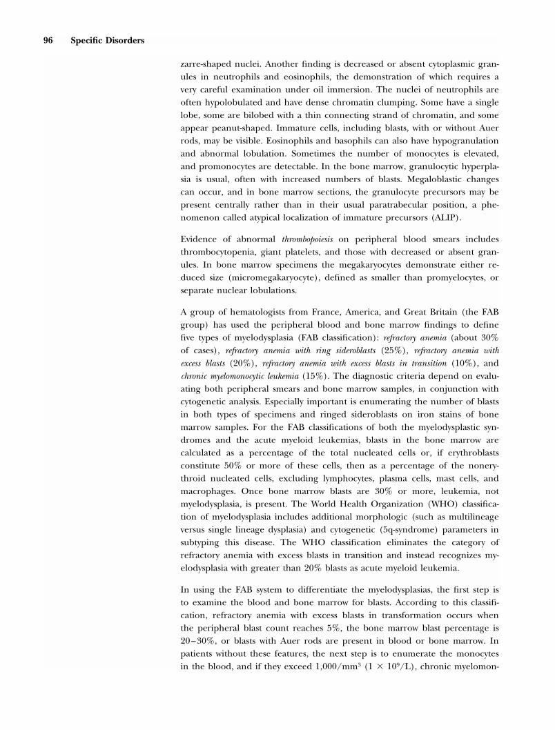
96 Specific Disorders
zarre-shaped nuclei. Another finding is decreased or absent cytoplasmic gran-ules in neutrophils and eosinophils, the demonstration of which requires avery careful examination under oil immersion. The nuclei of neutrophils areoften hypolobulated and have dense chromatin clumping. Some have a singlelobe, some are bilobed with a thin connecting strand of chromatin, and someappear peanut-shaped. Immature cells, including blasts, with or without Auerrods, may be visible. Eosinophils and basophils can also have hypogranulationand abnormal lobulation. Sometimes the number of monocytes is elevated,and promonocytes are detectable. In the bone marrow, granulocytic hyperpla-sia is usual, often with increased numbers of blasts. Megaloblastic changescan occur, and in bone marrow sections, the granulocyte precursors may bepresent centrally rather than in their usual paratrabecular position, a phe-nomenon called atypical localization of immature precursors (ALIP).
Evidence of abnormal thrombopoiesis on peripheral blood smears includesthrombocytopenia, giant platelets, and those with decreased or absent gran-ules. In bone marrow specimens the megakaryocytes demonstrate either re-duced size (micromegakaryocyte), defined as smaller than promyelocytes, orseparate nuclear lobulations.
A group of hematologists from France, America, and Great Britain (the FABgroup) has used the peripheral blood and bone marrow findings to definefive types of myelodysplasia (FAB classification): refractory anemia (about 30%of cases), refractory anemia with ring sideroblasts (25%), refractory anemia withexcess blasts (20%), refractory anemia with excess blasts in transition (10%), andchronic myelomonocytic leukemia (15%). The diagnostic criteria depend on evalu-ating both peripheral smears and bone marrow samples, in conjunction withcytogenetic analysis. Especially important is enumerating the number of blastsin both types of specimens and ringed sideroblasts on iron stains of bonemarrow samples. For the FAB classifications of both the myelodysplastic syn-dromes and the acute myeloid leukemias, blasts in the bone marrow arecalculated as a percentage of the total nucleated cells or, if erythroblastsconstitute 50% or more of these cells, then as a percentage of the nonery-throid nucleated cells, excluding lymphocytes, plasma cells, mast cells, andmacrophages. Once bone marrow blasts are 30% or more, leukemia, notmyelodysplasia, is present. The World Health Organization (WHO) classifica-tion of myelodysplasia includes additional morphologic (such as multilineageversus single lineage dysplasia) and cytogenetic (5q-syndrome) parameters insubtyping this disease. The WHO classification eliminates the category ofrefractory anemia with excess blasts in transition and instead recognizes my-elodysplasia with greater than 20% blasts as acute myeloid leukemia.
In using the FAB system to differentiate the myelodysplasias, the first step isto examine the blood and bone marrow for blasts. According to this classifi-cation, refractory anemia with excess blasts in transformation occurs whenthe peripheral blast count reaches 5%, the bone marrow blast percentage is20–30%, or blasts with Auer rods are present in blood or bone marrow. Inpatients without these features, the next step is to enumerate the monocytesin the blood, and if they exceed 1,000/mm3 (1 � 109/L), chronic myelomon-
40413 Tkachuk PM Gordon(SAUNM) INTERACTIVE LEFT
top of RHbase of RH
top of textbase of text
shortstandardlong

Specific Disorders 97
ocytic leukemia is present. In patients without these features, further classifi-cation rests on the number of blasts and ringed sideroblasts. With peripheralblasts of 1% or bone marrow blasts of 5%, the diagnosis is refractory anemiawith excess blasts. If these features are absent but ringed sideroblasts accountfor more than 15% of the erythroblasts, the diagnosis is refractory anemiawith ringed sideroblasts. The remaining cases are classified as refractory ane-mia (or refractory cytopenia when anemia is absent but thrombocytopenia orneutropenia is present).
40413 Tkachuk PM Gordon(SAUNM) INTERACTIVE RIGHT
top of RHbase of RH
top of textbase of text
shortstandardlong
In hematologic practice, the diagnosis of myelodysplastic syndrome is made(1) clinically, as a diagnosis of last resort for primary bone marrow-basedcytopenia(s), (2) pathologically, or (3) cytogenetically. In most cases of mye-lodysplastic syndrome the diagnosis rests on a combination of the above. It isdifficult to standardize criteria for making any diagnosis based purely on acytological basis, but it is still useful to follow some universal guidelines forwhat is meant by “dysplastic” and how one comes to make a morphologicdiagnosis of primary myelodysplasia. Well-preserved and adequately stainedmarrow spicules are essential. A history of (1) previous exposure to myelo-toxic agents (including chemotherapy, radiation, and other toxins) or (2)nutritional deficiencies (vitamin B12 and folate) are causes of secondary mye-lodysplastic syndrome to be excluded before considering a diagnosis of pri-mary myelodysplastic syndrome. A useful rule for making a morphologicdiagnosis of myelodysplastic syndrome is finding dysplastic changes in at leasttwo out of three hematopoietic lineages (myeloid, megakaryocytic, and ery-throid). In the early stages of myelodysplastic syndrome and in most cases atthe time of initial diagnosis, dysplastic changes are almost never found in allthe cells of a particular lineage but only in a minority. Accordingly, theexaminer should carefully assess many marrow cells from all three lineageswhen evaluating a marrow specimen for myelodysplastic syndrome. The mostcharacteristic dysplastic changes in the myeloid line, best seen in the periph-eral smear, include hypolobulated nuclei and hypogranular cytoplasm. Char-acteristic dysplastic changes in the erythroid lineage, seen in the marrow, areasymmetric multinucleation, irregular nuclear contours, other bizarre nuclearcontour abnormalities, and ringed sideroblasts with iron staining. Microme-gakaryocytes (smaller than a promyelocyte) and forms displaying clearly sepa-rate nuclear lobulations are recognized dysplastic findings in megakaryocytes.
Many patients with myelodysplastic syndrome eventually develop acute leuke-mia, nearly always myelogenous, which may appear suddenly or may graduallyevolve over several weeks to months. The likelihood varies according to thetype of myelodysplastic syndrome present; the descending order of risk isrefractory anemia with excess blasts in transition, refractory anemia withexcess blasts, chronic myelomonocytic leukemia, refractory anemia withringed sideroblasts, and refractory anemia. The chance of developing leuke-mia is quite high in the first three types, much lower in the latter two.

98 Specific Disorders
Slide 118. Myelodysplasia: Pseudo-Pelger-HuetCell
A common finding in the myelodysplastic and myelo-proliferative syndromes is the pseudo-Pelger-Huetcell. It consists of bilobed neutrophils, typically with asmall bridge connecting the two lobes. The cells re-semble those in the autosomal dominant disorder ofthe Pelger-Huet anomaly, but in the acquired form,the number of affected neutrophils is generallylower, and additional dysplastic features are presentin the granulocytes or other cell lines. In this slide apseudo-Pelger-Huet cell is seen on the left. A thinstring of chromatin connects its two lobes (pince-nezappearance; see slide 24). The white cell on the rightis a normal band with a twisted nucleus.
Slide 119. Myelodysplastic Syndromes:Peripheral Blood
This slide, from a patient with chronic myelomono-cytic leukemia, shows a nucleated erythrocyte that isdysplastic in having a lobulated nucleus. The whitecell is a bizarre, hypersegmented monocyte. Plateletsare diminished.
40413 Tkachuk PM Gordon(SAUNM) INTERACTIVE LEFT
top of RHbase of RH
top of textbase of text
shortstandardlong
Slide 120. Myelodysplastic Syndromes: BoneMarrow Aspirate
Despite the decreased numbers of red cells, platelets,and white cells in the peripheral smear in these dis-orders, the aspirate is usually hypercellular. Dysplasiain the hematopoietic lines is the most importantfinding. In this slide, from a patient with chemother-apy-induced myelodysplasia, the megakaryocyte in theright upper area is small and shows hypolobulationand sparse, poorly granulated cytoplasm. A cluster ofdysplastic, hypogranular, and hypolobulated myeloidprecursors with abnormally condensed nuclei appearsin the lower left. Myeloid maturation is absent.

Specific Disorders 99
Slide 121. Myelodysplasia: Bone MarrowAspirate
This bone marrow aspirate illustrates dysplasia in sev-eral cells. On the left of the slide is a cell with refrac-tile granules that partially cover a single nucleus.This is a dysplastic, unilobular eosinophil whose gran-ules are less intense than usual. The cell with abun-dant vacuolated cytoplasm on the right is a dysplasticmicromegakaryocyte with only a single-lobed nucleus.
Slide 122. Refractory Anemia with RingedSideroblasts: Bone Marrow Aspirate, IronStain
In this disease, the peripheral smear usually demon-strates macrocytic red cells, but often with a smallerpopulation of microcytic, hypochromic cells—a di-morphic anemia. Basophilic stippling, Pappenheimerbodies, and nucleated red cells may be present. Thebone marrow aspirate typically discloses erythroid hy-perplasia with dysplastic changes. The other cell linesare usually normal, and blasts are less than 5%. Oniron stain, ringed sideroblasts constitute more than15% of the nucleated red cells. Ringed sideroblastshave iron granules arranged in a ring around thenucleus rather than in a random distributionthroughout the cytoplasm (see slide 62). This slideshows two ringed sideroblasts.
40413 Tkachuk PM Gordon(SAUNM) INTERACTIVE RIGHT
top of RHbase of RH
top of textbase of text
shortstandardlong
Slide 123. Refractory Anemia with ExcessBlasts: Bone Marrow Aspirate
In this disorder, blasts represent 1–5% of the periph-eral white cells or 5–20% of the bone marrow cells.On peripheral smear, in addition to the presence ofblasts, pancytopenia is common, and dysplastic fea-tures affect the different cell types. The bone marrowaspirate shows hypercellularity, with dysplasia usuallypresent in all lines, and the defining increase inblasts. In this slide, at least four of the cells areblasts. At the left is a small, dysplastic megakaryocytewith separated nuclear lobules.

100 Specific Disorders
Slide 124. Chronic Myelomonocytic Leukemia
This diagnosis requires a monocyte count of �1 �109/L (�1,000/mm3), with up to 20% blasts in thebone marrow and no more than 5% blasts in theperipheral blood. In the bone marrow promonocytesare often increased, and neutrophils are frequentlynumerous on the peripheral smear. The monocytesoften appear normal, but some have bizarre shapesor hypersegmented nuclei and increased cytoplasmicbasophilia. Dysplasia occurs in the other cell lines aswell. This slide shows a dysplastic nucleated red cellwith a pyknotic nucleus, displaying irregular bordersand three atypical monocytes, reflecting the pro-found peripheral monocytosis in this patient. Howell-Jolly bodies are present in several red cells.
40413 Tkachuk PM Gordon(SAUNM) INTERACTIVE LEFT
top of RHbase of RH
top of textbase of text
shortstandardlong

White Cell Disorders
LEUKEMIAS
Acute Leukemias
The proliferation of undifferentiated bone marrow cells of either lymphoidor myeloid origin produces acute lymphocytic (ALL) or acute myeloid leuke-mia (AML), respectively. Acute leukemia represents a clonal expansion ofabnormal immature cells that often have diagnostically significant chromo-somal abnormalities such as translocations. At the time of presentation, leuke-mia cells have usually replaced the marrow cavity and spilled out into theblood. The term acute with regard to leukemia no longer denotes a rapidfatal outcome but refers to the undifferentiated state of the leukemia cells. Infact, most patients present with nonspecific symptoms such as fatigue, leth-argy, and weight loss. Older people with acute leukemia may complain ofdyspnea, angina, and dizziness related to anemia. Approximately one-third ofpatients have serious infections at the time of diagnosis, and, with the excep-tion of AML-M3, rarely do serious bleeding problems pose a problem at thatpoint.
Acute leukemia is slightly more common in white men and has been corre-lated with high socioeconomic status. AML, which constitutes approximately80% of all acute leukemias in adults, is a disease of the elderly, with itsincidence rising after age 50. ALL, on the other hand, occurs primarily inchildren and young adults, with a median age in adults ranging from 25 to40 years. Risk factors for acute leukemia include previous exposure to ioniz-ing radiation, benzene, and certain classes of chemotherapeutic drugs, in-cluding alkylating agents and topoisomerase II-inhibitors. Patients with Downsyndrome are at increased risk of developing acute leukemia. Other predis-posing conditions include rare hereditary chromosome breakage (Bloom’ssyndrome and Fanconi’s anemia) and immunodeficiency (Bruton-type X-linked agammaglobulinemia and hereditary ataxia-telangiectasia) syndromes.
At the time of presentation, the peripheral blood usually shows anemia,neutropenia, and thrombocytopenia, although the total white cell count ishighly variable. Circulating blasts are apparent in the peripheral blood inapproximately 80% of cases. So-called aleukemic leukemia, a term used when
40413 Tkachuk PM Gordon(SAUNM) INTERACTIVE RIGHT
top of RHbase of RH
top of textbase of text
shortstandardlong

102 Specific Disorders
circulating blasts are absent, is slightly more common in AML than ALL.Important features to assess in the peripheral blood of newly diagnosed acuteleukemia include (1) signs of intravascular coagulation (which suggest acutepromyelocytic leukemia, AML-M3), such as a microangiopathic picture withsevere anisopoikilocytosis, microcytes, red cell fragments, schistocytes, andprofound thrombocytopenia; (2) hyperleukocytosis (especially in AML-3); (3)signs of underlying myelodysplasia, such as hypolobulated and hypogranularneutrophils; and (4) Auer rods in circulating blasts.
After a complete history and physical examination, all patients with acuteleukemia should have slides of their peripheral blood and representativebone marrow specimens scrutinized. In addition, all those with ALL and AMLwith monocytic differentiation (AML-M4 and AML-M5) should undergo lum-bar puncture to examine the cerebrospinal fluid (CSF) for the presence ofblasts. A diagnosis of acute leukemia rests upon demonstrating an increasednumber of immature hematopoietic cells and then assigning a particularlineage, generally either myeloid or lymphoid, to the abnormal leukemic cellpopulation. The most widely used classification is that originally proposed bythe French-American-British (FAB) Cooperative Group in 1976 (Br J Haematol1976;33:452–8). The World Health Organization has incorporated much ofthe FAB classification into an expanded version ( J Clin Oncol 1999;17:3835–49). A diagnosis of acute leukemia is made after examining Wright’s- orWright’s-Giemsa stained bone marrow aspirate smears and peripheral bloodalong with a hematoxylin-eosin (H&E)-stained clot section and biopsy. Inaddition, in all newly diagnosed cases of acute leukemia, cytogenetic analysisand histochemical and flow immunophenotyping should be done on thecells.
The diagnosis of acute leukemia is usually not subtle. Bone marrow examina-tion most commonly reveals a hypercellular specimen with sheets of blasts.The normal marrow constituents—the erythroid, maturing myeloid, andmegakaryocytes—are usually markedly diminished in number. Occasionally,most often in cases of high-grade myelodysplasia (refractory anemia withexcess blasts), the marrow exhibits atypical morphological features. In thesecases, overall marrow cellularity may be low, the erythroid series markedlyincreased, populations of atypical or dysplastic megakaryocytes expanded, andextensive marrow fibrosis seen. In addition, so-called hypoplastic variants ofacute leukemia, usually AML, exist, characterized by low total marrow cellu-larity but relatively high numbers of blasts. Rarely, acute leukemia can arisefrom an extramedullary focus, usually a blast crisis complicating a preexistingmyeloproliferative syndrome, and marrow samples are nonrepresentative.
Nearly always, a diagnosis of acute leukemia can be made by recognizingincreased blasts and determining the lineage by histochemical and/or surfacephenotype studies. A diagnosis of de novo acute leukemia requires that atleast 30% of the total nucleated bone marrow cells be blasts, with threeexceptions. If the marrow displays displastic erythroid hyperplasia (erythro-blasts � 50% of total cells) and if �30% of nonerythroid cells are blasts, adiagnosis of AML with erythroid differentiation (AML-M6) is made. Another
40413 Tkachuk PM Gordon(SAUNM) INTERACTIVE LEFT
top of RHbase of RH
top of textbase of text
shortstandardlong

Specific Disorders 103
exception is in myelodysplastic syndrome (MDS) when myeloblasts are �20%of total marrow cells. This condition, formerly called “refractory anemia withexcess blasts-in-transformation” (RAEB-T) is now recognized as AML. Thethird exception is acute promyelocytic leukemia (AML-M3), in which abnor-mal promyelocytes rather than increased blasts constitute the majority ofmarrow cells. A blast is defined morphologically as a cell that is medium tolarge in size, usually has a high nuclear : cytoplasm ratio, possesses an openchromatin pattern with or without nucleoli, and typically has sparse baso-philic-staining cytoplasm. Fine azurophilic (bright red/purple) cytoplasmicgranulation is sometimes present in blasts (so-called type II blasts). Unless itcontains an Auer rod, a blast cannot be classified as either myeloid orlymphoid without ancillary histochemical or phenotypic studies. Once in-creased blasts are recognized, the origin—either lymphoid or myeloid—mustbe determined to make a diagnosis of ALL or AML, respectively. The recom-mended approach is to first rule in AML by showing positive cytoplasmicstaining with myeloid granule–specific stains such as myeloperoxidase (orSudan Black) and monocytic-specific stain such as �-naphthyl acetate esterase.If �3% of the blasts are positive with the myeloid granule–specific stain,immunophenotyping by flow cytometry is recommended. Since blasts of onetype of AML, AML with minimal evidence of myeloid differentiation (AML-M0), are negative with these myeloid granule–specific stains, negative stain-ing does not exclude a diagnosis of AML. For this reason, flow immunophen-otyping is indicated to rule out AML-M0 if the myeloid-specific histochemicalstains are negative.
Acute Myeloid Leukemias
The FAB classification of acute myeloid leukemias (sometimes called acutenonlymphocytic leukemia, or ANLL) depends largely on a 500-cell differen-tial count of a bone marrow specimen. The FAB classification includes seventypes of acute myeloid leukemia defined by the nature of the blast cells;usually these cells are also present on the peripheral blood smear, allowing apresumptive diagnosis to be made. This system recognizes three types ofblasts. Type I are myeloblasts lacking cytoplasmic granules but possessingprominent nucleoli, a central nucleus, and uncondensed chromatin patterns.Type II blasts resemble type I cells and retain a central nucleus but have afew primary (azurophilic) cytoplasmic granules. Type III cells have more than20 azurophilic cytoplasmic granules but do not display a Golgi zone, which ischaracteristic of promyelocytes.
In addition to the abnormalities in white cells, anemia and thrombocytopeniaare common, as are morphologic changes in the erythrocytes and platelets onthe peripheral smear. Anisocytosis, poikilocytosis, basophilic stippling, andnucleated red cells are often present among the erythrocytes. Leukemic cellsmay sometimes ingest red cells (erythrophagocytosis). In the platelet series,giant or agranular platelets and circulating micromegakaryocytes may bevisible.
40413 Tkachuk PM Gordon(SAUNM) INTERACTIVE RIGHT
top of RHbase of RH
top of textbase of text
shortstandardlong
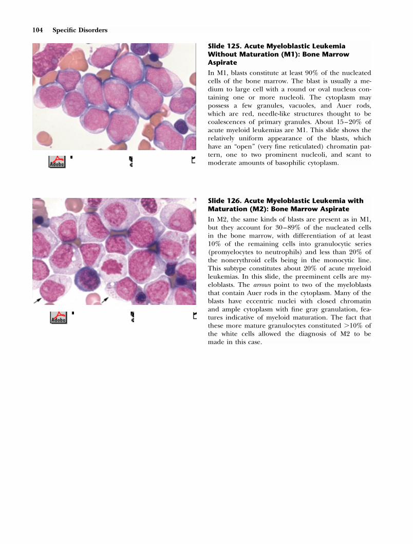
104 Specific Disorders
Slide 125. Acute Myeloblastic LeukemiaWithout Maturation (M1): Bone MarrowAspirate
In M1, blasts constitute at least 90% of the nucleatedcells of the bone marrow. The blast is usually a me-dium to large cell with a round or oval nucleus con-taining one or more nucleoli. The cytoplasm maypossess a few granules, vacuoles, and Auer rods,which are red, needle-like structures thought to becoalescences of primary granules. About 15–20% ofacute myeloid leukemias are M1. This slide shows therelatively uniform appearance of the blasts, whichhave an “open” (very fine reticulated) chromatin pat-tern, one to two prominent nucleoli, and scant tomoderate amounts of basophilic cytoplasm.
Slide 126. Acute Myeloblastic Leukemia withMaturation (M2): Bone Marrow Aspirate
In M2, the same kinds of blasts are present as in M1,but they account for 30–89% of the nucleated cellsin the bone marrow, with differentiation of at least10% of the remaining cells into granulocytic series(promyelocytes to neutrophils) and less than 20% ofthe nonerythroid cells being in the monocytic line.This subtype constitutes about 20% of acute myeloidleukemias. In this slide, the preeminent cells are my-eloblasts. The arrows point to two of the myeloblaststhat contain Auer rods in the cytoplasm. Many of theblasts have eccentric nuclei with closed chromatinand ample cytoplasm with fine gray granulation, fea-tures indicative of myeloid maturation. The fact thatthese more mature granulocytes constituted �10% ofthe white cells allowed the diagnosis of M2 to bemade in this case.
40413 Tkachuk PM Gordon(SAUNM) INTERACTIVE LEFT
top of RHbase of RH
top of textbase of text
shortstandardlong

Specific Disorders 105
Slide 127. Acute Hypergranular PromyelocyticLeukemia (M3): Bone Marrow Aspirate
In this leukemia, the blasts are usually �30% of nu-cleated bone marrow cells, but the typical morphol-ogy of the predominant cell allows the diagnosis tobe made with confidence. The abnormal promyelo-
cyte is larger than a normal myeloblast; has an oval,kidney-shaped, folded, or bilobed nucleus; and pos-sesses a cytoplasm replete with large, sometimes gi-ant, red or purplish granules that may obscure thenucleus. The cytoplasm commonly has abundantAuer rods as well. A variant form, M3V, has cells withreniform, lobulated, or convoluted nuclei and cyto-plasm that may be either agranular or sparsely butfinely granulated. These two forms of M3 constituteabout 10% of acute myeloid leukemias. This slideshows promyelocytes with folded and bilobed nuclei.The four in the center of the slide with impressiveaccumulations of Auer rods are sometimes called“faggot cells,” because their contents resemble a bun-dle of sticks. A single erythroid precursor lies abovethe promyelocytes. Most cases of M3 and M3V AMLhave a balanced reciprocal translocation betweenchromosomes 15 and 17, t(15;17)(q22;q21), that canbe detected by cytogenetic fluorescence in situ hy-bridization (FISH) analyses.
40413 Tkachuk PM Gordon(SAUNM) INTERACTIVE RIGHT
top of RHbase of RH
top of textbase of text
shortstandardlong
Slide 128. Acute Myelomonocytic Leukemia(M4): Peripheral Blood
In this disorder, blasts constitute 30–89% of the nu-cleated bone marrow cells. The granulocytic compo-
nent (including myeloblasts) and the monocytic com-ponent (monoblasts, promonocytes, monocytes) eachaccount for at least 20% of the bone marrow cells. Inthe peripheral blood, monocytes are often very nu-merous, reaching levels of more than 5 � 109/L(5,000/mm3). The predominant cells are monoblastsand promonocytes. The monoblast is a large cellwhose round or convoluted nucleus has lacy chroma-tin and one or more nucleoli, which may be large;the cytoplasm is ample, basophilic, often vacuolated,and sometimes granulated. Auer rods are occasionallypresent. In promonocytes as compared to mono-blasts, the nucleoli are smaller, the nuclei are oftenindented, the cytoplasm has more numerous gran-ules, and its color is more basophilic. M4 accountsfor about 15–20% of acute myeloid leukemia. Thisslide from a peripheral smear shows two monocytesand two blasts, reflecting the dual populations ofblasts, myeloid and monocytic, typical for this subtypeof AML.

106 Specific Disorders
Slide 129. Acute Monocytic Leukemia (M5):Bone Marrow Aspirate
In M5, blasts constitute at least 30% of the bonemarrow’s nucleated cells, and the monocytic compo-nent is at least 80% of the nonerythroid cells. M5 issubdivided into M5a (acute monoblastic leukemia), ifmonoblasts account for at least 80% of the bonemarrow monocytic component, and M5b, if they areless than that percentage. This slide, from a patientwith M5, shows numerous monoblasts with lacy chro-matin, round or oval nuclei, nucleoli, and gray cyto-plasm.
Slide 130. Acute Monocytic Leukemia (M5):Bone Marrow Aspirate, Esterase Stain
Cytochemical stains can confirm the cell type of thisleukemia, since the monocyte and its precursors reactwith the nonspecific esterase stains, �-naphthyl ace-
tate esterase or �-naphthyl butyrate esterase. In this
slide, the red material in the cytoplasm of the cellsindicates a positive reaction for nonspecific esterases.
40413 Tkachuk PM Gordon(SAUNM) INTERACTIVE LEFT
top of RHbase of RH
top of textbase of text
shortstandardlong
Slide 131. Acute Myeloid Leukemia withProminent Erythroid Differentiation (M6):Bone Marrow Aspirate
In M6 (erythroleukemia), dysplastic erythroid precur-sors must constitute at least 50% of the nucleatedcells in the bone marrow and granulocytic blastsmust account for at least 30% of the nonerythroidcells or at least 20% of nonerythroid cells using theFAB or WHO classifications, respectively. The eryth-roblasts usually show substantial dysplasia, includingcytoplasmic vacuolization, bizarre nuclear features,multinuclearity, megaloblastic changes, and karyor-rhexis (rupture of the nucleus). M6 represents about3–4% of acute myeloid leukemias. This slide showsseveral erythroblasts, two with multinuclearity, twowith cytoplasmic vacuoles, and some with megaloblas-tic changes, characterized by large nuclei with loosechromatin in cells with polychromatophilic cyto-plasm.

Specific Disorders 107
Acute Lymphoblastic Leukemia
According to the French-American-British (FAB) classification, the diagnosisof acute lymphoblastic leukemia (ALL) depends on demonstrating that atleast 30% of the nucleated cells in a count of 500 bone marrow cells arelymphoblasts, when evaluated by special studies. The blasts in ALL are nega-tive with histochemical stains that demonstrate cells of myeloid lineage—myeloperoxidase, Sudan black, and the esterases. Most ALL blasts show char-acteristic “blocks” of cytoplasmic PAS (periodic acid-Schiff). Immunopheno-typing demonstrates that 75% of all cases of ALL have a B-cell lineage andabout 25% are T-cells.
Slide 132. Acute Megakaryoblastic Leukemia(M7)
In M7, blasts constitute at least 30% of the bonemarrow nucleated cells, and they are identifiable asmegakaryoblasts by their morphology or by otherstudies. These pleomorphic cells are often indistin-guishable from other blasts on Romanowsky stains,but their true identity may be suspected when theyhave cytoplasmic projections or budding of plateletsor when large, bizarre platelets or increased numbersof megakaryocytes are also present. Immunopheno-typic studies, however, are necessary to confirm thatthe cells are genuinely megakaryoblasts. M7 consti-tutes about 2–4% of acute myeloid leukemias. Thisslide demonstrates one blast, which contains a largenucleus, one nucleolus, and scant basophilic cyto-plasm from which rounded projections emanate.These projections suggest that the blast is in themegakaryocyte line, recapitulating the budding ofplatelets that normally occurs.
40413 Tkachuk PM Gordon(SAUNM) INTERACTIVE RIGHT
top of RHbase of RH
top of textbase of text
shortstandardlong
Slide 133. Acute Lymphoblastic Leukemia:Bone Marrow Aspirate
The predominant cell is small, possesses a regularnucleus with fairly homogeneous chromatin, and hasa high nuclear : cytoplasm ratio. Nucleoli are absentor inconspicuous. Most cases of ALL have cells ofthis type. This aspirate demonstrates cells of fairlyuniform size, containing very fine nuclear chromatin,absent nucleoli, and sparse cytoplasm.

108 Specific Disorders
Slide 134. Acute Lymphoblastic Leukemia:Bone Marrow Aspirate
In some cases of acute lymphoblastic leukemia theblasts are larger and more variable, commonly haveprominent nucleoli, and possess more abundant, of-ten vacuolated, cytoplasm. The nuclei are irregular inoutline and may be indented, folded, or cleft. Com-pared to the previous slide, the cells in this bonemarrow aspirate are more pleomorphic, with coarsernuclear chromatin and greater irregularity in nuclearshape. In some cells, the cytoplasm is more substan-tial. Prominent nucleoli are present.
Slide 135. Lymphoma with Cells in thePeripheral Blood
Sometimes, the malignant cells of lymphomas mayappear in large numbers within the peripheral circu-lation. These cases resemble acute lymphoblastic leu-kemia. Typically, the cells are large, and their cyto-plasm is basophilic and commonly vacuolated. Theregular-shaped, round, or oval nuclei may containone or more nucleoli and uniform, dense, and finely-stippled chromatin. Vacuoles can occur in the nu-cleus, as demonstrated in this aspirate.
40413 Tkachuk PM Gordon(SAUNM) INTERACTIVE LEFT
top of RHbase of RH
top of textbase of text
shortstandardlong
CHRONIC LYMPHOCYTIC LEUKEMIA
Chronic lymphocytic leukemia is a malignant disorder in which the primaryproblem is not an excessive production of abnormal lymphocytes but a pro-tracted life span, causing their progressive accumulation. The cause is adefect in the steps that lead to programmed death, or apoptosis. In �95% ofcases the leukemic lymphocyte is a B-cell. This disorder, the most commoncause of leukemia in the United States, accounting for 25–30% of all cases, isprimarily a disease of older adults, with the median age at diagnosis beingabout 55. Many patients are asymptomatic at presentation, the disease beingdiscovered on routine blood tests. When symptoms develop, the most com-mon are weakness, fatigue, and weight loss. In patients with hypogammaglob-ulinemia, frequent or severe bacterial infections may occur. The physicalexamination is often normal, but the commonest finding is enlargement ofthe cervical, axillary, and inguinal lymph nodes, which are firm, nontender,and movable. Hepatomegaly and splenomegaly, varying from mild to massive,may develop.
The diagnostic criteria for CLL are (1) an absolute blood lymphocytosis�5 � 109/L (5,000/mm3), with morphologically mature cells and lastingmore than 4 weeks; (2) at least 30% lymphocytes in a normocellular or

Specific Disorders 109
hypercellular marrow; and (3) a monoclonal B-cell phenotype of the predom-inant lymphocytes, with low levels of surface immunoglobulins and CD5 posi-tivity. The lymphocytes appear mature on peripheral blood smear, but dis-rupted cells (“smudge cells”) are common, indicating increased fragility, withdamage to the cells occurring during preparation of the slide. Some patientshave anemia, either from decreased production (because excessive lympho-cytes in the bone marrow impair erythropoiesis) or from an autoimmunehemolytic anemia that occurs in 10–25% of patients sometime during theirdisease. The antibody is usually a warm-reactive, polyclonal IgG. Rarely, animmune-related red cell aplasia occurs. Thrombocytopenia can also developfrom marrow suppression or, uncommonly, from an immune mechanism.Various staging systems exist that provide prognostic information. Patientswith only lymphocytosis have a lengthy period of survival. When lymph nodeenlargement or hepatosplenomegaly accompanies the lymphocytosis, theprognosis is intermediate. Those with anemia or thrombocytopenia have theworst outcome. Other laboratory findings associated with a poor prognosisinclude increased serum LDH levels, hypoalbuminemia, and hypercalcemia.
Late complications of CLL include bacterial, viral, or fungal infections relatedto hypogammaglobulinemia, diminishing cell-mediated immunity, or neutro-penia from the disease or its treatment. In about 1–10% of cases the disor-der transforms into a non-Hodgkin’s lymphoma (Richter’s syndrome), usuallyof the large-cell variety, often developing suddenly with fever or other sys-temic symptoms and enlarging lymph nodes. In about 10% of patients withCLL, the disease transforms into prolymphocytic leukemia, with the lympho-cyte changing to a large cell with nucleoli, convoluted nuclei, and immaturechromatin. Rarely, patients develop paraneoplastic pemphigus, with blisteringand ulceration of the skin and mucous membranes.
monly disintegrate. In these smear or smudge cells, thecytoplasm is lost and the nucleus spreads out.
Some of the cells may be prolymphocytes, whichare larger than lymphocytes, have more abundant cy-toplasm, possess a prominent nucleolus, and have amore condensed chromatin. Cases of CLL in whichthe prolymphocytes constitute 10–55% of the lym-phoid cells are called CLL/PL. In others, lymphoidcells may vary from small to large, but prolympho-cytes are less than 10%. These two variants of CLLare called chronic lymphocytic leukemia, mixed celltypes. When prolymphocytes exceed 55% of the lym-phoid cells, the disorder is called prolymphocytic leu-kemia (see slide 138).
When autoimmune hemolytic anemia complicatesCLL, polychromatophilia and spherocytosis appearon the smear. Autoimmune thrombocytopenia mayalso occur. In this slide, lymphocytes, which consti-tute all the white cells except for one neutrophil, arefairly uniform in size and shape; a few smudge cellsare scattered throughout the slide.
40413 Tkachuk PM Gordon(SAUNM) INTERACTIVE RIGHT
top of RHbase of RH
top of textbase of text
shortstandardlong
Slide 136. Chronic Lymphocytic Leukemia
In CLL, the lymphocytes usually appear normal, butthey may be large or have deeply cleft nuclei (Reidercells). They sometimes contain coarse, clumped chro-matin, and the cytoplasm may be vacuolated. Withpreparation of the blood film, lymphocytes com-

110 Specific Disorders
Slide 137. Chronic Lymphocytic Leukemia:Bone Marrow Biopsy
Patterns of bone marrow involvement include inter-stitial, nodular, diffuse, and mixed, which is a combi-nation of nodular and interstitial. The type of patternpresent has some prognostic significance, with thediffuse type having the most aggressive course. Nodu-lar infiltrates are closely packed aggregates of cellsthat are usually not paratrabecular in location. In theinterstitial pattern the lymphocytes intermingle withhematopoietic cells. In this slide, the bone marrowbiopsy demonstrates the diffuse pattern.
Slide 138. B-Cell Prolymphocytic Leukemia
When neoplastic prolymphocytes account for �55%of the lymphoid cells, the disorder is called prolym-phocytic leukemia. T- and B-cell prolymphocytic leu-kemias are separate disorders. In the B-cell type thecells are larger than the lymphocytes in CLL, theirnuclei have prominent nucleoli, and sometimes thecytoplasm is more abundant. The chromatin is mod-erately condensed. In the T-cell variety, the cells aremore pleomorphic; the nuclear shape is irregular,often knobby; nucleoli are less prominent; the cyto-plasm is scanty; and blebs may protrude from it. Thisslide reveals the characteristic appearance of prolym-phocytes. In some, the cytoplasm is absent, and nu-cleoli are very prominent. In many cases this disordermay be an accelerated form of CLL, which oftenprecedes it.
40413 Tkachuk PM Gordon(SAUNM) INTERACTIVE LEFT
top of RHbase of RH
top of textbase of text
shortstandardlong

Specific Disorders 111
OTHER LEUKEMIAS
Hairy Cell Leukemia
Hairy cell leukemia accounts for about 2% of adult leukemias, with a male-female ratio of about 4:1 and an average age at diagnosis of approximately50–55 years. The major clinical findings arise from infiltration of the bonemarrow and spleen by a distinctive B-lymphocyte, the “hairy cell,” causingpancytopenia and splenomegaly. The clinical features of pancytopenia in-clude fatigue and other complaints related to anemia; easy bruising, pete-chiae, and ecchymoses from decreased platelets; and infections because ofneutropenia, typically from gram-positive or gram-negative bacteria. Patientswith hairy cell leukemia, however, also have an especial increased susceptibil-ity to nontuberculous mycobacteria and fungi. Toxoplasma gondii, Pneumocystiscarinii, Listeria monocytogenes, and various viruses may also cause infections,suggesting a diminished cell-mediated immunity. Indeed, nearly all patientshave decreased numbers of monocytes. Palpable splenomegaly, sometimes toenormous dimensions, occurs in about 80–90% of patients. The liver isenlarged in approximately 30–40%. Hairy cell leukemia is the most commoncause of paraneoplastic vasculitis, characterized by arthritis, palpable purpura,nodular skin lesions, and fever.
Slide 139. Hairy Cell Leukemia
Most patients have pancytopenia at presentation.Hairy cells are present in about 85% of peripheralsmears. Larger than most other lymphocytes, with adiameter of 10–20 �m, these mononuclear cells havea round, oval, reniform, or dumbbell shape. The nu-cleus is often eccentric, round, or oval and containsdiffuse chromatin. Nucleoli, when present, are smalland inconspicuous. The abundant pale blue-gray andagranular cytoplasm has an uneven border with nu-merous irregular thin projections resembling hairs.This slide is taken from a thick part of the smear,accounting for the rouleaux, but white cells are oftenmost abundant in these areas. All the cells are hairycells, with easily visible cytoplasmic projections. No-tice the diffuse chromatin in the nuclei and the gray-blue cytoplasm.
40413 Tkachuk PM Gordon(SAUNM) INTERACTIVE RIGHT
top of RHbase of RH
top of textbase of text
shortstandardlong

112 Specific Disorders
Slide 140. Hairy Cell Leukemia: TRAP Stain
Acid phosphatase activity is present in several types ofblood cells. One form is an acid phosphatase resis-tant to tartrate. A tartrate-resistant acid phosphatase(TRAP) stain is positive in �95% of cases of hairycell leukemia. Flow cytometric testing for surfacemarkers specific for hairy cell leukemia, however, hasreplaced the TRAP stain as the preferred diagnostictest. In this slide, the hairy cell shows the red colorthat defines a positive TRAP reaction, which is absentfrom the two other myeloid cells. Cells are occasion-ally TRAP-positive in other disorders, such as infec-tious mononucleosis and chronic lymphocytic leu-kemia.
Slide 141. Hairy Cell Leukemia: Bone MarrowBiopsy
Bone marrow aspiration is frequently difficult or im-possible in hairy cell leukemia because of associatedfibrosis. On the biopsy, involvement by malignantcells may be patchy, focal, or diffuse. The abundantcytoplasm creates a substantial space between the nu-clei of adjacent hairy cells, producing an appearancethat resembles fried eggs. The nuclei, which demon-strate low mitotic activity, are round to oval and maybe indented or bilobed. Reticulin fibrosis occurs inthe areas of the hairy cells, its extent being propor-tional to the number of malignant cells present. Insome cases the normal hematopoietic cells, especiallygranulocytic precursors, are markedly reduced, occa-sionally to the point of resembling aplastic anemia. Acareful search for the hairy cells is then necessary toestablish the proper diagnosis. This slide demon-strates infiltration of the hairy cells in the paratrabec-ular area. These are benign-appearing lymphoid cellswith uniformly spaced nuclei because of the substan-tial cytoplasm characteristic of hairy cells.
40413 Tkachuk PM Gordon(SAUNM) INTERACTIVE LEFT
top of RHbase of RH
top of textbase of text
shortstandardlong

Specific Disorders 113
Sezary’s Syndrome
In 1938 Sezary and Bouvrain described a syndrome of pruritus, erythroderma,and hyperconvoluted lymphoid cells in the peripheral blood. This disorder,now considered a subset of cutaneous T-cell lymphoma, has an average age atonset of 55 and a male-female ratio of 2:1. Sezary’s syndrome may be theinitial manifestation of this malignant disorder or, more commonly, it devel-ops in a patient with preceding cutaneous abnormalities of mycosis fungo-ides. These include scaly, erythematous macules and patches (the “eczema-tous” stage); sharply demarcated, scaly, and indurated erythematous toviolaceous, elevated lesions (the “plaque” stage); or nodules and masses (the“tumor” stage). When Sezary’s syndrome occurs, the skin becomes diffuselyred and scaly, sometimes with infiltration and elevation of the facial struc-tures (“leonine facies”), including the eyebrows, cheeks, and forehead. Pal-mar and plantar hyperkeratosis, hair loss, and abnormal fingernails are otherdermatologic features. Constitutional findings include fever, chills, and weightloss. Sezary cells, which are activated T-cells, have hyperchromic and convo-luted nuclei. They are present in varying numbers in Sezary’s syndrome, theirpopulation often changing dramatically over short periods of time and theirenumeration differing considerably among observers. Their presence signifiesa poor prognosis. These cells, however, may also occur in cutaneous T-celllymphomas in the absence of erythroderma and in benign skin diseases,including eczemas and the phenytoin hypersensitivity syndrome.
Slide 142. Sezary’s Syndrome
In Sezary’s syndrome the cells have a high nuclear :cytoplasm ratio and may be small (8–10 �m in diam-eter) or large (15–20 �m). The nuclei are convo-luted, resembling the surface of a brain (“cerebri-form”), and have dense chromatin. Nucleoli arecommonly absent or inconspicuous, but may be visi-ble in the large-cell variety. The cytoplasm is typicallybasophilic, agranular, and often vacuolated. The lym-phoid cell in this slide is big, has scanty cytoplasm,and contains a large cleft, cerebriform nucleus.
40413 Tkachuk PM Gordon(SAUNM) INTERACTIVE RIGHT
top of RHbase of RH
top of textbase of text
shortstandardlong

114 Specific Disorders
LYMPHOMAS
The lymphomas constitute a heterogeneous group of neoplastic lymphocyticdisorders, usually arising from lymph nodes. Less commonly they originate inother tissues, including the bone marrow, gut, spleen, liver, and skin. Theyare classified as either Hodgkin’s disease or non-Hodgkin’s lymphoma(NHL), the latter further divided into B-cell and T-cell types. Bone marrowexamination is commonly part of the initial staging of lymphoma and isvaluable in assessing the response to treatment, including chemotherapy,radiation, or bone marrow transplantation. Marrow involvement by lymphomais usually from disseminated disease, but a significant discrepancy often existsbetween the morphology seen in the lymph nodes and that in trephinemarrow biopsies. For this reason, accurate classification of lymphomas solelyby the bone marrow findings is usually not possible. With marrow involve-ment, the lymphoma is best detected by generous bilateral iliac crest trephinebiopsies, but other samples to evaluate include touch imprints, aspiratesmears, clot sections, and peripheral blood smears. Aspirated material permitsadditional studies, including immunophenotyping, cytogenetics, and molecu-lar assays. Manual disaggregation of fresh trephine biopsies into tissue culturemedia also provides specimens for ancillary tests such as flow cytometry andcytogenetics.
Bone marrow involvement by lymphoma is common. Over 80% of patientswith low-grade variants of B-cell NHL have lymphoma in the marrow at thetime of presentation. The standard method to evaluate bone marrow frompatients with lymphoma is to examine thin-cut, H&E-stained specimens fromformalin-fixed, paraffin-embedded core biopsy specimens. Biopsies are bestviewed at low-power magnification to discern focal disruptions of overallarchitecture, such as aggregates primarily comprising lymphoid cells or ab-normal collections of inflammatory cells (lymphocytes, plasma cells, histio-cytes, and eosinophils). Other abnormal architectural patterns detected atlow power include many manifestations of Hodgkin’s disease, namely, foci ofnecrosis, fibrosis, or granulomatous inflammation.
Bone marrow involvement by NHL typically consists of a diffuse effacementof normal marrow architecture or of discrete, neoplastic lymphoid aggregates.Compared to the benign aggregates described below, the neoplastic ones aremore numerous, larger, and composed entirely of monotonous, medium tolarge lymphoid cells with irregular nuclear contours. Lymphoid aggregatesthat reside exclusively in paratrabecular locations indicate marrow involve-ment by follicular center–derived B-cell NHL. If the morphological findingsare suspicious on H&E stains, flow cytometry can often readily confirm bonemarrow involvement by demonstrating abnormal lymphoma-specific immuno-phenotypes (such as surface light chain restriction in B-cell NHL).
40413 Tkachuk PM Gordon(SAUNM) INTERACTIVE LEFT
top of RHbase of RH
top of textbase of text
shortstandardlong

Specific Disorders 115
Benign lymphoid aggregates are common in marrow biopsy sections. Theyare more frequent in the elderly and are often associated with other disor-ders, such as infections, autoimmune diseases, and myeloproliferative syn-dromes. Benign lymphoid aggregates are usually small, well-circumscribedaggregates consisting of a moderately pleomorphic population of small, regu-lar lymphocytes admixed with other inflammatory cells, including macro-phages and plasma cells. They are often found near small blood vessels andvirtually never in paratrabecular locations.
Slide 143. Non-Hodgkin’s Lymphoma:Peripheral Blood
Sometimes lymphoma cells are visible on peripheralsmears in low numbers. This finding is usually seenin one of two clinical settings: (1) primary involve-ment of the bone marrow or spleen by a chroniclymphoproliferative disorder, or (2) secondary in-volvement of blood from disseminated node- or ex-tranodal-based NHL. The former includes CLL, pro-lymphocytic lymphoma, hairy cell leukemia, andlarge granular lymphocytic leukemia. This bloodsmear, an unusual example of peripheral lymphocy-tosis (or “leukemic phase”) attributable to numerouscirculating marginal zone B-cell lymphoma cells, con-tains six atypical lymphoid cells with monocytoid fea-tures.
40413 Tkachuk PM Gordon(SAUNM) INTERACTIVE RIGHT
top of RHbase of RH
top of textbase of text
shortstandardlong
Slide 144. Splenic Lymphoma with VillousLymphocytes: Peripheral Blood
This indolent, low-grade B-cell lymphoma characteris-tically occurs in elderly men, who have splenomegaly,little or no lymph node enlargement, mild anemiaand thrombocytopenia, and a moderate leukocytosiscomposed of atypical lymphocytes. These cells areslightly larger than normal small lymphocytes andhave a round or oval nucleus with condensed chro-matin and, often, a nucleolus. The cytoplasm issparse to moderate and basophilic, with villous pro-jections that are often bipolar—that is, occurring attwo sites 180 degrees apart. Plasmacytoid lymphocytesmay also occur. Most patients have positive marrowsamples, with focal involvement more frequent thandiffuse. This peripheral blood smear demonstrates alymphocyte with a slightly oval nucleus, moderatelycondensed chromatin, and numerous, somewhat bul-bous projections from the gray-blue cytoplasm.

116 Specific Disorders
Slide 145. Non-Hodgkin’s Lymphoma: BoneMarrow Aspirate
This slide is a medium-power view of an aspirate thatshows a proliferation of small and medium-sizedmononuclear cells with high nuclear : cytoplasmic ra-tios and round, regular nuclei with closed chromatin.These abnormal lymphoid cells constitute the major-ity of the marrow cells in this field. A megakaryocyteis seen on the right, but most of the erythroid andmyeloid series are markedly reduced in number. Thispatient suffered from large-cell non-Hodgkin’s lym-phoma.
Slide 146. Non-Hodgkin’s Lymphoma: BoneMarrow Aspirate
Non-Hodgkin’s lymphoma is a morphologically het-erogeneous group of disorders that often include cel-lular components with both benign and malignantappearances. Because aspirate smears may not repre-sent the bone marrow cavity as a whole, morphologi-cal interpretation to assess marrow involvement bylymphoma is best done in conjunction with biopsyinterpretation. Often histochemical and immuno-phenotypical analysis, in addition to routine Roma-nowsky stains, are necessary to evaluate these cases.This slide, from an anaplastic large-cell lymphoma(an aggressive T-cell lymphoma), shows a very largemalignant cell with irregular nuclear borders, multi-ple large nucleoli, and vacuolated basophilic cyto-plasm. Accompanying this large bizarre cell is an-other malignant vacuolated lymphoma cell, and tothe right are three plasma cells (short horizontal ar-rows) and two atypical lymphocytes (long vertical ar-rows).
40413 Tkachuk PM Gordon(SAUNM) INTERACTIVE LEFT
top of RHbase of RH
top of textbase of text
shortstandardlong
Slide 147. Burkitt’s Lymphoma: Bone MarrowAspirate and Biopsy
Burkitt’s lymphoma is an aggressive B-cell lympho-proliferative disorder that usually arises in extranodalsites. In adults, the nonendemic variety predominatesand most often presents with abdominal pain andswelling. The left side of this composite slide showsan aspirate smear with eight Burkitt lymphoma cellshaving characteristic morphology: an open chromatinpattern with several prominent nucleoli and amplevacuolated, immature, blue cytoplasm. On the rightside of the slide is the corresponding biopsy speci-men showing monotonous sheets of atypical lym-phoid cells with conspicuous nucleoli and ample ba-sophilic, vacuolated cytoplasm.

Specific Disorders 117
Slide 148. Non-Hodgkin’s Lymphoma: Biopsy
Non-Hodgkin’s lymphoma involves the bone marrowin approximately one-third to one-half of patients atthe time of diagnosis. Microscopic patterns of in-volvement can be focal, interstitial, or diffuse, and,depending on the type of lymphoma, may have prog-nostic significance. This slide shows a paratrabecularpattern typical of non-Hodgkin’s lymphoma arisingfrom follicular center B-cells, in which the malignantlymphocytes disrupt marrow architecture by formingdiscrete nodules around bony trabeculae.
Slide 149. Non-Hodgkin’s Lymphoma: BoneMarrow Biopsy
Bone marrow involvement by non-Hodgkin’s lym-phoma is usually apparent as either discrete atypicallymphoid aggregates or as effacement with a diffuseincrease in atypical lymphoid cells. The cellular com-position is highly variable and may not even repre-sent the cell type seen in the lymph nodes fromwhich the lymphoma originated. The bone marrowlymphomatous nodule may be composed entirely of amonotonous lymphoid cell or may include other in-flammatory cells such as eosinophils, macrophages,and plasma cells. Compared to normal benign lym-phoid aggregates (see slide 84), lymphomatous nod-ules are usually numerous, large, poorly circum-scribed, and, in follicular center–derived B-cellvarieties, paratrabecular in location. This slide, froma patient with diffuse large-cell non-Hodgkin’s lym-phoma, is an example of nodular involvement in themarrow, showing three lymphomatous nodules, twoparatrabecular and one midtrabecular in location.
40413 Tkachuk PM Gordon(SAUNM) INTERACTIVE RIGHT
top of RHbase of RH
top of textbase of text
shortstandardlong
Slide 150. Non-Hodgkin’s Lymphoma: BoneMarrow Biopsy
Non-Hodgkin’s lymphomas constitute a diverse spec-trum of diseases. They are neoplastic proliferations ofboth B- and T-cells with quite varied morphologicalfeatures on microscopic examination, ranging fromheterogeneous collections of benign hematopoieticcells that accompany many of the T-cell lymphomasto a monotonous proliferation of highly atypical largelymphoid cells characteristic of the large B-cell lym-phomas. This slide from a biopsy specimen showsnumerous large atypical lymphoid cells in a paratra-becular location.

118 Specific Disorders
Slide 151. Non-Hodgkin’s Lymphoma: BoneMarrow Biopsy
This high-power view of a biopsy specimen from apatient with diffuse large-cell non-Hodgkin’s lym-phoma shows prominent numbers of highly atypicallarge lymphoid cells with vesicular (or “bubbly”)chromatin and one or two nucleoli. These large lym-phoma cells have highly irregular nuclear contourswith an overall highly pleomorphic appearance—allfeatures of a malignant cell. In the background, smalllymphocytes are scattered diffusely throughout.
Slide 152. Hodgkin’s Lymphoma: Bone MarrowBiopsy
Hodgkin’s lymphoma involves the marrow in about10% of cases at the time of the initial diagnosis. Themorphological appearance is most often of a fibroticprocess accompanied by a reactive collection of dif-ferent inflammatory cells, including small and largelymphocytes, histiocytes, eosinophils, and plasmacells. The diagnostic malignant cell of Hodgkin’s lym-phoma, the Reed-Sternberg cell, is often present, butmore commonly atypical large histiocytic cells pre-dominate. The Reed-Sternberg cell is a large multi-nucleated cell containing inclusion-like, pink nucleoliwith abundant clear cytoplasm. Because of the largeamount of fibrosis present, attempts at bone marrowaspiration are often unsuccessful (dry taps). Thisslide is a low-power view showing disruption of nor-mal marrow architecture by an obliterative fibroticprocess with a single hypercellular focus in the upperleft portion of the slide composed primarily of smalllymphocytes. The bony trabeculae are thickened, acommon reaction associated with fibrosis.
40413 Tkachuk PM Gordon(SAUNM) INTERACTIVE LEFT
top of RHbase of RH
top of textbase of text
shortstandardlong
Slide 153. Hodgkin’s Disease: Bone MarrowBiopsy
This hypercellular biopsy demonstrates a mixture ofinflammatory cells and spindle-shaped fibroblasts.The inflammatory cells include small lymphocyteswith slightly irregular dark-staining nuclei, plasmacells, eosinophils, and larger atypical cells with openvesicular nuclei—atypical Reed-Sternberg cells.

Specific Disorders 119
PLASMA CELL DISORDERS
Multiple Myeloma
Multiple myeloma is a malignancy of plasma cells of unknown cause, al-though radiation exposure is a predisposing factor in some patients. Themedian age at diagnosis is about 65, and it is about twice as common inblacks as whites. Its clinical manifestations arise from (1) the presence ofnumerous malignant plasma cells, usually in the bone marrow; (2) the effectsof a monoclonal protein in the blood or urine produced by the malignantcells; or (3) the immunologic deficiencies caused when the immunoglobulinproduction by remaining normal plasma cells is inadequate.
This disorder commonly causes several bone lesions, which gave rise to itsname, multiple myeloma (tumor of the bone marrow). Some patients, how-ever, have only a single lesion, and others have diffuse osteopenia, ratherthan discrete areas of osteolysis, because of the widespread presence of malig-nant cells throughout the bones. These skeletal abnormalities develop be-cause the malignant cells produce substances that cause bone absorption andimpair bone formation. One clinical manifestation is bone pain, which canoccur from vertebral compression fractures or from osteolytic lesions in othersites. On radiographs, these lytic areas often have a sharp margin, especiallyin the skull and long bones. In the pelvis, ribs, and vertebrae, their radiologicappearance may be more mottled and indistinct. Another complication ofbone involvement is hypercalcemia, causing lethargy, confusion, and constipa-tion. In addition, the presence of polyuria, nausea, and vomiting from theincreased calcium can lead to intravascular volume depletion and renal fail-ure. In about two-thirds of patients, the presence of malignant plasma cells inthe bone marrow impairs erythropoiesis, causing anemia.
Tumors of plasma cells can also form outside the marrow (extramedullaryplasmacytoma), presenting as masses in the skin, lymph nodes, liver, spleen,
Slide 154. Hodgkin’s Disease: Bone MarrowAspirate
This slide from a case of Hodgkin’s lymphoma involv-ing the marrow demonstrates a diagnostic binucleateReed-Sternberg cell with characteristic inclusion-typenucleoli and clear-staining cytoplasm, an unusualfinding in aspirates.
40413 Tkachuk PM Gordon(SAUNM) INTERACTIVE RIGHT
top of RHbase of RH
top of textbase of text
shortstandardlong

120 Specific Disorders
and other locations. They may be solitary and unassociated with any otherevidence of multiple myeloma.
In nearly all patients the myeloma cells produce an abnormal monoclonal(M) protein, which is IgG in about 60%, IgA in 20%, and light chains only(� in about two-thirds; in about one-third) in 20%. Immunoelectrophoresisand immunofixation of blood and urine reveals an M protein in 99% ofpatients. A serious complication of this protein is renal failure, typicallycaused by the formation in the renal tubules of casts consisting of immuno-globulin and free light chains (Bence Jones protein), which about 80% ofpatients with multiple myeloma excrete in the urine. M proteins can alsocause glomerular damage in some patients. In fewer than 10% of patientsthese paraproteins, usually IgA, form enough polymers to cause hyperviscos-ity, producing decreased visual acuity, bleeding, and neurologic manifesta-tions such as dizziness, headache, confusion, vertigo, and ataxia. Excess lightchains may result in AL amyloidosis, present in as many as 35% of patientswith multiple myeloma and usually manifested by the carpal tunnel syndromeand proteinuria. In the absence of amyloid, a symmetrical sensorimotor pe-ripheral neuropathy can develop from the toxic effect of some paraproteinson peripheral nerves. This complication particularly occurs in a syndromethat can include hepatosplenomegaly, lymph node enlargement, diabetesmellitus, erectile dysfunction, gynecomastia, diffuse cutaneous hyperpigmenta-tion, and lesions in the bones that are sclerotic rather than lytic. This disor-der is called osteosclerotic myeloma, Crow-Fukase syndrome, or POEMS, anacronym for polyneuropathy, organomegaly, endocrinopathy, M-protein, andskin changes.
A diminution in normal immunoglobulin production can cause an increasedsusceptibility to infections, especially from two bacteria with polysaccharidecapsules, Streptococcus pneumoniae and Hemophilus influenzae. Infections withgram-negative bacilli are also common, sometimes arising from neutropeniacaused by disease progression or chemotherapy.
In one scheme, the diagnosis of multiple myeloma depends on the presenceof one major and one minor criteria or three minor criteria. The majorcriteria are (1) plasmacytoma on tissue biopsy; (2) plasma cells constituting atleast 30% of the marrow cells; (3) a monoclonal serum protein (IgG � 3.5g/dL; IgA � 2 g/dL); or urine secretion of Bence Jones protein of at least 1g/24 hours. The minor criteria are (1) marrow plasmacytosis of 10–29%; (2)monoclonal proteins present in less than the quantity specified in the majorcriteria; (3) lytic bone lesions; (4) decreased uninvolved immunoglobulins(IgM � 50 mg/dL; IgA � 100 mg/dL; IgG � 600 mg/dL).
Because the plasma cells in multiple myeloma produce increased immuno-globulin (paraprotein), which is acidic and takes up the basophilic stain, thebackground color on slides of peripheral blood may be bluer than usual. Theparaprotein causes red cells to adhere, and the resulting rouleaux may bevisible on those portions of the smear where erythrocytes are normally sepa-
40413 Tkachuk PM Gordon(SAUNM) INTERACTIVE LEFT
top of RHbase of RH
top of textbase of text
shortstandardlong

Specific Disorders 121
rated. Anemia is usual, and the red cells are ordinarily normochromic andnormocytic, but occasionally macrocytic. Leukoerythroblastosis, the presenceof nucleated erythrocytes and immature granulocytes, is sometimes present. Afew plasmacytoid lymphocytes and plasma cells are often detectable. Thebone marrow characteristically discloses increased plasma cells, which mayappear cytologically normal or have features of altered maturation, such as ahigh nuclear : cytoplasmic ratio, nucleoli, a diffuse nuclear chromatin pattern,bilobed nuclei, and mitoses. Some plasma cells can have peripheral cytoplas-mic eosinophilia (flame cells). Inclusions in the plasma cells represent in-creased immunoglobulin synthesis: large, homogeneous hyaline materialcalled Russell bodies when in the cytoplasm and Dutcher bodies when in thenucleus. Plasma cells containing numerous inclusions are called Mott, grape,or morular cells. None of these findings is specific for multiple myeloma; allmay be present in benign plasmacytosis.
On biopsy, the patterns of involvement are interstitial, with the myeloma cellsinterspersed with the normal hematopoietic cells; nodular, in which masses ofcells usually occur away from the paratrabecular areas; and diffuse or“packed” marrow, with solid areas in which the plasma cells replace fat andnormal hematopoietic components.
Plasma cell leukemia is arbitrarily defined as an absolute plasma cell count inthe blood of �2,000/mm3 (2 � 109/L) associated with a neoplastic prolifera-tion of monoclonal plasma cells. It occurs in about 1–4% of cases of multiplemyeloma, either as the presenting feature or as a transformation of a previ-ously diagnosed case.
Slide 155. Multiple Myeloma: Bone MarrowAspirate
On bone marrow aspirates, plasma cells are usuallyincreased and may constitute nearly all the cells.Their appearance may be normal, but atypical fea-tures are often present, including large size, mitoticfigures, multinuclearity, nuclear-cytoplasmic asyn-chrony in maturation, prominent nucleoli, phagocy-tosis of other cells by the plasma cells, and absenceof the Golgi zone. In this slide, nearly all the cellsare plasma cells.
40413 Tkachuk PM Gordon(SAUNM) INTERACTIVE RIGHT
top of RHbase of RH
top of textbase of text
shortstandardlong

122 Specific Disorders
Slide 156. Multiple Myeloma: Bone MarrowAspirate
In this slide, many of the nucleated cells are neoplas-tic plasma cells. Nucleoli are often prominent, andthe Golgi zone varies from prominent to absent. Sev-eral are Mott cells with numerous cytoplasmic inclu-sions, and in one cell in the left upper corner, theseinclusions obscure the cytoplasm altogether.
Slide 157. Multiple Myeloma: Bone MarrowBiopsy
This biopsy demonstrates a pleomorphic populationof plasma cells. Several are Mott cells with intracyto-plasmic inclusions. Note the characteristic eccentricnuclei in the plasma cells.
40413 Tkachuk PM Gordon(SAUNM) INTERACTIVE LEFT
top of RHbase of RH
top of textbase of text
shortstandardlong
Slide 158. Plasma Cell Leukemia
In plasma cell leukemia, the circulating plasma cellsmay appear normal, but often have the atypical fea-tures described in slide 155. This blood smear dis-closes rouleaux. Several pleomorphic lymphoid cellsare recognizable as plasma cells by the coarselyclumped nuclear chromatin, the eccentric nuclei,and the bluish-gray color of the cytoplasm. Chroma-tin may clump in several sites at the periphery of thenucleus, causing it to resemble the markings on aclock face. A spectrum of pleomorphic lymphoidcells is present, ranging from large, atypical plasmacy-toid forms to normal small lymphocytes.

Specific Disorders 123
Waldenstrom’s Macroglobulinemia
Waldenstrom’s macroglobulinemia, which accounts for about 2% of adulthematologic malignancies, is a clonal disorder of B-cells in which excessiveamounts of IgM are produced. It occurs almost exclusively in older adults,with a median age at diagnosis in the mid-60s and a slight male predomi-nance. Constitutional clinical features of fatigue and weight loss are common.The main manifestations of the disease develop from (1) tissue infiltration bymalignant B-cells, especially in the bone marrow, lymph nodes, and spleen;(2) the effects of excessive circulating IgM, including hyperviscosity, cryoglobu-linemia, and cold-agglutinin anemia; and (3) the results of excessive tissueIgM. Neuropathy, glomerular disease, or amyloidosis can occur. Lymph nodeenlargement and splenomegaly are present in about 20–40% of patients.Unusual areas of tumor invasion include the lung, gastrointestinal tract, andskin.
Hyperviscosity causes several problems. Visual blurring and decreased acuityare accompanied by funduscopic findings of dilated and tortuous retinalveins, with segmented dilations (“sausage links” or “boxcars”), hemorrhages,exudates, and papilledema. Neurologic manifestations, probably from vascularsludging, include fatigue, dizziness, headache, deafness, tinnitus, nystagmus,confusion, vertigo, and ataxia. Bleeding, manifested by epistaxis, oral mucosalhemorrhage, or purpura, is frequent and arises from capillary damage due tohyperviscosity as well as from IgM interactions with platelets and coagulationfactors. The plasma volume is commonly increased, sometimes leading tocongestive heart failure and cerebral hemorrhages. Cryoglobulins may causeRaynaud’s phenomenon, cold urticaria, arthralgias, purpura, peripheral neu-ropathy, renal failure, and vascular occlusion, while cold agglutinins canproduce a hemolytic anemia.
Tissue deposition of IgM in nerves can cause a chronic, predominantly demy-elinating sensorimotor peripheral neuropathy, which occurs in about 5–10%of patients. Glomerular deposition of IgM may lead to proteinuria, which isusually mild and reversible. Amyloidosis develops in less than 5% of patients,producing the nephrotic syndrome, renal failure, cardiac failure, and pulmo-nary involvement.
A normocytic, normochromic anemia is common and has several causes,including bone marrow infiltration, hemolysis, bleeding, and dilution fromhyperviscosity-related increased plasma volume. On blood smear, rouleauxformation is typically present, red cell agglutination may occur, and erythro-phagocytosis may be visible. Lymphocytes are often increased and may resem-ble plasma cells (“plasmacytoid lymphocytes”).
IgM levels are typically �1g/dL. Other laboratory findings include an in-creased erythrocyte sedimentation rate, often exceeding 100 mm/hr; hyper-uricemia; hyperviscosity, usually with IgM levels �3g/dL; hyponatremia; andhypercalcemia in about 4%.
40413 Tkachuk PM Gordon(SAUNM) INTERACTIVE RIGHT
top of RHbase of RH
top of textbase of text
shortstandardlong

124 Specific Disorders
Slide 159. Waldenstrom’s Macroglobulinemia:Bone Marrow Aspirate
As in multiple myeloma, the excessive amount of im-munoglobulin may give an increased bluish color tothe background and cause rouleaux formation onthe peripheral blood smear. Because the paraproteinis IgM, substantial red cell agglutination may alsooccur. The erythrocytes are normochromic and nor-mocytic. In many patients lymphocytes are increased;these appear normal or have plasmacytoid features.In this bone marrow aspirate, two atypical plasmacells are present; one, on the right, has a prominentnucleolus. A plasmacytoid lymphocyte (arrow) ap-pears to the left of them. Three normal small lym-phocytes lie on the left side.
Systemic Amyloidosis
Amyloidosis occurs from the extracellular deposition of homogeneous proteinmolecules that aggregate to form nonbranching fibrils that, when stainedwith Congo red, give an apple-green birefringence under polarized light. Onelectron microscopy, they have a �-pleated sheet pattern, thought to beresponsible for the staining characteristics. Several amyloid types exist, eachderiving from normal human protein precursors. Light chains, about 2–3times as frequently as �, can polymerize to form an amyloid called AL(amyloid, light-chain-related), which causes primary amyloidosis. Amyloid fi-brils in secondary amyloidosis and in familial Mediterranean fever consist ofamyloid A, which arises from portions of a circulating serum protein (SAA)produced by hepatocytes. Levels of SAA rise significantly with inflammation,and amyloid A is most commonly seen in chronic inflammatory conditions.Some systemic amyloidoses are hereditary, and the amyloid in them formsfrom abnormal types of transthyretin, a plasma protein that carries thyroxineand retinol-binding protein. Other precursor proteins in various amyloidsinclude gelsolin, present in the cytoplasm; lysozyme, an antibacterial sub-stance found in several body fluids; and �2-microglobulin, a light chain onthe surface of cells expressing MHC class 1 antigen that is catabolized by theproximal tubular cells and accumulates to cause amyloid in patients withchronic renal failure undergoing dialysis. The diagnosis of any of these disor-ders requires demonstration of amyloid in tissue and subsequently immuno-histochemical staining to identify the specific type present.
The most common variety, primary amyloidosis (AL), occurs in males abouttwice as commonly as in females and is usually diagnosed at about age 60.Sometimes the origin of the light chains is multiple myeloma or Walden-strom’s macroglobulinemia. The most common symptoms are weakness, fa-tigue, and weight loss. The clinical manifestations depend on which organscontain amyloid; virtually any may be involved. Infiltration of the tongue can
40413 Tkachuk PM Gordon(SAUNM) INTERACTIVE LEFT
top of RHbase of RH
top of textbase of text
shortstandardlong

Specific Disorders 125
cause macroglossia. With renal deposition, proteinuria leading to hypoalbu-minemia and peripheral edema is common. Renal failure may occur, some-times abruptly. Amyloid deposition in the heart may cause conduction de-fects, arrhythmias, and congestive heart failure from a restrictivecardiomyopathy with normal systolic function but impaired diastolic filling. Asymmetrical, predominantly sensory, peripheral neuropathy can occur, caus-ing paresthesias and numbness, primarily in the lower extremities. Amyloiddeposition in the carpal tunnel can compress the median nerve, leading tothe carpal tunnel syndrome. Accumulation of amyloid in the autonomicnervous system can produce orthostatic hypotension, gastrointestinal motilitydisorders, impotence, abnormal sweating, and bladder dysfunction. Involve-ment of the liver causes hepatomegaly, sometimes with portal hypertension,and amyloid deposition in the spleen produces splenomegaly, sometimesleading to splenic rupture and hypofunction. With infiltration of the gastroin-testinal tract, malabsorption, hemorrhage, protein loss, obstruction, and ul-ceration may occur. Cutaneous findings include nodules, tumors, and pur-pura, which arises from vascular fragility caused by amyloid infiltration of thevessel walls and impaired coagulation because factor X binds to amyloid. Acommon site of ecchymoses is periorbital.
Anemia, when present, is usually mild, but a blood smear may show signs ofsplenic hypofunction—thrombocytosis and Howell-Jolly bodies. Serum andurine electrophoresis and immunofixation identify a monoclonal protein inabout 90–95% of patients. Bone marrow examination shows normal or mildlyincreased numbers of plasma cells, unless multiple myeloma is present. Onbone marrow biopsy, amyloid deposits are detectable in about 50–60% ofpatients, either in the walls of small blood vessels or in extravascular spaces.They are homogeneous pink areas that demonstrate apple-green birefrin-gence with polarized light after staining with Congo red. In most cases of ALamyloidosis, fine-needle subcutaneous fat aspirates of the abdominal wall orbone marrow biopsy can establish the diagnosis. If these are negative, biopsyof the rectal mucosa or of a clinically involved organ is appropriate.
In most systemic amyloidoses other than AL, the organs involved include theheart, kidney, and peripheral nervous system; the bone marrow is usuallyunaffected. Secondary amyloidosis is an exception. This disorder occurs inchronic inflammatory conditions, with amyloid formed from serum amyloid A(SAA), an acute-phase reactant that increases in inflammation. In the UnitedStates, the most common cause is rheumatoid arthritis, but other diseasesseen in this country include osteomyelitis, bronchiectasis, inflammatory boweldisease, renal cell carcinoma, familial Mediterranean fever, and other rheu-matic diseases, such as ankylosing spondylitis, psoriatic arthritis, and systemiclupus erythematosus. The organ most commonly involved is the kidney, caus-ing proteinuria and renal insufficiency. The gastrointestinal tract is clinicallyaffected in about 20%, with diarrhea, decreased motility, and malabsorptionbeing the major manifestations. Sites where biopsies are commonly positiveinclude thin-needle aspiration of subcutaneous abdominal fat, bone marrow(about 50%), rectum, kidney, stomach or small bowel, and liver.
40413 Tkachuk PM Gordon(SAUNM) INTERACTIVE RIGHT
top of RHbase of RH
top of textbase of text
shortstandardlong

126 Specific Disorders
Slide 160. Light Chain (AL) Amyloidosis: BoneMarrow Biopsy
In AL amyloidosis, the peripheral blood smear maybe unremarkable or may show rouleaux and en-hanced background staining because of high immu-noglobulin levels. The bone marrow cells may benormal, or plasma cells may be increased. In thisbone marrow biopsy specimen, pink-staining materialinfiltrates the vessel wall; special stains demonstratedthat it was amyloid.
40413 Tkachuk PM Gordon(SAUNM) INTERACTIVE LEFT
top of RHbase of RH
top of textbase of text
shortstandardlong

Platelet Disorders
THROMBOTIC THROMBOCYTOPENIC PURPURA
In this disease, platelet thrombi form in the arterioles and capillariesthroughout the body. No predisposing factors are usually apparent, but somemedications such as ticlopidine and cyclosporine seem to cause it. Womenare more frequently affected than men, and most patients are between 20and 60 years of age. The pathogenesis appears to be related to the presencein the blood of large polymers of von Willebrand’s factor, which is secretedby the vascular endothelial cells into the circulation. Ordinarily an enzymebreaks these substances down, but in thrombotic thrombocytopenic purpurathis proteinase is absent or inactivated by an antibody. The large polymersthat accumulate cause platelet clumping, which occludes the microvasculatureand damages erythrocytes as they travel through the vessels.
Five major clinical features occur in this disease—fever, neurologic abnor-malities, renal failure, thrombocytopenia, and microangiopathic hemolyticanemia; the last two are essential to the diagnosis. The most common find-ings at presentation are bleeding, usually cutaneous, in the form of petechiaeor ecchymoses, but sometimes mucosal, such as epistaxis, melena, or hemato-chezia; fatigue (probably attributable to anemia); neurologic symptoms; andabdominal pain. The neurologic abnormalities are often multifocal and tran-sient; these include headache, confusion, seizures, visual changes (usuallyfrom hemorrhage into the retina or vitreous), cranial nerve palsies, and focalfindings such as aphasia and paresis. The source of the abdominal pain isuncertain, but it may arise from organ ischemia or hemorrhage. Renal mani-festations consist of proteinuria, hematuria, casts, and usually mild elevationsin serum creatinine and urea nitrogen levels. Acute, often oliguric, renalfailure can occur. Heart failure, arrhythmias, and conduction abnormalitiesmay develop, probably from myocardial ischemia due to thrombi in thecoronary vessels.
40413 Tkachuk PM Gordon(SAUNM) INTERACTIVE RIGHT
top of RHbase of RH
top of textbase of text
shortstandardlong
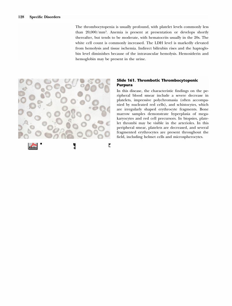
128 Specific Disorders
The thrombocytopenia is usually profound, with platelet levels commonly lessthan 20,000/mm3. Anemia is present at presentation or develops shortlythereafter, but tends to be moderate, with hematocrits usually in the 20s. Thewhite cell count is commonly increased. The LDH level is markedly elevatedfrom hemolysis and tissue ischemia. Indirect bilirubin rises and the haptoglo-bin level diminishes because of the intravascular hemolysis. Hemosiderin andhemoglobin may be present in the urine.
Slide 161. Thrombotic ThrombocytopenicPurpura
In this disease, the characteristic findings on the pe-ripheral blood smear include a severe decrease inplatelets, impressive polychromasia (often accompa-nied by nucleated red cells), and schistocytes, whichare irregularly shaped erythrocyte fragments. Bonemarrow samples demonstrate hyperplasia of mega-karyocytes and red cell precursors. In biopsies, plate-let thrombi may be visible in the arterioles. In thisperipheral smear, platelets are decreased, and severalfragmented erythrocytes are present throughout thefield, including helmet cells and microspherocytes.
40413 Tkachuk PM Gordon(SAUNM) INTERACTIVE LEFT
top of RHbase of RH
top of textbase of text
shortstandardlong

Infections
BACTERIA
To be visible on a regular peripheral blood smear, bacteria must reach adensity of about 105 organisms/mL of blood. Even if a slide is made from abuffy coat (a concentrated preparation of white cells and platelets made aftercentrifugation of blood), bacteria are generally detectable only when theirnumbers exceed about 104/mL of blood. This intensity of bacteremia rarelyoccurs: in most patients with positive blood cultures, �500 bacteria/mL arepresent. Occasionally, however, when overwhelming sepsis develops, orga-nisms are visible on a regular peripheral blood smear. Many different typeshave been seen, including Staphylococcus aureus, Neisseria meningitidis, Streptococ-cus pneumoniae, and various gram-negative bacilli.
Such high-level bacteremia can occur in asplenic hosts infected with Streptococ-cus pneumoniae, a gram-positive diplococcus. Since both this organism and thegram-negative diplococcus, Neisseria meningitidis, stain blue with Romanowskydyes, they can be differentiated on the Wright stain only by their shape andsize. Pneumococci are oval and lie end-to-end; meningococci are larger, ap-pear kidney-shaped, and lie side-by-side. A Gram stain of the smear candetermine whether the organism is gram-positive or gram-negative.
Slide 162. Pneumococcal Bacteremia
A pair of oval-shaped organisms lying end-to-end (ar-row) is visible in the cytoplasm of the lower white cell(a band) in this asplenic patient with bacteremiafrom Streptococcus pneumoniae. The upper granulocyteshows extensive cytoplasmic vacuolization, a findingoften present in severe infections.
40413 Tkachuk PM Gordon(SAUNM) INTERACTIVE RIGHT
top of RHbase of RH
top of textbase of text
shortstandardlong

130 Specific Disorders
Slide 163. Meningococcal Bacteremia
Two pairs of bacteria lie in the cytoplasm of thisneutrophil, which also has vacuoles. Each set of orga-nisms consists of two kidney-shaped bacteria lyingside-by-side. Blood cultures yielded Neisseria meningiti-dis.
Slide 164. Borreliosis (Relapsing Fever)
Relapsing fever is an infection by certain Borrelia spe-cies that consists of recurrent episodes of chills, fever,headache, and fatigue lasting a few days, followed bya few days of apyrexia. During the febrile episodesthe Borrelia spirochetes causing this disease are usu-ally visible on routine blood smears, which is the bestprocedure in establishing the diagnosis. The orga-nisms are 7–20 �m long and about 0.7 �m in diame-ter. Borrelia recurrentis, which has no animal reservoir,is spread from person to person by body lice andexists in Africa, China, and the Peruvian Andes. Tick-borne disease, whose major reservoirs are wild ro-dents, occurs in the western United States as well asmany other countries throughout the world and iscaused by several different Borrelia species. In thisslide, two contiguous threadlike spiral organisms arevisible.
40413 Tkachuk PM Gordon(SAUNM) INTERACTIVE LEFT
top of RHbase of RH
top of textbase of text
shortstandardlong
Slide 165. Ehrlichiosis
The genus Ehrlichia consists of obligate intracellulargram-negative bacteria that can be transmitted byticks to humans from their mammalian hosts, whichinclude horses, deer, and dogs. On Romanowskystains of the peripheral blood, these organisms maybe visible inside the cytoplasm of granulocytes ormonocytes, depending on the species of Ehrlichia.They appear, often in vacuoles, as dark blue bacteriain loose or dense aggregates called morulae becauseof their resemblance to mulberries. Infection withthese organisms causes fever, headache, myalgias, in-creased liver enzymes, and progressive pancytopenia.In this patient with granulocytic ehrlichiosis, a mor-ula is present just to the left of the central lobe ofthis neutrophil.

Specific Disorders 131
VIRUSES
Infectious Mononucleosis
The Epstein-Barr virus, part of the herpesvirus group, infects most humanssometime during their lives. In young children, infection with this organismis either asymptomatic or lacks distinctive clinical features, but in older chil-dren and adults a characteristic syndrome, infectious mononucleosis, devel-ops. In tropical countries, most infections occur before age 10, and infectiousmononucleosis is rare. In more affluent societies, however, a substantial per-centage of adolescents and young adults are susceptible, and the diseasecommonly occurs between the ages of 17 and 25 years. The organism istransmitted by saliva, usually from a carrier rather than someone acutelyinfected. Kissing is probably the most frequent mode of infection, and theincubation period is about 3–7 weeks.
The commonest symptoms of infectious mononucleosis are anorexia, fatigue,sweats, fever, nausea, headache, sore throat, and dysphagia, in various combi-
Slide 166. Mycobacteria: Bone Marrow Biopsy
Disseminated infection with various Mycobacterium spe-cies, including M. tuberculosis, typically causes feverand anemia, but other cytopenias, leukemoid reac-tions (marked increase in neutrophils), monocytosis,thrombocytosis, and leukoerythroblastosis may alsooccur. Bone marrow specimens may establish the di-agnosis by demonstrating granulomatous inflamma-tion with organisms visible on special stains, or byyielding the bacteria on culture. In this patient withmiliary tuberculosis, a granuloma lies in the center ofthe field, and some caseous necrosis (the pink granu-lar material) is visible within it.
40413 Tkachuk PM Gordon(SAUNM) INTERACTIVE RIGHT
top of RHbase of RH
top of textbase of text
shortstandardlong
Slide 167. Mycobacteria: Special Stains ofBone Marrow Aspirate
In this patient with AIDS, fever, and pancytopenia,an acid-fast stain of a bone marrow aspirate revealsnumerous beaded acid-fast (red) bacilli within thecytoplasm of macrophages. Culture of the specimenyielded Mycobacterium avium-intracellulare.

132 Specific Disorders
nations and sequences. Fever is frequent and sometimes reaches 40�C ormore. Bilateral lymph node enlargement, especially affecting the cervicalchain, but also commonly the axillary and inguinal nodes, is nearly universalsometime in the course of illness. The pharynx is usually inflamed, often witha patchy exudate, and erythematous palatal macules may appear. Periorbitaledema may develop. Splenomegaly is detectable in about 50–75% of patients,typically in the second week of illness, and may cause left upper quadrantdiscomfort or early satiety. Hepatomegaly is apparent in a minority of pa-tients.
40413 Tkachuk PM Gordon(SAUNM) INTERACTIVE LEFT
top of RHbase of RH
top of textbase of text
shortstandardlong
The Epstein-Barr virus infects epithelial cells in the pharynx and B-cells. Theabnormal (“atypical”) lymphocytes visible on a blood smear, however, areactivated T-cells that the infection has provoked. The leukocyte count istypically between 10,000 and 20,000/mm3, with 60% or more being mononu-clear cells, of which at least 10% are the “atypical” lymphocytes. Mild throm-bocytopenia (100,000–150,000/mm3) is common. Bone marrow examination,rarely necessary, is typically unremarkable or reveals generalized hyperplasia.
Other laboratory findings include abnormal liver tests, such as mild elevationsin transaminases, alkaline phosphatase, and bilirubin. Serum LDH levels arecharacteristically increased. Serologic tests for specific antibodies to severalantigens on the Epstein-Barr virus can establish the diagnosis of infectiousmononucleosis. More commonly, the confirmatory test is the detection of theheterophil antibody, an IgM that causes agglutination of sheep and horseerythrocytes. It usually becomes positive by the second or third week of illnessand disappears in 4–8 weeks. A rapid, reliable slide test is the Monospot.
Hematologic complications occur in �5% of cases. A hemolytic anemia withpositive cold agglutinins (IgM) is usually mild. Severe thrombocytopenia,neutropenia, and marrow aplasia are rare. Occasionally infectious mononu-cleosis leads to a virus-associated hemophagocytic syndrome characterized byfever, pancytopenia, generalized lymph node enlargement, and hepatospleno-megaly. Bone marrow examination reveals hypercellular or hypocellular he-matopoiesis and erythrophagocytosis by histiocytes.
Serious nonhematologic complications may also ensue, but are rare. Thespleen may rupture, usually following trauma, sometimes apparently mild.Upper airway obstruction can develop from pharyngeal edema or enlarge-ment of tonsils and other lymphatic tissue. Liver failure occasionally occurs,as may neurologic complications, including Guillain-Barre syndrome, periph-eral neuropathies, aseptic meningitis, meningoencephalitis, cerebellar ataxia,cranial nerve palsies, and optic neuritis. Pericarditis and myocarditis rarelydevelop. For the vast majority of patients, however, infectious mononucleosisis a mild, self-limited disease that resolves within a few weeks, leaving nosequelae.

Specific Disorders 133
Slide 168. Infectious Mononucleosis
In infectious mononucleosis, a large number of atypi-cal lymphocytes are usually visible. They are pleomor-phic and vary in size, often being large. The nucleiare oval, kidney-shaped, or lobulated; frequently con-
tain nucleoli; and may have diffuse, reticular, or par-tially condensed chromatin. The cytoplasm is oftendark blue, sometimes granular or foamy, and com-monly exhibits vacuolization. Where the atypical lym-phocytes contact other cells, indentations may occurin the cytoplasm, with increased darkness of theedges. In most cases of infectious mononucleosis,lymphocytes constitute over half of the white cells,and at least 20% of these are atypical. When cold-antibody (IgM) hemolytic anemia occurs, the redcells may agglutinate or form rouleaux. Thrombocy-topenia may also develop. Other viruses, especiallycytomegalovirus, can cause a similar hematologic pic-ture. This slide, from a patient with infectious mono-nucleosis, shows two large atypical lymphocytes, inwhich the cytoplasm is bluish-gray and the nuclei arelarge and oval-shaped. One normal small lymphocyteappears on the lower right, demonstrating its smallersize, rounded nucleus, and scant cytoplasm. Throm-bocytopenia is also apparent.
40413 Tkachuk PM Gordon(SAUNM) INTERACTIVE RIGHT
top of RHbase of RH
top of textbase of text
shortstandardlong
Human Immunodeficiency Virus (HIV) Infection
The clinical constellations of HIV infection are covered extensively in theinfectious disease literature. Lymphocyte (particularly T-cell) numbers andfunctions undergo progressive impairment. These can lead to secondary in-fections by organisms that usually are nonpathogens, secondary malignancies,abnormal immune-mediated phenomena such as immune thrombocytopenicpurpura, neutropenia and hypoproliferative anemia. Anti-HIV medicationsand antibiotics can also affect the blood and marrow. Marrow abnormalitiesin HIV infection are either nonspecific or attributable to secondary infectionor an HIV-related neoplasia such as lymphoma or Kaposi’s sarcoma. Thenonspecific marrow findings in HIV infection include plasmacytosis, lympho-cytosis (often seen as aggregates in biopsy), megaloblastosis, myelodysplasia,serous atrophy and fibrosis.

134 Specific Disorders
Slide 170. Histoplasma capsulatum: BoneMarrow Aspirate
When it causes disseminated disease, Histoplasma cap-sulatum may be visible within phagocytic cells in thebone marrow. The organisms are present as yeasts,which are round or oval, single-cell fungi that repro-duce by budding. They take up Romanowsky stains,but are often better seen with periodic acid–Schiff(PAS) or Gomori’s methenamine silver stains. In thisaspirate, the yeasts are visible within the cytoplasm ofthe phagocyte in the center of the field.
40413 Tkachuk PM Gordon(SAUNM) INTERACTIVE LEFT
top of RHbase of RH
top of textbase of text
shortstandardlong
FUNGI
Slide 169. Parvovirus: Bone Marrow Aspirate
Parvoviruses are the smallest DNA-containing virusesthat infect mammalian cells. The only known humanpathogen in this group is B19, which infects mostpeople at some time during their lives, either pro-ducing no symptoms or an illness consisting of avarying combination of fever, myalgia, arthritis, andrash, sometimes in the form of erythema infectiosum,a febrile illness with a characteristic erythema overthe cheeks (“slapped cheeks”) and a relative circum-
oral pallor. Concurrently or a few days later, a lacyerythema often appears on the trunk and extremities.Rarely, pruritic or painful, rapidly progressive swell-ing and erythema develop on the hands and feet.The rash includes papules and purpura and hassharp margins at the wrists and ankles. This disorderis called “papular-purpuric gloves and socks syn-drome.”The virus infects and destroys erythrocyte precur-
sors in the bone marrow, causing red cell aplasia,which is usually short-lived and not clinically appar-ent unless the patient has chronic hemolysis fromsuch disorders as sickle cell anemia or hereditaryspherocytosis. Even a brief cessation of red cell pro-duction in these circumstances leads to a rapid wors-ening of anemia, and this organism is the most com-mon cause of community-acquired aplastic crises. Inimmunodeficiency diseases, such as AIDS, the infec-tion may become chronic, causing a sustained redcell aplasia. The characteristic bone marrow findingsare a decrease in erythrocyte precursors and the pres-ence of giant proerythroblasts, one of which is seenas the upper cell in this slide. They typically haveintranuclear inclusions, four of which are present inthis example. The cytoplasm is usually basophilic andcontains vacuoles.
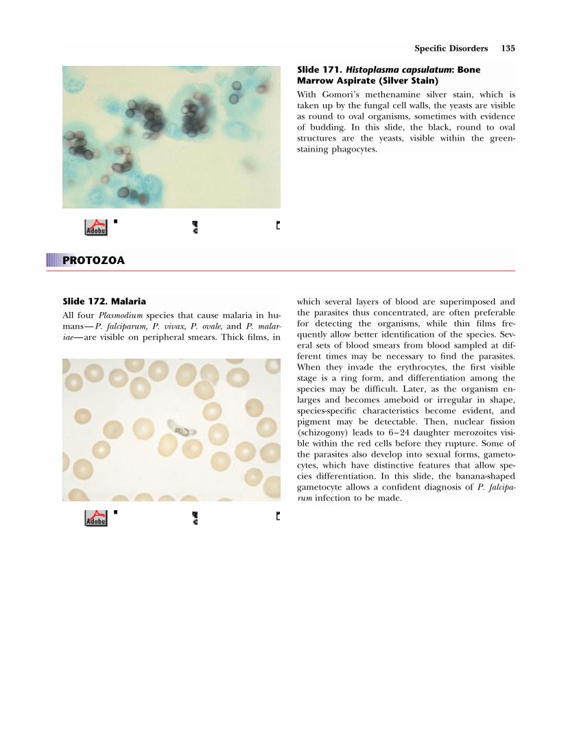
Specific Disorders 135
Slide 172. Malaria
All four Plasmodium species that cause malaria in hu-mans—P. falciparum, P. vivax, P. ovale, and P. malar-iae—are visible on peripheral smears. Thick films, in
which several layers of blood are superimposed andthe parasites thus concentrated, are often preferablefor detecting the organisms, while thin films fre-quently allow better identification of the species. Sev-eral sets of blood smears from blood sampled at dif-ferent times may be necessary to find the parasites.When they invade the erythrocytes, the first visiblestage is a ring form, and differentiation among thespecies may be difficult. Later, as the organism en-larges and becomes ameboid or irregular in shape,species-specific characteristics become evident, andpigment may be detectable. Then, nuclear fission(schizogony) leads to 6–24 daughter merozoites visi-ble within the red cells before they rupture. Some ofthe parasites also develop into sexual forms, gameto-cytes, which have distinctive features that allow spe-cies differentiation. In this slide, the banana-shapedgametocyte allows a confident diagnosis of P. falcipa-rum infection to be made.
40413 Tkachuk PM Gordon(SAUNM) INTERACTIVE RIGHT
top of RHbase of RH
top of textbase of text
shortstandardlong
PROTOZOA
Slide 171. Histoplasma capsulatum: BoneMarrow Aspirate (Silver Stain)
With Gomori’s methenamine silver stain, which istaken up by the fungal cell walls, the yeasts are visibleas round to oval organisms, sometimes with evidenceof budding. In this slide, the black, round to ovalstructures are the yeasts, visible within the green-staining phagocytes.

136 Specific Disorders
Slide 174. Babesiosis
Babesia microti, a protozoan that infects mice, is trans-mitted between hosts by ticks. Infected humans maybe asymptomatic, but, especially in asplenic hosts, fe-ver, myalgias, and hemolytic anemia can occur. Theorganisms are intraerythrocytic, ringed parasites thatare 2–3 �m in diameter, have 1–2 chromatin dots,and may assume basket shapes. More than one or-ganism can be present in a red cell; when four havetheir ends in contact, they form a pathognomonicMaltese cross appearance. Babesia organisms differfrom those of malaria in that the ring forms aresmaller, pigment is absent, and schizonts and game-tocytes do not exist. In this slide, several of the eryth-rocytes contain the parasite; one in the right centercontains two protozoa, while the large red cell to theleft of center has four.
40413 Tkachuk PM Gordon(SAUNM) INTERACTIVE LEFT
top of RHbase of RH
top of textbase of text
shortstandardlong
Slide 173. Malaria
This slide demonstrates several red cells containingring forms. The nucleus of the ring form is small—achromatin dot that stains red to purple—and thecytoplasmic ring, which stains blue, is slender. In oneerythrocyte (arrow), two ring forms are present,which strongly suggests that this is P. falciparum, sincethe occurrence of multiple ring forms in one erythro-cyte is rare in other species. Other features that cansuggest P. falciparum are the small size and thinnessof the rings, the presence of double chromatin dots,the absence of cells containing merozoites, appliqueforms (in which the ring parasites appear next to thered cell membrane), 5% or more of red cells in-volved, and the pathognomonic banana-shaped game-tocyte, shown in the previous slide. Differentiation ofthis species from the others is extremely important,since P. falciparum malaria, a medical emergency, canbe rapidly fatal without prompt, appropriate therapy.The other forms of malaria are rarely lethal.

Specific Disorders 137
Slide 176. Toxoplasmosis
Toxoplasma gondii exists in three forms: tachyzoites,which indicate acute, active infection; bradyzoites incysts, which signify latent infection; and sporozoitesin oocysts, which are present only in the intestines of
cats, the definitive hosts, or in soil contaminated bytheir feces. Humans become infected primarily byingesting material contaminated with oocysts or meatcontaining cysts, especially pork or lamb. From thesesources the tachyzoites are released in the intestines,enter the bloodstream or the lymphatics, and travelto sites where they invade tissues. Occasionally thetachyzoites are visible in the peripheral blood, butmore often they or the cysts that form from them aredetectable in various tissues, including the bone mar-row. The tachyzoite is oval or crescent-shaped, about3 by 7 �m in size, with a central nucleus and oneend that is pointed, the other round. They may bepresent within any cell except non-nucleated erythro-cytes. Cysts are about 10–100 �m in diameter andcontain numerous bradyzoites, which are nearly iden-tical to the tachyzoites in appearance. In this slide,two megaloblastic nucleated red cells are visible. Inthe one in the right upper corner, four tachyzoiteslie in the cytoplasm, indenting the nucleus.
40413 Tkachuk PM Gordon(SAUNM) INTERACTIVE RIGHT
top of RHbase of RH
top of textbase of text
shortstandardlong
Slide 175. Trypanosoma cruzi
Infection with Trypanosoma cruzi, T. rhodesiense, and T.gambiense is often detectable by examining thin orthick blood smear preparations. American trypanoso-miasis (Chagas’ disease) is caused by T. cruzi, an or-ganism found in Central and South America andtransmitted by bites of reduviid bugs. T. rhodesienseand T. gambiense, which are morphologically identical,cause African trypanosomiasis or sleeping sickness,are found only on that continent, and are transmit-ted by tsetse flies. The large round purplish structureis the nucleus; the wavy thread is the flagellum,joined to the body of the organism by an undulatingmembrane; and the small violet dot to which theflagellum attaches at the end of the parasite is thekinetoplast. Differences in sizes of the organisms andthe characteristics of their structures allow discrimi-nation between T. cruzi, present in this slide, and theAfrican species.

138 Specific Disorders
FILARIA
Adult filarial parasites live in subcutaneous tissues or lymphatics and repro-duce sexually to generate microfilaria, which may be visible in the blood-stream or bone marrow aspirates. Some (Wuchereria bancrofti, Brugia malayi,Mansonella perstans) characteristically release the microfilaria at night, some(Loa loa) during the day, and others (Mansonella ozzardi) at any time.
Slide 178. Wuchereria bancrofti: Bone MarrowAspirate
Filariasis due to Wuchereria bancrofti, a mosquito-bornedisease, is widespread throughout tropical countries.The microfilariae have encircling sheaths and areabout 200–300 �m in length and 8 �m in diameter.The purple circles throughout the organism arecalled body nuclei. The size and curvature of themicrofilaria, the presence or absence of a sheath, thenature of the body nuclei, and the characteristics ofthe tail distinguish among the various species of mi-crofilariae. In this slide, the microfilaria lies coiledamong numerous erythrocytes.
40413 Tkachuk PM Gordon(SAUNM) INTERACTIVE LEFT
top of RHbase of RH
top of textbase of text
shortstandardlong
Slide 177. Toxoplasmosis: Bone MarrowAspirate
In this bone marrow aspirate, a cyst is present withnumerous bradyzoites inside. The cyst wall does notstain well with Romanowsky dyes, but it is easily visi-ble with periodic acid–Schiff or silver stains.

Miscellaneous Disorders
HEMATOLOGIC EFFECTS OF ALCOHOLISM
Excessive alcohol intake may produce hematologic abnormalities indirectlybecause of associated malnutrition (e.g., folic acid deficiency), liver disease(e.g., spur-cell anemia), or gastrointestinal hemorrhage (i.e., anemia fromacute bleeding or from iron deficiency caused by chronic blood loss). Alco-hol, however, has direct hematologic effects as well, including several bonemarrow findings. Vacuolization of the hematopoietic precursors, especially inthe cytoplasm of proerythroblasts, occurs frequently with high intake, appear-ing within 1 week of the onset of heavy drinking and vanishing a few daysafter abstinence. Vacuoles can form in the nuclei of proerythroblasts and thecytoplasm of promyelocytes. Reversible hypocellularity of the bone marrow ormegaloblastic changes in the absence of other causes occasionally occur, andthe marrow may also show an unexplained increase in plasma cells. Ringedsideroblasts may form, and these commonly disappear days to weeks followingsobriety. The mechanism may involve impaired pyridoxine or iron metabo-lism or a direct effect of alcohol on heme biosynthesis. Occasionally a macro-cytic or microcytic anemia develops in association with the sideroblasts.
About 60% of alcoholics have a macrocytosis attributable to alcohol alone,with the MCV usually 100–110 fl. The characteristic findings of macro-ovalo-cytes and hypersegmented neutrophils seen in folate or vitamin B12 deficiencyare absent, as is anemia, unless another disorder coexists. The macrocytosispersists until 2–4 months after abstinence. Target cells and stomatocytes mayalso be visible on blood smears. Excessive alcohol intake sometimes causesthrombocytopenia, which usually subsides within a few days of sobriety, butmay “overcorrect” to cause a transient thrombocytosis in 1–3 weeks. Thenumber of megakaryocytes in the bone marrow may be normal or reduced.
40413 Tkachuk PM Gordon(SAUNM) INTERACTIVE RIGHT
top of RHbase of RH
top of textbase of text
shortstandardlong

140 Specific Disorders
Severe neutropenia may occur in alcoholics, typically associated with a severebacterial infection. In these patients the bone marrow demonstrates hypocel-lularity with decreased mature granulocytes. Other reasons for neutropeniainclude folate deficiency and hypersplenism. Neutropenia without an appar-ent cause other than alcoholism may also occur; in these patients the marrowis usually normal except for vacuoles in the erythroid and granulocyte precur-sors. Excessive alcohol intake can also cause lymphocytopenia.
Slide 179. Effects of Alcoholism: Bone MarrowAspirate
The two proerythroblasts in this slide, from a patientwith alcoholism, show numerous cytoplasmic vacu-oles.
40413 Tkachuk PM Gordon(SAUNM) INTERACTIVE LEFT
top of RHbase of RH
top of textbase of text
shortstandardlong
METASTATIC CANCER
Many malignancies metastasize to the bone marrow. Among adults, the mostcommon sites of origin are carcinomas of the lung, breast, and prostate. Asingle trephine bone marrow biopsy recovers malignant cells in about one-third to one-half of cases in which osseous metastases are present. Obtainingbilateral iliac crest specimens increases the yield. About one-half to three-quarters of patients with positive biopsies have a positive bone marrow aspi-rate. This lower diagnostic rate arises partly from the frequent presence offibrosis (desmoplastic response) around malignant cells, which makes themdifficult to aspirate. In less than half of the cases of bone marrow metastases,leukoerythroblastosis occurs on the peripheral blood smear; its presence cor-relates more with the amount of marrow fibrosis than with the extent ofmalignancy. In many patients with bone marrow metastases, the peripheralblood smear is completely normal.
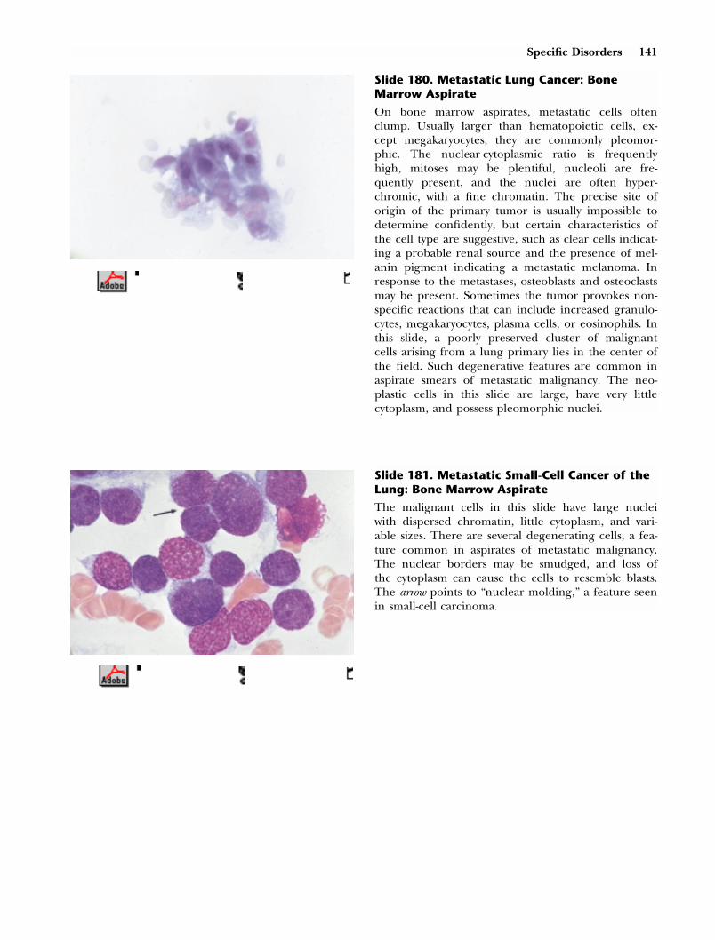
Specific Disorders 141
Slide 180. Metastatic Lung Cancer: BoneMarrow Aspirate
On bone marrow aspirates, metastatic cells oftenclump. Usually larger than hematopoietic cells, ex-cept megakaryocytes, they are commonly pleomor-phic. The nuclear-cytoplasmic ratio is frequentlyhigh, mitoses may be plentiful, nucleoli are fre-quently present, and the nuclei are often hyper-chromic, with a fine chromatin. The precise site oforigin of the primary tumor is usually impossible todetermine confidently, but certain characteristics ofthe cell type are suggestive, such as clear cells indicat-ing a probable renal source and the presence of mel-anin pigment indicating a metastatic melanoma. Inresponse to the metastases, osteoblasts and osteoclastsmay be present. Sometimes the tumor provokes non-specific reactions that can include increased granulo-cytes, megakaryocytes, plasma cells, or eosinophils. Inthis slide, a poorly preserved cluster of malignantcells arising from a lung primary lies in the center ofthe field. Such degenerative features are common inaspirate smears of metastatic malignancy. The neo-plastic cells in this slide are large, have very littlecytoplasm, and possess pleomorphic nuclei.
Slide 181. Metastatic Small-Cell Cancer of theLung: Bone Marrow Aspirate
The malignant cells in this slide have large nucleiwith dispersed chromatin, little cytoplasm, and vari-able sizes. There are several degenerating cells, a fea-ture common in aspirates of metastatic malignancy.The nuclear borders may be smudged, and loss ofthe cytoplasm can cause the cells to resemble blasts.The arrow points to “nuclear molding,” a feature seenin small-cell carcinoma.
40413 Tkachuk PM Gordon(SAUNM) INTERACTIVE RIGHT
top of RHbase of RH
top of textbase of text
shortstandardlong

142 Specific Disorders
STORAGE DISEASES
Gaucher’s Disease
Gaucher’s disease is an autosomal recessive disorder found predominantlybut not exclusively in people of Ashkenazi Jewish descent. The homozygousstate causes a deficiency in a lysosomal enzyme, glucocerebrosidase, neededto degrade glucocerebroside, which is released from dying cells such as neu-trophils. Macrophages ingest the insoluble glucocerebroside, and the accumu-lation of macrophages in liver, spleen, bone marrow, and occasionally otherorgans leads to the clinical features. Of the three kinds of Gaucher’s disease,type 1, the “adult” form, is the most common; its manifestations can becomeapparent in childhood or later in life. The spleen is often massive and may
Slide 182. Metastatic Breast Carcinoma: BoneMarrow Biopsy
Metastases are diffuse or focal on bone marrow bi-opsy, and, especially when the infiltrate is large, cen-tral necrosis can occur. Osteolytic tumors lead to lossof bony trabeculae. With osteoblastic metastases, suchas from prostate and breast cancer, new bone forma-tion and numerous osteoblasts may be prominent.Adenocarcinomas originating from any site can formglands and mucin. In this slide, from a patient withmetastatic breast carcinoma, large sheets of malig-nant cells cluster in glandlike aggregates.
40413 Tkachuk PM Gordon(SAUNM) INTERACTIVE LEFT
top of RHbase of RH
top of textbase of text
shortstandardlong
Slide 183. Metastatic Adenocarcinoma withFibrosis: Bone Marrow Biopsy
Some malignancies provoke substantial fibrosis whenthey involve bone marrow. This “desmoplastic” re-sponse is most common with tumors originating fromthe breast, lung, stomach, and prostate. In this slide,several foci of malignant cells form glandlike struc-tures within a large area of fibrous tissue above thebony trabecula. (The fibrosis should be comparedwith that in slide 87.) Because of the fibrosis, theperipheral smear may exhibit the findings of a “my-elophthisic” disorder, as occurs when abnormal tissueinfiltrates normal marrow. These changes includeleukoerythroblastosis (see slide 29), polychromato-philia, and teardrop cells.

Specific Disorders 143
cause abdominal discomfort and pancytopenia. Chronic fatigue is frequent.Bone pain, sometimes severe and often the most prominent symptom, canarise from infarction or pathologic fractures, primarily from osteolytic lesionsin the long bones but also in the ribs, pelvis, and vertebrae. A characteristicearly change is an osteolytic lesion in the distal femur that resembles anErlenmeyer flask. Aseptic necrosis of the femoral heads can occur. Hepaticenlargement, sometimes causing abdominal discomfort, is also common, andliver fibrosis can lead to portal hypertension.
A normochromic, normocytic anemia is frequently seen, but usually the he-matocrit exceeds 25. Leukopenia is typically mild, but thrombocytopenia canbe severe, leading to bleeding complications. With hepatic involvement, liverenzyme levels rise and the prothrombin time lengthens. Serum acid phospha-tase is usually increased. Gaucher cells, characteristically present in the bonemarrow, liver, and spleen, have small eccentric nuclei surrounded by a crin-kled or striated cytoplasm containing the glucocerebroside. The definitiveway to establish the presence of Gaucher’s disease is to measure glucocere-brosidase activity in the patient’s leukocytes or fibroblasts or to detect theknown mutations in the patient’s DNA.
Slide 184. Gaucher’s Disease: Bone MarrowAspirate
Many patients with Gaucher’s disease have no hema-tologic abnormalities. When abnormalities do occur,they result from hypersplenism, which can cause pan-cytopenia or an isolated reduction in any cell line.Since the definitive diagnosis rests on the studiesmentioned above, bone marrow samples are unneces-sary, but they usually reveal the characteristicGaucher cell, which has a small, eccentric nucleusand an abundant pale-blue cytoplasm with a crinkledor striated appearance. Cells indistinguishable onlight microscopy from Gaucher cells sometimes occurin other disorders, such as lymphomas, chronic gran-ulocytic leukemia, and multiple myeloma, because in-creased cell turnover exceeds the normal cataboliccapacity of glucocerebrosidase. Usually the underly-ing diagnosis is evident from the abundance of tu-mor cells in the bone marrow sample. In this slide,three typical Gaucher cells are present.
40413 Tkachuk PM Gordon(SAUNM) INTERACTIVE RIGHT
top of RHbase of RH
top of textbase of text
shortstandardlong

144 Specific Disorders
SARCOIDOSIS
In sarcoidosis, a disease of unknown cause, the involved organs containnoncaseating granulomas, sometimes accompanied by fibrosis. This disordercan occur in virtually any part of the body, but the vast majority of patientshave disease in the thoracic cavity, affecting the lung parenchyma and lymphnodes in the mediastinum and hila, usually bilaterally. Other areas commonlyinvolved include the eyes and skin. The usual age at onset is between 20 and40, with rates about 10 times higher among blacks than whites in the UnitedStates. The disease is generally more common in females than males, but itsfrequency varies considerably in countries throughout the world. Sarcoidosisis more common in northern than southern Europe and is relatively unusualin Asia and South America, where it is more prevalent in colder than warmerclimates. The duration and prognosis relate to the onset and extent ofdisease: patients with the abrupt onset of erythema nodosum or with asymp-tomatic bilateral hilar lymph node enlargement tend to have a short, self-limited course, while those with insidious onset and extensive extrapulmonaryinvolvement often have progressive fibrosis of the lungs and other organs.
Hematologic abnormalities are common in sarcoidosis. Anemia occurs inabout 5–30% of cases; its cause may be hypersplenism, present in approxi-mately 15% of patients; anemia of chronic disease; granulomas in the bonemarrow, causing replacement or suppression of hematopoietic cells; immune-related hemolysis (rarely); or apparently unrelated conditions, such as irondeficiency. The red cells are usually normochromic and normocytic, and theanemia typically resolves with systemic corticosteroid therapy. Neutropenia(defined as �3,000 neutrophils/mm3) occurs in about 30–40% of patients,but is usually mild. The most likely cause is the presence of bone marrowgranulomas. Lymphopenia (�1,500 lymphocytes/mm3) develops in 30–50%of cases, commonly with reduced T-lymphocyte numbers and CD4/CD8 ra-tios, and may originate as lymphocytes migrate out of the circulation intoareas of inflammation. Eosinophilia (�250 eosinophils/mm3) develops in 5–35% of patients through unknown mechanisms. Monocytosis (�800 mono-cytes/mm3) occurs in about 10% of cases. Thrombocytopenia, an uncommonfinding, can arise from hypersplenism or immunologic destruction.
The bone marrow biopsy discloses granulomas in about one-half of patientswith anemia. Usually disease is clinically evident in many other sites, espe-cially in the thoracic cavity, but chest films are sometimes normal. Typicallythe granulomas are numerous and well-formed but occupy only a smallamount of the bone marrow specimen. Langhans’ giant cells may be appar-ent, but necrosis is very rare and should strongly suggest another diagnosis.
40413 Tkachuk PM Gordon(SAUNM) INTERACTIVE LEFT
top of RHbase of RH
top of textbase of text
shortstandardlong

Specific Disorders 145
Slide 185. Sarcoidosis: Bone Marrow Biopsy
This slide discloses several noncaseating granulomaswith giant cells admixed with reactive inflammatorycells, including a prominent population of small lym-phocytes.
Slide 186. Splenectomy: Peripheral Smear
In the first week after splenectomy, neutrophilia iscommon, but it generally abates. Mild to moderateleukocytosis may persist, usually because of increasedlymphocytes and monocytes rather than neutrophils.Beginning about one week after splenectomy, manypatients have marked thrombocytosis, sometimes upto 2,000 � 109/L (2 million/mm3), but the plateletcount then typically falls to levels of about 500–1,000 � 109/L, which may persist for years. Plateletsmay also be larger than normal. Changes in theerythrocytes are universal. Howell-Jolly bodies arepresent in varying numbers in all patients, and Pap-penheimer bodies are also frequent. The red cellsurface area increases, commonly leading to targetcells. Abnormal cells that the spleen ordinarily re-moves, such as acanthocytes, spherocytes, and bizarre-shaped erythrocytes, are often present. Similarly,polychromatophilic cells are numerous; the spleenordinarily removes many of these from the circula-tion until they mature. In this slide, several Howell-Jolly bodies are present, along with target cells,spherocytes, and polychromatophilic erythrocytes.This patient also had hypothyroidism, which may ex-plain the macrocytosis.
40413 Tkachuk PM Gordon(SAUNM) INTERACTIVE RIGHT
top of RHbase of RH
top of textbase of text
shortstandardlong
FINDINGS FOLLOWING SPLENECTOMY

146 Specific Disorders
SYSTEMIC MAST CELL DISEASE
In this disorder, excessive mast cells infiltrate various organs, most frequentlythe skin, bone marrow, liver, lymph nodes, spleen, and gastrointestinal tract.About 65% of cases occur in childhood, and in most of these patients thedisease resolves or markedly improves during adolescence. When the diseasebegins in adults, the average age at onset is about 60, and both sexes areequally affected. A classification of systemic mastocytosis defines four groups:indolent disease, by far the most common form; mastocytosis associated witha hematologic disorder; aggressive lymph node involvement with blood eosin-ophilia; and mast cell leukemia. Skin lesions in the form of urticaria pigmen-tosa may be present in any of the four forms. Characteristic are numerousreddish-brown, macular or slightly raised lesions several millimeters in diame-ter, primarily on the trunk and proximal extremities. They may cause pruri-tus and dermographism. A pathognomonic feature is Darier’s sign, the devel-opment of a wheal on rubbing or scratching the lesion.
In indolent disease, the other findings related to infiltration of organs bymast cells are skeletal disease, hepatosplenomegaly, lymph node enlargement,malabsorption from small intestinal invasion, and hematologic abnormalitiesresulting from bone marrow involvement. Skeletal disease includes diffuseosteopenia or focal sclerotic or lytic lesions; these bony abnormalities may beasymptomatic or may cause pain or fractures. Many patients have some labo-ratory evidence of fat malabsorption, but it is usually insufficiently severe toproduce significant clinical findings. Excessive mast cells in the bone marrow,present in about 90% of patients, result in anemia in about 30–50%, throm-bocytopenia in 15–20%, leukopenia in 15–20%, leukocytosis in 20–30%, andeosinophilia in up to 40%.
Other manifestations of indolent disease originate from the systemic effectsof mast cell mediators, either stored within cytoplasmic granules or producedat the time of mast cell stimulation. These include histamine; eicosanoids,such as prostaglandin 2; and cytokines, including tumor necrosis factor andvarious interleukins. Their hemodynamic effects may cause syncope or flush-ing, and their gastrointestinal effects include diarrhea and increased gastricacidity, which sometimes leads to peptic ulcers of the stomach and duode-num. Patients may have constitutional complaints such as fever, malaise, andweight loss, while neurologic effects include headaches and confusion. Manyof the symptoms caused by mast cell mediators appear or worsen with exer-cise, heat, alcohol, narcotics, salicylates, and anticholinergic agents.
In the second type of mast cell disease, patients have a concurrent hemato-logic condition, most commonly a myelodysplastic or myeloproliferative dis-ease. Other associated malignancies have included Hodgkin’s disease or non-Hodgkin’s lymphoma and various leukemias, some secondary to a previouslyunderlying hematologic disease. Fever, weight loss, gastrointestinal com-
40413 Tkachuk PM Gordon(SAUNM) INTERACTIVE LEFT
top of RHbase of RH
top of textbase of text
shortstandardlong

Specific Disorders 147
plaints, pruritus, dermographism, and flushing are more common in this typethan in the indolent form. The prognosis for the patient depends primarilyon the associated disease, however, rather than on the mastocytosis itself.
The third type of systemic mast cell disease is an aggressive disorder, withlymph node enlargement affecting both peripheral and intra-abdominal sites,hepatosplenomegaly, and peripheral eosinophilia, leading to a markedlyshortened survival. Ascites may be present.
The fourth type, mast cell leukemia, is defined by the presence of at least10% mast cells, many hypogranular or multinucleated, among the circulatingnucleated cell population. Skin lesions are usually absent, and the patientstypically have fever, weight loss, weakness, hepatosplenomegaly, and gastroin-testinal symptoms of abdominal pain, vomiting, and diarrhea.
Evaluation of a patient for the presence of systemic mast cell disease shouldinvolve a careful skin examination and biopsies of suspicious lesions; bonemarrow aspiration and biopsy; serum tryptase level determinations (oftenincreased); and 24-hour urine collections for mediators such as histamine.Biopsies of clinically involved tissues such as lymph nodes or intestinal mu-cosa may demonstrate increased populations of mast cells, whose reliabledetection requires special stains, such as Giemsa and toluidine blue, ormonoclonal antibodies to tryptase or chymase.
Slide 187. Systemic Mast Cell Disease: BoneMarrow Aspirate, Toluidine Blue Stain
In systemic mast cell disease the peripheral blood isoften normal, but increased numbers of some cellsmay occur, such as eosinophils, basophils, monocytes,neutrophils, and platelets. Mast cells are visible in theperipheral blood in patients with mast cell leukemia.On the aspirate, mast cells are often increased andmay be cytologically normal, exhibiting the character-istic purplish granules in the cytoplasm and a centralnucleus. Sometimes, however, they are atypical inhaving a lobulated nucleus or decreased granules.Mast cells stain, as in this slide, with a metachromaticpreparation, toluidine blue, and appear as heavilygranulated cells, commonly in the bone marrowstroma.
40413 Tkachuk PM Gordon(SAUNM) INTERACTIVE RIGHT
top of RHbase of RH
top of textbase of text
shortstandardlong

148 Specific Disorders
Slide 188. Systemic Mast Cell Disease: BoneMarrow Biopsy
On biopsy, the mast cells commonly provoke exten-sive fibrosis and are most frequent around blood ves-sels, adjacent to bony trabeculae, or in small nodules.Occasionally, mast cells diffusely replace the bonemarrow. They are usually spindle- or oval-shaped cellswith clear cytoplasm. Eosinophils, lymphocytes, andplasma cells may accompany the mast cells. In otherportions of the marrow, increases in granulocytes,eosinophils, and megakaryocytes are common. In thisslide, spindle-shaped mast cells lie below the bonytrabecula.
40413 Tkachuk PM Gordon(SAUNM) INTERACTIVE LEFT
top of RHbase of RH
top of textbase of text
shortstandardlong