proton radiography - nsc.ruancient.hydro.nsc.ru/srexpl/out/protony/2006_lanl.pdf · proton...
TRANSCRIPT

32 Los Alamos Science Number 30 2006
Proton Radiography
32 Los Alamos Science Number 30 2006
Proton RadiographyChristopher Morris, John W. Hopson, and Philip Goldstone
The United States stopped nuclear testing in 1992, and since then it has become increasingly important to develop predictive models for the behavior of materials driven by high explosives. The primary experimental tools to observe that dynamic behavior have been based on x-radiography, the imaging technique used during the earliest days of the Manhattan Project. Over the last decade, however, a new imaging technique has been developed that uses high-energy protons, rather than x-rays, to radiograph materials during dynamic experiments. Proton radiography allows researchers to make short mov-ies and obtain much more detailed information on the motions and densities of materi-als when driven by shock compression than was ever possible before. The penetrating power, or long mean free path, of protons and the ability to focus them are opening up new opportunities for quantitative experiments, accurate model development, and designer training that will revolutionize how the U.S. nuclear stockpile is stewarded in the future.
pRad article(NC).indd 32 1/24/06 8:40:00 AM

Number 30 2006 Los Alamos Science 33
Proton Radiography
Number 30 2006 Los Alamos Science 33
The technique of focusing protons for radiography was first demonstrated in 1995
(Figure 1). The protons came from the P3W pion channel in the pion experimental area (Area-A) of the Los Alamos Neutron Science Center (LANSCE) linear accelerator. Since
then, major progress has been made in developing techniques for dynamic imaging that have made proton radiography (pRad) an important contributor to the weapons pro-gram. Approximately 30 small-scale dynamic experiments are performed per year using the line C facility at
LANSCE shown in the opening pho-tograph. For the first time, movies of up to 32 frames can be made of explo-sively driven experiments, allowing new phenomena to be observed and quantified. (Just as visualization is a critical tool for designers using advanced simulation and computing,
Figure 1. Demonstrating pRad with Magnetic FocusingA beam of protons (188 MeV in energy) from the P3W channel at LAMPF was sent through an object—a 6-mm-thick steel plate with the words LANL P-RAD machined halfway through—and the positions and trajectories of the transmitted protons were recorded by a layered proton detector (see diagram). (a) With nothing between the object and the detector, we obtained a blurred radiograph (green) showing the positions of the protons as they entered the detector, but by projecting the proton trajectories recorded at the detector back to the object, the letters on the sign became visible (red radiograph). (b) The purple radiograph (inverted image) was obtained by placing a triplet of quadrupole magnets between the object and the detector and directly recording the positions of the protons entering the detector. Because the magnets act like a proton lens, focusing the protons at the detector, they allow a clear image to be recorded. Magnetic focusing makes flash radiography possible because each proton does not need to be individually measured.
(a)
At the detector Projected to the object After a lens
Object DetectorLens(b)
pRad article(NC).indd 33 1/24/06 8:40:21 AM

34 Los Alamos Science Number 30 2006
Proton Radiography
seeing multiple frames adds a new dimension to understanding complex dynamic phenomena.) New capabili-ties are also being added to the line C facility continuously. Most recently, a powder gun drive has been commis-sioned, and a new magnifying lens is currently under construction.
In addition to the ongoing pro-gram at LANSCE, experiments with higher proton energies have been conducted at the Alternating Gradient Synchrotron (AGS) at Brookhaven National Laboratory (BNL). These experiments have shown that pRad could, in the future, enable a revolutionary improvement in data from hydrotests compared with the impressive capabilities of the dual-axis radiographic hydrotest facil-ity (DARHT), the best flash x-ray machine in the world.
In this article, we discuss the con-tributions of pRad to stockpile stew-ardship and discuss how it emerged as a result of the interaction of basic science activities at LANSCE with nuclear weapons research.
Nuclear Weapons
Modern nuclear weapons in the U.S. stockpile use two stages to develop high yield-to-weight ratios. The first stage, the primary, works by using high explosives to compress a fissionable core, or “pit,” to a super-critical state in which it can sustain a chain reaction. The pit is filled with deuterium-tritium (d-t) gas, and the pit’s implosion, along with the onset of the fission reactions, heats the d-t gas to the point at which the d-t atoms undergo fusion reactions. In turn, neutrons released from the d-t fusion reactions produce additional fission reactions and amplify the energy released from the primary. This process of using fusion reactions to enhance the energy release is called “boost.”
Although the basic physical pro-cesses involved are relatively straight-forward, there are many subtleties that can lead to uncertainties in predicting the performance of a nuclear primary. The pressures generated in a conver-gent explosion far exceed those that are available for study in static labora-tory experiments. The high pressures, forces, and accelerations involved drive instabilities at material inter-faces that are difficult to predict with numerical simulations. The materials used in nuclear weapons are quite complicated in their behavior. For all these reasons, it is important to obtain data on the response of these materi-als in conditions and configurations that are close to the working condi-tions of a primary in order to develop and validate models and calculations. Obtaining such data will improve the predictive capability of our advanced simulation and computing models, reduce remaining uncertainties within those models, and help us ensure the safety and reliability of the stockpile without new nuclear tests.
A number of experiments must focus on isolating and closely study-ing individual processes or the combined effects of some of those
processes to improve the underlying physics models of materials response. Much of the work with pRad at LANSCE is along these lines. The results from such fundamental or semi-integrated experiments (some-times called “small-scale”) help lead to validated science-based models that can be incorporated into the computer codes used to predict the entire weap-ons system. It is vital that the under-standing embodied in these codes be accurate.
Other experiments must be more integral in nature and must more closely mock up the full set of pro-cesses and interactions that occur in a primary implosion (but, of course, without producing a nuclear explo-sion). One can obtain valuable data by replacing the fissile material with a surrogate, in a geometry that closely matches that of a primary (Figure 2). These large, integral implosion experiments are called hydrotests (hydrodynamic tests) because, at high pressures, the material flows like water. Electrical pins and flash x-ray radiography have been used to study hydrotest experiments since the development of the first plutonium weapons during World War II. The
Figure 2. Schematic of an Implosion AssemblyThis schematic of implosion shows high explosives surrounding a fissile core. Detonation of the high explosive produces a converging shock wave that compresses the core to a supercritical state.
Subcritical mass
Chemical explosive (HE)
Compressed
supercritical mass
Implosion
pRad article(NC).indd 34 1/24/06 8:40:22 AM

Number 30 2006 Los Alamos Science 35
Proton Radiography
most powerful x-ray machine ever built, DARHT, is the primary diag-nostic for hydrotests in the United States. Results from hydrotests, along with past nuclear-test data, are vital for benchmarking and test-ing simulation models to ensure that the underlying science within these models is sufficiently accurate and complete. And although hydrotests use surrogate materials to prevent a nuclear explosion, precise data from these tests do allow direct inference of the initial conditions for the nuclear performance of a primary, including boost. That knowledge itself can sig-nificantly constrain predictions and can substantially increase designer confidence.
Current pRad experiments at LANSCE are playing an increasingly important role in developing the sci-ence for primary predictive capability. In addition, in the future, higher-energy pRad could provide a new, quantitative, and much more capable diagnostic for hydrotest experiments, meeting stringent requirements for establishing the initial conditions for boost.
pRad Principles
Radiographic information is obtained by measuring the inten-sity of the shadow of an object in a beam of penetrating radiation. If the radiation is attenuated over too short a distance, only the outer edges of the object can be measured, and if it is attenuated over too long a distance, there is no shadow. Indeed, during the Manhattan Project, flash x-radiogra-phy was used to measure the outside edge of an imploded core made of a heavy-metal surrogate in order to test the high-explosive drive for the implosion. X-rays have since been used in sophisticated hydrotests and “small-scale” research into shock- and high-explosive-driven phenomena.
However, high-energy proton beams offer an almost ideal radiographic probe for studying the physics related to primary implosion phenomena because their mean free path (or aver-age distance traveled between colli-sions) can be tailored to allow seeing inside almost any experiment. Proton radiography offers new power and finesse for imaging such experiments.
Protons interact with matter in several ways. Each of these ways can be used to advantage for radiography. At high energies, protons interact with atomic nuclei primarily through the nuclear force (the short-range “strong” force that binds nuclei into a tight, compact shape) and less so through the Coulomb force (the long-range electrostatic force between charges). They also interact with electrons through the Coulomb force.
Because the cross section of the atomic nucleus is small (πr2), high-energy protons travel a much longer distance in matter than even the most penetrating x-rays. This property makes them well suited for radio-graphing thick objects. High-energy protons that interact directly with a nucleus through the nuclear force are usually scattered through large angles. They are thus scattered out of the beam, and their energy is significantly reduced. In other words, protons undergo hard scattering much like x-rays in x-radiography, but they have a longer mean-free path than x-rays. This property makes them ideal for transmission radiography.
Every proton that passes by a given nucleus, even if not close to it, is given a push by the Coulomb force. The sum of all the small pushes from nuclei leads to changes in direction and therefore diffusion of the incident angle. A theory for how to treat this angle diffusion was developed by Enrico Fermi in the 1930s. Coulomb multiple scattering from nuclei can make the net mean-free path for protons shorter than
the nuclear mean-free path alone by using an angle collimator at a Fourier point (angle focus) in the lens, a fea-ture that allows the mean free path to be adjusted to match nearly any experiment. It is this feature of proton interactions that has enabled the pRad program at LANSCE to address many different physics problems with pro-tons of the same energy.
A Coulomb interaction also occurs between protons and electrons in the material. Because the electrons have very small mass compared with the protons, the interaction causes large changes in electron directions and velocities but only incremen-tal changes in proton direction and energy. In the 1930s, Hans Bethe and Felix Block developed a theory that shows how the Coulomb interaction with electrons leads to a net force that results in a drag, slowing down and eventually stopping the protons.
Brief History of pRad
Although the motivation for devel-oping pRad came from the weapons program, it is a remarkable fact that all the techniques, ideas, and equip-ment that were synthesized into this new technology are a legacy of the nuclear physics program at the Clinton P. Anderson Los Alamos Meson Physics Facility (LAMPF), the predecessor of LANSCE. Proton radiography would never have been developed had it not been for the colocation of basic research with clas-sified, national-defense research and development. Strong basic-research efforts at the weapons laboratories can continue to provide personnel and feed innovative technologies that will be used for solving the difficult prob-lems of stockpile stewardship in the future, and the emergence of pRad is just one of many important examples of how this outcome is achieved.
The basic idea behind pRad
pRad article(NC).indd 35 1/24/06 8:40:22 AM

36 Los Alamos Science Number 30 2006
Proton Radiography
goes back to Andreas Koehler from Harvard, who pointed out in the 1960s that the statistical fluctuations in proton range could be used to make very-high-contrast radiographs of objects if the proton beam energy was adjusted so that the proton range was just equal to the thickness of an object being radiographed. The range strag-gling in some conditions is only a few percent of the total range. Because of this narrow variation in range, the transmission of a proton beam can vary by 100 percent with just a few percent change in the thickness of an object. This high contrast meant that radiographs could be made using low radiation doses (beam intensities multiplied by irradiation times) when compared with conventional x-ray radiography, but the position resolu-tion would be poor. Coulomb multiple scattering of protons leads to blur in the radiographs that is about an order of magnitude larger than that in radio-graphs made using x-rays because the latter travel on straight paths between interactions.
Ken Hanson implemented the idea of range radiography using proton beams at LAMPF. Hanson was able to extend the dynamic range over which this type of radiography could be applied by stopping the protons in a thick detector after the object rather than in the object itself and using the distances traveled in the detector to measure the energy remaining in the transmitted protons. In this way, varia-tions in the thickness (areal density) of the object up to the detector thick-ness could be optimally radiographed. This energy-loss radiography provided better position resolution even using a low dose because the average proton energy in the object was higher, more protons were transmitted, and each transmitted proton provided radio-graphic information.
As one of us (Chris Morris) recalls, in the early 90s, the weapons program funded a study of neutron radiography
led by Victor Gavron. One important idea to come from that study was that the long mean-free path of high-energy protons, neutrons, and other hadrons (particles that interact through the nuclear force) made them ideal for performing radiography on objects typically encountered in the nuclear weapons program. Steve Sterbenz picked up on this idea and explored the possibility of using neutrons to radiograph hydrotests. However, the available neutron flux, even if the intense pulses from the Proton Storage Ring (PSR) were used to produce the neutrons, would be insufficient for obtaining images during the short tim-escales of a hydrotest. Gerry Garvey, then head of LAMPF, on hearing this argument, immediately asked, “Why not use protons?”
Because they are charged par-ticles, protons bend as they move through magnetic fields, and they can therefore be focused by magnetic lenses. The technology of focusing and bending proton trajectories using magnets, or the optics of charged particles, is central to the operation of modern particle accelerators, and many physics experiments performed at LANSCE also required expertise in the optics of charged particles. For example, the high-resolution spectrometer (HRS) at LAMPF, one of the premier charged-particle spec-trometers in the world, was tuned by optically imaging the low-intensity proton line in the focal plane using a phosphor and an intensified charge-coupled-device camera. Jerry Nolen, who developed this tuning technique, thus demonstrated that it was possible to image low intensities of protons with position resolution approaching 100 micrometers.
When one of us (Chris Morris) put all these ideas together, it became obvious that high-energy proton beams—within the existing state of the accelerator art—could provide a breakthrough in dynamic materials
experiments and hydrotest diagnos-tics. The final steps that led to the development of pRad occurred when Tom Mottershead and John Zumbro developed a magnetic lens design that provided good position resolution over the entire field of view required for radiography, and Nick King adapted a detector system based on fast imaging that had been developed for other weapons applications.
The development of pRad bears out the vision that Louis Rosen had more than forty years ago—by col-locating basic research with program-matic work, LAMPF/LANSCE would be of great benefit to the Laboratory’s mission. Although over time, the LANSCE mission has increased its focus on national security, the impor-tance of a clear engagement between defense research and the broad front of fundamental science has remained unchanged, and LANSCE contin-ues to play an important role in that regard.
pRad and the Physics of Implosion
Proton radiography is arguably the most valuable and versatile single technique available to interrogate the hydrodynamic aspects of primary physics. Many physical regimes and processes become operative as a weapons implosion proceeds. They include the initiation and detonation of the chemical high explosive, the complex response of the metal com-ponents to intense shock waves, the extremely high rate of deformation and compression of the fissionable components during the supercritical assembly, and the fundamental hydro-dynamics and hydrodynamic instabili-ties that are driven by these extreme conditions. For each condition, it is necessary to develop and validate explicit hydrodynamic physics mod-els that can be implemented in new
pRad article(NC).indd 36 1/24/06 8:40:22 AM

Number 30 2006 Los Alamos Science 37
Proton Radiography
simulation computer codes from the Advanced Simulation and Computing (ASC) Program. The physics models must capture critical hydrodynamic behaviors with high accuracy and are the main drivers for setting the design of most weapons physics experiments.
Though many techniques are (and must be) used, it remains experimen-tally challenging to interrogate the critical state variables and stress-strain response in the interior of materials under dynamic stress. Modeling of many processes depends on accu-rately capturing the time evolution of those state variables and stress-strain responses on a microsecond scale. With its ability to penetrate and accu-rately image the interior of highly compressed components, as well as its highly flexible and precisely record-able pulsed format, pRad is uniquely suited to providing the necessary data for weapon certification codes and models. At present, pRad is being applied to a number of key scien-tific questions that address stockpile stewardship goals. These include the detailed detonation behavior of insen-sitive high explosives; the dynamic material response to shock loading, including material failure; and experi-ments relating to understanding mate-rials dynamics and conditions late in the process of implosion.
High-Explosive Detonations
Detonation fronts move through high explosives with velocities near 8 millimeters per microsecond (mm/µs). The combination of chemistry and shock physics needed to describe the detonation process is not com-pletely understood and is difficult to model. Most calculations use param-eterized geometric models to describe the detonation of high explosives. Proton radiography has been used to diagnose a number of experiments used to study the detonation process
in both conventional and insensitive high explosives.
The complex mix of shock physics and chemistry that occurs in a detona-tion front in high explosives has not yet been modeled from fundamental (atomic-scale) physics principles. However, physical models exist, and improvements continue to be devel-oped that incorporate more complete (and complex) physics. For example, the pressure profile through the deto-nation front and the propagation of the front can be adequately described with some simplifying assumptions in a parameterized model. Movies made with pRad provide a way to establish parameters, as well as develop and check these models.
A number of pRad experiments on detonation physics performed by Eric Ferm are revealing in this regard. They include studies of a detonation front turning around a corner as it propagates from a narrow cylinder of high explosive into a wider one, rate sticks for measuring the veloc-ity of the detonation front, colliding detonation fronts showing reflected shock waves propagating through detonation products, and failure cone experiments for determining the radius at which the detonation fails to propagate. Some time sequences from these experiments are shown in Figure 3. The failure cone and corner-turning experiments allow predictions of detonation front propagation to be checked, and the rate stick and col-liding-wave experiments provide data on the equation of state of detonation products over a wide range of pres-sures. In particular, the velocity of the reflected shock waves in the detona-tion products determines a shock Hugoniot in the high-explosive prod-ucts. The experimental results shown in Figure 3—for example, the dead zone in the corner-turning experi-ment—challenge even the best current models of high-explosive detonation.
Material Failure
When the pressure wave pro-duced by a high-explosive detonation impacts a metal surface, the metal can be accelerated to velocities that exceed the sound speed in the metal. This phenomenon results in the for-mation and propagation of shock waves in the metal. The shock waves reflect from interfaces and surfaces putting the material alternately under compression and tension, and in many cases, under considerable shear. The pressures induced in these processes can exceed the strength of the metal and can lead to phase changes, as well as tensional and shear failure. These phenomena have been studied for well over a century. However, the richness of the physical processes involved and the complexity of the materials make it a challenge to develop microscopic models based on the fundamental forces of nature. We need data to guide and validate improvements to the various approaches to modeling these phenomena.
A set of experiments has been performed with pRad to study how shocked metals fail when a shock wave is reflected from a free metal surface and the resulting rarefac-tion wave puts material in tension. Experiments were driven with Taylor waves (the shape of a shock wave produced by high explosives) and with plane waves (waves produced by a high-explosive-driven flyer plate or a projectile from a gun). A composite of experiments showing the richness of metal failure from shock-induced tension is displayed in Figure 4.
Fragmentation, another form of material failure, occurs when met-als are stretched (or strained) at very high rates. Figure 5 shows a series of experiments performed with pRad to study this phenomenon. Several sequences of metal failing in stretch-ing modes are shown. In Figure 5(a), a half-cylinder of titanium is being
pRad article(NC).indd 37 1/24/06 8:40:23 AM

38 Los Alamos Science Number 30 2006
Proton Radiography
Figure 3. Studies of High-Explosive DetonationThese radiographic time sequences show a set of experiments aimed at studying the propagation of a detonation wave in a high-explosive and the densities and pressures of the detonation products. (Left to right) The images record detonation fronts turning a corner, traveling along a rate stick, colliding, and propagating in a failure cone, respectively. The images were enhanced by Abel inversion and therefore show volume densities (gm/cm3). (This figure is courtesy of Eric Ferm.)
Corner turningT
ime
Rate stick Colliding fronts Failure core
1.42 µs
2.86 µs
4.29 µs
7.15 µs
8.58 µs
1.42 µs
2.86 µs
4.29 µs
7.16 µs
5.72 µs
8.58 µs
7.52 µs
9.67 µs
11.10 µs
13.96 µs
12.53 µs
15.39 µs
1.63 µs
2.16 µs
2.70 µs
3.77 µs
3.24 µs
4.30 µs
Undetonated HE
Detonation
wave
Detonation
wave
Detonation
products
Detonation
wave
Reflected
shock
waves
Detonation
waves
Expected
failure radius
pRad article(NC).indd 38 1/24/06 8:40:30 AM

Number 30 2006 Los Alamos Science 39
Proton Radiography
Figure 4. Spall from Metal Plates Driven by a Planar Shock WaveThese data, courtesy of D. Holtkamp, show the “spall” type of metal failure that occurs when metals are put in tension by shock waves that reflect from a free surface and produce rarefaction waves. (Left to right) The metal samples undergoing spall are 6-mm-thick copper, 12-mm-thick copper, 12-mm-thick tin, and 4.4-mm-thick tantalum. In the leading part of the experiment, layers break off from the free surface as a result of spall. Later these layers become disorganized and appear to be broken up. Each experiment was driven by a plane-wave high-explosive driver.
6-mm copper
Spall layers
Spall layers
Melt and cavitation
12-mm copper 12-mm tin 4.4-mm tantalum
pRad article(NC).indd 39 1/24/06 8:40:32 AM

40 Los Alamos Science Number 30 2006
Proton Radiography
rapidly expanded by detonation of a cylindrical charge of high explosives placed inside the titanium cylinder. As the explosive expands radially, the cylinder fragments in a bacon strip pattern that seems to be character-istic of uniaxial (cylindrical) strain. In Figure 5b, a hemisphere of a ura-nium alloy is being expanded with a hemispherical high-explosive charge. Here, the failure results in irregular cornflake-shaped fragments.
These experiments demonstrate the richness and complexity of the response of real materials at high strain rate. These data are part of a program of model development aimed at a better understanding and predic-tive capability for the materials and conditions encountered in weapon systems.
Instabilities and Ejecta
Another very interesting field of research with important applications in weapons is the study of instabili-
ties. When a dense fluid is accelerated by a light fluid (Rayleigh-Taylor) or when an interface between two fluids is impulsively driven (Richtmyer-Meshkov), the interface is unstable. As the instability grows, the two flu-ids mix. Modeling instability growth in fluids is numerically difficult. For solid interfaces, even predicting the onset of instability growth becomes difficult because material strength sta-bilizes the surfaces.
Instability growth has been radio-graphed with pRad in several experi-ments. Two examples are shown in Figure 6. The first two time sequences in Figure 6a show different views of a jet growing vertically from a slot cut in an aluminum disc-shaped target as a shock wave from a high-explosive charge located underneath the disc propagates in orthogonal directions. The sequence in Figure 6b shows the development of a Richtmyer-Meshkov instability in a thin tin target containing a wavy, or sine-wave-shaped, free sur-face. That sine-wave-shaped perturba-tion was machined into the flat surface
as a seed for the growth of the instabil-ity. The tin plate was then struck by a flyer plate driven by a high explosive, and the rate of instability growth was then determined from the sequence of radiographs shown in the figure.
Small Implosion Experiments
In addition to these fundamental and semi-integral small-scale experi-ments, we have also performed some small implosion experiments with pRad at LANSCE, using experimental con-figurations and high-explosive charges suitable to the constraints of that facility. Although these integral experiments cannot be described in detail here, they have provided valuable and relevant information on the evolution of materi-als dynamics in implosions.
High-Energy pRad
Planning done within the nuclear weapons program has shown that the
Detonation
front
Bacon
striping
Corn
flaking
Figure 5. Fragmentation Failure of Metal Driven by Shock Waves from High-Explosive Charges(a) Detonation of an explosive charge placed in a titanium half-cylinder causes rapid cylindrical expansion. The failure of the half-cylinder of titanium leads to bacon striping, or fragmentation along the axial direction of the cylinder (data
are courtesy of P. Rightly). (b) A hemispherical shell of uranium–6% niobium is being spherically expanded by detonation of a high-explosive charge (data are courtesy of K. Prestrige). The rapid expansion causes formation of a cornflake pattern of fragmentation.
pRad article(NC).indd 40 1/24/06 8:40:33 AM

Number 30 2006 Los Alamos Science 41
Proton Radiography
800-million-electron-volt (MeV) pRad facility at LANSCE is needed to sup-port many of the long-term research goals of the weapons laboratories, and many years of experiments have already been identified to support specific stewardship deliverables. However, in addition to the need for the capability at 800 MeV, our col-leagues are considering the potential—and potential need—for an extension to 20 giga–electron volts (GeV).
Full-scale hydrotest radiography with pRad requires higher energy than is available at LANSCE. A series of experiments has been performed using the high-energy protons available at the AGS at BNL with several goals in mind: developing the techniques needed for high-energy pRad, dem-onstrating the capabilities of pRad for interrogating full-scale hydrotests, and making some direct comparisons with DARHT. Much of this work is classified, but the conclusions are rather remarkable and can be given here. The quality of flash radiography with protons is so much better for thick hydrotest objects than even that obtained from DARHT that it would take about 100 times more x-ray dose than DARHT can currently deliver to
31.096 µs
30.02 µs
28.950 µs
27.876 µs
26.803 µs
25.730 µs
24.657 µs
Figure 6. Ejecta and Instability Experiments (a) These two sequences show the development of a metal jet from a slot in an aluminum target looking along the slot (left) and across the slot (right). The jet is driven by shock waves from a high-explosive charge located underneath the plate. (b) The growth of a “classic” Richtmyer-Meshkov instability from a tin target is shown. It contains a sinusoidally shaped free surface. The instability is driven by the impact of a high-explosive-driven flyer plate moving down in this figure. [The data in (a) are
courtesy of K. Prestridge and in (b), courtesy
of W. Buttler.]
pRad article(NC).indd 41 1/24/06 8:40:35 AM

42 Los Alamos Science Number 30 2006
Proton Radiography
obtain the same radiographic detail. Even more important than the quality is the quantitative nature of pRad.
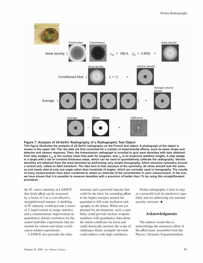
A series of density reconstruc-tions of an unclassified radiographic test object is shown in Figure 7. The radiographs have been calibrated to measure material densities using data taken on step wedges. In the set of tests shown, the uniformity and com-position of the high-density material limited the precision of the density reconstructions to about 2 percent. In classified experiments done sub-sequently, an accuracy of better than 1 percent has been attained for density reconstructions from pRad. This level of precision for density measure-ments is nearly an order of magnitude better than that obtained from thick object radiography using x-rays and meets requirements established nearly a decade ago for a next generation of hydrotest radiography machines beyond DARHT.
The high effective dose, quantita-tive density reconstructions, submil-limeter position resolution, and ease with which time sequences can be radiographed at frame rates in excess of 5 million frames per second make pRad the obvious choice for any next-generation flash-radiography machine beyond DARHT. This finding has led to the studies described below.
A 20-GeV Capability
Motivated by the success of the pRad experiments done at BNL, a recent study was made to examine the design parameters and estimate the costs of a proton synchrotron at an energy of 20 GeV that could be applied to quantitative radiography for the weapons stockpile. Siting options at both Los Alamos National Laboratory and the Nevada Test Site (NTS) were studied, and design fea-sibility and cost estimates were deter-mined.
If the mission requirement for such a capability were established, a choice of a Los Alamos site could take advantage of the existing accel-erator infrastructure at LANSCE. The 800-MeV LANSCE linear accelerator could be used as an injector to the synchrotron ring, which would save the time and money needed to build and commission a new accelerator. In addition, the existing infrastruc-ture of trained people and equipment would simplify commissioning a new accelerator. Studies indicate that a 20-GeV synchrotron ring would be smaller in its longest dimension than the existing kilometer-long linear accelerator that forms the core of LANSCE today.
Notional high-level requirements for a 20-GeV capability were syn-thesized from a combination of the results from the AGS experiments and from requirement studies carried out over the last decade. They are listed in Table I.
The number of pulses is driven by the need to measure density to infer criticality (as calculated for a hypothetical equivalent experiment that used nuclear material). Although large numbers of pulses are avail-able with pRad, extensive studies by Kevin Buescher, John Hopson, and Wayne Slattery have shown that four pulses spaced at a minimum of 200 nanoseconds are sufficient (DARHT-2 is intended to produce four pulses, a new state of the art for x-ray machines). A fifth pulse was added to the design requirements so that early-time phenomena can be studied simultaneously with late-time
configurations. A 20-GeV ring can provide up to 10 pulses, limited by the circumference of the synchrotron. The proton dose in Table I is twice that used in the validation experi-ments described above. This increase is enough to allow a two-Gaussian imaging mode in which part of the beam would be used to image small radii in the object and the remainder would be used for full-field imaging.
Summary
Proton radiography is a highly versatile invention that was born from the interaction between defense mission research and basic science. Experiments similar to the examples described here and some others have added quantitative data that have impacted near-term nuclear weapons stockpile assessment and certifica-tion. They have also added qualitative insights that would have been hard to obtain without the resolution and mul-tiple images pRad provides. This tech-nology is an important complement to other dynamic materials research and to DARHT. Ensuring that pRad will continue to provide data for the next two decades is one of the key reasons that the Laboratory has proposed to refurbish LANSCE. We project a need for 20 to 30 pRad experiments annu-ally based on outyear program plan-ning, past experience in performing the required work, and the schedule that these experiments need to meet to deliver data for model validation and certification milestones.
Currently included within the LANSCE refurbishment proposal,
Table I. High-Energy Radiography Requirements
Number of pulses >5
Minimum pulse spacing ~200 ns
Protons per pulse 2 × 10¹¹
Time format Individual pulse extraction
pRad article(NC).indd 42 1/24/06 8:40:36 AM
Number 30 2006 Los Alamos Science 43
Proton Radiography
the H– source intensity at LANSCE that feeds pRad can be increased by a factor of 2 in a cost-effective, straightforward manner. A doubling of H– intensity would provide a factor of 2 improvement in image statistics and a commensurate improvement in quantitative density resolution for the scaled hydrolike experiments that are needed for current and future certifi-cation-related experiments.
LANSCE also provides the infra-
structure and a powerful injector that could be the basis for extending pRad to the higher energies needed for quantitative full-scale hydrotest radi-ography in the future. While not yet planned for development, such a capa-bility could provide nuclear weapons modelers with quantitative data about the initial conditions for boost and could drastically increase the scope of challenges future stockpile stewards could address with high confidence.
Proton radiography is here to stay as a powerful tool for predictive capa-bility and for addressing our national security missions. n
Acknowledgments
The authors would like to acknowledge the enormous effort of the pRad team, assembled from the Physics, Dynamic Experimentation,
Figure 7. Analysis of 24-GeV/c Radiography of a Radiographic Test ObjectThis figure illustrates the analysis of 24 GeV/c radiography on the French test object. A photograph of the object is shown in the upper left. The raw data are first corrected for a number of experimental effects, such as beam shape and detector and camera response. Then, the transmission radiograph is inverted to give areal densities with data obtained from step wedges (λw is the nuclear mean free path for tungsten, and χ0 is an empirical radiation length). A step wedge is a target with a set of constant thickness steps, which can be used to quantitatively calibrate the radiography. Volume densities are obtained from the areal densities by performing very simple tomography, which assumes symmetry around a central axis, called an Abel transform. The idea here is that, because of the symmetry, all views should look the same, so one needs data at only one angle rather than hundreds of angles, which are normally used in tomography. The results of many measurements have been combined to obtain an estimate of the uncertainties in each measurement. In the end, we have shown that it is possible to measure densities with a precision of better than 1% by using this straightforward procedure.
Areal density
Conditioned Abel
Average ( ) =
( , = 1) =
w = 168.4, 0 = 5.806) =
Overburden Transmission Areal density
Areal density
Average Model
=–
Difference
Volume density
Average volumedensity
( , ,
pRad article(NC).indd 43 1/24/06 8:40:37 AM

44 Los Alamos Science Number 30 2006
Proton Radiography
and Applied Physics Divisions, over the past decade. LANSCE has done an impressive job providing beam with 100 percent reliability for the 215 pRad shots that have been fired to date. We are also enormously grateful for the freedom to develop pRad and the funding that has been provided for the effort by the nuclear weapons program at the Laboratory.
Further Reading
Alford, O. J., P. D. Barnes Jr., A. K. Chargin, W. D. Dekin, E. P. Hartouni, J. N. Hockman et al. 1999. The Scrounge-Atron: A Phased Approach to the Advanced Hydrotest Facility Utilizing Proton Radiography. In Proceedings of the IEEE Particle Accelerator Conference. Vol. 4, p. 2590. Piscataway, NJ: IEEE.
Aufderheide, M. B., H.-S. Park, E. P. Hartouni, P. D. Barnes, D. M. Wright, R. M. Bionta et al. 1999. Proton Radiography as a Means of Material Characterization. In AIP Conference Proceedings 9th International Symposium on Nondestructive Characterization of Materials. Edited by R. E. Green. Vol. 497, p. 706. Melville, NY: AIP.
Aufderheide, M. B., D. M. Slone, and A. E. Schach von Wittenau. 2001. HADES, a Radiographic Simulation Code. In AIP Conference Proceedings on Review of Progress in Quantitative Nondestruction Evaluation. Edited by D. O. Thompson and D. E. Chimenti. Vol. 557 (pt. A and B), p. 507. Melville, NY: AIP.
Baker, S. A., L. J. Castellano, P. A. Flores, B. C. Frogget, W. Lewis, P. T. Nedrow et al. 1997. Large Format Imaging System. In Proceedings of the SPIE - The International Society for Optical Engineering 22nd International Congress on High-Speed Photography and Photonics. Edited by D. L. Paisley. Vol. 2869, p. 929. Bellingham, WA: SPIE.
Charpak, G. 1978. Applications of Proportional Chambers to Some Problems in Medicine and Biology. Nucl. Instrum. Methods 156: 1.
Cochrane Jr., J. C., and P. J. Turchi. 2002. Design and Performance of a Current Transformer for Efficient Liner Implosions. IEEE International Conference on Plasma Science. p. 233. Piscataway, NJ: IEEE.
Cookson, J. A., B. H. Armitage, and A. T. Ferguson. 1972. Proton Radiography. Non-Destr. Test. 5 (4):225.
Cunningham, G., and C. Morris. 2003. The Development of Flash Radiography at Los Alamos National Laboratory. Los Alamos Science 28: 76.
Curry, J., and V. W. Steward. 1978. Establishment of a Beam Line at the Fermi National Accelerator Laboratory for Proton Radiography. Med. Phys. 5: 188.
Ellard, G. A., P. T. Gammon, H. S. Helmy, and R. J. W. Rees. 1972. Radiography with 160 MeV Protons. Nature 239 (5368): 157.
Ferm, E. N., C. L. Morris, J. P. Quintana, P. Pazuchanic, H. Stacy, J. D. Zumbro et al. 2002. Proton Radiography Examination of Unburned Regions in PBX 9502 Corner Turning Experiments. In AIP Conference Proceedings on Shock Compression of Condensed Matter. Edited by M. D. Furnish, Y. Horie, and N. N. Thadhani. Vol. 620 (pt. 2), p. 966. Melville, NY: AIP.
Gorbunov, V. I., V. P. Ivakin, B. A. Kononov, and A. L. Lukin. 1976. Flaw Detection with Medium-Energy Protons. Defektoskopiya—Sov. J. Non-Destr. Test. 12 (3): 338.
Harms, A. A., and L. D. Molson. 1974. Nuclear Radiation Conversion Enhancement with Inhomogeneous Converters and Intensifiers. Nucl. Instrum. Methods 119: 389.
Hartouni, E., and C. L. Morris. 2000. Proton Radiography. Beam Line 30 (1): 20.
Hogan, G. E. 1999. PC DAQ, A Windows-Based DAQ System. In Proceedings of the IEEE Conference on Real-Time Computer Applications in Nuclear Particle and Plasma Physics. Edited by S. C. Schaller. p. 160. Piscataway, NJ: IEEE.
Hogan, G. E., K. J. Adams, K. R. Alrick, J. F. Amann, J. G. Boissevain, M. L. Crow et al. 1999. Proton Radiography. In Proceedings of the IEEE Particle Accelerator Conference. Edited by A. Luccio and W. MacKay. Vol. 1, p. 579. Piscataway, NJ: IEEE.
Jones, D. T. L. 1982. Proposed Medical Applications of the National Accelerator Center Facilities. S. Afr. J. Sci. 78 (4): 149.
King, N. S. P., E. Ables, K. Adams, K. R. Alrick, J. F. Amann, S. Balzar et al. 1999. An 800-MeV Proton Radiography Facility for Dynamic Experiments. Nucl. Instrum. Methods Phys. Res. A, Accel. Spectrom. Detect. Assoc. Equip. 424 (1): 84.
King, N. S. P., S. Baker, S. Jaramillo, K. Kwiatkowski, S. Lutz, G. E. Hogan et al. 2003. Imaging Detector Systems for Soft X-Ray and Proton Radiography. In Proceedings of the SPIE—The International Society for Optical Engineering 25th International Congress on High-Speed Photography and Photonics. Edited by C. Cavailler, G. P. Haddleton, and M. Hugenschmidt. Vol. 4948, p. 610. Bellingham, WA: SPIE.
Kleinfelder, S. 2004. High-Speed, High-Sensitivity, Low-Noise Scientific CMOS Image Sensors. In Proceedings of the SPIE—Conference on Microelectronics: Design, Technology, and Packaging. Edited by D. Abbott, K. Eshraghian, C. A. Musca, D. Pavlidis, and N. Weste. Vol. 5274, p. 194. Bellingham, WA: SPIE.
Kleinfelder, S., K. Kwiatkowski, Y. Chen, and A. Shah. 2003. Multi-Million Frames/S Sensor Circuits for Pulsed-Source Imaging. IEEE Nucl. Sci. Symp. Conf. Rec. 3: 1504.
Koehler, A. M. 1968. Proton Radiography. Science 160: 303.
Koehler, A. M. 1970. Proton Radiography. Phys. Med. Biol. 15 (1): 181.
Koehler, A. M. 1972. Medical Treatment and Diagnosis Using 160 MeV Protons. In AIP Conference Proceedings. Vol. 9, p. 586. Melville, NY: AIP.
Kwiatkowski, K., J. F. Beche, M. T. Burks, G. Hart, G. E. Hogan, P. F. Manfredi et al. 2002. Development of Multi-Frame Detectors for Ultrafast Radiography with 800 MeV Protons. IEEE Trans. Nucl. Sci. 49 (1): 293.
Kwiatkowski, K., N. S. P. King, J. Lyke, J. F. Beche, G. E. Hogan, C. Kapusta et al. 2003. Development of a Multi-Frame Optical Imaging Detector for Proton Radiography at LANL. In Proceedings of the SPIE—The International Society for Optical Engineering 25th International Congress on High-Speed Photography and Photonics. Vol. 4948, p. 616. Bellingham, WA: SPIE.
pRad article(NC).indd 44 1/24/06 8:40:38 AM

Number 30 2006 Los Alamos Science 45
Proton Radiography
Kwiatkowski, K., J. Lyke, R. Wojnarowski, C. Kapusta, S. Kleinfelder, and M. Wilke. 2003. 3-D Stacked Electronics Assembly for High-Performance Imaging Detectors. IEEE Nucl. Sci. Symp. Conf. Rec. 1: 63.
Kwiatkowski, K., J. C. Lyke, R. J. Wojnarowski, J. F. Beche, R. Fillion, C. Kapusta et al. 2003. 3-D Interconnect Architecture for High-Bandwidth Massively Paralleled Imager. Nucl. Instrum. Methods Phys. Res. A, Accel. Spectrom. Detect. Assoc. Equip. 509 (1–3): 200.
Martin, R., M. Foss, J. Moenich, and R. Lari. 1975. The Proton Diagnostic Accelerator. IEEE Trans. Nucl. Sci. ns-22 (3): 1804.
Mottershead, C. T., and J. D. Zumbro. 1998. Magnetic Optics for Proton Radiography. In Proceedings of the IEEE Particle Accelerator Conference. Edited by M. Comyn, M. K. Craddock, M. Reiser, and J. Thomson. Vol. 2, p. 1397. Piscataway, NJ: IEEE.
Mottershead, T., D. Barlow, B. Blind, G. Hogan, A. Jason, F. Merrill et al. 2003. Design and Operation of a Proton Microscope for Radiography at 800 MeV. In Proceedings of the IEEE Particle Accelerator Conference. Edited by J. Chew, P. Lucas, and S. Webber. Vol. 1, p. 702. Piscataway, NJ: IEEE.
Neri, F., H. A. Thiessen, and P. L. Walstrom. 1998. Synchrotrons and Beamlines for Proton Radiography. In Proceedings of the IEEE Particle Accelerator Conference. Edited by M. Comyn, M. K. Craddock, M. Reiser, and J. Thomson. Vol. 3, p. 3788. Piscataway, NJ: IEEE.
Neri, F., and P. L. Walstrom. 2005. A Simple Empirical Forward Model for Combined Nuclear and Multiple Coulomb Scattering in Proton Radiography of Thick Objects. Nucl. Instrum. Methods Phys. Res. Sect. B 229: (3–4): 425.
Pedroni, E., R. Bacher, H. Blattmann, T. Bohringer, A. Coray, A. Lomax et al. 1995. The 200-MeV Proton Therapy Project at the Paul Scherrer Institute: Conceptual Design and Practical Realization. Med. Phys. 22: 37.
Prichard, B. A. 1993. A Proposed Proton Therapy Facility at the SSC. Nucl. Instrum. Methods Phys. Res. Sect. B 79 (1–4): 895.
Saudinos, J. 1977. A New Medical Application: Radiography by Nuclear Scattering. Bull. Info. Sci. Tech. 224–225: 175.
Schneider, U., and E. Pedroni. 1994. Multiple Coulomb Scattering and Spatial Resolution in Proton Radiography. Med. Phys. 21 (11): 1657.
———. 1995. Proton Radiography as a Tool for Quality Control in Proton Therapy. Med. Phys. 22 (4): 353.
———. 1996. Quantitative Proton Radiography. In Quantitative Imaging in Oncology: 19th L. H. Gray Conference on Quantitative Imaging in Oncology. Edited by K. Faulkner, B. Carey, A. Crellin, and R. M. Harrison. p. 61. London: British Institute of Radiology.
Schneider, U., J. Besserer, and P. Pemler. 2001. On Small Angle Multiple Coulomb Scattering of Protons in the Gaussian Approximation. Z. Med. Phys. 11: 110.
Schneider, U., M. Dellert, E. Pedroni, P. Pemler, J. Besserer, M. Moosburger et al. 2003. Quantitative Proton Radiography of an Animal Patient. In Proceedings of the SPIE - The International Society for Optical Engineering. Edited by M. K. Yaffe and L. E. Antonuk. Vol. 5030, p. 585. Bellingham, WA: SPIE.
Schneider, U., and A. Tourovsky. 1998. Range-Uncertainty Imaging for Obtaining Dose Perturbations in Proton Therapy. IEEE Trans. Nucl. Sci. 45: 2309.
Schultz, J. H., T. Antaya, J. V. Minervini, A. L. Radovinsky, B. A. Smith, R. J. Camille et al. 2003. The Advanced Hydrotest Facility (AHF) Large Bore Quadrupole Focusing Magnet System. IEEE Trans. Appl. Supercond. 13 (2): 1343.
Steward, V. W. 1976. Proton (and Other Heavy Charged Particle) Radiography in Medical Diagnosis. IEEE Trans. Nucl. Sci. ns-23 (1): 577.
———. 1979. Proton (Heavy Ion) Radiography in Medical Diagnosis. IEEE Trans. Nucl. Sci. ns-26 (2): 2257.
Steward, V. W., and A. M. Koehler. 1973. Proton Radiography as a Diagnostic Tool. Phys. Med. Biol. 18 (4): 591.
———. 1974. Proton Radiography in the Diagnosis of Breast Carcinoma. Radiology 110 (1): 217.
———. 1974. Proton Radiography of a Human Brain Tumor Within Skull—Preliminary Report. Surg. Neurol. 2 (4): 283.
West, D. 1975. The Potential of Proton Radiography. In Proceedings of the 7th International Conference on Cyclotrons and Their Applications. Edited by W. John. p. 503. Basel, Switzerland: Birkhäuser Verlag.
Wilson, R. 1972. Medical Uses of the Harvard University Cyclotron: An Overall View. In AIP Conference Proceedings. Vol. 9, p. 578. Melville, NY: AIP.
Yates, G. J., K. L. Albright, K. R. Alrick, R. A. Gallegos, J. Galyardt, N. T. Gray et al. 1998. An Intensified/Shuttered Cooled CCD Camera for Dynamic Proton Radiography.In Proceedings of the SPIE—The International Society for Optical Engineering Conference on Digital Solid State Cameras: Designs and Applications. Edited by G. M. Williams. Vol. 3302, p. 140. Bellingham, WA: SPIE.
Ziock, H. J., K. J. Adams, K. R. Alrick, J. F. Amann, J. F. Boissevain, M. L. Crow et al. 1998. The Proton Radiography Concept. In Proceedings of PIXEL98, International Pixel Detector Workshop. Edited by D. Anderson and S. Kwan. p. 233. Batavia, IL: Fermi Nat. Accel. Lab.
Ziock, H. J., K. R. Alrick, R. A. Gallegos, J. Galyardt, N. T. Gray, G. E. Hogan et al. 1998. Detector Development for Dynamic Proton Radiography. In Proceedings of PIXEL98, International Pixel Detector Workshop. Edited by D. Anderson and S. Kwan. p. 221. Batavia, IL: Fermi Nat. Accel. Lab.
pRad article(NC).indd 45 1/24/06 8:40:38 AM