protocols microbiological analysis in ...analysis in pharmaceutical industry 2 table of contents...
TRANSCRIPT
Inspired by knowledge
PROTOCOLS
ALL PROCEDURES ACCORDING TO EUROPEAN PHARMACOPEIA
MICROBIOLOGICAL ANALYSIS IN
PHARMACEUTICALINDUSTRY

2
Table of Contents
MICROBIOLOGICAL WATER TESTING _______________ 04 Ph. Eur. 9.0
STERILITY TESTING _______________________________ 05 Procedure according to Ph. Eur. 2.6.1
NON-STERILE PRODUCT TESTING Procedure according to Ph. Eur. 2.6.12
TOTAL MESOPHILIC ENUMERATION ___________________ 07
ESCHERICHIA COLI _________________________________ 09
BILE-TOLERANT GRAM-NEGATIVE BACTERIA ___________ 10
SALMONELLA _____________________________________ 11
STAPHYLOCOCCUS AUREUS _________________________ 12
PSEUDOMONAS AERUGINOSA ______________________ 13
CLOSTRIDIUM _____________________________________ 14
CANDIDA _________________________________________ 15

3
The pharmaceutical industry is one of the world's most influential business sectors, comprised of public and private organizations dedicated exclusively to the research, manufacture, marketing and distribution of pharmaceutical chemicals for human and animal health.
Regulating the analysis and control of medications in this sector is extremely important both in the development of new drugs and in the control of existing products in order to ensure their quality, effectiveness and safety.
The European Pharmacopoeia is the official legal instrument in the EU established to ensure the quality of this type of products. It includes quality standards for active principles, general standards for formulation, and general standards for manufacturing medications. It is made up by Expert Committees of the countries involved and other countries, as well as the WHO, participate as observers.
This standard considers the microbial analysis of pharmaceutical products, raw materials used, water, air, equipment or packaging material, among others, which are vital to minimize the type and number of microorganisms present. Those products of parenteral administration or for use in eyes must be sterile.
Drugs are considered contaminated when it exceeds a limit of opportunistic or objectionable pathogenic microorganisms, which have the capacity to limit the effectiveness of the product, when they present toxic metabolites or physical or chemical deterioration. The ineffective dose varies between species and even between individuals.
This guide will describe the culture media and the procedures formulated according to the Pharmacopoeia required to perform:
• Sterility testing
• Non-sterile product testing
• Microbiological count testing
• Microbiological water testing

4
Microbiological Water Testing
Water is the most commonly used substance in the pharmaceutical industry, both for production, as an ingredient in the product or in one of the manufacturing stages; and for washing equipment. Microbiological control of water is of vital importance at entrance to the treatment plant, purification, storage, and distribution stages since microorganisms can proliferate throughout any stage described above, affecting its quality.
Water samples should be analyzed to avoid changes in the microbial count process. Once the sample has been collected, it must be stored at a temperature between 2 and 10ºC. The count must be performed within 8 hours and the analysis of coliform bacteria within 30 hours. Sampling, reception and analysis records must demonstrate that the established timelines are met.
Water for pharmaceutical use can be classified according to use and method of production.
• Water for injections: Used as a vehicle (bulk injection water) or as a substances/preparations diluent for preparing medications to be administered parenterally.
• Purified water: Used for preparing medications that require sterility and apyrogenicity, except those authorized.
• Water for preparation of extracts: For preparing medication extracts.
• Highly purified water: Its purpose is the preparation of medicinal products, for which a high biological quality is required (except those cases in which water is used for injectables).
Bibliography
American Public Health Association (1985) Standard Method for the Enumeration of Water and Wasterwater.European Pharmacopeia 9.3.
Determine by means of a filter membrane with a pore size not exceeding 0.45 µm. Culture in R2A Agar (CAT. 1071)
Incubation at 30-35 ºC // 5 days
PURIFIED WATER: 100 CFU/ml
HIGHLY PURIFIED WATER: 10 CFU/100 ml
Use at least 200 ml of sample
WATER FOR INJECTIONS: 10 CFU/ 100 ml
Use at least 200 ml of sample
Determine by means of a filter membrane with a pore size not exceeding 0.45 µm. Culture in Trypticasein Soy Agar (TSA) (CAT. 1068)
Incubation at 30-35 ºC // 5 days
WATER FOR PREPARATION OF EXTRACTS:
100 CFU/100 ml
STORED PURIFIED WATER:
100 CFU/ 100 ml

5
Sterility Testing
Introduction
The purpose of the sterility test is to verify the absence of contamination by microorganisms in products that, according to pharmacopoeias, are required to be sterile, whether they have been sterilized or prepared aseptically. A satisfactory result indicates the absence of contaminating microorganisms in the sample examined under test conditions. To achieve such conditions, the test environment must be adapted to the way in which the sterility test is performed. These working conditions are regularly monitored by means of an appropriate sampling of the working area.
This test appeared for the first time in the British Pharmacopoeia in 1932, and later in the United States Pharmacopoeia in 1936 for sterile solutions. Over the years, constant changes have been made, which have led to the improvement of techniques for detecting contamination in this type of products.
Bibliography
Brewer. JAMA, 115. 1940. Vera. J. Bact. 47:59, 1944.Pittman. J. Bact. 51:19, 1946.
European Pharmocopoeia 9.3.
Aerobic, anaerobes, and fungus growth promotion test
Inoculate in containers separated by microorganism Thioglycolate Liquid Medium (CAT. 1508) with < 100 CFU of each species described above.
Inoculate in containers separated by microorganism Trypticasein Soy Broth (TSB) (CAT. 1224) with < 100 CFU of each species described above.
AEROBIC BACTERIA (Staphylococcus aureus, Pseudomonas aeruginosa)
ANAEROBIC BACTERIA (Clostridium sporogenes)
AEROBIC BACTERIA (Bacillus subtilis)
FUNGUS (Candida albicans, Aspergillus brasiliensis)
Incubate bacteria for no more than 3 days, and fungi and yeasts for no more than 5 days.
6
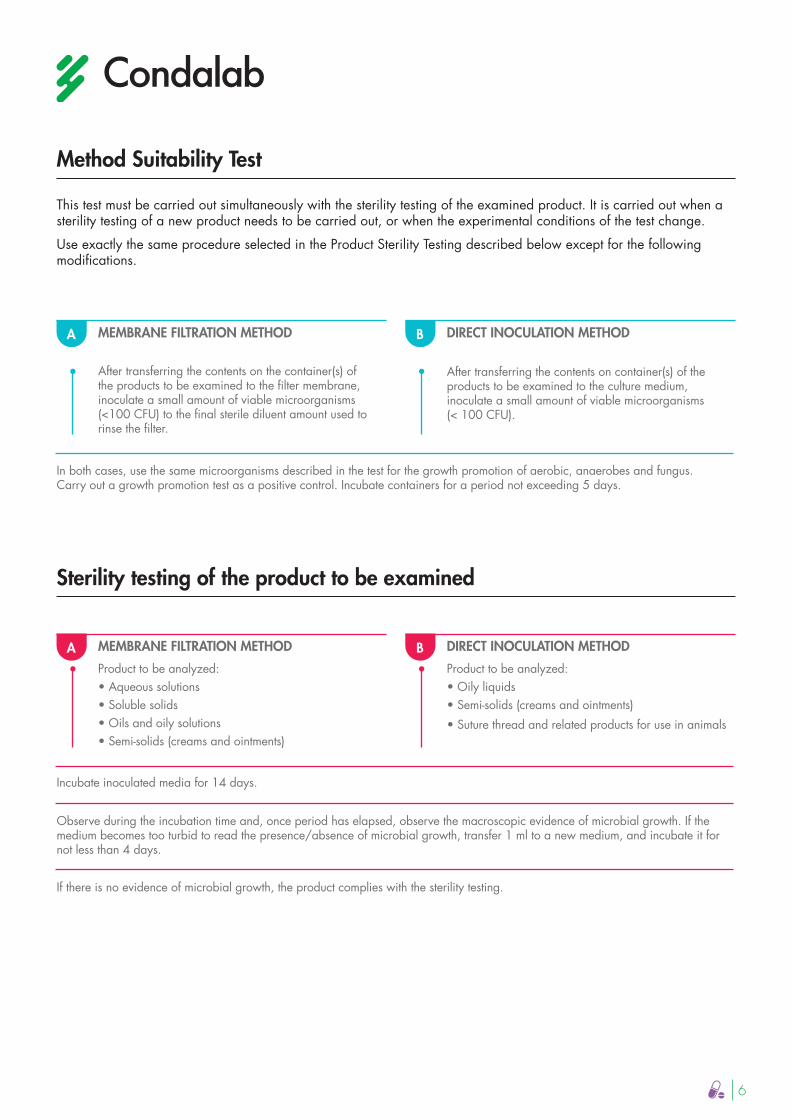
This test must be carried out simultaneously with the sterility testing of the examined product. It is carried out when a sterility testing of a new product needs to be carried out, or when the experimental conditions of the test change.
Use exactly the same procedure selected in the Product Sterility Testing described below except for the following modifications.
Method Suitability Test
MEMBRANE FILTRATION METHOD
After transferring the contents on the container(s) of the products to be examined to the filter membrane, inoculate a small amount of viable microorganisms (<100 CFU) to the final sterile diluent amount used to rinse the filter.
A DIRECT INOCULATION METHOD
After transferring the contents on container(s) of the products to be examined to the culture medium, inoculate a small amount of viable microorganisms (< 100 CFU).
B
In both cases, use the same microorganisms described in the test for the growth promotion of aerobic, anaerobes and fungus.Carry out a growth promotion test as a positive control. Incubate containers for a period not exceeding 5 days.
Sterility testing of the product to be examined
MEMBRANE FILTRATION METHOD
Product to be analyzed:• Aqueous solutions• Soluble solids• Oils and oily solutions• Semi-solids (creams and ointments)
A DIRECT INOCULATION METHOD
Product to be analyzed:• Oily liquids• Semi-solids (creams and ointments)
• Suture thread and related products for use in animals
B
Incubate inoculated media for 14 days.
Observe during the incubation time and, once period has elapsed, observe the macroscopic evidence of microbial growth. If the medium becomes too turbid to read the presence/absence of microbial growth, transfer 1 ml to a new medium, and incubate it for not less than 4 days.
If there is no evidence of microbial growth, the product complies with the sterility testing.

7
MicrobiologicalI count testTAMC (Total Aerobic Microbial Count) &; TYMC (Total Yeast and Mold Counts)
The tests described in this section allow quantitative enumeration of mesophilic bacteria, total aerobic microbial (TAMC) and fungus count, total combined yeasts and mold count (TYMC), which can grow under aerobic conditions
Bibliography
Standard Methods for the Examination of Dairy Products. 11th Edition. APHA., Inc. New York, 1960.
European Pharmocopoeia 9.3.
SAMPLE PREPARATION
At a concentration of 1:10, prepare the sample in Buffered Peptone Water (CAT. 1401) at pH 7.0 and homogenize.
This stock solution can be supplemented with neutralizersand surfactants
A - MEMBRANE FILTRATION B - PLATE COUNT METHODS B.1 POUR PLATE METHOD
MICROORGANISMSAEROBIC TOTALS
Transfer 10 ml of the solution ofstock to the filter membrane(pore size 0.45 µm)
MOLDS ANDYEASTS TOTAL
Transfer 10 ml of the solution ofstock to the filter membrane(pore size 0.45 µm)
Wash each filter membrane 3x100 ml of Buffered Peptone Water (CAT. 1401) at pH 7.0
Calculate the number of CFU per gram or milliliter of product
MICROORGANISMS AEROBIC TOTALS
Transfer the membrane to aTrypticasein Soy Agar (TSA)(CAT. 1068)Incubation at 30-35 ºC // 5 days
MOLDS ANDYEASTS TOTALS
Transfer the membrane toSabouraud Dextrose Agar Plates (CAT. 1024)Incubation at 20-25 ºC //5-7 days
SAMPLE PREPARATION
At a concentration of 1:10, prepare the sample in Buffered Peptone Water (CAT. 1401) at pH 7.0 and homogenize.
This stock solution can be supplemented with neutralizers and surfactants
MICROORGANISMSAEROBIC TOTALS
Transfer 1 ml of the stock solution to at least one empty and sterilized Petri dish.Add 15-20 ml of Trypticasein Soy Agar (TSA) (CAT. 1068) at < 45 ºC Incubation at 30-35 ºC // 5 days
MOLDS ANDYEASTS TOTAL
Transfer 1 ml of the stock solution to at least one empty and sterilized Petri dish. Add 15-20 ml of Sabouraud Dextrose Agar (CAT. 1024) at < 45ºC Incubation at 20-25 ºC // 5-7 days
Select the plates corresponding to the given dilution and presenting the highest number of colonies lower than 250 for TAMC and 50 for TYMC.
Find the arithmetic mean of the counts by culture medium.
Calculate the number of CFU per gram or milliliter of product

8
B - PLATE COUNT METHODSB.2 SURFACE METHOD
SAMPLE PREPARATIONAt a concentration of 1:10, prepare the sample in Buffered Peptone Water (CAT. 1401) at pH 7.0and homogenize.
This stock solution can be supplemented with neutralizersand surfactants
MICROORGANISMSAEROBIC TOTALS
Transfer at least 0.1 ml of the stock solution to a plate of Trypticasein Soy Agar (TSA) (CAT. 1068) Incubation at 30-35 ºC // 120 hours
TOTAL MOLDSAND YEASTS
Transfer at least 0.1 ml of the stock solution to a Sabouraud Dextrose Agar plate (CAT.1024)Incubation at 20-25 ºC //5-7 days
Calculate the number of CFU per gram or milliliter of product
SAMPLE PREPARATION
At a concentration of 1:10, prepare the sample in Buffered Peptone Water (CAT. 1401) at pH 7.0 and homogenize.
This stock solution can be supplemented with neutralizers and surfactants
C - MOST LIKELY NUMBER METHOD (TAMPC)
CONFIRMATIONTubes with turbidity
Perform a subculture in the same broth or on a plate of Trypticasein Soy Agar (TSA) (CAT.1068). Determine the Most Probable Number of microorganisms by gram or milliliter of product
CONFIRMATIONTubes without turbidity
Determine the Most Probable Number of microorganisms per gram or milliliter of product
Make two serial dilutions from the stock solution of 1 ml / dilution in two tubes of 9 ml each of Trypticasein Soy Broth (TSB) (CAT. 1224)
Using the initial three dilutions, carry out three further dilutions with at least 3 tubes/dilution in a concentration of 1:10 in Trypticasein Soy Broth (TSB) (CAT. 1224) Incubation at 30-35 ºC/120 hours.
Using the initial three dilutions, carry out three further dilutions with at least 3 tubes / dilution in a concentration of 1:10 in Trypticasein Soy Broth (TSB) (CAT. 1224) Incubation at 30-35 ºC/ max. 3 days.

9
Escherichia coli
IntroductionThis microorganism belongs to the Enterobacteriaceae family; it is a gram-negative, mobile, non-sporulating bacillus. In addition, it is lactose-positive and negative oxidase. Escherichia coli can be distinguished from other coliforms by its ability to produce indole from tryptophan or by the production of the ß-glucuronidase enzyme. These characteristics are used for selective isolation and confirmation in various analysis processes.
These ubiquitous bacteria are located in the intestine of humans and warm-blooded animals. Their high presence in the intestinal tract and feces make it a marker that indicates bad hygienic practices or fecal contamination during the manufacture or handling of drugs. Their presence is used as a marker that other pathogenic organisms of fecal origin may be present, especially in products meant for oral consumption and natural raw materials.
Today, the tests also contemplate the concentration of Escherichia coli according to the European Pharmacopoeia.
Bibliography
MacConkey J. H. 5:33. 1905. Joseph Md. State. Dept. Health. Procedures, 1960. European Pharmocopoeia 9.3.
Chils, E., and L. A. Allen. 1953. Improved methods for determining the most probable number of Bacterium coli and of Enterococcus faecalis. J. Hyg.Camb. 51:468-477.
PRIMARY ENRICHMENT
Spread 10 ml of the stock solution or the quantity corresponding to 1 g/1 ml of the product in 100 ml of Trypticasein Soy Broth (TSB) (CAT. 1224) and homogenize.Incubation at 30-35 ºC // 18-24 hours
Method
SAMPLE PREPARATION
At a concentration of 1:10, prepare the sample using at least 1 g/ 1 ml of the product to be examined in Buffered Peptone Water (CAT. 1401) and homogenize.
This stock solution can be supplemented with neutralizers and surfactants.
RESULTS INTERPRETATION
The growth of colonies in the medium indicates the possible presence of E. coli. Confirmation by identification test. The product passes the test if no colonies are observed or if identification tests return negative
SELECTIVE ENRICHMENT
Spread again 1 ml of Trypticasein Soy Broth in 100 ml of MacConkey Broth (CAT.1210)Incubation at 42-44 ºC // 24-48 hours
SELECTIVE ISOLATION
Perform subcultures on Agar MacConkey (CAT. 1052). Incubation at 30-35 ºC // 18-72 hours
10
Bile-tolerantgram-negative bacteria
Introduction
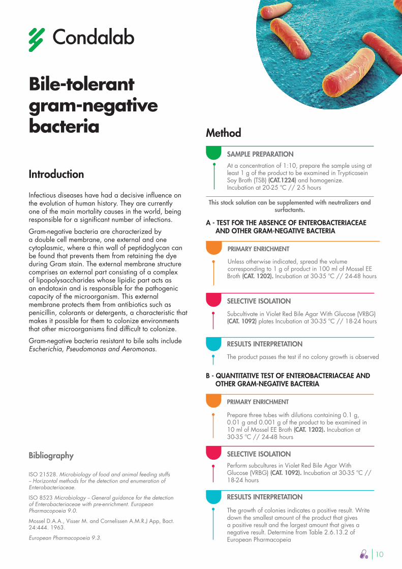
Infectious diseases have had a decisive influence on the evolution of human history. They are currently one of the main mortality causes in the world, being responsible for a significant number of infections.
Gram-negative bacteria are characterized by a double cell membrane, one external and one cytoplasmic, where a thin wall of peptidoglycan can be found that prevents them from retaining the dye during Gram stain. The external membrane structure comprises an external part consisting of a complex of lipopolysaccharides whose lipidic part acts as an endotoxin and is responsible for the pathogenic capacity of the microorganism. This external membrane protects them from antibiotics such as penicillin, colorants or detergents, a characteristic that makes it possible for them to colonize environments that other microorganisms find difficult to colonize.
Gram-negative bacteria resistant to bile salts include Escherichia, Pseudomonas and Aeromonas.
Bibliography
ISO 21528. Microbiology of food and animal feeding stuffs -- Horizontal methods for the detection and enumeration of Enterobacteriaceae.
ISO 8523 Microbiology -- General guidance for the detection of Enterobacteriaceae with pre-enrichment. European Pharmacopoeia 9.0.
Mossel D.A.A., Visser M. and Cornelissen A.M.R.J App, Bact. 24:444. 1963.
European Pharmacopoeia 9.3.
Method
PRIMARY ENRICHMENT
Unless otherwise indicated, spread the volume corresponding to 1 g of product in 100 ml of Mossel EE Broth (CAT. 1202). Incubation at 30-35 ºC // 24-48 hours
PRIMARY ENRICHMENT
Prepare three tubes with dilutions containing 0.1 g, 0.01 g and 0.001 g of the product to be examined in 10 ml of Mossel EE Broth (CAT. 1202). Incubation at 30-35 ºC // 24-48 hours
SAMPLE PREPARATION
At a concentration of 1:10, prepare the sample using at least 1 g of the product to be examined in Trypticasein Soy Broth (TSB) (CAT.1224) and homogenize.Incubation at 20-25 ºC // 2-5 hours
This stock solution can be supplemented with neutralizers and surfactants.
SELECTIVE ISOLATION
Subcultivate in Violet Red Bile Agar With Glucose (VRBG) (CAT. 1092) plates Incubation at 30-35 ºC // 18-24 hours
SELECTIVE ISOLATION
Perform subcultures in Violet Red Bile Agar With Glucose (VRBG) (CAT. 1092). Incubation at 30-35 ºC // 18-24 hours
RESULTS INTERPRETATION
The product passes the test if no colony growth is observed
RESULTS INTERPRETATION
The growth of colonies indicates a positive result. Write down the smallest amount of the product that gives a positive result and the largest amount that gives a negative result. Determine from Table 2.6.13.2 of European Pharmacopeia
A - TEST FOR THE ABSENCE OF ENTEROBACTERIACEAE AND OTHER GRAM-NEGATIVE BACTERIA
B - QUANTITATIVE TEST OF ENTEROBACTERIACEAE AND OTHER GRAM-NEGATIVE BACTERIA

11
Salmonella
Introduction
Salmonella is a very diverse family of microorganisms, including some 2,300 distinct serotypes. It is one of the four main causes of diarrheal diseases. This group of bacteria occurs in the form of gram-negative bacillus (as they belong to the enormous group of Enterobacteriaceae), they are facultative anaerobic and mobile by means of flagella. In addition, they are glucose fermenters but not lactose fermenters and do not produce urease.
It is estimated that salmonellosis affects tens of millions of people worldwide every year and causes more than 100,000 deaths. This type of bacteria is the reason for millions of hospitalizations worldwide, and this number may increase up to 30 times if infections that are not diagnosed are included, as they can usually cause a normal diarrhea process. It behaves as a facultative intracellular pathogen that, depending on the serotype, inoculum, virulence factors expressed by the strain, the host involved, and the immune status of the patient can cause from a medium to severe gastrointestinal infection to a systemic infection.
It is widely distributed in nature and as normal flora of the intestinal tract in animals and humans. Unlike other microorganisms that cause gastrointestinal diseases, it may be commonly found in natural raw materials, making it vitally important to research them, especially those of animal origin.
Bibliography
Rollender, W. U. Beckford; R.D. Belsky, B. Krostoff (1969) Comparison of Xylose Lysine desoxycholate agar and MacConkey agar for the isolation of Salmonella and Shigella from clinical specimens (tech. Bull. Reg. Med. Tech, 39 (1) 8-p).
European Pharmacopoeia. 9.3.
Method
SELECTIVE ENRICHMENT
Spread 0.1 ml of Trypticasein Soy Brothin 10 ml of Rappaport Vassidialis Broth (CAT. 1414)Incubation at 30-35 ºC // 18-24 hours
SAMPLE PREPARATION
At a concentration of 1:10, prepare the sample using at least 10 g / 10 ml of the product to be examined in Trypticasein Soy Broth (TSB) (CAT. 1224) and homogenize. Incubation at 30-35 ºC // 18-24 hours
Trypticasein Soy Broth can be supplemented withneutralizers and surfactants.
SELECTIVE ISOLATION
Perform subcultures on XLD Agar (Xylose Lysine Desoxycholate Agar) (CAT. 1080)Incubation at 30-35 ºC // 18-48 hours
RESULTS INTERPRETATION
All red colonies with/without black nucleus are susceptible of Salmonella Confirmation by means of an identification test. The product passes the test if no colonies of the types described are observed or if the confirmation tests are negative

12
Staphylococcus aureusIntroduction
It's a bacterium that belongs to the Staphylococcaceae family. They are facultative anaerobic cocci grouped in clusters, gram-positive, immobile and not sporulating. Among its most distinctive characteristics is the production of virulence factors such as coagulases and catalases that are used for confirmatory tests after isolation. It is a microorganism widely distributed worldwide and acts as a commensal in the human epithelium and mucous membranes. It is often found in the mouth, blood, mammary glands, intestine, genitourinary tract, and airways.
It is also an opportunistic pathogen, causing acute and pyogenic infections which, if untreated, can spread to the surrounding tissue or to other organs via bacteremia. Among the types of infections caused by this microorganism are pneumonia, osteomyelitis, and acute endocarditis, among others. The presence of Staphylococcus and, particularly, S. aureus genus in raw material or pharmaceutical or cosmetic product, indicates that the source of contamination may be human, i.e. operators. These microorganisms can be transported through dust, skin, clothing, and moisture droplets generated by moving, talking, and sneezing.
Bibliography
McColloch Am. J. Vet. Research, 8:173. 1947. Velilla, Faber, and Pelczar Am. J. Vet. Research, 8:275. 1947. Chapman, G.H. 1945 J. Bact. 50:201-203.
European Pharmacopoeia. 9.3.
Method
PRIMARY ENRICHMENT
Spread 10 ml of the stock solution or the quantity corresponding to 1 g/1 ml of the product in100 ml of Trypticasein Soy Broth (TSB) (CAT. 1224) and homogenise.Incubation at 30-35 ºC // 18-24 hours
SAMPLE PREPARATION
At a concentration of 1:10, prepare the sample using at least 1 g/ 1 ml of the product to be examined in Trypticasein Soy Broth (TSB) (CAT.1224) / Buffered Peptone Water (CAT.1401) at pH 7.2 and homogenize.
This stock solution can be supplemented with neutralizers and surfactants.
SELECTIVE ISOLATIONPerform subcultures in Mannitol Salt Agar (MSA) (Chapman Medium) (CAT. 1062). Incubation at 30-35 ºC // 18-72 hours
RESULTS INTERPRETATION
The growth of white/white colonies surrounded by a yellow zone indicates the possible presence of Staphylococcus aureus. Confirmation by means of an identification test. The product passes the test if no colonies of the types described are observed or if the confirmation tests are negative.

13
Pseudomonas aeruginosaIntroduction
It's a bacterium that belongs to the Pseudomonadaceae family. They are straight or curved bacilli, strictly aerobic, gram-negative, mobile and non-sporulating. In addition, they are positive catalase, using this characteristic for later identification. This group of bacteria is capable of producing different pigments such as pyocyanin and uorescein. Therefore, different culture media have been developed to promote the production of these pigments and thus be able to identify the different species.
This microorganism combines its adaptability to different ecosystems with a wide variety of virulence factors, and the spectrum of diseases it can cause ranges from mild infection to sepsis. During its mechanism of action it attaches to epithelial cells by means of polar pili and, once attached, it produces proteases, hemolysins, exotoxins and endotoxins that damage tissues.
The P. aeruginosa infection is often related to water and aqueous solutions contamination. One of the most severe infections caused by this microorganism is endocarditis due to intravenous administration of medications, when injected into the patient and dissolved or suspended in watery vehicles. Hence, the importance of detecting its presence in pharmaceutical products that are to be administered by inhalation and ocular route or in aqueous vehicles. Cosmetics may also be susceptible to this type of contamination, especially liquids and creams.
In addition, it can colonize water purification systems by the formation of biofilms, structures very difficult to eliminate with the use of salinizing agents. Its remarkable resistance to antimicrobials is one of the world's greatest concerns.
Bibliography
King, Ward and Raney. J. Lab. and Clin. Med. 44:301. 1954. Brown and Lowbury. J. Clin. Path. 18:752. 1965. Lowbury. J. Clin. Path. 4:66. 1951. Lowbury and Collins. J. Clin. Path. 8:47. 1955.
European Pharmacopoeia. 9.3.
Method
PRIMARY ENRICHMENT
Spread 10 ml of the stock solution or the quantity corresponding to 1 g/1 ml of the product in100 ml of Trypticasein Soy Broth (TSB) (CAT. 1224) and homogenise. Incubation at 30-35 ºC // 18-24 hours
SAMPLE PREPARATION
At a concentration of 1:10, prepare the sample using at least 1 g of the product to be examined in Buffered Peptone Water (CAT. 1401) at pH 7 and homogenize
This stock solution can be supplemented with neutralizers and surfactants.
SELECTIVE ISOLATION
Perform subcultures in Cetrimide Agar (CAT. 1102)Incubation at 30-35 ºC for 18-72 hours
RESULTS INTERPRETATION
The growth of colonies in the medium indicates the possible presence of Pseudomonas aeruginosa. Confirmation by means of an identification test. The product passes the test if no colonies are observed or if the confirmation tests are negative

14
Clostridia
Introduction
They are a class of bacteria belonging to the Firmicutes phylum class. They are characterized by being mostly gram-positive anaerobes bacilli. Most genuses do not form spores, but some do, such as Clostridium, one of the best known.
These bacteria are widely distributed in nature, mainly in the soil and in the intestinal tract of many animal species, including man, where they may also be found as normal flora on the skin and mucous membranes of the nasopharynx, pharynx, mouth, urethra, and vagina. They can cause both endogenous and exogenous infections, affecting any region of the body, provided the conditions in the tissues are favorable. They are fundamentally involved in abscesses of any organ, colitis, appendicitis, bacteremia, chronic otitis media, crackling cellulitis, dental and oral infections, endocarditis and endometritis.
The presence of anaerobic microorganisms, especially those belonging to the Clostridium genus is usually found in natural raw materials, powder, starch, etc. And also in medicinal herbs and phytotherapeutic medications.
Bibliography
Ellner, Stossel, Drakeford and Vasi. AM J. Clin. Path. 45:502-504. 1966.
European Pharmacopoeia. 9.3.
Method
PRIMARY ENRICHMENT
Spread 10 ml, separately, of each portion of the stock solution in 100 ml of Reinforced Clostridial Medium (CAT. 1007). Incubation at 30-35 ºC // 48 hours
SELECTIVE ISOLATION
Perform subcultures in Columbia Agar (CAT. 1104)Incubation under anaerobic conditions at 30-35 ºC for 48-72 hours
RESULTS INTERPRETATION
The growth of colonies (with/without spores) gives a negative reaction to catalase indicating the presence of Clostridia. Confirmation by identification test
SAMPLE PREPARATION
At a concentration of 1:10, prepare the sample using at least 2 g/2 ml of the product to be examined in Trypticasein Soy Broth (TSB) (CAT. 1224) / Buffered Peptone Water (CAT. 1401) at pH 7.2 and homogenize
This stock solution can be supplemented with neutralizers and surfactants.
INACTIVATION SPORES
Divide the sample into 2 tubes of at least 10 ml. Heat 1 portion to 80 ºC/10 min and cool quickly. Do not heat the other portion

15
Candida albicans
Introduction
These are yeasts belonging to the Saccharomycetaceae family and the Candida genus. These fungi develop in a unicellular form and are human saprophytic. It is very common to find them in oral cavities, gastrointestinal tract and vaginal area. Although they have a very important function in the fermentation of certain sugars for the body, there are times that this microorganism acts as an opportunistic pathogen, being able to cause superficial or systemic candidiasis in weakened people, in newborns and in old people with a deficient immune system.
The products with the highest susceptibility to fungal contamination are ophthalmic solutions, ointments, suppositories, salves and in cosmetics, soaps, powders, and others that contain nutrients rich in carbohydrates and fatty acids.
It is therefore necessary to analyze these pharmacological products in order to identify the presence of this microorganism and thus avoid the consequences of a possible infection by C. albicans in the consumer.
Bibliography
Beuchat, L.R., J.E Corry, A.D King, Jr. And J.I Pitt (ed) 1986) Methods for the mycological examination of food. Plenum Pres, New York European Pharmacopoeia. 9.3.
Sabouraud, Ann. Dermat and Syphilol 1892-3. Gerog J. Lab.CLin. Med. 67;355 1953.
Method
PRIMARY ENRICHMENT
Spread 10 ml of the stock solution or the quantity corresponding to 1 g/1 ml of the product in 100 ml of Sabouraud Dextrose Broth (CAT. 1205) and homogenize.Incubation at 30-35 ºC // 3-5 days
SELECTIVE ISOLATION
Perform subcultures in Sabouraud Dextrose Agar plates (CAT.1024). Incubation at 30-35 ºC // 24-48 hours
RESULTS INTERPRETATION
The growth of white colonies in the medium indicates the possible presence of Candida albicans. Confirmation by means of an identification test. The product passes the test if these colonies are not observed or if confirmatory tests return negative
SAMPLE PREPARATION
At a concentration of 1:10, prepare the sample in Trypticasein Soy Broth (TSB) (CAT.1224) / Buffered Peptone Water (CAT. 1401) at pH 7.2 and homogenise
This stock solution can be supplemented with neutralizers and surfactants

[email protected] | www.condalab.com