protocol to guide the assessment of microwave tissue ... · protocol to guide the assessment of...
TRANSCRIPT
MSAC website: www.msac.gov.au
MEDICAL SERVICES ADVISORY COMMITTEE
Protocol to guide the assessment of microwave
tissue ablation for primary and secondary lung
cancer
MSAC Application 1403
January 2016
MSAC website: www.msac.gov.au Page i
Table of Contents
1 Title of application ..................................................................................................................... 1
2 Purpose of application ............................................................................................................... 1
3 Intervention – proposed medical service ................................................................................... 2
3.1 Description of the proposed medical service ....................................................................... 2
3.2 Registered trademark ......................................................................................................... 3
3.3 Proposed clinical setting...................................................................................................... 3
3.4 Service delivery ................................................................................................................... 3
4 Co-dependent information ........................................................................................................ 4
5 Population eligible for the proposed medical service ................................................................ 5
5.1 Medical condition relevant to the service ............................................................................ 5
5.2 Proposed patient population(s) ........................................................................................... 6
5.3 Defining surgical operability ................................................................................................ 2
5.4 Expected utilisation ............................................................................................................. 4
5.5 Evidence for the population that would benefit from this service ........................................ 6
6 Comparator ............................................................................................................................... 7
7 Clinical management algorithm ................................................................................................. 4
7.1 Current and proposed clinical practice ................................................................................ 4
8 Expected health outcomes......................................................................................................... 9
8.1 Expected patient-relevant health outcomes ........................................................................ 9
8.2 Potential risks to patients .................................................................................................... 9
9 Clinical claim for the proposed intervention ............................................................................ 10
9.1 Clinical claim ..................................................................................................................... 10
9.2 Economic evaluation ......................................................................................................... 11
10 Decision analytic ...................................................................................................................... 12
11 Fee for the proposed medical service ...................................................................................... 14
11.1 Type of funding proposed for this service .......................................................................... 14
11.2 Direct costs associated with the proposed service ............................................................. 16
11.3 Proposed fee ..................................................................................................................... 17
12 Regulatory information and registered trademark .................................................................. 17
13 Healthcare resources ............................................................................................................... 18
14 Questions for public funding.................................................................................................... 18
15 References ............................................................................................................................... 25

MSAC website: www.msac.gov.au Page ii
List of Terms
AHRQ Agency for Healthcare and Research Quality
ANZCTR Australian and New Zealand Clinical Trials Registry
CHART Continuous hyperfractionated accelerated radiotherapy
CMA Canadian Medical Association
CRD Centre for Reviews and Dissemination
CT Computed tomography
DLCO Diffusing capacity of the lungs for carbon monoxide
ECOG Eastern Cooperative Oncology Group
FEV1 Forced expiratory volume
FRANZCR Fellowship of the Royal Australian and New Zealand College of Radiologists
IGRT Image-guided radiation therapy
IMRT Intensity-modulated radiation therapy
IRSA Interventional Radiology Society of Australasia
MBS Medicare Benefits Schedule
MTA Microwave tissue ablation
NICE National Institute for Health and Care Excellence
NHMRC National Health and Medical Research Council
NSCLC Non-small cell lung cancer
PET Positron emission tomography
RANZCR Royal Australian and New Zealand College of Radiologists
RFA Radiofrequency ablation
SBRT Stereotactic body radiation therapy
SCLC Small cell lung cancer
SEER Surveillance Epidemiology End Results (SEER) Summary Staging
SIGN Scottish Intercollegiate Guidelines Network
US Ultrasound

Page 1 of 46 MSAC website: www.msac.gov.au
1 Title of application
Microwave tissue ablation (MTA) for primary and secondary lung cancer
2 Purpose of application
MTA delivers electromagnetic radiation percutaneously in order to produce cell death via
coagulative necrosis. MTA can be used to provide potentially curative tumour ablation in
patients with early stage non-small cell lung cancers (NSCLC) who are not candidates for
surgical resection (Dupuy 2013). While less developed in the literature, MTA may also be used
as a potentially curative treatment in specific secondary lung tumours, and for symptom relief
in palliative care.
Surgical resection is currently the best treatment for survival and local control of early stage
NSCLC. However, Dupuy (2013) reported that over 15 per cent of all patients and 30 per cent
of patients aged over 75 years, with technically resectable lung cancer, were not candidates
for surgical resection, which limits their therapeutic management options (Dupuy 2013). This
may be due to poor cardiopulmonary function, cardiovascular limitations, advanced age and
other comorbidities (Dupuy 2013). Some patients, although operable, elect not to have major
invasive surgery associated with lengthy hospital stays and protracted recovery. The primary
treatment options in these patients include radiotherapy, chemoradiotherapy, and tissue
ablation.
Radiofrequency soft tissue ablation (RFA) of the lung has been available for patients with
unresectable lung cancer since 1998 with promising mid-and long-term survival data, (Ambrogi
et al 2011; Simon et al 2007) mainly for tumours smaller than 3cm in longest diameter and
located in the outer third of the lungs. This technique utilises ~480kHz wavelength in the
radiofrequency spectrum. In contrast, MTA uses a higher frequency and shorter wavelength
electromagnetic energy (up to 2.45GHz). Owing to these technical advantages, MTA offers
shorter ablation times, larger and more predictable ablation zones, higher and more
homogenous temperatures during ablation, and heat dissipation that is not limited by
desiccated or charred tissue (Swan et al 2012). Furthermore, MTA is less susceptible to any
heat sink effect – circulating blood causing an undesired local tissue cooling – thus carrying a
lower risk of local tumour recurrence as compared to RFA.
Compared to conventional radiotherapy, where the total radiation dose is administered over
usually 25-30 sessions, MWA is a single treatment event where patients are usually observed
overnight and discharged the day after the procedure.
The applicant has advised that MTA is currently used in both the public and private settings as
a replacement for RFA; however, there is no current Medicare Benefits Schedule (MBS) service
for either MTA or RFA in the proposed populations. As such, patients in the private setting
must meet the full cost of treatment. As the service is predominantly performed in public
hospitals, a listing on the MBS could lead to cost shifts from States and Territories to the
Commonwealth without additional benefits.
There are currently no systematic reviews of MTA for lung ablation available. The narrative
review by Dupuy et al (2013) provides background context to the use of MTA in treating lung
tumours. MTA for lung lesions has not previously been considered by MSAC.
Page 2 of 46 MSAC website: www.msac.gov.au
3 Intervention – proposed medical service
3.1 Description of the proposed medical service
MTA is a thermo-ablative technique that uses high frequency electromagnetic energy to
produce large ablation volumes in short procedure times (up to ten minutes per ablation
cycle), with high accuracy and predictability (Dupuy 2009). Microwaves are the part of the
electromagnetic spectrum with frequencies ranging from 900 to 2450 MHz, lying between
infrared radiation and radio waves (Banik et al 2003). Microwave is a non-ionising radiation
and therefore does not contain sufficient energy per quantum to ionise (or completely remove
an electron from) atoms or molecules. Consequently, microwave does not induce DNA
damage in individual cells (Banik et al 2003; Ong et al 2009). When microwave radiation hits
water molecules in tissue, they oscillate between two to five billion times per second,
generating heat from the friction and subsequently leading to cell death through coagulation
necrosis (Lu et al 2001; Ong et al 2009; Simon et al 2005).
In clinical application of MTA, a thin microwave antenna is positioned in the centre of the
tumour (Ong et al 2009). These antennas are straight applicators with active tips ranging in
length from 0.6 to 4.0 cm, they can be single, dual or triple antenna which are simultaneously
activated, and have either a straight or looped configuration affecting ablation volume
(Meredith et al 2005; Yu et al 2006).
A microwave generator then emits electromagnetic waves at a frequency of up to 2.45 GHz,
with powers ranging from 20W to 140W through the non-insulated portion of the antenna to
surrounding tissue (Dong et al 2003; Seki et al 2000). The microwave field allows for direct and
uniform deposition of energy into tissue several centimetres from the antenna, rather than
relying upon current flow and resistive heating. Tumours in this field are treated to over 60°C
to achieve coagulation necrosis (Swan et al 2012). The average ablation duration ranges
between 60 and 300 seconds (Kuang et al 2007). Lower frequency microwave radiation at
0.915 GHz can theoretically be applied at a power of 45W, requiring longer duration of
ablation (Simon et al 2005; Yu et al 2006).
In the context of pulmonary lesions, MTA is administered percutaneously with computed
tomography (CT). US guidance is suitable for chest wall tumours, or tumours with broad
pleural contact (He et al 2006). However, it is rarely used and for the purposes of this
application MTA is considered to be administered with CT. Within Australia, available MTA
systems are either 902 – 928 MHz or 2400 – 2500 MHz. Independent clinical feedback has
indicated that both systems have the same indication profile, but that high powered systems
are considered superior owing to their ability to conduct larger ablations in shorter times.
Clinical input suggests that MTA of lung tumours is ideally suited to tumours that do not
exceed 4.5 to 5.0 cm, which accounts for a 0.5 cm circumferential safety margin. In terms of
the maximum number of lesions suitable for MTA per-procedure, a soft rule of maximally 5
lesions per hemithorax has been widely adopted;(Gillams et al 2013; Smith and Jennings 2015)
however, the best long-term survival rates are achieved in patients with up to 2 pulmonary
metastases no larger than 3cm in diameter (de Baere et al 2015).
Page 3 of 46 MSAC website: www.msac.gov.au
3.2 Registered trademark
The application for the proposed service is not limited to a registered trademark, but
encompasses the technique of MTA more broadly. There are currently four MTA systems
available in Australia, including:
Acculis MTA system, sponsored by N Stenning and Co Pty Ltd.
Avecure Microwave Ablation/Coagulation System, sponsored by Aurora BioScience Pty
Ltd.
Emprint™ Ablation System with Thermosphere™ Technology, sponsored by Covidien
Pty Ltd.
Amica microwave hyperthermia system, sponsored by Culpan Medical Pty Ltd.
Further details of the regulatory status and technical specifications of these devices are
provided in section 12.
3.3 Proposed clinical setting
Inpatient or outpatient, tertiary centres
Major complications are a rare but severe consequence of MTA procedures. In order to
effectively manage major complications, vascular interventional radiology, cardiothoracic
surgery and intensive care units should be accessible. These services are typically only
available in specialised tertiary centres, and are not accessible in stand-alone private radiology
clinics. Therefore, MTA is provided in radiology departments within larger public or private
hospitals, with patients either being kept overnight or in a day surgery setting. A chest X-ray is
required within 3-4 hours after the procedure to monitor complications. If no complications
are observed patients may be discharged on the same day. Patients may be admitted as
inpatients for overnight observation to monitor perioperative complications. If patients
remain stable they can be discharged the following day.
3.4 Service delivery
Percutaneous MTA is provided by interventional radiologists familiar with pulmonary
interventions. Interventional radiology is a clinical subspecialty of radiology, which involves
the conduct of minimally invasive procedures under image guidance. Radiologists completing
the Fellowship of the Royal Australian and New Zealand College of Radiologists (FRANZCR)
qualification are considered competent to perform interventional radiology procedures. The
Interventional Radiology Society of Australia (IRSA) defines two tiers of intervention radiology
competence (IRSA 2015):
Tier A: includes basic diagnostic angiography and interventional techniques including
angiography, nephrostomy, abscess drainage and biopsy. Tier A falls within the scope
of requirements of RANZCR Fellowship training and any individual with FRANZCR may
perform them.
Tier B: includes a number of more complex interventional procedures such as neuro-
interventional procedures and oesophageal and duodenal stent placement etc. For
these procedures accreditation is based on proof of a certain number of procedures
performed at IRSA/RANZCR accredited sites.
Page 4 of 46 MSAC website: www.msac.gov.au
No formal requirements beyond FRANZCR are currently required to perform MTA procedures.
However hospitals may apply their own credentialling standards to determine that the
radiologist is competent and permitted to perform procedures. It is preferable, but not
formally required, that interventional radiologists wishing to conduct MTA procedures conduct
prior bench work or observation of procedures.
The pre-procedure patient preparation is similar to that for a CT-guided lung biopsy, added by
the requirement of booking an overnight bed. Patients may be contraindicated for MTA if they
have tumours abutting the hilum, large blood vessels or bronchi, severe coagulation disorders,
or recently used anticoagulants (Schneider et al 2013; Simon and Dupuy 2005).
MTA is administered percutaneously, under CT image guidance to localise and position a thin
microwave antenna into the centre of the target tumour (Simon et al 2005). A microwave
generator emits electromagnetic waves at 915 MHz or 2.45 GHz through the non-insulated
portion of the antenna to the surrounding tissue. This results in the surrounding dipole water
molecules needing to constantly realign with the electric field, thus generating heat into the
targeted tumour, inducing cellular death (Simon et al 2005; Swan et al 2013). During the
procedure patients may receive conscious sedation or general anaesthesia.
The size, shape, location and vascular supply of the target lesion have an influence on the
power and time required to complete an ablation. A single ablation is usually performed in less
than 8 minutes, while overlapping ablations required in larger target lesions may add up to a
total ablation time of 15-20 minutes. The procedure as a whole – including patient positioning
and anaesthesia – typically takes between 1-1.5 hours.
A routine follow-up chest X-ray is performed 3-4 hours after the procedure, generally followed
by a limited CT scan of the ablated area the morning after the procedure. The limited CT scan
aims to assess the final thermal damage at the ablation site and potential salient
complications (described in section 8.2); this scan is the baseline scan for comparison of
future. Without complications requiring further action, the patient can be discharged after this
CT scan.
Clinical feedback recommends routine CT imaging follow-up be performed at three, six and 12
months after ablation and yearly thereafter (Liu and Steinke 2013). However, a recent
literature review conducted by Cancer Australia concluded that optimal post-operative follow-
up remain contentious (Cancer Australia 2013).
4 Co-dependent information
There are no co-dependant services. MTA requires image guidance to locate the lesions to be
ablated. The current wording of the proposed item indicates that this imaging is to be included
in the proposed items (wording states “including any associated imaging services”).

Page 5 of 46 MSAC website: www.msac.gov.au
5 Population eligible for the proposed medical service
5.1 Medical condition relevant to the service
Lung cancer is a major contributor to cancer-related mortality and burden of disease in
Australia. It was the leading cause of cancer-related mortality in 2014 – accounting for 18.3
per cent of all cancer deaths (8,630 deaths) – and was the fifth most common primary cancer
in Australia (excluding non-melanoma skin cancers) (AIHW 2014). Lung cancer was responsible
for 9.4 per cent of new cancer diagnoses in 2014 (11,580 cases), with an estimated age-
standardised (Australia 2001) incidence rate of 54.8 cases per 100,000 men and 33.2 cases per
100,000 women (AIHW 2014).
The high mortality rate associated with lung cancer is reflected in the current estimates of 5-
year relative survival. In 2007-2011, the 5-year relative survival at diagnosis was 14.3 per cent
(AIHW 2014). There is a strong correlation between age and relative survival, with a sharp
decline in 5-year relative survival between patients aged 15-24 (76%) and 25-44 (29%),
followed by a more gradual decline towards patients aged 75+ (8.7%) (AIHW 2014). However,
the relative survival of lung cancer depends on the aetiology of the lesion.
Primary lung cancer
There are two broad categories of primary lung cancer: small cell lung cancer (SCLC) and non-
small cell lung cancer (NSCLC). SCLCs accounted for 12.3 per cent (1,140 cases) of all lung
cancers in 2007, and are derived from neuroendocrine precursor cells in the bronchi and
bronchioles. They are characterised by aggressive progression and spread throughout the
body (AIHW 2011). Due to the manner in which SCLC progresses, patients with this form of
cancer may not be suitable candidates for surgical resection and are often managed with
palliative care. As a result, patients with SCLC are not considered to be appropriate candidates
for MTA and are not included in the eligible patient populations.
In contrast, NSCLC accounted for 62.6 per cent (6,095 cases) of lung cancers in 2007, and may
be derived from a range of bronchial epithelial progenitor cells. The main forms of NSCLC
include squamous cell carcinoma, adenocarcinoma, and large cell carcinoma (AIHW 2011).
They are characterised by slower growth and metastatic spread compared to SCLC (AIHW
2011). Due to their slower rate of progression, NSCLC may be amenable to curative
treatments, including surgical resection, radiotherapy, and chemoradiotherapy. Based on data
from the United States, it is estimated that 16.1 per cent of NSCLC in males and 19.6 per cent
of NSCLC in females remains localised at the time of diagnosis (AIHW 2011).
Secondary lung cancer
Secondary lung cancers are metastases from primary malignancies elsewhere in the body. The
lungs are the second most common site of metastases. Breast, colorectal, lung, kidney, head
and neck, and uterine cancers are the most common primary tumours with lung metastasis at
autopsy (Seo et al 2001). Colorectal cancer, which accounts for 10 per cent of all cancers,
accounts for 15 per cent of all cases of pulmonary metastases (Hirakata et al 1993). In total, 20
per cent of metastatic disease is isolated to the lungs.
Page 6 of 46 MSAC website: www.msac.gov.au
The presence of pulmonary metastases tends to indicate advanced, disseminated disease;
however, it can occasionally be an isolated event. The patients’ prognosis depends on the
primary tumour and whether it is under control as well as whether the pulmonary metastatic
spread is an isolated event or part of disseminated disease. The applicant has suggested that
sarcomas, thyroid, renal, head & neck cancers tend to metastasise predominantly or
exclusively to the lung. In the setting of metastases confined to the lung with the primary
tumour under control, the patient may be eligible for curative therapy.
5.2 Proposed patient population(s)
There are three proposed population groups eligible for MTA of primary or secondary lung
cancers. These groups include:
1. Patients with early stage NSCLC who are not eligible for surgical resection, and who
are receiving treatment with curative intent.
2. Patients with pulmonary metastases, in whom the primary tumour is under control,
and who are receiving treatment with curative intent (oligometastatic disease).
3. Patients with NSCLC or pulmonary metastases, who are receiving palliative treatment.
PASC feedback suggests that Population Two should be stratified into two groups at the
assessment phase with respect to their primary tumours: those with sarcoma (bone and soft
tissue) and those with non-sarcoma primaries.
MTA is primarily intended to be used in patients with early stage NSCLC who are not
candidates for surgical resection. This group includes 15 per cent of all NSCLC patients, and 30
per cent of NSCLC patients over the age of 75 (Dupuy 2013). As lung cancer patient
demographics are changing, with increasing age at time of diagnosis, invasive and costly
therapies are becoming less attractive (Dupuy 2013). Factors that influence whether a patient
is a candidate for surgery are discussed in Section 5.3.
MTA may also be used in patients with pulmonary metastases where the number and site of
metastases, or previous lung surgery, precludes them from further surgery (Hiraki and
Kanazawa 2012).
It is necessary to specify different clinical management algorithms and PICO criteria for each of
these populations as the appropriate comparator for each group differs according to disease
stage and treatment intent. This has flow on effects for the expected health outcomes of each
patient group.
Page 2 of 46 MSAC website: www.msac.gov.au
5.3 Defining surgical operability
Factors that influence whether a patient is a candidate for surgical resection include (Dupuy
2009; Lanuti et al 2012; Lee et al 2013):
Anatomical suitability
Fitness for surgery
Local recurrence after previous surgery, radiotherapy or thermal ablation
Patient willingness to undergo surgery.
An assessment of surgical resectability involves determining the anatomical suitability of the
lesion, as well as the ability of the patient to withstand surgery and the loss of the resected
lung. In some instances a patient may be deemed unresectable due to unwillingness to
undergo surgery. Some considerations around the anatomical characteristics of disease that
are not amenable to surgery, as well as the major considerations of the patient’s ability to
withstand surgery, are described below.
Anatomical suitability
An unresectable tumour is one that cannot be removed completely through surgery. The
decision about the anatomic suitability of a primary tumour for curative resection depends
upon the absence of significant mediastinal or distant spread as identified by CT, positron
emission tomography (PET), bronchoscopy or mediastinoscopy (Gould 2006). Generally,
patients with stage I and II disease, and some patients with stage IIIA disease, are considered
to have surgically curable disease (British Thoracic Society 2001). The presence of distant
metastases, stage IIIB disease and stage IV disease are usually indicative of unresectability.
However, at any stage there are particular characteristics of the primary tumour that affect
the ability of surgery to achieve complete resection. Features of the primary tumour that can
indicate unresectability include (Quint 2004):
significant mediastinal fat invasion
invasion of a vital mediastinal structure
combination invasion of the chest wall and mediastinal lymph node metastases
proven ipsilateral mediastinal lymph node metastases with bulky lymph nodes or
extracapsular nodal tumours
patients with metastases in contralateral hilar, contralateral mediastinal, ipsilateral or
contralateral scalene or supraclavicular lymph.

Page 3 of 46 MSAC website: www.msac.gov.au
Fitness for surgery
Postoperative complications and morbidity related to pulmonary resections can be significant,
necessitating a thorough investigation of a patient’s ability to withstand both surgery and the
loss of the resected lung. In the context of thermal ablation of pulmonary lesions, expert
advice suggests that comorbidities most often preclude patients from having surgery in the
setting of primary early stage NSCLC. The number and distribution of metastases, which may
leave the patient with too little functional lung if resected, are also considered. Overall
suitability for surgery depends on the presence of risk factors and the extent of the planned
surgery. The management of patients and the assessment of operative suitability will be based
on clinical judgement of the risks and benefits informed by the patient age, pulmonary
function, cardiovascular fitness, weight loss, performance status and nutrition.
Age
Age alone is not a contradiction to lobectomy or wedge resection, particularly in early disease
(Gould 2006). Guidelines from the British Thoracic Society state that surgery for stage I and
stage II disease can be as effective in patients over 70 years as in younger patients, but note
that pneumonectomy is associated with a higher mortality risk in the elderly (age >70) (British
Thoracic Society 2001). Age may be considered a factor in deciding suitability for
pneumonectomy, especially in octogenarians with more than one adverse prognostic
comorbidity (Tammemagi et al 2004).
Pulmonary function
Poor respiratory function can be indicative of increased risk of perioperative morbidity and
mortality (Datta and Lahiri 2003). It can also indicate the possibility of postoperative poor
quality of life secondary to respiratory insufficiency. Risks are related to the pre-existing
pulmonary function of the patient and to the extent of the planned surgery. Pulmonary
function is evaluated by reviewing the predicted postoperative values for Forced Expiratory
Volume (FEV1) and diffusing capacity of the lung for carbon monoxide (DLCO); in patients with
predicted postoperative values for FEV1 and DLCO less than 40 per cent of normal for age,
further testing is required. A maximal oxygen uptake (VO2 max) of less than 15 ml/kg is
considered a contraindication for surgery (Datta and Lahiri 2003). These may vary according to
the extent of the planned surgery.
Cardiovascular fitness
The risk of myocardial infarction or death within 30 days of non-cardiac surgery is increased by
the presence of pre-existing coronary artery disease. Major cardiovascular risk factors include:
acute or recent myocardial infarction with evidence of important ischaemic risk by clinical
symptoms or non-invasive study; unstable or severe angina; decompensated heart failure;
significant arrhythmia; and severe valvular disease (British Thoracic Society 2001; Gould 2006).
Guidelines on the evaluation of perioperative cardiovascular risk in non-cardiac surgery are
available (Fleisher et al 2015).
Page 4 of 46 MSAC website: www.msac.gov.au
Weight loss, performance status and nutrition
In patients with a history of recent weight loss, poor nutritional status and poor performance
status on the Eastern Cooperative Oncology Group (ECOG) scale the prognosis is poor (Oken et
al 1982). These factors are associated with advanced disease and poor overall outlook. These
factors may be taken into account in considering a patient’s fitness for surgery (British
Thoracic Society 2001).
5.4 Expected utilisation
Depending on a centre’s catchment area, the applicant estimates that 20-35 pulmonary
ablations would be expected to be performed per site, per year. This estimate is based on data
from large tertiary hospitals currently conducting pulmonary RFA, including the Royal Perth
Hospital and the Royal Brisbane and Women’s Hospital. However, it is currently unclear how
many sites would need to be considered in estimates of overall utilisation if the proposed
service received MBS funding.
Beyond the above estimate of 20-35 ablations per site, it is difficult to substantiate the likely
number of patients that may be eligible for MTA. In 2007, there were 6,095 cases new cases of
NSCLC in Australia (AIHW 2011). Data from New South Wales, collected between 1995 and
2004, suggests that 29.6 per cent of staged lung cancers are localised (AIHW 2011). Based on
these data, it may be assumed that up to 1500 cases of primary NSCLC may be technically
eligible for MTA per year; however, these staging data are reported using the Surveillance
Epidemiology End Results (SEER) Summary Staging system, which does not account for tumour
size or additional factors that affect surgical resectability. This estimate also assumes that all
candidates for MTA based on stage will receive MTA instead of existing treatment modalities,
which may not be intended or likely.
It is unclear how often either MTA or RFA are used in current clinical practice. There is no
current MBS item for RFA of the lung, and we are not aware of any specific data points for
either RFA or MTA of the lung in the AIHW hospital procedures data cubes. We have identified
AIHW hospital procedures data for RFA of the liver in the following section:
Chapter 10, Procedures on digestive system
Subchapter 951−956, Liver
Block 956, Other procedures on liver
Procedure 50950−00: Radiofrequency ablation of the liver
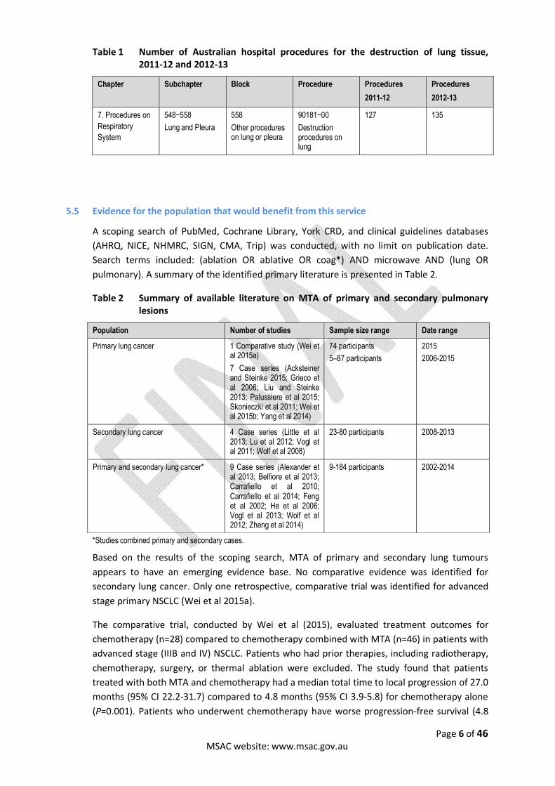
We did not identify a similar procedure for the lung. The closest description of a procedure that may be related to RFA or MTA of the lung appears to be procedure 90181-00: destruction procedures on lung. Destruction procedures of the lung from 2011-12 and 2012-13 are reported in

Page 5 of 46 MSAC website: www.msac.gov.au
Table 1.
Page 6 of 46 MSAC website: www.msac.gov.au
Table 1 Number of Australian hospital procedures for the destruction of lung tissue, 2011-12 and 2012-13
Chapter
Subchapter Block Procedure Procedures
2011-12
Procedures
2012-13
7. Procedures on
Respiratory
System
548−558
Lung and Pleura
558
Other procedures on lung or pleura
90181−00
Destruction procedures on lung
127 135
5.5 Evidence for the population that would benefit from this service
A scoping search of PubMed, Cochrane Library, York CRD, and clinical guidelines databases
(AHRQ, NICE, NHMRC, SIGN, CMA, Trip) was conducted, with no limit on publication date.
Search terms included: (ablation OR ablative OR coag*) AND microwave AND (lung OR
pulmonary). A summary of the identified primary literature is presented in Table 2.
Table 2 Summary of available literature on MTA of primary and secondary pulmonary lesions
Population Number of studies Sample size range Date range
Primary lung cancer
1 Comparative study (Wei et al 2015a)
7 Case series (Acksteiner and Steinke 2015; Grieco et al 2006; Liu and Steinke 2013; Palussiere et al 2015; Skonieczki et al 2011; Wei et al 2015b; Yang et al 2014)
74 participants
5–87 participants
2015
2006-2015
Secondary lung cancer
4 Case series (Little et al 2013; Lu et al 2012; Vogl et al 2011; Wolf et al 2008)
23-80 participants 2008-2013
Primary and secondary lung cancer*
9 Case series (Alexander et al 2013; Belfiore et al 2013; Carrafiello et al 2010; Carrafiello et al 2014; Feng et al 2002; He et al 2006; Vogl et al 2013; Wolf et al 2012; Zheng et al 2014)
9-184 participants 2002-2014
*Studies combined primary and secondary cases.
Based on the results of the scoping search, MTA of primary and secondary lung tumours
appears to have an emerging evidence base. No comparative evidence was identified for
secondary lung cancer. Only one retrospective, comparative trial was identified for advanced
stage primary NSCLC (Wei et al 2015a).
The comparative trial, conducted by Wei et al (2015), evaluated treatment outcomes for
chemotherapy (n=28) compared to chemotherapy combined with MTA (n=46) in patients with
advanced stage (IIIB and IV) NSCLC. Patients who had prior therapies, including radiotherapy,
chemotherapy, surgery, or thermal ablation were excluded. The study found that patients
treated with both MTA and chemotherapy had a median total time to local progression of 27.0
months (95% CI 22.2-31.7) compared to 4.8 months (95% CI 3.9-5.8) for chemotherapy alone
(P=0.001). Patients who underwent chemotherapy have worse progression-free survival (4.8

Page 7 of 46 MSAC website: www.msac.gov.au
months, 95% CI 3.9-5.8) compared to chemotherapy/MTA (10.9 months, 95% CI 5.1-16.7)
(P=0.001). The median overall survival within the follow-up period (mean follow up 21 months,
range 5.1-39.2) was not significantly different between the chemotherapy (17.3 months, 95%
CI 15.2-19.3) and chemotherapy/MTA (23.9 months, 95% CI 15.2-32.6) (P=0.14).
The limited comparative evidence also appears to be an issue for other potential treatment
options. A systematic review of local therapies for stage I and II lung cancers was conducted by
the Agency for Healthcare Research and Quality (AHRQ) in 2013 (Ratko et al 2013). The review
did not identify any comparative studies investigating the use RFA or and radiotherapy in this
population.
Only one ongoing clinical trial investigating the use of MTA for lung cancer was identified on
clinicaltrials.gov and the Australian and New Zealand Clinical Trials Registry (ANZCTR):
NCT01746810: IR-guided ablation (IRGA) combined with stereotactic ablative radiation
(SABR) for large lung tumours.
6 Comparator
The applicant has suggested that RFA is the appropriate comparator; however, this technology
is not widely diffused in the Australian healthcare system and is not currently associated with
an MBS item. Therefore, in addition to RFA there are several other treatments for patients
with primary and secondary lung cancer that can be considered comparators to MTA. These
comparators are first described broadly below with the specific comparators for each patient
population detailed at the end of this section.
Radiofrequency ablation (RFA)
RFA involves creating a closed circuit electrical current through the patient using grounding
pads. Ablation can occur at any point along the closed circuit resulting in unpredictable
ablation zones (Lloyd et al 2011). Unlike RFA, MTA produces localised, predictable ablation
volume shapes and sizes (Bhardwaj et al 2010). The unpredictable ablative nature of RFA may
potentially compromise healthy surrounding lung parenchyma. Further, as RFA requires an
electrical circuit, it is less effective in low electrical conductivity and high baseline impedance
areas such as lung parenchyma (Lee et al 2013). Brace and colleagues demonstrated in a swine
model that microwave energy is a more effective energy source compared with
radiofrequency for use in the lungs (Dupuy and Shulman 2010).
MTA has a steeper temperature gradient, with tissue temperatures reaching > 200 degrees
Celsius, and faster conduction than RFA (Simo et al 2013). This allows for larger ablation
volumes in faster times of 4-6 minutes in contrast to 12-20 minutes for single ablations
required for RFA (Swan et al 2013). Brace et al. found that MTA ablation zones were 25 per
cent larger in mean diameter, 50 per cent larger in cross sectional area and 133 per cent larger
in volume compared to RFA (Brace et al 2009).
MTA has a favourable safety profile compared to RFA as it does not involve electricity or
grounding pads. This eliminates the risk of pad site burns and potential malfunction of
implanted cardiac devices (Lee et al 2013; Schutt et al 2009). MTA is also less susceptible to
the “heat sink” effect due to its ability to reach high ablation temperatures in fast times

Page 8 of 46 MSAC website: www.msac.gov.au
(Dupuy and Shulman 2010). These properties provide an indication in clinical settings,
especially in pulmonary lesions, to move from RFA towards MTA.
Current best practice radiotherapy
The intent of radiotherapy is to achieve a cytotoxic dose of ionising radiation to the tumour
volume whilst attempting to minimize adverse effects of radiation on adjacent normal lung
tissue and thoracic structures. Radiotherapy modalities used in the treatment of NSCLC
include radical radiotherapy delivered in commonly employed regimens as well as continuous
hyperfractionated accelerated radiotherapy (CHART). Radical radiotherapy is an intensive
course of radiotherapy that may be used with curative intent. The course of treatment is
usually given for five days a week in sessions of 10-15 minutes with course between four and
seven weeks. CHART is an alternative method of delivering radical radiotherapy. CHART is
given three times a day for 12 consecutive days (NHS choices).
Stereotactic body radiation therapy (SBRT) may also be used in the treatment of NSCLC; it uses
advanced imaging techniques to deliver highly targeted radiation resulting in less damage to
healthy tissue. Currently, there are no items for SBRT listed on the MBS, and no available
evidence to suggest that SBRT would be superior to conventional radiation therapy for these
treatment populations. SBRT can be used to give single high dose radiation or several
fractionated radiation doses. One potential advantage of SBRT is that can be used to deliver
higher doses of radiation than is possible with other radiotherapy techniques. SBRT
treatments have the advantage of reducing the risk of damage to normal tissue. Guidelines
from the Alberta health services define a role for SBRT in stage I NSCLC who cannot undergo
surgery. These guidelines recommend SBRT for tumours five or less cm in size. Cancer
Australia guidelines do not cover the use of SBRT for this indication.
Cancer Australia guidelines state that (Cancer Council Australia Lung Cancer Guidelines
Working Party 2015):
For Stage I inoperable NSCLC,
“In patients with inoperable stage I NSCLC and good performance status, high dose
radiotherapy is an appropriate treatment option (Grade C). In patients with inoperable stage
I NSCLC, high dose radiotherapy to a total of 60 Gy (gray) in 30 fractions over six weeks is a
reasonable option. CHART may be used as an alternative to radical conventionally
fractionated RT, provided the appropriate resources are available.”
For Stage II inoperable NSCLC,
“Patients with inoperable stage II disease could be offered radiotherapy with curative intent.”
For stage III inoperable NSCLC
“For patients with good performance status and inoperable stage III NSCLC, the concurrent
administration of chemotherapy and radiotherapy is recommended (Grade A). It is
recommended that for patients with inoperable stage III NSCLC undergoing curative therapy
once daily thoracic radiotherapy to at least 60Gy in 2Gy/f plus chemotherapy is administered
(Grade B). For patients with stage III NSCLC who are suitable for curative therapy, but where
Page 9 of 46 MSAC website: www.msac.gov.au
chemotherapy is contra-indicated or refused, CHART may be used as an alternative to radical
conventionally fractionated radiotherapy (Grade B).”
Two additional forms of radiotherapy that may be applied are image-guided radiation therapy
(IGRT) and intensity-modulated radiation therapy (IMRT). In IGRT frequent imaging is used
during the course of radiation to improve the precision and accuracy of treatment. CT, MRI, US
and x-ray imaging may all be used for IGRT (Radiological Society of North America 2014). In
IMRT radiation is delivered in multiple small volumes; this allows the delivery of higher
radiation doses to focused regions of known malignancy whilst minimising radiation to
adjacent tissues. Treatment planning is conducted using 3D CT or MRI imaging (Radiological
Society of North America 2015).
Chemotherapy
Chemotherapy is a systemic treatment for cancer that is taken by mouth or injected into a
vein. It can be given as a combination of drugs (most often two). The National Institute for
Health and Care Excellence (NICE) recommends that chemotherapy should be offered to
patients with stage III NSCLC and good performance status with the aim of improving survival,
disease control and quality of life (NICE 2011). Chemotherapy can also be delivered before or
after radiotherapy as an adjuvant therapy. No recommendations about the role of
chemotherapy delivered before or after radiotherapy were identified for NSCLC. When
chemotherapy is delivered concurrently with radiotherapy it is called chemoradiotherapy. The
rationale for combining chemotherapy and radiotherapy is to combine the benefits of
locoregional control from radiotherapy with the benefits of chemotherapy in reducing the
risks of metastatic disease. With concurrent chemoradiation there is the potential for
chemotherapy, given during a course of radiotherapy, to enhance the effectiveness of
radiotherapy. NICE recommends the consideration of chemoradiotherapy for patients with
stage II or III NSCLC who are not suitable for surgery (NICE 2011). Cancer Australia guidelines
state that there is insufficient evidence to recommend routine use of chemotherapy along
with radiation for the treatment of patients with inoperable stage II NSCLC; however, the
guidelines also state that patients with inoperable stage II disease who have good
performance status and organ function may be considered for definitive concurrent chemo-
radiation with a platin-based regimen (this is based on data extrapolated from studies mainly
including inoperable stage III disease). For patients with inoperable stage III disease, the
guidelines state that the concurrent administration of chemotherapy and radiotherapy is
recommended for those with good performance status (Cancer Council Australia Lung Cancer
Guidelines Working Party 2015).Pulmonary Metastasectomy
Lung metastases from a primary extrapulmonary malignancy are often a manifestation of
widespread disease; however, some patients have metastases exclusive to the lung. In these
patients surgical resection of the pulmonary metastases can substantially prolong survival and
cure some patients. Surgical resection of secondary lung cancer is generally performed in
patients who:
have their primary tumour site controlled
have no uncontrollable extra-pulmonary disease
Page 2 of 46 MSAC website: www.msac.gov.au
all visible lung metastases, including bilateral disease, are resectable while leaving the
patient with adequate pulmonary reserve (Villeneuve and Sundaresan 2009).
A variety of surgical approaches for pulmonary metastasectomy have been described including
video-assisted thoracic surgery (VATS), posterolateral thoracotomy, median sternotomy,
clamshell (bilateral anterior thoracotomies with transverse sternotomy) and staged
procedures (Nichols 2014). The applicant has indicated that MTA should be considered in
selected patients with pulmonary metastases who are eligible for surgical resection.
Page 3 of 46 MSAC website: www.msac.gov.au
Comparators to MTA in early stage inoperable NSCLC with curative intent (Population One)
The applicant has indicated that MTA is indicated in the treatment of early stage NSCLC with
curative intent. The applicant has specified that this includes NSCLC T1a-T2b, N0, M0 (up to
and including stage IIa). For patients with unresectable NSCLC treatment options are
dependent upon the stage of cancer and patient characteristics such as performance status.
Treatments can be stand alone or multimodal and generally comprise radiotherapy alone or in
combination with chemotherapy. Comparators to MTA in this group include the following:
RFA
Current best practice radiotherapy with or without chemotherapy
Comparators to MTA for patients with lung metastases, in whom the primary tumour is
under control and who are receiving treatment with curative intent (oligometastatic
disease) (Population Two)
The applicant has indicated that MTA has a role in the definitive treatment of patients with
lung metastase(s) in whom the primary tumour is under control. In this patient group
comparators include the following:
RFA
Surgical resection (any technique)
Current best practice radiotherapy with or without chemotherapy
The applicant has indicated that thermal ablation can be considered in selected operable
patients with uni- or bi-lateral disease because it is less invasive, more tissue-sparing,
repeatable and can be performed in an outpatient setting or with an overnight stay, having
the least negative impact on quality of life.
Comparators to MTA for patients with NSCLC who are not eligible for surgical resection and
patients with pulmonary metastases who are receiving treatment with palliative intent
(Population Three)
MTA may have a role in treating patients with NSCLC with palliative intent. In this group, MTA
may assist with symptom control and decrease tumour burden in metastatic disease. In this
group the comparators to MTA include the following:
Conventional palliative therapy without MTA

Page 4 of 46 MSAC website: www.msac.gov.au
7 Clinical management algorithm
7.1 Current and proposed clinical practice
The following algorithm, Figure 1, shows the current management of unresectable, early stage
NSCLC. MTA is shown as an alternative to RFA and current best practice radiotherapy with or
without chemotherapy. In the proposed algorithm, Figure 2, MTA is shown as an alternative to
current best practice radiotherapy with or without chemotherapy.
Figure 3 shows the current clinical management algorithm for the management of pulmonary
metastases in patients with the primary cancer under control. In this algorithm MTA is an
alternative to RFA and radiotherapy with or without platinum-based chemotherapy in patients
who are not eligible for surgical resection. Figure 4, the proposed algorithm shows MTA as a
comparator both to radiotherapy with or without chemotherapy and as a comparator to
surgery in both bilateral and unilateral disease.
Figure 5 and Figure 6 show the current and proposed clinical management algorithms for the
palliative management of NSCLC and pulmonary metastases respectively. MTA is shown as an
additional treatment option to conventional palliative treatments for NSCLC and pulmonary
metastases.
In each of the proposed algorithms MTA is replacing RFA.
Figure 1 Current clinical management algorithm for the management of unresectable, early
stage NSCLC with curative intent (Population One)
*Stage IIA patients are considered to be unsuitable for SBRT. NSCLC = non-small cell lung
cancer. RFA = radiofrequency ablation. SBRT = stereotactic body radiotherapy
Page 5 of 46 MSAC website: www.msac.gov.au
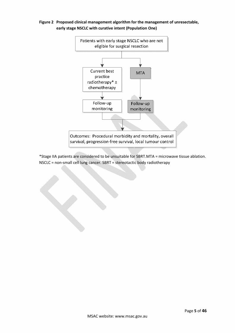
Figure 2 Proposed clinical management algorithm for the management of unresectable,
early stage NSCLC with curative intent (Population One)
*Stage IIA patients are considered to be unsuitable for SBRT.MTA = microwave tissue ablation.
NSCLC = non-small cell lung cancer. SBRT = stereotactic body radiotherapy
Page 6 of 46 MSAC website: www.msac.gov.au
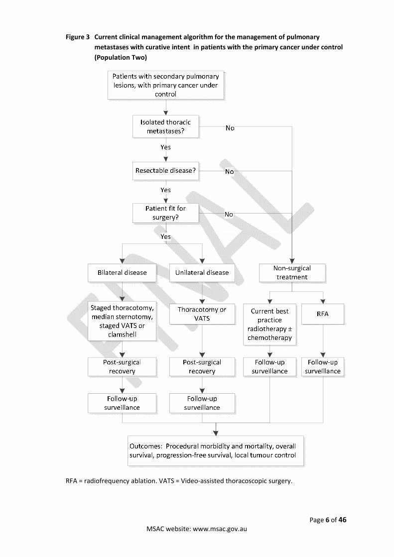
Figure 3 Current clinical management algorithm for the management of pulmonary
metastases with curative intent in patients with the primary cancer under control
(Population Two)
RFA = radiofrequency ablation. VATS = Video-assisted thoracoscopic surgery.

Page 7 of 46 MSAC website: www.msac.gov.au
Figure 4 Proposed clinical management algorithm for the management of pulmonary
metastases with curative intent in patients with the primary cancer under control
(Population Two)
MTA = microwave tissue ablation. VATS = video-assisted thorascopic surgery.

Page 8 of 46 MSAC website: www.msac.gov.au
Figure 5 Current clinical management algorithm for the palliative management of NSCLC and pulmonary metastases (Population Three)
NSCLC = non-small cell lung cancer
Figure 6 Proposed clinical management algorithm for the palliative management of NSCLC and pulmonary metastases (Population Three)
MTA = microwave tissue ablation. NSCLC = non-small cell lung cancer.
Page 9 of 46 MSAC website: www.msac.gov.au
8 Expected health outcomes
8.1 Expected patient-relevant health outcomes
The clinical literature suggests that the primary health outcome of relevance to patients
treated with curative intent is overall and disease-free survival. Secondary outcomes of
relevance to patients include disease control, recurrence and the need for re-ablation. If RFA is
an appropriate comparator, a further secondary outcome may include patient discomfort and
total treatment time. This is because the applicant states that MTA minimises patient
discomfort and enables considerably faster treatment times than RFA.
Primary effectiveness outcomes
The patient population is restricted to patients who are not candidates for surgical resection.
The treatment intent of MTA in this patient group is to extend patient life through destruction
of primary tumours or through local control of pulmonary metastases. Measures of survival
relevant to the patient population include: mortality rates from NSCLC or pulmonary
metastatic tumour at 1-,2-,3- and 5-years; overall survival; the survival rates at 1-,2-,3- and 5-
years; and, the recurrence free survival period and recurrence free survival rates.
In patients treated with palliative intent the primary outcomes are symptom control/relief and
median survival time.
Secondary effectiveness outcomes
Secondary effectiveness outcomes include measures of disease control and recurrence
including: local recurrence rates, 1-year local control rate, mean time to first recurrence, distal
metastases and tumour progression. There are a range of measures associated with
quantifying local control that would be relevant to this patient population and should be
included at the assessment phase. Other secondary effectiveness outcomes may include
procedural discomfort, total procedure time and length of patient hospital stays. Quality of life
measures should also be considered.
In patients treated with palliative intent the secondary outcomes include relative survival
rates.
8.2 Potential risks to patients
The primary safety concern with the proposed service is procedure-related mortality and
morbidity due to peri-operative complications. The applicant states that the complication rate
for MTA varies. Percutaneous MTA requires general anaesthesia or conscious sedation and
may therefore be associated with anaesthesia related adverse events. The procedure may also
involve exposure to CT, which carries an associated risk of ionising radiation exposure.
Potential adverse events that may arise as a result of MTA of the lung identified in the
literature include (Acksteiner and Steinke 2015; Alexander et al 2013; Belfiore et al 2013;
Carrafiello et al 2010; Carrafiello et al 2014; Feng et al 2002; Grieco et al 2006; He et al 2006;
Little et al 2013; Liu and Steinke 2013; Lu et al 2012; Palussiere et al 2015; Skonieczki et al
2011; Vogl et al 2011; Vogl et al 2013; Wei et al 2015a; Wei et al 2015b; Wolf et al 2012; Wolf
et al 2008; Yang et al 2014; Zheng et al 2014):
Page 10 of 46 MSAC website: www.msac.gov.au
Pneumothorax
Needle track implantation
Haemoptysis
Haemothorax
Skin burns
Broncho-pleural fistula
Rib fracture
Pneumonitis
Infection
Chest pain
Pain
Other adverse events
Post-ablation syndrome
9 Clinical claim for the proposed intervention
9.1 Clinical claim
The clinical claim associated with this application depends upon the intended use of, and
available treatment alternatives to MTA.
Clinical claim in patients with early stage inoperable NSCLC who are receiving treatment
with curative intent (Population One)
The applicant has indicated that MTA has a role in the definitive treatment of early stage
inoperable NSCLC. In these patients, guidelines recommend the use of radiotherapy including
SBRT or radical radiotherapy and chemoradiotherapy. MTA is intended to be offered as an
alternative to these therapies in selected patients. It is understood that the clinical claim
associated with the application for this patient group is that MTA offers equivalent
effectiveness outcomes to radiotherapy or chemoradiotherapy with an acceptable safety
profile.
Clinical claim in patients with lung metastase(s), in whom the primary tumour is under
control and who are receiving treatment with curative intent (Population Two)
In these patients the potential treatments for lung metastases depends on whether the
patient is suitable for surgical resection. In patients who are not suitable for surgical resection
the clinical claim is that MTA offers equivalent effectiveness to radiotherapy or
chemoradiotherapy with an acceptable safety profile.
In patients who are eligible for surgical resection the applicant has indicated that MTA can be
considered in selected operable patients with unilateral or bilateral disease, as it is much less
invasive, more tissue-sparing, repeatable and can be performed in an outpatient setting or
with an overnight stay, having the least negative impact on quality of life. Hence, the clinical
claim associated with patients in this group eligible for surgical resection is that MTA
demonstrates equivalent effectiveness to surgical resection with an acceptable safety profile.
Further to this the applicant claims that MTA offers certain benefits over surgical resection in
terms of invasiveness, repeatability and quality of life.

Page 11 of 46 MSAC website: www.msac.gov.au
Clinical claim in patients with NSCLC who are not eligible for surgical resection and patients
with pulmonary metastases who are receiving treatment with palliative intent (Population
Three)
MTA may have a role in treating patients with NSCLC with palliative intent. In these patients
chemotherapy and radiotherapy are the main treatment options. MTA may be offered as an
adjunct to radiotherapy and/or chemotherapy in these patients, as a means of de-bulking
prominent tumours for symptom relief. In this population, MTA may improve symptom relief
as opposed to conventional palliative therapies without MTA.
Clinical claim with respect to RFA in all patient groups
The applicant suggests there are significant treatment advantages of MTA over RFA, especially
in the setting of lung tumour ablation. MTA is arguably more controllable and considered a
safer procedure. MTA also offers larger, faster, more predictable ablation zones and higher
temperatures during ablation. This may result in lower local recurrence rates and better
patient-relevant health outcomes. Hence, in all the patient groups the applicant has suggested
that RFA is a treatment option and that MTA is superior to RFA in terms of effectiveness for all
patient groups and is associated with an acceptable safety profile.
9.2 Economic evaluation
The economic evaluation for the proposed service is informed by the following clinical claims:
Superior safety and effectiveness compared to RFA (Population One and Two).
Non-inferior effectiveness compared to surgery (Population Two) and current best
practice radiotherapy with or without chemotherapy (Population One and Two).
Superior safety compared to surgery (Population Two) and current best practice
radiotherapy with or without chemotherapy (Population One and Two).
In this context, the economic evaluation of the proposed service will be a cost-effectiveness
analysis/cost-utility analysis.
Page 12 of 46 MSAC website: www.msac.gov.au
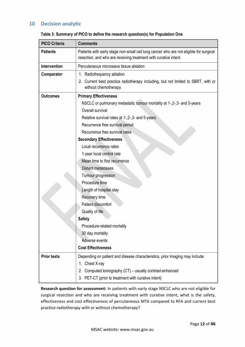
10 Decision analytic
Table 3: Summary of PICO to define the research question(s) for Population One
PICO Criteria Comments
Patients Patients with early stage non-small cell lung cancer who are not eligible for surgical
resection, and who are receiving treatment with curative intent.
Intervention Percutaneous microwave tissue ablation
Comparator 1. Radiofrequency ablation
2. Current best practice radiotherapy including, but not limited to SBRT, with or
without chemotherapy
Outcomes Primary Effectiveness
NSCLC or pulmonary metastatic tumour mortality at 1-,2-,3- and 5-years
Overall survival
Relative survival rates at 1-,2-,3- and 5-years
Recurrence free survival period
Recurrence free survival rates
Secondary Effectiveness
Local recurrence rates
1-year local control rate
Mean time to first recurrence
Distant metastases
Tumour progression
Procedure time
Length of hospital stay
Recovery time
Patient discomfort
Quality of life
Safety
Procedure-related mortality
30 day mortality
Adverse events
Cost Effectiveness
Prior tests Depending on patient and disease characteristics, prior imaging may include:
1. Chest X-ray
2. Computed tomography (CT) – usually contrast-enhanced
3. PET-CT (prior to treatment with curative intent)
Research question for assessment: In patients with early stage NSCLC who are not eligible for
surgical resection and who are receiving treatment with curative intent, what is the safety,
effectiveness and cost effectiveness of percutaneous MTA compared to RFA and current best
practice radiotherapy with or without chemotherapy?
Page 13 of 46 MSAC website: www.msac.gov.au
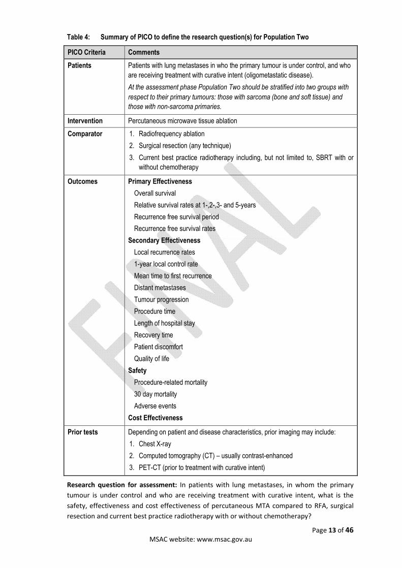
Table 4: Summary of PICO to define the research question(s) for Population Two
PICO Criteria Comments
Patients Patients with lung metastases in who the primary tumour is under control, and who
are receiving treatment with curative intent (oligometastatic disease).
At the assessment phase Population Two should be stratified into two groups with
respect to their primary tumours: those with sarcoma (bone and soft tissue) and
those with non-sarcoma primaries.
Intervention Percutaneous microwave tissue ablation
Comparator 1. Radiofrequency ablation
2. Surgical resection (any technique)
3. Current best practice radiotherapy including, but not limited to, SBRT with or
without chemotherapy
Outcomes Primary Effectiveness
Overall survival
Relative survival rates at 1-,2-,3- and 5-years
Recurrence free survival period
Recurrence free survival rates
Secondary Effectiveness
Local recurrence rates
1-year local control rate
Mean time to first recurrence
Distant metastases
Tumour progression
Procedure time
Length of hospital stay
Recovery time
Patient discomfort
Quality of life
Safety
Procedure-related mortality
30 day mortality
Adverse events
Cost Effectiveness
Prior tests Depending on patient and disease characteristics, prior imaging may include:
1. Chest X-ray
2. Computed tomography (CT) – usually contrast-enhanced
3. PET-CT (prior to treatment with curative intent)
Research question for assessment: In patients with lung metastases, in whom the primary
tumour is under control and who are receiving treatment with curative intent, what is the
safety, effectiveness and cost effectiveness of percutaneous MTA compared to RFA, surgical
resection and current best practice radiotherapy with or without chemotherapy?
Page 14 of 46 MSAC website: www.msac.gov.au
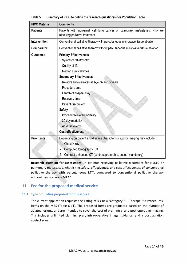
Table 5: Summary of PICO to define the research question(s) for Population Three
PICO Criteria Comments
Patients Patients with non-small cell lung cancer or pulmonary metastases, who are
receiving palliative treatment.
Intervention Conventional palliative therapy with percutaneous microwave tissue ablation
Comparator Conventional palliative therapy without percutaneous microwave tissue ablation
Outcomes Primary Effectiveness
Symptom relief/control
Quality of life
Median survival times
Secondary Effectiveness
Relative survival rates at 1-,2-,3- and 5-years
Procedure time
Length of hospital stay
Recovery time
Patient discomfort
Safety
Procedure-related mortality
30 day mortality
Adverse events
Cost effectiveness
Prior tests Depending on patient and disease characteristics, prior imaging may include:
1. Chest X-ray
2. Computed tomography (CT)
3. Contrast-enhanced CT (contrast preferable, but not mandatory)
Research question for assessment: In patients receiving palliative treatment for NSCLC or
pulmonary metastases, what is the safety, effectiveness and cost effectiveness of conventional
palliative therapy with percutaneous MTA compared to conventional palliative therapy
without percutaneous MTA?
11 Fee for the proposed medical service
11.1 Type of funding proposed for this service
The current application requests the listing of six new ‘Category 3 – Therapeutic Procedures’
items on the MBS (Table 6-11). The proposed items are graduated based on the number of
ablated lesions, and are intended to cover the cost of pre-, intra- and post-operative imaging.
This includes a limited planning scan, intra-operative image guidance, and a post ablation
control scan.
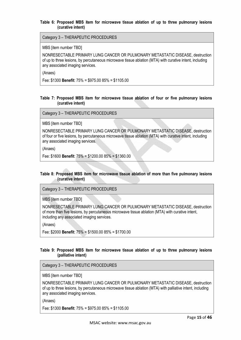
Page 15 of 46 MSAC website: www.msac.gov.au
Table 6: Proposed MBS item for microwave tissue ablation of up to three pulmonary lesions (curative intent)
Category 3 – THERAPEUTIC PROCEDURES
MBS [item number TBD]
NONRESECTABLE PRIMARY LUNG CANCER OR PULMONARY METASTATIC DISEASE, destruction of up to three lesions, by percutaneous microwave tissue ablation (MTA) with curative intent, including any associated imaging services.
(Anaes)
Fee: $1300 Benefit: 75% = $975.00 85% = $1105.00
Table 7: Proposed MBS item for microwave tissue ablation of four or five pulmonary lesions (curative intent)
Category 3 – THERAPEUTIC PROCEDURES
MBS [item number TBD]
NONRESECTABLE PRIMARY LUNG CANCER OR PULMONARY METASTATIC DISEASE, destruction of four or five lesions, by percutaneous microwave tissue ablation (MTA) with curative intent, including any associated imaging services.
(Anaes)
Fee: $1600 Benefit: 75% = $1200.00 85% = $1360.00
Table 8: Proposed MBS item for microwave tissue ablation of more than five pulmonary lesions (curative intent)
Category 3 – THERAPEUTIC PROCEDURES
MBS [item number TBD]
NONRESECTABLE PRIMARY LUNG CANCER OR PULMONARY METASTATIC DISEASE, destruction of more than five lesions, by percutaneous microwave tissue ablation (MTA) with curative intent, including any associated imaging services.
(Anaes)
Fee: $2000 Benefit: 75% = $1500.00 85% = $1700.00
Table 9: Proposed MBS item for microwave tissue ablation of up to three pulmonary lesions (palliative intent)
Category 3 – THERAPEUTIC PROCEDURES
MBS [item number TBD]
NONRESECTABLE PRIMARY LUNG CANCER OR PULMONARY METASTATIC DISEASE, destruction of up to three lesions, by percutaneous microwave tissue ablation (MTA) with palliative intent, including any associated imaging services.
(Anaes)
Fee: $1300 Benefit: 75% = $975.00 85% = $1105.00

Page 16 of 46 MSAC website: www.msac.gov.au
Table 10: Proposed MBS item for microwave tissue ablation of four or five pulmonary lesions (palliative intent)
Category 3 – THERAPEUTIC PROCEDURES
MBS [item number TBD]
NONRESECTABLE PRIMARY LUNG CANCER OR PULMONARY METASTATIC DISEASE, destruction of four or five lesions, by percutaneous microwave tissue ablation (MTA) with palliative intent, including any associated imaging services.
(Anaes)
Fee: $1600 Benefit: 75% = $1200.00 85% = $1360.00
Table 11: Proposed MBS item for microwave tissue ablation of more than five pulmonary lesions (palliative intent)
Category 3 – THERAPEUTIC PROCEDURES
MBS [item number TBD]
NONRESECTABLE PRIMARY LUNG CANCER OR PULMONARY METASTATIC DISEASE, destruction of more than five lesions, by percutaneous microwave tissue ablation (MTA) with palliative intent, including any associated imaging services.
(Anaes)
Fee: $2000 Benefit: 75% = $1500.00 85% = $1700.00
11.2 Direct costs associated with the proposed service
Clinical feedback suggests RFA is cheaper than MTA. The costs associated with RFA range
between $1,500 and $2,000 for consumables, as opposed to $2,200 and $2,900 for MTA.
Private health insurance usually covers the cost of the consumables; however, it is currently
understood that gap payments are charged on top of the cost of consumables. Many of the
following costs associated with MTA will need to be identified during the assessment phase:
MTA equipment – including: cost of machine $50,000, applicator $2,960, temperature
probe ($960), and other associated costs (source: application documents)
Interventional radiologist, time (percutaneous procedures)
Radiology suite usage
Other consumables, e.g. dressings
Anaesthetic
Follow-up imaging
Dedicated nursing staff for post-intervention care
Overnight stay in hospital

Page 17 of 46 MSAC website: www.msac.gov.au
11.3 Proposed fee
As the applicant has not suggested a proposed fee for MTA of pulmonary lesions, the
proposed fee has been adopted from Application 1402 (MTA of liver tumours). Application
1402 states:
“A $1300 fee for ablation of 2-3 lesions, a $1600 fee for ablation of 4-5 lesions and a $2000 fee
for ablation of >5 lesions. The higher fee for >5 lesions reflects the increased risk to the patients
such as collateral damage as well as more skill, time and expertise required of the physician to
ensure better patient outcomes”
According to the applicant the number of tumours treated alters the complexity of the
procedure. A graduated fee structure for the number of tumours treated should be supported
by evidence of increased complexity and increased clinical benefits. To determine the value of
a graduated fee, PASC has advised that the assessment phase should include a stratified
survival analysis based on the number of ablated lesions.
As there is no Medicare number for lung RFA, the maximum rebate that can be received in
private practice is $470.00 (MBS item 57341 for CT-guided interventions). The fee for RFA
services for liver [both percutaneous and open/laparoscopic (50952)] is $817.10. It should be
noted, the application claims MTA has a faster ablation time which would result in less time
overall spent in the radiology suite, and may impact on the cost of the procedure.
12 Regulatory information and registered trademark
The application refers to the Acculis MTA System with a single use microwave applicator,
which is registered to be used in Australia with N Stenning and Co Pty Ltd as the sponsor. In
addition to the Acculis MTA system, there are three additional MTA systems currently
available in Australia. Other devices registered in Australia include:
The Avecure Microwave Ablation / Coagulation System sponsored by Aurora
BioScience Pty Ltd (ARTG ID 200325) is listed on the ARTG for ablation/coagulation of
soft tissue. This device uses 902-928 MHz microwaves, and 32W.
The Emprint™ Ablation System with Thermosphere™ Technology - microwave
hyperthermia system (ARTG ID 226598), an intracorporeal microwave hyperthermia
applicator (ARTG ID 178369), and two hyperthermia microwave systems (ARTG IDs
152044, 178699) sponsored by Covidien Pty Ltd. The system is intended to be used for
percutaneous, laparoscopic, and intraoperative coagulation (ablation) of soft tissue.
This system uses 1.4-1.5 GHz and 100Watts.
The Amica microwave hyperthermia system (ARTG ID 212509), and an intracorporeal
microwave hyperthermia applicator (ARTG ID 212510) sponsored by Culpan Medical
Pty Ltd. For soft tissue pathologies such as solid tumours or hyperplasia of the liver,
kidney, lung, bone, breast, prostate, etc. The system uses 2.45 GHz and 20-140W of
power.
The Acculis MTA system involves thermal coagulation of soft tissue using 2.45GHz microwave
energy. The system consists of the Sulis VpMTA Generator, Acculis Local Control Station (LCS),

Page 18 of 46 MSAC website: www.msac.gov.au
Acculis MTA Applicators and optional MTA Temperature Probes. The ARTG listing or
registration number:
Temperature Probes (ARTG ID 174513): The temperature probes used with the Acculis
MTA System are intended to monitor the temperature of the probes at the point of
delivery of the microwave energy (i.e. at the point of tissue coagulation).
Trolley (ARTG ID 195697): A general-purpose trolley or conveyance designed for
transporting/supplying any kind of devices, medical equipment or goods within a
department or hospital. It may have one or more shelves
Applicator (ARTG ID 174514): The Single Use Microwave Applicator is intended to be
used with the Acculis MTA System for intraoperative coagulation of soft tissue.
Microwave Generator System (ARTG ID 157722): Treat lesions using microwave
hyperthermia
13 Healthcare resources
The healthcare resources related to the proposed service and comparator interventions are
outlined in Table 12.
14 Questions for public funding
None

Page 19 of 46 MSAC website: www.msac.gov.au
Table 12: List of resources to be considered in the economic analysis
Provider of resource
Setting in which resource is
provided
Proportion of patients
receiving resource
Number of units of resource per relevant time horizon per
patient receiving resource
Disaggregated unit cost
MBS
Item Safety nets*
Other government
budget
Private health insurer
Patient Total cost
Resources provided to identify eligible population
Diagnostic imaging (US, CT, CECT, MRI, FDG PET etc.)
Radiologists Radiology clinic or radiology department (hospital)
100%
Resources provided to deliver proposed intervention (MTA)
Machine cost ($50,000) Hospital Radiology clinic or radiology department
100%
Disposable probe ($2,960) Hospital Radiology clinic or radiology department
100%
Time to perform procedure (ablation time of 4-6 minutes per lesion, also time for patient positioning, anaesthetic administration)
Interventional radiologist
Radiology clinic or radiology department
100%
Image-guidance (CT or US) Interventional radiologist
Radiology clinic or radiology department
100%
Anaesthetic Anaesthetist Radiology clinic or radiology department
100%
Page 20 of 46 MSAC website: www.msac.gov.au
Provider of resource
Setting in which resource is
provided
Proportion of patients
receiving resource
Number of units of resource per relevant time horizon per
patient receiving resource
Disaggregated unit cost
MBS
Item Safety nets*
Other government
budget
Private health insurer
Patient Total cost
Resources provided in association with proposed intervention (MTA)
Aftercare Dedicated nursing staff
Radiology clinic or radiology department
100%
Follow-up imaging (cross-sectional) 6 weeks post-procedure
Radiologist/radiographer
Radiology clinic or radiology department
100%
Resources provided to deliver comparator 1 (RFA)
Machine cost ($40,000-$65,000**)
Hospital Out-patient 100%
Disposable probe ($1,700-$2,700)**
Hospital Out-patient 100%
Time to perform ablation (10-20 minutes)
Interventional radiologist or surgeon
Radiology clinic or radiology department
100%
Imaging (CT or US) Interventional radiologist or surgeon
Radiology clinic or radiology department
100%
Anaesthetic Anaesthetist Radiology clinic or radiology department
100%
Resources provided in association with comparator 1 (RFA)
Page 21 of 46 MSAC website: www.msac.gov.au
Provider of resource
Setting in which resource is
provided
Proportion of patients
receiving resource
Number of units of resource per relevant time horizon per
patient receiving resource
Disaggregated unit cost
MBS
Item Safety nets*
Other government
budget
Private health insurer
Patient Total cost
Aftercare Dedicated nursing staff
Radiology clinic or radiology department
100%
Follow-up imaging Radiologist/radiographer
Radiology clinic or radiology department
100% 24 hrs post procedure
Resources provided to deliver comparator 2 (Radiotherapy)
Simulation Radiation
oncologist
Outpatient 100% 15550 ($658.60), 15553, 15600, 15500
Dosimetry Radiation
oncologist
Outpatient 100% 15518 , 15521, 15524, 15527, 15530, 15533
Treatment Radiation
oncologist
Outpatient 100% 15000, 15006, 15100, 15106, 15112
Verification Radiation
oncologist
Outpatient 100% 15700, 15705, 15710
Resources provided in association with comparator 2 (Radiotherapy)
Aftercare 100%
Follow-up imaging Radiologist/radiographer
Outpatient 100%
Page 22 of 46 MSAC website: www.msac.gov.au
Provider of resource
Setting in which resource is
provided
Proportion of patients
receiving resource
Number of units of resource per relevant time horizon per
patient receiving resource
Disaggregated unit cost
MBS
Item Safety nets*
Other government
budget
Private health insurer
Patient Total cost
Resources provided to deliver chemotherapy (potential adjunct to the intervention and comparators 1 and 2)
Initial specialist consult specialist 100%
Chemotherapy planning Specialist/multi-disciplinary team
100%
Pathology monitoring 100%
Administering chemotherapy Nurse/specialist oncologist/haematologist
Outpatient or hospital
100%
Chemotherapy drug(s) Nurse/specialist oncologist/haematologist
Outpatient or hospital
100% Cisplatin, max safety net: $37.70
PBS, Dispensed price per maximum amount: $126.72
Max price to consumer: $37.70
Resources provided in association with chemotherapy (potential adjunct to the intervention and comparators 1 and 2)
Aftercare Dedicated nursing staff
Outpatient or hospital
100% 6-weekly follow-up intervals
Resources used to manage patients successfully treated with the proposed intervention
Follow-up imaging to confirm no tumour recurrence
100% of patient successfully treated
Follow-up treatment as required

Page 23 of 46 MSAC website: www.msac.gov.au
Provider of resource
Setting in which resource is
provided
Proportion of patients
receiving resource
Number of units of resource per relevant time horizon per
patient receiving resource
Disaggregated unit cost
MBS
Item Safety nets*
Other government
budget
Private health insurer
Patient Total cost
Follow-up palliative care as required
Resources used to manage patients who are unsuccessfully treated with the proposed intervention
Follow-up imaging to confirm incomplete ablation of tumour and/or tumour recurrence
100% of patients unsuccessfully treated
Re-staging of disease and treatment as determined according to current disease status
100% of patients unsuccessfully treated
Resources used to manage patients successfully treated with comparator 1
Follow-up imaging to confirm no tumour recurrence
100% of patient successfully treated
Follow-up treatment as required
Follow-up palliative care as required
Resources used to manage patients who are unsuccessfully treated with comparator 1
Follow-up imaging to confirm incomplete ablation of tumour and/or tumour recurrence
100% of patients unsuccessfully treated
at 3, 6 and 12 months and yearly thereafter

Page 24 of 46 MSAC website: www.msac.gov.au
Provider of resource
Setting in which resource is
provided
Proportion of patients
receiving resource
Number of units of resource per relevant time horizon per
patient receiving resource
Disaggregated unit cost
MBS
Item Safety nets*
Other government
budget
Private health insurer
Patient Total cost
Re-staging of disease and treatment as determined according to current disease status
100% of patients unsuccessfully treated
CECT – contrast enhanced CT. CT= computed tomography. MRI = magnetic resonance imaging. MBS = Medicare Benefits Scheme. MTA = microwave tissue ablation. NA = not applicable. RFA = radiofrequency ablation. US = ultrasound.
Page 25 of 46 MSAC website: www.msac.gov.au
15 References
Acksteiner, C & Steinke, K 2015, 'Percutaneous microwave ablation for early-stage non-small cell lung cancer (NSCLC) in the elderly: a promising outlook', J Med Imaging Radiat Oncol, vol.59(1), pp. 82-90.
Aihw 2011, Lung cancer in Australia: an overview, Canberra, Australia, viewed <Aihw 2011, Lung cancer in Australia: an overview, Canberra, Australia.
Aihw 2014, Cancer in Australia: An overview 2014, Canberra, Australia, viewed <Aihw 2014, Cancer in Australia: An overview 2014, Canberra, Australia.
Alexander, ES, Hankins, CA, Machan, JT, Healey, TT & Dupuy, DE 2013, 'Rib fractures after percutaneous radiofrequency and microwave ablation of lung tumors: incidence and relevance', Radiology, vol.266(3), pp. 971-8.
Ambrogi, MC, Fanucchi, O, Cioni, R, Dini, P, De Liperi, A, Cappelli, C, Davini, F, Bartolozzi, C & Mussi, A 2011, 'Long-term results of radiofrequency ablation treatment of stage I non-small cell lung cancer: a prospective intention-to-treat study', J Thorac Oncol, vol.6(12), pp. 2044-51.
American Joint Committee on Cancer 2009, Lung Cancer Staging, viewed 17 June 2015, <https://cancerstaging.org/references-tools/quickreferences/Documents/Lung%20Cancer%20Staging%20Poster%20Updated.pdf>.
Banik, S, Bandyopadhyay, S & Ganguly, S 2003, 'Bioeffects of microwave––a brief review', Bioresource Technology, vol.87(2), pp. 155-59.
Belfiore, G, Ronza, F, Belfiore, MP, Serao, N, Di Ronza, G, Grassi, R & Rotondo, A 2013, 'Patients' survival in lung malignancies treated by microwave ablation: our experience on 56 patients', Eur J Radiol, vol.82(1), pp. 177-81.
Bhardwaj, N, Strickland, AD, Ahmad, F, El-Abassy, M, Morgan, B, Robertson, GS & Lloyd, DM 2010, 'Microwave ablation for unresectable hepatic tumours: clinical results using a novel microwave probe and generator', Eur J Surg Oncol, vol.36(3), pp. 264-8.
Brace, CL, Hinshaw, JL, Laeseke, PF, Sampson, LA & Lee, FT, Jr. 2009, 'Pulmonary thermal ablation: comparison of radiofrequency and microwave devices by using gross pathologic and CT findings in a swine model', Radiology, vol.251(3), pp. 705-11.
British Thoracic Society 2001, 'BTS guidelines: guidelines on the selection of patients with lung cancer for surgery', Thorax, vol.56(2), pp. 89-108.
Cancer Australia 2013, Best practice approaches to lung cancer care: A review of the literature, Surry Hills, NSW, viewed
Cancer Council Australia Lung Cancer Guidelines Working Party 2015, Clinical practice guidelines for the treatment of lung cancer, Cancer Council Australia Lung Cancer Guidelines Working Party, viewed 16 June 2015, Cancer Council Australia Lung Cancer Guidelines Working Party 2015, Clinical practice guidelines for the treatment of lung cancer, Cancer Council Australia Lung Cancer Guidelines Working Party.
Carrafiello, G, Mangini, M, De Bernardi, I, Fontana, F, Dionigi, G, Cuffari, S, Imperatori, A, Lagana, D & Fugazzola, C 2010, 'Microwave ablation therapy for treating primary and secondary lung tumours: technical note', Radiol Med, vol.115(6), pp. 962-74.
Carrafiello, G, Mangini, M, Fontana, F, Ierardi, AM, De Marchi, G, Rotolo, N, Chini, C, Cuffari, S & Fugazzola, C 2014, 'Microwave ablation of lung tumours: single-centre preliminary experience', Radiol Med, vol.119(1), pp. 75-82.
Datta, D & Lahiri, B 2003, 'Preoperative evaluation of patients undergoing lung resection surgery', Chest, vol.123(6), pp. 2096-103.
De Baere, T, Auperin, A, Deschamps, F, Chevallier, P, Gaubert, Y, Boige, V, Fonck, M, Escudier, B & Palussiere, J 2015, 'Radiofrequency ablation is a valid treatment option for lung metastases: experience in 566 patients with 1037 metastases', Ann Oncol, vol.26(5), pp. 987-91.

Page 26 of 46 MSAC website: www.msac.gov.au
Dong, B, Liang, P, Yu, X, Su, L, Yu, D, Cheng, Z & Zhang, J 2003, 'Percutaneous sonographically guided microwave coagulation therapy for hepatocellular carcinoma: results in 234 patients', AJR Am J Roentgenol, vol.180(6), pp. 1547-55.
Dupuy, DE 2009, 'Microwave ablation compared with radiofrequency ablation in lung tissue-is microwave not just for popcorn anymore?', Radiology, vol.251(3), pp. 617-8.
Dupuy, DE 2013, 'Treatment of medically inoperable non-small-cell lung cancer with stereotactic body radiation therapy versus image-guided tumor ablation: can interventional radiology compete?', J Vasc Interv Radiol, vol.24(8), pp. 1139-45.
Dupuy, DE & Shulman, M 2010, 'Current status of thermal ablation treatments for lung malignancies', Semin Intervent Radiol, vol.27(3), pp. 268-75.
Feng, W, Liu, W, Li, C, Li, Z, Li, R, Liu, F, Zhai, B, Shi, J & Shi, G 2002, 'Percutaneous microwave coagulation therapy for lung cancer', Zhonghua Zhong Liu Za Zhi, vol.24(4), pp. 388-90.
Fleisher, LA, Fleischmann, KE, Auerbach, AD, Barnason, SA, Beckman, JA, Bozkurt, B, Davila-Roman, VG, Gerhard-Herman, MD, Holly, TA, Kane, GC, Marine, JE, Nelson, MT, Spencer, CC, Thompson, A, Ting, HH, Uretsky, BF & Wijeysundera, DN 2015, '2014 ACC/AHA guideline on perioperative cardiovascular evaluation and management of patients undergoing noncardiac surgery: executive summary: a report of the American College of Cardiology/American Heart Association Task Force on practice guidelines. Developed in collaboration with the American College of Surgeons, American Society of Anesthesiologists, American Society of Echocardiography, American Society of Nuclear Cardiology, Heart Rhythm Society, Society for Cardiovascular Angiography and Interventions, Society of Cardiovascular Anesthesiologists, and Society of Vascular Medicine Endorsed by the Society of Hospital Medicine', J Nucl Cardiol, vol.22(1), pp. 162-215.
Gillams, A, Khan, Z, Osborn, P & Lees, W 2013, 'Survival after radiofrequency ablation in 122 patients with inoperable colorectal lung metastases', Cardiovasc Intervent Radiol, vol.36(3), pp. 724-30.
Gould, G, Pearce, A., 2006, 'Assessment of suitability for lung resection', Continuing Education in Anaesthesia, Critical Care & Pain, vol.6(3), pp. 97-100.
Grieco, CA, Simon, CJ, Mayo-Smith, WW, Dipetrillo, TA, Ready, NE & Dupuy, DE 2006, 'Percutaneous image-guided thermal ablation and radiation therapy: outcomes of combined treatment for 41 patients with inoperable stage I/II non-small-cell lung cancer', J Vasc Interv Radiol, vol.17(7), pp. 1117-24.
He, W, Hu, XD, Wu, DF, Guo, L, Zhang, LZ, Xiang, DY & Ning, B 2006, 'Ultrasonography-guided percutaneous microwave ablation of peripheral lung cancer', Clin Imaging, vol.30(4), pp. 234-41.
Hirakata, K, Nakata, H & Haratake, J 1993, 'Appearance of pulmonary metastases on high-resolution CT scans: comparison with histopathologic findings from autopsy specimens', AJR Am J Roentgenol, vol.161(1), pp. 37-43.
Hiraki, T & Kanazawa, S 2012, 'Lung radiofrequency ablation: potential as a therapy to oligometastasis and oligorecurrence', Pulm Med, vol.2012pp. 196173.
Irsa 2015, Interventional Radiology Society of Australasia Guidelines for Credentialing for Interventional Radiology, viewed 4 September 2015, Irsa 2015, Interventional Radiology Society of Australasia Guidelines for Credentialing for Interventional Radiology,.
Kuang, M, Lu, MD, Xie, XY, Xu, HX, Mo, LQ, Liu, GJ, Xu, ZF, Zheng, YL & Liang, JY 2007, 'Liver cancer: increased microwave delivery to ablation zone with cooled-shaft antenna--experimental and clinical studies', Radiology, vol.242(3), pp. 914-24.
Lanuti, M, Sharma, A, Willers, H, Digumarthy, SR, Mathisen, DJ & Shepard, JA 2012, 'Radiofrequency ablation for stage I non-small cell lung cancer: management of locoregional recurrence', Ann Thorac Surg, vol.93(3), pp. 921-7; discussion 27-88.
Lee, WK, Lau, EW, Chin, K, Sedlaczek, O & Steinke, K 2013, 'Modern diagnostic and therapeutic interventional radiology in lung cancer', J Thorac Dis, vol.5 Suppl 5pp. S511-23.
Page 27 of 46 MSAC website: www.msac.gov.au
Little, MW, Chung, D, Boardman, P, Gleeson, FV & Anderson, EM 2013, 'Microwave ablation of pulmonary malignancies using a novel high-energy antenna system', Cardiovasc Intervent Radiol, vol.36(2), pp. 460-5.
Liu, H & Steinke, K 2013, 'High-powered percutaneous microwave ablation of stage I medically inoperable non-small cell lung cancer: a preliminary study', J Med Imaging Radiat Oncol, vol.57(4), pp. 466-74.
Lloyd, DM, Lau, KN, Welsh, F, Lee, KF, Sherlock, DJ, Choti, MA, Martinie, JB & Iannitti, DA 2011, 'International multicentre prospective study on microwave ablation of liver tumours: preliminary results', HPB (Oxford), vol.13(8), pp. 579-85.
Lu, MD, Chen, JW, Xie, XY, Liu, L, Huang, XQ, Liang, LJ & Huang, JF 2001, 'Hepatocellular carcinoma: US-guided percutaneous microwave coagulation therapy', Radiology, vol.221(1), pp. 167-72.
Lu, Q, Cao, W, Huang, L, Wan, Y, Liu, T, Cheng, Q, Han, Y & Li, X 2012, 'CT-guided percutaneous microwave ablation of pulmonary malignancies: Results in 69 cases', World J Surg Oncol, vol.10pp. 80.
Meredith, K, Lee, F, Henry, MB, Warner, T & Mahvi, D 2005, 'Microwave ablation of hepatic tumors using dual-loop probes: results of a phase I clinical trial', J Gastrointest Surg, vol.9(9), pp. 1354-60.
Nice 2011, Lung cancer: The diagnosis and treatment of lung cancer, viewed <guidance.nice.org.uk/cg121>.
Nichols, FC 2014, 'Pulmonary metastasectomy: role of pulmonary metastasectomy and type of surgery', Curr Treat Options Oncol, vol.15(3), pp. 465-75.
Oken, MM, Creech, RH, Tormey, DC, Horton, J, Davis, TE, Mcfadden, ET & Carbone, PP 1982, 'Toxicity and response criteria of the Eastern Cooperative Oncology Group', Am J Clin Oncol, vol.5(6), pp. 649-55.
Ong, SL, Gravante, G, Metcalfe, MS, Strickland, AD, Dennison, AR & Lloyd, DM 2009, 'Efficacy and safety of microwave ablation for primary and secondary liver malignancies: a systematic review', Eur J Gastroenterol Hepatol, vol.21(6), pp. 599-605.
Palussiere, J, Lagarde, P, Auperin, A, Deschamps, F, Chomy, F & De Baere, T 2015, 'Percutaneous lung thermal ablation of non-surgical clinical N0 non-small cell lung cancer: results of eight years' experience in 87 patients from two centers', Cardiovasc Intervent Radiol, vol.38(1), pp. 160-6.
Quint, LE 2004, 'Lung cancer: assessing resectability', Cancer Imaging, vol.4(1), pp. 15-8. Radiological Society of North America 2014, Image-guided radiation therapy (IGRT), viewed 1
October 2015, Quint, LE 2004, 'Lung cancer: assessing resectability', Cancer Imaging, vol.4(1), pp. 15-8..
Radiological Society of North America 2015, Intensity-modulated radiation therapy (IMRT), viewed 1 October 2015 Radiological Society of North America 2015, Intensity-modulated radiation therapy (IMRT),.
Ratko, T, Vats, V, Brock, J, Ruffner, B, Jr & Aronson, N 2013, Local nonsurgical therapies for stage I and symptomatic obstructive non-small-cell lung cacner, Rockville, MD, viewed <Ratko, T, Vats, V, Brock, J, Ruffner, B, Jr & Aronson, N 2013, Local nonsurgical therapies for stage I and symptomatic obstructive non-small-cell lung cacner.
Schneider, T, Heussel, CP, Herth, FJ & Dienemann, H 2013, 'Thermal ablation of malignant lung tumors', Dtsch Arztebl Int, vol.110(22), pp. 394-400.
Schutt, DJ, Swindle, MM, Bastarrika, GA & Haemmerich, D 2009, 'Sequential activation of ground pads reduces skin heating during radiofrequency ablation: initial in vivo porcine results', Conf Proc IEEE Eng Med Biol Soc, vol.2009pp. 4287-90.
Seki, T, Tamai, T, Nakagawa, T, Imamura, M, Nishimura, A, Yamashiki, N, Ikeda, K & Inoue, K 2000, 'Combination therapy with transcatheter arterial chemoembolization and percutaneous microwave coagulation therapy for hepatocellular carcinoma', Cancer, vol.89(6), pp. 1245-51.

Page 28 of 46 MSAC website: www.msac.gov.au
Seo, JB, Im, JG, Goo, JM, Chung, MJ & Kim, MY 2001, 'Atypical pulmonary metastases: spectrum of radiologic findings', Radiographics, vol.21(2), pp. 403-17.
Simo, KA, Tsirline, VB, Sindram, D, Mcmillan, MT, Thompson, KJ, Swan, RZ, Mckillop, IH, Martinie, JB & Iannitti, DA 2013, 'Microwave ablation using 915-MHz and 2.45-GHz systems: what are the differences?', HPB (Oxford), vol.15(12), pp. 991-6.
Simon, CJ & Dupuy, DE 2005, 'Current role of image-guided ablative therapies in lung cancer', Expert Rev Anticancer Ther, vol.5(4), pp. 657-66.
Simon, CJ, Dupuy, DE, Dipetrillo, TA, Safran, HP, Grieco, CA, Ng, T & Mayo-Smith, WW 2007, 'Pulmonary radiofrequency ablation: long-term safety and efficacy in 153 patients', Radiology, vol.243(1), pp. 268-75.
Simon, CJ, Dupuy, DE & Mayo-Smith, WW 2005, 'Microwave ablation: principles and applications', Radiographics, vol.25 Suppl 1pp. S69-83.
Skonieczki, BD, Wells, C, Wasser, EJ & Dupuy, DE 2011, 'Radiofrequency and microwave tumor ablation in patients with implanted cardiac devices: is it safe?', Eur J Radiol, vol.79(3), pp. 343-6.
Smith, SL & Jennings, PE 2015, 'Lung radiofrequency and microwave ablation: a review of indications, techniques and post-procedural imaging appearances', Br J Radiol, vol.88(1046), pp. 20140598.
Swan, R-Z, Tsirline, V, Sindram, D, Martinie, J-B & Iannitti, D-A 2012, 'Fundamentals of microwave physics: application to hepatic ablation', Journal of Microwave Surgery, vol.30pp. 25-40.
Swan, RZ, Sindram, D, Martinie, JB & Iannitti, DA 2013, 'Operative microwave ablation for hepatocellular carcinoma: complications, recurrence, and long-term outcomes', J Gastrointest Surg, vol.17(4), pp. 719-29.
Tammemagi, CM, Neslund-Dudas, C, Simoff, M & Kvale, P 2004, 'In lung cancer patients, age, race-ethnicity, gender and smoking predict adverse comorbidity, which in turn predicts treatment and survival', J Clin Epidemiol, vol.57(6), pp. 597-609.
Villeneuve, PJ & Sundaresan, RS 2009, 'Surgical management of colorectal lung metastasis', Clin Colon Rectal Surg, vol.22(4), pp. 233-41.
Vogl, TJ, Naguib, NN, Gruber-Rouh, T, Koitka, K, Lehnert, T & Nour-Eldin, NE 2011, 'Microwave ablation therapy: clinical utility in treatment of pulmonary metastases', Radiology, vol.261(2), pp. 643-51.
Vogl, TJ, Worst, TS, Naguib, NN, Ackermann, H, Gruber-Rouh, T & Nour-Eldin, NE 2013, 'Factors influencing local tumor control in patients with neoplastic pulmonary nodules treated with microwave ablation: a risk-factor analysis', AJR Am J Roentgenol, vol.200(3), pp. 665-72.
Wei, Z, Ye, X, Yang, X, Huang, G, Li, W, Wang, J & Han, X 2015a, 'Microwave ablation plus chemotherapy improved progression-free survival of advanced non-small cell lung cancer compared to chemotherapy alone', Med Oncol, vol.32(2), pp. 464.
Wei, Z, Ye, X, Yang, X, Zheng, A, Huang, G, Li, W, Ni, X, Wang, J & Han, X 2015b, 'Microwave ablation in combination with chemotherapy for the treatment of advanced non-small cell lung cancer', Cardiovasc Intervent Radiol, vol.38(1), pp. 135-42.
Wolf, FJ, Aswad, B, Ng, T & Dupuy, DE 2012, 'Intraoperative microwave ablation of pulmonary malignancies with tumor permittivity feedback control: ablation and resection study in 10 consecutive patients', Radiology, vol.262(1), pp. 353-60.
Wolf, FJ, Grand, DJ, Machan, JT, Dipetrillo, TA, Mayo-Smith, WW & Dupuy, DE 2008, 'Microwave ablation of lung malignancies: effectiveness, CT findings, and safety in 50 patients', Radiology, vol.247(3), pp. 871-9.
Yang, X, Ye, X, Zheng, A, Huang, G, Ni, X, Wang, J, Han, X, Li, W & Wei, Z 2014, 'Percutaneous microwave ablation of stage I medically inoperable non-small cell lung cancer: clinical evaluation of 47 cases', J Surg Oncol, vol.110(6), pp. 758-63.
Yu, NC, Lu, DS, Raman, SS, Dupuy, DE, Simon, CJ, Lassman, C, Aswad, BI, Ianniti, D & Busuttil, RW 2006, 'Hepatocellular carcinoma: microwave ablation with multiple straight and loop

Page 29 of 46 MSAC website: www.msac.gov.au
antenna clusters--pilot comparison with pathologic findings', Radiology, vol.239(1), pp. 269-75.
Zheng, A, Wang, X, Yang, X, Wang, W, Huang, G, Gai, Y & Ye, X 2014, 'Major complications after lung microwave ablation: a single-center experience on 204 sessions', Ann Thorac Surg, vol.98(1), pp. 243-8.
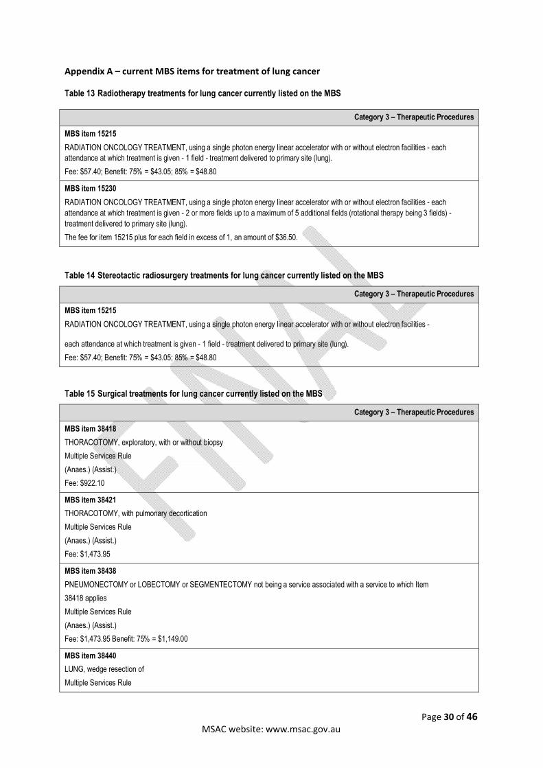
Page 30 of 46 MSAC website: www.msac.gov.au
Appendix A – current MBS items for treatment of lung cancer
Table 13 Radiotherapy treatments for lung cancer currently listed on the MBS
Category 3 – Therapeutic Procedures
MBS item 15215
RADIATION ONCOLOGY TREATMENT, using a single photon energy linear accelerator with or without electron facilities - each
attendance at which treatment is given - 1 field - treatment delivered to primary site (lung).
Fee: $57.40; Benefit: 75% = $43.05; 85% = $48.80
MBS item 15230
RADIATION ONCOLOGY TREATMENT, using a single photon energy linear accelerator with or without electron facilities - each
attendance at which treatment is given - 2 or more fields up to a maximum of 5 additional fields (rotational therapy being 3 fields) -
treatment delivered to primary site (lung).
The fee for item 15215 plus for each field in excess of 1, an amount of $36.50.
Table 14 Stereotactic radiosurgery treatments for lung cancer currently listed on the MBS
Category 3 – Therapeutic Procedures
MBS item 15215
RADIATION ONCOLOGY TREATMENT, using a single photon energy linear accelerator with or without electron facilities -
each attendance at which treatment is given - 1 field - treatment delivered to primary site (lung).
Fee: $57.40; Benefit: 75% = $43.05; 85% = $48.80
Table 15 Surgical treatments for lung cancer currently listed on the MBS
Category 3 – Therapeutic Procedures
MBS item 38418
THORACOTOMY, exploratory, with or without biopsy
Multiple Services Rule
(Anaes.) (Assist.)
Fee: $922.10
MBS item 38421
THORACOTOMY, with pulmonary decortication
Multiple Services Rule
(Anaes.) (Assist.)
Fee: $1,473.95
MBS item 38438
PNEUMONECTOMY or LOBECTOMY or SEGMENTECTOMY not being a service associated with a service to which Item
38418 applies
Multiple Services Rule
(Anaes.) (Assist.)
Fee: $1,473.95 Benefit: 75% = $1,149.00
MBS item 38440
LUNG, wedge resection of
Multiple Services Rule

Page 31 of 46 MSAC website: www.msac.gov.au
Category 3 – Therapeutic Procedures
(Anaes.) (Assist.)
Fee: $1,103.75 Benefit: 75% =
MBS item 38441
RADICAL LOBECTOMY or PNEUMONECTOMY including resection of chest wall, diaphragm, pericardium, or formal
mediastinal node dissection
Multiple Services Rule
(Anaes.) (Assist.)
Fee: $1,746.40 Benefit: 75% = $1,361.40
Multiple Services Rule: Note T8.3.
Procedure Performed with Local Infiltration or Digital Block
It is to be noted that where a procedure is carried out with local infiltration or digital block as the means of anaesthesia, that
anaesthesia is considered to be part of the procedure and an additional benefit is therefore not payable.
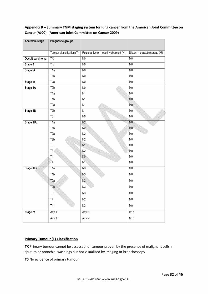
Page 32 of 46 MSAC website: www.msac.gov.au
Appendix B – Summary TNM staging system for lung cancer from the American Joint Committee on
Cancer (AJCC). (American Joint Committee on Cancer 2009)
Anatomic stage Prognostic groups
Tumour classification (T) Regional lymph node involvement (N) Distant metastatic spread (M)
Occult carcinoma TX N0 M0
Stage 0 Tis N0 M0
Stage IA T1a
T1b
N0
N0
M0
M0
Stage IB T2a N0 M0
Stage IIA T2b
T1a
T1b
T2a
N0
N1
N1
N1
M0
M0
M0
M0
Stage IIB T2b
T3
N1
N0
M0
M0
Stage IIIA T1a
T1b
T2a
T2b
T3
T3
T4
T4
N2
N2
N2
N2
N1
N2
N0
N1
M0
M0
M0
M0
M0
M0
M0
M0
Stage IIIB T1a
T1b
T2a
T2b
T3
T4
T4
N3
N3
N3
N3
N3
N2
N3
M0
M0
M0
M0
M0
M0
M0
Stage IV Any T
Any T
Any N
Any N
M1a
M1b
Primary Tumour (T) Classification
TX Primary tumour cannot be assessed, or tumour proven by the presence of malignant cells in
sputum or bronchial washings but not visualized by imaging or bronchoscopy
T0 No evidence of primary tumour
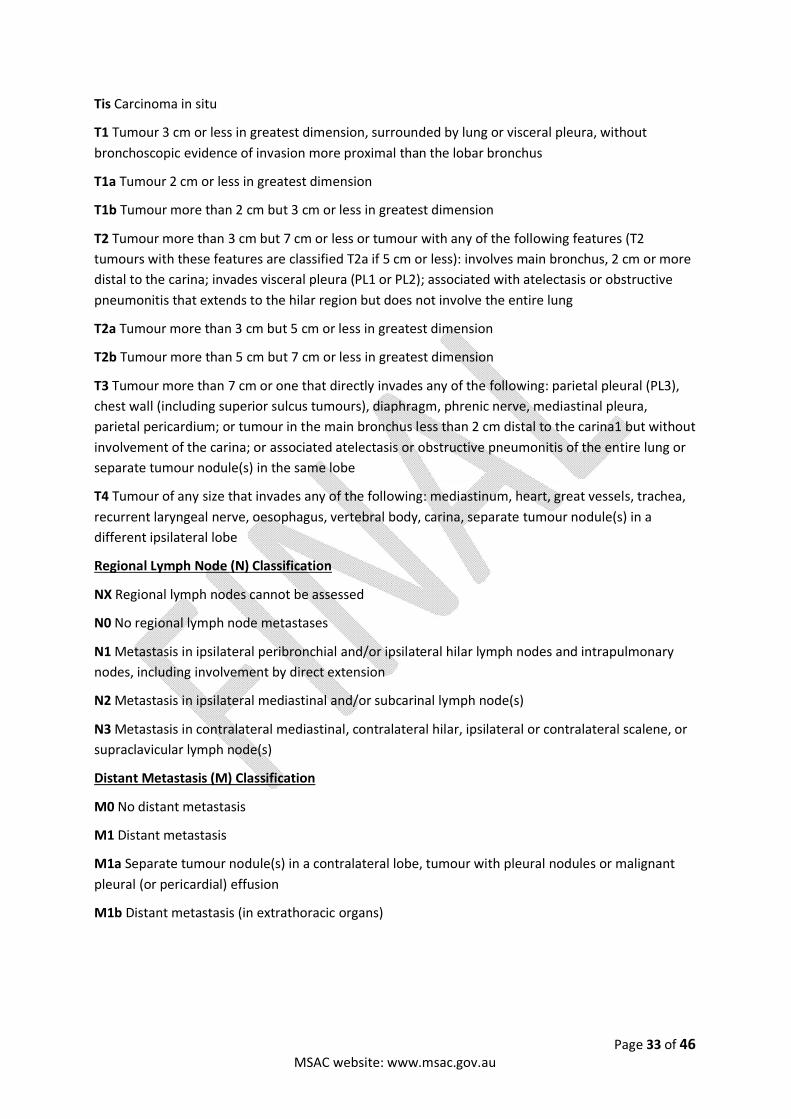
Page 33 of 46 MSAC website: www.msac.gov.au
Tis Carcinoma in situ
T1 Tumour 3 cm or less in greatest dimension, surrounded by lung or visceral pleura, without
bronchoscopic evidence of invasion more proximal than the lobar bronchus
T1a Tumour 2 cm or less in greatest dimension
T1b Tumour more than 2 cm but 3 cm or less in greatest dimension
T2 Tumour more than 3 cm but 7 cm or less or tumour with any of the following features (T2
tumours with these features are classified T2a if 5 cm or less): involves main bronchus, 2 cm or more
distal to the carina; invades visceral pleura (PL1 or PL2); associated with atelectasis or obstructive
pneumonitis that extends to the hilar region but does not involve the entire lung
T2a Tumour more than 3 cm but 5 cm or less in greatest dimension
T2b Tumour more than 5 cm but 7 cm or less in greatest dimension
T3 Tumour more than 7 cm or one that directly invades any of the following: parietal pleural (PL3),
chest wall (including superior sulcus tumours), diaphragm, phrenic nerve, mediastinal pleura,
parietal pericardium; or tumour in the main bronchus less than 2 cm distal to the carina1 but without
involvement of the carina; or associated atelectasis or obstructive pneumonitis of the entire lung or
separate tumour nodule(s) in the same lobe
T4 Tumour of any size that invades any of the following: mediastinum, heart, great vessels, trachea,
recurrent laryngeal nerve, oesophagus, vertebral body, carina, separate tumour nodule(s) in a
different ipsilateral lobe
Regional Lymph Node (N) Classification
NX Regional lymph nodes cannot be assessed
N0 No regional lymph node metastases
N1 Metastasis in ipsilateral peribronchial and/or ipsilateral hilar lymph nodes and intrapulmonary
nodes, including involvement by direct extension
N2 Metastasis in ipsilateral mediastinal and/or subcarinal lymph node(s)
N3 Metastasis in contralateral mediastinal, contralateral hilar, ipsilateral or contralateral scalene, or
supraclavicular lymph node(s)
Distant Metastasis (M) Classification
M0 No distant metastasis
M1 Distant metastasis
M1a Separate tumour nodule(s) in a contralateral lobe, tumour with pleural nodules or malignant
pleural (or pericardial) effusion
M1b Distant metastasis (in extrathoracic organs)