protocol for the examination of specimens from patients ... · hypopharyngeal squamous cell...
TRANSCRIPT

Protocol for the Examination of Specimens From Patients With Cancers of the Larynx Version: Larynx 4.0.0.1 Protocol Posting Date: June 2017 Includes pTNM requirements from the 8th Edition, AJCC Staging Manual For accreditation purposes, this protocol should be used for the following procedures AND tumor types: Procedure Description Resection Includes specimens designated larynx, supraglottis, glottis, and
subglottis. Tumor Type Description Carcinoma Includes squamous cell carcinoma, neuroendocrine carcinoma, and
minor salivary gland carcinoma Mucosal Melanoma
This protocol is NOT required for accreditation purposes for the following: Procedure Biopsy Primary resection specimen with no residual cancer (eg, following neoadjuvant therapy) Cytologic specimens
The following tumor types should NOT be reported using this protocol: Tumor Type Hypopharyngeal squamous cell carcinoma (consider the Pharynx protocol) Sarcoma (consider the Soft Tissue protocol) Lymphoma (consider the Hodgkin or non-Hodgkin Lymphoma protocols)
Authors Raja R. Seethala, MD*; Ilan Weinreb, MD*; Martin J Bullock, MD; Diane L. Carlson, MD; Robert L. Ferris, MD, PhD; Louis B. Harrison, MD; Jonathan B. McHugh, MD; Jason Pettus, MD; Mary S. Richardson, MD; Jatin Shah, MD; Lester D.R. Thompson, MD; Bruce M. Wenig MD With guidance from the CAP Cancer and CAP Pathology Electronic Reporting Committees. * Denotes primary authors. All other contributing authors are listed alphabetically.
© 2017 College of American Pathologists (CAP). All rights reserved. For Terms of Use please visit www.cap.org/cancerprotocols

Head and Neck • Larynx 4.0.0.1
Accreditation Requirements This protocol can be utilized for a variety of procedures and tumor types for clinical care purposes. For accreditation purposes, only the definitive primary cancer resection specimen is required to have the core and conditional data elements reported in a synoptic format. • Core data elements are required in reports to adequately describe appropriate malignancies. For
accreditation purposes, essential data elements must be reported in all instances, even if the response is “not applicable” or “cannot be determined.”
• Conditional data elements are only required to be reported if applicable as delineated in the protocol. For instance, the total number of lymph nodes examined must be reported, but only if nodes are present in the specimen.
• Optional data elements are identified with “+” and although not required for CAP accreditation purposes, may be considered for reporting as determined by local practice standards.
The use of this protocol is not required for recurrent tumors or for metastatic tumors that are resected at a different time than the primary tumor. Use of this protocol is also not required for pathology reviews performed at a second institution (ie, secondary consultation, second opinion, or review of outside case at second institution). Synoptic Reporting All core and conditionally required data elements outlined on the surgical case summary from this cancer protocol must be displayed in synoptic report format. Synoptic format is defined as: • Data element: followed by its answer (response), outline format without the paired "Data element:
Response" format is NOT considered synoptic. • The data element should be represented in the report as it is listed in the case summary. The response for
any data element may be modified from those listed in the case summary, including “Cannot be determined” if appropriate.
• Each diagnostic parameter pair (Data element: Response) is listed on a separate line or in a tabular format to achieve visual separation. The following exceptions are allowed to be listed on one line:
o Anatomic site or specimen, laterality, and procedure o Pathologic Stage Classification (pTNM) elements o Negative margins, as long as all negative margins are specifically enumerated where applicable
• The synoptic portion of the report can appear in the diagnosis section of the pathology report, at the end of the report or in a separate section, but all Data element: Responses must be listed together in one location
Organizations and pathologists may choose to list the required elements in any order, use additional methods in order to enhance or achieve visual separation, or add optional items within the synoptic report. The report may have required elements in a summary format elsewhere in the report IN ADDITION TO but not as replacement for the synoptic report ie, all required elements must be in the synoptic portion of the report in the format defined above. CAP Laboratory Accreditation Program Protocol Required Use Date: March 2018
CAP Larynx Protocol Summary of Changes Version 4.0.0.1 The following data elements were modified: Regional Lymph Nodes pN: Modified pN2b and pN2c for “Metastases” and pN3, pN3b to include “a single contralateral node of any size and ENE(+)”
2

CAP Approved Head and Neck • Larynx 4.0.0.1
Surgical Pathology Cancer Case Summary Protocol posting date: June 2017 LARYNX (SUPRAGLOTTIS, GLOTTIS, SUBGLOTTIS): Select a single response unless otherwise indicated. Procedure (select all that apply) ___ Excision ___ Endolaryngeal excision ___ Transoral laser excision (glottis) ___ Supraglottic laryngectomy ___ Supracricoid laryngectomy ___ Vertical hemilaryngectomy (specify side): ____________________________ ___ Partial laryngectomy (specify type): ____________________________ ___ Total laryngectomy ___ Neck (lymph node) dissection (specify): ____________________________ ___ Other (specify): ____________________________ ___ Not specified Tumor Site (Note A) ___ Larynx, supraglottis + ___ Epiglottis, lingual aspect + ___ Epiglottis, laryngeal aspect + ___ Aryepiglottic folds + ___ Arytenoid(s) + ___ False vocal cord + ___ Ventricle ___ Larynx, glottis + ___ True vocal cord + ___ Anterior commissure + ___ Posterior commissure + ___ With subglottic extension ___ Larynx, subglottis ___ Other (specify): ____________________________ ___ Not specified Transglottic Extension ___ Present ___ Not identified Tumor Laterality (select all that apply) ___ Right ___ Left ___ Midline ___ Not specified Tumor Focality ___ Unifocal ___ Multifocal ___ Cannot be determined Tumor Size Greatest dimension (centimeters): ___ cm + Data elements preceded by this symbol are not required for accreditation purposes. These optional elements may be
clinically important but are not yet validated or regularly used in patient management.
3
CAP Approved Head and Neck • Larynx 4.0.0.1
+ Additional dimensions (centimeters): ___ x ___ cm ___ Cannot be determined (explain): ____________________________ Histologic Type (Note B) Squamous Cell Carcinoma and Variants (select all that apply) ___ Squamous cell carcinoma, conventional (keratinizing) ___ Squamous cell carcinoma, nonkeratinizing ___ Acantholytic squamous cell carcinoma ___ Adenosquamous carcinoma ___ Basaloid squamous cell carcinoma ___ Papillary squamous cell carcinoma ___ Spindle cell squamous cell carcinoma ___ Verrucous squamous cell carcinoma ___ Lymphoepithelial carcinoma Carcinomas of Minor Salivary Glands ___ Mucoepidermoid carcinoma, low grade ___ Mucoepidermoid carcinoma, intermediate grade ___ Mucoepidermoid carcinoma, high grade ___ Adenoid cystic carcinoma, tubular pattern#
+ Specify percentage of solid component: ____% ___ Adenoid cystic carcinoma, cribriform pattern#
+ Specify percentage of solid component: ____% ___ Adenoid cystic carcinoma, solid pattern#
+ Specify percentage of solid component: ____% ___ Adenocarcinoma, not otherwise specified, low grade ___ Adenocarcinoma, not otherwise specified, intermediate grade ___ Adenocarcinoma, not otherwise specified, high grade # Note: If multiple patterns present, select predominant pattern unless solid pattern is greater than 30%, in which case should select solid pattern. Neuroendocrine Carcinoma ___ Well-differentiated neuroendocrine carcinoma (typical carcinoid tumor) ___ Moderately differentiated neuroendocrine carcinoma (atypical carcinoid tumor) ___ Poorly differentiated neuroendocrine carcinoma, small cell type ___ Poorly differentiated neuroendocrine carcinoma, large cell type ___ Combined (or composite) neuroendocrine carcinoma with (specify types): ____________________________ ___ Mucosal melanoma ___ Carcinoma, type cannot be determined ___ Other histologic type not listed (specify): ____________________________ Histologic Grade (Note C) (required for squamous cell carcinoma only) ___ G1: Well differentiated ___ G2: Moderately differentiated ___ G3: Poorly differentiated ___ Other (specify): ____________________________ ___ GX: Cannot be assessed + Tumor Extension (other structures/spaces involved) + Specify: ____________________________ Margins (select all that apply) (Notes D and E) ___ Cannot be assessed + Data elements preceded by this symbol are not required for accreditation purposes. These optional elements may be
clinically important but are not yet validated or regularly used in patient management.
4
CAP Approved Head and Neck • Larynx 4.0.0.1
___ Uninvolved by invasive tumor Distance from closest margin (millimeters): ____ mm
Specify location of closest margin, per orientation, if possible: ____________________________ + Location and distance of other close margins: ____________________________
___ Involved by invasive tumor Specify margin(s), per orientation, if possible: ____________________________ ___ Uninvolved by high-grade dysplasia/in situ disease# Distance from closest margin (millimeters): ____ mm
Specify location of closest margin, per orientation, if possible: ____________________________ ___ Involved by high-grade dysplasia/in situ disease# Specify margin(s), per orientation, if possible: ____________________________ # Note: Applicable only to squamous cell carcinoma and histologic variants, and required only if margins uninvolved by invasive carcinoma. Lymphovascular Invasion ___ Not identified ___ Present ___ Cannot be determined Perineural Invasion (Note F) ___ Not identified ___ Present ___ Cannot be determined Regional Lymph Nodes (Note G) ___ No lymph nodes submitted or found Lymph Node Examination (required only if lymph nodes present in specimen) Number of Lymph Nodes Involved: ________ ___ Number cannot be determined (explain): ____________________________ Number of Lymph Nodes Examined: _______ ___ Number cannot be determined (explain): ____________________________ Lymph Node Metastasis (required for all histologies except mucosal melanoma, and only if lymph nodes involved) Laterality of Lymph Nodes Involved ___ Ipsilateral (including midline) ___ Contralateral ___ Bilateral ___ Cannot be determined Size of Largest Metastatic Deposit (centimeters): ____ cm Extranodal Extension (ENE) ___ Not identified ___ Present + Distance from lymph node capsule (millimeters): _____ mm +___ ENEma (>2 mm) +___ ENEmi (≤2 mm) ___ Cannot be determined Pathologic Stage Classification (pTNM, AJCC 8th Edition) (Note H)
+ Data elements preceded by this symbol are not required for accreditation purposes. These optional elements may be
clinically important but are not yet validated or regularly used in patient management.
5

CAP Approved Head and Neck • Larynx 4.0.0.1
Note: Reporting of pT, pN, and (when applicable) pM categories is based on information available to the pathologist at the time the report is issued. Only the applicable T, N, or M category is required for reporting; their definitions need not be included in the report. The categories (with modifiers when applicable) can be listed on 1 line or more than 1 line. Note: The phrases in italics include clinical findings required for AJCC staging. This clinical information may not be available to the pathologist. However, if known, these findings should be incorporated into the pathologic staging. TNM Descriptors (required only if applicable) (select all that apply) ___ m (multiple primary tumors) ___ r (recurrent) ___ y (posttreatment) For All Carcinomas Primary Tumor (pT) ___ pTX: Primary tumor cannot be assessed ___ pTis: Carcinoma in situ For the Supraglottis ___ pT1: Tumor limited to one subsite of supraglottis with normal vocal cord mobility ___ pT2: Tumor invades mucosa of more than one adjacent subsite of supraglottis or glottis or region outside
the supraglottis (eg, mucosa of base of tongue, vallecula, medial wall of pyriform sinus) without fixation of the larynx
___ pT3: Tumor limited to larynx with vocal cord fixation and/or invades any of the following: postcricoid area, preepiglottic space, paraglottic space, and/or inner cortex of thyroid cartilage
___ pT4 Moderately advanced or very advanced ___ pT4a: Moderately advanced local disease. Tumor invades through the outer cortex thyroid cartilage and/or
invades tissues beyond the larynx (eg, trachea, soft tissues of neck including deep extrinsic muscle of tongue, strap muscles, thyroid, or esophagus)
___ pT4b: Very advanced local disease. Tumor invades prevertebral space, encases carotid artery, or invades mediastinal structures
For the Glottis ___ pT1: Tumor limited to the vocal cord(s) (may involve anterior or posterior commissure) with normal mobility ___ pT1a: Tumor limited to one vocal cord ___ pT1b: Tumor involves both vocal cords ___ pT2: Tumor extends to supraglottis and/or subglottis and/or with impaired vocal cord mobility ___ pT3: Tumor limited to the larynx with vocal cord fixation and/or invasion of paraglottic space and/or inner
cortex of the thyroid cartilage ___ pT4: Moderately advanced or very advanced ___ pT4a: Moderately advanced local disease. Tumor invades through the outer cortex of the thyroid cartilage
and/or invades tissues beyond the larynx (eg, trachea, cricoid cartilage, soft tissues of neck including deep extrinsic muscle of the tongue, strap muscles, thyroid, or esophagus)
___ pT4b: Very advanced local disease. Tumor invades prevertebral space, encases carotid artery, or invades mediastinal structures
For the Subglottis ___ pT1: Tumor limited to subglottis ___ pT2: Tumor extends to vocal cord(s) with normal or impaired mobility ___ pT3: Tumor limited to larynx with vocal cord fixation and/or invasion of paraglottic space and/or inner cortex
of the thyroid cartilage ___ pT4: Moderately advanced or very advanced ___ pT4a: Moderately advanced local disease. Tumor invades cricoid or thyroid cartilage and/or invades tissues
beyond the larynx (eg, trachea, soft tissues of neck including deep extrinsic muscles of the tongue, strap muscles, thyroid, or esophagus)
___pT4b: Very advanced local disease. Tumor invades prevertebral space, encases carotid artery, or invades mediastinal structures
+ Data elements preceded by this symbol are not required for accreditation purposes. These optional elements may be
clinically important but are not yet validated or regularly used in patient management.
6
CAP Approved Head and Neck • Larynx 4.0.0.1
Regional Lymph Nodes (pN)# (Note G) ___ pNX: Regional lymph nodes cannot be assessed ___ pN0: No regional lymph node metastasis ___ pN1: Metastasis in a single ipsilateral lymph node, 3 cm or smaller in greatest dimension and ENE(-) ___ pN2: Metastasis in a single ipsilateral lymph node, 3 cm or smaller in greatest dimension and ENE(+);
or larger than 3 cm but not larger than 6 cm in greatest dimension and ENE(−); or metastases in multiple ipsilateral lymph nodes, none larger than 6 cm in greatest dimension and ENE(−); or in bilateral or contralateral lymph node(s), none larger than 6 cm in greatest dimension and ENE(−)
___ pN2a: Metastasis in a single ipsilateral node, 3 cm or smaller in greatest dimension and ENE(+); or metastasis in a single ipsilateral node, larger than 3 cm but not larger than 6 cm in greatest dimension and ENE(−)
___ pN2b: Metastases in multiple ipsilateral nodes, none larger than 6 cm in greatest dimension and ENE(−) ___ pN2c: Metastases in bilateral or contralateral lymph node(s), none larger than 6 cm in greatest dimension
and ENE(−) ___ pN3: Metastasis in a lymph node, larger than 6 cm in greatest dimension and ENE(−);
or metastasis in a single ipsilateral node, larger than 3 cm in greatest dimension and ENE(+); or multiple ipsilateral, contralateral, or bilateral lymph nodes and any with ENE(+); or a single contralateral node of any size and ENE(+)
___ pN3a: Metastasis in a lymph node larger than 6 cm in greatest dimension and ENE(−) ___ pN3b: Metastasis in a single ipsilateral node, larger than 3 cm in greatest dimension and ENE(+);
or multiple ipsilateral, contralateral, or bilateral lymph nodes and any with ENE(+); or a single contralateral node of any size and ENE(+)
# Midline nodes are considered ipsilateral nodes. Note: Pathological ENE should be recorded as ENE(−) or ENE(+). Note: Measurement of the metastatic focus in the lymph nodes is based on the largest metastatic deposit size, which may include matted or fused lymph nodes. Distant Metastasis (pM) (required only if confirmed pathologically in this case) ___ pM1: Distant metastasis Specify site(s), if known: ____________________________ For Mucosal Melanoma Primary Tumor (pT) ___ pT3: Tumors limited to the mucosa and immediately underlying soft tissue, regardless of thickness or
greatest dimension; for example, polypoid nasal disease, pigmented or nonpigmented lesions of the oral cavity, pharynx, or larynx
___ pT4: Moderately advanced or very advanced disease ___ pT4a: Moderately advanced disease
Tumor involving deep soft tissue, cartilage, bone, or overlying skin ___ pT4b: Very advanced disease
Tumor involving brain, dura, skull base, lower cranial nerves (IX, X, XI, XII), masticator space, carotid artery, prevertebral space, or mediastinal structures
Regional Lymph Nodes (pN) ___ pNX: Regional lymph nodes cannot be assessed ___ pN0: No regional lymph node metastases ___ pN1: Regional lymph node metastases present Distant Metastasis (pM) (required only if confirmed pathologically in this case) ___ pM1: Distant metastasis present
Specify site(s), if known: ____________________________
+ Data elements preceded by this symbol are not required for accreditation purposes. These optional elements may be
clinically important but are not yet validated or regularly used in patient management.
7

CAP Approved Head and Neck • Larynx 4.0.0.1
+ Additional Pathologic Findings (select all that apply) (Note I) + ___ None identified + ___ Dysplasia, low grade (includes hyperplasia, keratosis, and mild dysplasia) + ___ Dysplasia, high grade (includes moderate to severe dysplasia, and carcinoma in situ) + ___ Squamous papilloma, solitary + ___ Squamous papillomatosis + ___ Inflammation (specify type): ____________________________ + ___ Squamous metaplasia + ___ Colonization, fungal + ___ Colonization, bacterial + ___ Other (specify): ____________________________ + Ancillary Studies
Note: For reporting molecular testing and other cancer biomarker testing results, the CAP Head and Neck Biomarker Template should be used. Pending biomarker studies should be listed in the Comments section of this report.
+ Comment(s)
+ Data elements preceded by this symbol are not required for accreditation purposes. These optional elements may be
clinically important but are not yet validated or regularly used in patient management.
8

Background Documentation Head and Neck • Larynx 4.0.0.1
Explanatory Notes Scope of Guidelines The reporting of laryngeal cancer is facilitated by the provision of a case summary illustrating the features required for comprehensive patient care. However, there are many cases in which the individual practicalities of applying such a case summary may not be straightforward. Common examples include finding the prescribed number of lymph nodes, trying to determine the levels of the radical neck dissection, and determining if isolated tumor cells in a lymph node represent metastatic disease. Case summaries have evolved to include clinical, radiographic, morphologic, immunohistochemical, and molecular results in an effort to guide clinical management. Adjuvant and neoadjuvant therapy can significantly alter histologic findings, making accurate classification an increasingly complex and demanding task. This protocol tries to remain simple while still incorporating important pathologic features as proposed by the American Joint Committee on Cancer (AJCC) cancer staging manual, the World Health Organization (WHO) classification of tumors, the TNM classification,1 the American College of Surgeons Commission on Cancer, and the International Union on Cancer (UICC). This protocol is to be used as a guide and resource, an adjunct to diagnosing and managing cancers of the larynx in a standardized manner. It should not be used as a substitute for dissection or grossing techniques and does not give histologic parameters to reach the diagnosis. Subjectivity is always a factor, and elements listed are not meant to be arbitrary but are meant to provide uniformity of reporting across all the disciplines that use the information. It is a foundation of practical information that will help to meet the requirements of daily practice to benefit both clinicians and patients alike. A. Anatomic Sites and Subsites for the Larynx (Figure 1) Supraglottis Epilarynx, including marginal zone
Suprahyoid epiglottis, including tip, lingual (anterior), and laryngeal surfaces Aryepiglottic fold, laryngeal aspect Arytenoid Supraglottis, excluding epilarynx Infrahyoid epiglottis Ventricular bands (false cords) Ventricle Glottis Vocal cords Anterior commissure Posterior commissure Subglottis The protocol applies to all carcinomas arising at these sites.2 The piriform sinus represents part of the hypopharynx which expands bilaterally and forward around the sides of the larynx and lies between the larynx and the thyroid cartilage. Cancers of the piriform sinus are included in the protocol on pharynx cancers. Anatomic Compartments (Figure 1) The anatomic compartments of the larynx include: 1. Supraglottic larynx extending from the tip of the epiglottis to a horizontal line passing through the apex of the ventricle; structures included in this compartment are the epiglottis (lingual and laryngeal aspects), aryepiglottic folds, arytenoids, false vocal cords and the ventricle. 2. Glottic region, which extends from the ventricle to approximately 0.5 cm to 1.0 cm below the free level of the true vocal cord and includes the anterior and posterior commissures and the true vocal cord. 3. Subglottic larynx, which extends approximately 1.0 cm below the level of the true vocal cord to the inferior rim of the cricoid cartilage.
9
Background Documentation Head and Neck • Larynx 4.0.0.1
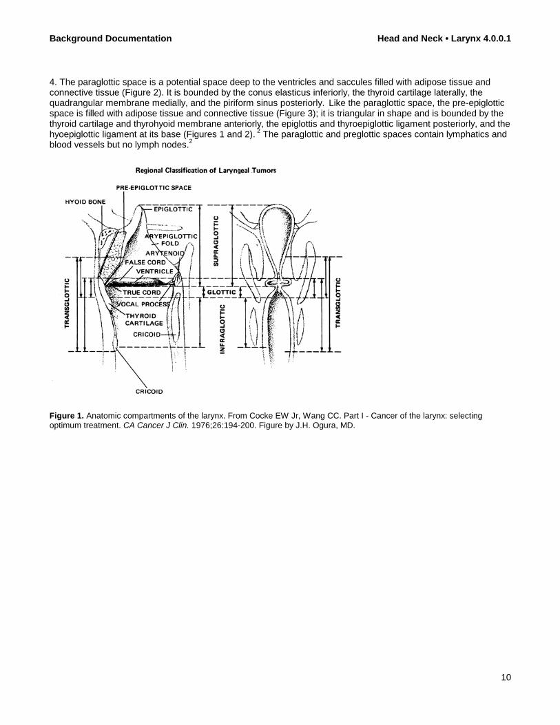
4. The paraglottic space is a potential space deep to the ventricles and saccules filled with adipose tissue and connective tissue (Figure 2). It is bounded by the conus elasticus inferiorly, the thyroid cartilage laterally, the quadrangular membrane medially, and the piriform sinus posteriorly. Like the paraglottic space, the pre-epiglottic space is filled with adipose tissue and connective tissue (Figure 3); it is triangular in shape and is bounded by the thyroid cartilage and thyrohyoid membrane anteriorly, the epiglottis and thyroepiglottic ligament posteriorly, and the hyoepiglottic ligament at its base (Figures 1 and 2). 2 The paraglottic and preglottic spaces contain lymphatics and blood vessels but no lymph nodes.2
Figure 1. Anatomic compartments of the larynx. From Cocke EW Jr, Wang CC. Part I - Cancer of the larynx: selecting optimum treatment. CA Cancer J Clin. 1976;26:194-200. Figure by J.H. Ogura, MD.
10

Background Documentation Head and Neck • Larynx 4.0.0.1
Figure 2. The paraglottic space. From World Health Organization Classification of Tumours: Pathology and Genetics of Head and Neck Tumours. Lyon, France: IARC Press; 2005. Reproduced with permission.
Figure 3. The pre-epiglottic space. From World Health Organization Classification of Tumours: Pathology and Genetics of Head and Neck Tumours. Lyon, France: IARC Press; 2005. Reproduced with permission. Site-Specific Carcinomas 1. Supraglottic squamous cell carcinoma represents a squamous cell carcinoma that involves the structures of the supraglottic larynx, including the epiglottis (laryngeal and lingual surfaces), aryepiglottic folds, arytenoids, false vocal cords, and ventricles.
11

Background Documentation Head and Neck • Larynx 4.0.0.1
2. Glottic squamous cell carcinoma represents a squamous cell carcinoma that involves the structures of the glottis, including the true vocal cords, and the anterior and posterior commissures. 3. Subglottic squamous cell carcinoma represents a squamous cell carcinoma that involves the subglottis, which begins 1 cm below the apex of the ventricle to its inferior border represented by the rim of the cricoid cartilage. 4. Transglottic carcinomas represent a carcinoma that crosses the ventricles in a vertical direction arising in either the glottic or supraglottic larynx B. Histologic Type A modification of the WHO classification of carcinomas of the larynx is shown below.3,4 This list may not be complete. This protocol applies to carcinomas and melanomas and does not apply to lymphomas or sarcomas. Carcinomas of Larynx Squamous cell carcinoma Squamous cell carcinoma, conventional (keratinizing)
Squamous cell carcinoma, nonkeratinizing Adenosquamous carcinoma Basaloid squamous cell carcinoma Papillary squamous cell carcinoma Spindle cell squamous cell carcinoma Verrucous squamous cell carcinoma Lymphoepithelial carcinoma
Neuroendocrine carcinoma Well-differentiated neuroendocrine carcinoma (typical carcinoid tumor)
Moderately differentiated neuroendocrine carcinoma (atypical carcinoid tumor) Poorly differentiated neuroendocrine carcinoma, small cell type Poorly differentiated neuroendocrine carcinoma, large cell type Combined (or composite) neuroendocrine carcinoma#
# Represents a carcinoma showing combined features of small cell neuroendocrine carcinoma associated with a squamous or adenocarcinomatous component.3 Carcinomas of Minor Salivary Glands
Adenoid cystic carcinoma Mucoepidermoid carcinoma Adenocarcinoma, not otherwise specified Other# # Please refer to the CAP protocol for major salivary glands for detailed listing of other tumor types. Mucosal Melanoma C. Histologic Grade For histologic types of carcinomas that are amenable to grading, 3 histologic grades are suggested, as shown below. For conventional squamous cell carcinoma, histologic grading as a whole does not perform well as a prognosticator.5 Nonetheless, it should be recorded when applicable, as it is a basic tumor characteristic. Selecting either the most prevalent grade or the highest grade for this synoptic protocol is acceptable. Variants of squamous cell carcinoma (ie, verrucous, basaloid, etc) have an intrinsic biologic potential and currently do not appear to require grading. Grade 1 Well differentiated Grade 2 Moderately differentiated Grade 3 Poorly differentiated Grade X Cannot be assessed
12

Background Documentation Head and Neck • Larynx 4.0.0.1
The histologic (microscopic) grading of salivary gland carcinomas has been shown to be an independent predictor of behavior and plays a role in optimizing therapy. Further, there is often a positive correlation between histologic grade and clinical stage.6-9 However, most salivary gland carcinoma types have an intrinsic biologic behavior and attempted application of a universal grading scheme is merely a crude surrogate.8 Thus a generic grading scheme is no longer recommended for salivary gland carcinomas.10 Carcinoma types for which grading systems exist and are relevant are incorporated into histologic type. The 3 major categories that are amenable to grading include adenoid cystic carcinoma, mucoepidermoid carcinoma, and adenocarcinoma, not otherwise specified.7,8,11 In some carcinomas, histologic grading may be based on growth pattern, such as in adenoid cystic carcinoma, for which a histologic high-grade variant has been recognized based on the percentage of solid growth.11 Those adenoid cystic carcinomas showing 30% or greater of solid growth pattern are considered to be histologically high-grade carcinomas. The histologic grading of mucoepidermoid carcinoma includes a combination of growth pattern characteristics (eg, cystic, solid, neurotropism) and cytomorphologic findings (eg, anaplasia, mitoses, necrosis).12-
14 Adenocarcinomas, not otherwise specified, do not have a formalized grading scheme and are graded intuitively based on cytomorphologic features.8 D. Surgical Margins The definition of a positive margin is somewhat controversial given the varied results from prior studies.15,16 However, overall, several studies support the definition of a positive margin to be invasive carcinoma or carcinoma in situ/high-grade dysplasia present at margins (microscopic cut-through of tumor).17 Furthermore, reporting of surgical margins should also include information regarding the distance of invasive carcinoma, carcinoma in situ, or high-grade dysplasia (moderate to severe) from the surgical margin. Tumors with “close” margins also carry an increased risk for local recurrence.16,17 The definition of a “close” margin is not standardized as the effective cut-off varies between studies and between anatomic subsites. Commonly used cut points to define close margins are 5 mm in general and 2 mm with respect to glottic larynx.16 However, values ranging from 3 mm to 7 mm have been used with success,16,18 and for glottic tumors as low as 1 mm.19 Thus, distance of tumor from the nearest margin should be recorded. Reporting of surgical margins for carcinomas of the minor salivary glands should follow those used for squamous cell carcinoma of larynx. Dysplasia The types of intraepithelial dysplasia of the upper aerodigestive tract (UADT) include nonkeratinizing (“classic”) dysplasia and keratinizing dysplasia. Of the 2 types of dysplasias, the keratinizing dysplasias are significantly more common than the nonkeratinizing dysplasias. The current WHO advocates a 2-tiered approach with low-grade and high-grade dysplasia categories (see below under Note M).20 High-grade dysplasia at a margin is regarded and reported as a positive margin, while low-grade dysplasia is not. E. Orientation of Specimen Complex specimens should be examined and oriented with the assistance of the operating surgeon(s). Direct communication between the surgeon and pathologist is a critical component in specimen orientation and proper sectioning. Whenever possible, the tissue examination request form should include a drawing or photograph of the resected specimen showing the extent of the tumor and its relation to the anatomic structures of the region. The lines and extent of the resection can be depicted on preprinted adhesive labels and attached to the surgical pathology request forms. F. Perineural Invasion Traditionally, the presence of perineural invasion (neurotropism) is an important predictor of poor prognosis in head and neck cancer of virtually all sites.21 The presence of perineural invasion (neurotropism) in the primary cancer is associated with poor local disease control and regional control, as well as being associated with metastasis to regional lymph nodes.21 Further, perineural invasion is associated with decrease in disease-specific survival and overall survival.21 There is conflicting data relative to an association between the presence of perineural invasion and the development of distant metastasis, with some studies showing an increased association with distant metastasis, while other studies showing no correlation with distant metastasis.21 The relationship between perineural invasion and prognosis is independent of nerve diameter.22 Additionally, emerging evidence suggests that extratumoral perineural invasion may be more prognostically relevant.23 Although
13

Background Documentation Head and Neck • Larynx 4.0.0.1
perineural invasion of small unnamed nerves may not produce clinical symptoms, the reporting of perineural invasion includes nerves of all sizes including small peripheral nerves (ie, less than 1 mm in diameter). Aside from the impact on prognosis, the presence of perineural invasion also guides therapy. Concurrent adjuvant chemoradiation therapy has been shown to improve outcomes in patients with perineural invasion (as well as in patients with extranodal extension and bone invasion).24,25 Given the significance relative to prognosis and treatment, perineural invasion is a required data element in the reporting of head and neck cancers. G. Lymph Nodes Measurement of Tumor Metastasis The cross-sectional diameter of the largest lymph node metastasis (not the lymph node itself) is measured in the gross specimen at the time of macroscopic examination or, if necessary, on the histologic slide at the time of microscopic examination.24,25 Special Procedures for Lymph Nodes The risk of regional (cervical neck) nodal spread from cancers varies based on anatomic subsite. At the current time, no additional special techniques are required other than routine histology for the assessment of nodal metastases. Immunohistochemistry and polymerase chain reaction (PCR) to detect isolated tumor cells are considered investigational techniques at this time. Regional Lymph Nodes (pN0): Isolated Tumor Cells Isolated tumor cells (ITCs) are single cells or small clusters of cells not more than 0.2 mm in greatest dimension. While the generic recommendation is that for lymph nodes with ITCs found by either histologic examination, immunohistochemistry, or nonmorphologic techniques (eg, flow cytometry, DNA analysis, PCR amplification of a specific tumor marker), they should be classified as N0 or M0, respectively.26,27 Evidence for the validity of this practice in head and neck squamous cell carcinoma and other histologic subtypes is lacking. In fact, rare studies relevant to head and neck sites indicate that isolated tumor cells may actually be a poor prognosticator in terms of local control.28 Lymph Node Number Histologic examination of a selective neck dissection specimen will ordinarily include 6 or more lymph nodes. Histologic examination of a radical or modified radical neck dissection specimen will ordinarily include 10 or more lymph nodes in the untreated neck. Classification of Neck Dissection 1. Radical neck dissection 2. Modified radical neck dissection, internal jugular vein and/or sternocleidomastoid muscle spared 3. Selective neck dissection (SND), as specified by the surgeon (Figure 4), defined by dissection of less than the
5 traditional levels of a radical and modified radical neck dissection. The following dissections are now under this category29-31:
a. Supraomohyoid neck dissection b. Posterolateral neck dissection c. Lateral neck dissection d. Central compartment neck dissection 4. Superselective neck dissection (SSND), a relatively new term defined by dissection of the fibrofatty elements
of 2 or less levels32 5. Extended radical neck dissection, as specified by the surgeon For purposes of pathologic evaluation, lymph nodes are organized by levels as shown in Figure 4.
14

Background Documentation Head and Neck • Larynx 4.0.0.1
Figure 4. The six sublevels of the neck for describing the location of lymph nodes within levels I, II, and V. Level IA, submental group; level IB, submandibular group; level IIA, upper jugular nodes along the carotid sheath, including the subdigastric group; level IIB, upper jugular nodes in the submuscular recess; level VA, spinal accessory nodes; and level VB, the supraclavicular and transverse cervical nodes. From: Flint PW, et al, eds. Cummings Otolaryngology: Head and Neck Surgery. 5th ed. Philadelphia, PA; Saunders: 2010. Reproduced with permission © Elsevier. In order for pathologists to properly identify these nodes, they must be familiar with the terminology of the regional lymph node groups and with the relationships of those groups to the regional anatomy. Which lymph node groups surgeons submit for histopathologic evaluation depends on the type of neck dissection they perform. Therefore, surgeons must supply information on the types of neck dissections that they perform and the details of the local anatomy in the specimens they submit for examination or, in other manners, orient those specimens for pathologists. If it is not possible to assess the levels of lymph nodes (for instance, when the anatomic landmarks in the excised specimens are not specified), then the lymph node levels may be estimated as follows: level II, upper third of internal jugular (IJ) vein or neck specimen; level III, middle third of IJ vein or neck specimen; level IV, lower third of IJ vein or neck specimen, all anterior to the sternocleidomastoid muscle. Level I. Submental Group (Sublevel IA) Lymph nodes within the triangular boundary of the anterior belly of the digastric muscles and the hyoid bone. Level I. Submandibular Group (Sublevel IB) Lymph nodes within the boundaries of the anterior and posterior bellies of the digastric muscle and the body of the mandible. The submandibular gland is included in the specimen when the lymph nodes within this triangle are removed. Level II. Upper Jugular Group (Sublevels IIA and IIB) Lymph nodes located around the upper third of the internal jugular vein and adjacent spinal accessory nerve extending from the level of the carotid bifurcation (surgical landmark) or hyoid bone (clinical landmark) to the skull base. The posterior boundary is the posterior border of the sternocleidomastoid muscle, and the anterior boundary is the lateral border of the stylohyoid muscle. Level III. Middle Jugular Group Lymph nodes located around the middle third of the internal jugular vein extending from the carotid bifurcation superiorly to the omohyoid muscle (surgical landmark), or cricothyroid notch (clinical landmark) inferiorly. The posterior boundary is the posterior border of the sternocleidomastoid muscle, and the anterior boundary is the lateral border of the sternohyoid muscle.
15
Background Documentation Head and Neck • Larynx 4.0.0.1
Level IV. Lower Jugular Group Lymph nodes located around the lower third of the internal jugular vein extending from the omohyoid muscle superiorly to the clavicle inferiorly. The posterior boundary is the posterior border of the sternocleidomastoid muscle, and the anterior boundary is the lateral border of the sternohyoid muscle. Level V. Posterior Triangle Group (Sublevels VA and VB) This group comprises predominantly the lymph nodes located along the lower half of the spinal accessory nerve and the transverse cervical artery. The supraclavicular nodes are also included in this group. The posterior boundary of the posterior triangle is the anterior border of the trapezius muscle, the anterior boundary of the posterior triangle is the posterior border of the sternocleidomastoid muscle, and the inferior boundary of the posterior triangle is the clavicle. Level VI. Anterior (Central) Compartment Lymph nodes in this compartment include the pre- and paratracheal nodes, precricoid (Delphian) node, and the perithyroidal nodes, including the lymph nodes along the recurrent laryngeal nerve. The superior boundary is the hyoid bone, the inferior boundary is the suprasternal notch, the lateral boundaries are the common carotid arteries, and the posterior boundary by the prevertebral fascia. Level VII. Superior Mediastinal Lymph Nodes Metastases at level VII are considered regional lymph node metastases; all other mediastinal lymph node metastases are considered distant metastases. Lymph node groups removed from areas not included in the above levels, eg, scalene, suboccipital, and retropharyngeal, should be identified and reported from all levels separately. Midline nodes are considered ipsilateral nodes. Extranodal Extension The status of cervical lymph nodes is the single most important prognostic factor in aerodigestive cancer. All macroscopically negative or equivocal lymph nodes should be submitted in toto. Grossly positive nodes may be partially submitted for microscopic documentation of metastasis. Reporting of lymph nodes containing metastasis should include whether there is presence or absence of extranodal extension (ENE),33 which is now part of N staging. This finding consists of extension of metastatic tumor, present within the confines of the lymph node, through the lymph node capsule into the surrounding connective tissue, with or without associated stromal reaction. A distance of extension from the native lymph node capsule is now suggested (but not yet required) with the proposed stratification of ENE into ENEma (>2 mm) and ENEmi (≤2 mm).34-37 However, pitfalls in the measurement (ie, in larger, matted lymph nodes, in nodes post fine-needle aspiration, and in nodes with near total replacement of lymph node architecture), and the disposition of soft tissue deposits is still not resolved. In general, absence of ENE in a large (>3 cm) lymph node, especially with traversing fibrous bands, should be viewed with skepticism. Soft tissue deposits for lymph node metastases based on limited studies appear to be the equivalent of a positive lymph node with ENE and should be recorded as such.38 H. TNM and Stage Groupings The protocol recommends the TNM staging system of the American Joint Committee on Cancer.2 There are no significant alterations in the 8th edition to T stage of larynx. However, extranodal extension (ENE) is included in N stage. In essence, pathologic ENE(+) will increase the nodal category by 1. The 8th edition of the AJCC staging of head and neck cancers includes mucosal melanomas; this does not show significant changes from the 7th edition. Approximately two-thirds of mucosal melanomas arise in the sinonasal tract, one-quarter are found in the oral cavity, and the remainder occur only sporadically in other mucosal sites of the head and neck. Even small cancers behave aggressively with high rates of recurrence and death. To reflect this aggressive behavior, primary cancers limited to the mucosa are considered T3 lesions. Advanced mucosal melanomas are classified as T4a and T4b. The anatomic extent criteria to define moderately advanced (T4a) and very advanced (T4b) disease are given below. The AJCC staging for mucosal melanomas does not provide for the histologic definition of a T3 lesion; as the majority of mucosal melanomas are invasive at presentation, mucosal-based melanomas (T3 lesions) include those lesions that involve either the epithelium and/or lamina propria of the
16

Background Documentation Head and Neck • Larynx 4.0.0.1
involved site. Rare examples of in situ mucosal melanomas occur, but In situ mucosal melanomas are excluded from staging, as they are extremely rare.39 By AJCC/UICC convention, the designation “T” refers to a primary tumor that has not been previously treated. The symbol “p” refers to the pathologic classification of the TNM, as opposed to the clinical classification, and is based on gross and microscopic examination. pT entails a resection of the primary tumor or biopsy adequate to evaluate the highest pT category, pN entails removal of nodes adequate to validate lymph node metastasis, and pM implies microscopic examination of distant lesions. Clinical classification (cTNM) is usually carried out by the referring physician before treatment during initial evaluation of the patient or when pathologic classification is not possible. Pathologic staging is usually performed after surgical resection of the primary tumor. Pathologic staging depends on pathologic documentation of the anatomic extent of disease, whether or not the primary tumor has been completely removed. If a biopsied tumor is not resected for any reason (eg, when technically unfeasible) and if the highest T and N categories or the M1 category of the tumor can be confirmed microscopically, the criteria for pathologic classification and staging have been satisfied without total removal of the primary cancer. TNM Descriptors For identification of special cases of TNM or pTNM classifications, the “m” suffix and “y” and “r” prefixes are used. Although they do not affect the stage grouping, they indicate cases needing separate analysis. The “m” suffix indicates the presence of multiple primary tumors in a single site and is recorded in parentheses: pT(m)NM. The “y” prefix indicates those cases in which classification is performed during or following initial multimodality therapy (ie, neoadjuvant chemotherapy, radiation therapy, or both chemotherapy and radiation therapy). The cTNM or pTNM category is identified by a “y” prefix. The ycTNM or ypTNM categorizes the extent of tumor actually present at the time of that examination. The “y” categorization is not an estimate of tumor prior to multimodality therapy (ie, before initiation of neoadjuvant therapy). The “r” prefix indicates a recurrent tumor when staged after a documented disease-free interval, and is identified by the “r” prefix: rTNM. Additional Descriptors Residual Tumor (R) Tumor remaining in a patient after therapy with curative intent (eg, surgical resection for cure) is categorized by a system known as R classification, shown below. RX Presence of residual tumor cannot be assessed R0 No residual tumor R1 Microscopic residual tumor R2 Macroscopic residual tumor For the surgeon, the R classification may be useful to indicate the known or assumed status of the completeness of a surgical excision. For the pathologist, the R classification is relevant to the status of the margins of a surgical resection specimen. That is, tumor involving the resection margin on pathologic examination may be assumed to correspond to residual tumor in the patient and may be classified as macroscopic or microscopic according to the findings at the specimen margin(s). T Category Considerations Supraglottis. Normal vocal cord mobility (T1), fixation of the larynx (T2), and vocal cord fixation (T3) may only be determined clinically. Glottis. Normal vocal cord mobility (T1), impaired vocal cord mobility (T2), and vocal cord fixation (T3) may only be determined clinically.
17

Background Documentation Head and Neck • Larynx 4.0.0.1
Subglottis. Normal or impaired vocal cord mobility (T2) and vocal cord fixation (T3) may only be determined clinically. I. Dysplasia of the Upper Aerodigestive Tract (UADT) In contrast to the uterine cervix in which the nonkeratinizing (“classic”) form of epithelial dysplasia is most common resulting in a reproducible and clinically useful grading scheme of mild, moderate, and severe dysplasia (ie, carcinoma in situ), the majority of the UADT mucosal lesions fall under the designation of keratinizing dysplasias, for which the aforementioned criteria are not as easily applied. The current approach indicated by the WHO for laryngeal precursor lesions is to use a 2-tiered classification of low-grade and high-grade dysplasia. Hyperplasias and keratoses without atypia as well as those with mild dysplasia are categorized as “low grade,” while lesions with moderate and severe dysplasia as well as carcinoma in situ are considered “high grade.”20 Separation of carcinoma in situ as a distinct category (ie, 3 tiers) is an acceptable option, but data are limited to support this. 40 Morphologic criteria for the classification of laryngeal precursor lesions are summarized below.20 Low-grade dysplasia (including previous category of mild dysplasia)
Architectural criteria
Stratification is preserved: transition of basal cells or augmented basal/parabasal cell layer with perpendicular orientation to the basement membrane to prickle cells horizontally oriented in the upper part Spinous layer: spectrum of changes ranging from increased spinous layer in the whole thickness up to changes in which prickle cells are seen only in the upper epithelial half Basal/parabasal layer: spectrum of changes, from 2-3 unchanged layers to augmentation of basal and parabasal cells in the lower half of the epithelium
Cytologic criteria
At most minimal cellular atypia Parabasal cells; slightly increased cytoplasm compared to basal cells, enlarged nuclei, uniformly distributed chromatin, no intercellular bridges Rare regular mitoses in or near basal layer Few dyskeratotic cells present
High-grade dysplasia (including previous category of moderate and severe dysplasia, and carcinoma in situ)
Architectural criteria*
Abnormal maturation Variable degrees of disordered stratification and polarity in as much as the whole epithelium Altered epithelial cells usually occupying from half to the entire epithelial thickness Two subtypes: keratinizing (spinous-cell type) and nonkeratinizing (basal-cell type) Variable degree of irregularly shaped rete (bulbous, downwardly extending), with an intact basement membrane No stromal alterations
Cytologic criteria* Easily identified conspicuous cellular and nuclear atypia, including marked variation in size and shape, marked variation in stain intensity with frequent hyperchromasia, nucleoli increased in number and size
18

Background Documentation Head and Neck • Larynx 4.0.0.1
Increased N:C ratio Increased mitoses at or above the suprabasal level, with or without atypical forms Dyskeratotic and apoptotic cells are frequent throughout the entire epithelium
* Complete loss of stratification and polarity and/or severe cytological atypia and atypical mitoses qualifies as carcinoma in situ if a 3-tiered system is used. The risk off malignant progression in low-grade dysplasia is ~1.6%, while that of high-grade dysplasia is ~12.5%. Limited data suggest that the specific designation of carcinoma in situ carries a risk of 40%.20 References 1. Gress DM, Edge SB, Greene FL, et al. Principles of cancer staging. In: Amin MB, ed. AJCC Cancer
Staging Manual. 8th ed. New York, NY: Springer; 2017. 2. Patel SG, Lydiatt WM, Glastonbury CM, et al. Larynx. In: Amin MB, ed. AJCC Cancer Staging Manual. 8th
ed. New York, NY: Springer; 2017. 3. El-Naggar AK. Introduction. In: El-Naggar AK, Chan JKC, Grandis JR, Takata T, Slootweg PJ, eds. WHO
Classification of Head and Neck Tumours. 4th ed. Geneva, Switzerland: WHO Press; 2017:160-162. 4. Slootweg PJ, Grandis JR. Tumours of the hypopharynx, larynx and trachea and parapharyngeal space. In:
El-Naggar AK, Chan JKC, Grandis JR, Takata T, Slootweg PJ, eds. WHO Classification of Head and Neck Tumours. 4th ed. Geneva, Switzerland: WHO Press; 2017:78-81.
5. Barnes L, Eveson JW, Reichart P, Sidransky D. WHO histological classification of tumours of the oral cavity and oropharynx. In: Barnes L, Eveson JW, Reichart P, Sidransky D, eds. World Health Organization Classification of Tumours: Pathology and Genetics of Head and Neck Tumours. Lyon: IARC; 2005:164.
6. Spiro RH, Thaler HT, Hicks WF, Kher UA, Huvos AH, Strong EW. The importance of clinical staging of minor salivary gland carcinoma. Am J Surg. 1991;162(4):330-336.
7. Spiro RH, Huvos AG, Strong EW. Adenocarcinoma of salivary origin. Clinicopathologic study of 204 patients. Am J Surg. 1982;144(4):423-431.
8. Seethala RR. Histologic grading and prognostic biomarkers in salivary gland carcinomas. Adv Anat Pathol. 2011;18(1):29-45.
9. Kane WJ, McCaffrey TV, Olsen KD, Lewis JE. Primary parotid malignancies: a clinical and pathologic review. Arch Otolaryngol Head Neck Surg. 1991;117(3):307-315.
10. Lydiatt WM, Mukherji SK, O'Sullivan B, Patel SG, Shah JP. Major salivary glands. In: Amin MB, ed. AJCC Cancer Staging Manual. 8th ed. New York, NY: Springer; 2017.
11. Szanto PA, Luna MA, Tortoledo ME, White RA. Histologic grading of adenoid cystic carcinoma of the salivary glands. Cancer. 1984;54(6):1062-1069.
12. Seethala RR, Dacic S, Cieply K, Kelly LM, Nikiforova MN. A reappraisal of the MECT1/MAML2 translocation in salivary mucoepidermoid carcinomas. Am J Surg Pathol. 2010;34(8):1106-1121.
13. Brandwein MS, Ivanov K, Wallace DI, et al. Mucoepidermoid carcinoma: a clinicopathologic study of 80 patients with special reference to histological grading. Am J Surg Pathol. 2001;25(7):835-845.
14. Auclair PL, Goode RK, Ellis GL. Mucoepidermoid carcinoma of intraoral salivary glands. Evaluation and application of grading criteria in 143 cases. Cancer. 1992;69(8):2021-2030.
15. Brandwein-Gensler M, Teixeira MS, Lewis CM, et al. Oral squamous cell carcinoma: histologic risk assessment, but not margin status, is strongly predictive of local disease-free and overall survival. Am J Surg Pathol. 2005;29(2):167-178.
16. Hinni ML, Ferlito A, Brandwein-Gensler MS, et al. Surgical margins in head and neck cancer: a contemporary review. Head Neck. 2012.
17. Alicandri-Ciufelli M, Bonali M, Piccinini A, et al. Surgical margins in head and neck squamous cell carcinoma: what is 'close'? Eur Arch Otorhinolaryngol. 2012;270(10):2603-2609
18. Liao CT, Chang JT, Wang HM, et al. Analysis of risk factors of predictive local tumor control in oral cavity cancer. Ann Surg Oncol. 2008;15(3):915-922.
19. Ansarin M, Santoro L, Cattaneo A, et al. Laser surgery for early glottic cancer: impact of margin status on local control and organ preservation. Arch Otolaryngol Head Neck Surg. 2009;135(4):385-390.
19
Background Documentation Head and Neck • Larynx 4.0.0.1
20. Gale N, Hille JJ, Jordan RC, Nadal A, Williams MD. Dysplasia. In: El-Naggar AK, Chan JKC, Grandis JR, Takata T, Slootweg PJ, eds. WHO Classification of Head and Neck Tumours. 4th ed. Geneva, Switzerland: WHO Press; 2017:91-93.
21. Smith BD, Haffty BG. Prognostic factoris in patients with head and neck cancer. In: Harrison LB, Sessions RB, Hong WK, eds. Head and Neck Cancer: A Multidisciplinary Approach. Philadelphia, PA: Lippincott Williams and Wilkins; 2009:51-75.
22. Fagan JJ, Collins B, Barnes L, D'Amico F, Myers EN, Johnson JT. Perineural invasion in squamous cell carcinoma of the head and neck. Arch Otolaryngol Head Neck Surg. 1998;124(6):637-640.
23. Miller ME, Palla B, Chen Q, et al. A novel classification system for perineural invasion in noncutaneous head and neck squamous cell carcinoma: histologic subcategories and patient outcomes. Am J Otolaryngol. 2012;33(2):212-215.
24. Cooper JS, Pajak TF, Forastiere AA, et al. Postoperative concurrent radiotherapy and chemotherapy for high-risk squamous-cell carcinoma of the head and neck. N Engl J Med. 2004;350(19):1937-1944.
25. Bernier J, Domenge C, Ozsahin M, et al. Postoperative irradiation with or without concomitant chemotherapy for locally advanced head and neck cancer. N Engl J Med. 2004;350(19):1945-1952.
26 Sobin LH, Gospodarowicz MK, Wittekind CH, eds. TNM Classification of Malignant Tumours. New York, NY: Wiley-Liss; 2009.
27. Singletary SE, Greene FL, Sobin LH. Classification of isolated tumor cells: clarification of the 6th edition of the American Joint Committee on Cancer Staging Manual. Cancer. 2003;98(12):2740-2741.
28. Broglie MA, Haerle SK, Huber GF, Haile SR, Stoeckli SJ. Occult metastases detected by sentinel node biopsy in patients with early oral and oropharyngeal squamous cell carcinomas: Impact on survival. Head Neck. 2013;35(5):660-666.
29. Ferlito A, Robbins KT, Shah JP, et al. Proposal for a rational classification of neck dissections. Head Neck. 2011;33(3):445-450.
30. Robbins KT, Shaha AR, Medina JE, et al. Consensus statement on the classification and terminology of neck dissection. Arch Otolaryngol Head Neck Surg. 2008;134(5):536-538.
31. Seethala RR. Current state of neck dissection in the United States. Head Neck Pathol. 2009;3(3):238-245. 32. Suarez C, Rodrigo JP, Robbins KT, et al. Superselective neck dissection: rationale, indications, and
results. Eur Arch Otorhinolaryngol. 2013. 33. Ebrahimi A, Gil Z, Amit M. International Consortium for Outcome Research (ICOR) in Head and Neck
Cancer. Primary tumor staging for oral cancer and a proposed modification incorporating depth of invasion: an international multicenter retrospective study. JAMA Otolaryngol Head Neck Surg. 2014;140(12):1138-1148.
34. Ridge JA, Lydiatt WM, Patel SG, et al. Lip and oral cavity. In: Amin MB, ed. AJCC Cancer Staging Manual. 8th ed. New York, NY: Springer; 2017.
35. Ebrahimi A, Clark JR, Amit M, et al. Minimum nodal yield in oral squamous cell carcinoma: defining the standard of care in a multicenter international pooled validation study. Ann Surg Oncol. 2014;21(9):3049-3055.
36. Prabhu RS, Hanasoge S, Magliocca KR, et al. Extent of pathologic extracapsular extension and outcomes in patients with nonoropharyngeal head and neck cancer treated with initial surgical resection. Cancer. 2014;120(10):1499-1506.
37. Dunne AA, Muller HH, Eisele DW, Kessel K, Moll R, Werner JA. Meta-analysis of the prognostic significance of perinodal spread in head and neck squamous cell carcinomas (HNSCC) patients. Eur J Cancer. 2006;42(12):1863-1868.
38. Jose J, Moor JW, Coatesworth AP, Johnston C, MacLennan K. Soft tissue deposits in neck dissections of patients with head and neck squamous cell carcinoma: prospective analysis of prevalence, survival, and its implications. Arch Otolaryngol Head Neck Surg. 2004;130(2):157-160.
39. Lydiatt WM, Brandwein-Gensler MS, Kraus DH, Mukherji SK, Ridge JA, Shah JP. Mucosal melanoma of the head and neck. In: Amin MB, ed. AJCC Cancer Staging Manual. 8th ed. New York, NY: Springer; 2017.
40. Gale N, Blagus R, El-Mofty SK, et al. Evaluation of a new grading system for laryngeal squamous intraepithelial lesions--a proposed unified classification. Histopathology. 2014;65(4):456-464.
20