protocol for breeding drosophila to teach homeobox genetics and the history and importance of model...
TRANSCRIPT

A Drosophila Protocol for Teaching Homeobox Genetics and Homeotic Mutations
by Robin Dirksen
1

Lesson Overview
This protocol is designed to introduce homeotic mutations and homeoboxes genetics in addition to traditional Mendelian dominant/recessive inheritance. Students will apply principles of population genetics and statistical analysis to the data they collect on their own crosses. The lab will hopefully provide them with concepts that will push them to ask deeper questions about how model organisms, and Drosophila in particular, are used to study questions about human diseases. The discussion portion will hopefully give students an understanding of the relevance of model organisms to their own lives.
Fly life cycle and reproduction p. 3
Homeobox and homeotic genetics p. 4
History of homeobox research p. 5
Teacher Preparation p. 11
Student protocol p. 13
Extensions and Glossary p. 24
References p. 26
TimelineThe student portion of the lab takes approximately 30 days and the instructor portion takes approximately 50 days. The actual classroom time required performing the experiments, data collection, and analysis can be done in four 90 minute blocks, and additional time independent of class for students to come in according to their schedules to attend to the flies. The increased time required by the instructor is to establish a stock population large enough for the students to have adequate flies for their crosses.
ObjectivesStudents will--apply knowledge to form hypotheses, correctly use equipement and apply lab techniques.-evaluate findings based on experimentation and communicate those findings in writing, both qualitatively and quantitatively.-discuss the value and motivation of using model organisms in research-develop a basic understanding of homeobox genetics and developmental regulation in organisms, and an appreciation for the universality of many genes across organisms
2

Background for the Instructor
Classification of the virtuous and splendid Fruit FlyDomain: EukaryaKingdom: AnimaliaPhylum: ArthropodaClass: Insecta Order: DipteraFamily: DrosophilidaeGenus: Drosophila (dew lover)species: melanogaster (dark gut)
Drosophila Life Cycle and Reproduction
The average life span of a Lab fly is 26 days for a female, and 33 days for a male. (Under crowded conditions this may be reduced to 12 days. Also mutant flies generally have a shorter life span.) There are four phases to the life cycle: egg, 3 larval (instar) stages, pupa, and adult. The timing of development is highly influenced by temperature: 10 days from egg to adult at room temperature (25°C); 13 days at 20°C; 90 days at 15°C.
3

Flies may mate more than one once, and fertilized eggs are laid generally after the third day of the female’s adult life, the eggs are .5 mm long. Larvae hatch out in 22 hours, and grow and feed for four days, (longer at lower temperatures). The larvae are transparent, and during the third instar stage of larval development, larvae can be seen crawling up the sides of the culture container in preparation for pupation. At 25°C the pupa stage lasts about 4 days. At this time one can see dark projections called pupal horns, off the anterior end. These are the spiracles (outside opening of the respiratory tubes) turned inside out. (It is important to notice this when doing salivary gland extraction activities, because this procedure is best done during the third instar, but before the appearance of pupal horns.)
The pupal case forms, darkens, and hardens and lasts for 4-6 days, during which time metamorphosis occurs. Larval tissues are broken down (except for the brain and a few other tissues), and imaginal discs (pockets of cells stored in the larvae) develop into adult organs. There is a disc for each leg, wing, eye, antennae etc. The understanding of how genes act to control the development of imaginal discs, has gotten clearer with continuing research and has illuminated much about human embryology.
Finally the pupa is ready to eclose (emerge) into the adult stage. At first, before the cuticle has darkened and hardened, the newly emerged female adult looks pale and puffy. (When crossing flies of different genotypes, it is important to use virgin females, and this is one way to visually separate out virgins in a lab population.) Adult Drosophila males and females can be easily distinguished. Males are smaller, with a rounded, blackened tip to their abdomen. Females have a pointed abdomen, with a pattern of even dark bands, (see images).
Homeoboxes and Homeotic Genes
Breeding flies is a great way to teach homeotic and homeobox genetics, the topic may be better saved as a conclusion to their breeding activities. Let’s start with the definitions-
Homeobox: genes that encode proteins that bind and regulate the expression of DNA in multicellular organisms. Genes containing homeoboxes are present in the genomes of many organisms from fruit flies to humans i.e., all eukaryotic genomes, and are associated with cell differentiation and bodily segmentation. Homeoboxes are DNA sequences containing about 180 nucleotides that encode for corresponding sequences of usually 60 amino acids, called homeodomains, found in proteins that bind DNA and regulate gene transcription, determining when those particular groups of genes are expressed.
Homeotic: any of a family of genes that results in a significant change in the embryonic development of a body part that is homologous to one usually found elsewhere. Mutations (defining genes) with a phenotype in which a given cell develops along a
4

pathway normally followed by a different cell type that can change the fate of an imaginal disk in insect development.
Homeoboxes are about regulation, or timing of patterns of development in animals, fungi and plants (genes that are mostly transcription regulators). Homeotic means that something has been changed into the likeness of something else and homeotic genes can be thought of as genetic switches that turn different programs of cellular differentiation on or off.
In summary, homeotic genes encode transcription factors that control the expression of genes responsible for particular anatomical structures, such as wings, legs, and antennae. The homeotic genes include a 180 nucleotide sequence called the homeobox, which is translated into a 60 amino acid domain, called the homeodomain. The homeodomain is involved in DNA binding, as shown in the images below.
History
A group of mutations called homeotic genes were first discovered In the 1920’s. However, it was not until 10-20 years later in the 1940’s at Cold Spring Harbor with the beginnings of molecular biology, that scientists began to understand the structure of homeotic genes. In the late 40’s and 50’ Edward B. Lewis revealed that regulatory genes control the body plan of the fly, segment by segment. Lewis found colinearity in time and space between the order of the genes in the bithorax complex and their affected regions in the segments.
Christiane Nüsslein-Volhard and Eric F. Wieschaus identified and classified 15 genes of key importance that controlled the development of the embryo. Their approach was to create mutations at random then screen large numbers of flies for recessive lethals affecting
5

various stages of early embryogenesis. They established 27,000 lines containing mutated chromosomes and characterized 139 mutations affecting embryogenesis. The original 15 genes were: cubitus interruptus, wingless, gooseberry, hedgehog, fused, patch, paired, even-skipped, odd-skipped, barrel, runt, engrailed, Kruppel, knirps, and hunchback.
Christiane Nüsslein-Volhard, Eric F. Wieschaus and Edward B. Lewis were awarded the Nobel Prize in Physiology or Medicine in 1995. This earlier work led to the eventual sequencing and cloning of these genes in the 1970’s.
Edward B. Lewis at Caltech
Through the years it has been found that homeotic mutations were not caused by one gene, but by a complex of several genes that are very close to each other on the chromosome (so much so that crossing over doesn’t happen). They encode transcription factors which switch on cascades of other genes. Humans have many Hox genes located on chromosomes 7, 17, 12 and 2.
For example, HoxA and HoxD genes specify segment identitiy along the limb axis. They are typically found in an organized cluster and the order of the genes in the cluster correlates to the order of the regions they affect and the timing in which they are affected. As a consequence, mutations in the cluster result in changes in the affected regions. When one gene is lost, the segment becomes more anterior, and a gain becomes more posterior. Mutations to homeobox genes can produce easily visible phenotypes. The apterous mutation is controlled by another family of homeoboxes called LIM.
6

The homeodomain protein motif is highly conserved across vast evolutionary distances. The functional equivalence of homeotic proteins can be demonstrated by the fact that a fly can function perfectly well with a chicken homeotic protein in place of its own. This means that, despite having a last common ancestor that lived over 670 million years ago, a given homeotic protein in chickens and that in flies are so similar, that they can actually take each other's place.
Drosophila’s Contribution in Researching Human Disease is Undeniable
The sequencing of the Drosophila genome provides an unparalleled opportunity to compare human disease gene counterparts in the fly genome. Approximately, 178 out of 287 human disease genes (62%) appear to be conserved in the fly. Drosophila maintains human paralogs for neurological, hematological, endocrine, renal and immune diseases, as well as genes for cancer and malformation and metabolic syndromes.
7

This next image is a ClustalW alignment of the human and Drosophila Menin proteins. The fly protein is 34% identical and 47% similar to the human protein over its entire length. Mutations in menin are found in a familial endocrine cancer characterized by varying combinations of tumors in the parathyroid glands, the pancreatic islets, the anterior pituitary, as well as a variety of other tissues. This is just one example of the orthologs shared between humans and Drosophila.
8

9
Antennepedia is a famous example of homeotic genetics and is one of the mutations the students will be working with. Antennapedia activates Ubx (Ultrabithorax), Hox protein genes that specify the structures of the 2nd thoracic segment, which contains a leg and a wing, and represses genes involved in eye and antenna formation. Thus, legs and wings, but not eyes and antennae, will form wherever the Antennapedia protein is located. This mutation is located on chromosome 3, position 47.5. Ubx influences midgut, central nervous system, peripheral nervous system, leg, and haltere development.
The other mutation the students will work with is apterous, a homeobox gene that with a recessive mutation, results in flies with either no wings (apt), or vestigial wings (vv). It is interesting because the organization of the dorsal and ventral compartments of the wing involves complex signaling pathways and cells in the dorsal compartment express the homeobox gene apterous (apt), while the ventral compartment cells do not; hence marking the dorsal/ventral boundary. It is a gene that encodes a protein of the LIM homeodomain family.
Many transcription factors of this class have been conserved during evolution; however, the functional significance of their structural conservation is generally not known. ap is best known for its fundamental role as a dorsal selector gene required for patterning and growth of the wing, but it also has other important functions required for neuronal fasciculation, fertility, and normal viability. Mouse (mLhx2) and human (hLhx2) ap orthologs have been isolated, and used in transgenic animals to investigate the conservation of the ap protein during evolution. It was found that the human protein LHX2 is able to regulate correctly ap target genes in the fly, causes the same phenotypes as ap when ectopically produced, and most importantly rescues ap mutant phenotypes as efficiently as the fly protein. There are also striking similarities in the expression patterns of the Drosophila and murine genes. Both mLhx2 and ap are expressed in the respective nerve cords, eyes, olfactory organs, brain, and limbs. These results demonstrate the conservation of ap protein function across phyla and argue that aspects of its expression pattern have also been conserved from a common ancestor of insects and vertebrates.
10

Instructor Lab Preparation
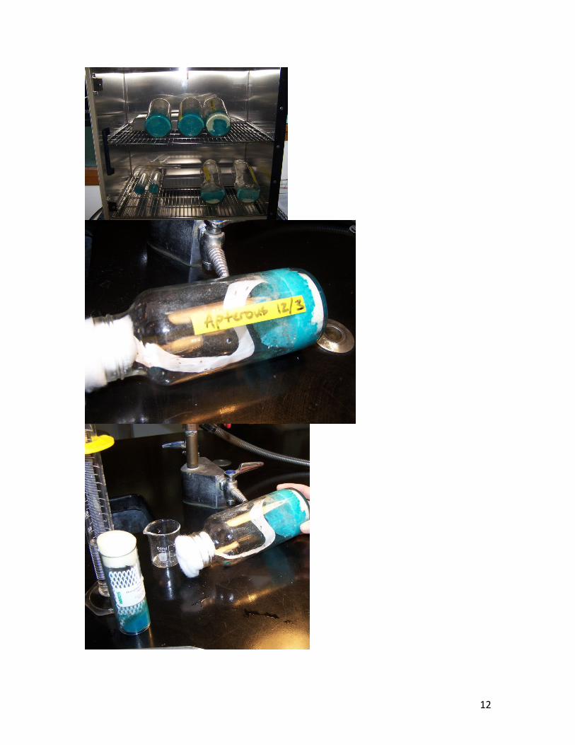
Subculturing the shipped flies When flies arrive subculture the flies using the techniques in the student lab so that there are about 5-10 males and 5-10 females in each new tube. If the shipped tubes have ample flies an alternative method from the student anesthesia procedure is to prepare large culture bottles. They can be subcultured without sexing them, it is likely that you will have pregnant, or reproductively viable females and males in the tubes. The technique is to gently tap the shipped tube so the flies move, but do not stick, to the bottom of the tube, quickly remove the foam top, and let them crawl into the larger subculture tube you have prepared. For stock cultures, larger bottles can be used to house larger populations, whatever the size, media should be 1/5th to 2/5th the volume of the bottle (see photo on the following page). Hydrate with an equal volume of water, what I have discovered since these photos were taken is to hydrate the media at an angle to prevent water from pooling, and to keep the media from falling down when the bottles are later put in the incubator on their sides. It also helps to incubate the bottles on their sides with one end elevated. Media should sit for a few minutes, adding water if necessary. The surface should be moist with a shiny appearance and there should be no spaces in the media. If the media is not completely hydrated, robust culture is compromised. Add a few grains of yeast, but no more to the media. Label them with the date and phenotypes, and incubate. Also, place the original flies in the incubator and as additional flies eclose they may also be subcultured. When the stock culture population is large enough for the class, virgin females must be obtained for the student vials so that when calculating ratios of F1
and F2 offspring recessive traits will accurately be reflected. Females do not mate for about 8-10 hours after hatching and can be obtained and placed into separate vials. Enough flies for groups of 2 or 3 to have 10-20 virgin mutants and 10-20 wild males must be reared. To maintain an ongoing stock, subculture your flies every 10-14 days.
11
12

Virgin Collecting Methods
Removal: remove all flies from the vial, after 8-10 hours collect all females present in the original vial and place in a fresh vial and wait 2-3 days to ensure no larvae are present. (Females tend to eclose early in the morning.)
Visual: virgin females are much larger than older females and are lightly pigmented, a dark green spot may be visible un the underside of the abdomen (their most recent meal).
Temperature cycling (not tried by this author): cultures at 18°C slow development and the female will not mate until 16 hours after eclosure. Removing flies in the afternoon/evening and placing the vials in an 18°C incubator produces about 98% females.
Make equipment available for students in a dedicated location: students will need a place where they can get supplies and do their work at their convenience when they need to as their schedules allow. You will need to provide a morgue for spent flies which can be a bottle with mineral oil or alcohol. You can use water if you dispose of them daily.
except for anaesthesia: put anaesthesia into a dropper bottle/s and provide it only when it is to be used by the students. It can potentially be “huffed” and therefore needs to be controlled by the instructor.
What to do with spent tubes: if plastic tubes are used it is best to dispose of them after they are used because of the contagious nature of mites and fungi. Plastic can be autoclaved (20 minutes and 121°C and 15 psi) or washed in a 10% bleach solution, however both methods make the tubes opaque, which makes it difficult to see into the tube. Tubes can be placed in the freezer to kill flies and thrown away. If they are glass dispose of the flies and media, rinse and autoclave or bleach.
Culture Notes:Although flies can tolerate 25°C (77°F), that is sort of a high end for them, and 20°C (68°F) is the lower end. I set my incubator at 22°C to be safe, sometimes the inside of the tube may be slighter warmer than the incubator from the fermentation of the medium. Lower temperatures prolong the life cycle, higher temperatures increase sterility and reduce viability. When getting your incubator to temp. keep in mind that if it’s digital it can take several hours to stabilize. Flies should not be kept in direct sunlight.
This author also learned that using rubber bungs is most definitely not a good idea. While it ensures that no flies will escape, it does ensure suffocation, which luckily did not happen, but the epiphany did wake me up in the middle of the night.
13

Fly Wrangling 101 How to Subculture and Breed Your Flies
Objectives• Develop fly handling skills and culture techniques• Apply computational methods for analyzing data• Reinforce student understanding of Mendelian genetics
Materials
• Drosophila food medium• Lg. glass cuvettes• Clipboard and task/date/name sheet• Lab Notebook for each group• Sterile cotton • Horsehair brushes• Filter paper (should be the appropriate size to fit inside a Petri dish, as well as
strips for anaesthesia) or white index cards• Dissecting scope• Petri dishes (also preferably glass)• Funnel• Anesthesia (there are several brands readily available in catalogs)• Nets for tubes (or filter paper)• Incubator for holding constant temperature if possible
Procedure
1. Prepare two culture tubes for your crosses. Make sure that your label includes:• the generation, e.g. P for parents (this will be your first generation that you’ve
placed into your new tubes), F1, F2, etc. for subsequent offspring.• the date• the type of mating in the cross
2. The amount of media used depends on the culture tube. In a standard tube use about 10 mls of dry media and equal parts water. If you’re using a glass cuvette use 5 mls of media and equal parts water. You will want the media to remain at the bottom of the vial, avoid chunks of media on the sides of the tube. Simply pour the water into the tube, give it a few minutes to soak into the media. If the media is still dry you may add a bit more water, (the larvae prefer the media on the wet side, rather than too dry).
3. Put a piece of filter paper in your Petri dish, and tape a strip of filter paper to the inside of the lid. (In case your flies wake-up before you’re done with them you can put a drop of anaesthesia on the paper taped to the lid.)
14

4. Obtain vials of parental flies from your teacher.
5. Record the vial number and parental cross marked on the vial in your notebook and start your datasheets. When working with your flies have your notebook and your datasheets with you.
6. Put a couple of drops of Fly Nap on the cotton at the bottom of the sleep box. (A cotton swab can also be inserted in the tube with the flies instead of using a sleep box).
Open the sleep box and put your fly tubes in the box, put the lid on the box, open the lid on with as little an opening as possible and using forceps, take the cotton swabs out of your parent tubes. It is important to keep your tubes on their sides as much as possible when handling flies that are asleep. If not the flies will fall into the wet medium, adhere to the medium and drown.
When the flies STOP MOVING (usually ~ 2 min.) they are sufficiently anaesthetized, they will die if left in the anaesthesia too long, (when the wings stand out at an angle, the flies are dead). Gently dump the flies into the viewing dish and observe and record the phenotypes and sexes of the flies (it is difficult to look for banding in newly hatched flies as the pigments are not well developed). IF YOUR FLIES START WAKING UP PUT A COUPLE OF DROPS OF ANAESTHESIA ON THE FILTER PAPER TAPED TO THE LID OF A PETRI DISH AND COVER YOUR FLIES. In your journal describe eye color, number and size of wings, or any unusual placement of body structures.
7. Place a five or six wild males and a five or six mutant females into each of your culture tubes, keeping your tubes on their sides using your paintbrush, and put your tubes into the incubator.
8. After seven days remove the parent flies from the mating bottle by tapping them into the sleep box and anesthetizing them until dead. Put them into a Petri and look at them under the dissecting scope, in your notebook draw (feel free to use colored pencils) a male, a female and a mutant, label the drawing (wild or mutant, male and female). Also, label the parts of the body (insects have a head, a thorax and an abdomen). When you are finished with your drawing discard flies into the morgue and place tubes back in the incubator. Record what you did and the date in your notebook.
9. When the flies begin to emerge, examine them and record the characteristics. This is the F1 generation. If the mutation is recessive, none of the F1 should exhibit the mutation. If any do, one of the P females was not a virgin and the culture should not be used in the rest of the experiment. Go back to step 1.
10. Prepare two or three new culture bottles, (properly labeled). Place 5-6 F1 males and females into two or three culture bottles and place in the incubator.
15

After 7 days euthanize the F1 flies and discard them.
When the F2 generation eclose euthanize them and record phenotypes
Before turning in your journal make sure it contains: a drawing and description of wild male, female, and mutant flies, and a drawing of the life cycle of Drosophila. At the end of your journal, pose at least three questions you would like to investigate.
16

Identifying Flies
Female on the left has an elongated posterior with thinner pigment bands, male on the right, the abdomen of the male has a black tip and a more round posterior
Male on the left, (note the sex combs on the forelegs) female on the right
17

Our mutations
1. Antennepedia (that means legs on yer noggin, or legheadedness) it’s a _ mutation,
Here’s the homeodomain antennepedia protein
18

2. and the last one on the right, Apterous (missing wings, oops), with some other mutated-winged friends.
19

Collecting Data
1. About 7 days after starting cross, remove parents to prevent breeding betweengenerations and to insure data collection from one generation only.
2. Data collection from an experimental cross is begun the day after the progeny first emerge. Usually flies are phenotyped and counted every other day for about 8 days to insure inclusion of mutants and the sex with slower developmental rates (females often appear sooner than males).
Genetic Notation Used in Describing Crosses
A fly with red eyes and other normal traits is called wild type and is designated by a +. The + refers here to all the traits (the entire phenotype). However, a + can also refer to an allele (locus).
A fly with a heritable trait different from wild type is considered a mutant. Mutations at particular loci are designated by letters derived from the descriptive name of the mutation. Abbreviations for recessive mutations are written entirely in lower case letters, whereas abbreviations for dominant mutations begin with capital letters. For our flies, we will use the symbols +,+ for the wild fly to indicate that wild alleles are dominant and a,a for the apterous fly indicating that the apterous allele genotype is recessive.
(During the initial stages of an inheritance study when the dominance relationships of alleles are unknown, the problem of deciding how to abbreviate the name for the mutation can be avoided by using a combination of letters and superscripts to designate a particular allele. For example, a mutant autosomal trait can be denoted as Am, while its wildtype counterpart can be denoted as A+. Similarly, an X-linked trait can be denoted as Xm for the mutant allele, and X+ for the wild type allele.)
20
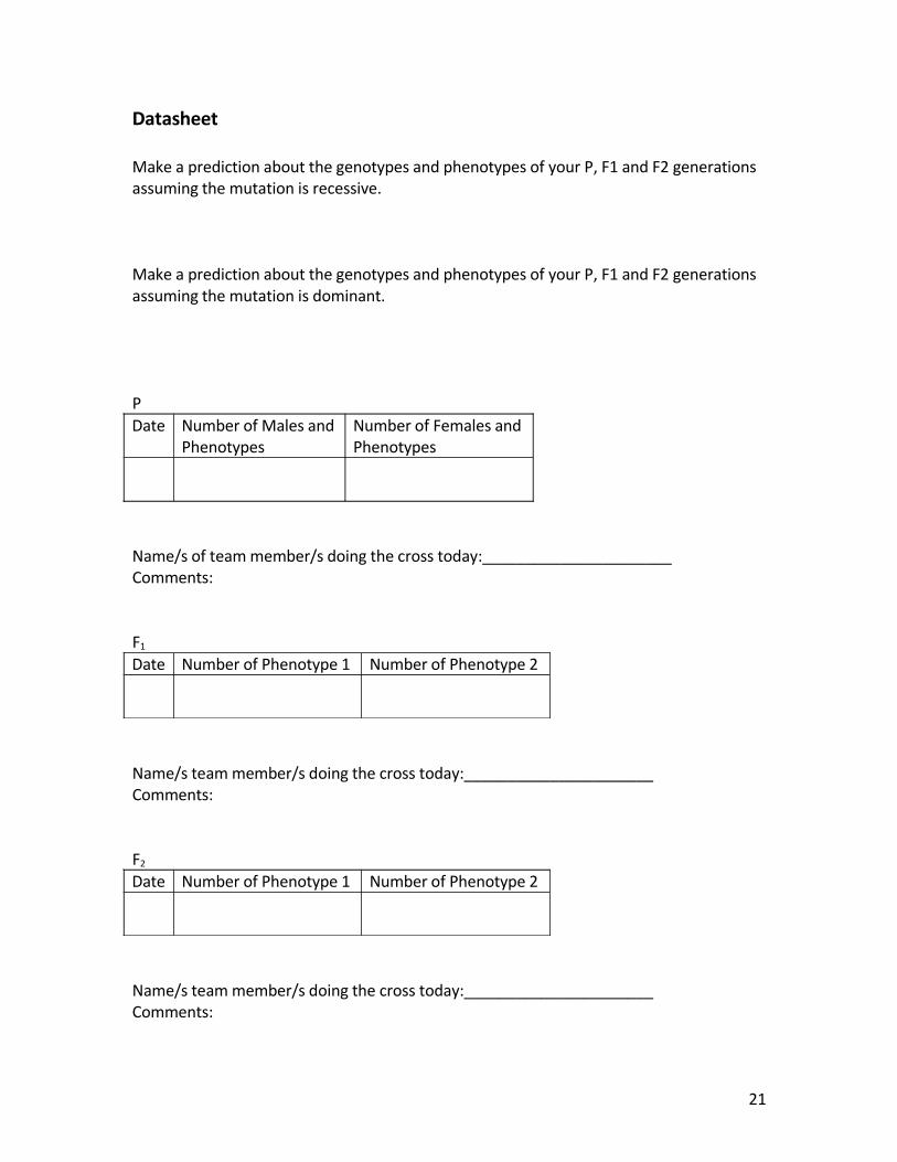
Datasheet
Make a prediction about the genotypes and phenotypes of your P, F1 and F2 generations assuming the mutation is recessive.
Make a prediction about the genotypes and phenotypes of your P, F1 and F2 generations assuming the mutation is dominant.
PDate Number of Males and
PhenotypesNumber of Females andPhenotypes
Name/s of team member/s doing the cross today:______________________Comments:
F1
Date Number of Phenotype 1 Number of Phenotype 2
Name/s team member/s doing the cross today:______________________Comments:
F2
Date Number of Phenotype 1 Number of Phenotype 2
Name/s team member/s doing the cross today:______________________Comments:
21

Analysis
Hardy-Weinberg
The Hardy-Weinberg equation will allow you to estimate the approximate percentages and gene frequencies of homozygous dominant, heterozygous and recessive genotypes of your flies.
The equation is p2 + 2pq + q2, where p2 will represent the homozygous dominant genotype, q2 will represent the recessive genotype and 2pq will represent the heterozygous individuals.
Here’s an example of how your will use the equation:
Total fly population- 278Number of wilds- 190 (these show the dominant phenotype)Number of mutants- 88 (the recessive phenotype)
The percent of each : Dominant – (p2 + 2pq ) 190/278 x 100% = 68.35%, as a frequency .6835 Recessive- (q2 ) 88/278 x 100% = 31.66%, as a frequency .3166, q = .563
In order to find the frequency of heterozygotes we have to find p. p = 1-q, p = 1 - .563 = .437 p2 = .1912pq = 2(.437)(.563)
To estimate the number of homozygous flies, multiply the frequency of p2 by the total population.278(.191) = 53.09, or 53
the estimated number of heterozygous individuals, multiply the frequency of 2pq by the total population278(.492) = 136.8, or 137
find the expected number of recessives by multiplying the total population by .25278 x .25 = 69.5
Now that you know the observed and expected we can use something called a Chi-square, which will let us evaluate the dataset.
The chi-square test is used in two similar but distinct circumstances:
22

a. for estimating how closely an observed distribution matches an expected
distribution - we'll refer to this as the goodness-of-fit test
b. for estimating whether two random variables are independent.
Chi SquareΧ2 = ∑ (observed x frequency – expected x frequency) 2
expected x frequency
The funny x looking thing is just the Greek letter “chi.” The expected is other Greek letter, sigma, which in statistics means “sum.”
Lastly, to determine the significance level we need to know the "degrees of freedom." In the case of the chi-square goodness-of-fit test, the number of degrees of freedom is equal to the number of terms used in calculating chi-square minus one. There were two terms in the chi-square for this problem - therefore, the number of degrees of freedom is one.
df P = 0.05 P = 0.01 P = 0.001
1 3.84 6.64 10.83
2 5.99 9.21 13.82
3 7.82 11.35 16.27
4 9.49 13.28 18.47
5 11.07 15.09 20.52
Report your statistical results in the Results section of your lab report.
23

Extensions
• Research function and expression of human Hox genes.Research of homeobox families and classes http://homeodb.cbi.pku.edu.cn/families.php?og=Drosophila
• Evolution of homeobox genes• Evo-devo• Up-regulation and down-regulation of genes• Virtual Apterous Lab
Glossary of Terms
imaginal disc: epithelial infoldings in the larvae of holometabolous insects (e.g. Lepidoptera, Diptera) that rapidly develop into adult appendages (legs, antennae, wings etc.) during metamorphosis from larval to adult form. During larval development, imaginal discs grow inside the larva. Development of the adult from the imaginal disc entails complex signaling interactions that divide the disc into distinct anterior, posterior, dorsal, and ventral compartments. At metamorphosis, the larva forms a pupa, inside which the larval tissues are reabsorbed and the imaginal tissues undergo extensive morphogenetic movements to form adult structures.
homeotic genes: in general homeotics are thought of as genetic switches that control the choice between different developmental pathways, also known as Hox genes, specifying the anterior-posterior axis and segment identity during early development of metazoan organisms. They are critical for the proper placement and number of embryonic segment structures (such as legs, antennae and eyes). The first genes found to encode homeodomain proteins were Drosophila developmental control genes, in particular homeotic genes, from which the name "homeo"box was derived. However, many homeobox genes are not homeotic genes; the homeobox is a sequence motif (a nucleotide or amino-acid sequence pattern that is widespread and has, or is conjectured to have, a biological significance), while "homeotic" is a functional description for genes that cause homeotic transformations."
homeobox: a fragment of DNA of about 180 basepairs (not counting introns), found in homeobox genes. A homeobox is a DNA sequence found within genes that are involved in the regulation of patterns of development (morphogenesis) in animals, fungi and plants. Genes that have a homeobox are called homeobox genes and form the homeobox gene family. Homeobox genes encode transcription factors which typically switch on cascades of other genes.
LIM-homeobox genes: The primary structure of LIM-homeobox genes has been remarkably conserved through evolution. A host of new data has been derived from
24

mutational analysis in diverse organisms, such as nematodes, flies and vertebrates. These studies have revealed a prominent involvement of LIM-homeodomain proteins in tissue patterning and differentiation, and their function in neural patterning is evident in all organisms studied to date. LIM genes act in a variety of developmental contexts, and display functional similarities across all organisms studied All LIM genes have expression and function in the nervous system (but some act elsewhere too). LIM genes determine correct axonal arrangements: sensory, motor or inter neurons. LIM genes may share overlapping functions in distinct cell types, might regulate common sets of downstream target genes But some LIM genes have quite specific roles: mec-3 regulates touch-neuron-specific genes (C. elegans),
apterous: wingless, a LIM-homeodomain protein that is expressed in dorsal cells and acts as a selector gene to divide the disc into dorsal and ventral compartments
homeodomain: in eukaryotes, homeodomains induce cellular differentiation by initiating the cascades of coregulated genes required to produce individual tissues and organs. The DNA-binding domain, binds DNA in a specific manner, is usually about 60 amino acids in length, and is encoded by the homeobox. The homeodomain fold is a protein structural domain that binds DNA or RNA and is thus commonly found in transcription factors. Most of the time, homeodomain proteins act in the promoter region of their target genes as complexes with other transcription factors, often also homeodomain proteins. The fold consists of a 60-amino acid helix-turn-helix structure in which three alpha helices are connected by short loop regions [1]. The N-terminal two helices are antiparallel and the longer C-terminal helix is roughly perpendicular to the axes established by the first two. It is this third helix that interacts directly with DNA. Homeodomain folds are found exclusively in eukaryotes but have high homology to lambda phage proteins that alter the expression of genes in prokaryotes.
Hox genes: Hox genes are a subgroup of homeobox genes. In vertebrates these genes are found in gene clusters on the chromosomes. In mammals four such clusters exist, called Hox clusters. The gene name "Hox" has been restricted to name Hox cluster genes in vertebrates. Only genes in the HOX cluster should be named Hox genes. So note: homeobox genes are NOT Hox genes, Hox genes are a subset of homeobox genes.
Hox cluster: The term Hox cluster refers to a group of clustered homeobox genes, named Hox genes in vertebrates, that play important roles in pattern formation along the anterior-posterior body axis. In fact, the first homeobox genes discovered where those of the Drosophila homeotic gene clusters, i.e. the "Antennapedia complex" and the "Bithorax complex", which summarily are referred to as HOM-C (homeotic complex). This HOM-C complex in Drosophila is the evolutionary homolog of the vertebrate Hox clusters and the evolutionarily related homeobox gene clusters in other animals (i.e. chordates, insects, nematodes, etc.) are now also called HOX clusters.
25

References
Photos of flies http://biol.org/DrosPics.htm
Culturing and life cycle, Pete Geiger, The Biology Projecthttp://biology.arizona.edu/sciconn/lessons2/geiger/intro2.htmhttp://biology.arizona.edu/sciconn/lessons2/geiger/intro.htm
Antennapedia Interactive Fly http://www.sdbonline.org/fly/segment/antenap3.htm
http://www.sdbonline.org/fly/segment/antenap1.htm#Bio
Apterous Virtual Fly Lab http://bioweb.wku.edu/courses/Biol114/Vfly1.asp
Apterous Homeodomain, Marco Milán, et al, European Molecular Biology Laboratory, http://www.sciencedirect.com/science?_ob=ArticleURL&_udi=B6T9H-4C2F8SJ-1&_user=10&_rdoc=1&_fmt=&_orig=search&_sort=d&view=c&_acct=C000050221&_version=1&_urlVersion=0&_userid=10&md5=1f829451d5bddf51e8e0f6a2508ee546
Homeobox Database Homepage maintained by Thomas R. Bürglin http://homeodb.cbi.pku.edu.cn/
Homeobox Database Drosophila main page, homeobox by family http://homeodb.cbi.pku.edu.cn/
Other homeobox information from http://faculty.pnc.edu/pwilkin/homeobox.htmlhttp://www.cbt.ki.se/groups/tbu/homeo.html
Antennapedia Homeobox http://homeodb.cbi.pku.edu.cn/family_info.php?spf=t&spfm=ANTP&sbfm=&og=All
Hox Genes Department of Biochemistry & Cell Biology Rice University Journal of Biological Chemistry http://www.jbc.org/cgi/reprint/M312842200v1.pdf
Developmental Regulatory Networks http://www.wwnorton.com/college/biology/devbio/chaptersummary/ch15.htm
Human-Fly genes that are shared A Survey of Human Disease Gene Counterparts in the Drosophila Genome, Mark E. Fortini, et al PMCID: PMC2180233 Fig 1. disease genes shared Fig 2 Blast table, included in protocolhttp://www.pubmedcentral.nih.gov/articlerender.fcgi?artid=2180233
26

Autism and Hox http://instruct1.cit.cornell.edu/courses/bioap475/Autism%203_11_02.pdf
Edward Lewis http://www.genetics.org/cgi/reprint/168/4/1773
27