proteomic analysis of regenerated rabbit lenses reveal ... · proteomic analysis of regenerated...
TRANSCRIPT
Proteomic analysis of regenerated rabbit lenses reveal crystallinexpression characteristic of adult rabbits
Xialin Liu,1 Min Zhang,1 Yuhua Liu,1 Pratap Challa,2 Pedro Gonzalez,2 Yizhi Liu1
1Zhongshan Ophthalmic Center, State Key Laboratory of Ophthalmology, Sun Yat-sen University, Guangzhou, China; 2Departmentof Ophthalmology, Duke University, Durham, NC
Purpose: To explore lens crystallin characteristics and morphology of rabbit regenerated lenses in comparison with wildtype natural lenses by means of proteomic analysis and histological assay.Methods: The lens regeneration model of the New Zealand rabbit was established, and lens regeneration was observedby slit lamp examination and photography. A histological assay was evaluated under light microscopy and transmissionelectron microscopy (TEM). Protein samples of regenerated lenses were collected from experimental rabbit eyes 2, 4, and16 weeks after surgery. Two-dimensional gel electrophoresis (2-DE) was performed. Image analyses was done using theImageMaster 2D Elite 3.01 software package. The protein spots were trypsinized and identified by matrix-assisted laserdesorption/ionization-time-of-flight-mass spectrometry.Results: Lens regeneration began in the periphery of the capsule bag about one to two weeks after the surgery andproceeded to regenerate toward the center. The regenerated lens appeared spherical in shape with a fairly translucentcortical structure and a nuclear opacity. Histological findings showed that the remnant lens epithelial cells differentiateat the lens capsule equator and new lens fibers form in a concentric pattern in a manner similar to that observed in naturallenses. However, TEM showed morphological changes in the epithelial cells of the regenerated lenses as compared withnatural lenses. 2-D electrophoresis revealed that the patterns of protein spots from regenerated lenses (two weeks, fourweeks, and 16 weeks) were analogous to those of 16-week-old natural lenses but were substantially different from thoseof two-week-old natural lenses, particularly when the two-week-old regenerated lenses were compared with the two-week-old natural lenses.Conclusions: Proteomic analysis revealed that crystallin expression in regenerated rabbit lenses was analogous to that ofnatural lenses of adult rabbits but was different from that of very young rabbits (two weeks old), and TEM revealed thepresence of morphological changes in the epithelial cells of regenerated lenses. These results suggest that the regrowth oflens materials in the lens capsule after endocapsular phacoemulsification might actually represent the regeneration of“mature” lens substances, which have led us to the conclusion that the regenerative process does not exactly mimicembryonic development.
Cataract phacoemulsification surgery and intraocularlens implantation is highly successful. However, it isassociated with accommodation dysfunction and variouscomplications [1]. Since 1827 [2], spontaneous regenerationof the lens following extracapsular extraction has beenextensively studied in rabbits as well as other mammals[3-9]. Reports of cell regrowth in animal lenses led us toconsider whether regeneration of human lenses mighteventually be possible [10]. Therefore, we have undertakenstudies to further understand the process of regeneration in therabbit lens.
It has been demonstrated that after lens substanceevacuation, the remnant lens epithelial cells differentiate atthe lens capsule equator and new lens fibers form first in theequatorial region. They then align with each other in a
Correspondence to: Yizhi Liu, MD, Ph.D., Zhongshan OphthalmicCenter of Sun Yat-sen University, State Key Laboratory ofOphthalmology; 54S Xianlie, Guangzhou, Guangdong, 510060,China; Phone: (86)-20-87330293; FAX: (86)-20-87333271; email:[email protected]
concentric pattern in a manner similar to that observed inembryonic and young rabbits [11].The newly formed lenscontains all of the major crystallin classes, although severalspecific crystallin subunits have been found to be absent orpresent in abnormally low concentrations [12].Embryonically, lens development involves a process ofcontinuous proliferation and differentiation of lens epithelialcells. As in embryonic development, regeneration of rabbitlens proceeds by cellular proliferation and differentiationalong the capsule [13,14]. It has been hypothesized that lensepithelial cells at the equatorial zone may have features ofstem cells, i.e. the ability to proliferate and differentiate intolens fibers and finally form a completely regenerated lens [8,15-18]. Recent studies, however, have reported that even stemcells are not exempt from aging [19-22]. Therefore, we areinterested in exploring whether adult lens epithelial cells canreally regenerate lens substance by mimicking the process ofthe lens development.
Given the similarity between the processes ofdevelopment and regeneration of rabbit lens [3,5,6,13], we
Molecular Vision 2008; 14:2404-2412 <http://www.molvis.org/molvis/v14/a277>Received 9 August 2008 | Accepted 11 December 2008 | Published 19 December 2008
© 2008 Molecular Vision
2404

have studied the histological features and the profile of theprotein composition of regenerated lens materials andcompared them with the natural lens materials from rabbits ofdifferent ages. A perfect regenerated lens should have thehealthy appearance and histological arrangement of a newregenerated lens as well as an accurate protein composition.In this study, we established a lens regeneration model in theNew Zealand rabbit as previously reported [14,23], observedthe process of regeneration and the histological features, andperformed proteomic analysis to explore the characteristics ofthe lens proteins.
METHODSEstablishment of regenerated rabbit lens model: All theexperimental protocols using animals strictly adhered to theARVO Statement for the Use of Animals in Ophthalmic andVision Research under an approved animal protocol.Following Gwon’s method, lens extraction was performed byendocapsular phacoemulsification on the experimental eyesof 12-week-old New Zealand albino rabbits (weighing 1.5–2.5 kg) [14,23]. Following lens extraction, all treated eyesreceived topical 1% tropicamide and 0.3% tobramycin fourtimes daily for seven days. Lens regeneration was evaluatedby slit lamp examination, and photographs were taken.Rabbits were divided into groups of three, which were thensacrificed at different times (two weeks, four weeks, and 16weeks). The regenerated lens from each rabbit was dissectedcarefully and stored under different conditions, dependingupon the analysis techniques to be used. Controls were naturallenses from 2-, 4-, and 16-week-old wild-type rabbits.Sample preparation for histopathological assay:
Paraffin-embedded tissue section preparation—Thewhole, dissociated, regenerated lenses and natural controllenses were fixed in 10% neutral buffered formalin. Tissuewas processed in an automatic tissue processor overnight andwas dehydrated in reagent grade alcohol, cleared with xylene,and infiltrated with paraffin. The paraffin embedded tissuewas sectioned at 5 µm and stained with hematoxylin and eosin.
Tissue preparation for transmission electronmicroscopy—Lens capsules were removed from the lenscortex. The capsule specimens were immediately placed in a1% glutaraldehyde/4% formaldehyde solution and sent to thepathology laboratory. Post-fixation was performed in osmiumtetroxide. The capsule specimens were dehydrated inincreasing concentrations of ethyl alcohol and embedded inresin. Ultrathin, 100 nm sections were stained with uranylacetate and lead citrate, and transmission electron microscopy(TEM) was performed with a Philip CM-10 electronmicroscope (Philip, Eindhoven, The Netherlands). The TEMslides were reviewed in a masked fashion.Sample preparation for protein analysis: Lens samples werecollected from experimental regenerated lenses at differenttimes (2, 4, and 16 weeks after surgery) and from naturalcontrol lenses. The capsules of the lenses were removed, and
the lens materials were pooled and ground to fine powder witha mortar in the presence of liquid nitrogen. The powder wasthen dissolved in lysis buffer that contained 7 M urea, 2 Mthiourea, 2% w/v 3-[(3-Cholamidopropyl)dimethylammonio]propanesulfonic acid(CHAPS), 1% dithiothreitol (DTT), 2%V/V carrierampholyte, pH 3–10, and protease inhibitor cocktail mix(Boehringer Mannheim GmbH, Mannheim, Germany) andthen spun at 12,000 rpm at 4 °C for 20 min. The supernatantwas collected and stored at −70 °C until further use. Proteinconcentration was measured by the Bradford assay [24].Two-dimensional electrophoresis: The proteins of all sampleswere first characterized using 13% SDS–PAGE. Theconcentration of the sample was 2 mg/ml, and the amount was20 μl/band. The gels were stained with Coomassie brilliantblue R-250 (Sigma-Aldrich Corp., St. Louis, MO). Two-dimensional gel electrophoresis (2-DE) was performed asdescribed by Gorg [25] using precast immobilized pHgradient (IPG) strips (immobiline DryStrip pH3-10 NL,18cm;GE Healthcare Life Science, Piscataway, NJ) in the firstdimension (isoelectric focusing) according to themanufacture’s instructions, and SDS–PAGE in the seconddimension. Total protein (150 μg) was loaded on each IPGstrip. After separation, the strips were immediatelyequilibrated two times for 15 min each time; in the first 15min, the strips were equilibrated with 50 mM Tris-HCl, pH8.8, 6 M urea, 30% glycerol, 2% SDS, and DTT (0.5% W/V).In the second 15 min step, 4.5% w/v iodoacetamide, but notDTT was added to the equilibration solution.to alkylate thiols.The separation in the second dimension was performed using13% SDS–PAGE gel in the Protein II device (Bio-Rad,Hercules, CA). The strips were held in place with 0.5%agarose dissolved in SDS/Tris/glycine containing runningbuffer, and electrophoresis was performed at a constantcurrent (30 mA/gel) at 16 °C. After electrophoresis, gels werestained with Coomassie brilliant blue R-250.Image analysis: The stained 2-D gels were imaged using theImageScanner (Amersham Pharmacia Biotech). Images weredigitized and evaluated with ImageMaster 2D Elite 3.01software (Amersham Pharmacia Biotech). Image analysis wasconducted for spot detection, matching, backgroundsubtraction, normalization, and isoelectric point/molecularweight calibration.Mass spectrometry: Trypsin in-gel digestion was performedas described by Rosenfeld et al. [26]. Briefly, gel spots wereexcised from the stained gel and cut into 1–2 mm2 slices thendestained with 25 mM ammonium bicarbonate/50%acetonitrile (Fisher Scientific, Springfield, NJ) andlyophilized with a SpeedVac Plus SC110A vacuumconcentrator (Savant, Holbook, NY). The gel was rehydratedin trypsin solution (Boehringer Mannheim). The ratio ofenzyme to protein was about 1:20 [27]. After overnightincubation at 37 °C, peptides were eluted with 5%
Molecular Vision 2008; 14:2404-2412 <http://www.molvis.org/molvis/v14/a277> © 2008 Molecular Vision
2405
trifluoroacetic acid (TFA) at 40 °C for 1 h followed by 5%TFA/50% acetonitrile elution until the gel slices becamewhite. The eluate was collected in an Eppendorf tube andlyophilized with SpeedVac Plus SC110A. The peptidemixture was dissolved with 0.5% TFA for mass spectrometryanalysis. The peptide mixture with matrix solution, α-cyano-4-hydroxycinnamic acid (CHCA; Sigma, St. Louis,MO), was measured on matrix-assisted laser desorption/ionization-time-of-flight-mass spectrometry (MALDI-TOF-MS [Reflex III; Bruker, Billerica, MA]) fitted with N2 lasers.The protein search was performed on the Matrix Science website with the search parameters set as follows: enzyme:trypsin, mass values: monoisotopic, Peptide Mass Tolerance:±0.5 Da, Peptide Charge State: 1+, Max Missed Cleavages:1.
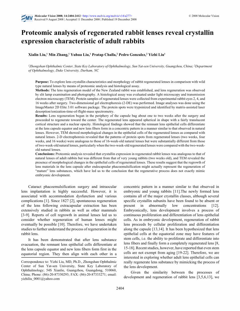
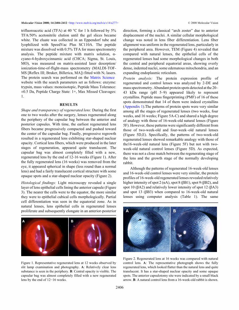
RESULTSShape and transparency of regenerated lens: During the firstone to two weeks after the surgery, lenses regenerated alongthe periphery of the capsular bag between the anterior andposterior capsules. With time, the earliest regenerated lensfibers became progressively compacted and pushed towardthe center of the capsular bag. Finally, progressive regrowthresulted in a regenerated lens exhibiting star-shaped nuclearopacity. Cortical lens fibers, which were produced in the laterstages of regeneration, appeared quite translucent. Thecapsular bag was almost completely filled with a new,regenerated lens by the end of 12-16 weeks (Figure 1). Afterthe fully regenerated lens (16 weeks) was removed from theeye, it appeared spherical in shape (less round than a normallens) and had a fairly translucent cortical structure with someopaque spots and a star-shaped nuclear opacity (Figure 2).
Histological findings: Light microscopy revealed a singlelayer of lens epithelial cells lining the anterior capsule (Figure3). The nearer the cells were to the equator, the more similarthey were to epithelial cubical cells morphologically. Partialcell differentiation was seen in the equatorial zone. As innatural lenses, lens epithelial cells in regenerated lensesproliferate and subsequently elongate in an anterior-posterior
Figure 1. Representative regenerated lens at 12 weeks observed byslit lamp examination and photography. A: Relatively clear lenssubstance is seen in the periphery. B: Central opacity is visible. Thecapsular bag was almost completely filled with a new regeneratedlens by the end of 12−16 weeks.
direction, forming a classical “arch zoster” due to anteriordisplacement of the nuclei. A similar cellular morphologicalchange was noted in lens fiber differentiation. Lens fiberalignment was uniform in the regenerated lens, particularly inthe peripheral area. However, TEM (Figure 4) revealed thatcompared with natural lenses, the epithelial cells of theregenerated lenses had some morphological changes in boththe central and peripheral equatorial areas, showing overlydense, indented nuclei, some edematous mitochondria, and anexpanding endoplasmic reticulum.Protein analysis: The protein expression profile ofregenerated and control lenses was analyzed by 2-DE andmass spectrometry. Abundant protein spots detected at the 20–43 kDa range (pH 5–9) appeared likely to representcrystallins. Peptide mass fingerprinting (PMF) of 16 of thesespots demonstrated that 14 of them were indeed crystallins(Appendix 1).The patterns of protein spots were very similaramong all the stages of regenerated lenses (two weeks, fourweeks, and 16 weeks; Figure 5A-C) and shared a high degreeof analogy with those of 16-week-old natural lenses (Figure5F). However, these patterns were significantly different fromthose of two-week-old and four-week–old natural lenses(Figure 5D,E). Specifically, the patterns of two-week-oldregenerated lenses showed remarkable analogy with those ofthe16-week-old natural lens (Figure 5F) but not with two-week-old natural control lenses (Figure 5D). As expected,there was not a close match between the regenerating stage ofthe lens and the growth stage of the normally developingrabbit.
Although the patterns of regenerated 16-week-old lensesand 16-week-old control lenses were very similar, the proteinprofiles of 16-week-old regenerated lenses revealed relativelyhigher intensity of spot 2 (αA), spot 8 (βB1), spot 9 (βB2), andspot 10 (βA2) and relatively lower intensity of spot 12 (βA3)and spot 13 (βB3) when compared to 16-week-old naturallenses using computer analysis (Table 1). The same
Figure 2. Regenerated lens at 16 weeks was compared with naturalcontrol lens. A: The representative photograph shows the fullyregenerated lens, which looked flatter than the natural lens and quitetranslucent. It has a star-shaped nuclear opacity and some opaquespots. The anterior capsulotomy site were indicated by a small blackarrow. B: A natural control lens from a 16-week-old rabbit is shown.
Molecular Vision 2008; 14:2404-2412 <http://www.molvis.org/molvis/v14/a277> © 2008 Molecular Vision
2406

magnitude changes in the intensity of some spotscorresponding to several crystallins were also observedamong 2-, 4-, and 16-week-old regenerated lenses.
DISCUSSIONGwon and coworkers [11-14,28] have extensively studiedrabbit lens regeneration, observing the regenerative processin the New Zealand albino rabbit after endocapsular lensextraction. They studied the histological changes and proteincomposition of regenerated lenses, showing that regeneratedrabbit lenses differentiate normally at the equatorial zone andproduce α-, β-, and γ-crystallins in proportions similar to thoseof natural lenses [11,12]. Based on these observations, they
proposed that lens regeneration mirrors the stages seen inembryonic development.
We followed Gwon’s methods to establish the rabbit lensregeneration models. Relatively clear regenerated lenses butwith a nuclear opacity and some opaque spots in the cortexwere formed within 12–16 weeks after surgery. Similar towhat was reported by Gwon et al. [11,13], we found by lightmicroscopy that remnant lens epithelial cells differentiate atthe lens equator and new lens fibers aligned in a concentricpattern, which is histologically similar to that of the early stageof lens development. However, TEM revealed lens equatorialepithelial cells with morphological changes including overlydense, indented nuclei, edematous mitochondria, andexpanding endoplasmic reticulum. Although more complete
Figure 3. Histological characteristics ofregenerated lens seen by lightmicroscopy. A: A single layer of the lensepithelial cell lines the anterior capsule.Cells are similar to epithelial cubicalcells morphologically. B: Partial celldifferentiation is seen in the equatorialzone. C: Proliferated epithelial cellshave elongated in an anterior-posteriordirection, forming a classical “archzoster” due to anterior displacement ofthe nuclei. D: Lens fiber alignment wasuniformly arranged in the regeneratedlens, particularly at the periphery.
Molecular Vision 2008; 14:2404-2412 <http://www.molvis.org/molvis/v14/a277> © 2008 Molecular Vision
2407

Figure 4. Representative transmission electron micrograph of epithelial cells in a regenerated lens. Morphological changes are seen both atthe peripheral equatorial (A) and central (B) areas, including overly dense and indented nuclei, edematous mitochondria, and an expandingendoplasmic reticulum when compared to natural lenses (C,D). Magnification is 8,000X).
Molecular Vision 2008; 14:2404-2412 <http://www.molvis.org/molvis/v14/a277> © 2008 Molecular Vision
2408

analysis will be needed to draw definitive conclusions, theseresults suggest changes usually associated with cellularsenescence and not with the normal lens developmentalprocess.
More importantly, proteomic 2-DE with peptide massfingerprinting identification showed that although theregenerated lens indeed contains all the major α-, β-, γ -crystallins, there was not a close match between the proteinexpression pattern of the lens substance in the regeneratingstage of lens and the pattern in the growth stage of the normallydeveloping lens.
The rabbits selected for surgery were about 12 weeks old.We began to count the lens regeneration time when the surgerywas performed. For example, two weeks after surgery, werecorded a regenerated lens as two weeks old, although therabbit’s age was 14 weeks. Four weeks after surgery, the rabbitage is actually 16 weeks, but we recorded the regenerated lensas four weeks, and so on. We compared 2-, 4-, and 16-week-old regenerated lens substances with natural lens substancesfrom rabbits aged two weeks, four weeks, and 16 weeks. 2-DE showed that the protein spots of all regenerated lenses (twoweeks, four weeks, and 16 weeks) were remarkably analogousto those of natural clear lenses from adult 16-week-old rabbits.The similarity was particularly evident in the major crystallinsubunit fractions. Interestingly, however, these fractions werenot at all analogous to natural lenses of earlier developing age
rabbits, particularly when the two-week-old natural lenseswere compared with the two-week-old regenerated lenses.
During the development of the natural lens, the proteinexpression pattern is in a continuous changing process.However, there is a more noticeable level of change from thenewborn stage or very early developing stage to adult stages[29-32]. Although we did not follow the changes in the proteinexpression over time in the regenerated lens, the proteinexpression pattern of the early regeneration stage (such astwo-week-old regenerated lens) showed clear analogy withthose of adult rabbit natural lens (16 weeks old) with respectto crystallin expression. The protein patterns are very similaramong all the stages of regeneration (two weeks, four weeks,and 16 wks) from early regeneration time to late regenerationtime. The reason could be that even for the early regeneratinglens, the rabbit’s actual age is mature enough. Therefore, thenew regenerating lens substance has the similar proteinpattern with the natural adult lens but such a dissimilar proteinpattern from newborn or early developing lens. Moreover,although 2-D gels show a similarity in protein expressionpattern between 16-week-old regenerated lenses (rabbit age28 wks) and 16-week-old natural lenses (rabbit age 16 weeks),further quantitative comparison revealed that the amounts ofcrystallin subunit spots in 16-week-old regenerated lensmaterials (rabbit age 28 weeks) were different from thosefound in the natural lens materials of 16-week-old rabbits.However, the real meaning of the differences will require
Figure 5. Two-dimensional electrophoresis photography of regenerated lens. A-C: Patterns of protein spots in regenerated lenses (two weeks,four weeks, and 16 weeks). D-F: Patterns of protein spots of natural lenses (two weeks, four weeks, and 16 weeks). The protein patterns arevery similar among all stages of regeneration (two weeks, four weeks, and 16 weeks) and shared a high degree of analogy with those of 16-week-old natural lenses (F). However, these patterns were significantly different from those of two-week-old or four-week-old natural lenses(D,E).
Molecular Vision 2008; 14:2404-2412 <http://www.molvis.org/molvis/v14/a277> © 2008 Molecular Vision
2409

further investigation because the similar magnitude of theincreases and decreases of some crystalline subunit spots werealso observed among 2-, 4-, and 16-week-old regeneratedlenses.
In summary, rabbit lens regeneration is the result of pre-equatorial epithelial cell proliferation and differentiation,regardless TEM showed the morphological changes of thenuclei and organelles in lens epithelial cells, consistent withthose previously reported that have lead to the hypothesis thatlens regeneration mimics the process of lens development.However, proteomic analysis revealed that the protein profileof regenerated lenses (even two-week-old regenerated lenses)was not analogous to the one existing in the natural lenses inthe early developing stage (two-week-old rabbits) but sharesa much clearer similarity with profile of the natural lenses ofadult rabbits (16 weeks old). This finding suggests that theregrowth of lens materials in the lens capsule afterendocapsular phacoemulsification might actually representthe regeneration of “mature” lens substances. Our results haveled us to the conclusion that the regeneration process does notexactly mimic embryogenesis. More studies are needed tounderstand the synthesis of lens crystallin proteins as well asposttranslational modification changes in the regenerativeprocess. Such understanding is critical for tissue engineeringefforts aimed at regenerating fully functional clear lensesrather than old cataractous lenses.
ACKOWLEDGMENTSThis work was supported by grants from the National NaturalScience Foundation of China to Liu, Yizhi (Number30070802) and to Xialin Liu (Number 30500554). The
authors wish to thank Dr. Bing Chen (ZhongshanOphthalmic Center, Guangzhou, China) for her technicalassistance. The funders had no role in the study design,data collection and analysis, decision to publish, or preparation of the manuscript.
REFERENCES1. Arango JL, Margo CE. Wound complications following
cataract surgery. A case-control study. Arch Ophthalmol1998; 116:1021-4. [PMID: 9715681]
2. Cocteau L. Reproduction of crystallin. Journal de PhysiologieExperimental et Pathologique 1827; 1:30-44.
3. Stewart DS, Espinasse PG. Lens regeneration in the rabbit.Nature 1966; 212:214-5. [PMID: 5972226]
4. Kessler J. Lens refilling and regrowth of lens substance in therabbit eye. Ann Ophthalmol 1975; 7:1059-62. [PMID:1180461]
5. Stewart DS, Espinasse PG. Regeneration of the lens of the eyein the rabbit. Nature 1959; 183:1815. [PMID: 13834580]
6. Stone LS. The Regeneration Of The Crystalline Lens. InvestOphthalmol 1965; 4:420-32. [PMID: 14340159]
7. Call MK, Grogg MW, Del Rio-Tsonis K, Tsonis PA. Lensregeneration in mice: implications in cataracts. Exp Eye Res2004; 78:297-9. [PMID: 14729361]
8. Gwon A. Lens regeneration in mammals: a review. SurvOphthalmol 2006; 51:51-62. [PMID: 16414361]
9. Lois N, Taylor J, McKinnon AD, Forrester JV. Posteriorcapsule opacification in mice. Arch Ophthalmol 2005;123:71-7. [PMID: 15642815]
10. Olson CM. If rabbits can regenerate the eye's lens, will sciencefind a way for humans to do it? JAMA 1989; 261:16-7.[PMID: 2908977]
11. Gwon AE, Gruber LJ, Mundwiler KE. A histologic study of lensregeneration in aphakic rabbits. Invest Ophthalmol Vis Sci1990; 31:540-7. [PMID: 2318593]
TABLE 1. CHANGES IN NORMALIZED SPOT VOLUME OF CRYSTALLIN SUBUNITS IN REGENERATED RABBIT LENS.
Spot No. Crystallin name Reg1 Reg2 Reg3 Control1 /* 2.12 3.32 1.89 2.112 αA 11.41 12.31 17.47 10.153 /* 3.43 2.02 4.52 3.414 βA4 7.82 6.32 5.34 4.525 βA3 (contains: βA1) 5.16 4.39 5.72 5.656 βB3 5.81 5.8 6.13 5.567 αB 2.02 1.12 1.22 1.458 βB1 5.53 4.05 7.33 4.579 βB2 14.58 10.89 13.63 9.3610 βA2 4.78 5.32 7.03 5.0311 βA3 1.40 1.43 1.28 1.3412 β A3 0.77 1.67 1.16 1.8813 βB3 0.13 0.19 0.09 1.7414 λ 5.27 6.43 5.39 6.4115 αB 14.77 12.11 14.16 13.8216 γC / 1.43 1.28 1.68
Normalized spot volume of each crystallin subunit is an average of three or four gels of each group. A spot is calculated bydividing its volume by the total volume and multiply by 100. Reg1: lenses from a two-week-old regenerated lens; Reg2: lensesfrom a four-week-old regenerated lens, Reg3: lenses from a 16-week-old regenerated lens; Control: natural lenses from a 16-week-old rabbit. The asterisk indicates that the protein spot was not identified as crystalline.
Molecular Vision 2008; 14:2404-2412 <http://www.molvis.org/molvis/v14/a277> © 2008 Molecular Vision
2410

12. Gwon A, Enomoto H, Horowitz J, Garner MH. Induction of denovo synthesis of crystalline lenses in aphakic rabbits. ExpEye Res 1989; 49:913-26. [PMID: 2612588]
13. Gwon A, Gruber L, Mantras C, Cunanan C. Lens regenerationin New Zealand albino rabbits after endocapsular cataractextraction. Invest Ophthalmol Vis Sci 1993; 34:2124-9.[PMID: 8491562]
14. Gwon A, Gruber LJ, Mantras C. Restoring lens capsule integrityenhances lens regeneration in New Zealand albino rabbits andcats. J Cataract Refract Surg 1993; 19:735-46. [PMID:8271170]
15. Tsonis PA, Del Rio-Tsonis K. Lens and retina regeneration:transdifferentiation, stem cells and clinical applications. ExpEye Res 2004; 78:161-72. [PMID: 14729349]
16. Wolosin JM, Budak MT, Akinci MA. Ocular surface epithelialand stem cell development. Int J Dev Biol 2004; 48:981-91.[PMID: 15558489]
17. Zhou M, Leiberman J, Xu J, Lavker RM. A hierarchy ofproliferative cells exists in mouse lens epithelium:implications for lens maintenance. Invest Ophthalmol Vis Sci2006; 47:2997-3003. [PMID: 16799045]
18. Xia X, Zhang X. A hypothesis of lens stem cell involvement incataract formation. Ann Ophthalmol 1999; 31:18-20.
19. Liu J, Finkel T. Stem cell aging: what bleach can teach. Nat Med2006; 12:383-4. [PMID: 16598279]
20. Chambers SM, Goodell MA. Hematopoietic stem cell aging:wrinkles in stem cell potential. Stem Cell Rev 2007;3:201-11. [PMID: 17917133]
21. Geiger H, Rennebeck G, Van Zant G. Regulation ofhematopoietic stem cell aging in vivo by a distinct geneticelement. Proc Natl Acad Sci USA 2005; 102:5102-7. [PMID:15788535]
22. Kamminga LM, de Haan G. Cellular memory andhematopoietic stem cell aging. Stem Cells 2006; 24:1143-9.[PMID: 16456126]
23. Gwon AE, Jones RL, Gruber LJ, Mantras C. Lens regenerationin juvenile and adult rabbits measured by image analysis.
Invest Ophthalmol Vis Sci 1992; 33:2279-83. [PMID:1607239]
24. Kruger NJ. The protein protocols handbook. Totowa, NJ,Human Press, 1996.
25. Gorg A, Obermaier C, Boguth G, Harder A, Scheibe B,Wildgruber R, Weiss W. The current state of two-dimensionalelectrophoresis with immobilized pH gradients.Electrophoresis 2000; 21:1037-53. [PMID: 10786879]
26. Rosenfeld J, Capdevielle J, Guillemot JC, Ferrara P. In-geldigestion of proteins for internal sequence analysis after one-or two-dimensional gel electrophoresis. Anal Biochem 1992;203:173-9. [PMID: 1524213]
27. Wan J, Wang J, Cheng H, Yu Y, Xing G, Oiu Z, Qian X, He F.Proteomic analysis of apoptosis initiation induced by all-transretinoic acid in human acute promyelocytic leukemia cells.Electrophoresis 2001; 22:3026-37. [PMID: 11565797]
28. Gwon A, Kuszak J, Gruber LJ. Intralenticular implant study inpigmented rabbits: opacity lensmeter assessment. J CataractRefract Surg 1999; 25:268-77. [PMID: 9951676]
29. Ueda Y, Duncan MK, David LL. Lens proteomics: theaccumulation of crystallin modifications in the mouse lenswith age. Invest Ophthalmol Vis Sci 2002; 43:205-15.[PMID: 11773033]
30. Lampi KJ, Ma Z, Hanson SR, Azuma M, Shih M, Shearer TR,Smith DL, Smith JB, David LL. Age-related changes inhuman lens crystallins identified by two-dimensionalelectrophoresis and mass spectrometry. Exp Eye Res 1998;67:31-43. [PMID: 9702176]
31. Lampi KJ, Shih M, Ueda Y, Shearer TR, David LL. Lensproteomics: analysis of rat crystallin sequences and two-dimensional electrophoresis map. Invest Ophthalmol Vis Sci2002; 43:216-24. [PMID: 11773034]
32. Bloemendal H, de Jong W, Jaenicke R, Lubsen NH, SlingsbyC, Tardieu A. Ageing and vision: structure, stability andfunction of lens crystallins. Prog Biophys Mol Biol 2004;86:407-85. [PMID: 15302206]
Molecular Vision 2008; 14:2404-2412 <http://www.molvis.org/molvis/v14/a277> © 2008 Molecular Vision
2411
Appendix 1. Rabbit crystallins subunits identified by peptide massfingerprinting.
To access the data, click or select the words “Appendix1.” This will initiate the download of a compressed (pdf)archive that contains the file.
Molecular Vision 2008; 14:2404-2412 <http://www.molvis.org/molvis/v14/a277> © 2008 Molecular Vision
The print version of this article was created on 13 December 2008. This reflects all typographical corrections and errata to thearticle through that date. Details of any changes may be found in the online version of the article.
2412