proteins serve a variety of functions. transport –myoglobin transports o 2 throughout muscles....
TRANSCRIPT

Proteins serve a variety of functions.
• Transport– Myoglobin transports O2 throughout muscles.
– Hemoglobin transports O2 in blood.
• Structural– Actin forms microfilaments in cells.– Tubulin dimers constitute microtubules.– Keratin filaments constitute the bulk of animal
hair.– Collagen is a major protein in connective
tissue.© 2014 John Wiley & Sons, Inc. All rights reserved.

Proteins serve a variety of functions.
• Motor function– Myosin interacts with actin to facilitate
muscular movement.– Kinesin moves along microtubules to support
a variety of cellular functions.
• Other functions of proteins– Catalysis– Immunity– Regulation of gene expression
© 2014 John Wiley & Sons, Inc. All rights reserved.

KEY CONCEPTS: Section 5-1
• O2 binds to the heme group of myoglobin such that binding is half-maximal when the oxygen concentration is equal to the dissociation constant.
• The similarities in structure and sequence between myoglobin and hemoglobin indicate a common evolutionary origin.
© 2014 John Wiley & Sons, Inc. All rights reserved.

Why focus on myoglobin and hemoglobin?
Sickled red blood cellHealthy red blood cell
• O2 transport is critical for sustaining life.
• Hemoglobin mutation can possibly lead to disease.
• Characteristics about O2 binding to myoglobin and hemoglobin are observed in many areas of biochemistry.
© 2014 John Wiley & Sons, Inc. All rights reserved.

Myoglobin is a classical globular protein.
Space-filling representation Ribbon diagram with heme in purple
What is heme?
© 2014 John Wiley & Sons, Inc. All rights reserved.

Heme is a prosthetic group.• Prosthetic group =
organic molecule bound to protein that aids protein function
• Heme is a porphyrin that chelates iron for oxygen transport.
© 2014 John Wiley & Sons, Inc. All rights reserved.

Myoglobin transports O2 via the Fe in heme.
O2
• His residues play a key role in anchoring both O2 and iron.
• Anemia is often treated with iron supplements or an iron-rich diet.
© 2014 John Wiley & Sons, Inc. All rights reserved.

How can binding of O2 to myoglobin be described?
Mb + O2 MbO2
K = [MbO2][Mb]
[MbO2]
© 2014 John Wiley & Sons, Inc. All rights reserved.

Myoglobin binds to O2 in a hyperbolic trend.
Fractional Saturation (Y):the proportion of myoglobin
molecules that have bound O2
Y = Bound Mb
Total Mb
[MbO2][Mb] +
[MbO2]Y =
pO2K +
pO2Y =
Equation of a hyperbolic curve
© 2014 John Wiley & Sons, Inc. All rights reserved.

O2 binds to the heme group of myoglobin such that binding is
half-maximal when the oxygen concentration
is equal to the dissociation constant.
Hyperbolic data is common in biochemistry!© 2014 John Wiley & Sons, Inc. All rights reserved.

Remember!
• Proteins have four possible levels of structure.– Primary sequence
– Secondary: alpha helices and beta sheets
– Tertiary: 3D fold
– Quaternary: interaction of multiple subunits
© 2014 John Wiley & Sons, Inc. All rights reserved.

Mb and Hb are only ~18% identical in primary sequence.
Invariant Identical in all Identical in Hb
© 2014 John Wiley & Sons, Inc. All rights reserved.

Mb and Hb are similar in their secondary and tertiary structures.
Myoglobin α-Subunit of Hemoglobinβ-Subunit of Hemoglobin
Heme
Even though myoglobin and hemoglobin haveonly ~18% identical residues, their secondary and tertiary structures overlap almost perfectly when superimposed!
Hb has quaternary structure, but Mb
does not.
© 2014 John Wiley & Sons, Inc. All rights reserved.

The similarities in structure and sequence between myoglobin and
hemoglobin indicate a common evolutionary origin.
© 2014 John Wiley & Sons, Inc. All rights reserved.

How to Express Affinity in Biochemistry?
• Kd = dissociation constant
• Mb + O2 reaction
• Fractional saturation
• Plot of fractional saturation vs. pO2
© 2014 John Wiley & Sons, Inc. All rights reserved.

KEY CONCEPTS: Section 5-1
• O2 can bind cooperatively to hemoglobin as the protein shifts from the deoxy to the oxy conformation.
• The Bohr effect and BPG modulate hemoglobin function in vivo.
© 2014 John Wiley & Sons, Inc. All rights reserved.

Oxygen binds cooperatively to Hb.
Dotted line represents O2
binding to myoglobin (hyperbola).
Solid line represents O2
binding to hemoglobin (sigmoid).
Note: Sigmoidal data are
indicative of cooperativity.
Cooperativity: Binding of O2 to one subunit induces easier binding to other subunits.
© 2014 John Wiley & Sons, Inc. All rights reserved.


Bohr Effect and O2 TransportWhat is happening biochemically when you breathe?
From Metabolism
+ H2O
© 2014 John Wiley & Sons, Inc. All rights reserved.

As pH , O2 affinity
© 2014 John Wiley & Sons, Inc. All rights reserved.
From Metabolism
+ H2O


BPG decreases Hb’s O2 affinity.
Lower O2 affinity
Fra
ctio
nal
Sat
ura
tio
n o
f O
2
BPG binds only to the tense (deoxy)
conformation of Hb.
© 2014 John Wiley & Sons, Inc. All rights reserved.


KEY CONCEPTS: Section 5-2
• Globular actin subunits associate in a double chain to form a microfilament.
• The growth and regression of actin filaments can change a cell’s shape.
• Microtubules are hollow tubes built from tubulin dimers.
© 2014 John Wiley & Sons, Inc. All rights reserved.

Microfilaments are polymers of actin.
Actin monomer
© 2014 John Wiley & Sons, Inc. All rights reserved.

Globular actin subunits associate in a double chain to form a
microfilament.
Actin monomer
Polymerization
© 2014 John Wiley & Sons, Inc. All rights reserved.

α and β-Tubulin form dimers.
© 2014 John Wiley & Sons, Inc. All rights reserved.
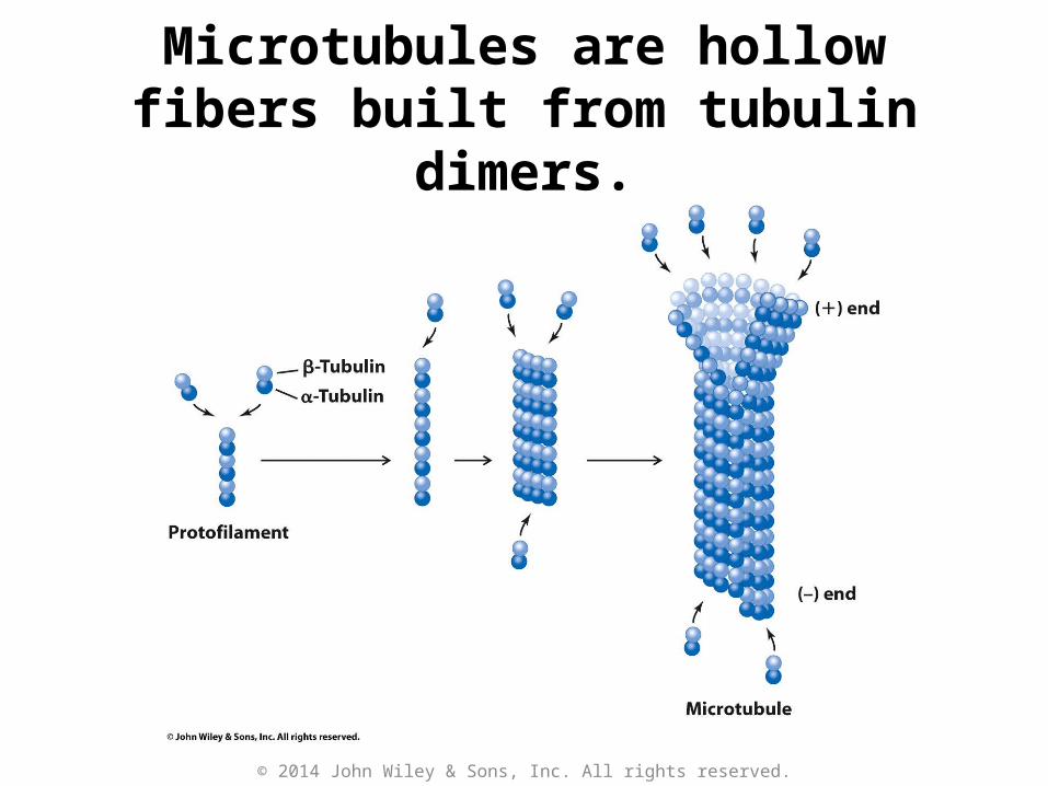
Microtubules are hollow fibers built from tubulin dimers.
© 2014 John Wiley & Sons, Inc. All rights reserved.

Cryoelectron microscopy reveals tubular structure of a microtubule.
© 2014 John Wiley & Sons, Inc. All rights reserved.

Microtubules can be observed in dividing cells.
Microtubules are shown in greenfluorescence.
Chromosomes are detected inblue fluorescence.
© 2014 John Wiley & Sons, Inc. All rights reserved.

KEY CONCEPTS: Section 5-2
• Intermediate filaments are long-lasting fibrous proteins consisting of coiled α helices.
• Three left-handed Gly-rich helical polypeptides form the collagen triple helix.
© 2014 John Wiley & Sons, Inc. All rights reserved.

Keratin is an intermediate filament.
Keratin forms acoiled-coil structureshown in the three
representationshere.
Backbone Stick Space-filling
© 2014 John Wiley & Sons, Inc. All rights reserved.

Collagen
• Gly-Pro-xxx repeat discourages a-helices or b-sheets
• Triple helix packs Gly in center• Pro is modified for H-bonding• Triple helices bound together to
make strong fibrils for hair/skin


Collagen is a triple helix.
© 2014 John Wiley & Sons, Inc. All rights reserved.

Collagen is covalently
cross-linked.
Cross-linking
stabilizes collagen’s structure.
Oxidation Oxidation
© 2014 John Wiley & Sons, Inc. All rights reserved.

Collagen has a noteworthy sequence.
• Every 3rd amino acid = Gly
• ~30% of remaining amino acids are proline or hydroxyproline.
© 2014 John Wiley & Sons, Inc. All rights reserved.

The Arrangement of Collagen
Fibrils in Various Tissues.

KEY CONCEPTS: Section 5-3
• The motor protein myosin couples the steps of ATP hydrolysis to conformational changes, resulting in muscle contraction.
• Kinesin transports cargo by moving processively along a microtubule track.
© 2014 John Wiley & Sons, Inc. All rights reserved.

Myosin has two heads and a long tail.
© 2014 John Wiley & Sons, Inc. All rights reserved.

Myosin binds to ATP.
© 2014 John Wiley & Sons, Inc. All rights reserved.

ATP hydrolysis drives the physical movement of myosin along an actin
filament.
© 2014 John Wiley & Sons, Inc. All rights reserved.

Kinesin is a microtubule-associated protein.
Vesicle (cargo) binding region
© 2014 John Wiley & Sons, Inc. All rights reserved.

Kinesin transports cargo by moving processively along a microtubule
track.
© 2014 John Wiley & Sons, Inc. All rights reserved.

Kinesin transports cargo by moving processively along a microtubule
track.
© 2014 John Wiley & Sons, Inc. All rights reserved.

Kinesin transports cargo by moving processively along a microtubule
track.
© 2014 John Wiley & Sons, Inc. All rights reserved.

Kinesin transports cargo by moving processively along a microtubule
track.
© 2014 John Wiley & Sons, Inc. All rights reserved.
