protein-oligonucleotide conjugation kit - solulink · catalog # s-9011-1 1 version 07.11.2013...
TRANSCRIPT

Catalog # S-9011-1 1
Version 07.11.2013
Protein-Oligonucleotide Conjugation Kit
Technical ManualCatalog # S-9011-1
Note: This protocol and any documents linked below can be downloaded from the appropriate category in the Solulink Library athttp://www.solulink.com/library.

Catalog # S-9011-1 2
Disclaimer
The products offered here are for research use only. Any commercial application will require a license
from Solulink. The Solulink Conjugation System is patented and has multiple patents pending. Please
contact Solulink for information regarding licensing information. Solulink products and methods may be
covered by one or more of the following United States patents Nos. 6,686,461, 6,800,728, 7,102,024,
7,173,125, 7,462,689 and other pending patent applications. Information in this manual is subject to
change without notice and does not constitute a commitment on the part of Solulink, Inc. It is supplied
on an “as is” basis without any warranty of any kind, either explicit or implied. Information may be
changed or updated in this manual at any time. This document may not be copied, transferred,
reproduced, disclosed, or duplicated, in whole or in part, without the prior written consent of Solulink,
Inc. This documentation is proprietary information and protected by the copyright laws of the United
States and international treaties. The manufacturer of this documentation is Solulink, Inc
Safety Information
WARNING – CHEMICAL HAZARD. Some chemicals used can be potentially hazardous, and can cause
injury or illness.
• Read and understand the Material Safety Data Sheets (MSDS) available at Solulink.com before you
store, handle, or work with any chemicals or hazardous materials.
• Minimize contact with and inhalation of chemicals. Wear appropriate personal protective equipment
when handling chemicals (e.g. safety glasses, gloves, or clothing). For additional safety guidelines
consult the MSDS.
• Check regularly for chemical leaks or spills. If a leak or spill occurs, follow the manufacturer’s clean-up
procedures as recommended in the MSDS.
• Comply with all local, state/provincial, or national laws and regulations related to chemical storage,
handling and disposal.

Catalog # S-9011-1 3
Table of Contents
I. Introduction -------------------------------------------------------------------------------------------- 4a. Product Description -------------------------------------------------------------------------- 4b. The Solulink Bioconjugation Method ---------------------------------------------------- 4
II. Protein-Oligonucleotide Conjugates: a Review ------------------------------------------------- 5III. Accessing 4FB-modified Oligonucleotides ------------------------------------------------------- 6IV. The Keys to Successful Conjugation --------------------------------------------------------------- 7V. Kit Components- --------------------------------------------------------------------------------------- 7VI. Additional Equipment/Reagents Required ------------------------------------------------------ 7VII. Protocols------------------------------------------------------------------------------------------------- 8
a. Desalt/Buffer Exchange of the Protein/Oligo ------------------------------------------ 9b. Amino Oligo Modification Protocol with S-4FB -------------------------------------- 10c. Protein Modification Protocol with S-HyNic ----------------------------------------- 15d. HyNic-Protein – 4FB-Oligo Conjugation Protocol ----------------------------------- 17
VIII. Analysis of the Conjugation Protein with Oligonucleotide ------------------------------- 19IX. Purification --------------------------------------------------------------------------------------------- 20X. Kit Stability --------------------------------------------------------------------------------------------- 20
XI. Troubleshooting -------------------------------------------------------------------------------------- 21XII. References --------------------------------------------------------------------------------------------- 22

Catalog # S-9011-1
I. Introduction
a. Product Description
The Protein-Oligo Conjugation Kit is designed to conjugate a protein with an oligonucleotide. It includesall of the necessary components and protocols for easy and specific crosslinking of any protein with anyamino-oligo from 20 to 100 bases in length. This kit is flexible so that researchers with little or noconjugation experience can make their own custom protein-oligo conjugate to suit their needs.
The SoluLink bioconjugation method converts more than 95% of protein to conjugate when four moleequivalents of oligo are added. High conversion rates, coupled with the unique UV traceable bondformed during crosslinking, allows for easy purification and identification of the conjugate from theexcess oligo using size exclusion purification methods such as HPLC.
b. The SoluLinK Bioconjugation Method
The Protein-Oligo Conjugation Kit uses SoluLink’s superior bioconjugation method to prepare protein-oligonucleotide conjugates in 3 easy-to-perform steps (Figure 1). The first step is the modification of theoligo with our 4FB crosslinker, followed by the formation of the HyNic modified protein. Finally, simplemixing of the two modifiedbiomolecules will result in theformation of a stable, UV-traceablebond formed by the reaction of aHyNic modified protein with a 4FBmodified oligonucleotide.
This technology has many practicaladvantages compared to previouscrosslinking methods:
1. The reaction is high yielding.Routine yields of conjugate are 50-80% based on starting protein.
2. The reaction is efficient: Only 3-4mole equivalents of oligo arenecessary for the protein, >90% ofthe protein is conjugated.
3. The conjugate bond is extremelystable: The conjugate bond isstable to 92OC and pH 2.0-10.0.
4. The reaction conditions are mildand do not cause any proteindenaturation: Unlike thiol-based conjugaticompromise the activity of proteins by cleaves disulfide bonds intact. No metals, o
Figure 1: Schematic representation of the three step process to prepare an antibody-
oligonucleotide conjugate using SoluLink’s Bioconjugation chemistry. Initially an
antibody is modified with S-HyNic to incorporate HyNic groups and subsequently the
4
on protocols, where reducing reagents are required that canleaving disulfide bonds, the HyNic-4FB conjugation couplexidation or reducing reagents are required.
HyNic-modified antibody is reacted with a 4FB-modified oligonucleotide.
Catalog # S-9011-1 5
5. The conjugation is traceable spectrophotometrically. The HyNic-4FB conjugate bond is UV traceable-it absorbs at 354 nm and has a molar extinction coefficient of 29000.
6. The modifications of both the HyNic linker on the protein and the 4FB linker on the oligonucleotideare quantifiable using colorimetric assays. The reproducibility of any reaction is facilitated by accuratecharacterization of all components. The Molar Substitution Ratio (MSR) of linker groups, i.e. thenumber of HyNic linkers per protein, can be quantified colorimetrically. This kit contains all thereagents necessary to determine the MSRs for both the protein and the oligo.
II. Protein-Oligonucleotide Conjugates: A Review
The diversity and specificity of proteins combined with the specificity of hybridization ofoligonucleotides results in unlimited numbers of specific protein detection reagents whose applicationsare addressed below.
The use of oligo-protein conjugates was initially demonstrated by Sano et al.1 for protein detection by atechnique called immuno-PCR (Polymerase Chain Reaction) where a 100mer oligo/antibody conjugatewas allowed to bind to its ligand and amplified by PCR demonstrating extremely sensitive proteindetection. Since this initial publication there has been a need for a straight forward, efficient and highyielding method for the preparation of these conjugates.
The first generation immuno-PCR protocol was plagued by high background due to non-specific bindingof the conjugate and the extreme sensitivity of PCR. This has been overcome by the Proximal LigationAssay (PLA) developed by Fredriksson and Lundegren.2 In the PLA assay, two antibodies to differentepitopes are conjugated to a 40mer 5’-phosphorylated oligonucleotide through the 3’-end and 60meroligonucleotide conjugated through its 5’-terminus. The two oligo/antibody conjugates are incubatedwith the sample, allowed to bind to their respective epitopes, the mixture is washed and then incubatedwith a ‘splint’ oligo that hybridizes across the two oligonucleotides that is subsequently ligated.Following ligation, PCR is performed on the ligated oligo generating a quantifiable signal. In subsequentwork the oligo/antibody conjugates used by Fredriksson et al. and others used conjugates prepared bySoluLink using the HyNic-4FB Conjugation Method. 3-6 Kozlov et al. 7 also describe the use ofoligonucleotide/antibody conjugates for the sensitive detection of proteins.
Additionally, oligonucleotide/antibody conjugates have been used for capture of antigens andsubsequent addressing to antibody arrays for multiplex detection of proteins as well for cell sorting onthe same diagnostic platform.8,9 Oligonucleotide/protein conjugates have been also been used invaccines to increase adjuvanticity using CpG oligonucleotide/protein conjugates.10-12

Ca
III. Accessing 4FB-modified Oligonucleotides
Stable and disulfide-cleavable 4FB oligonucleotides can be obtained in several ways:
1. 5’-4FB oligonucleotide
a. 4FB-phosphoramidite: 4FB-Phosphoramidite (1; Figure 2) is available for incorporationof 5’-4FB groups during oligonucleotide solid phase synthesis. Standard couplingprotocols are used and the yields are similar to any amino modifier. The 4FBPhosphoramidite may be purchased directly (SoluLink catalog #S-1005) or you mayorder 5’-4FB oligonucleotides directly from SoluLink.
b. 5’-amino oligonucleotides: 5’-amino oligonucleotides may be converted to 5’-4FBmodified oligonucleotides in a straight forward high yielding modification step with S-4FB (2; Figure 2).
The Protein-Oligo Conjugation Kit includes S-4FB and all the reagents and materials required toconvert a 5’-amino oligonucleotide to a 5’-4FB-oligonucleotide.
2. 3’-4FB oligonucleotide:3’-Amino oligonucleotides are converted to 3’-4FB modified oligonucleotides in an easy, highyielding modification step with S-4FB (2; Figure 2).The Protein-Oligo Conjugation Kit includes S-4FB and all the reagents and disposables requiredto convert a 3’-amino oligonucleotide to a 3’-4FB-oligonucleotide.
3. 5’- and 3’-4FB disulfide-cleavable oligonucleotides: 5’- and 3’-amino oligonucleotides may beconverted to disulfide-cleavable oligonucleotides using S-SS-4FB (3; Figure 2) in an easy, highyielding modification step. This product with protocol is available separately (SoluLink catalog#S-1037).
O
H
HN
O
4FB-phosphoramidite(S3034-100 or S3034-250)
O
H
O
ON
O
O
O
H
O
NH
OP
N
O
NC
NH2
S-4FB (S-1004-105 and-010)
NH
O
O
SS
H
O
ON
O
O
3’- or 5’-amino modified oligonucleotide
S-SS-4FB(S1037-010)
2
31
Figure 2: Schematic representation of the conversion of an amino-modified oligonucleotide to a 4FB-oligonucleotide withS-4FB (2) (top) and structures of 4FB-phosphoramidite (1) and S-SS-4FB (3), the reagent used to convert an amino-oligonucleotide to a 4FB-SS-oligonucleotide.
talog # S-9011-1 6

Catalog # S-9011-1 7
IV. The Keys to Successful Conjugation
The following are three crucial requirements that must be fulfilled for a reproducibly successfulpreparation of a protein/oligonucleotide conjugate using Solulink’s bioconjugation technology:
1. Desalting: Prior to modification, the starting protein must be thoroughly desalted, removing allamine contaminants, and exchanged into 1X Modification Buffer.
2. Protein concentration: The recommended concentration of the protein (1 – 5mg/ml) must beadhered to in all steps.
3. Molar substitution ratio: The Molar ratio of the HyNic on the protein and the 4FB on the oligomust be determined and within the desired range before continuing to the next step.
V. Kit Components
NOTES:1) For convenience all kit components can be stored at 4
OC
If precipitate is present in buffers on storage at 4OC redissolve by warming to 37
OC before
using2) 10X Modification Buffer: 1.0 M phosphate, 1.5 M NaCl, pH 8.03) 10X Conjugation Buffer: 1.0 M phosphate, 1.5 M NaCl, pH 6.04) 10X TurboLink Catalyst Buffer: 100 mM aniline, 100 mM phosphate, 150 mM NaCl, pH 6.0
VI. Equipment/Regents Required But Not Provided
Variable-speed bench-top microcentrifugeSpectrophotometer or Plate ReaderProtein concentration assay reagents such as BCA or Bradford assays
Component Component # Size Storage
S-HyNic S-9011-1-01 2 X 0.5 mg Desiccated
S-4FB S-9011-1-02 2 X 1.0 mg Desiccated
10X Modification Buffer S-9011-1-03 1.5 mL 4oC
10X Conjugation Buffer S-9011-1-04 1.5 mL 4oC
10X TurboLink Catalyst buffer S-9011-1-05 1.0 mL 4oC
7kDa 0.5 mL Zeba Columns S-9011-1-06 12 4oC
Anhydrous DMF S-9011-1-07 1.5 mL Desiccated
0.5 mM 2-Hydrazinopyridine Reagent S-9011-1-09 0.5 mL 4oC
0.5 mM 2-Sulfobenzaldehyde Reagent S-9011-1-10 0.5 mL 4oC
7kDa 2 mL Zeba Columns S-9011-1-13 2 4oC
10X PBS S-9011-1-14 1.5 mL 4oC
2.0 mL Collection tubes S-9011-1-15 12 RT
Oligo Resuspension Solution S-9011-1-16 1.0 mL 4oC

Catalog # S-9011-1 8
VII. Protocols
Certain strict parameters:
1. Protein molecular weight range 25,000 – 900,000 Daltons
2. Protein concentration range 1.0 – 5.0 mg/ml; Mass of protein to be modified range 50 –
650 µg; protein reaction volume range 50 – 130 µL
3. Oligonucleotide size range 20 – 100 bases
4. Oligonucleotide concentration range 0.3 – 0.6 OD260/µL; Mass of oligo to be modified
range 15 – 40 OD260; Oligo reaction volume range 30 – 60 µL
a. Desalt/Buffer Exchange of the Protein or oligo
Protein and oligo must be completely desalted into 1X Modification Buffer (dilute from 10XModification Buffer) before they are modified with S-HyNic or S-4FB. SoluLink recommends theuse of ZebaTM Desalt Spin Columns (provided) to desalt the protein and oligo. These rapid spincolumns are recommended because they do not significantly dilute the sample during desaltingand recover 85-90% of the protein or oligo.
Your protein
Concentrate the sample to 50-130 µL and 1 – 5 mg/mL
orDilute to 50 – 130 µL if sampleconcentration is more than 5 mg/mL.
Is the protein in liquid or solid form?
LiquidIs the protein
concentration known?
Determine proteinconcentration (BCABradford or A280nm)
Is the concentrationbetween 1 – 5 mg/mL?
Is the volumebetween 50 – 130µL?
Is the available massbetween 0.05 – 0.65 mg?
Add 1x modification buffer tobring the protein concentrationto 1 – 5 mg/mL and volumebetween 50 – 130 µL.
Desalt with 0.5 mL Zeba column.
Yes
Yes
Yes
Yes
Yes
Yes
Yes
Solid
No
No
No
Figure 3. Flow-chart used for guiding a user to the start of the conjugation process.

Catalog # S-9011-1
Included in this kit are 0.5 mL Zeba Spin Des
of 130 µL. Therefore up to 0.65 mg of a 5 msolution of oligo can be desalted. As this kitcolumns are included; one to initially desaltdesalt and exchange the modified protein inoligo into 1x Modification buffer and two toConjugation buffer. This kit also includes twocapacity of 50 – 150 µL or 150-700 µL, respeconjugates into PBS storage buffer after con
Zeba Desalting Protocol
0.5mL ZebaTM Spin Column Preparation an
1. Remove spin column’s bottom closure a
2. Place spin column in a 2.0 mL microcent
3. Centrifuge at 1,500 x g for 1 minute to re
4. Place a mark on the side of the column wcolumn in the microfuge with the mark f
5. Add 300 µL of required buffer to the topminute; discarding the flow through from
6. Repeat step 5 two additional times, disca
7. Column is now ready for sample loading.
Sample Loading
1. Place column in a new 2.0 mL microceof sample to the center of the compac
2. Centrifuge column at 1,500 x g for 2 mafter use.
3. Confirm protein or oligo concentration
0.5mL
2.0mL
Figure 4. ZebaTM
Desalt Spin Columnsused to desalt protein and Oligo
9
alt columns (Figure 4) that have a maximum capacity
g/mL solution of protein or 40 OD of a 0.6 OD/Lhas been designed for two conjugations, ten Zebathe protein with 1x Modification buffer and one toto 1x Conjugation buffer. One to initially desalt thedesalt and exchange the modified oligo into 1x0.5 mL or 2 mL ZebaTM desalting columns that have a
ctively, to desalt the final two protein/oligojugation depending on the final reaction volume.
d Sample Loading
nd loosen the top cap (do not remove cap).
rifuge collection tube.
move storage solution.
here the compacted resin is slanted upward. Placeacing outward in all subsequent centrifugation steps.
of the resin bed. and centrifuge at 1,500 x g for 1the collection tube.
rding buffer from the collection tube.
ntrifuge tube, remove cap and slowly apply 50-130µLted resin bed.
inutes to collect desalted sample. Discard column
and now ready for next step.
Catalog # S-9011-1 10
b. Amino Oligo modification Protocol with S-4FB (Calculator Worksheet 1)
A. Enter Amino-Oligo Information into Conjugation Calculator
1. Enter the following amino-oligo parameters directly from the Oligo vendor’s Certificate ofAnalysis into the Protein-Oligonucleotide Conjugation Calculator worksheet 1 - amine-oligonucleotide modification calculator on section I.
a) Oligonucleotide nameb) OD260 units supplied by vendorc) Oligonucleotide molar extinction coefficient (liter cm-1 mol-1)d) Oligonucleotide molecular weight (Daltons)e) Nanomoles per OD260 as listed on the product data sheet
Note: If the certificate of analysis is not available, the necessary information may be obtained byentering the oligo sequence into the Oligo Analyzer tool on IDT’s website at the followingaddress:
http://www.idtdna.com/analyzer/Applications/OligoAnalyzer/Default.aspx#
B. Resuspend Amino-Oligo
1. Ensure at least 15 OD260 units of oligo are available for modification—this information can befound on the oligo product data sheet supplied by the vendor.
2. Place the vial containing lyophilized oligonucleotide in a microcentrifuge and centrifuge at 15,000x g for 15 seconds to pellet the lyophilizate at the bottom of the tube. If the tube containsbetween 15 and 25 OD260 units of oligo, add 50 µL of Oligo Resuspension Solution to the tube. Ifthe tube contains more than 25 OD260 units of oligo, add a sufficient volume of OligoResuspension Solution to create a 0.5 OD260/µL solution (example: if there are 30 OD260 units ofoligo, add 60 µL of Oligo Resuspension Solution to create a 0.5 OD260/µL solution).
Note- 30 - 60 µL of oligo solution is using to modify with S-4FB
3. Allow the pellet to re-hydrate for 1 minute, then vortex the solution on medium speed for 10seconds to assist dissolution. This process will need to be repeated several times until no un-dissolved lyophilizate remains. Briefly spin the tube to ensure the entire solution is pooledtogether at the bottom, and then proceed to step C to measure the amino-oligo concentration.
C. Measure Amino-Oligo Concentration on a Spectrophotometer
The actual amino-oligo concentration can be measured either on a conventionalspectrophotometer or micro-volume UV-VIS scanning spectrophotometer (e.g.,NanoDropTM
spectrophotometer). When using a conventional spectrophotometer, a quartz or UV-transparentplastic cuvette is required.

Catalog # S-9011-1 11
NanoDropTMSpectrophotometer (models ND-1000 and ND-2000)
1. Determine the concentration (OD260/L) of the resuspended amino-oligo on a NanoDropTM asfollows:
Note - The definition of the OD260/L of oligo is the A260 value of 1 L of oligo solution in 1000Lof H2O at 1 cm path length.
a) In a microcentrifuge tube, prepare a 1:200 dilution of the dissolved amino-oligo by
transferring 2 L oligo with a calibrated P-2 pipette into 398L molecular gradeH2O. Vortex well to mix.
b) Select the “Nucleic Acid” menu option on the NanoDrop and initialize theinstrument using molecular grade water (NanoDrop 1000 only).
c) Clean the sample pedestal and blank the instrument with 2 L molecular gradeH2O.
d) Measure the A260 of the 1:200 amino-oligo solution as displayed in the 10 mm pathlength window. Record the A260 value.
e) Divide this number by 5 (convert to 1:1000 dilution) to calculate the OD260/Lconcentration of the stock oligo solution.
2. Multiply the OD260/L value calculated in (e) above by the volume of initial oligo resuspensionsolution using to dissolve oligo pellet (step B2) and to determine the total OD260 units availableafter dissolution (do not enter this information into the calculator at this point).
3. Important: If less than 15 OD260 units are recovered after resuspension (from 2 above), obtainadditional amino-oligo. 40 OD260 is the maximum units of amino-oligo to be modified.
Conventional UV-VIS Spectrophotometer
1. Determine the concentration (OD260/L) of the resuspended amino-oligo using a quartz or UV-transparent cuvette and a spectrophotometer as follows:
a) In a microcentrifuge tube, prepare a 1:500 dilution of the dissolved amino-oligo by
transferring 2 L oligo with a calibrated P-2 pipette into 998L molecular gradeH2O. Vortex well to mix.
b) Blank the spectrophotometer at 260 nm using 1mL molecular grade H2O.
c) Measure the A260 of the 1:500 amino-oligo. Record the A260 value.
d) Divide this number by 2 (convert to 1:1000 dilution) to calculate the OD260/Lconcentration of the stock oligo solution.

Catalog # S-9011-1 12
2. Multiply the OD260/L value calculated in (d) above by the volume of initial oligo resuspensionsolution using to dissolve oligo pellet (step B2) and to determine the total OD260 units availableafter dissolution (do not enter this information into the calculator at this point).
3. Important: If less than 15 OD260 units are recovered after resuspension, obtain additionalamino-oligo. 40 OD260 is the maximum units of amino-oligo to be modified.
D. Buffer Exchange Amino-Oligo
1. Prepare a oligo desalting spin column by using 0.5mL ZebaTM column with 1x Modificationbuffer (dilute 10X stock buffer 1/10 with water) following the Zeba desalting procedure on page9.
2. Remove the spin column from the collection tube (discard the collection tube containing excessbuffer) and place the column in a new microcentrifuge tube. Slowly and carefully pipet exactly
30 - 60 L of oligo solution into the center of the resin bed. Be careful not to let the oligosolution contact the tube wall; it must channel down through the resin itself. Replace the capand loosen one-quarter turn.
3. Centrifuge the column again at 1,500 x g for 2 minutes to collect the desalted oligo andmeasure the volume with appropriate pipet. Enter this volume (µL) of desalted amino-oligorecovered into the oligo modification calculator (Section II).
4. Vortex the oligo solution to mix thoroughly. Repeat the concentration determination as
described in section C above. Enter the calculated OD260/L stock oligo concentration into theOligo Modification Calculator (Section II).
Note: Excess un-desalted amino oligo from Part D may be stored at -20°C or lower indefinitely.
E. Dissolve S-4FB Reagent
1. Briefly centrifuge the tube of S-4FB reagent at 15,000 x g to ensure that all material is at the
bottom of the tube. Add 40L Anhydrous DMF to the 1.0 mg vial of S-4FB reagent and vortex for20 seconds to re-suspend. Continue to periodically vortex until the pellet is completelydissolved. The sample may need to be pipetted up and down several times to dissolvecompletely. Briefly spin the completely dissolved reagent to the bottom of the tube.
F. Modify Amino-Oligo with S-4FB Reagent
1. After entering the volume of oligo recovered (step D3) and its concentration (step D4), thecalculator will determine the volume of DMF to add, as well as the volume of S-4FB in DMF toadd to modify the oligo (output on section III of the oligo modification calculator).
2. First, add the indicated volume (µL) of DMF to the oligo solution and briefly vortex to mix. Next,add the volume of dissolved S-4FB in DMF indicated to the amino-oligo and vortex vigorously tomix. Do not centrifuge the reaction mixture after the S-4FB reagent is added.

Catalog # S-9011-1 13
3. Incubate at room temperature for 2 hours to allow the reagent to react with the amino-oligo.
G. Removal of excess S-4FB
Five minutes prior to the end of the 4FB/oligo modification reaction, prepare two desaltingcolumns as follows:
1. Prepare two oligo desalting spin columns by using 0.5mL ZebaTM column with 1x Conjugation
buffer (dilute 10X stock buffer 1/10 with water) following the Zeba desalting procedure on
page 9.
2. Remove the spin columns from the collection tubes (discard the collection tubes containingexcess buffer) and place the columns in new microcentrifuge tubes and ready for using.
3. Before load to column, centrifuge the oligo-modification reaction from Part F at 15,000 x g for2 minutes to pellet any insoluble reaction by-products (Please note the increased spin speed of15,000 x g, rather than 1,500 x g that is used during the desalting process). In the next step, useonly the clear supernatant (which contains the 4FB oligo) in the desalting process, leaving thepellet (if any) in the tube.
4. Slowly and carefully pipet the entire modification reaction (except for precipitate, if any) ontothe center of only one of the spin columns. Be careful not to let the oligo solution contact thetube wall; it must channel down through the resin itself. Replace the cap and loosen one-quarter turn. Leave the other column on the bench top during the next step.
5. Centrifuge the column containing the modified oligo at 1,500 x g (not 15,000 x g) for 2 minutesto recover the desalted oligo into the microcentrifuge tube.
6. Immediately transfer the entire elute from step 5 to the other spin column and repeat thedesalting process by centrifuging at 1,500 x g for 2 minutes to a new microcentrifuge tube.This “double-desalting” will ensure that all traces of un-incorporated 4FB are removed from theoligo.
7. Measure the volume with a P-200 pipette. Vortex the solution to mix thoroughly beforeproceeding to Part H.
H. Measure 4FB-Oligo Concentration
Measure the 4FB-Oligo concentration as follows:
Using a NanoDropTM Spectrophotometer (models ND-1000 and ND-2000)
1. In a microcentrifuge tube prepare a 1:50 dilution of the 4FB-oligo by transferring 2 L oligo
with a calibrated P-2 pipette into 98 L molecular grade H2O. Vortex well to mix.
2. Select the “Nucleic Acid” menu option on the NanoDrop and initialize the instrument usingmolecular grade water (ND-1000 only).
Catalog # S-9011-1 14
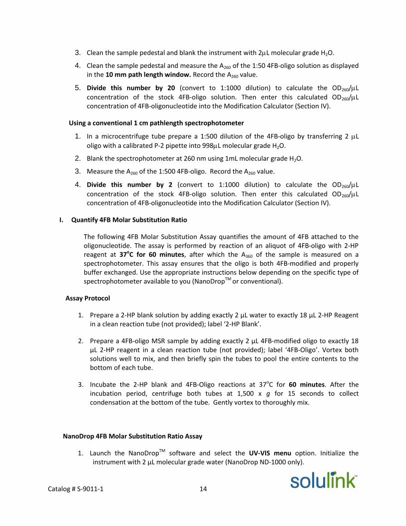
3. Clean the sample pedestal and blank the instrument with 2L molecular grade H2O.
4. Clean the sample pedestal and measure the A260 of the 1:50 4FB-oligo solution as displayedin the 10 mm path length window. Record the A260 value.
5. Divide this number by 20 (convert to 1:1000 dilution) to calculate the OD260/L
concentration of the stock 4FB-oligo solution. Then enter this calculated OD260/Lconcentration of 4FB-oligonucleotide into the Modification Calculator (Section IV).
Using a conventional 1 cm pathlength spectrophotometer
1. In a microcentrifuge tube prepare a 1:500 dilution of the 4FB-oligo by transferring 2 L
oligo with a calibrated P-2 pipette into 998L molecular grade H2O.
2. Blank the spectrophotometer at 260 nm using 1mL molecular grade H2O.
3. Measure the A260 of the 1:500 4FB-oligo. Record the A260 value.
4. Divide this number by 2 (convert to 1:1000 dilution) to calculate the OD260/L
concentration of the stock 4FB-oligo solution. Then enter this calculated OD260/Lconcentration of 4FB-oligonucleotide into the Modification Calculator (Section IV).
I. Quantify 4FB Molar Substitution Ratio
The following 4FB Molar Substitution Assay quantifies the amount of 4FB attached to theoligonucleotide. The assay is performed by reaction of an aliquot of 4FB-oligo with 2-HPreagent at 37oC for 60 minutes, after which the A360 of the sample is measured on aspectrophotometer. This assay ensures that the oligo is both 4FB-modified and properlybuffer exchanged. Use the appropriate instructions below depending on the specific type ofspectrophotometer available to you (NanoDropTM or conventional).
Assay Protocol
1. Prepare a 2-HP blank solution by adding exactly 2 µL water to exactly 18 µL 2-HP Reagentin a clean reaction tube (not provided); label ‘2-HP Blank’.
2. Prepare a 4FB-oligo MSR sample by adding exactly 2 µL 4FB-modified oligo to exactly 18µL 2-HP reagent in a clean reaction tube (not provided); label ‘4FB-Oligo’. Vortex bothsolutions well to mix, and then briefly spin the tubes to pool the entire contents to thebottom of each tube.
3. Incubate the 2-HP blank and 4FB-Oligo reactions at 37oC for 60 minutes. After theincubation period, centrifuge both tubes at 1,500 x g for 15 seconds to collectcondensation at the bottom of the tube. Gently vortex to thoroughly mix.
NanoDrop 4FB Molar Substitution Ratio Assay
1. Launch the NanoDropTM software and select the UV-VIS menu option. Initialize theinstrument with 2 µL molecular grade water (NanoDrop ND-1000 only).

Catalog # S-9011-1 15
2. Blank the NanoDropTMwith 2 µL 2-HP blank solution and clean the pedestal. Set the λ1 absorbance wavelength to read at 360 nm.
3. Place a 2 µL aliquot of the 4FB-Oligo MSR reaction on the pedestal and click the‘Measure’ icon. The 1.0 mm A360 absorbance will appear. Enter this value directly into theMSR calculator (Section V) using Protein-Oligonucleotide Conjugation Calculator oncalculator worksheet -1 to obtain MSR.
Conventional Spectrophotometer 4FB Molar Substitution Ratio Assay (100µL micro-cuvettemethod)
1. Prepare a 1:10 dilution of the 2-HP blank solution by adding 180 µL of molecular gradeH2O into the blank tube.
2. Prepare a 1:10 dilution of the 4FB-oligo MSR reaction solution by adding 180 µL ofmolecular grade H2O into the blank tube. Vortex both solutions well to mix well.
3. In a 1 cm, 100 µL quartz micro-cuvette, blank the spectrophotometer at λ = 360 nm with 100 µL of 1:10 diluted 2-HP blank.
4. Measure the A360 of the 1:10 4FB-Oligo MSR sample from step 2 above in the cuvette.Record the A360 and enter this value directly into the MSR calculator (Section V) usingProtein-Oligonucleotide Conjugation Calculator on calculator worksheet 1 to obtain MSR.
c. Protein Modification Protocol with S-HyNic (Calculator Worksheet 2)
Recommended Guidelines for Modifying Proteins with S‐HyNic
The modification process is a critical element of any conjugation project. For this reason, we have included
a more detailed discussion of this important step. For example, the number of functional groups
incorporated per protein molecule is commonly referred to as the molar substitution ratio (MSR). The final
MSR obtained after a modification reaction with S-HyNic is a function of several variables that include
protein concentration, number of available amino-groups on the protein (often related to M.W.), excess
linker equivalents added (e.g. 5X, 10X or 20X) and reaction pH. Table 1 presents the results of a study to
determine the level of HyNic incorporation on an antibody adding 5X, 10X and 20X equivalents of S-HyNic
at 1.0, 2.5 and 5.0 mg/mL of antibody concentration in 1X modification buffer.
Table 1: MSR of Modifying Bovine IgG with S-HyNic
Note - It is recommend that the MSR of HyNic-proteins is 4 - 8 for proteins greater than100,000 Daltons, and 2-4 for proteins equal to or less than 100,000 Daltons.
Protein concentration 5X HyNic 10X HyNic 20X HyNic
1.0 mg/mL 1.0 1.4 3.0
2.5 mg/mL 3.2 5.3 7.9
5.0 mg/mL 4.9 6.6 7.8

Catalog # S-9011-1 16
In general, as the protein concentration and number of linker equivalents are increased, the molar
substitution ratio increases. Caution is recommended since over-modification can dramatically change the
isoelectric point of the protein and result in precipitation of the protein or loss of biological activity. This is
especially critical with proteins <50 kD MW.
S-HyNic-Protein Modification Protocol (Calculator Worksheet 2)
1. Desalt protein to be modified with S-HyNic into 1X Modification Buffer (dilute 10X stock buffer 1/10 withwater) following the Zeba desalting procedure on page 9. Between 50 - 130 µL of protein solution at aconcentration between 1 - 5 mg/mL may be used.
2. On the S-HyNic Modification Calculator input the name, molecular weight, protein concentration,
volume of protein to be modified, and the mole equivalents of HyNic used to modify protein in the
green fields. Add the required volume of anhydrous DMF to a 0.5 mg vial of pre-weighed S-HyNic
reagent. Pipette the solution up and down to dissolve the pellet. The volume of DMF will be output in
the pink field. If the volume of DMF output is more than 700 L, two or more times dilution of the HyNic
reagent may be required.
For example if the volume of DMF output is 5857 L, then three times dilutions are required:
(1) Make a 58.57X stock solution by dissolving S-HyNic pellet in 100 L of DMF (5857L/100L = 58.57).
(2) Next, make a 5.857X S-HyNic solution by adding 10 L of 58.57X S-HyNic solution into 90 L of DMFand mix well (1:10 dilution).
(3) Third, make 1X S-HyNic solution by adding 10 L of 5.857X S-HyNic solution into 48.57 L of DMF andmix well (1:5.857 dilution).
3. If protein molecular weight is greater than 50,000 Daltons, add 2.0 L of S-HyNic reagent into the
desalted protein. If protein molecular weight is equal or less than 50,000 Daltons, add 3.0 L of S-HyNic
reagent into the desalted protein as calculated using the Protein-Oligonucleotide Conjugation
Calculator on calculator worksheet 2. Briefly vortex to mix.
4. Incubate the reaction at room temperature for 2.5 hours.
5. Proceed to desalt the HyNic-modified protein using 0.5 mL ZebaTM
column equilibrated with 1X
Conjugation buffer (dilute 10X stock buffer 1/10 with water) using the Zeba desalting protocol on page
9.
Determining the HyNic Molar Substitution Ratio (MSR)
After desalting with a ZebaTM spin column to remove excess HyNic from the modification reaction, the
protein concentration is determined by using either BCA or Bradford assay (the protein concentration
must not be determined by reading A280 on a spectrophotometer at this step because of the
contribution at A280 of HyNic itself). After the HyNic-protein concentration is determined proceed to
MSR assay.

Catalog # S-9011-1 17
Assay Protocol
1. Transfer 2 μL of HyNic-modified (desalted) protein solution (1-5 mg/mL in 1X Conjugation
Buffer) to a new 1.5 mL microfuge tube containing 18 μL of SBA working solution. Prepare
another reaction tube (blank) containing 2 μL of 1X Conjugation Buffer and 18 μL of 2-SBA
working solution.
2. Vortex to mix well, then incubate both reaction tubes at 37oC for 1 hour.
3. Remove the reaction tubes from 37oC and briefly centrifuge at 10,000 x g to collect any
condensate. Vortex both samples thoroughly before reading the A348 by one of the
following methods:
Method A: NanoDropTM Method
1. Launch the NanoDropTM software and select the UV-VIS menu option. Initialize the instrument with
2 μL water (NanoDropTM ND-1000 only).
2. Blank the NanoDropTM with 2 μL blank (Conjugation Buffer + SBA) solution and clean the pedestal.
3. Set the λ1 wavelength to 348nm. Place 2 μL of the HyNic-protein MSR reaction on the pedestal and
click the “Measure” icon. The 1.0 mm A348 absorbance will appear. Multiply this number by 10
(converts from 1 mm to 1 cm) and then enter this value into the HyNic MSR Calculator Protein-
Oligonucleotide Conjugation Calculator on calculator worksheet 2 to obtain MSR.
Method B: Cuvette Spectrophotometer (100 μL, 1‐cm micro‐cuvette)
1. Prepare a 1:10 dilution of blank (Conjugation Buffer + SBA) solution by adding 180 μL of deionized
water into the blank sample tube and mix well.
2. Prepare a 1:10 dilution of HyNic-protein MSR reaction solution by adding 180 μL of deionized water
into the HyNic-protein MSR reaction tube and mix well.
3. In a 1 cm, 100 μL quartz micro-cuvette, blank the spectrophotometer at 348nm with 100 μL of the
blank solution prepared in step 1 above.
4. Remove the blank solution and add 100 μL of the HyNic-protein MSR sample solution from step 2
above to the cuvette.
5. Record the 348nm absorbance value of the HyNic-protein MSR sample. Multiply this number by 10
(to account for the 10-fold dilution) and then enter this value into the HyNic MSR Calculator Protein-
Oligonucleotide Conjugation Calculator on calculator worksheet 2 to obtain MSR.
d. HyNic-Protein – 4FB-Oligo Conjugation Protocol (Calculator Worksheet 3)
1. Using the Protein-Oligonucleotide Conjugation Calculator on calculator Worksheet 3. On theHyNic-Protein/4FB-Oligo conjugation Calculator, input the name, molecular weight,

Catalog # S-9011-1 18
concentration and mass of HyNic- protein to be modified, and the name, molecular weight,concentration, MSR of 4FB-oligo and Extinction coefficient of oligo in the green fields.
2. The calculator will determine the volumes of HyNic-protein to combine with 4FB-oligo and thevolume of 10X TurboLink Catalyst Buffer required to add into the conjugation solution in the
blue field. If the volume of 4FB‐oligo output is less than 1 μL, the 4FB‐oligo (OD260/L) can be
diluted 10 fold with 1X Conjugation buffer. Be sure to change OD260/L by divided by 10.3. Incubate at room temperature for 2 -3 h or 4oC for overnight.
4. The conjugation reaction is now ready for purification by column chromatography, if required,or desalting into storage buffer.
5. The TurboLink™ Catalyst should be removed soon after the conjugation reaction is complete.
The reaction solution may either be purified by using chromatography immediately, or may be
desalted into storage 1X PBS buffer using either a 0.5 or 2 mL ZebaTM column based on the
conjugation reaction volume (0.5 ml ZebaTM column has a capacity of 50 – 150 μL; 2 ml ZebaTM
column has a capacity of 150 – 700 μL).
6. If using a 0.5 mL Zeba, proceed to buffer exchange the oligo/protein conjugated into 1X PBS
Buffer as described on page 9. For using the 2 mL ZebaTM column, please see the protocol below.
7. Determine final protein concentration using BCA or Bradford assay. Add bacteriostat (e.g. 0.05%
sodium azide) and/or a protein stabilizer (e.g. 0.5% BSA) if necessary, then store at 4oC.
ZebaTM Column Desalting Protocol
2 mL Zeba™ Spin Column Preparation and Sample Loading (150 – 700 µL)
1. Remove spin column bottom closure and loosen the top cap (do not remove cap).
2. Place spin column in a 15 mL conical collection tube.
3. Centrifuge at 1,000 x g for 2 minutes. Discard the flow through from the collection tube.
4. Place a mark on the side of the column where the compacted resin is slanted upward. Place column
in the 15 mL conical collection tube with the mark facing outward and away from the center of the
rotor in all subsequent centrifugation steps.
5. Add 1 mL of 1X PBS Buffer to the top of the resin bed and centrifuge at 1,000 x g for 2 minutes.
Discard the flow through from the collection tube.
6. Repeat step 5 an additional two times, discarding the flow through from the collection tube.
7. Place the ZebaTM column in a new 15 mL conical collection tube, remove cap, and slowly apply
sample onto the center of the compacted resin bed (150–700 µL).
Note- For sample volumes less than 350 µL, apply a 40 µL 1X PBS Buffer stacker to top of the resin
bed after the sample has fully absorbed to ensure maximal protein recovery. Avoid contact with
the sides of the column when loading.
8. Centrifuge column at 1,000 x g for 2 minutes to collect desalted sample.
9. Determine final protein concentration using BCA or Bradford assay. Add bacteriostat (e.g. 0.05%
sodium azide) and/or a protein stabilizer (e.g. 0.5% BSA) if necessary, then store at 4oC.

Catalog # S-9011-1 19
VIII. Analysis of the conjugated protein with Oligo
After completion of the conjugation reaction, a small aliquot of the crude reaction mixture is often
analyzed using a 10-12% Bis-Tris gel in an SDS (non-reducing) running buffer system. The amount of
crude sample loaded on these gels depends on the type and sensitivity of staining method being used.
For example, Coomassie blue stain can easily detect 2-4 micrograms of crude conjugate per lane; DNA
silver stain can detect 250-500ng of conjugate per lane. An appropriate protein marker is loaded side by
side, as well as HyNic-protein and 4FB-modified oligo as references. The protein/oligo conjugate may
migrate slowly in the gel and is visualized as slightly higher molecular weight species.
Protein-Oligo conjugation sample:Monoclonal mouse anti-human IgG1 (3mg/ml) was modified with 20 mole equivalents of S-HyNic. Oligo(75 bp) was modified with 20 mole equivalents of S-4FB. Then HyNic-mouse anti-human IgG1 wasconjugated with 3 moles equivalents of 4FB-oligo (75 bp) and purified with HPLC (SuperDex 200, 60min
program). Respectively, loading 1g or 450ng of conjugated on 10% SDS-PAGE, followed by coomassie orDNA silver stain.
Coomassie Stain:Lane 1. MW marker
Lane 2. Mouse anti-human IgG1 (2 g)Lane 3. Crude HyNic-mouse anti-human IgG1/4FB-oligo
Lane 4. Purified HyNic-mouse anti-human IgG1/4FB-oligo
Lane 5. Oligo (75 bp)
DNA Silver Stain:Lane 1. MW markerLane 2. Mouse anti-human IgG1Lane 3. Crude HyNic-mouse anti-human IgG1/4FB-oligo
Lane 4. Purified HyNic-mouse anti-human IgG1/4FB-oligo
Lane 5. Oligo (75 bp)
Figure 5. Sample of HyNic-protein/4FB-Oligo conjugation

Catalog # S-901
IX. Purification
All conjugation reactions will be a mixture of reaction products consisting of the desired conjugate along
with some un-conjugated 4FB-modified oligo. For this reason, the conjugation reactions are purified
using chromatographic methods to remove excess 4FB-modified oligo. Various FPLC or HPLC
chromatography workstations are available for this purpose (Figure 6). Solulink routinely purifies
conjugates using size exclusion. Other media such as metal chelate affinity chromatography or protein
A/G columns may be used where appropriate.
X. Kit Stabilit
XI. Troubles
Uno
S-Hy
4FB-
HyN
Puriwith
Figure 6: Purification of an antibody-20mer oligo conjugate by size exclusion HPLC(SuperDex 200; GE HealthCare). The initial peak is the desired conjugate followed byunconjugated oligonucleotide. Above each peak its UV spectrum- note the 354 nmabsorbance of the conjugate peak due to the formation of the conjugate bond.
1-1 20
y
hooting
Component Storage Stability
pened Kit 4OC Refer to Certificate of Analysis
Nic / S-4FB in DMF 4OC 1 day
oligonucleotide < -20OC > 1 year
ic-modified protein - Use immediately
fied protein-oligo conjugate0.05% azide 4OC 6 months
Catalog # S-9011-1 21
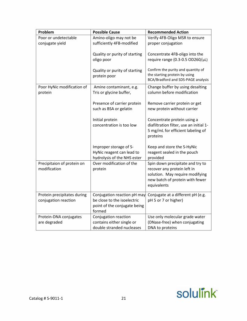
Problem Possible Cause Recommended Action
Poor or undetectableconjugate yield
Amino-oligo may not besufficiently 4FB-modified
Quality or purity of startingoligo poor
Quality or purity of startingprotein poor
Verify 4FB-Oligo MSR to ensureproper conjugation
Concentrate 4FB-oligo into therequire range (0.3-0.5 OD260/µL)
Confirm the purity and quantity ofthe starting protein by usingBCA/Bradford and SDS-PAGE analysis
Poor HyNic modification ofprotein
Amine contaminant, e.g.Tris or glycine buffer,
Presence of carrier proteinsuch as BSA or gelatin
Initial proteinconcentration is too low
Improper storage of S-HyNic reagent can lead tohydrolysis of the NHS ester
Change buffer by using desaltingcolumn before modification
Remove carrier protein or getnew protein without carrier
Concentrate protein using adiafiltration filter, use an initial 1-5 mg/mL for efficient labeling ofproteins
Keep and store the S-HyNicreagent sealed in the pouchprovided
Precipitaion of protein onmodification
Over modification of theprotein
Spin down precipitate and try torecover any protein left insolution. May require modifyingnew batch of protein with fewerequivalents
Protein precipitates duringconjugation reaction
Conjugation reaction pH maybe close to the isoelectricpoint of the conjugate beingformed
Conjugate at a different pH (e.g.pH 5 or 7 or higher)
Protein-DNA conjugatesare degraded
Conjugation reactioncontains either single ordouble stranded nucleases
Use only molecular grade water(DNase-free) when conjugatingDNA to proteins

Catalog # S-9011-1 22
XII. References1) Sano T, Smith CL, Cantor CR. Immuno-PCR: very sensitive antigen detection by
means of specific antibody-DNA conjugates. Science. 1992 258, 120-2.2) Fredriksson S, Gullberg M, Jarvius J, Olsson C, Pietras K, Gústafsdóttir SM, Ostman
A, Landegren U, Nat Biotechnol.. Protein detection using proximity-dependent DNAligation assays. 2002,20, 473-7.
3) Fredriksson S, Horecka J, Brustugun OT, Schlingemann J, Koong AC, Tibshirani Rand Davis RW, Multiplexed Proximity Ligation Assays to Profile Putative PlasmaBiomarkers Relevant to Pancreatic and Ovarian Cancer, Clinical Chemistry, 2008,54, 582-589.
4) Fredriksson S, Dixon W, Ji H, Koong AC, Mindrinos M and Davis RW, Multiplexedprotein detection by proximity ligation for cancer biomarker validation , NatureMethods 2007, 4, 327.
5) Jarvius M, Paulsson J, Weibrecht I, Leuchowius K-J, Andersson A-C, Wählby, MatsGullberg C, Botling J, Sjöblom T, Markova B, Östman A, Landegren U and SöderbergO, In situ detection of phosphorylated PDGF receptor β using a generalized proximity ligation method, Mol. And Cell. Proteomics 2007, 1500-9
6) Schallmeiner E, Oksanen E, Ericsson O, Spångberg L, Eriksson S, Stenman U-H,Pettersson K and Landegren U, Sensitive protein detection via triple-binderproximity ligation assays, Nature Methods 2007, 4, 135 – 137.
7) Kozlov IA, Melnyk PC, Stromsborg KE, Chee MS, Barker DL, Zhao C, Efficientstrategies for the conjugation of oligonucleotides to antibodies enabling highlysensitive protein detection, Biopolymers 2004, 73, 621.
8) Bailey RC, Kwong G, Radu CG, Witte ON, and Heath JR, DNA-Encoded AntibodyLibraries: A Unified Platform for Multiplexed Cell Sorting and Detection of Genesand Proteins, J. Amer. Chem. Soc. 2007, 129, 1959-1967.
9) Kattah MG, Coller J, Cheung RK, Oshidary N, Utz PJ, HIT: a versatile proteomicsplatform for multianalyte phenotyping of cytokines, intracellular proteins andsurface molecules. Nat Med. 2008 Nov;14, 1284-9.
10) Datta SK, Cho HJ, Takabayashi K, Horner AA, Raz E., Antigen- immunostimulatoryoligo-nucleotide conjugates: mechanisms and applications., Immunol Rev. 2004Jun;199:217-26.
11) Allergen-immunostimulatory oligodeoxynucleotide conjugate: a novel allergoid forimmunotherapy., Curr Opin Allergy Clin Immunol. 2002 2, 547-51.
12) Horner AA, Datta SK, Takabayashi K, Belyakov IM, Hayashi T, Cinman N, NguyenMD, Van Uden JH, Berzofsky JA, Richman DD, Raz E., Immunostimulatory DNA-based vaccines elicit multifaceted immune responses against HIV at systemic andmucosal sites., J Immunol. 2001, 167, 1584-91.