protein import, cholesterol transport and
TRANSCRIPT

ROLE OF PROTEIN-PROTEIN INTERACTIONS IN MITOCHONDRIAL
PROTEIN IMPORT, CHOLESTEROL TRANSPORT AND
STEROID BIOSYNTHESIS
A Dissertation submitted to the Faculty of the
Graduate School of Arts and Sciences of Georgetown University
in partial fulfillment of the requirement for the degree of
Doctor of Philosophy In Biochemistry and Molecular & Cellular Biology
By
Malena Beth Rone, B.S.
Washington, DC February 1, 2010

ii
Copyright 2010 by Malena Beth Rone
All Rights Reserved

iii
ROLE OF PROTEIN-PROTEIN INTERACTIONS IN MITOCHONDRIAL
PROTEIN IMPORT, CHOLESTEROL TRANSPORT AND
STEROID BIOSYNTHESIS
Malena Beth Rone, B.S.
Thesis Advisor: Vassilios Papadopoulos, DPharm, PhD.
ABSTRACT
Steroid synthesis is initiated by the transfer of cholesterol to the inner
membrane of the mitochondria where the conversion of cholesterol to pregnenolone
occurs through the C27 cholesterol side chain cleavage cytochrome P450 enzyme
(P450scc; CYP11A1). The rate of steroidogenesis is not regulated by the activity of
CYP11A1 but by the availability of substrate. As the process of trafficking
cholesterol to the mitochondria can occur through a series of cytosolic and
mitochondrial proteins and vesicular interactions, we propose that this delivery of
cholesterol into the mitochondria occurs through specific protein-protein interactions
that drive steroidogenesis.
This hypothesis was explored using MA-10 mouse Leydig cells, which undergo
steroidogenesis to produce progesterone. It was identified through cross-linking
studies that a protein complex formed at the outer mitochondrial membrane (OMM)
upon hormonal stimulation. This complex consisted of mitochondrial protein
translocator protein (TSPO, 18 kDa), Golgi protein PBR Associated Protein 7,

iv
(PAP7), cytosolic protein PKA-RIα, and mitochondrially targeted Steroidogenic
Acute Regulatory protein (StAR). TSPO assists with the translocation of cholesterol
from the OMM to the IMM and is an integral OMM protein; therefore, we decided to
study the mechanisms of its import and integration into the OMM. We identified
that the C-terminus and amino acids #103-109 functioned in targeting TSPO to the
mitochondria through the assistance of heat shock proteins (HSP). Translocase of
Outer Mitochondrial (TOM) Membrane protein Tom70 interacted with the HSP’s in
an ATP-dependant manner to import TSPO with the aid of theMetaxin1. From these
findings, we went on to study the protein-protein interactions formed by TSPO in the
mitochondria. Upon hCG treatment TSPO undergoes polymerization, identification
of a shift in TSPO molecular weight and proteins that associate with TSPO was
observed through Blue Native-PAGE and 2D-SDS PAGE immunoblot (western)
analysis after hCG treatment in MA-10 cells. From this we identified a BN-PAGE
shift in CYP11A1 from a lower to a higher molecular weight complex, suggesting it
interacts specifically with the polymerized TSPO, forming of a steroidogenic protein
complex.
In summary, these studies demonstrate that delivery of cholesterol into the
mitochondria and thus steroidogenesis are driven by a series of protein-protein
interactions.

v
ACKNOWLEDGEMENTS
As another wise Missourian once said, “Keep away from people who try to
belittle your ambitions. Small people always do that, but the really great make you
feel that you, too, can become great.” I would like to think my thesis mentor Dr.
Vassilios Papadopoulos for always making me feel that I can become, if I was not
already, great. If it was not for his guidance, support and understanding this thesis
would not have been possible.
I have a world of gratitude for all the members of the Papadopoulos/Culty
Lab, both at Georgetown and Montreal, for all they have taught me. Many special
thanks go to Dr. Jun Lui, for his training and scientific advice I will always be
grateful. .
Thanks and gratitude goes out to my thesis committee members, Dr. Elliot
Crooke, Dr. Paul Roepe, Dr. Richard Youle, and Dr. Jason Young for all of their
advice and guidance during these past seven years.
Words alone are not possibly enough to thank all my family and friends. To
my parents, Lewis and Martha, thank you for your continued love and support.
Without the values and morals you instilled in me I could not have become the
person I am today. To Justin, Rebecca and Hannah, they say siblings are mirrors of
your possibilities, thank you for reflecting so brightly. Many thanks go to Tyler,
Boxie and Val, for their friendship and support both then and now. And to Andrew,
thank you for helping me become not only a better scientist but also a better person.

vi
TABLE OF CONTENTS
Title Page Abstract ............................................................................................ iii Acknowledgements ........................................................................... v Table of Contents ............................................................................. vi List of Abbreviations ..................................................................... viii List of Figures and Tables ............................................................. xii 1. Introduction .................................................................................. 1
1.1. Cholesterol and Steroid Synthesis ....................................................... 1 1.2. Cholesterol Sources in the Cell ............................................................ 2
1.2.1. ER Cholesterol ........................................................................... 3 1.2.2. Plasma Membrane ...................................................................... 6 1.2.3. Lipid Droplets .......................................................................... 10
1.3. Targeting Cholesterol to the mitochondria ......................................... 11 1.3.1. Sterol Carrier Protein-2 ............................................................ 12 1.3.2. START Domain Protein ........................................................... 12
1.4. Importing Cholesterol into the Mitochondria .................................... 18 1.4.1. TSPO and Cholesterol .............................................................. 19 1.4.2. TSPO and Mitochondrial Proteins ........................................... 25
1.5. TSPO and Cytosolic Cholesterol Import ............................................ 27 1.5.1. TSPO and StAR ....................................................................... 27 1.5.2. TSPO and PAP7 ....................................................................... 29
1.6. Aims .................................................................................................... 33 2. Chapter 1. Protein-Protein Interactions Mediate Mitochondrial
Cholesterol Transport and Steroid Biosynthesis ....................... 34
2.1. Abstract ............................................................................................... 35 2.2. Introduction ......................................................................................... 36
vii
2.3. Material and Methods ......................................................................... 39 2.4. Results ................................................................................................. 41 2.5. Discussion ........................................................................................... 44
3. Chapter 2: Targeting and Insertion of the Cholesterol-Binding
Translocator Protein into the Outer Mitochondrial Membrane ................................................................................. 46
3.1. Abstract ............................................................................................... 47 3.2. Introduction ......................................................................................... 49 3.3. Material and Methods ......................................................................... 53 3.4. Results ................................................................................................. 61 3.5. Discussion ........................................................................................... 73
4. Chapter 3: Hormonal Induction of Mitochondrial Protein Complexes
and Steroid Formation in MA-10 Leydig Cells. ....................... 77
4.1. Abstract ............................................................................................... 78 4.2. Introduction ......................................................................................... 80 4.3. Material and Methods ......................................................................... 84 4.4. Results ................................................................................................. 90 4.5. Discussion ........................................................................................... 94
5. Summary and Conclusions ........................................................ 99 6. Tables/Figures ......................................................................... 110 7. References................................................................................ 151
viii
List of Abbreviations ACAT Acyl-coenzyme A:cholesterol acyltransferase
ACBD Acyl-coenzyme A binding domain containing
protein
ACTH Adrenocorticotropic hormone
AdR Adrenodoxin Reductase
Adx Adrenodoxin
AKAP A kinase anchoring proteins
ANT Anion Nucleotide Transporter
ANOVA Analysis of Variance
ATG Autophagy Related Proteins
Bal-A1 Balfiomycin A1
BN-PAGE Blue-native polyacrylamide gel electrophoresis
BRET Bioluminescence resonance energy transfer
cAMP cyclic AMP
CRAC Cholesterol-recognition amino acid consensus
CYP11A1 C27 cholesterol side chain cleavage cytochrome
p450 enzyme
DBI Diazepam Binding Inhibitor
DGAT2 Acyl CoA:diacylglycerol acyltransferase
DMEM Dulbecco’s Modified Eagle’s Medium

ix
DMEM/Ham’s F-12 Dulbecco’s Modified Eagle’s medium Ham’s F-12
ER Endoplasmic Reticulum
FBS Fetal Bovine Serum
FRET Förster resonance energy transfer
FSH Follicle-stimulating hormone
GD Geldanamycin
hCG Human chorionic gonadotropin
HDL High Density Lipoprotein
HMGR HMG-CoA reductase
HSL Hormone-sensitive lipase
Hsp Heat Shock Protein
IMM Inner mitochondrial membrane
LD Lipid Droplets
LDL Low Density Lipoprotein
LH Lutentizing Hormone
MAM Mitochondria associated membrane
MLN64 Metastatic lymph node 64 protein
MPTP Mitochondria permeability transition pore
MS Mass Spectrometry
NADP Nicotinamide adenine dinucleotide phosphate
NB Novobiocin

x
NMR Nuclear Magnetic Resonance
NPC Niemann-Pick disease, type C 1
ODNs Oligodeoxynucleotides
OMM Outer mitochondrial membrane
PAP7 PBR associated protein 7
PBR Peripheral-type Benzodiazepine Receptor
PBS Phosphate buffered saline
PI(3)K Phosphoinositide 3-kinase
PKA cAMP-dependent protein kinase
PVDF Polyvinylidene fluoride
RIα Regulatory subunit isoforms Iα and Iβ, IIα and IIβ
RIA Radioimmunoassay
ROS Reactive Oxygen Species
SBD Sterol-binding domain
SCP-2 Sterol Carrier Protein-2
SR-BI Scavenger Receptors Class B Type 1
SREBP Sterol regulatory element binding protein
StAR Steroidogenic Acute Regulatory protein
START Steroidogenic acute regulatory (StAR)-related lipid
transfer protein
TNN Triakontatetraneuropetide

xi
TOM Translocase of outer mitochondrial membrane
TPR Tetratriacopeptide Repeat
TSPO Translocator protein (18 kDa)
SAM Sorting and assembly machinery complex
SDS-PAGE Sodium Dodecyl Sulfate Polyacrylamide Gel
Electrophoresis
VDAC Voltage-dependant anion channel
WT Wortmannin
3-MA 3-methyladenine

xii
List of Figures and Tables
Figure 1. Trafficking of cholesterol to the mitochondria for
Steroidogenesis ............................................................................................... 110
Figure 2. Cholesterol-binding domains of StAR and TSPO ......................... 112
Figure 3. Cholesterol-binding domains of StAR and TSPO ......................... 116
Figure 4: Protein-protein interactions at the OMM ........................................ 118
Figure 5: Photo-crosslinking amino acids ..................................................... 120
Figure 6: Protein-protein interactions identified by cross-linking ................. 122
Figure 7: Identification of TSPO import complexes in the OMM ................. 124
Figure 8: TSPO import is dependent upon heat shock proteins and ATP for
import ...................................................................................................... 126
Figure 9: Identification of the TSPO amino acid sequence(s) responsible for
targeting the protein to the OMM................................................................... 128
Figure 10: TSPO protein regions and amino acids required for insertion
into the OMM. ................................................................................................ 130
Figure 11: TSPO import is dependent on Metaxin 1 .................................... 132
Figure 12: TSPO import is increased with cAMP stimulated mitochondria .. 134
Figure 13: TSPO import into the OMM ........................................................ 136
Figure 14: Blue-Native Page/2D SDS PAGE Gels identify
protein complexes .......................................................................................... 138
Figure 15: Cholesterol binding in BN-PAGE ................................................ 140

xiii
Figure 15: CYP11A1 probe is functional in determining activity in whole cells and
isolated mitochondria ..................................................................................... 142
Figure 17: CYP11A1 probe activity in the BN-PAGE gel ........................... 144
Table 1: Primers used for Q-PCR analysis ..................................................... 146 Table 2: Identification of proteins present in the 66- and 800 kDa mitochondrial
protein complexes by mass spectrometry in HeLa cells ................................ 147
Table 3: Identification of proteins present in the 66- and 800 kDa
mitochondrial protein complexes in control and treated MA-10 cells by mass
spectrometry ................................................................................................... 149
1
Introduction:
Cholesterol and Steroid Synthesis
Cholesterol is the sole precursor of steroids. Steroid synthesis is initiated at
the inner mitochondrial membrane (IMM), where the cytochrome P450 cholesterol
side chain cleavage enzyme (CYP11A1) catalyzes the conversion of cholesterol to
pregnenolone (1). Pregnenolone then enters the endoplasmic reticulum (ER) where
further enzymatic reactions occur to produce the final steroid products. It has been
shown that the translocation of cholesterol from the outer mitochondrial membrane
(OMM) to the IMM is the rate-limiting step in the production of all steroids (2;3).
Therefore, the ability of cholesterol to move into mitochondria to be available to
CYP11A1 determines the efficiency of steroid production.
The production of steroids is regulated by trophic hormones, specifically, the
adrenocorticotropic hormone (ACTH) in adrenocortical cells and luteinizing
hormone (LH) in testicular Leydig and ovarian cells (2;3). The presence of these
hormones activates the G protein-coupled receptors, which release the stimulatory
subunit, resulting in the activation of adenylyl cyclase and a rise in intracellular
cAMP (4). This increase in cAMP leads to an increase in lipid synthesis, protein
synthesis, and protein phosphorylation. All these process have been shown to play a
role in steroidogenesis and assist with cholesterol trafficking to the mitochondria.
Mitochondria are relatively cholesterol-poor organelles, with the majority of
cholesterol located in the OMM. In the mitochondria of steroidogenic cells the pool

2
of cholesterol available for steroidogenesis is segregated from the structural
membrane cholesterol and is bound to the cholesterol-binding domain of the
translocator protein (18 kDa, TSPO), formerly called the peripheral-type
benzodiazepine receptor (PBR) (5). It is from this site that cholesterol is released
under hormonal stimulation to move to the matrix side of the IMM, where the
cholesterol side chain cleavage enzyme CYP11A1, which will metabolize
cholesterol to pregnenolone, is located. This pathway will be discussed in detail later.
As the initial translocation of cholesterol from TSPO is not sufficient to
sustain the continuous production of elevated concentrations of steroids, additional
free cholesterol must be moved from intracellular stores to the mitochondria. This
intracellular cholesterol is known to come from three sources: i) de novo synthesis of
cholesterol in the endoplasmic reticulum (ER) ii) mobilization of cholesterol in the
plasma membrane with further uptake of circulating cholesterol esters from receptors
found on the plasma membrane and iii) mobilization of cholesterol in lipid droplets
(LD) (Fig. 1).
Cholesterol sources in the cell
With 65% to 80% of the total cellular cholesterol located in the plasma
membrane, comprising 20% to 25% of the total lipids present, cholesterol plays a
significant role in the structure and function of the plasma membrane. These
interactions affect the organization of proteins and lipids in the membrane, alter the
permeability of the membrane, and initiate the formation of lipid rafts (6). The

3
second highest concentration of cholesterol is found in the endosomal pathway, with
the majority found in the endosomal to trans-Golgi compartment (6). While the
majority of total cholesterol found in the cell is located in the plasma membrane, the
ER contains only 1% to 2% of the total cell cholesterol (7). This gradient provides a
mechanism for transport of cholesterol inside the cell from the ER to the plasma
membrane and allows it to be recycled back (8).
ER Cholesterol
Every cell in the body is capable of producing cholesterol, with
endogenous cholesterol production occurring at high rates in the liver and the brain
(9). Steroidogenic organs besides the brain, such as the adrenals, testis and ovaries,
are also known to synthesize cholesterol at high rates. The cholesterol synthesis
pathway was initially proposed to use acetate (Fig. 1A) upon the observation that
ingested deuterated acetic acid leads to the formation of deuterated acetic acid
cholesterol. This led to the proposal that intermediates of isoprene units to form
cholesterol (Fig 1B) (10). This model was confirmed when it was shown that two
molecules of acetyl-CoA condense to form acetoacetyl-CoA, another molecule of
acetyl-CoA then combines to form hydroxymethyglutaryl-CoA (HMG-CoA) a
reaction mediated by HMG-CoA synthase (Fig 1C) (11;12). The reduction of HMG-
CoA to mevalonate, the rate-limiting step in cholesterol synthesis, is catalyzed by the
enzyme HMG-CoA reductase (HMGR) (13). HMGR is anchored to the ER, an
organelle which functions as the cholesterol sensor of the cell. The ER regulates

4
endogenous cholesterol production primarily through the sterol regulatory element
binding protein (SREPB) complex (14;15). The SREBP proteins are translocated to
the Golgi upon a decrease in cholesterol and upon arrival SREBP is cleaved by two
proteases. The resulting N-terminus is an active transcription factor that translocates
to the nucleus. This results in an increase in the activity of cholesterol transcription
genes, including HMGR (16). As HMGR is the rate-limiting enzyme in cholesterol
synthesis, it is easily degraded under the high sterol conditions and can be inhibited
in low levels of ATP (17). HMGR is upregulated in the presence of hormones,
resulting in an increase in cholesterol production under hormonal stimulation (14).
The increase of cholesterol via this pathway though has not been shown to play a
primary role in steroid production (18).
After the irreversible step of mevalonate synthesis is complete, the mevalonate
is then converted into two isopenoid intermediates, isopentenyl pyrophosphate (IPP)
and dimethylallyl pyrophosphate (Fig 1D). Pyrophphosphomevalonate
decarboxylase catalyzes the formation of IPP from mevalonate requiring both
NADPH and ATP. IPP can be converted into dimethylallyl pyrophosphate by
isopentenyl pyrophosphate isomerase, these two compounds are then used to
generate geranyl pyrophosphate combined head to tail by prenyl transferase (Fig.
1D) (12). Addition of second IPP is added in a head to tail condensation by prenyl
transferase to the geranyl pyrophosphate to yield farnesyl pyrophosphate. These
reactions occurring to produce farnesyl pyrophosphate from mevalonate have been
5
shown to occur both in the peroxisomes and the cytosol(19). The formation of
squalene occurs with the head to head addition of two farnesyl pyrophosphates by
squalene synthase. This generation of squalene is actually the first committed step
for the synthesis of cholesterol, as previously the farnesyl pyrophosphate could be
used to farnesylate proteins and form ubiquinone amongst many other cellular
functions (20). The linearized squalene figure in Fig 1D shows the individual
additions of carbon from the isopenoid intermediates present in Figure 1D.
From the formation squalene (represented in circular form in Fig 1E) the
conversion to lanosterol proceeds via an oxygen-dependent epoxidation to give 2,3-
epoxysqualene, this reaction is catalyzed by squalene exposidase. This molecule then
undergoes enzymatic cycilization to yield lanosterol by 2,3-Oxidosqualene:lanosterol
cyclase (OSC) (Fig 1E) (21). The activity of OSC, whose expression is also
regulated by SREPBs, yields intermediates that control the liver X receptor (LXR).
The activity of the LXR affects the regulation of cholesterol efflux into the cell,
though which inhibition of OSC has shown to lower LDL present in the blood
stream, providing a new potential drug to suppress atherogenesis (21). The
conversion of lanosterol is a nineteen step process that requires both oxygen and
NADPH and release three methyl groups to produce the final product of cholesterol
(Fig. 1 E) (12).
As cholesterol synthesis is tightly regulated in the ER during hormonal
stimulation, cholesterol transport out of the ER is tightly controlled as well.

6
Cholesterol flux from the ER can occur through many pathways, including cytosolic
lipid transfer proteins, through intracellular compartments or passive diffusion
through contact sites. Contact sites are common between the ER and other
intracellular organelles, facilitating cholesterol flux out of the ER. ER-mitochondrial
contact sites have been identified in which mitochondria-associated membranes
(MAM) cluster with stacks of ER (22). The cholesterol generated in the ER could
potentially use this pathway for receiving steroidogenic cholesterol (Fig. 2, Pathway
1). Recently an ER protein, acyl CoA:diacylglycerol acyltransferase 2 (DGAT2)
was found associated with both LD and the mitochondria while still present in the
ER. As DGAT2 functions in the final stage of triglycerol synthesis it is possible that
this is a pathway for lipid transfer (23). Currently it is unknown whether an
interaction between the ER and mitochondria occurs in steroidogenic cells; therefore
further studies will be necessary to investigate the presence and function of such an
interaction in steroidogenesis.
Plasma Membrane
Cholesterol is primarily stored in the plasma membrane. Upon hormonal
stimulation there is increased cholesterol absorption through the plasma membrane.
When cholesterol is imported into the cell via the plasma membrane it greatly
increases the cholesterol content stored elsewhere in the cell. This was observed
when an increase of 50% in cellular cholesterol absorbed via the plasma membrane
resulted in a 10-fold increase in ER cholesterol (24). Currently, there are two known

7
pathways for this cholesterol absorption and import into the cell: a non-selective
endocytic pathway and a “selective” absorption pathway. In the non-selective
pathway LDL molecules are specifically bound and internalized via the LDL
receptors. Once the receptor has been internalized it fuses with the endosomal
pathway for distribution of the lipoproteins (Fig. 1, Pathway 2). The “selective”
pathway uses scavenger receptors class B type I (SR-BI) located at the plasma
membrane to bind both LDL and HDL. Through local binding mechanisms the
cholesterol present in the lipoproteins is transferred directly to the cell membrane
without absorption of the lipoprotein particles (25) (Fig. 1, Pathway 3). Further
analysis of these two pathways has shown that adrenal steroidogenesis is dependent
primarily on HDL cholesterol absorbed from the plasma membrane, primarily via the
SR-BI pathway (26).
The non-selective vesicular pathway is initiated primarily by LDL particles
binding to the LDL receptor, followed by the endocytosis and budding of clathrin-
coated pits into the cytoplasm (27) (Fig 1, Pathway 2). These vesicles fuse with
early endosomes, releasing their clathrin coats and allowing the LDL receptors to
cycle back to the membrane. This endosomal fusion and trafficking occur through
interactions with microtubules which are controlled through Rabs (28). Rabs are
small GTPases that regulate membrane traffic through binding at their active site,
currently there have been more then 60 proteins identified in mammalian cells
(29;30). The early endosomes bind to recycling endosomes coordinated by Rab5,

8
and then to late endosomes via Rab 7, to further distribute cholesterol throughout the
cell (29;31). The endosomes also undergo a decrease in pH from 7.4 at the plasma
membrane to 5.5 - 6 at the late endosome (32). This decrease in pH helps further
dissolve the absorbed lipoprotein and prepares the late endosomes to fuse with the
lysosomes.
It has been shown that the LDL receptor is not necessary for acute adrenal
steroidogenesis, suggesting that cholesterol absorbed via this pathway is not
necessarily used for steroidogenesis (33). However, in FSH- or FSH plus
androstenedione-treated granulosa cells the rate of LDL receptor absorption
increases while the time needed for the LDL to reach the lysosome decreases
compared to non-hormone-treated cells suggesting this pathway is used for
steroidogenesis (34). As endosomes contain a large percentage of the cytosolic
cholesterol present in the cell; they can function in the trafficking of intracellular
cholesterol to the mitochondria without first absorbing cholesterol from the plasma
membrane. This pathway could occur specifically through the cholesterol rich late
endosomes, shown to fuse with the lysosomes and the Golgi apparatus and
transiently interacting with the mitochondria, thus allowing for multiple sources of
cholesterol to be available to the mitochondria. Endosomal trafficking in the cell has
also been shown to be altered by cholesterol concentrations, specifically via Rab7,
suggesting a mechanism by which trafficking to the mitochondria could be regulated
(35).

9
The second pathway identified for cholesterol absorption and trafficking in
steroidogenic cells occurs through the action of the SR-BI receptor (36) (Fig 1,
Pathway 3). The SR-BI receptor is found in many tissues such as intestines,
macrophages, and endothelial cells, though it is expressed in highest concentrations
in steroidogenic tissues such as the adrenals, ovary, and testis (25). Unlike the LDL
receptor in which the apoprotein is absorbed, the SR-BI receptor forms a non-
aqueous channel that allows a large influx of cholesterol directly into the plasma
membrane (25). This non-aqueous channel has also been shown to be regulated in
the intestines through other proteins found in the plasma membrane, such as CD36
and other proteins yet to be identified (37). Suggesting this pathway might require a
complex, multiprotein interaction to regulate cholesterol absorption into the plasma
membrane. Because SR-BI absorbs free cholesterol from HDL and stores it in the
plasma membrane, this cholesterol can move spontaneously between bilayers and
membranes in the cell without the assistance of any proteins. This process is slow
and therefore not suggested as a pathway involved in the acute stimulation of
steroidogenesis (38). The esterified cholesterol absorbed from the SR-BI receptor
must be converted to free cholesterol before it can be used for steroidogenesis via a
cholesterol ester hydrolase (39). In steroidogenic cells cholesterol ester hydrolysis
is performed through hormone-sensitive lipase (HSL) (40). HSL becomes activated
when phosphorylated by cAMP; inhibition of HSL results in decreased
steroidogenesis in the adrenals and inhibits sperm production in the testis (41-43).
10
The cholesterol absorbed via the plasma membrane has been shown to be hydrolyzed
rapidly, presumably close to the plasma membrane, to form free cholesterol. Once
the cholesterol esters have undergone hydrolysis, the HSL can interact with various
cholesterol-binding proteins to direct the cholesterol to the OMM for steroidogenesis
(44). This pathway involving cholesterol-binding proteins will be discussed later in
detail.
Lipid Droplets
Lipid droplets (LD) are bounded by a phospholipid membrane and function as a
repository of cholesterol esters and triglycerides in the cell. It has been proposed that
LD form from the ER when excess neutral lipids bud off, although there is no direct
evidence to support this model (45). The cholesterol esters found in the LD are the
products of the ER enzyme acyl-coenzyme A:cholesterol acyltransferase (ACAT),
which becomes active in the presence of high levels of cholesterol. This enzyme
attaches an ester to the free cholesterol found in the ER, increasing the cholesterol
ester content present in the cell (46). Because lipids in the LD can be used for
various biological activities their size fluctuates depending upon the cell’s activity.
In steroidogenic cells, LDs are small to increase the surface area for lipid retrieval
(45).
The cholesterol esters present in the LD are also converted to free cholesterol in
the same manner as cholesterol absorbed via the SR-BI receptor, i.e., through HSL.
Transfer of steroidogenic cholesterol from intracellular organelles to the

11
mitochondria is thought to occur through cholesterol-binding proteins found in the
cytosol (Fig 1, Pathway 4) (47). Other mechanisms for lipid transport from the LD
have been demonstrated. Rab5, which localizes to early endosomes, has been shown
to interact with LD, suggesting a mechanism for cholesterol transfer from the LD to
the endosome and vice versa, allowing for an increase in cholesterol in the
endosomal pathway (48). Rab18 also associates with LD, regulating the contact
between the lipid droplet and the ER, which controls the flux of cholesterol during
lipolysis (49). Since LDs play an important role in regulating intracellular
cholesterol through storage, trafficking, and esterification, further studies are needed
to determine the endosome/LD interaction which would allow for transfer or fusion
of the cholesterol from the LDs into the early endosomal pathway for
steroidogenesis.
Targeting cholesterol to the mitochondria
The primary pathway for targeting cholesterol to the mitochondria has not
been definitively identified. Two pathways have been proposed: (i) the non-
vesicular pathway involving cholesterol-binding proteins transferring cholesterol
through the cytosol to the mitochondria, and (ii) a vesicular pathway characterized
by an increase in the fusion of vesicular membranes, such as endosomes and
lysosomes, which results in an increase in cholesterol targeted to the mitochondria
(50). An overlap between these two pathways is highly likely as cholesterol binding
proteins have been found on endosomes; this suggests that an increase in cholesterol
12
targeting to endosomes could also have a direct effect on cholesterol-binding
proteins which target cholesterol for transfer to the mitochondria.
Sterol carrier protein-2
Sterol carrier protein-2 (SCP-2) was one of the initial cholesterol binding
proteins identified; SCP-2 was shown to play a role in the intracellular transfer of
cholesterol, including from the lysosomal to mitochondrial membranes (51;52).
SCP-2 is found in tissues involved in cholesterol trafficking and oxidation, such as
the liver, intestines, adrenal, testis, and ovary, suggesting it could play a role in
steroidogenesis (53). Further studies showed SCP-2 increases cholesterol uptake and
transport throughout the cell while inhibiting the efflux of cholesterol from the cell
through the HDL receptor (54;55). Because no alteration in steroidogenesis was
observed in SCP-2 knock-out mice, it has been assumed that SCP-2 does not play a
primary role in steroidogenesis in vivo, though other interactions are still possible
(56). This focused attention on other identified cholesterol binding/transfer proteins.
START domain proteins
The steroidogenic acute regulatory (StAR)-related lipid transfer (START)
domain is an amino acid motif that has been proposed to play a role in cholesterol
and lipid binding (57) (Fig. 3A). The motif first was identified in the StAR protein,
discussed below, which has been shown to play a role in cholesterol transport to the
mitochondria for steroidogenesis. The 210-amino-acid sequence forms a beta sheet
core surrounded by two alpha helices, resulting in a hydrophobic channel that can

13
hold a sterol molecule capped by the C-terminal alpha helix (47). The structure
becomes stable upon binding of the cholesterol molecule, which has been shown to
bind at a 1:1 ratio (58).
MLN64, the second identified StART domain protein, was shown to be
upregulated in breast and ovarian cancers and found to have a C-terminal domain
with 37% amino acid identity and 50% amino acid similarity to the C-terminal
domain of StAR (59;60). Further studies confirmed that this was a START domain
that could bind cholesterol and transfer free cholesterol from sterol-rich vesicles to
acceptor membranes. MLN64 was found to be localized primarily on late
endosomes, integrating at the plasma membrane and functioning in the vesicular
trafficking of LDL cholesterol (61) (Fig 1). When the START domain was removed
from MLN64, cholesterol accumulated in the lysosomes and altered late endosome
trafficking (62). In COS-F2 cells, this accumulation of cholesterol suppressed
steroidogenesis, presumably by limiting the efflux of free cholesterol from the late
endosomes and lysosomes to the mitochondria.
MLN64 was also found to be associated with Niemann-Pick type C disease
protein 1 (NPC1) in the late endosomes (63). Niemann-Pick type C disease (NPC) is
a disorder characterized by the accumulation of LDL-derived unesterified cholesterol
in the late endosomes and lysosomes caused by mutations in either NPC1 or NPC2
(64). NPC1 gene expression has been shown to be responsive to cAMP and essential
for the normal development of the adrenals (65). NPC1 is found bound to late

14
endosomal membranes while NPC2 is found inside the late endosomes, early
lysosomes, and in the cytosol. The presence of NPC2 accelerates the transfer of free
cholesterol from late endosomes to lysosomes and cholesterol efflux to the plasma
membrane (66;67). NPC1 is necessary for cholesterol efflux from the late lysosome
for use in the cell (32). When steroidogenic human granulosa-lutein cells deficient
in NPC1 protein were used for studying cholesterol trafficking, steroidogenesis
levels decreased to levels seen with LDL-deficient media (68). This observation
suggests that the NPC1/late endosome pathway is used primarily for LDL-derived
steroidogenesis. As NPC1 and NPC2 function in the efflux of cholesterol from the
endosome, this pathway could also interact with MLN-64 for steroidogenic purposes.
Steroidogenic acute regulatory (StAR) protein was first identified due to its
rapid phosphorylation and protein expression soon after the addition of hormones
and cAMP in steroidogenic cells (69-71). Expression was confirmed in the adrenal
cortex, testis, and ovary and later in the brain and placenta, suggesting a connection
between StAR protein expression and steroid production (72;73). Transfection of
StAR expression vectors in both mouse Leydig MA-10 cells and COS F2 cells,
which contain the components of CYP11A1, was found to increase steroidogenesis
(74;75).
There have been many models proposed for the function of StAR in
steroidogenesis (76). It was suggested early that StAR assists with the transfer of
cholesterol to the mitochondria, although no clear mechanism was identified.

15
Because StAR contains an N-terminal mitochondrial-targeting sequence, which is
cleaved from its active molecular weight of 37 kDa to its inactive weight of 30 kDa
upon import into the mitochondrion, it was proposed that this targeting sequence
could assist with the formation of a mitochondrial “contact site”. This would be
accomplished by the interaction of StAR with mitochondrial protein import
complexes found on both the OMM and IMM and allow the N-terminus to form a
linker connecting the membranes. Cholesterol would then be able to flow from the
OMM to the IMM and interact with CYP11A1. However, removal of the N-
terminus targeting sequence in the construct N-62 StAR and transfection into
steroidogenic cells had no effect on steroidogenesis, suggesting this is not the
mechanism by which StAR facilitates cholesterol transfer (77). It was later
observed that N-62 StAR was able to insert cholesterol into cytosolic membranes
other than the mitochondria, suggesting that the primary function of the N-terminal
sequence is not to form mitochondrial contact sites but to limit StAR’s cholesterol
targeting abilities solely to the OMM (78).
The effect of StAR on mitochondrial steroidogenesis was further confirmed
by fusing mitochondrial translocases to the N-62 StAR construct. Fusion of N-62
StAR with Tim9, a mitochondrial inner membrane space protein, or Tim40, an IMM
protein, resulted in no increase in steroidogenesis while fusion of N-62 StAR with
Tom20, an OMM protein, resulted in maximal production of steroids (79). This
observation demonstrated that StAR’s site of action was confined to the OMM and

16
that, once imported, StAR does not stimulate steroidogenesis. Because StAR
interacts only transiently with the OMM before being imported, the amount of time
StAR spends at the OMM would be able to alter the rate of steroid production. This
was shown when a StAR/StAR construct, which is imported at a slower rate than
wild-type StAR, was shown to increase pregnenolone production over both wild-type
and N-62 StAR construct levels in COS-F2 cells (79). This finding shows that StAR
functions primarily at the OMM, possibly activating a pathway of cholesterol
transport for steroidogenesis.
Another proposed model built to explain StAR’s activity suggested that it
could function as an intermembrane cholesterol shuttle, moving cholesterol from the
outer to the inner mitochondria membrane one molecule at a time during StAR’s
import (58). Tsujishita and Hurley proposed that during StAR’s import into the
mitochondria several molecules of cholesterol could be transferred through the
import of one StAR molecule through transient openings. Several issues were raised
with the model, including the lack of understanding of how StAR could both bind
cholesterol and then release it in the IMM space. It was also uncertain how StAR
would reside in the IMM space as it does not have a mitochondria targeting sequence
for the inner mitochondria membrane space.
One of the more current proposed models is the “molten globule” model
identified through the studying of the StAR’s C-terminus. Removal of the last 10 C-
terminal amino acids resulted in decreased steroidogenesis, while removal of the 28

17
C-terminal amino acids resulted in a biologically inactive protein, suggesting that
StAR’s mode of action was occurring at the C-terminus. Further studies showed that
the C-terminus forms a sterol-binding domain (SBD) with which a cholesterol
molecule is proposed to interact. The SBD forms a pocket that prevents the release
of the bound cholesterol molecule, which can occur only following a conformational
change. This is proposed to occur through the “molten globule” configuration in
which tertiary structures are removed, allowing the remaining secondary structures to
undergo a conformational shift and allow the cholesterol molecule to enter the
mitochondria. The molten globule model has been proposed to occur through the
protonation of the C-terminus at the OMM. Since binding of cholesterol to the SBD
in the StAR protein has not been definitively demonstrated, it is still not clear if this
is the mechanism by which cholesterol is transferred to the OMM. It should be
noted that the cholesterol-binding activity of StAR is independent of its activity at
the OMM, because StAR mutant R182L can still function in the binding and transfer
of cholesterol in isolated liposomes, although this results in an increase in cholesterol
present in the cell (80). This suggests that cholesterol binding is necessary but not
sufficient for StAR’s function on the OMM.
Further homology modeling and biophysical studies recently indicated the
existence of a two-state model (81). The first and open state of the model proposes
that the C-terminal alpha-helix 4 of StAR, acting primarily as a gating mechanism to
the cholesterol binding site, undergoes partial unfolding allowing cholesterol to bind.

18
This resulting cholesterol bound state, in theory, would lead to the stabilization and
the refolding of alpha-helix 4; resulting in a well-defined tertiary structure (81). This
stable tertiary structure could be necessary for protein-protein interactions formed for
cholesterol transfer at the surface of the mitochondria, suggesting that both StAR
conformation and cholesterol binding are necessary for its proper steroidogenic
activity.
These models of StAR’s function have been able to provide many
descriptions of cholesterol transport to the mitochondria which can relate to the
function of START proteins. As six other START-domain families have been
identified, such as StarD4, StarD5, and StarD6, the understanding of how the
START domains function can be applied to these proteins as well (82). This can
have far reaching effects, as compared to StAR and MLN64; StarD4, StarD5, and
StarD6 lack an organelle-targeting sequence and therefore are thought to be
cytoplasmic. StarD4 and StarD5 are widely expressed, while StarD6 is found
primarily in the testis (57). This ability of the START domain to target and transport
cholesterol from multiple sources, including the plasma membrane, ER, and
endosomes, could ensure a large source of cholesterol for steroidogenesis.
Importing Cholesterol into the Mitochondria
Cholesterol successfully imported via the plasma membrane or accessed in
LDs and transported to the OMM remains segregated in the OMM until translocation

19
to the IMM. This, the rate-limiting step in steroidogenesis, has been suggested to
occur primarily through TSPO.
TSPO and cholesterol
TSPO was first identified by the presence of radiolabeled diazepam binding in the
kidney (83) and it was later found to be present in most tissues of the body (84). It
was proposed that TSPO plays a role in steroidogenesis when ligand-binding studies
revealed increased expression of TSPO in steroidogenic tissues and subcellular
localization studies indicated that it was primarily localized to the OMM (84-87).
TSPO was recognized using a benzodiazepine drug ligand specific for the
GABAA receptor in the central nervous system; however, subsequent identification
of TSPO-specific ligands allowed it to be differentiated from the GABAA receptor.
This was accomplished by use of the isoquinoline carboxamide PK 11195, which
binds with nanomolar affinity to TSPO but has no affinity for the GABAA receptor
(88). Many endogenous TSPO ligands exist in the cell, with porphyrins being able
to bind TSPO with high nanomolar affinity (89). The diazepam binding inhibitor
(DBI) is another endogenous ligand. This 10 kDa protein, which also binds the
GABAA receptor with low affinity, is expressed in many tissues but is primarily
expressed in steroidogenic tissues where it is localized in the cytosol in contact with
the OMM (90). Naturally processed peptides of DBI, octadecaneuropeptide (ODN,
DBI33-50) and triakontatetraneuropetide (TNN, DBI17-50), expressed in a hormone-
dependent manner, were functional in binding TSPO in the brain, adrenal, and testis
20
(83;91). DBI and its peptides stimulated mitochondrial pregnenolone formation.
When DBI expression was suppressed in the presence of antisense oligonucleotides
MA-10 Leydig cells failed to respond to hormonal stimulation and steroid production
was inhibited.
Early experiments in multiple steroidogenic cell systems showed that
pregnenolone production was stimulated upon exposure of the cells to TSPO ligands
(92;93). When these experiments were repeated in isolated mitochondria incubated
with TSPO ligands a similar increase in pregnenolone was observed (85;93). This
increase was not seen in mitoplasts, mitochondria devoid of their OMM and
therefore deficient in TSPO. To determine the effect of TSPO ligands on the
mitochondria, cholesterol content in the OMM and the IMM was measured both
before and after TSPO ligand treatment (94). This study revealed that ligand
binding to TSPO induced the translocation of cholesterol from the OMM to the IMM
and confirmed that TSPO participates in the binding and release of cholesterol at the
OMM, an initial step in the production of steroids.
To further confirm TSPO’s role in cholesterol binding and translocation a
bacterial expression system was devised because bacteria contain no endogenous
cholesterol. E. coli were transformed with an inducible mouse cDNA TSPO vector,
resulting in fully expressed TSPO. Ligand-binding experiments were performed to
verify that the bacterial TSPO possessed the same pharmacological binding
properties as native TSPO; this was confirmed when bacterial TSPO bound both

21
cholesterol and PK 11195 with nanomolar affinities (95). When bacterial TSPO
was incubated with radiolabeled steroids, time- and temperature-dependent uptake of
cholesterol was seen in the protoplasts although no uptake of other steroids was seen.
When the bacterial cholesterol-loaded membranes were treated with PK 11195, the
cholesterol was released (95). These findings confirm that TSPO functions as a
cholesterol translocator and suggest that TSPO might further function as a
cholesterol sink, holding cholesterol until it is released by the binding of a ligand.
To identify the basis of the interaction of TSPO with cholesterol, molecular
modeling and site-directed mutagenesis were used to identify potential binding sites.
Previous studies had shown that TSPO spans the OMM in five alpha helices,
composed of approximately 21 amino acids each. The 3-D models produced
suggested the five alpha helixes come together to form a channel with a hydrophilic
but uncharged interior surface (95) (Fig. 3B). It was shown that the interior of the
channel could bind a cholesterol molecule that had not been significantly modified,
suggesting that TSPO could function as a transporter of cholesterol to the IMM. To
identify the cholesterol-binding domain several deletion constructs were generated.
A region on the C-terminus (∆153-169) was identified as necessary for cholesterol
binding by virtue of the mutant’s reduced ability to take up cholesterol, although PK
11195 binding was unaltered (96). Further site-directed mutagenesis experiments
identified the specific amino acids necessary for cholesterol binding, yielding a
CRAC (cholesterol-recognition amino acid consensus) domain (Fig. 3C & D). The
22
CRAC domain showed the high nanomolar affinity for cholesterol that had been
observed in other proteins interacting with cholesterol (96). These data suggest that
the C-terminus of TSPO plays an important role in the uptake and translocation of
cholesterol into the IMM.
To confirm the role of TSPO in ligand-binding and cholesterol translocation,
a peptide antagonist was developed using a random seven-mer peptide library
attached to an HIV-TAT domain (97). The TAT domain allows receptor-
independent entry of the protein or peptide attached to the sequence (98). The
random seven-mer peptides attached to the TAT domain were incubated with MA-10
Leydig cells and peptides eluted with ligand Ro5-4864. It was shown that the
domain STXXXXP, specifically STPHSTP, competed with the highest efficiency for
the ligand-binding domain (99). This was further confirmed when hormone-induced
steroidogenesis was shown to be inhibited by this peptide in a dose-dependent
fashion. The CRAC domain was then also fused to the TAT domain, allowing entry
into MA-10 cells in a dose-dependent manner (96). This was shown to inhibit
steroidogenesis through a dominate-negative effect by altering the translocation of
cholesterol from the mitochondria to the TAT-CRAC domain. These data confirmed
the importance of the C-terminus in the binding and translocation of cholesterol. In
both cases the production of steroids from 22R-hydroxycholesterol was not altered,
demonstrating that the peptide affected only TSPO and its ability to bind cholesterol
and endogenous ligands(96;99).

23
From these experiments it was shown that TSPO is a high-affinity
cholesterol- and drug ligand-binding protein. It functions in the translocation of
cholesterol from the OMM to the IMM in the presence of its ligands. It should be
mentioned that due to the important role of cholesterol in mammalian cells and the
diverse localization of TSPO in many tissues, TSPO may play a more extensive role
in the cell, participating in targeting cholesterol to mitochondria for membrane
biogenesis and also cholesterol transport in the cell. This idea gained further support
when treatment of both steroidogenic and non-steroidogenic cells with TSPO ligands
resulted in a redistribution of cholesterol from the plasma membrane to LD (100),
suggesting a possible role for TSPO in the intracellular regulation and trafficking of
cholesterol, independent of cell type.
The next step was to determine if TSPO was solely responsible or if other
proteins could be assisting with this translocation of cholesterol in the mitochondria.
To do this the R2C rat Leydig cell line, derived from rat Leydig tumors and shown to
constitutively produce steroids (101), was used. The TSPO gene was disrupted by
homologous recombination, resulting in a dramatic decrease in steroid production to
10% of control values (102). However, when 22R-hydroxycholesterol, a hydrophilic
CYP11A1 substrate that can pass directly into the IMM, was added the levels of
steroid production returned to normal (102). The role of TSPO in steroidogenesis
was further verified when a TSPO knock-out mouse model proved to be embryonic

24
lethal (103), demonstrating that TSPO is not only necessary for steroidogenesis but it
also plays a critical role in early embryonic development.
Hormonal stimulation in steroidogenic cells initiates the transfer of
cholesterol from the OMM to the IMM. As TSPO is suggested to play a role in this
process, the effect of hormones on TSPO activity was examined. Upon the addition
of the gonadotropin hCG in MA-10 Leydig cells, TSPO was shown to cluster in
groups of four to six molecules(104;105). This clustering results in increased ligand
binding, cholesterol dispersal into the IMM, and steroid production and has been
shown to increase the formation of contact sites. The clustering of TSPO caused by
hCG can be inhibited by the addition of a cAMP-dependent protein kinase (PKA)
inhibitor, suggesting that localization and clustering of TSPO is a cAMP-inducible
event (106). Because antibodies against TSPO recognize immunoreactive proteins
of molecular weight greater than 18 kDa, it has been suggested these were polymers
of TSPO formed through the hormonally induced clustering. The clusters of TSPO
have been shown to be due to the formation of permanent dityrosine bonds (107).
Bond formation is achieved through the generation of reactive oxygen species (ROS)
triggered by the presence of hormones in Leydig cells. This has been further
confirmed to follow the pathway of induction of cAMP-induced and PKA-dependent
ROS formation via the mitochondrial respiration complex I (108).
To better understand the role of drug ligands in cholesterol transport in the
mitochondria NMR analyses were performed. The results showed that the alpha

25
helical structure of TSPO was present in the monomer form, while the overall
tertiary structure was somewhat less structured (109). The presence of the drug
ligand PK 11195 stabilized TSPO, providing a more stable environment for the
translocation of cholesterol to the IMM.
Interactions of TSPO and mitochondrial proteins:
Once cholesterol has been bound to TSPO it is committed to use in
steroidogenesis. Because TSPO is located primarily at mitochondrial contact sites
(110), it has been suggested that TSPO does not function alone in the OMM. Native
TSPO in digitonin solubilized mitochondrial extracts elutes on gel filtration column
chromatography in a digitonin containing buffer as a 200- to 240 kDa complex
(unpublished results) while cross-linked TSPO solubilized with digitonin elutes at
170 to 210 kDa (111;112). Studies identifying proteins in these complexes showed
TSPO to be eluting at 18, 36, and 54 kDa, presumably representing the monomer and
polymers of TSPO induced by hCG. Other proteins eluted were identified as
voltage-dependent anion channel (VDAC), adenine nucleotide transporter (ANT),
and unidentified proteins at 60 kDa (Fig 4A) (113;114).
VDAC is an OMM channel-forming protein that regulates the passage of ions
and small molecules through the OMM. This function determines membrane
potential, thus assisting with the regulation of apoptosis and cell metabolism (115).
VDAC’s interaction with ANT forms the mitochondrial permeability transition pore
(MPTP), which is located primarily at mitochondrial contact sites and regulates the

26
cell’s response to apoptotic events. ANT’s role in the MPTP has been shown not to
be essential though it is believed that it might function in a regulatory manner (116).
Its interactions with TSPO are currently unknown though it has recently been shown
that ANT can also bind an identified TSPO ligand, protoporphyrin IX, and transport
it into the mitochondrial matrix (117). It has been suggested that interactions of
TSPO, VDAC, and ANT might modulate the cell’s response to apoptotic signals.
Because TSPO has been shown to be involved in ROS production, it has also been
suggested that TSPO might function in the apoptotic response (118). It has also been
suggested that MPTP can alter steroidogenic rates as it is known that Leydig cell
mitochondria need to be fully functional for steroidogenesis and that the opening and
closing of the MPTP alters the cells’ ability to produce steroids (unpublished data).
As the number of contact sites can be modulated (increased) by hormone treatment;
which in theory, could increase cholesterol transport between the OMM and the
IMM, the permeability of the mitochondria could also affect steroidogenesis,
regulated in part by TSPO (119).
Modeling has shown that VDAC binds cholesterol and it has been
independently demonstrated that VDAC influences cholesterol distribution in the
mitochondria (114;120;121). It is also known that PK 11195 affects TSPO tertiary
structure by stabilizing the protein in the cell(109). TPSO’s loss of flexibility as a
result of binding to VDAC could translate into a more rigid VDAC, altering the
respiratory state and possibly cholesterol distribution in the mitochondria as well.

27
TSPO and cytosolic cholesterol import
TSPO and StAR
Steroidogenic cholesterol is targeted to the mitochondria though proteins
containing the StART domain - StAR and MLN64 - as mentioned earlier. For this
cholesterol to be used effectively for steroidogenesis it must interact with TSPO.
Because TSPO and StAR have been shown to interact by FRET (122), but not
BRET (123) analysis, this might be a pathway through which cholesterol can be
transferred.
To determine how TSPO and StAR interact at the OMM, antisense
oligodeoxynucleotides (ODNs) were used to reduce expression of the two proteins.
When StAR expression was reduced, hCG-stimulated MA-10 Leydig cells stopped
producing progesterone after 20 minutes, while in TSPO-depleted cells
steroidogenesis was inhibited after ten minutes(124). Together these results show
that both TSPO and StAR function in steroidogenesis; the difference in time of arrest
of steroidogenesis was attributed to the presence of cholesterol on the OMM
available to be used for steroidogenesis. Since we were unable to demonstrate a
direct physical StAR/TSPO interaction we searched for a functional one. Thus,
StAR expression was examined in TSPO-depleted cells. It was shown that StAR
was not processed from the 37 kDa cytosolic protein to the mature
intramitochondrial 30 kDa protein that is normally seen under hormonal stimulation
(124). This was further confirmed when a peptide antagonist shown to bind to the

28
cholesterol-binding domain of TSPO also inhibited the intramitochondrial formation
of the 30 kDa StAR (99;124). Based on these results it was suggested that TSPO
plays a direct role in the import of StAR into the IMM and that StAR is dependent
upon TSPO for its activity.
To test this hypothesis, TSPO-depleted mitochondria were transfected with a
Tom/StAR construct that would be targeted to and imported into the OMM.
Previously it had been shown that this construct increased progesterone production
two-fold in MA-10 cells; however, no effect was seen on steroid production in
TSPO-depleted mitochondria (79;124). TSPO was then reincorporated into the
isolated mitochondria, restoring the ability of the mitochondria to produce
pregnenolone. This effect was seen with both Tom/StAR and StAR constructs.
Based on separate analysis and reintroduction of these two proteins into the cell
system, it was proposed that StAR’s primary function is in the hormone-induced
transport of cholesterol to the OMM while TSPO regulates the translocation of
cholesterol into the IMM.
Acute stimulation of steroidogenesis in vivo results in measurable hormone
production within minutes. However, in vitro this effect is not observed until 10 - 20
minutes, which is interesting because StAR protein synthesis shows a lag of 20 - 30
min after hormone stimulation (125). As previously mentioned, steroidogenic
cholesterol can be found bound to TSPO in the OMM, suggesting that this time
frame would deplete TSPO of cholesterol and allow StAR to then replenish

29
cholesterol concentrations. Limiting StAR protein expression and, therefore, its
activity in the cell, would allow for cholesterol to be specifically targeted to the
mitochondria for steroidogenesis and inhibit excess cholesterol transport. This
delayed protein expression also confirms that StAR does not act alone on the OMM
in steroidogenesis.
StAR has been shown to cycle sufficiently rapidly to transfer 400 molecules
of cholesterol per minute into adrenal cells (126). However, experimental
observations have demonstrated that the stoichiometry of cholesterol transfer is 1.82
molecules of cholesterol per minute in isolated mitochondria (127), suggesting that
further modification in the cell is needed for maximal StAR activity. One proposed
mechanism for this modification is cholesterol binding to the SBD, which has been
shown to be necessary for StAR’s function (128). StAR binds cholesterol with an
affinity of 32 nM (129), although binding at an affinity of 95 µM has recently been
reported (130). Because StAR rapidly shuttles cholesterol through the OMM, high-
affinity binding of cholesterol to StAR would not favor this transfer. It is also
important to note that this affinity is substantially lower than the affinity of 5 nM of
TSPO for cholesterol (131), suggesting a possible mechanism by which TSPO
removes cholesterol from StAR when it is in close proximity to the OMM.
TSPO and PAP7
To determine if other proteins are necessary for importing cholesterol into the
mitochondria a yeast-two hybrid screen was performed with TSPO as the bait. This

30
approach demonstrated the association of several PBR-associated proteins (PAPs)
with TSPO, with PAP7 demonstrating the most compelling interaction (132). PAP7
was found to have an expression pattern similar to TSPO and was localized to the
Golgi and mitochondria. Interestingly, in another yeast-two hybrid screen with the
regulatory subunit RIα of PKA as the bait, PAP7 was also identified (132). These
data provided useful insight into the function of PAP7 because PKA phosphorylates
proteins in a hormone-specific manner through the activation of cAMP. When
cAMP levels rise the proteins bind to the two regulatory subunits of PKA, RI and RII
and their isoforms (Iα and Iβ, IIα and IIβ), which release the two catalytic subunits;
these are then able to phosphorylate specific serine and threonine residues, activating
select proteins (Fig. 4 B). It was then confirmed that PAP7 binds both TSPO and
PKA-RIα in vitro in MA-10 mouse Leydig cells (132). Overexpression of PAP7 was
shown to stimulate progesterone production in MA-10 cells and transfection of the
TSPO- and PKA-Riα-binding domain of PAP7 (a.a. 228 - 445) significantly inhibits
steroidogenesis (132;133). These results further confirm PAP7’s role in
steroidogenesis via interaction with TSPO and PKA-RIα. In these studies we
identified an acyl-coA binding motif in PAP7, similar to the one identified in DBI
(133). More recently, DBI and PAP7 were renamed acyl-coenzyme A binding
domain containing 1 (ACBD1) and 3 (ACBD3) proteins, respectively (Fan, Liu,
Culty and Papadopoulos, manuscript submitted).

31
Since PAP7 is known to bind PKA-RIα, it was suggested that PAP7
functions as an A Kinase Anchoring Protein (AKAP). AKAPs are a family of
proteins known to recruit the PKA holoenzyme into proximity to its substrate,
confining its activity (134). In this process, PAP7 is presumed to bring PKA-RIα
into closer proximity to the proteins mediating cholesterol transport and thus
steroidogenesis, playing a role in regulating their activity via phosphorylation. This
mechanism would allow the signaling mechanism for steroidogenesis to be localized
to certain areas on the mitochondria, limiting the concentration of protein needed for
maximal stimulation. Thus, as the concentration of trophic hormone needed to
stimulate maximal cAMP production is about 15 times higher than that needed to
maximally stimulate testosterone secretion (135) such a mechanism would allow
maximal steroid formation in the presence of submaximal cAMP accumulation.
Targeting of PKA-RIα close to mitochondrial TSPO would result in the localization
and amplification of its ability to phosphorylate proteins involved in steroidogenesis.
It is known that cloned rat, bovine, and murine TSPO are phosphorylated in
the C-terminal domain, although a phosphorylation site has not been identified in
human TSPO (136). StAR becomes rapidly phosphorylated upon the addition of
trophic hormones in all species investigated to date (137). Based on these
observations it was proposed that PAP7 anchors PKA-RIα, facilitating the
phosphorylation of StAR and possibly TSPO, in a cAMP-dependent manner (Fig
32
4B). This mechanism would allow the activity of StAR to be regulated and activated
by proteins localized to the OMM, controlled by proteins known to anchor to TSPO.
To determine if the proteins under investigation interact at the OMM, COS-
F2-130 and MA-10 cells were transfected with TSPO, StAR, PAP7, and PKA-RIα.
The transfection of the four proteins together in the non-steroidogenic cells induced
an increase in steroid production greater than that induced by each individual protein
alone, suggesting that these proteins form a complex that performs a necessary role
in steroidogenesis. Hormonal stimulation of non-transfected Leydig cells induced a
greater increase in steroid synthesis, suggesting that the complex of proteins is still
dependent upon stimulation through cAMP and not on the amount of proteins
present. The interactions between TSPO, StAR, and PKA-RIα were subsequently
analyzed by microscopy. These studies indicated that, upon hormonal stimulation,
PAP7 translocated from the Golgi to the mitochondria (138) and StAR translocates
to the mitochondria, where PKA-RIα and PAP7 colocalize with TSPO (138). These
studies indicate that a protein complex is formed at the mitochondria to assist with
the translocation of cholesterol into the mitochondria.

33
Aims: Many advances have been made in identifying protein-protein interactions
necessary for steroidogenesis. From the initial stages of cholesterol import into the
plasma membrane, through lipoprotein binding to receptors, to the actual
translocation of cholesterol into the IMM for pregnenolone production, each step has
been shown to be tightly controlled and regulated though these interactions. Despite
progress in understanding the cellular and molecular mechanisms underlying the
hormonal regulation of cholesterol transport into mitochondria and, thus, of
steroidogenesis, there are several issues that remain to be addressed. The primary
issue, and one this thesis will address, is the molecular mechanisms necessary to
regulate cholesterol trafficking to the mitochondria for steroidogenesis. This thesis
addresses the hypothesis that protein-protein interactions in distinct cellular
compartments dictate the flux of cholesterol in steroidogenic cells. This hypothesis
will be addressed through the completion of three aims. The first aim addresses how
a series of proteins, TSPO, StAR, PKA-RIα and PAP7, all interact and form a
complex at the OMM to promote cholesterol transport. As cholesterol is dependant
on TSPO for delivery to the IMM, the second aim proposes the import of TSPO into
the OMM is regulated by protein-protein interactions as well, which has implications
for steroidogenesis. The third aim proposes that in addition to the TSPO-StAR-PKA-
RIa-PAP7 complex formed on the OMM, protein-protein complexes occurring in the
mitochondria assist with the transfer of cholesterol from the OMM to the IMM and
its conversion to pregnenolone.

34
CHAPTER 1: Identification of Protein-Protein
Interactions that Mediate Mitochondrial Cholesterol Transport and Steroid Biosynthesis
The work herein has been published in Jun Liu, Malena B. Rone, and Vassilios Papadopoulos. Protein-Protein Interactions Mediate Mitochondrial Cholesterol Transport and Steroid Biosynthesis. Journal of Biological Chemistry 2006; 281: 38879-38893

35
Abstract: Transfer of cholesterol to the inner mitochondrial membrane is the rate limiting step
in the production of steroids. As mitochondria are cholesterol poor organelles, the
delivery of supplemental cholesterol to the mitochondria is a necessary and essential
element of acute steroidogenesis. In the following study, we examine the protein-
protein interactions that occur after hormonal stimulation, confirming the formation
of a macromolecule signaling complex. This complex consists of the mitochondrial
translocator protein (TSPO, 18 kDa), Golgi protein PAP7, the regulatory subunit RIα
of cAMP- dependant protein kinase A (PKA-RIα) and the Steroidogenic Acute
Regulatory (StAR) protein. This macromolecule signaling complex was also shown
to contain 7-azi-5α-cholestan-3β-ol (photo-cholesterol) when it is incubated in the
media. This demonstrates that the protein complex that forms on the outer
mitochondria membrane contains cholesterol and a hormone dependant protein,
StAR. From these observations, we conclude that this protein complex formation is
necessary for steroidogenesis, playing a role in targeting cholesterol to the
mitochondria.

36
Introduction:
Cholesterol is the sole precursor for steroidogenesis, a process that is initiated
at the mitochondria with the transfer of cholesterol from the OMM to the inner
mitochondrial membrane (IMM) where it is converted to pregnenolone by C27
cholesterol side chain cleavage cytochrome p450 enzyme (P450scc; CYP11A1)
(2;3;135). The rate limiting step in this process has been shown not to be the activity
of the enzyme Cyp11A1, but substrate availability. As mitochondria are cholesterol
poor organelles, cholesterol must be imported into the mitochondria for acute
steroidogenesis to continue to occur. This is proposed to be accomplished through
specific protein-protein interactions that occur in the cell, specifically between
mitochondrial translocator protein (TSPO, 18 kDa), Golgi protein PAP7, the
regulatory subunit RIα of cAMP- dependant protein kinase A (PKA-RIα) and the
Steroidogenic acute regulatory protein (StAR), which will be discussed below.
The most studied non-vesicular mechanism of cholesterol transport to the
mitochondria occurs through the Steroidogenic Acute Regulatory (StAR) protein, a
37 kDa protein whose expression parallels steroid production in adrenals and
gonadal cells in exposure to trophic hormones (69-71). Newly synthesized StAR
contains an N-terminus mitochondrial targeting sequence which targets the protein to
the mitochondria where its activity is proposed to occur. Upon import into the
mitochondrial matrix the N-terminus is cleaved, producing the 30 kDa inactive
protein. StAR’s C-terminus contains a StAR-related lipid Transfer (START) domain,

37
an amino acid motif shown to function in cholesterol and lipid binding. As StAR’s
mechanism in cholesterol transport is currently not known, it is proposed to act
through the C-terminus START domain in which cholesterol is transferred from the
protein to the OMM for further import into the mitochondria.
TSPO has been shown to bind cholesterol present in the OMM. Upon
treatment of steroidogenic cells with ligands binding specific to TSPO this
cholesterol is converted into pregnenolone in both whole cells and isolated
mitochondria. It has also been shown that after hormonal stimulation, TSPO’s
distribution in the mitochondria is altered to form clusters of 4-6 molecules, which
increase ligand binding and translocation of cholesterol to the IMM. Moreover, in
TSPO-depleted cells steroidogenesis is greatly reduced and StAR is not processed
into the 30 kDa form.
To further identify proteins that interact with TSPO, a yeast-two hybrid
screen was performed using TSPO as the bait and several PBP-associated proteins
(PAPs) were identified. Of the identified PAP proteins, PAP7 showed the same
tissue specificity as TSPO and localized to mitochondria and the Golgi. If was
further identified that PAP7 bound PKA-RIα in a yeast-two hybrid screen using
PKA-RIα as the bait. From these observations, it was proposed that PAP7 could
function by bring PKA-RIα into closer proximity to the mitochondria, where it
would interact with proteins shown to play a role in steroidogenesis, specifically

38
StAR. Formation of a scaffolding complex that could then control the rate which
steroidogenesis occurs by regulating the activity of the components of this complex.
In this study we use MA-10 cells which produce maximal progesterone
production in two hours following hormonal stimulation. We show through the use
of cross-linkable amino acids that this complex, consisting of TSPO, PAP7, PKA-RI,
and StAR, forms in both hormonally- and non-hormonally-stimulated cells. We also
show that when incubated with cross-linkable cholesterol, it is present in the
complex, implying that cholesterol can be delivered to the mitochondria through this
complex.

39
Material and Methods:
Cell Culture: MA-10 Leydig cells were a gift from Mario Ascoli (University of
Iowa, Ames) and were maintained in DMEM/Ham’s F12 (50:50) supplemented with
5% fetal bovine serum (FBS) and 2.5% horse serum at 37 °C and 3.7% CO2. COS-
F2-130 cells were a gift from W.L. Miller and were maintained in Dulbecco’s
Modified Eagle’s medium supplemented with 10% heat-inactivated fetal calf serum.
cDNA and Plasmid Construction: cDNA sequences from Pap7, Tspo, PkaIα, and
Star genes were obtained as previously stated (138). Briefly, total RNA from MA-
10 cells was isolated using TRIzol reagent (Invitrogen), reversed transcribed and
amplified with Advantage 2 PCR polymerase (BD Bioscience). The resulting
products were sequenced at the Georgetown Sequencing Core Facility and were
inserted into the stated expression vectors (BD Bioscience).
Photo-cholesterol: A UV spectrum of the sample was taken with 1mL of .5mg/ml
solution of photomethionine used, identifying a peak at 345 nm. The half-life of the
photomethionine was then determined by exposing the sample to UV-irradiation
through a UV light 100-watt mercury lamp (Olypmus) with a peak at 365 (Dapi
filter) on IX51 Olympus microscope, a distance of 4 cm from the source to the
sample, and measuring the decay of the absorption peak. The UV-irradiation time
was determined at four times the half life of the sample, 16 minutes.
Transient Transfection and Photolabeling: MA-10 cells were plated in a 6-well
plate and 24 hours later were transfected with pEGFP-TSPO and media deficient in

40
both leucine and methionine. Photoleucine and photomethionine, generated by Dr.
Thiele (Max Plank Institute, Dresden, Germany) were added to the media at a
concentration of 4mM and incubated for 24 hours. In some cases 5 µCi (83.5pmol)
of 7-azi-5α-[3,5,6-3H] cholestan-3β-ol ([3H]photocholesterol), American
Radiolabeled Chemicals Inc. (St. Louis, MO)) was added to the media along with the
photolabled amino acids, photo-leucine and photo-methionine, and incubated for 24
hours. After 24 hours, cells were treated with 50ng/mL hCG for the stated periods of
time, washed with PBS and irradiated with UV light 100-watt mercury lamp
(Olypmus) with a peak at 365 nm for 16 minutes
Cells were then harvested and run on a 4-20% SDS-Page gel, transferred and
probed using either antibodies for TSPO, Pap7, StAR, VDAC1, ANT, or GAPDH.
COS-F2-130 were also transfected 24 hours after plating in a 6-well plate with StAR
cDNA by Lipofectamine 2000 (Invitrogen), photoleucine and photomethionine were
both added as stated before. After 24 hours, the cells were washed and irradiated
with the UV light, as stated before, harvested and run on a SDS-Page gel, transferred
and analyzed as stated previously.

41
Results:
Cross-linking amino acids identifies high molecular weight complex: TSPO,
PAP7, PKA-RIα, and StAR have been shown to play a role in steroidogenesis;
therefore, to confirm these proteins interact at the OMM, photoactivatable amino
acids, specifically leucine and methionine, were used (Fig 5A). These amino acids
are functionally incorporated in proteins by the addition to the media, which can then
be cross-linked by exposure to UV ligh., The cross-linked proteins can then be
visualized via immunoblotting, allowing identification of protein-protein interactions
(139). The immediate cross-linking of proteins permits the identification of transient
protein-protein interactions, which normally could not be observed using classical
procedures such as immunoprecipitation. To examine the presence of transient
protein-protein interactions in steroidogenic cells, twenty four hours after
transfection of PAP7 in the presence of photo-amino acids in MA-10 cells were
exposed to UV light and immunoprecipiated with anti-GFP. A 100 kDa complex
containing both PKA-RIα and TSPO was identified that was only present with the
addition of photo-leucine in the presence of cAMP (Fig 5B).
To further confirm complex formation in response to stimulation of
steroidogenesis, StAR cDNA was transfected into COS-130-F2 cells, a stably
transfected cell line expressing a CYP11A1-adrenodoxin-adrenodoxin reductase
fusion protein, in the presence of photo-amino acids. After UV cross linking the

42
amino acids, immunoblot analysis identified a 210 kDa complex containing
immunoreactive TSPO and StAR proteins (Figure 6A).
The presence of StAR suggested that this complex plays a role in
steroidogenesis, we next determined if cholesterol was present in the identified
complex. MA-10 cells were transfected with GFP-TSPO in the presence of photo-
amino acids and after 24 hours, radiolabeled [3H]7-azi-5α-cholestan-3β-ol
([3H]photo-cholesterol) was added to the media for an additional 24 hours. The
[3H]photo-cholesterol functions similarly to the photoactivatable amino acids, cross-
linking upon exposure to UV light. Following 24 hours of exposure, the MA-10 cells
were stimulated with 50 ng/ml hCG to stimulate steroidogenesis and harvested. To
determine the amount of photo-cholesterol that had incorporated into the protein
complex, the GFP-TSPO was immunoprecipitated and liquid scintillation
spectrometry was performed (Figure 6B). The results show a three-fold induction of
photo-cholesterol present in the protein complex over basal after hormonal
stimulation.
To demonstrate what other protein are present in the identified high
molecular weight complex, MA-10 Leydig cells grown for 24 hours in the presence
of photo-amino acids were treated with hCG between 10 and 120 min, and cross-
linked under UV light. Immunoblot analysis of the proteins identified a 240 kDa
protein complex in which TSPO, PAP7, PKA-RIα, and VDAC were observed to be
associated in a time-dependent manner following hCG exposure (Fig 6C). StAR

43
levels peaked after a 30-min treatment with hCG and decreased after two hours of
stimulation, while VDAC levels increased slightly and then also decreased at two
hours (Fig 6D).
44
Discussion:
The results presented here demonstrate that a protein complex interacting at
the OMM plays a role in the delivery of cholesterol to the mitochondria for optimal
steroidogenesis. Protein complex formations are an important and necessary cellular
mechanism for the propagation of signals in the cell. A prominent example is the
way cAMP achieves signal specificity, cAMP stimulates the activity of protein
kinase A (PKA), which phosphorylates downstream signaling substrates. In recent
years, we have come to understand that PKA achieves signaling specificity through
interactions with A kinase anchoring proteins (AKAPs), which bind to PKA and
target it to specific substrates (140). We propose that hormonal signaling in
steroidogenic cells achieves cAMP signaling specificity through the formation of a
protein complex between TSPO, StAR, Pap7 and PKA-RIα. The formation of this
protein complex can ensure the fidelity and amplification of cAMP signal by
localizing all necessary proteins that play a role in further enhancing the cAMP
signaling and assisting with the transfer cholesterol to the mitochondria. We propose
that TSPO functions by anchoring PAP7. The anchoring of PAP7 to the OMM
provides a scaffolding mechanism for PKA-RIα to bind. This activity brings PKA-
RIa close to the OMM and allows it to interact with StAR, phosphorylating it and
increasing its activity. As TSPO, PAP7 and PKA-RIα protein concentrations stay
relatively constant during the two hour time frame steroidogenesis is monitored, the
data demonstrate that these proteins can make up a basal complex located on the

45
OMM. As the levels of StAR protein change during the examined period (Fig 6C,
D), we propose that the hormonally-regulated StAR activates system. PKA-RIα
plays a role in the phosphorylation of StAR, which has been shown to increase its
activity at the OMM, demonstrating that the formation and function of the protein
complex at basal state can also regulate the activity of complex (141). StAR has
been shown to be dependent upon VDAC for import (142), therefore it is interesting
to note that VDAC also undergoes depletion during the hCG response parallel to
StAR (Fig 6C, D). This finding suggests that VDAC could play a role in the acute
response to hormonal stimulation, assisting in the binding of StAR to the OMM.
The complex formation would then allow StAR to be phosphorylated, stimulating
steroidogenesis. These experiments demonstrated that the inducible hormone
complex of steroidogenic proteins, interact to facilitate the transfer of cholesterol
from the OMM to the IMM. This complex occurs at the OMM, not at the IMM, as
confirmed by presence of VDAC and the absence of ANT in the cross-linked
complex.

46
CHAPTER 2:
TARGETING AND INSERTION OF THE CHOLESTEROL-BINDING
TRANSLOCATOR PROTEIN INTO THE OUTER
MITOCHONDRIAL MEMBRANE
The work herein has been published in: Malena Rone, Jun Liu, Josip Blonder, Xiaoying Ye, Timothy D. Veenstra, Jason C. Young and Vassilios Papadopoulos. Targeting and Insertion of the Cholesterol-Binding Translocator Protein into the Outer Mitochondrial Membrane. Biochemistry 2009; 48(29): 6909-20
47
Abstract: Translocator protein (18 kDa, TSPO) is an outer mitochondrial membrane (OMM)
protein necessary for cholesterol import and steroid production. We reconstituted
the mitochondrial targeting and insertion of TSPO into the OMM to analyze the
signals and mechanisms required for this process. Initial studies indicated a
formation of a mitochondrial 66 kDa complex through Blue Native-PAGE analysis.
The formation of this complex was found to be dependent on the presence of ATP
and the cytosolic chaperone Hsp90. Through mutational analysis we identified two
areas necessary for TSPO targeting, import, and function: amino acids 103-108
(Schellman motif), which provide the necessary structural orientation for import, and
the cholesterol-binding C-terminus required for insertion. Although the Translocase
of the Outer Mitochondria Membrane (TOM) complex proteins Tom22 and Tom40
were present in the OMM, the TOM complex did not interact with TSPO. In search
of proteins involved in TSPO import, complexes known to interact with TSPO were
analyzed by mass spectrometry. The 66 kDa complex formation was found to be
dependent on an identified protein, Metaxin 1, for formation and TSPO import.
TSPO import into steroidogenic cell mitochondria was increased following treatment
of the cells with cAMP. These findings suggest that the initial targeting of TSPO to
mitochondria is dependent upon the presence of cytosolic chaperones interacting
with the import receptor Tom70. The C-terminus plays an important role in
targeting TSPO to mitochondria whereas its import into the OMM is dependent upon

48
the presence of the Schellman motif. Final integration of TSPO into the OMM
occurs via its interaction with Metaxin 1. TSPO import into steroidogenic cell
mitochondria is regulated by cAMP.

49
Introduction:
Translocator protein (18 kDa, TSPO), previously known as the peripheral-
type benzodiazepine receptor, is an 18 kDa, high affinity, cholesterol- and drug-
binding protein that is located in the outer mitochondria membrane (OMM). TSPO
appears to contain five α-helixes that span the OMM and assist with the transport of
cholesterol from intracellular stores into the mitochondrial matrix, the rate-limiting
step in steroid biosynthesis (5;109;131). TSPO has been shown to interact with the
voltage-dependent anion channel (VDAC) and the adenine nucleotide translocase
(ANT) comprising the mitochondria permeability transition pore (MPTP), which is
located at the contact site between the inner and outer mitochondrial membrane
(110;113;114;118;143). This mitochondrial localization at the contact site influences
many of the biological functions in which TSPO participates, including cholesterol
transport, protein import, cell proliferation, and apoptosis (5;100;124;131;144-148).
Due to its key role in these cellular functions, the targeting and insertion of TSPO
into the OMM is thought to be tightly regulated.
Like the majority of mitochondrial proteins, TSPO is genetically encoded in
the nucleus, translated in the cytosol, and then imported into mitochondria (149-
151). Unlike many mitochondrial proteins of the matrix and inner mitochondria
membrane, OMM proteins such as TSPO, do not have cleavable presequences for
mitochondrial targeting. Instead, these proteins are targeted to the OMM through
internal amino acid sequences (152). To date, OMM targeting signals have not been

50
defined or predicted in multimembrane spanning proteins and the signal or signals
within TSPO are unknown.
The Translocase of the Outer Mitochondria Membrane (TOM) complex is a
protein complex composed of receptors that recognize mitochondrial proteins for
import and an aqueous pore for the translocation of proteins across the membrane
(153). The TOM complex includes the key components Tom22 and Tom40, the
latter of which forms the translocation pore (154). Additional import complexes
further direct the protein to its correct location in the OMM, inner mitochondrial
membrane (IMM), intermembrane space, and mitochondrial matrix. The TOM
receptor protein, Tom70, has been shown to loosely associate with the TOM
complex and is important for the import of IMM metabolite carriers that have
internal targeting sequences as well as larger hydrophobic proteins (155). Tom70
functions as a docking protein for both Hsc70 and Hsp90 through a central
tetratriacopeptide repeat (TPR) domain (156), while its C-terminus is thought to bind
mitochondrial proteins during import. Cytosolic chaperones, in particular Hsc70 and
Hsp90, have been shown to assist with mammalian mitochondrial import,
maintaining the newly made protein in a soluble, import-competent state (156;157).
The chaperones that bind the mitochondrial protein dock onto Tom70; the
mitochondrial protein is then transferred in an ATP-dependent manner to the core
TOM complex for translocation (158).

51
The import of OMM proteins is an active area of investigation, as these
proteins appear to use a diverse array of import mechanisms. C-terminal, tail-
anchored OMM proteins, such as Bax and Bcl-xL, which span the OMM once by a
transmembrane α-helix, appear to require neither chaperones nor ATP for insertion
(159). β-barrel OMM proteins, such as VDAC, require an additional OMM protein
complex in addition to the TOM complex, the sorting and assembly machinery
(SAM) complex, for correct insertion(160-162). The proposed mammalian SAM
complex proteins include Sam50, the pore forming protein of the complex, Metaxin
1, which assists with protein integration into the OMM, and Metaxin 2, a cytosolic
protein shown to bind to Metaxin 1(163-165). The stoichiometry and stability of the
SAM complex has not yet been firmly established. As TSPO appears to span the
OMM via five α-helixes; it is unknown whether TSPO is inserted directly into the
OMM through the TOM complex or requires sorting through the SAM complex.
Previously, the results of Otera, et al. (166) have shown that TSPO does not
use the traditional protein insertion pathway, but do not identify the pathway
necessary for import. Our results presented herein further these studies by
demonstrating that (i) during translocation to the OMM, TSPO interacts with
cytosolic chaperones to facilitate an interaction with TOM70, (ii) there are specific
amino acids necessary for the targeting of TSPO to the OMM, and (iii) once
targeting is complete, TSPO insertion into the OMM is mediated through Metaxin 1,
a member of the SAM complex. Interestingly, TSPO import into mitochondria was

52
increased following treatment of hormone-responsive steroidogenic Leydig cells
with cAMP, an event that parallels increased cholesterol transport and steroid
formation by the cells (124). These findings support the existence of a novel three-
step integration pathway for OMM proteins and suggest that protein import into
steroid synthesizing mitochondria might be a cAMP- and thus hormone-regulated
process.

53
Material and Methods
Cell Culture: HeLa cells (Lombardi Comprehensive Cancer Center Cell Culture
Facility, Georgetown University), a well-established model used to study protein
import into mitochondria, were maintained in DMEM supplemented with 10% FBS
at 37 °C and 6% CO2. MA-10 mouse tumor Leydig cells, a well established model to
study cholesterol transport into mitochondria and steroidogenesis, were a gift from
Mario Ascoli (University of Iowa, Ames) and were maintained in DMEM/Ham’s
F12 (50:50) supplemented with 5% fetal bovine serum (FBS) and 2.5% horse serum
at 37 °C and 3.7% CO2. In some experiments, confluent MA-10 cells were used to
evaluate the effect of cAMP on mitochondria import. MA-10 cells were treated for
two hours with 1mM 8-Bromo cAMP (Biomol, Farmingdale, NY) prior to
mitochondrial isolation.
Plasmid Construction: The mouse TSPO cDNA coding sequence (114) was
subcloned into pEGFP (CLONTECH Laboratories, Inc., Otsu, Shiga, Japan) at the
Sac1 and BamHI sites (pEGFP-TSPO). For construction of TSPO with a truncated
N-terminus, the QuickChange Site-Directed Mutagenesis Kit (Stratagene; La Jolla,
CA) was used to generate a Sac1 mutation in the open reading frame of TSPO at the
specified region. The vector was then digested with Sac1, gel purified, and ligated.
TSPO with a C-terminal truncation was generated by creating a sequence verified
stop codon mutation at amino acids 151 and 157. For generating TSPO constructs
carrying various deletions, the appropriate regions of TSPO were amplified by PCR,

54
gel purified, ligated, and reinserted into the pEGFP vector. The fusion sites were
verified by sequencing.
Confocal Microscopy: Confocal experiments were performed as stated previously
(138). Briefly, MA-10 cells were grown on cover glass bottom dish, (Fluorodish,
WPI; Sarasota, FL) until 70% confluence. Plasmid constructs were transfected using
Lipofectamine 2000 (Invitrogen; Carlsbad, CA). After 24 hours, cells were stained
with 50nM Mitotracker CMX (Molecular Probes; Carlsbad, CA) for thirty minutes
and viewed through an Olympus Fluoview FV1000 Laser Confocal Microscope.
Mitochondria Isolation: Mitochondria were isolated by differential centrifugation as
previously described (94). Briefly, HeLa cells or confluent MA-10 cells were washed
twice with PBS, harvested in Buffer A (10mM Hepes-KOH, pH 7.5, .2 M Mannitol,
.07 M Sucrose, 1mM EDTA , 1x Complete Protease Inhibitor Cocktail Tablet
(Roche, Switzerland)) using a cell lifter, and centrifuged at 500 x g for 10 minutes.
The cell pellet was resuspended in 5 volumes of Buffer A, incubated at 4°C for 10
minutes and then centrifuged at 500 x g for 10 minutes. The cell pellet was
resuspended in 5 volumes Buffer B (40 mM Hepes-KOH, pH 7.5, 500mM sucrose,
160 mM Potassium Acetate and 10mM Magnesium Acetate, 1x Complete Protease
Inhibitor Cocktail Tablets) and homogenized using an electric potter (glass-Teflon)
for 10 passes. Once complete, cells were centrifuged at 500 × g for 10 min. The cell
pellet was resuspended in 5 volumes Buffer B with a glass-glass homogenizer (20
passes) and centrifuged at 500 × g for 10 min. The supernatant was pooled and

55
centrifuged at 10,000 × g for 10 minutes at 4 °C to form a mitochondrial pellet. The
mitochondrial pellet was resuspended in 1mL Buffer B and centrifuged at 10,000 × g
for 10 minutes to enrich mitochondrial purity. Once complete, the mitochondria
were resuspended in mitochondria import buffer (3% BSA, 250 mM sucrose, 5 mM
MgCl2, 80 mM KCl, 10 mM MOPS-KOH, pH 7.2, 5 mM ADP, and 10mM succinate
(Sigma, St. Louis), 1x Complete Protease Inhibitor Cocktail Tablets) to give a final
concentration of 1 mg/mL mitochondria for BN-Page import and 5mg/mL
mitochondria for sodium carbonate extraction. Mitochondria were kept on ice until
use for no longer than 1 hour.
Protein Import: Radiolabeled TSPO was generated using the TNT® T7 Quick
Coupled Transcription/Translation System (Promega; Madison, WI) in the presence
of [35S]-methionine (Amersham Biosciences; Piscataway, NJ) as performed
previously (124) for one hour at 30°C. Once complete, the reaction was terminated
by the addition of one volume of 2xTT buffer (20 mM Hepes-KOH pH7.5, 500mM
sucrose, 80 mM KOAc, 5mM MgOAc2, 1mM Methionine). 5 µl of the TNT
reaction was added to 50 µg isolated mitochondria in import buffer for the stated
times. Mitochondria were centrifuged at 10,000 × g for 10 minutes, solubilized with
1% digitonin buffer (20 mM Tris-Cl, 0.1 mM EDTA, 50 mM NaCl, 10% w/v
glycerol, 1% digitonin (Invitrogen) and 1 mM PMSF) for 20 minutes on ice, and
centrifuged at 10,000 × g for 10 minutes. One-half of each sample was digested with

56
250 µg/ml proteinase K (Qiagen; Dusseldorf, Germany) at 4 °C for 10 minutes while
the other half of the sample remained untreated.
Blue Native (BN)-PAGE: BN-PAGE was performed as described by Simpson (2).
BN-PAGE loading dye (5% w/v Coomassie Brilliant Blue G-250, 500 mM є-amino-
n-caproic acid, and 160 mM BisTris, pH 7.0) was added to the sample supernatants,
loaded onto a 4-16% native gel (Invitrogen), and run at 130 V. Once complete, the
gel was transferred using a semi-dry transfer apparatus for one hour at 25 V. The
PVDF membrane was then fixed and dried. Then membrane was exposed to a
multipurpose phosphor screen for 1-7 days and analyzed by phosphorimaging using
the Cyclone Storage Phosphor System (Packard BioScience; Waltham, MA). Once
complete, the membrane was then used for immunodetection. Image analysis was
performed by Multi Gauge V3.0 from FujiFilm.
Carbonate Extraction: Mitochondrial protein import was analyzed as described by
Fujiki et al. (167). After import, mitochondria were incubated at 0.5 mg/ml in 180 µl
of 0.1 M Na2CO3 (pH 10.5) at 4 °C for 30 minutes. Forty µl of sucrose buffer (500
mM sucrose and 0.1 M Na2CO3, pH 10.5) was added to the top of the sample and
centrifuged at 180,000 × g for 30 minutes at 4 °C in a Beckman Coulter TLA-100
rotor. Trichloroacetic acid precipitation was performed on the supernatant, and both
pellet and supernatant were analyzed by SDS-PAGE.
Nickel-Sepharose Pull Down Assay: The phosphate carrier (PiC) was also generated
from cell free transcription/translation reactions as stated previously (168).

57
Radiolabeled TSPO and PiC were diluted 10-fold with reticulocyte lysate and
incubated with either geldanamycin or novobiocin. Tom70-H3 was then added to the
TNT reaction of either PiC or TSPO (156). After five minutes, apyrase was added to
terminate the incubation reaction and 50µl of GTI buffer (100mM KOAc, 20mM
Hepes-KOH pH7.5, 5mM MgOAc2, 20mM imidazole, 0.1% Triton X-100) was
added. Nickel-Sepharose slurry was added to the mixture and incubated on an
orbital shaker at 4°C for 30 minutes. The resulting reactions contained 5% rectulate
lysate reaction of TSPO or PiC containing the nickel-Sepharose slurry, the negative
control reaction did not contain Tom70-H3, positive control, geldanamycin, and
novobiocin treated samples contained 5µM Tom70-H3 and either 18µM
geldanamycin or 1mM novobiocin. The beads were then washed twice with GTI
buffer and once with GI buffer (lacking Triton X-100). The protein was eluted with
40µl LLB with EDTA and separated by SDS-PAGE.
Steroid Biosynthesis: Steroid analysis was preformed as previously described (138).
Briefly, MA-10 cells were plated in 24-well plates at a density of 50,000 cells/well.
Cells were transfected after 24 hours with various GFP-TSPO constructs. Culture
media were collected 24-48 hours post-transfection. In certain experiments, cells
were washed with serum-free media and treated with 50 ng/ml hCG for two hours to
determine the hormone-responsiveness of the cells. At the end of the incubation,
media were collected. To determine maximal steroid production, the hydrosoluble
substrate 22R-hydroxycholesterol (10 µM) was added for two hours to the cells

58
cultured in serum-free media. Progesterone production was measured by RIA in the
media. Anti-progesterone antiserum was from MP-Biomedicals (Solon, OH) and
[1,2,6,7-3H(N)]-progesterone (specific activity: 17.5 Ci/mmol) was from
PerkinElmer Life Sciences.
In-gel Digestion and MS Analysis: Selected protein spots from HeLa extracts
separated by BN-Page followed by 2D-SDS-PAGE were subjected to an in-gel
digestion procedure as described elsewhere (169). Resulting in-gel digests were
desalted using C18 Zip Tips (Millipore) before analysis by nano-flow reversed-phase
liquid chromatography (nanoRPLC) using an Agilent 1100 LC system (Agilent
Technologies, Inc., Paolo Alto, CA) coupled online to a linear ion trap (LIT) mass
spectrometer (LTQ, Thermo Scientific, San Jose, CA). Reversed-phase separations
were performed using 75 µm i.d. × 360 µm o.d. × 10 cm long capillary columns
(Polymicro Technologies Inc., Phoenix, AZ) that were slurry packed in-house with a
5-µm, 300 Å pore size Jupiter C-18 silica bonded stationary phase (Phenomenex
Torrance, CA). After being injected with 5 µl of sample, the column was washed for
20 minutes with 98% solvent A (0.1% formic acid in water, vol/vol) and peptides
were eluted using a linear gradient of 2% solvent B (0.1% formic acid in 100%
acetonitrile, vol/vol) to 85% solvent B for 110 minutes at a constant flow rate of 250
nl/min. The LIT-MS was operated in a data-dependent mode in which each full MS
scan was followed by seven MS/MS scans where the most abundant peptide
molecular ions were dynamically selected for collision-induced dissociation (CID)

59
using a normalized collision energy of 36%. The temperature of the heated capillary
and electrospray voltage (applied on column base) was 180 ˚C and 1.7 kV,
respectively. The CID spectra were searched against a non-redundant human protein
database using SEQUEST (Thermo Scientific, San Jose, CA) and results were
tabulated for each identified peptide/protein.
Transfection with siRNA: Metaxin 1 siRNAs
(5’-GCGCUGUCCUCAGAAUAAACCUGTT-3’;
5’-CGUAAAGAGAAGUAUAAUGCCGACT-3’;
5’- GGAUAGACGCCAAGAACUAUGUGGA-3’) were purchased from IDT
(Coralville, IA) in a TriFecta kit. A hypoxanthine-guanine
phosphoribosyltransferase (HPRT)-targeted positive control and a scrambled
negative control were obtained from the same provider. MA-10 cells cultured in
100-mm dishes were transfected with 30 µl Lipofectamine RNAiMAX (Invitrogen)
and 20 nM total of either the control siRNA duplex or protein-targeted duplexes for
48 hours. At the end of the treatment, media was changed and 3 days later cells were
harvested and mitochondria were isolated for import studies.
Real-Time qPCR: 1x10⁵ cells were harvested from the control and 8-Bromo cAMP
treated MA-10 cells and used for RNA isolation to measure stated mRNA levels.
RNA was isolated with an RNeasy Mini Kit (Qiagen) with optional DNase
Digestion. The cDNA was then generated from Advantage RT-for-PCR kit
(Clontech), using100ng RNA incubated with oligo(dT) primer. Analysis was
60
performed with 7900HT Sequence Detection Agents (Applied Biosystems), primers
used are stated in Table 1, TaqManTM was from Applied Biosystems (Foster City,
CA). All sequences were normalized to HPRT.
Statistics: Statistical analysis was performed using Prism version 4.0 (GraphPad
Software, San Diego, CA). Group means were compared using student's t test or
two-way ANOVA test followed by a Bonferroni column test. Data are presented as
mean ± sem, p < 0.05 was considered significant.
Materials: Antibodies specific for TOM22 (Sigma), VDAC, Cox IV, Tom40
(Abcam; Cambridge, MA), Metaxin 1 (BD Bioscience; San Jose, CA) GapDH
(Trevigen, Gaithersburg, MD) were purchased from the various vendors. StAR
antibody was a generous gift from Dr. DB Hales (170). A specific rabbit polyclonal
antibody was raised against the purified cytosolic fragment of human Tom70. The
TSPO polyclonal antibody was developed as previously described (96).

61
Results
Import of TSPO –To identify the import pathway of TSPO, we incubated cell-free,
radiolabeled, in vitro translated TSPO with isolated HeLa cell mitochondria and
monitored the formation of radiolabeled protein complexes through BN-PAGE by
phosphoimaging. We have previously shown that the incubation of TSPO with
isolated mitochondria results in import of a fully functional protein, capable of
binding and transporting cholesterol in isolated steroidogenic mitochondria from
MA-10 cells with consequent generation of steroids (124). As the TSPO import
reaction with isolated mitochondria proceeded, we observed the presence of a
radioactive band migrating at approximately 66-kDa that increased in intensity with
incubation time (Fig. 7A). A denaturing 2D SDS-PAGE of the BN-PAGE import
reactions produced an 18 kDa radioactive band, further confirming TSPO import into
the mitochondria (data not shown). The presence of the TOM complex in the
mitochondria was assessed by immunoblot analysis of proteins separated on BN-
PAGE gels, electrotransferred on membranes, and blotted using antibodies against
Tom22 and Tom40. Both of these antibodies recognized immunoreactive proteins
that migrated at 440 kDa, thus indicating the presence of the core TOM complex at
the appropriate size though TSPO was not present in the complex. These data were
confirmed in experiments in which the major import receptor Tom20 was knocked
down; its absence failed to affect TSPO import into mitochondria (data not shown).
We further probed native membranes with an affinity-purified, anti-TSPO peptide-

62
specific polyclonal antibody. Immunoreactive proteins were identified to migrate at
300 kDa and 600 kDa. It should be noted that HeLa cells are known to have low
levels of endogenous TSPO. In parallel studies using the rich-in-TSPO MA-10 cell
mitochondria we identified immunoreactive TSPO at 66 kDa, 300 kDa and 600 kDa
(data not shown). As mature TSPO is known to form polymers (107) and also
associate with various other IMM and OMM proteins (113;114), these large
complexes likely contain both homo- and/or hetero-polymers of TSPO. The 66 kDa
protein complex formed by newly imported TSPO was not detected in HeLa cells by
immunoblot analysis as seen of steady-state endogenous TSPO. This may be due to
the low levels of TSPO generated by the TNT radiolabeling kit, it must be
remembered that TSPO antibodies directed towards the mature peptide are unlikely
to recognize protein morphology altered during mitochondrial import.
To confirm TSPO import into isolated mitochondria, we performed a proteinase K
digestion to degrade non-imported proteins. Protein import was performed at 4ºC
and 33ºC and import reactions were terminated at 0 and 30 minutes. Samples were
then divided into two sets, with and without proteinase K treatment. With the 4ºC
import reactions, the amount of 66 kDa complex was greatly reduced relative to
reactions at 33°C. Proteinase K digestion of the 4°C reactions removed the 66 kDa
protein complexes that were observable in untreated samples (Fig. 7B). In contrast,
with the permissive 33 °C-import reaction, the 66 kDa complex was resistant to

63
proteinase treatment (Fig. 7B). These data suggest that the 66 kDa complex, under
permissive conditions, is fully incorporated into mitochondrial membranes.
Further confirmation of TSPO membrane integration was obtained by carbonate
extraction. Here TSPO was imported under permissive conditions and mitochondria
were purified. These re-isolated mitochondria were treated with sodium carbonate to
remove proteins that were associated with, but not integrated into, the OMM. Upon
carbonate treatment and centrifugation, the majority of TSPO was found in the pellet
with the mitochondrial membrane fraction, implying full incorporation of the protein
into the OMM (Fig. 7C). As a positive control, the carbonate extraction experiment
was performed with radiolabeled Tom70, an integral OMM protein, which displayed
the same association profile as TSPO (Fig. 7C). In contrast, the soluble matrix
protein Hsp60 was mostly found in the extracted supernatant fraction. Thus, the
HeLa mitochondria have the necessary machinery to drive authentic TSPO import.
TSPO associates with HSP90 and Tom70 – Heat shock proteins (HSPs), including
Hsp90, are known to play an important role in delivering proteins to the OMM for
import, interacting with Tom70 and assisting translocation in an ATP dependent
manner (158). Therefore, to determine if TSPO interacts with Hsp90 for import into
the OMM we incubated the purified C-terminal fragment of Hsp90 (C90) with TSPO
import reactions into isolated mitochondria. C90 has previously been shown not to
interact with mitochondrial proteins before import but does bind stably to Tom70,
which outcompetes the HSP90 interaction and inhibits Tom70-dependent import

64
(156). The addition of C90, resulted in a large reduction in TSPO import compared
to control (Fig. 8A, C), suggesting that import is dependent upon Hsp90 interaction.
To further test this hypothesis, we performed a series of BN-PAGE experiments.
Considering that the chaperone-Tom70 pathway requires ATP for the function of
Hsp90 or Hsc70, we depleted ATP from import reactions using apyrase. BN-PAGE
analysis of the samples indicated that the membrane-integrated 66 kDa complex was
reduced dramatically in the early stages of import (Fig. 8B). As Hsp90 seemed to
function in TSPO targeting to Tom70, we next used the specific Hsp90 inhibitors
geldanamycin (GD) and novobiocin (NB) to directly confirm the role of Hsp90 in
TSPO import. GD obstructs the N-terminal ATP-binding domain of Hsp90 and
inhibits the ATPase activity of the protein, resulting in a stalled complex and
decreased translocation across the OMM (158;171). NB interferes with the targeting
of proteins to the mitochondria by binding near the C-terminus of Hsp90, inhibiting
substrate binding (172). NB also interferes with the docking of Hsp90 to Tom70.
As shown in Figures 8B & C, NB and GD significantly reduced import of TSPO
compared to control (p<0.01 by ANOVA). NB shows the greatest decrease in
import; this is expected as NB would prevent TSPO from binding to Tom70 and
associating with the mitochondria. GD is decreased, though not to the same extent as
NB, caused by the prevention of cleavage of ATP, thus stalling TSPO on the
mitochondrial membrane.

65
To further confirm that TSPO interacts with Tom70 for import, radiolabeled TSPO
was incubated with the His-tagged Tom70 cytosolic fragment and then co-
precipitated with nickel-sepharose, reconstituting the targeting step as previously
published. TSPO was recovered from the nickel-sepharose only when His-tagged
Tom70 was present, indicating that TSPO interacts with Tom70 for import (Fig. 8D).
GD and NB were incubated separately with the His-tagged Tom70 fragment and
radiolabeled TSPO, and Tom70-associated material analyzed. GD did not inhibit the
binding of TSPO to Tom70 as the recovery levels were similar to the control (Fig.
8D). This finding was similar to that observed with Hsp90-dependent precursor
proteins, as GD only inhibits the ATP-dependant release of TSPO from the bound
chaperone-Tom70 complex. In contrast, NB did prevent TSPO from associating
with Tom70. Again, this finding matched the behavior of Hsp90-dependent
precursors. Our results suggest that Hsp90 functions to target TSPO to the Tom70
import receptor by a mechanism typical of the chaperone-Tom70 pathway.
N-terminus is necessary but not sufficient to target TSPO to the mitochondria - To
identify the OMM-targeting sequence(s) of TSPO, we tested the localization of GFP-
fused TSPO in MA-10 mouse Leydig cells. TSPO was tagged on either the N- or C-
terminus to confirm that the presence of GFP did not interfere with the localization
of the protein to mitochondria (data not shown). We then chose a construct in which
GFP was linked at the N-terminus of TSPO to ensure that the cholesterol-binding
domain on the C-terminus of the protein (96;173) would not be directly affected by

66
the presence of the GFP moiety. A series of deletion constructs were generated to
determine whether the N-terminus is important for mitochondrial targeting (Fig. 9).
Constructs in which the first 7, 28, 48, 68, and 85 amino acids were removed showed
only a slight decrease in TSPO co-localization with the mitochondria, labeled with
the Mitotracker CMX dye (Fig. 9A-F). Removal of amino acids 1-110 resulted in a
loss of co-localization of TSPO with mitochondria and appearance of a diffuse
pattern, consistent with an import defect (Fig. 9G).
As this region appeared necessary for TSPO import, we next examined the
importance of amino acids found between residues 88-110. A Schellman motif was
predicted between amino acids 103-108. This motif, often found terminating α-
helical secondary structures, is stabilized through hydrophobic interactions between
amino acids around a signature glycine residue (174). In TSPO, the Schellman motif
is predicted to reside in the second intermembrane space loop, bridging the third &
fourth predicted transmembrane helices (Fig. 9a). Removal of these amino acids
(∆103-108) resulted in focal aggregates of GFP-TSPO in areas adjacent to the
mitochondria, as seen through a decrease in co-localization with Mitotracker (Fig.
9H). The point mutation of glycine 106 to alanine (G106A), that disrupts the
Schellman motif by reducing flexibility, resulted in a similar profile as seen with
TSPO∆103-108 (Fig. 9I). The Schellman motif between amino acids 103-108 of
TSPO thus appears to be critical for proper folding and/or membrane insertion.
Though its role in localization and import remain open to investigation, the

67
juxtamitochonrial location of aggregates suggests that the proteins are targeted, but
not capable of import.
C-terminus is necessary for targeting to the OMM - As roughly two-thirds of the
TSPO protein from the N-terminus can be removed without affecting the localization
of the remaining protein to mitochondria, we then questioned whether the removal of
the C-terminus affects TSPO localization. Generation of ∆151-169 and ∆157-169
amino acid-deletion constructs caused a decrease in the localization of the protein to
mitochondria with an increase in aggregation of GFP-TSPO (Fig. 9J, K).
Considering that the cholesterol-binding domain of TSPO is present within the
deleted region in TSPO ∆151-169, it was possible that this domain could play a role
in the targeting of TSPO to the OMM. However, removal of region 148-157, which
contains the cholesterol-binding domain, did not alter the TSPO/mitochondria co-
localization pattern (Fig. 9L), suggesting that the domain does not play a role in
TSPO import. Because the degree of TSPO/mitochondria co-localization decreases
with the removal of the C-terminus and although the cholesterol binding domain
plays no role, it could be that some other feature of the C-terminus, such as the
overall secondary structure, could play a role in TSPO localization with the OMM. A
comparison of the Schellman motif and C-terminal sequences was made, revealing
two conserved areas of high leucine content, amino acids 112-114 and 136, 137, and
141 (Fig. 10A). Point mutations of both sets of leucines (Leucine112-114 and

68
Leucine136,137,141) into alanines did not alter TSPO co-localization with mitochondria
(Fig. 9M, N).
Inhibition of import of TSPO mutants - In the above-described GFP-
TSPO/mitochondria co-localization experiments, several constructs displayed
reduced degrees of co-localization. To confirm that reduced co-localization reflects
reduced TSPO import into mitochondria, BN-PAGE of in vitro import reactions was
performed using [35S]-TSPO mutant constructs. Notably, the Schellman motif
mutant ∆103-108 could not be imported to the 66 kDa complex (Fig. 10B). The
import of TSPO carrying the G106A point mutation also resulted in the greatly
decreased import of the 66 kDa protein complex, although not to the extent seen with
the ∆103-108 TSPO construct (Fig. 10B). The ability to import was then tested for
the TSPO constructs in which the C-terminal domains, 151-169 and 157-169, were
removed. Both deletions clearly abolished the import of the 66 kDa complex in
isolated mitochondrial (Fig. 10C). To determine whether the alteration of the
structure of the C-terminus results in the inhibition of TSPO import, the Leucine112-
114 and Leucine136,137,141 TSPO point mutants were analyzed by BN-PAGE.
Leucine112-114 did not show an effect on TSPO import (Fig. 10D). However, the
Leucine136,137,141 TSPO mutant showed a significant decrease in the import of the 66
kDa complex (Fig. 10D) although its level of synthesis in vitro was similar to the
Leucine112-114 mutant (Fig. 10E). This result differs from the results of confocal
microscopy in which the Leucine112-114 TSPO mutant was found to co-localize with

69
mitochondria. This apparent discrepancy could be due to differences between the in
situ and in vitro situations, including the highly concentrated intracellular
environment, which favor TSPO import and alleviate partial import defects. Overall,
the in vitro and live cell results support an important role of the Schellman motif and
sequences within the C-terminus of TSPO for import.
Functional evaluation of TSPO import in steroid biosynthesis - As TSPO controls
the rate-limiting step in the production of steroids, cholesterol tranport into
mitochondria, increasing the TSPO protein concentration in the OMM by transient
transfection with a TSPO construct was shown to increase progesterone production
by MA-10 mouse Leydig cells in culture (138). To investigate the functional role of
altered import of TSPO mutants previously evaluated by confocal microscopy and
BN-PAGE analyses, we measured progesterone production in MA-10 cells
transfected with wild-type and mutated TSPO constructs (Fig. 9). As the constitutive
expression of TSPO in MA-10 cells was not silenced, a basal level of progesterone
production was observed. However, transfection of MA-10 cells with GFP-TSPO
resulted in a marked 2.5-fold increase in progesterone production (Fig. 10F; p<0.01
by Student’s t-test), consistent with previous findings (138). Transfection with
TSPO in which the Schellman motif was mutated (G106A) did not affect basal
progesterone formation but did not cause a similar increase seen by transfection with
wild-type GFP-TSPO. The results obtained with the G106A TSPO mutant support
the data obtained in situ by confocal microscopy and in vitro using BN-PAGE,

70
indicating that such mutants are not imported and therefore not functional in
cholesterol transport. The Leucine136,137,141 mutant showed progesterone levels near
basal levels, suggesting that while this mutant may be imported into the OMM, it
was not functional in the transport of cholesterol into the mitochondrial matrix. C-
terminal mutants also failed to increase progesterone production, consistent with an
import defect. The finding that removal of the cholesterol-binding domain in the
TSPO∆151-169 construct resulted in the reduction of basal progesterone production
is in agreement with previous findings (124;173).
Metaxin 1 is necessary for TSPO import - Considering the data presented above that
TSPO does not stably associate with the core TOM complex during import, we
undertook a detailed analysis of the identification of proteins involved. Using a
combination of BN-PAGE and 2D-SDS-PAGE followed by mass spectrometric
analysis of proteins present in all spots separated from the 66 kDa complex, as well
as the stable 800 kDa complex, we identified a number of proteins of interest (Table
2). Among the identified proteins present in the 66 kDa protein complex was
Metaxin 1. Metaxin 1 was shown previously to be present in MA-10 cells and be
induced by hormone-treatment in a manner parallel to steroid synthesis (175).
Probing BN-PAGE immunoblots with an anti-Metaxin 1 antibody showed that the
protein identified migrated at a similar molecular size to [35S]-TSPO (Fig. 11A).
Metaxin 1 is the mammalian homolog of yeast Sam37 (also called Mas37/Tom37), a
component of the SAM complex known to function in the stable integration of

71
OMM β-barrel proteins (162;165). Metaxin 1’s primary role in the OMM is to
function as a receptor in the SAM complex (176). It was possible that Metaxin 1
could play a role in TSPO import and to address this, an siRNA duplex that targets
Metaxin 1 was used to knock down its expression. Mitochondria were isolated from
MA-10 cells that had been treated for 5 days with 20 nM of either a siRNA complex
toward Metaxin 1 or a scrambled negative control. Figure 11C shows that the
siRNA successfully reduced Metaxin 1 protein levels compared to the scrambled
siRNA control. Moreover, the basal levels of the inner mitochondrial membrane
protein COXIV were unaffected by Metaxin 1 silencing, though endogenous TSPO
levels were slightly decreased. BN-PAGE of import into the mitochondria from
Metaxin1 knockdown cells showed that the rate of TSPO import at the 66 kDa
complex was diminished, compared to mitochondria from scrambled siRNA and
untreated cells (Fig. 11B). This data, quantified in Figure 11D (p<0.05 by ANOVA),
suggests that Metaxin 1 is involved in TSPO import into the OMM.
Cyclic-AMP stimulated mitochondria increases TSPO import: Steroidogenesis is
initiated upon the transfer of cholesterol from the OMM to the IMM where it
interacts with CYP11A1 to be converted into pregnenolone. This transfer of
cholesterol is activated via a cAMP-dependent pathway, resulting in an increase in
protein synthesis, protein phosphorylation and lipid synthesis (177). To determine if
activation of steroidogenesis would alter mitochondrial protein import, MA-10 cells
were incubated with 1mM cAMP for two hours, the point in which maximal rates of

72
steroid production are seen in vitro, and the mitochondria were isolated (138). Upon
initiation of TSPO mitochondrial import, a 1.7-fold increase of 66 kDa TSPO was
observed in mitochondria isolated from cAMP-treated cells within the first 5 minutes
followed by a 2.2- and 1.8-fold induction over basal at 15 and 30 min, respectively
(Fig. 12A, B). The effect of cAMP was statistically significant (p<0.05 by t-test). To
determine if gene and protein expression levels were altered both qPCR and western
blot analysis were performed. qPCR results showed no increase upon cAMP
treatment of the Tom40 pore for OMM protein import and Tim22 for IMM protein
import. Metaxin 1 showed a slight but not significant increase upon cAMP
stimulation (Fig. 12C). While the 2 hour cAMP treatment is too short for most
transcriptionally-based changes in protein expression, translation- or degradation-
based regulation can cause faster effects. However, upon immunoblot analysis there
were no significant increases seen in any mitochondrial proteins including Metaxin1
(Fig. 12D). StAR protein expression, which is a cAMP-induced protein, was also
measured to confirm the 8-bromo cAMP activity. StAR mRNA levels were increase
by 10-fold (data not shown) and there was also a pronounced induction of StAR
protein levels (Fig. 12D). This suggests that while the mechanisms necessary for
mitochondrial import are being up-regulated, the increase in TSPO import is not due
to an increase in mitochondria import proteins. It is possible however that changes in
the composition of protein complexes and protein-protein interactions needed for
import could occur.
73
Discussion
In the above experiments, we determined the pathway for the targeting and insertion
of the multi-membrane spanning protein, TSPO, into the OMM. A schematic
representation of the steps of the process is shown in Figure 13. The initial targeting
of TSPO to mitochondria has been shown to be dependent on the presence of the
cytosolic chaperones Hsp90 and most likely Hsc70 as well as their interactions with
Tom70 upon arrival at the OMM. As this first step shows, these early interactions of
the cytosolic chaperones and Tom70 depend on the proper binding of TSPO to the
chaperones. Targeting of chaperone-TSPO complexes to Tom70 is the second step.
The Tom70 complex formed at this stage may be relatively unstable and although
not detected in the BN-PAGE, it can be reconstituted with purified Tom70. The third
step of TSPO import into the OMM is insertion into the membrane. This is a
temperature and ATP-dependent process involving the release of TSPO from the
chaperones and Tom70. Metaxin1 functions at this step to boost the kinetics of
insertion. After insertion, TSPO becomes resistant to proteinase K. The third step
requires formation of specific secondary structures including the Schellman motif,
which probably aids integration of the third and fourth transmembrane domains of
TSPO into the OMM. In steroidogenic and hormone-responsive MA-10 Leydig cells,
the rate of import into the isolated mitochondria can be accelerated following
treatment with cAMP (Fig. 13; yellow arrow). These conformational requirements
suggest that the 66 kDa complex contains TSPO in a native folded state. The native

74
complex is most likely a homo-oligomer of TSPO as has previously been observed.
The 66 kDa complex then matures into the final complexes of up to 800 kDa which
are observed in steady-state mitochondria of endogenous TSPO. These complexes
may include the known TSPO interacting proteins VDAC and ANT, further
identified by mass spectrometry to be present in this complex, as well as other
proteins. This process of TSPO import constitutes a novel pathway for protein import
into the OMM.
As mentioned earlier, our results broadly support the conclusion of Otera et al. (166)
that TSPO is inserted into mitochondria via a novel pathway. The authors identified
a 66 kDa TSPO complex by BN-PAGE upon import into HeLa mitochondria. They
also demonstrated that Tom22, Tom20, Tom40, and Sam50 were not involved in
TSPO import whereas Tom70 played a role in TSPO targeting to mitochondria, in
agreement with our findings. They hypothesized the importance of other
unidentified mitochondrial proteins for TSPO and other protein import. We now
confirm these findings and extend them by providing evidence that Hsp90 and
Metaxin1 also function in the targeting and insertion of TSPO into the OMM.
Although the role of heat shock proteins in TSPO mitochondrial import was
previously discussed, the interaction was described as ATP-independent, a
conclusion that is not supported by our data. Our demonstration of ATP-dependent
import is in fact consistent with established work on the Tom70 pathway and with
current models of Hsp90 and Hsc70 activity.
75
We furthermore report that Metaxin1 and the C-terminus of TSPO are critical for
TSPO insertion into the OMM. These findings might explain the mechanism of
import of a number of proteins into the OMM. Sam37 has been shown to play a role
in the import of α-helical, C-terminal-targeted TOM proteins, such as Tom22 and
small Tom proteins. This finding suggests that the interaction of TSPO via its C-
terminus with Metaxin 1 is necessary for its complete import into the OMM. Slight
alteration of the α-helix of the C-terminus could interfere with this import, as seen
with the inhibition of Leucine136,137,141 mutant import by BN-PAGE. Metaxin 2, a
cytosolic protein shown to interact with Metaxin 1 at the OMM, could also play a
role in import. Moreover, we identified the amino acids critical for insertion of the
protein into the OMM and demonstrated the functional significance of TSPO
insertion and import into mitochondria of steroidogenic cells in which TSPO, a
cholesterol-binding protein, functions in cholesterol import, the rate-determining step
in steroid biosynthesis.
The final step of TSPO import is the functional integration into the OMM. Antibody
detection of TSPO on the BN gel membrane identified a major 800 kDa complex
while not recognizing the protein migrating at 66 kDa either due to limited amounts
of the protein or accessibility of the antibody to the antigen. We propose that the 800
kDa complex represents the biologically functional population of TSPO in the
OMM. Mass spectrometric analysis of these complexes revealed that at the 66 kDa
complex in HeLa cells, in addition to Metaxin 1, VDAC1 was identified. As

76
metaxin1 assists with β-barrel protein insertion into the OMM, it is possible the close
association of TSPO with VDAC could form here, allowing a foundation for a larger
protein complex to form. Mass spectrometric analysis of the 800 kDa complex
identified the isoforms of VDAC and anion nucleotide transporter (ANT). As it is
known that TSPO physically and functionally interacts with VDAC (113;114;138)
and ANT and that they are part of the mitochondrial permeability transition pore
(MPTP) (148), we propose that these complexes represent the contact sites between
the OMM and the IMM, a place where TSPO is concentrated (110). As contact sites
have been suggested to play a role in cholesterol transport, apoptosis, energy
metabolism and protein transport; further understanding of the proteins involved in
this area could lead to a deeper understanding of the function of contact sites (178-
180). Interestingly, at the 800 kDa protein complex, mass spectrometry identified
other known mitochondrial proteins, that is, fatty acid synthase and apolipoprotein
AI and AII, likely participating in cholesterol transport into mitochondria, needed for
steroid formation in steroidogenic cells and membrane biogenesis in proliferating
cells.
In conclusion, the findings reported herein using as a model TSPO support the
existence of a novel three-step integration pathway for OMM proteins and suggest
that such protein import might be a cAMP-regulated process.

77
CHAPTER 3:
HORMONAL INDUCTION OF MITOCHONDRIAL PROTEIN
COMPLEXES AND STEROID FORMATION IN MA-10 LEYDIG CELLS:
IDENTIFICATION OF A MITOCHONDRIAL PROTEIN COMPLEX
CONTAINING HORMONE-INDUCED CHOLESTEROL SIDE CHAIN
CLEAVAGE ACTIVITY
78
Abstract:
Cholesterol transfer from the outer mitochondrial membrane (OMM) to the
inner mitochondrial membrane (IMM) is the rate-limiting and hormone-sensitive
step in the acute regulation of steroid hormone synthesis. The hormone-induced
transfer of cholesterol to the OMM is accomplished by the formation of a multimeric
protein complex anchored at the OMM by the translocator protein (18 kDa; TSPO)
and composed of the cytosolic proteins; steroidogenic acute regulatory protein,
TSPO-associated protein PAP7 and PAP7-associated regulatory subunits of the
cAMP-dependent protein kinase, as well as the TSPO-associated voltage-dependent
anion channel located at the OMM/IMM contact sites. To evaluate how upon
hormonal stimulation cholesterol reaches the IMM enzyme CYP11A1, which
cleaves the side chain of cholesterol and initiates steroid synthesis, we analyzed the
presence of such complexes in control and hormone-treated MA-10 Leydig cells. We
used blue native polyacrylamide gel electrophoresis followed by immunoblot
analysis and mass spectrometry to identify protein complexes in control cells, TSPO
was found in 66 and 800 kDa protein complexes. Many proteins that belong to both
the OMM and IMM were found in the 800 kDa complex suggesting that it may be
part of the OMM/IMM contact sites. A 140 kDa complex containing CYP11A1 was
identified as well. Upon hormone treatment, however, CYP11A1 was found in the
800 kDa complex. To determine whether the CYP11A1-containing complexes are
functional we devised a method using a chemically synthesized CYP11A1 probe,

79
cholesterol resorufin in which cleavage of cholesterol to form pregnenolone by
CYP11A1 releases the fluorescent resorufin. The results obtained demonstrated that
under basal conditions the 140 kDa complex contains a functional CYP11A1. Upon
hormonal stimulation, this activity moves to the 800 kDa complex and
steroidogenesis begins. Taken together these results suggest that acute hormone-
induced steroidogenesis begins at the 800 kDa mitochondrial OMM/IMM contact
site complex.

80
Introduction:
The transfer of cholesterol from the outer mitochondrial membrane (OMM)
to the inner mitochondrial membrane (IMM) is the hormone-sensitive and rate-
limiting step in the production of steroids. This transfer of cholesterol allows for its
conversion to pregnenolone by C27 cholesterol side chain cleavage P450 enzyme
(CYP11A1). The activity of CYP11A1 was presumed to be the rate-limiting step in
steroidogenesis until identification that membrane permeable, hydroxylated analogs
of cholesterol, such as 22R-hydroxycholesterol, could produce steroids in the
absence of hormone stimulation (181;182). These findings demonstrated that
CYP11A1 was fully active in control cells and the availability of the substrate
cholesterol and its transfer rate to the IMM, where CYP11A1 and its auxiliary
proteins adrenodoxin and adrenodoxin reductase are located was the rate limiting
reaction. Therefore, for hormones to rapidly produce the large volume of steroids
needed to supply the body a mechanism must occur that facilitates the transfer of
cholesterol from OMM to IMM.
As discussed in chapter 1, the hormone-induced transfer of cholesterol to the
OMM is accomplished by the formation of a multimeric protein complex anchored at
the OMM by the translocator protein (18 kDa; TSPO) and composed of the cytosolic
steroidogenic acute regulatory protein (STAR), TSPO-associated protein PAP7 and
PAP7-associated regulatory subunits of the cAMP-dependent protein kinase (PKA-

81
RIα and β, as well as the TSPO-associated voltage-dependent anion channel (VDAC)
located at the OMM/IMM contact sites (138).
StAR was initially identified to play a role in steroidogenesis as its synthesis
parallels hormone-induced steroid formation (183;184). StAR’s N-terminus
contains a mitochondrial targeting sequence while its C-terminus contains a StAR-
Related Lipid Transfer (START) domain that binds cholesterol at 30nM affinity,
suggesting a mechanism in which it could transfer cholesterol to the mitochondria
(76;127). StAR is imported into the mitochondria, due to which it was proposed that
it could function in the transferring of cholesterol to the IMM (128). Though, further
analysis identified that StAR is primarily active at the OMM and import of the
mature protein into mitochondria ends its activity (124). These results suggest that
although StAR plays an important role in transferring cholesterol to the OMM, it
may not be involved in the transfer of cholesterol to the IMM.
TSPO is a high affinity cholesterol binding protein, which spans the OMM in
five a-helixes (83;185). TSPO was identified to assist with the transfer of cholesterol
into the OMM and from OMM to IMM (94). It was identified that upon exposure to
TSPO drug ligands in steroidogenic cells and isolated mitochondria production of
steroids is increased (83;85;186). Targeted deletion of TSPO in Leydig cells stops
cholesterol transport into the mitochondria and to the IMM (102). Upon cellular
exposure to hCG, TSPO has been shown to polymerize , experiments have shown
that dimerized TSPO has lower affinity for cholesterol and higher affinity for TSPO

82
ligands, suggesting a mechanism for cholesterol translocation into the IMM (107)
(131).
TSPO has been previously shown to interact with mitochondrial proteins
Voltage Dependant Anion Channel (VDAC) and Adenine Nucleotide Translocase
(ANT) at the OMM known form an IMM/OMM contact sites (113;114). The
number of contact sites increase with hormone treatment and it is proposed that this
increase plays a role in steroidogenesis by limiting the space cholesterol has to travel
between the OMM and IMM (187).
TSPO has been also shown to interact with the Golgi protein PAP7 which
rapidly moves to mitochondria following hormone treatment of the cells (138). PAP7
acts as an A-kinase anchoring protein (132;133)bringing PKARIα, RIIα and to a
lesser extend RIIβ, but not RIβ, to mitochondria (133;188). Though, PAP7’s role is
critical for steroid formation, as shown in knock down studies, the evidence
accumulated suggest a role in regulating cholesterol movement to OMM but no role
in the transfer of cholesterol to the IMM has been identified (132).
In a recent study, we evaluated the mechanism mediating TSPO import into
mitochondria (189). TSPO lacks a classical mitochondrial targeting signal sequence.
Using site directed mutagenesis, confocal microscopy, Blue-Native Polyacrylamide
Gel Electrophoresis (BN-PAGE), and mass spectrometry (MS) we demonstrated that
the initial targeting of TSPO to mitochondria is dependent upon the presence of
cytosolic chaperones interacting with the import receptor Tom70. The C-terminus

83
plays an important role in targeting TSPO to mitochondria whereas its import into
the OMM is dependent upon the presence of the Schellman motif. Final integration
of TSPO into the OMM occurs via its interaction with Metaxin 1(189). TSPO import
into steroidogenic cell mitochondria is regulated by cAMP (189). Interestingly, in
these studies that TSPO initially enters OMM at a 66 kDa protein complex.
However, at the end the biologically functional population of TSPO in the OMM is
found in a 800 kDa complex (189). MS analysis of the 800 kDa complex identified
the isoforms of VDAC and ANT (189).
Using the hormone responsive steroidogenic MA-10 mouse tumor Leydig
cells we evaluated the hormonal dependence of the protein composition of the
mitochondrial complexes, their ability to import cholesterol and serve as self
contained independent steroidogenic units forming pregnenolone. The results
obtained suggest that acute hormone-induced steroidogenesis begins at the 800 kDa
mitochondrial OMM/IMM contact site complex.

84
Material and Methods
Materials: Antibodies specific for TOM22 (Sigma), VDAC, Cox IV, Tom40
(Abcam; Cambridge, MA), Metaxin 1 (BD Bioscience; San Jose, CA) GapDH
(Trevigen, Gaithersburg, MD) were purchased from the various vendors. StAR
antibody was a generous gift from Dr. DB Hales (170). The TSPO polyclonal
antibody was developed as previously described (96).
Cell Culture: MA-10 Leydig cells were a gift from Mario Ascoli (University of
Iowa, Ames) and were maintained in DMEM/Ham’s F12 (50:50) supplemented with
5% fetal bovine serum (FBS) and 2.5% horse serum at 37 °C and 3.7% CO2.
CYP11A1 Probe: The fluorogenic probe 22-phenoxazonoxy-5-cholene-3-beta-ol was
synthesized in a four step chemical synthesis (US patent 5,208,332 with
modifications). 1g of 3-beta-acetoxy-23, 24-bisnor-5-cholenic acid (compound 1)
(Steraloids, Inc., Wilton, NH) was dissolved in 5mL of methylene chloride cooled to
0-5°C. 900 mg of tert-butoxy Thionyl chloride was added to the mixture and stirred
for 20 h. After a liquid/liquid extraction using NaCl solution, the organic phase was
dried. 2.3 mL of a 1.0 M tri-tert-butoxyaluminhydride in tetrahydrofuran was added
under argon and cooled to -65°C. 1 h after addition, the mixture was extracted with
NaCl/HCl solution. The dry organic phase contains Compound I, 3-beta-acetoxy-5-
cholene-22-ol, purified on a silica chromatography column. 175 mg of p-
toluenesulfonyl chloride was added to Compound I dissolved in pyridine. The
mixture was stirred for 20 h at room temperature. 3-beta-acetoxy-22-p-

85
toluenesulfonyl-5-cholene (compound II) was obtained after a liquid/liquid
extraction diethyl ether/HCl,NaCl. 200 mg of resorufin was added to Compound II in
DMSO and stirred under argon at 55°C for 10 days. 3-beta-acetoxy-22-
phenoxazonoxy-5-cholene (compound III) was obtained purified by silica
chromatography column as small orange needles. Compound III was suspended in
5% KOH/methanol containing 1% water and refluxed for 30 min under argon. After
washing the mixture with HCl/water, fluorogenic probe 22-phenoxazonoxy-5-
cholene-3-beta-ol (compound IV) was obtained as an orange solide crystallized in
methylene chloride/hexane. After synthesis, the sample was dissolved in 10mg/mL
ethanol, sonicated and filtered through a .22µm filter. Concentration was determined
via spectrophotometer and 1:200 sample was added to the isolated mitochondria or
cells.
Mitochondria Isolation: Mitochondria were isolated by differential centrifugation as
previously described (94). Briefly, HeLa cells or confluent MA-10 cells were washed
twice with PBS, harvested in Buffer A (10mM Hepes-KOH, pH 7.5, .2 M Mannitol,
.07 M Sucrose, 1mM EDTA , 1x Complete Protease Inhibitor Cocktail Tablet
(Roche, Switzerland)) using a cell lifter, and centrifuged at 500 x g for 10 minutes.
The cell pellet was resuspended in 5 volumes of Buffer A, incubated at 4°C for 10
minutes and then centrifuged at 500 x g for 10 minutes. The cell pellet was
resuspended in 5 volumes Buffer B (40 mM Hepes-KOH, pH 7.5, 500mM sucrose,
160 mM Potassium Acetate and 10mM Magnesium Acetate, 1x Complete Protease
86
Inhibitor Cocktail Tablets) and sonicated for 10 seconds. Once complete, cells were
centrifuged at 500 × g for 10 min. The supernatant was pooled and centrifuged at
10,000 × g for 10 minutes at 4 °C to form a mitochondrial pellet. The mitochondrial
pellet was resuspended in 1mL Buffer B and centrifuged at 10,000 × g for 10
minutes to enrich mitochondrial purity; this step was repeated until desired purity
was achieved. Once complete, the mitochondria were resuspended in either
mitochondria import buffer (3% BSA, 250 mM sucrose, 5 mM MgCl2, 80 mM KCl,
10 mM MOPS-KOH, pH 7.2, 5 mM ADP, and 10mM succinate (Sigma, St. Louis),
1x Complete Protease Inhibitor Cocktail Tablets) to give a final concentration of 1
mg/mL mitochondria for BN-Page and isolated steroidogenic assays.
Blue Native (BN)-PAGE: BN-PAGE was performed as described by Simpson (190).
BN-PAGE loading dye (5% w/v Coomassie Brilliant Blue G-250, 500 mM є-amino-
n-caproic acid, and 160 mM BisTris, pH 7.0) was added to the sample supernatants,
loaded onto a 4-16% native gel (Invitrogen), and run at 130 V. Once complete, the
gel was transferred using a semi-dry transfer apparatus for one hour at 25 V. The
PVDF membrane was then fixed and dried. If necessary the membrane was exposed
to a multipurpose phosphor screen for 1-7 days and analyzed by phosphorimaging
using the Cyclone Storage Phosphor System (Packard BioScience; Waltham, MA).
Once complete, the membrane was then used for immunodetection. Image analysis
was performed by Multi Gauge V3.0 from FujiFilm.

87
Two Dimensional (2D)-SDS-PAGE: A lane from the one-dimensional, BN-PAGE
gel was removed and incubated with reducing solution (50 mM dithiothreitol and 1×
NuPAGE LDS Sample Buffer, Invitrogen) for 30 minutes. The solution was
removed and replaced with Alkylating Solution (Invitrogen) for 30 minutes prior to
the addition of Quenching Solution (Invitrogen) for 15 minutes. The gel strip was
placed on the top of a new gel for SDS-PAGE, overlaid with NuPAGE LDS Sample
Buffer, and run at 120 V. The gel was transferred and analyzed as described above
for BN-PAGE.
Steroidogenesis assays. Steroid producing assays were performed by incubating
either mitochondria or BN-PAGE spots in Steroidogenic buffer (250mM Sucrose,
10mM Potassium Phosphate Buffer, pH 7.0, 15mM Triethanolamine-HCl, 20mM
KCl, 5mM MgCl2, 5µM Trilostane), initiating steroidogenesis by adding 1/10
volume of Buffer C (150mM Isocitrate, 5mM NADP) to the mitochondria for stated
time points (191). The CYP11A1 probe was incubated with the mitochondria or gel
spots before the addition of Buffer C for stated time points. Mitochondria were kept
on ice until use for no longer than 1 hour. MA-10 cells stimulated with 1mM 8-
Bromo cAMP were treated for two hours prior to isolation (Biomol, Farmingdale,
NY).
Photo-cholesterol: 5 µCi (83.5pmol) of 7-azi-5α-[3,5,6-3H] cholestan-3β-ol
([3H]photocholesterol, American Radiolabeled Chemicals Inc. (St. Louis, MO)
incubated with 50 ug of mitochondria isolated from control or cells treated with

88
1mM of dibutyryl cAMP for thirty minutes. Half of the sample was cross-linked
with UVP 3UV Ultraviolet Lamp (Pierce) at (365nm, 8 Watts) for 16 minutes, as
determined by the half-life of the photocholesterol, on ice 30mm from the source to
the sample. The other half of the sample was placed in the dark. Both samples were
solubilized with digitonin and run on a BN-PAGE gel. Samples were transferred to a
PVDF membrane and exposed to a phosphoimager screen for 1-3 days.
In-gel Digestion and MS Analysis: Selected protein spots from MA-10 mitochondria
extracts separated by BN-Page followed by 2D-SDS-PAGE were subjected to an in-
gel digestion procedure as described elsewhere (189). Resulting in-gel digests were
desalted using C18 Zip Tips (Millipore) before analysis by nano-flow reversed-phase
liquid chromatography (nanoRPLC) using an Agilent 1100 LC system (Agilent
Technologies, Inc., Paolo Alto, CA) coupled online to a linear ion trap (LIT) mass
spectrometer (LTQ, Thermo Scientific, San Jose, CA). Reversed-phase separations
were performed using 75 µm i.d. × 360 µm o.d. × 10 cm long capillary columns
(Polymicro Technologies Inc., Phoenix, AZ) that were slurry packed in-house with a
5-µm, 300 Å pore size Jupiter C-18 silica bonded stationary phase (Phenomenex
Torrance, CA). After being injected with 5 µl of sample, the column was washed for
20 minutes with 98% solvent A (0.1% formic acid in water, vol/vol) and peptides
were eluted using a linear gradient of 2% solvent B (0.1% formic acid in 100%
acetonitrile, vol/vol) to 85% solvent B for 110 minutes at a constant flow rate of 250
nl/min. The LIT-MS was operated in a data-dependent mode in which each full MS

89
scan was followed by seven MS/MS scans where the most abundant peptide
molecular ions were dynamically selected for collision-induced dissociation (CID)
using a normalized collision energy of 36%. The temperature of the heated capillary
and electrospray voltage (applied on column base) was 180 ˚C and 1.7 kV,
respectively. The CID spectra were searched against a non-redundant human protein
database using SEQUEST (Thermo Scientific, San Jose, CA) and results were
tabulated for each identified peptide/protein.

90
Results:
BN-PAGE separation and identification of proteins involved in steroid production:
MA-10 mouse Leydig cells were treated with 50ng/mL hCG for two hours. After
treatment, cells treated and untreated were harvested and the mitochondria isolated
and solubilized with digitonin. BN-PAGE gels were run at 4°C over night,
transferred to membranes and probed for proteins known to play a role in
steroidogenesis. Blotting against TSPO identified a large complex at 66 kDa and a
higher molecular weight complex at 800 kDa; a slight increase in the 800 kDa
complex was observed upon hCG treatment (Fig. 14A). CYP11A1 was show to form
a complex at 140 kDa with no initial change seen between the control and
mitochondria from hCG-treated cells, also present at this complex was ANT (Fig.
14A). VDAC showed a laddering effect at high molecular weights from 240 kDa to
800 kDa. CoxIV, mitochondrial cytochrome C subunit, was present around 440 kDa.
Adrenodoxin reductase was present around 50 kDa (Fig. 14A).
2D-SDS-PAGE analysis of protein complexes separated by BN-PAGE: Upon
completion of the BN-PAGE gel, a lane of gel was removed and proteins were
separated by 2D-SDS-PAGE to further identify protein changes in mitochondria
from control and hCG-treated MA-10 cells. At 66 kDa TSPO was identified to only
be present at the 18 kDa monomeric form, while at 800 kDa it was present only in
polymeric forms (Fig 14B). This suggests that monomeric TSPO present in the
mitochondria runs at 66 kDa while the polymer is primarily present at 800 kDa. A

91
shift in CYP11A1 (Fig. 14C) and VDAC (Fig 14E) from the lower to higher
molecular weights complexes was seen following hCG treatment of the cells and
compared to control. Mitochondrial HSP60 immunoreactivity also showed an
increase in the hCG-treated samples as well (Fig. 14D).
Identification of protein complexes through MS: The 66, 140 and 800 kDa protein
complexes identified by BN-PAGE and 2D-SDS-PAGE followed by immunoblot
analysis to contain proteins involved in steroidogenesis were further analyzed by
MS. Table 3 shows a list of proteins identified in each complex and the number of
times they are identified in each sample from the mitochondria isolated from control
and hormone treated MA-10 Leydig cells (Table 3).
Cholesterol is bound by the 66 kDa complex: To determine where cholesterol could
be bound on the mitochondria, azido- [3H]photocholesterol, was added to isolated
mitochondria and incubated for 30 minutes. The isolated mitochondria were
crosslinked with UV light or for control, left in a dark area. The samples were then
centrifuged and solubilized with digitonin and run on a BN-PAGE. As the
cholesterol molecules are chemically cross-linked to the interacting protein this
cholesterol should not be displaced by the digitonin. The results showed that only the
66 kDa band, where TSPO is located, bound the cross-linked cholesterol (Fig. 15). It
is also weakly present on the non-crosslinked samples; as this demonstrates the
efficiency of the TSPO monomer in binding cholesterol. Fig 15 also shows that when
mitochondria from hormone treated cells were used the amount of crosslinked

92
cholesterol was reduced, possibly due to isotopic dilution from endogenous
steroidogenic cholesterol.
Cholesterol-resorufin functions as a marker for CYP11A1 activity: CYP11A1 was
found in the BN-PAGE isolated complexes together with adrenodoxin reductase. To
determine whether CYP11A1 in these complexes is active and whether these
complexes can function as independent steroidogenic units we devised a method to
measure CYP11A1 activity. Cholesterol was conjugated to resorufin dye which upon
cleavage it becomes fluorescent with measurable activity at 595nm (Fig. 16A). The
cholesterol-resorufin probe was found to work well with MA-10 cells (Fig. 16B)
where after incubation for 24 hours with the probe stimulation with hCG resulted in
a time-dependent increase in side chain cleavage of cholesterol, marker of probe
availability at CYP11A1. The effect was fast with a significant specific induction
less than 10 minutes upon addition of the hormone. Upon incubation of the probe
with isolated mitochondria a dramatic peak was seen 20 min upon addition of the
pore (Fig. 16C).
The BN-PAGE complexes at 66,140, 440 and 800 kDa from control and
hormone treated samples were removed from the gel and incubated in steroidogenic
buffer containing NADP, ATP, isocitrate and 10uL of resorufin-cholesterol in
200uL. The 66 and 440 kDa complexes showed no CYP11A1 activity under control
or hCG treatment. Analysis of the 140 kDa gel spot, where CYP11A1 was shown to
be primarily concentrated in controls, indicated a time-dependent CYP11A1 activity
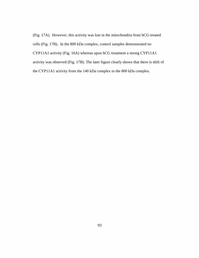
93
(Fig. 17A). However, this activity was lost in the mitochondria from hCG-treated
cells (Fig. 17B). In the 800 kDa complex, control samples demonstrated no
CYP11A1 activity (Fig. 16A) whereas upon hCG treatment a strong CYP11A1
activity was observed (Fig. 17B). The later figure clearly shows that there is shift of
the CYP11A1 activity from the 140 kDa complex to the 800 kDa complex.

94
Discussion:
BN-PAGE is a technique that allows the separation of protein-protein
complexes based solely on size; the use of digitonin functions by binding to
cholesterol and precipitating; creating holes in the membrane (192). This technique
has been shown to allow protein complexes to remain active in the gel due to the
weak solubization that still leaves protein-lipid interactions intact (193). We used
this method to answer many questions in steroidogenesis where lipid-protein and
protein-protein interactions are the regulating mechanisms. Specifically, it allows the
identity of the entry site of cholesterol into mitochondria as well as the site where it
is metabolized.
In previous experiments, we demonstrated the formation of a hormone-
dependant protein complex in the mitochondria conferring the ability to import
cholesterol into mitochondria and form steroids (138).Through BN-PAGE membrane
analysis we showed that TSPO forms complexes at both 66 kDa and 800 kDa
molecular sizes and further 2D-SDS-PAGE analysis demonstrated that the 66 kDa
complex contained only TSPO monomers whereas the 800 kDa complex contained
polymers. Upon treatment of Leydig cells with hCG a shift was seen in TSPO from
monomers at 66 kDa to polymers at 800 kDa, confirming the observation the that
TSPO polymerization occurs after exposure to hormones (104;107) . This change
may reflect the previous reported hormone-induced changes in radioligand binding
characteristics of TSPO (194). Based on these data we proposed that the 800 kDa

95
complex is the site of the functional TSPO (189) and the site of cholesterol transfer
from OMM to IMM site function in the transfer of cholesterol to the IMM for
steroidogenesis.
Detailed analysis by MS and immunoblotting identified a number of proteins
in the 140 and 800 kDa complexes including VDAC, ANT, CYP11A1, Tim44 and
Tim50 as well as other proteins that belong both to OMM and IMM. These data
suggest that these complexes may represent OMM/IMM contact sites, the place that
has been postulated for years to be the site where steroidogenic cholesterol crosses
from OMM to IMM bypassing the aqueous intermembrane space and loaded onto
CYP11A1. In mitochondria from hormone-treated Leydig cells CYP11A1 migrated
towards the higher molecular weight complex and the same was true for most of the
proteins examined. These results suggest that upon hormone treatment the 800 kDa
complex becomes the major complex, containing a functional TSPO and high levels
of CYP11A1. Interestingly, the 78 kDa glucose-regulated protein precursor (GRP
78) was also identified in the 800 kDa complex. This protein is the also precursor of
the steroidogenesis activating polypeptide shown to activate adrenal steroidogenesis
(195).
Despite the four decades-long efforts to identify the mechanisms underlying
the hormonal-induction of cholesterol transport into mitochondria, there is no
information about the actual site of cholesterol entry into these organelles. It is clear
that this site must be in an environment allowing for its segregation from the

96
structural cholesterol of the OMM. In an effort to identify this site, mitochondria
were isolated from MA-10 cells, incubated with the photoactivatable 3H-
photocholesterol, and exposed to UV. Proteins separated by BN-PAGE migrating at
66 kDa bound specifically cholesterol. Immunobloting demonstrated the co-
localization of the cholesterol binding site with TSPO at the 66 kDa complex where
TSPO enters into mitochondria (189). Mitochondria from hormone-treated cells
showed less photocholesterol binding probably due to isotopic dilution by the
endogenous cholesterol accumulating in the site. We have proposed that TSPO
segregates the steroidogenic pool of cholesterol from the structural cholesterol
present in OMM (131). Hormonal stimulation of the cells results in TSPO
polymerization move to the 800 kDa complex whereas in the same time the affinity
for cholesterol falls (107) so that cholesterol can now become available for
steroidogenesis.
To examine whether the 800 kDa complex is functional in steroidogenesis we
set up a system to measure in-vitro BN-PAGE and in test tube as well as in cells
CYP11A1 activity. In 1990, Marrone et al. reported the synthesis of a cholesterol-
resorufin probe with activity in isolated mitochondria and cells (196;197). This
probe was not available and we synthesized a new probe following the basic steps
outlined by the authors (US patent 5,208,332) with some modifications. Incubation
of cholesterol-resorufin with the gel spot together with the necessary cofactors
NADP, isocitrate and ADP resulted in the oxidation of the cholesterol-resorufin

97
molecule by CYP11A1 producing pregenolone and resorufin. Considering the
minute amounts of enzyme and materials available within the gel spot it would have
been impossible to measure pregnenolone or isocaproic acid levels to determine
CYP11A1 activity without the help of the resorufin fluorescent dye. The ability of
the 140 kDa complex to produce steroids was lost upon hCG stimulation, suggesting
the enzyme or protein necessary to oxidize cholesterol were no longer present or
present in its active form. Instead the 800 kDa mitochondrial complex from hCG
treated gained the ability to rapidly metabolize the CYP11A1, suggesting the
formation of a larger functional molecular weight complex in the mitochondria upon
hCG stimulation. None of the 66 kDa, 440 kDa mitochondrial complexes isolated
from control and hCG-treated cells were able to metabolize the cholesterol-resorufin
probe.
Once cholesterol is translocated to the IMM, its conversion to pregnenolone
occurs with CYP11A1 but also with the auxiliary electron transferring proteins,
adrenodoxin and adrenodoxin reductase. These proteins function in the transfer of
electrons from NADPH to the substrate (198). A ternary complex of adrenodoxin
(Adx), adrenodoxin reductase (AdR), and CYP11A1 has been shown to form in the
presence of cholesterol and phospholipids (199). Immunoblot analysis indicated that
adrenodoxin reductase was not associated with the BN-PAGE complexes, although
AdR was found in both the 140 and 800 kDa complexes by MS. A possible reason

98
AdR was not observed by immunoblot analysis the epitope was masked due to
complex formation in the 140 and 800 kDa complexes.
Because many IMM proteins were identified by MS at the 140 kDa complex,
such as Tim44 and Tim50 with adrenodoxin reductase, CYP11A1, ANT, and others,
we propose that this protein complex is primarily located in the IMM. This
observation would support the findings that the CYP11A1 is active in the IMM (1)
Upon solubilization of the mitochondria, analysis by BN-PAGE exposes this basal
complex. Control cells also have an 800 kDa complex but with minimal function.
Following hCG treatment IMM/OMM contact sites are formed and many proteins
move into the 800 kDa complex. MS analysis of the 800 kDa complex identified
proteins and enzymes identified known to be present at both the ER and Golgi
apparatus, suggesting a mechanism that would allow a further increase of cholesterol
targeted to the OMM and transferred to the IMM through this newly formed
OMM/IMM contact sites. We propose that the formation of an intra-mitochondrial
contact sites form following hormonal stimulation as previously suggested by
Stevens et al. (187). This contact sites may also contain proteins present at the
established Mitochondrial-ER contact site (MAM) and the Golgi apparatus which in
turn would bring cholesterol and proteins that could function in cholesterol transfer
at the OMM and steroidogenesis.

99
SUMMARY AND CONCLUSIONS
The first metabolic step of steroidogenesis occurs at the inner mitochondrial
membrane (IMM), where the cytochrome P450 cholesterol side chain cleavage
enzyme (CYP11A1) catalyzes the conversion of cholesterol to pregnenolone (1).
The transfer of cholesterol from the outer to the inner mitochondrial membrane is the
rate-limiting step in hormone-induced steroid formation. To ensure that this step is
achieved efficiently; free cholesterol present at the outer mitochondrial membrane is
transferred to the inner membrane. This is accomplished through a series of steps
that involve various intracellular organelles and proteins such as the translocator
protein (18 kDa, TSPO) and steroidogenic acute regulatory (StAR) protein. TSPO is
a high-affinity drug- and cholesterol-binding mitochondrial protein. StAR is a
hormone-induced mitochondria-targeted protein that has been shown to promote
cholesterol transfer into mitochondria. Through the assistance of proteins, such as the
cAMP-dependent protein kinase regulatory subunit Iα (PKA-RIα) and TSPO-
associated protein, PAP7, cholesterol is transferred to and docked at the outer
mitochondrial membrane. The association of TSPO with the outer/inner
mitochondrial membrane contact sites drives the intramitochondrial cholesterol
transfer and subsequent steroid formation. The hypothesis examined in this thesis is
that delivery of cholesterol into the mitochondria, and thus steroidogenesis, is driven
by a series of protein-protein interactions.

100
Initial studies with CYP11A1 proposed that this enzyme, which is
responsible for cleaving the cholesterol side chain, was the rate limiting step in the
production of steroids. This observation was shown to be incorrect as upon the
addition of cycloheximide, a protein synthesis inhibitor, steroidogenesis was shown
to be inhibited in the adrenals, though cholesterol was able to be loaded onto the
OMM (70). These results suggested that the ability of cholesterol to be transferred
from the OMM to the IMM is the primary control step in the production of steroids
and therefore necessary for the activity of CYP11A1 (200). Further experiments
were performed using a membrane permeable cholesterol analog, 22R-
hydroxycholesterol, which can bind to the active site of CYP11A1 without having to
being translocated across the OMM and was able to be metabolized to pregnenolone
(201). This suggested that the activity of CYP11A1 was not the rate limiting step in
steroidogenesis, but that the transfer of cholesterol to IMM is the rate limiting step of
steroidogenesis with CYP11A1’s activity being dependant upon the ability of
cholesterol to be transferred to the IMM. These findings resulted in the focus of
steroid production shifting from the study of CYP11A1 to identification proteins that
function in cholesterol transport to the mitochondria resulting in steroid formation.
In 1977, TSPO was initially identified in the kidney and later found to be
present at high concentrations in steroidogenic cells (5). As it was shown to be
present primarily in the OMM and have high cholesterol binding affinity, it was
suggested it could play a role in steroidogenesis by assisting with the translocation of

101
cholesterol from the OMM to the IMM (94). Its role in steroidogenesis was
confirmed after decreasing TSPO protein expression in mouse Leydig cells, resulted
in reduced steroid production as compared to control; these findings were further
confirmed in adrenal cells (102). These experiments demonstrated the importance of
TSPO in cholesterol transfer to the IMM, providing a mechanism for cholesterol
transport that was located on the OMM to the IMM.
Studies have demonstrated that only 20-40% of adrenal mitochondrial
cholesterol is depleted upon initiation of steroidogenesis, suggesting there is a
mitochondrial pool of cholesterol present for steroidogenesis which is utilized upon
hormonal stimulation (202). As this mitochondrial pool of cholesterol is utilized,
more cholesterol must then be targeted to the mitochondria for steroidogenesis. The
previous results demonstrated that cycloheximide, which inhibits protein synthesis,
also inhibited steroidogenesis implying there was a “labile protein factor” that played
a role in steroid production. It was suggested that this protein bound the cholesterol
needed for steroidogenesis to continue, interacting with the mitochondria upon tropic
hormone activation and assisting with the transfer into the IMM. In 1994, a 32-kDa
protein whose expression correlated with the initiation of steroid synthesis was
identified, cloned and named the Steroidogenic Acute Regulatory protein (StAR)
(74). StAR was shown to contain both an N-terminus mitochondrial targeting
sequence and a C-terminus cholesterol binding domain, suggesting a mechanism for
which it could function in targeting cholesterol to the mitochondria (76;78). Initial

102
studies on the mature 30-kDa form of StAR showed that it is necessary for
steroidogenesis and upon reduction in cAMP-dependant StAR expression,
steroidogenesis was inhibited (124). It was further shown however, that it was not
the 30-kDa form, but rather the precursor 37-kDa protein that was important for
steroidogenesis and it was acting outside the OMM (79). These results, which
identified the mechanism of StAR’s action at the OMM, laid the foundation for the
role of protein-protein interactions in steroidogenesis by providing the linking factor
for cholesterol transport from the cytosol to the mitochondria.
To determine then if StAR played a role in transferring cholesterol to TSPO
for import into the mitochondria several studies were performed and no physical
interactions between TSPO and StAR could be identified (123). A functional
interaction was shown, however, through antisense knockdown of either StAR or
TSPO; each protein was necessary for steroidogenesis, and TSPO was found to be
necessary for mitochondrial processing of the StAR protein (124). These
observations led to the proposal that other proteins must function in cholesterol
transport to the OMM and in steroidogenesis. Through yeast two-hybrid screens it
was identified that TSPO interacts with PBR associated protein 7, PAP7; moreover,
PAP7 was additionally shown to interact with PKA-R1α, a cAMP-dependant protein
kinase. The identification of these two proteins provided the foundation for a
hypothesis that a scaffolding complex forms at the OMM and is necessary for
steroidogenesis (133). While this scaffolding complex was shown to interact

103
through yeast two hybrid assays and confocal microscopy, no physical interactions
could be identified to confirm its presence at the OMM.
Chapter 1 of this thesis addressed the question of a mitochondrial scaffolding
complex. We focused upon demonstrating the formation of the protein complex of
TSPO, StAR, PAP7 and PKA-RIα, called the transduceosome, at the OMM.
Through the use of photoactivatable amino acids, the presence of the
transduceosome at the OMM, was confirmed. This was seen after incubation of the
photoactivable amino acid and crosslinking; western blot analysis identified TSPO,
StAR, VDAC, PAP7 and PKA-RIα all present at a high molecular weight complex.
As StAR requires phosphorylation for its activity at the OMM, the tranduceosome
brings StAR in close proximity PKA-RIα for phosphorylation.
Upon incubation of radiolabeled cholesterol in the media with MA-10 cells,
it was further identified that cholesterol was also present in the tranduceosome
complex. This provided another mechanism of action in the trafficking of
cholesterol to the mitochondria for use in steroidogenesis. The identification of this
novel mechanism shifted the assumption held previously that cholesterol transported
from intracellular stores into mitochondria is mediated by a single cholesterol
transport protein, StAR but now to the presence of a multi-protein scaffolding
complex, the tranduceosome. This proposal lays the foundation for a novel
mechanism of cholesterol delivery to the mitochondria, both for the basal and
hormonally-stimulated cells, as this complex was also identified at the basal state on
104
the OMM. As the tranduceosome could be utilized for the targeting of non-
steroidogenic cholesterol to the mitochondria, the basal state could also provide the
scaffolding mechanism for the amplification of cholesterol transport upon hormone
stimulation for StAR to act upon. Through the tranduceosome, StAR could amplify
the basal state of cholesterol transfer in to the mitochondria increasing the amount of
cholesterol available for transfer CYP11A1. Further experiments could analyze the
role of cholesterol in this complex; specifically the mechanisms by which it is
transferred.
As the tranduceosome protein complex is anchored around the OMM protein
TSPO, in chapter 2, we wanted to determine the protein-protein interactions that are
necessary for the import of TSPO into the OMM. Cytosolic proteins, Heat Shock
Protein (HSP) 70 and 90, were found to function in the targeting of TSPO to the
mitochondria. Also necessary for TSPO targeting to the OMM were the C-terminus
amino acids #151-169 and amino acids #103-109. Amino acids 103-109 form a
Schellman motif, which functions in maintaining the correct morphology for TSPO
import and protein-protein interactions. Upon targeting of TSPO to the
mitochondria, the HSP’s interact with the Translocase of Outer Mitochondrial
protein 70, TOM70. TSPO is then transferred to OMM protein Metaxin1 in an ATP-
dependant manner. Metaxin1 assists with TSPO’s final integration into the
membrane; this complex has been shown to form a 66kDa on a BN-PAGE. The rate

105
of import of TSPO at the 66kDa was shown to be increased upon hormone
stimulation, suggesting TSPO’s import is regulated by hormonal stimulation.
The data gathered herein provides a novel pathway for the insertion of multi-
spanning, alpha helical OMM proteins into the mitochondria, as previously no
pathway had yet been identified. Protein-protein interactions between the TOM and
the SAM protein complexes is a common pathway used for β-barrel protein
insertion, though it was unknown if these two protein complexes are also used for α-
helical protein insertion. The identification of the use of the receptor from the TOM
complex, TOM70, and a protein from the SAM complex, Metaxin1, suggests there
are protein-protein interactions between the two complexes that are used for the
membrane insertion of α-helical proteins. Therefore, this pathway for protein
insertion maybe more common or function in the import of more OMM proteins than
previously presumed (164).
Further study of the import of TSPO demonstrated that its import was
increased with hormonally stimulated isolated mitochondria as compared to control.
Moreover, the amounts of lipids in the mitochondria are altered in response to
hormonal stimulation; for example the amounts of cardiolipin and cholesterol are
increased, which could alter the rate of TSPO import. It has been suggested that the
import of mitochondrial proteins alters the regulation of mitochondrial lipids (203).
Therefore it would be interesting to know, if increased import would occur with all
mitochondrial proteins or only OMM proteins like TSPO. It has also been shown

106
that the presence of cardiolipin can stabilize protein import complexes located both
at the IMM and OMM, increasing the ability of TSPO to be imported into OMM
through the stabilization of the TOM or the SAM complex (204). Beside cardiolipin,
cholesterol levels in the OMM and IMM are increased upon hormonal stimulation; it
is unknown what effect this could have on protein-protein interactions necessary for
TSPO import, as cholesterol is known to decrease membrane fluidity and alter
membrane permeability. To further understanding of this event, it will be important
to see the rate of change of lipids present in the OMM, specifically cholesterol and
cardiolipin. If these lipids are altered, it would be interesting to determine what
decreasing the concentrations of cardiolipin or cholesterol would alter the import of
TSPO.
Once TSPO has been successfully imported into the mitochondria, as shown
in chapter 2, it interacts with other proteins present in the OMM. In chapter 3, we
continued the study of TSPO’s protein-protein interactions occurring in MA-10 cells
treated with hCG. This allowed us to identify protein-protein interactions that were
occurring in a hormone-dependent manner as compared to control. TSPO was
identified in a BN-PAGE gel at 66kDa in a monomer form and at 800kDa in polymer
form. As polymerization of TSPO occurs upon hormonal stimulation (107),
correlating with steroid synthesis, this findingsuggested that the 800kDa complex
may be functional in steroidogenesis. Immuoblotting of BN-PAGE gels identified
that CYP11A1 was present at 140kDa along with ANT. Upon hCG treatment, it was

107
shown that CYP11A1 was present at the 140kDa complex and at higher molecular
weights upon stimulation upto the 800kDa complex. To examine whether these
complexes contained CYP11A1 activity we used a chemically-modified cholesterol,
resorufin, which upon cleavage by CYP11A1 released a fluorescent probe and
pregnenolone. These experiments demonstrated that 140kDa basal complex was
capable of producing steroids while the 140kDa complex that had been exposed to
hCG was not. The reverse was seen of the 800kDa complex with the basal complex
was not capable of producing steroids while the hCG 800kDa was, suggesting a shift
in proteins necessary for steroidogenesis from 140kDa to the 800kDa complex.
The data gathered from both the BN-PAGE western blot analysis and the
resorufin dye identifies of a protein complex present in the mitochondria that has
been shown to be functional in steroid production. As previously mentioned, the rate
limiting step in the production of steroid hormones is the transfer of cholesterol from
the OMM to the IMM, with functional CYP11A1 located at the IMM and segregated
from the cholesterol. The identification of functional CYP11A1 present at 140kDa
on BN-PAGE interacting with another IMM protein, ANT, suggests this is the
complex that present at basal state. Upon addition of the cholesterol in the gel spot
at 140kDa basal state the complex is capable of producing pregnenolone. Upon
hormonal stimulation the steroidogenic complex is not present at 140kDa but shifts
to 800kDa, where CYP11A1 can now interacts with OMM proteins, VDAC and
TSPO, both proteins present in the tranduceosome and components of mitochondrial

108
contact sites (110). It is the presence of the OMM contacting the IMM would allow
for easy transfer of cholesterol to the IMM and interaction with CYP11A1.
Contact sites are dynamic localization of where OMM and IMM meet and
have previously been proposed to function in cholesterol transport to CYP11A1 in
the IMM (205). The formation of a contact site would allow the direct transfer of
cholesterol from one membrane to another while removing the inner mitochondrial
membrane space the hydrophobic cholesterol would have to cross. Contact sites
between VDAC and ANT have previously been proposed to control rates of
apoptosis and energy metabolism, in which these cellular functions can have several
affects on steroid synthesis (206). This work demonstrates the formation of contact
sites in the mitochondria for steroidogenesis as confirmed through both mass
spectrometry and western blot analysis. The lack of this contact site as seen from
control mitochondrial as compared to hormonally treated mitochondrial
demonstrates how important these intramitochondrial formations are for steroid
synthesis.
Further analysis of these contact sites can be used to identify the lipid
composition necessary for steroid formation. For example, the amount of cardioplin,
which has already been implicated for TSPO import, has also been shown to be
important in CYP11A1:adrenodoxin complex formation and function (207). .
Further analysis through mass spectrometry can also be used to determine which
enzymes and proteins are necessary for the generation of steroids, specifically

109
assisting with the formation of NADPH for the reduction of cholesterol and to
identify which other protein complexes can help function in contact site formation.
The results in this thesis confirm and extend our understanding of the importance of
protein-protein interactions in steroidogenesis from mitochondrial import,
cholesterol transport and steroid biosynthesis. Mitochondrial protein import of
TSPO was shown to be dependant upon the novel pathway of protein interactions of
heat shock proteins with mitochondrial protein import receptor TOM70 and Metaxin
1. Its import helps sustain the role TSPO plays in the initiation and maintenance of
steroidogenesis at the OMM and cholesterol transport into the IMM. This ability of
cholesterol transport was demonstrated to be dependant upon the formation of a
hormone-induced protein complex present between the inner and outer mitochondrial
membrane consisting of TSPO, VDAC, ANT and CYP11A1. The supply of
cholesterol to the mitochondria for steroidogenesis is maintained by a signaling
mechanism between TSPO, StAR, VDAC, PAP7 and PKA-RIα, functioning in the
transport of cholesterol to the mitochondria from the cytosol. The identification and
confirmation of these protein-protein interactions further confirm their important role
in the cell for steroidogenesis; from the cytosol to the outer to the inner
mitochondrial membrane and finally to CYP11A1 and steroid production.

110
Figure 1. Trafficking of cholesterol to the mitochondria for steroidogenesis.
Pathway 1: Cholesterol synthesized in the ER is trafficked to the Golgi apparatus
where it can be targeted to the mitochondria via PAP7 protein for steroidogenesis.
Passive diffusion from the ER to the mitochondria, shown here with the
corresponding cholesterol molecules present on both organelles, is another possible
pathway for steroidogenic cholesterol to be transferred to the mitochondria.
Pathway 2: Low density lipoprotein (LDL), containing cholesterol, binds to the
LDL receptor and is trafficked through the endosomal pathway. MLN-64 assists
with the transfer of cholesterol to the mitochondria from the late endosomes and
lysosomes for use in steroidogenesis. NPC1 and NPC2 associate with MLN-64 for
cholesterol transfer out of the lysosomes, although it is not known if this cholesterol
is used for steroidogenesis. Pathway 3: Cholesterol is transferred from high density
lipoprotein (HDL) to the plasma membrane by the SR-BI receptor. Hormone-
sensitive lipase (HSL) converts esterified cholesterol from the plasma membrane to
free cholesterol, which can be used for steroidogenesis. Pathway 4: HSL also
interacts with esterified cholesterol present in the lipid droplets (LD), which converts
esterified cholesterol to free cholesterol for use in steroidogenesis as well. Free
cholesterol from the LD can interact with lipid-binding proteins present in the
cytosol for delivery to the mitochondria.

111
Figure 1:

112
Figure 2: A) The structure of acetate (Carbon is grey, Oxygen is red). B) Structure
of Isoprene, basic carbon backbone structure used to produce cholesterol. C) Initial
steps of cholesterol synthesis with the conversion of Acetyl-CoA to Acetoacetyl-
CoA by thiolase, enzymatic addition of each molecule to form HMG-CoA by HMG-
CoA synthase and further reduction to mevalonate by HMG-CoA reductase. D)
Dimethylallyl pyrophosphate and Isopentyl pyrophosphate (IPP) are interconverted
by IPP isomerase with one molecule of each joined head to tail to produce geranyl
pyrophosphate. Another molecule of IPP is joined head to tail by prenyl
pyrophosphate to the geranyl pyrophosphate to produce farnesyl pyrophosphate. The
catalyzing of two farnesyl pyrophosphate in head to head condensation occurs
though squalene synthase to produce squalene. E) Squalene is converted to 2,3-
Oxidosqualene by squalene epoxidase and further is converted to lanosterol through
squalene oxidocyclase. The conversion of lanosterol to cholesterol occurs through
19 steps not demonstrated here.

113

114

115

116
Figure 3 Cholesterol-binding domains of StAR and TSPO. A) Human StAR
protein modeled with cholesterol (brown) present in the sterol-binding domain (PDB
ID code: 2I93). Homology models are deduced via the Swiss-model services under
project (optimize) mode using the crystal structures of human MLN64 (PDB ID
code: 1EM2) as template (http://swissmodel.expasy.org/SWISS-MODEL.html). B)
Molecular model of TSPO's five alpha helices in the presence of cholesterol,
demonstrating a pore accommodating a cholesterol molecule. Homology modeling
of mouse TSPO was performed using apolipophorin-III from Manduca sexta as
template (PDB ID code: 1EQ1) (208). (C) Ribbon diagram of mTSPO, showing the
five helices as well as the CRAC domain, consisting of Leu/Tyr/Arg residues, and
cholesterol (top view). (D) Docking model of cholesterol to the TSPO CRAC
domain. The accessible surface of the peptide and cholesterol molecules are
represented in red and blue, respectively (reprinted with permission from (173)).

117
Figure 3:

118
Figure 4: Protein-protein interactions at the OMM. A) Proposed basal-state
protein-protein interactions of TSPO, VDAC, and ANT. B)Transduceosome
complex formed after acute hormonal stimulation. The TSPO, PAP7 (ACBD3), and
VDAC complex recruits PKA-RIα, which is shown binding to PAP7 (ACBD3). The
accumulation of cAMP activates PKA-RIα, releasing the catalytic subunits and
phosphorylating StAR present at the OMM. StAR interacts with both TSPO and
VDAC and is imported into the IMM. Cholesterol is imported into the IMM with
the assistance of the transduceosome complex and the presence of DBI; there the
cholesterol interacts with the CYP11A1 side chain cleavage enzyme to be converted
to pregnenolone. ANT is represented in the IMM as identified from previous
isolated complexes with TSPO.

119
Figure 4: A) B)
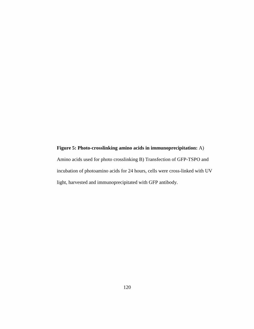
120
Figure 5: Photo-crosslinking amino acids in immunoprecipitation: A)
Amino acids used for photo crosslinking B) Transfection of GFP-TSPO and
incubation of photoamino acids for 24 hours, cells were cross-linked with UV
light, harvested and immunoprecipitated with GFP antibody.

121
Figure 5:

122
Figure 6: Protein-protein interactions identified by cross-linking. COS-F2-130
cells transfected with StAR cDNA were cultured with media supplemented with
photoactivable amino acids as described under “Experimental Procedures.” Cells
were washed and irradiated with UV light, and proteins were analyzed by
electrophoresis followed by immunoblotting with anti-TSPO and anti-StAR antisera
(top panel). MA-10 cells were cultured with media supplemented with
photoactivable amino acids (P-amino acids). At the end of the incubation, cells were
treated with 50 ng/ml hCG for the indicated periods of time. Cells were washed and
irradiated with UV light as described under “Experimental Procedures.” Cells were
collected, and proteins were separated by electrophoresis followed by
immunoblotting with anti-TSPO (A, B, and C), anti-StAR (A and C), anti-PAP7,
anti-PKARIα, anti-VDAC1, anti-ANT, or anti-glyceraldehyde-3-phosphate
dehydrogenase (GAPDH) antisera (C). Data (means; S.E. <20% of means) obtained
from three independent experiments were analyzed by image analysis. D, MA-10
cells were transfected with pEGFP-Tspo. After 48 h, cells were incubated with
[3H]photocholesterol together with the artificial amino acids and incubated for 24 h.
Cells were treated with hCG and irradiated with UV light as described above. At the
end of the treatment, cells were harvested, and cell lysates were subjected to
immunoprecipitation with anti-GFP antibodies followed by immunoblotting with
anti-TSPO antisera. Radioactivity content in the immunoprecipitates was measured
by liquid scintillation spectrometry.

123
Figure 6:

124
Figure 7: Identification of TSPO import complexes in the OMM. A) Isolated
mitochondria from HeLa cells were incubated with in vitro-transcribed/translated
[35S]-TSPO. The BN-PAGE gel was transferred to PVDF and exposed to a
multipurpose phosphor screen (left panel) or blotted with antisera against TSPO,
Tom 40, and Tom22 (right panel). B) In vitro-transcribed/translated TSPO import
into mitochondria examined at either 4 °C or 33 °C; at the stated time points, one-
half of the import reaction was treated with proteinase K and both samples were
analyzed by BN-PAGE. C) In vitro-transcribed/translated [35S]-TSPO and [35S]-
Tom70 were incubated with HeLa cell mitochondria and treated with sodium
carbonate on ice for 30 minutes. After treatment, solubilized mitochondria were
centrifuged and the pellet and supernatant were separated by SDS-PAGE, the
membrane was blotted with anit-HSP-60.

125
Figure 7:

126
Figure 8: TSPO import is dependent upon heat shock proteins and ATP for import.
A) Effects of C90 incubation with TSPO. TSPO was imported as described above
(control). B) Effect of ATP depletion, 18µM geldanamycin, and 1mM novobiocin
on TSPO import. C) Quantification of imported TSPO. Results shown are
means±sem from three independent experiments. D) Nickel-sepharose pull-down
assay of 35S-TSPO and 35S-PIC. When indicated geldanamycin or novobiocin were
added to the reaction mixture.

127
Figure 8:

128
Figure 9: Identification of the TSPO amino acid sequence(s) responsible for
targeting the protein to the OMM. MA-10 cells were transfected with either wild-
type GFP-TSPO or various constructs of GFP-TSPO, stained with Mitotracker CMX
and visualized by confocal microscopy. Each panel shows the construct used, the
GFP staining, the mitochondrial staining, and the merged image. A) Wild-type
TSPO with GFP linked to the N-terminus. Cells were also transfected with GFP-∆1-
7-TSPO (B), GFP- ∆1-28-TSPO (C), GFP-∆1-48-TSPO (D), GFP- ∆1-68-TSPO (E),
GFP- ∆1-85-TSPO (F), GFP- ∆1-110-TSPO (G), GFP-∆157-169-TSPO (H) GFP-
∆151-169-TSPO (I). The cholesterol binding domain, GFP-∆148-157-TSPO (J), or
the Schellman motif, GFP-∆103-108-TSPO (K) were removed and transfected.
Construct GFP-G106A-TSPO (L) point mutation altering the Schellman motif and
constructs GFP-Leucine112-114-TSPO (M) and GFP-Leucine137,138,141-TSPO
(N) were also transfected as described under experimental methods.

129
Figure 9:
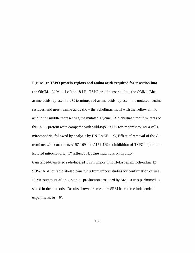
130
Figure 10: TSPO protein regions and amino acids required for insertion into
the OMM. A) Model of the 18 kDa TSPO protein inserted into the OMM. Blue
amino acids represent the C-terminus, red amino acids represent the mutated leucine
residues, and green amino acids show the Schellman motif with the yellow amino
acid in the middle representing the mutated glycine. B) Schellman motif mutants of
the TSPO protein were compared with wild-type TSPO for import into HeLa cells
mitochondria, followed by analysis by BN-PAGE. C) Effect of removal of the C-
terminus with constructs ∆157-169 and ∆151-169 on inhibition of TSPO import into
isolated mitochondria. D) Effect of leucine mutations on in vitro-
transcribed/translated radiolabeled TSPO import into HeLa cell mitochondria. E)
SDS-PAGE of radiolabeled constructs from import studies for confirmation of size.
F) Measurement of progesterone production produced by MA-10 was performed as
stated in the methods. Results shown are means ± SEM from three independent
experiments (n = 9).

131
Figure 10:

132
Figure 11: TSPO import is dependent on Metaxin 1. A) MA-10 mitochondria
incubated with in vitro-transcribed/translated TSPO analyzed by BN-PAGE, and
blotted with anti-metaxin 1 antibody. B) In vitro-transcribed/translated TSPO
incubated with mitochondria isolated from MA-10 cells control, Metaxin 1-depleted,
and MA-10 cells treated with scrambled siRNAs. C) Immunoblot analysis for
VDAC, mitofilin, TSPO and metaxin1 in mitochondria isolated form MA-10 cells
treated with the various siRNAs. D) Quantification of imported TSPO. Results
shown are means±sem from three independent experiments

133
`Figure 11:

134
Figure 12: TSPO import is increased with cAMP stimulated mitochondria. A)
Mitochondria isolated from MA-10 cells incubated with +/- 1mM cAMP were
incubated with radiolabeled TSPO, analyzed via BN-PAGE. B) Quantification of
imported TSPO, results shown are means±sem from three independent experiments.
C) Western blot of MA-10 cell incubated with +/- 1mM cAMP. D) QPCR analysis
of MA-10 RNA +/- 1mM cAMP

135
Figure 12:

136
Figure 13: TSPO import into the OMM. We have outlined the steps necessary
for TSPO targeting and insertion into the OMM. TSPO is targeted to the OMM
through its interactions with cytosolic chaperones, Hsc70 and Hsp90. At the OMM,
TSPO interacts with Tom70 and is released from the chaperones in an ATP-
dependent manner. TSPO insertion into the OMM is dependent upon Metaxin 1
(Mtx1) and TSPO structure, where both the C-terminus and the Schellman motif
(amino acids 103-108) are necessary to form a 66 kDa complex. Once import is
complete, TSPO associates with other protein complexes found in the OMM
identified previously as VDAC and ANT.

137
Figure 13:

138
Figure 14: Blue-Native Page Gels identify protein complexes. Mitochondria were
isolated from MA-10 cells that were treated or untreated for two hours in the
presence of 50ng/mL hCG. Upon isolation, samples were solubilized with 1%
digitonin and run on a BN-PAGE gel. Immunoblot analysis identified TSPO at
66kDa and 800kDa, Cyp11A1 at 140kDa, VDAC laddering at 242kDa, 440 kDa,
600kDa and 800kDa., COXIV at 480kDa, and Adrenodoxin Reductase (AdR)
present below 66kDa. B) Upon the removal of a lane from the BN-PAGE gel and
incubation in SDS, an SDS 2D-PAGE gel was run; identifying TSPO at an 18kDa
monomer at 66kDa while at 156 kDa a polymer at the BN-PAGE 800kDa is present.
C)Cyp11A1 formed a band across the gel at 60kDa. D)HSP60 was present at 60kDa
E) VDAC is present at 30kDa. .

139
Figure 14:

140
Figure 15: Lipids present in the identified protein complexes. Isolated
mitochondria solubilized with digitonin and run on BN-PAGE gel, control and hCG
treated. Radiolabeled cholesterol binding to TSPO both in the presence and absence
of cross-linking UV light in also with control and hCG treated mitochondria.

141
Figure 15:

142
Figure 16: Cyp11A1 probe is active in whole cells and isolated mitochondria.
A) Synthesis of Cyp11A1 Probe B) MA-10 cells were incubated with or without 5uL
of the Cyp11A1 probe for 24 hours after of which 1mM 8-bromo cAMP was added
to the half the samples. Measurement at 595nm showed an increase in the
fluorescence in the cAMP treated cells with the Cyp11A1 probe. C) MA-10 cells
control or treated with 1mM 8-bromo-cAMP, after the mitochondria were isolated
and incubated with 5uL ethanol, 5uL dye or control

143
Figure 16 :

144
Figure 17: Cyp11A1 probe shows activity in the BN-PAGE gel. Isolated
mitochondria from 50ng/mL hCG treated or untreated MA-10 cells were run on a
BN-PAGE gel and spots previously identified to contain proteins necessary for
steroidogenesis were isolated. Samples were incubated with steroidogenic import
buffer, NADP, and Isocitrate. A) Control samples show fluorescent activity at the
140kDa gel spot while no other samples did. B) hCG treated MA-10 cells showed
activity at the 800kDa spot while no other areas showed fluorescent activity.
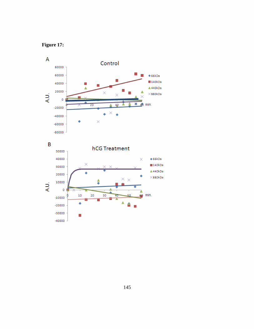
145
Figure 17:
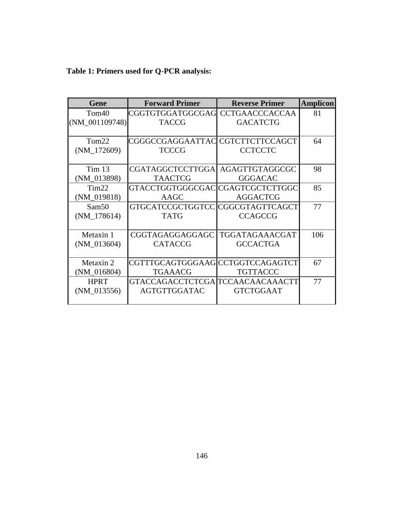
146
Table 1: Primers used for Q-PCR analysis:
Gene Forward Primer Reverse Primer Amplicon Tom40
(NM_001109748) CGGTGTGGATGGCGAG
TACCG
CCTGAACCCACCAAGACATCTG
81
Tom22 (NM_172609)
CGGGCCGAGGAATTACTCCCG
CGTCTTCTTCCAGCTCCTCCTC
64
Tim 13 (NM_013898)
CGATAGGCTCCTTGGATAACTCG
AGAGTTGTAGGCGCGGGACAC
98
Tim22 (NM_019818)
GTACCTGGTGGGCGACAAGC
CGAGTCGCTCTTGGCAGGACTCG
85
Sam50 (NM_178614)
GTGCATCCGCTGGTCCTATG
CGGCGTAGTTCAGCTCCAGCCG
77
Metaxin 1 (NM_013604)
CGGTAGAGGAGGAGCCATACCG
TGGATAGAAACGATGCCACTGA
106
Metaxin 2 (NM_016804)
CGTTTGCAGTGGGAAGTGAAACG
CCTGGTCCAGAGTCTTGTTACCC
67
HPRT (NM_013556)
GTACCAGACCTCTCGAAGTGTTGGATAC
TCCAACAACAAACTTGTCTGGAAT
77

147
Table 2: Identification of proteins present in the 66- and 800 kDa mitochondrial protein complexes by mass spectrometry in HeLa cells. The 66- and 800 kDa mitochondrial protein complexes separated by BN-PAGE and 2D SDS-PAGE were analyzed by mass spectrometry; some of the major identified proteins are shown.

148
Table 2:
Protein Sequences Identified 66 kDa Metaxin1 RSLASPGISPGPLTATIGGAVAGGGPR Nonspecific lipid-transfer protein
K.LQNLQLQPGNAKL K.IGGIFAFK.V K.ANLVFKEIEK.K
Voltage-dependent anion channel (VDAC 1)
TDEFQLHTNVNDGTEFGGSIYQK R.WTEYGLTFTEK.W K.LTFDSSFSPNTGK.K
800 kDa Adenine nucleotide translocator (ANT 1,2,3)
K.LLLQVQHASK.Q R.YFPTQALNFAFK.D R.AAYFGIYDTAK.G K.DFLAGGIAAAISK.T
Apolipoprotein A-I K.VSFLSALEEYTK.K Apolipoprotein A-II K.SPELQAEAK.S Fatty acid synthase K.VVVQVLAEEPEAVLK.G
R.LQVVDQPLPVR.G K.VGDPQELNGITR.A
Mitofilin R.ELDSITPEVLPGWK.G Voltage-dependent anion channel (VDAC 1,3)
R.VTQSNFAVGYK.T K.LSQNNFALGYK.A
66 and 800 kDa Annexin A2 K.SALSGHLETVILGLLK.T
R.QDIAFAYQR.R K.TPAQYDASELK.A K.GVDEVTIVNILTNR.S
Phosphatidylinositol 4,5-biphosphate-dependent ARF1 GTPase-activating protein
K.SFDLISHNR.T R.QEEIDESDDDLDDK.P K.NGILTISHATSNR.Q K.SFDLISHNR.T

149
Table 3: Identification of proteins present in the 66 and 800 kDa mitochondrial protein complexes in control and treated MA-10 cells by mass spectrometry. The 66- and 800 kDa mitochondrial protein complexes separated by BN-PAGE and 2D SDS-PAGE were analyzed by mass spectrometry; some of the major identified proteins are shown.

150
Table 3: Protein:
# of sequences identified Control
# of sequences identified hCG
66 kDa Ras-related proteins: Rab 1A, Rab 5C, Rab 7, Rab 14
0 8
14-3-3 Proteins: Epsilon, Gamma 4 9 Steroidogenic Proteins: CYP11A1, Acyl-CoA-binding protein 1 (ACBP1)
3 7
Mitochondrial import proteins: Tim 8, Tim13,Tim 23
2 2
Various proteins: Oxysterol-binding protein-related protein 1
1 2
140 kDa Steroidogenic Proteins: CYP11A1, NADPH-Adrenodoxin reductase Adenine nucleotide translocator (ANT 1,3)
20 32
Mitochondrial import proteins: Tim 8, Tim13,Tim50, Tim 44, Tom20
14 15
800 kDa Steroidogenic Proteins: CYP11A1 2 8 ER-Mitochondria Contact Site: Inositol 1,4,5-trisphosphate receptor, VDAC1, 78 kDa glucose-regulated protein precursor (GRP 78)
0 4
Golgi-associated retrograde protein (GARP) complex:VPS52, VPS53
0 3
Mitophilin Complex associating proteins: Mitofilin, Coiled-coil-helix-coiled-coil-helix domain-containing protein 3, DnaJ homolog subfamily C member 11
12 22
Mitochondrial morphology protein: SH3 domain GRB2-like protein B2 (Endophilin-B2), VDAC
3 2
Various proteins: Apolipoprotein A-IV, Calnexin Heat shock 70 kDa protein, Superoxide dismutase
4 2

151
Reference List 1. Hall, P. F. (1985) Vitam. Horm. 42, 315-368
2. Simpson, E. R. and Waterman, M. R. (1983) Can. J. Biochem. Cell Biol. 61, 692-707
3. Jefcoate, C. (2002) J. Clin. Invest 110, 881-890
4. Garren, L. D., Gill, G. N., Masui, H., and Walton, G. M. (1971) Recent Prog. Horm. Res. 27, 433-478
5. Papadopoulos, V., Baraldi, M., Guilarte, T. R., Knudsen, T. B., Lacapere, J. J., Lindemann, P., Norenberg, M. D., Nutt, D., Weizman, A., Zhang, M. R., and Gavish, M. (2006) Trends Pharmacol. Sci. 27, 402-409
6. Ikonen, E. (2008) Nat. Rev. Mol. Cell Biol. 9, 125-138
7. Warnock, D. E., Roberts, C., Lutz, M. S., Blackburn, W. A., Young, W. W., Jr., and Baenziger, J. U. (1993) J. Biol. Chem. 268, 10145-10153
8. Mukherjee, S., Zha, X., Tabas, I., and Maxfield, F. R. (1998) Biophys. J. 75, 1915-1925
9. Dietschy, J. M. and Turley, S. D. (2004) J. Lipid Res. 45, 1375-1397
10. Ponticorovo, L., RITTENBERG, D., and Bloch, K. (1949) J. Biol. Chem. 179, 839-842
11. Bloch, K. (1952) Harvey Lect. 48, 68-88
12. Voet, D. and Voet, J. G. (1995) Biochemistry, 2nd ed Ed., J. Wiley & Sons, New York
13. Brown, M. S. and Goldstein, J. L. (1980) J. Lipid Res. 21, 505-517
14. Goldstein, J. L., Bose-Boyd, R. A., and Brown, M. S. (2006) Cell 124, 35-46
15. Raghow, R., Yellaturu, C., Deng, X., Park, E. A., and Elam, M. B. (2008) Trends Endocrinol. Metab 19, 65-73
16. Ness, G. C. and Chambers, C. M. (2000) Proc. Soc. Exp. Biol. Med. 224, 8-19

152
17. Sato, R., Goldstein, J. L., and Brown, M. S. (1993) Proc. Natl. Acad. Sci. U. S. A 90, 9261-9265
18. Plump, A. S., Erickson, S. K., Weng, W., Partin, J. S., Breslow, J. L., and Williams, D. L. (1996) J. Clin. Invest 97, 2660-2671
19. Biardi, L. and Krisans, S. K. (1996) J. Biol. Chem. 271, 1784-1788
20. Do, R., Kiss, R. S., Gaudet, D., and Engert, J. C. (2009) Clin. Genet. 75, 19-29
21. Huff, M. W. and Telford, D. E. (2005) Trends Pharmacol. Sci. 26, 335-340
22. Mannella, C. A., Buttle, K., Rath, B. K., and Marko, M. (1998) Biofactors 8, 225-228
23. Stone, S. S., Levin, M. C., Zhou, P., Han, J., Walther, T. C., and Farese, R. V., Jr. (2008) J. Biol. Chem.
24. Lange, Y., Ye, J., Rigney, M., and Steck, T. L. (1999) J. Lipid Res. 40, 2264-2270
25. Connelly, M. A. and Williams, D. L. (2003) Trends Endocrinol. Metab 14, 467-472
26. Verschoor-Klootwyk, A. H., Verschoor, L., Azhar, S., and Reaven, G. M. (1982) J. Biol. Chem. 257, 7666-7671
27. Ungewickell, E. J. and Hinrichsen, L. (2007) Curr. Opin. Cell Biol. 19, 417-425
28. Murray, J. W. and Wolkoff, A. W. (2003) Adv. Drug Deliv. Rev. 55, 1385-1403
29. Jordens, I., Marsman, M., Kuijl, C., and Neefjes, J. (2005) Traffic. 6, 1070-1077
30. Schultz, J., Doerks, T., Ponting, C. P., Copley, R. R., and Bork, P. (2000) Nat. Genet. 25, 201-204
31. Grosshans, B. L., Ortiz, D., and Novick, P. (2006) Proc. Natl. Acad. Sci. U. S. A 103, 11821-11827

153
32. Mukherjee, S. and Maxfield, F. R. (2004) Biochim. Biophys. Acta 1685, 28-37
33. Kraemer, F. B., Shen, W. J., Patel, S., Osuga, J., Ishibashi, S., and Azhar, S. (2007) Am. J. Physiol Endocrinol. Metab 292, E408-E412
34. Foster, J. D., Strauss, J. F., III, and Paavola, L. G. (1993) Endocrinology 132, 337-350
35. Chen, H., Yang, J., Low, P. S., and Cheng, J. X. (2008) Biophys. J. 94, 1508-1520
36. Acton, S., Rigotti, A., Landschulz, K. T., Xu, S., Hobbs, H. H., and Krieger, M. (1996) Science 271, 518-520
37. Werder, M., Han, C. H., Wehrli, E., Bimmler, D., Schulthess, G., and Hauser, H. (2001) Biochemistry 40, 11643-11650
38. Hamilton, J. A. (2003) Curr. Opin. Lipidol. 14, 263-271
39. Connelly, M. A., Kellner-Weibel, G., Rothblat, G. H., and Williams, D. L. (2003) J. Lipid Res. 44, 331-341
40. Kraemer, F. B. and Shen, W. J. (2002) J. Lipid Res. 43, 1585-1594
41. Yeaman, S. J. (2004) Biochem. J. 379, 11-22
42. Kraemer, F. B., Shen, W. J., Harada, K., Patel, S., Osuga, J., Ishibashi, S., and Azhar, S. (2004) Mol. Endocrinol. 18, 549-557
43. Osuga, J., Ishibashi, S., Oka, T., Yagyu, H., Tozawa, R., Fujimoto, A., Shionoiri, F., Yahagi, N., Kraemer, F. B., Tsutsumi, O., and Yamada, N. (2000) Proc. Natl. Acad. Sci. U. S. A 97, 787-792
44. Shen, W. J., Patel, S., Natu, V., Hong, R., Wang, J., Azhar, S., and Kraemer, F. B. (2003) J. Biol. Chem. 278, 43870-43876
45. Murphy, S., Martin, S., and Parton, R. G. (2008) Biochim. Biophys. Acta
46. Chang, T. Y., Chang, C. C., and Cheng, D. (1997) Annu. Rev. Biochem. 66, 613-638
47. Strauss, J. F., III, Kishida, T., Christenson, L. K., Fujimoto, T., and Hiroi, H. (2003) Mol. Cell Endocrinol. 202, 59-65

154
48. Liu, P., Bartz, R., Zehmer, J. K., Ying, Y. S., Zhu, M., Serrero, G., and Anderson, R. G. (2007) Biochim. Biophys. Acta 1773, 784-793
49. Martin, S., Driessen, K., Nixon, S. J., Zerial, M., and Parton, R. G. (2005) J. Biol. Chem. 280, 42325-42335
50. Soccio, R. E. and Breslow, J. L. (2004) Arterioscler. Thromb. Vasc. Biol. 24, 1150-1160
51. Schroeder, F., Atshaves, B. P., McIntosh, A. L., Gallegos, A. M., Storey, S. M., Parr, R. D., Jefferson, J. R., Ball, J. M., and Kier, A. B. (2007) Biochim. Biophys. Acta 1771, 700-718
52. Gallegos, A. M., Schoer, J. K., Starodub, O., Kier, A. B., Billheimer, J. T., and Schroeder, F. (2000) Chem. Phys. Lipids 105, 9-29
53. Gallegos, A. M., Atshaves, B. P., Storey, S. M., Starodub, O., Petrescu, A. D., Huang, H., McIntosh, A. L., Martin, G. G., Chao, H., Kier, A. B., and Schroeder, F. (2001) Prog. Lipid Res. 40, 498-563
54. Baum, C. L., Reschly, E. J., Gayen, A. K., Groh, M. E., and Schadick, K. (1997) J. Biol. Chem. 272, 6490-6498
55. Moncecchi, D., Murphy, E. J., Prows, D. R., and Schroeder, F. (1996) Biochim. Biophys. Acta 1302, 110-116
56. Seedorf, U., Raabe, M., Ellinghaus, P., Kannenberg, F., Fobker, M., Engel, T., Denis, S., Wouters, F., Wirtz, K. W., Wanders, R. J., Maeda, N., and Assmann, G. (1998) Genes Dev. 12, 1189-1201
57. Alpy, F. and Tomasetto, C. (2005) J. Cell Sci. 118, 2791-2801
58. Tsujishita, Y. and Hurley, J. H. (2000) Nat. Struct. Biol. 7, 408-414
59. Tomasetto, C., Regnier, C., Moog-Lutz, C., Mattei, M. G., Chenard, M. P., Lidereau, R., Basset, P., and Rio, M. C. (1995) Genomics 28, 367-376
60. Moog-Lutz, C., Tomasetto, C., Regnier, C. H., Wendling, C., Lutz, Y., Muller, D., Chenard, M. P., Basset, P., and Rio, M. C. (1997) Int. J. Cancer 71, 183-191
61. Alpy, F., Stoeckel, M. E., Dierich, A., Escola, J. M., Wendling, C., Chenard, M. P., Vanier, M. T., Gruenberg, J., Tomasetto, C., and Rio, M. C. (2001) J. Biol. Chem. 276, 4261-4269

155
62. Zhang, M., Liu, P., Dwyer, N. K., Christenson, L. K., Fujimoto, T., Martinez, F., Comly, M., Hanover, J. A., Blanchette-Mackie, E. J., and Strauss, J. F., III (2002) J. Biol. Chem. 277, 33300-33310
63. Strauss, J. F., III, Liu, P., Christenson, L. K., and Watari, H. (2002) Steroids 67, 947-951
64. Sturley, S. L., Patterson, M. C., Balch, W., and Liscum, L. (2004) Biochim. Biophys. Acta 1685, 83-87
65. Gevry, N. Y. and Murphy, B. D. (2002) Endocr. Res. 28, 403-412
66. Xu, Z., Farver, W., Kodukula, S., and Storch, J. (2008) Biochemistry 47, 11134-11143
67. Infante, R. E., Wang, M. L., Radhakrishnan, A., Kwon, H. J., Brown, M. S., and Goldstein, J. L. (2008) Proc. Natl. Acad. Sci. U. S. A 105, 15287-15292
68. Watari, H., Blanchette-Mackie, E. J., Dwyer, N. K., Sun, G., Glick, J. M., Patel, S., Neufeld, E. B., Pentchev, P. G., and Strauss, J. F., III (2000) Exp. Cell Res. 255, 56-66
69. Espenshade, P. J. and Hughes, A. L. (2007) Annu. Rev. Genet. 41, 401-427
70. Pon, L. A. and Orme-Johnson, N. R. (1986) J. Biol. Chem. 261, 6594-6599
71. Pon, L. A., Hartigan, J. A., and Orme-Johnson, N. R. (1986) J. Biol. Chem. 261, 13309-13316
72. Lin, D., Sugawara, T., Strauss, J. F., III, Clark, B. J., Stocco, D. M., Saenger, P., Rogol, A., and Miller, W. L. (1995) Science 267, 1828-1831
73. Sierra, A. (2004) J. Neuroendocrinol. 16, 787-793
74. Clark, B. J., Wells, J., King, S. R., and Stocco, D. M. (1994) J. Biol. Chem. 269, 28314-28322
75. Sugawara, T., Holt, J. A., Driscoll, D., Strauss, J. F., III, Lin, D., Miller, W. L., Patterson, D., Clancy, K. P., Hart, I. M., Clark, B. J., and . (1995) Proc. Natl. Acad. Sci. U. S. A 92, 4778-4782
76. Miller, W. L. (2007) Biochim. Biophys. Acta 1771, 663-676

156
77. Arakane, F., Sugawara, T., Nishino, H., Liu, Z., Holt, J. A., Pain, D., Stocco, D. M., Miller, W. L., and Strauss, J. F., III (1996) Proc. Natl. Acad. Sci. U. S. A 93, 13731-13736
78. Kallen, C. B., Billheimer, J. T., Summers, S. A., Stayrook, S. E., Lewis, M., and Strauss, J. F., III (1998) J. Biol. Chem. 273, 26285-26288
79. Bose, H. S., Lingappa, V. R., and Miller, W. L. (2002) Endocr. Res. 28, 295-308
80. Baker, B. Y., Epand, R. F., Epand, R. M., and Miller, W. L. (2007) J. Biol. Chem. 282, 10223-10232
81. Barbar, E., LeHoux, J. G., and Lavigne, P. (2009) Mol. Cell Endocrinol. 300, 89-93
82. Soccio, R. E., Adams, R. M., Romanowski, M. J., Sehayek, E., Burley, S. K., and Breslow, J. L. (2002) Proc. Natl. Acad. Sci. U. S. A 99, 6943-6948
83. Papadopoulos, V. (1993) Endocr. Rev. 14, 222-240
84. Anholt, R. R., De Souza, E. B., Oster-Granite, M. L., and Snyder, S. H. (1985) J. Pharmacol. Exp. Ther. 233, 517-526
85. Papadopoulos, V., Mukhin, A. G., Costa, E., and Krueger, K. E. (1990) J. Biol. Chem. 265, 3772-3779
86. Anholt, R. R., Aebi, U., Pedersen, P. L., and Snyder, S. H. (1986) Biochemistry 25, 2120-2125
87. Woods, M. J., Zisterer, D. M., and Williams, D. C. (1996) Biochem. Pharmacol. 51, 1283-1292
88. Le, F. G., Perrier, M. L., Vaucher, N., Imbault, F., Flamier, A., Benavides, J., Uzan, A., Renault, C., Dubroeucq, M. C., and Gueremy, C. (1983) Life Sci. 32, 1839-1847
89. Verma, A., Nye, J. S., and Snyder, S. H. (1987) Proc. Natl. Acad. Sci. U. S. A 84, 2256-2260
90. Bovolin, P., Schlichting, J., Miyata, M., Ferrarese, C., Guidotti, A., and Alho, H. (1990) Regul. Pept. 29, 267-281
157
91. Papadopoulos, V., Berkovich, A., and Krueger, K. E. (1991) Neuropharmacology 30, 1417-1423
92. Mukhin, A. G., Papadopoulos, V., Costa, E., and Krueger, K. E. (1989) Proc. Natl. Acad. Sci. U. S. A 86, 9813-9816
93. Barnea, E. R., Fares, F., and Gavish, M. (1989) Mol. Cell Endocrinol. 64, 155-159
94. Krueger, K. E. and Papadopoulos, V. (1990) J. Biol. Chem. 265, 15015-15022
95. Lacapere, J. J., Delavoie, F., Li, H., Peranzi, G., Maccario, J., Papadopoulos, V., and Vidic, B. (2001) Biochem. Biophys. Res. Commun. 284, 536-541
96. Li, H., Yao, Z., Degenhardt, B., Teper, G., and Papadopoulos, V. (2001) Proc. Natl. Acad. Sci. U. S. A 98, 1267-1272
97. Scott, J. K. and Smith, G. P. (1990) Science 249, 386-390
98. Fawell, S., Seery, J., Daikh, Y., Moore, C., Chen, L. L., Pepinsky, B., and Barsoum, J. (1994) Proc. Natl. Acad. Sci. U. S. A 91, 664-668
99. Gazouli, M., Han, Z., and Papadopoulos, V. (2002) J. Pharmacol. Exp. Ther. 303, 627-632
100. Falchi, A. M., Battetta, B., Sanna, F., Piludu, M., Sogos, V., Serra, M., Melis, M., Putzolu, M., and Diaz, G. (2007) Neuropharmacology 53, 318-329
101. Freeman, D. A. (1987) Endocrinology 120, 124-132
102. Papadopoulos, V., Amri, H., Li, H., Boujrad, N., Vidic, B., and Garnier, M. (1997) J. Biol. Chem. 272, 32129-32135
103. Papadopoulos, V., Amri, H., Boujrad, N., Cascio, C., Culty, M., Garnier, M., Hardwick, M., Li, H., Vidic, B., Brown, A. S., Reversa, J. L., Bernassau, J. M., and Drieu, K. (1997) Steroids 62, 21-28
104. Boujrad, N., Vidic, B., and Papadopoulos, V. (1996) Endocrinology 137, 5727-5730
105. Papadopoulos, V., Boujrad, N., Ikonomovic, M. D., Ferrara, P., and Vidic, B. (1994) Mol. Cell Endocrinol. 104, R5-R9

158
106. Privalle, C. T., Crivello, J. F., and Jefcoate, C. R. (1983) Proc. Natl. Acad. Sci. U. S. A 80, 702-706
107. Delavoie, F., Li, H., Hardwick, M., Robert, J. C., Giatzakis, C., Peranzi, G., Yao, Z. X., Maccario, J., Lacapere, J. J., and Papadopoulos, V. (2003) Biochemistry 42, 4506-4519
108. Raha, S., Myint, A. T., Johnstone, L., and Robinson, B. H. (2002) Free Radic. Biol. Med. 32, 421-430
109. Murail, S., Robert, J. C., Coic, Y. M., Neumann, J. M., Ostuni, M. A., Yao, Z. X., Papadopoulos, V., Jamin, N., and Lacapere, J. J. (2008) Biochim. Biophys. Acta 1778, 1375-1381
110. Culty, M., Li, H., Boujrad, N., Amri, H., Vidic, B., Bernassau, J. M., Reversat, J. L., and Papadopoulos, V. (1999) J. Steroid Biochem. Mol. Biol. 69, 123-130
111. Doble, A., Ferris, O., Burgevin, M. C., Menager, J., Uzan, A., Dubroeucq, M. C., Renault, C., Gueremy, C., and Le Fur, G. (1987) Mol. Pharmacol. 31, 42-49
112. Riond, J., Vita, N., Le Fur, G., and Ferrara, P. (1989) FEBS Lett. 245, 238-244
113. McEnery, M. W., Snowman, A. M., Trifiletti, R. R., and Snyder, S. H. (1992) Proc. Natl. Acad. Sci. U. S. A 89, 3170-3174
114. Garnier, M., Dimchev, A. B., Boujrad, N., Price, J. M., Musto, N. A., and Papadopoulos, V. (1994) Mol. Pharmacol. 45, 201-211
115. Shoshan-Barmatz, V., Keinan, N., and Zaid, H. (2008) J. Bioenerg. Biomembr. 40, 183-191
116. Kokoszka, J. E., Waymire, K. G., Levy, S. E., Sligh, J. E., Cai, J., Jones, D. P., MacGregor, G. R., and Wallace, D. C. (2004) Nature 427, 461-465
117. Azuma, M., Kabe, Y., Kuramori, C., Kondo, M., Yamaguchi, Y., and Handa, H. (2008) PLoS. ONE. 3, e3070
118. Veenman, L., Shandalov, Y., and Gavish, M. (2008) J. Bioenerg. Biomembr. 40, 199-205

159
119. Golani, I., Weizman, A., Leschiner, S., Spanier, I., Eckstein, N., Limor, R., Yanai, J., Maaser, K., Scherubl, H., Weisinger, G., and Gavish, M. (2001) Biochemistry 40, 10213-10222
120. Campbell, A. M. and Chan, S. H. (2007) Arch. Biochem. Biophys. 466, 203-210
121. Hiller, S., Garces, R. G., Malia, T. J., Orekhov, V. Y., Colombini, M., and Wagner, G. (2008) Science 321, 1206-1210
122. West, L. A., Horvat, R. D., Roess, D. A., Barisas, B. G., Juengel, J. L., and Niswender, G. D. (2001) Endocrinology 142, 502-505
123. Bogan, R. L., Davis, T. L., and Niswender, G. D. (2007) J. Steroid Biochem. Mol. Biol. 104, 61-67
124. Hauet, T., Yao, Z. X., Bose, H. S., Wall, C. T., Han, Z., Li, W., Hales, D. B., Miller, W. L., Culty, M., and Papadopoulos, V. (2005) Mol. Endocrinol. 19, 540-554
125. Ariyoshi, N., Kim, Y. C., Artemenko, I., Bhattacharyya, K. K., and Jefcoate, C. R. (1998) J. Biol. Chem. 273, 7610-7619
126. Artemenko, I. P., Zhao, D., Hales, D. B., Hales, K. H., and Jefcoate, C. R. (2001) J. Biol. Chem. 276, 46583-46596
127. Tuckey, R. C., Headlam, M. J., Bose, H. S., and Miller, W. L. (2002) J. Biol. Chem. 277, 47123-47128
128. Roostaee, A., Barbar, E., LeHoux, J. G., and Lavigne, P. (2008) Biochem. J. 412, 553-562
129. Petrescu, A. D., Gallegos, A. M., Okamura, Y., Strauss, J. F., III, and Schroeder, F. (2001) J. Biol. Chem. 276, 36970-36982
130. Mathieu, A. P., Fleury, A., Ducharme, L., Lavigne, P., and LeHoux, J. G. (2002) J. Mol. Endocrinol. 29, 327-345
131. Lacapere, J. J. and Papadopoulos, V. (2003) Steroids 68, 569-585
132. Li, H., Degenhardt, B., Tobin, D., Yao, Z. X., Tasken, K., and Papadopoulos, V. (2001) Mol. Endocrinol. 15, 2211-2228

160
133. Liu, J., Li, H., and Papadopoulos, V. (2003) J. Steroid Biochem. Mol. Biol. 85, 275-283
134. Dodge-Kafka, K. L., Bauman, A., and Kapiloff, M. S. (2008) Handb. Exp. Pharmacol. 3-14
135. Catt, K. J., Harwood, J. P., Clayton, R. N., Davies, T. F., Chan, V., Katikineni, M., Nozu, K., and Dufau, M. L. (1980) Recent Prog. Horm. Res. 36, 557-662
136. Whalin, M. E., Boujrad, N., Papadopoulos, V., and Krueger, K. E. (1994) J. Recept. Res. 14, 217-228
137. Poderoso, C., Maloberti, P., Duarte, A., Neuman, I., Paz, C., Maciel, F. C., and Podesta, E. J. (2008) Mol. Cell Endocrinol.
138. Liu, J., Rone, M. B., and Papadopoulos, V. (2006) J. Biol. Chem. 281, 38879-38893
139. Suchanek, M., Radzikowska, A., and Thiele, C. (2005) Nat. Methods 2, 261-267
140. Tasken, K. and Aandahl, E. M. (2004) Physiol Rev. 84, 137-167
141. Dyson, M. T., Jones, J. K., Kowalewski, M. P., Manna, P. R., Alonso, M., Gottesman, M. E., and Stocco, D. M. (2008) Biol. Reprod. 78, 267-277
142. Bose, M., Whittal, R. M., Miller, W. L., and Bose, H. S. (2008) J. Biol. Chem. 283, 8837-8845
143. Galiegue, S., Tinel, N., and Casellas, P. (2003) Curr. Med. Chem. 10, 1563-1572
144. Galiegue, S., Casellas, P., Kramar, A., Tinel, N., and Simony-Lafontaine, J. (2004) Clin. Cancer Res. 10, 2058-2064
145. Wright, G. and Reichenbecher, V. (1999) Exp. Cell Res. 246, 443-450
146. Levin, E., Premkumar, A., Veenman, L., Kugler, W., Leschiner, S., Spanier, I., Weisinger, G., Lakomek, M., Weizman, A., Snyder, S. H., Pasternak, G. W., and Gavish, M. (2005) Biochemistry 44, 9924-9935

161
147. Shoukrun, R., Veenman, L., Shandalov, Y., Leschiner, S., Spanier, I., Karry, R., Katz, Y., Weisinger, G., Weizman, A., and Gavish, M. (2008) Pharmacogenet. Genomics 18, 977-988
148. Veenman, L., Papadopoulos, V., and Gavish, M. (2007) Curr. Pharm. Des 13, 2385-2405
149. Stojanovski, D., Johnston, A. J., Streimann, I., Hoogenraad, N. J., and Ryan, M. T. (2003) Exp. Physiol 88, 57-64
150. Rehling, P., Brandner, K., and Pfanner, N. (2004) Nat. Rev. Mol. Cell Biol. 5, 519-530
151. Neupert, W. and Herrmann, J. M. (2007) Annu. Rev. Biochem. 76, 723-749
152. Rapaport, D. (2003) EMBO Rep. 4, 948-952
153. Rapaport, D. (2005) J. Cell Biol. 171, 419-423
154. Kunkele, K. P., Heins, S., Dembowski, M., Nargang, F. E., Benz, R., Thieffry, M., Walz, J., Lill, R., Nussberger, S., and Neupert, W. (1998) Cell 93, 1009-1019
155. Chan, N. C., Likic, V. A., Waller, R. F., Mulhern, T. D., and Lithgow, T. (2006) J. Mol. Biol. 358, 1010-1022
156. Young, J. C., Hoogenraad, N. J., and Hartl, F. U. (2003) Cell 112, 41-50
157. Bhangoo, M. K., Tzankov, S., Fan, A. C., Dejgaard, K., Thomas, D. Y., and Young, J. C. (2007) Mol. Biol. Cell 18, 3414-3428
158. Fan, A. C., Bhangoo, M. K., and Young, J. C. (2006) J. Biol. Chem. 281, 33313-33324
159. Lan, L., Isenmann, S., and Wattenberg, B. W. (2000) Biochem. J. 349, 611-621
160. Pfanner, N., Wiedemann, N., Meisinger, C., and Lithgow, T. (2004) Nat. Struct. Mol. Biol. 11, 1044-1048
161. Paschen, S. A., Neupert, W., and Rapaport, D. (2005) Trends Biochem. Sci. 30, 575-582

162
162. Kozjak-Pavlovic, V., Ross, K., Benlasfer, N., Kimmig, S., Karlas, A., and Rudel, T. (2007) EMBO Rep. 8, 576-582
163. Kozjak, V., Wiedemann, N., Milenkovic, D., Lohaus, C., Meyer, H. E., Guiard, B., Meisinger, C., and Pfanner, N. (2003) J. Biol. Chem. 278, 48520-48523
164. Armstrong, L. C., Saenz, A. J., and Bornstein, P. (1999) J. Cell Biochem. 74, 11-22
165. Armstrong, L. C., Komiya, T., Bergman, B. E., Mihara, K., and Bornstein, P. (1997) J. Biol. Chem. 272, 6510-6518
166. Otera, H., Taira, Y., Horie, C., Suzuki, Y., Suzuki, H., Setoguchi, K., Kato, H., Oka, T., and Mihara, K. (2007) J. Cell Biol. 179, 1355-1363
167. Fujiki, Y., Hubbard, A. L., Fowler, S., and Lazarow, P. B. (1982) J. Cell Biol. 93, 97-102
168. Zara, V., Palmieri, F., Mahlke, K., and Pfanner, N. (1992) J. Biol. Chem. 267, 12077-12081
169. Wilm, M., Shevchenko, A., Houthaeve, T., Breit, S., Schweigerer, L., Fotsis, T., and Mann, M. (1996) Nature 379, 466-469
170. Hales, K. H., Diemer, T., Ginde, S., Shankar, B. K., Roberts, M., Bosmann, H. B., and Hales, D. B. (2000) Endocrinology 141, 4000-4012
171. Young, J. C. and Hartl, F. U. (2000) EMBO J. 19, 5930-5940
172. Marcu, M. G., Chadli, A., Bouhouche, I., Catelli, M., and Neckers, L. M. (2000) J. Biol. Chem. 275, 37181-37186
173. Jamin, N., Neumann, J. M., Ostuni, M. A., Vu, T. K., Yao, Z. X., Murail, S., Robert, J. C., Giatzakis, C., Papadopoulos, V., and Lacapere, J. J. (2005) Mol. Endocrinol. 19, 588-594
174. Viguera, A. R. and Serrano, L. (1995) J. Mol. Biol. 251, 150-160
175. Li, W., Amri, H., Huang, H., Wu, C., and Papadopoulos, V. (2004) J. Androl 25, 900-913
176. Walther, D. M. and Rapaport, D. (2009) Biochim. Biophys. Acta 1793, 42-51

163
177. Rone, M. B., Fan, J., and Papadopoulos, V. (2009) Biochim. Biophys. Acta
178. Thomson, M. (1998) Horm. Metab Res. 30, 16-28
179. Brdiczka, D. G., Zorov, D. B., and Sheu, S. S. (2006) Biochim. Biophys. Acta 1762, 148-163
180. Reichert, A. S. and Neupert, W. (2002) Biochim. Biophys. Acta 1592, 41-49
181. Lambeth, J. D., Kitchen, S. E., Farooqui, A. A., Tuckey, R., and Kamin, H. (1982) J. Biol. Chem. 257, 1876-1884
182. STONE, D. and HECHTER, O. (1954) Arch. Biochem. 51, 457-469
183. Stocco, D. M. and Clark, B. J. (1996) Endocr. Rev. 17, 221-244
184. Strauss, J. F., III, Kallen, C. B., Christenson, L. K., Watari, H., Devoto, L., Arakane, F., Kiriakidou, M., and Sugawara, T. (1999) Recent Prog. Horm. Res. 54, 369-394
185. Gavish, M., Bachman, I., Shoukrun, R., Katz, Y., Veenman, L., Weisinger, G., and Weizman, A. (1999) Pharmacol. Rev. 51, 629-650
186. Papadopoulos, V., Nowzari, F. B., and Krueger, K. E. (1991) J. Biol. Chem. 266, 3682-3687
187. Stevens, V. L., Tribble, D. L., and Lambeth, J. D. (1985) Arch. Biochem. Biophys. 242, 324-327
188. Liu, J., Matyakhina, L., Han, Z., Sandrini, F., Bei, T., Stratakis, C. A., and Papadopoulos, V. (2003) FASEB J. 17, 1189-1191
189. Rone, M. B., Liu, J., Blonder, J., Ye, X., Veenstra, T. D., Young, J. C., and Papadopoulos, V. (2009) Biochemistry 48, 6909-6920
190. Simpson, R. J. (2003) Proteins and proteomics a laboratory manual, Cold Spring Harbor Laboratory Press, Cold Spring Harbor, NY
191. Papadopoulos, V., Berkovich, A., Krueger, K. E., Costa, E., and Guidotti, A. (1991) Endocrinology 129, 1481-1488
192. Reisinger, V. and Eichacker, L. A. (2008) J. Proteomics. 71, 277-283

164
193. Reisinger, V. and Eichacker, L. A. (2007) Proteomics. 7 Suppl 1, 6-16
194. Boujrad, N., Gaillard, J. L., Garnier, M., and Papadopoulos, V. (1994) Endocrinology 135, 1576-1583
195. Pedersen, R. C. and Brownie, A. C. (1987) Science 236, 188-190
196. Marrone, B. L., Simpson, D. J., Yoshida, T. M., Unkefer, C. J., Whaley, T. W., and Buican, T. N. (1991) Endocrinology 128, 2654-2656
197. Asem, E. K., Simpson, D. J., and Marrone, B. L. (1992) J. Steroid Biochem. Mol. Biol. 43, 479-487
198. Payne, A. H. and Hales, D. B. (2004) Endocr. Rev. 25, 947-970
199. Kimura, T. (1981) Mol. Cell Biochem. 36, 105-122
200. Lambeth, J. D. and Stevens, V. L. (1984) Endocr. Res. 10, 283-309
201. CHAUDHURI, A. C., HARADA, Y., SHIMIZU, K., GUT, M., and DORFMAN, R. I. (1962) J. Biol. Chem. 237, 703-704
202. Simpson, E. R., Jefcoate, C. R., Brownie, A. C., and Boyd, G. S. (1972) Eur. J. Biochem. 28, 442-450
203. Tamura, Y., Endo, T., Iijima, M., and Sesaki, H. (2009) J. Cell Biol. 185, 1029-1045
204. Gebert, N., Joshi, A. S., Kutik, S., Becker, T., McKenzie, M., Guan, X. L., Mooga, V. P., Stroud, D. A., Kulkarni, G., Wenk, M. R., Rehling, P., Meisinger, C., Ryan, M. T., Wiedemann, N., Greenberg, M. L., and Pfanner, N. (2009) Curr. Biol. 19, 2133-2139
205. Martinez, F., Kiriakidou, M., and Strauss, J. F., III (1997) Endocrinology 138, 2172-2183
206. Vyssokikh, M. Y. and Brdiczka, D. (2003) Acta Biochim. Pol. 50, 389-404
207. Kisselev, P., Tuckey, R. C., Woods, S. T., Triantopoulos, T., and Schwarz, D. (1999) Eur. J. Biochem. 260, 768-773
208. Wang, J., Sykes, B. D., and Ryan, R. O. (2002) Proc. Natl. Acad. Sci. U. S. A 99, 1188-1193