protein engineering approaches for antibody...
TRANSCRIPT

1
http://journals.tubitak.gov.tr/biology/
Turkish Journal of Biology Turk J Biol(2019) 43: 1-12© TÜBİTAKdoi:10.3906/biy-1809-28
Protein engineering approaches for antibody fragments: directed evolution and rational design approaches
Merve ARSLAN1,2,*, Dilara KARADAĞ1,*, Sibel KALYONCU1,**1İzmir Biomedicine and Genome Center, İzmir, Turkey
2İzmir Biomedicine and Genome Institute, Dokuz Eylül University, İzmir, Turkey
1. IntroductionHundreds of therapeutic antibodies and their derivatives are being manufactured and tested in clinical trials. Currently, there are more than 65 monoclonal antibodies approved on the market for the treatment of various diseases, mostly cancer. The rate of antibody therapeutics receiving their first approvals has been increasing over the last decade. Last year, 10 antibodies were approved in either the European Union or the United States and this number is expected to increase in the upcoming years (Kaplon and Reichert, 2018).
The first technology that was used to produce therapeutic antibodies was mouse hybridoma technology (Frenzel et al., 2017). With this technology, therapeutic monoclonal antibodies (mAbs) are obtained via the fusion of murine B cells and myeloma cells. However, there are some limitations in the use of these mAbs in humans, especially the immune response against murine mAbs (human antimouse antibody response) (Qin and Li, 2014). To overcome this problem, several approaches were
developed by utilizing recombinant DNA technology, such as chimerization (replacement of the constant regions of the murine antibodies with homologous human sequences), which generally reduces the affinity and deteriorates biophysical properties of mAbs. Therefore, it is essential to apply affinity maturation and protein engineering approaches after this process. More importantly, there are known reproducibility problems related to the hybridoma technique where sequence information is lost and features of mAbs cannot be improved with many available in vitro systems (Bradbury and Pluckthun, 2015).
Approximately 90% of approved antibody drugs are full-length (IgG) and the rest are antibody fragments (mostly Fab formats), where all or some parts of constant regions are eliminated while the essential antigen binding region is preserved. It is very well known that antibody fragments usually show similar binding properties as their full-length versions with even better biophysical properties (Nelson, 2010). Compared to full-length antibodies, antibody fragments have many advantages for therapeutic
Abstract: The number of therapeutic antibodies in preclinical, clinical, or approved phases has been increasing exponentially, mostly due to their known successes. Development of antibody engineering methods has substantially hastened the development of therapeutic antibodies. A variety of protein engineering techniques can be applied to antibodies to improve their affinity and/or biophysical properties such as solubility and stability. Antibody fragments (where all or some parts of constant regions are eliminated while the essential antigen binding region is preserved) are more suitable for protein engineering techniques because there are many in vitro screening technologies available for antibody fragments but not full-length antibodies. Improvement of biophysical characteristics is important in the early development phase because most antibodies fail at the later stage of development and this leads to loss of resources and time. Here, we review directed evolution and rational design methods to improve antibody properties. Recent developments in rational design approaches and antibody display technologies, and especially phage display, which was recently awarded the 2018 Nobel Prize, are discussed to be used in antibody research and development.
Key words: Antibody, antibody fragment, directed evolution, rational design, protein engineering, phage display, yeast surface display, affinity, biophysical properties
Received: 07.09.2018 Accepted/Published Online: 09.01.2019 Final Version: 07.02.2019
Review Article
* These authors contributed equally to this work ** Correspondence: [email protected]
This work is licensed under a Creative Commons Attribution 4.0 International License.
ARSLAN et al. / Turk J Biol
2
use: (i) lower immunogenicity due to lack of constant regions, (ii) higher tumor penetration, (iii) cheaper and larger scale production with bacteria, and (iv) availability of various in vitro display technologies to improve several characteristics of antibodies. Today, the number of antibody fragments in clinical trials and on the market is increasing faster than before due to their advantages. Because most of the directed evolution approaches are only available for antibody fragments, improvement of full-length antibodies is usually conducted in their antibody fragment format, and then those improved fragments are converted back to full-length antibody format (Xiao et al., 2017).
Protein engineering techniques such as directed evolution and rational design approaches to discover and/or improve antibodies are becoming more popular both in the biopharmaceutical industry and research environments. Applying these techniques in the early discovery phase is important because it is high-throughput and there is full control of protein sequence during the development phase of biotherapeutics.
2. Antibody display technologies as directed evolution approachesFor the past 40 years, hybridoma technology has been used extensively to produce traditional monoclonal antibodies for research and diagnostics. Recently, a number of advanced methods called display technologies have emerged as fast and high-throughput alternatives. Phage display technology is the first radical in vitro approach that allowed to produce human antibodies without any need for immunization. In this technique, antibody fragments are fused to a capsid protein of the phage and thus expressed on the surface of the virus (García Merino, 2011; Chiu and Gilliland, 2016). Although phage display is the most common antibody display technique, today several recombinant display technologies are available and basically classified in two categories: in vitro display technologies (phage display, ribosome-mRNA display) and in vivo display technologies (bacterial, yeast, and mammalian cell-surface display) (Sergeeva et al., 2006; Harel Inbar and Benhar, 2012; Brodel et al., 2018).2.1. In vitro display technologies2.1.1. Phage DisplayThe phage display technique was first discovered in 1985 by George P Smith, who was one of three recipients of the 2018 Nobel Prize in chemistry for this discovery (Smith, 1985). This was an important step to develop new approaches for generation of mAbs. In this technique, a protein gene is fused to a gene encoding a capsid protein of the virus and the fused gene is inserted into a single-stranded DNA of the phage (Karimi et al., 2016; Ledsgaard et al., 2018). Basically, two types of capsid proteins are preferred; the
first one is pIII that allows to fuse larger proteins and the other one is pVIII. The most commonly used phages for phage display are the filamentous ones (M13, Fd, and f1), which are in the Ff family and have the ability to infect only the strains of Escherichia coli containing F conjugative plasmids (Li and Caberoy, 2010; Loset and Sandlie, 2012; Karimi et al., 2016; Gustafson et al., 2018; Kiguchi et al., 2018; Ledsgaard et al., 2018; Teixeira and Gonzalez-Pajuelo, 2018).
Two different application systems have been developed for phage display. In the first system, a protein sequence is used as an insert and is fused to a capsid gene of the virus. With this system, the desired protein is expressed within the genome of the virus. In the other more preferred system, a different plasmid called a phagemid is used and the expression of the desired protein is separated from the phage replication. Phagemids also include replication origins of E. coli and a phage, a specific selection marker, and specific tags that help detection and purification of the desired protein (Li and Caberoy, 2010; Loset and Sandlie, 2012; Teixeira and Gonzalez-Pajuelo, 2018).
The phage display technique was first applied for the variable fragments of antibodies and many different antibody fragment formats have been displayed by this technique. The antibody fragments that are displayed by this technique are usually scFv (a single-chain variable fragment) or Fab (antigen-binding fragment), and nowadays the most popular ones are VH (nanobody, heavy variable domain of the antibody) (Teixeira and Gonzalez-Pajuelo, 2018). It is easy to convert these fragments to full-length antibodies by recombinant DNA technology, if needed.
The phage display technique is carried out by a process of in vitro repeated cycles typically named biopanning or phage display selections (Figure 1). This process includes the following steps: (1) incubation: binding of the antibody library repertoire to the antigen; (2) washing: elimination of the nonspecific binders; and (3) elution and amplification: obtaining antibodies binding to the antigen specifically for further cycles or for screening. Although in the first cycle of biopanning the whole antibody repertoire is exposed to the antigen, depending on the fragment type of the antibody and the phage display, 2–4 cycles of selections are generally performed to enrich the specific binders. Evaluation of the success of each cycle of the process and enrichment is possible by comparing the phage titers after elution steps against a blank that does not include antigen, or alternatively it can be tested by ELISA (enzyme-linked immunosorbent assay) (Hairul Bahara et al., 2013; Chan et al., 2014; Ledsgaard et al., 2018; Teixeira and Gonzalez-Pajuelo, 2018).
The most powerful advantages of phage display are its small size and high diversity (antibody libraries up to

ARSLAN et al. / Turk J Biol
3
1011 clones), which allow to obtain antibody fragments with the desired affinity and biophysical properties. Also, this technique is preferred in both research areas and the biopharmaceutical industry due to its library diversity, ease of use, and low cost (Liu et al., 2017; Teixeira and Gonzalez-Pajuelo, 2018). For example, belimumab (market name Benlysta) was discovered and improved by phage display and it is used to treat adults with active systemic lupus erythematosus (Stohl and Hilbert, 2012). As a better known example, adalimumab (market name Humira) was discovered by phage display and it is widely used for rheumatoid arthritis treatment (Bain and Brazil, 2003). The number of antibodies discovered and/or optimized by phage display has been exponentially increasing over the last decade due to the many advantages listed above (Nixon et al., 2014).2.1.2. Ribosome and mRNA displayRibosome and mRNA display techniques are cell-free and this feature separates them from other display platforms. They have high molecular diversity (antibody libraries ranging from 1012 to 1014 clones) and enable isolation of antibodies that show affinities at pM level. Both techniques include the same basic features, such as in vitro transcription and translation steps (Harel Inbar and Benhar, 2012).
Ribosome-display technology was first reported in a patent application in 1991. While the mRNA encoding antibody library is translated in vitro, the translated
peptide and corresponding mRNA remain attached to the ribosome (Figure 1). By this means, the peptide–ribosome–mRNA (PRM) complex can be selected along with the sequence information of the desired antibody by affinity purification techniques. The most powerful aspect of this technique is its large size of library, which is not limited by the cell transformation efficiency. On the other hand, the ribosome amount and the existence of unrelated mRNA molecules are the main limitations of this technique (Hanes and Pluckthun, 1997). Groves et al. compared phage display and ribosome display to generate scFvs to a specific antigen (Groves et al., 2014). They found that scFvs affinity-matured by ribosome display had more structural diversity in the HCDR3 and VH-VL interface regions.
In the mRNA-display technique, first the antibody DNA library is transcribed to mRNA. Then mRNA is ligated to a linker, which is a DNA sequence linked to puromycin. Thereafter, the mRNA-linker-puromycin complex is translated. Puromycin first binds to the A-site of the ribosome, then attacks the P-site and the nascent peptide is transferred to puromycin, resulting in the mRNA-linker-puromycin-antibody fragment complex. The complex is then reverse-transcribed and the selection process is performed. After the selection step, ss-DNA is obtained by hydrolyzing the complementary mRNA via high pH, and the desired DNA sequence is amplified by PCR (Figure 1) (Sergeeva et al., 2006; Jijakli et al., 2016;
In vitro In vivo
Library
Screening
Displayed antibody fragment Puromycin linker Antigen-coated surface Inhibitory element Conjugated antigen scFv displayed on Aga2
scFv displayed on Lpp-OmpA’ chimera scFv displayed on PDGFR
Ribosome
P MammalianCell
Binding
Wash
Elution
Clone Selection
AntigenBinding
SelectionEnrichment
Yeast
Phage
FACSMACS
scFv P
Bacteria
Translated mRNA
Figure 1. Antibody display technologies. General schematic for in vitro and in vivo display techniques.

ARSLAN et al. / Turk J Biol
4
Liu et al., 2017). One of the limitations of this technique is low efficiency of mRNA-protein conjugates. Nagumo et al. overcame this problem by unexpected substitution mutations around the start codon of antibodies (Nagumo et al., 2016). These mutations destabilized the mRNA secondary structure and this somehow led to a better formation of conjugates and higher protein expression.2.2. In vivo display technologies2.2.1. Bacterial surface displayThe bacterial surface display technique was developed as a potential alternative to phage display (Sergeeva et al., 2006). The use of bacteria as a display system was first reported by George Georgiou’s group in 1993 (Georgiou et al., 1993). They first used the Lpp-OmpA’ chimera to display two specific scFvs on the outer membrane of the gram-negative bacterium E. coli. Several years later, a new approach was developed by the same group called APEx (anchored periplasmic expression) (Jeong et al., 2007). With this second system, scFvs were displayed in the periplasmic space anchored to the inner membrane of E. coli. For isolation of antigen-specific clones, flow cytometry was used for both applications. Due to the technological shortcoming of the FACS (fluorescence activated cell sorting) of that time, library size was limited and thereby this technique was basically used for the evolution of the preexisting antibodies (Harel Inbar and Benhar, 2012). This technique is more commonly used to display functional enzymes, antigens, and especially polypeptide libraries (up to 1011 library size) (Sergeeva et al., 2006; Liu et al., 2017). For example, to identify peptide ligands specific for VEGF, bacteria-displayed peptide libraries were constructed and screened (Liu et al., 2017).2.2.2. Yeast surface displayYeast surface display was first demonstrated by Dane Wittrup’s group using Saccharomyces cerevisiae to display antibody repertoires (Harel Inbar and Benhar, 2012). Yeast surface display is a powerful technique that allows to obtain antibodies with desired affinity, specificity, and stability. In this technique, scFvs that consist of VH and VL regions and a polypeptide linker binding them together are displayed. On yeast, scFvs are fused to the adhesion subunit of the yeast agglutinin protein Aga2p, which is bound to Aga1p via a disulfide bond and this complex attaches the scFv to the yeast cell wall and finally the desired antibody fragment is identified by FACS (Figure 1) (Feldhaus and Siegel, 2004; Chao et al., 2006; Liu et al., 2017; Mei et al., 2017). This technique is commonly used for antibody display and it has several advantages: (i) use of FACS to monitor equilibrium activity statistics of the sample; (ii) offering easy secretion and purification; and (iii) using yeast cells, which can perform posttranslational modification. On the other hand, it allows to display up to
109 copies of scFv, which is a limitation as compared with the other display platforms such as phage display (Chao et al., 2006; Harel Inbar and Benhar, 2012). Also, the best known disadvantage of yeast surface display is slower growth rate and lower transformation efficiency compared to both phage and bacteria surface display techniques (Mei et al., 2017).2.2.3. Mammalian surface displayMammalian surface display was developed by Ira Pastan’s group in 2006 (Ho et al., 2006). They used this technique to display an scFv library fused to the N-terminal transmembrane domain of human platelet-derived growth factor receptor (PDGFR) on the surface of HEK-293T cells and were able to isolate high-affinity anti-CD22 antibodies. Mammalian cell display has powerful aspects for the isolation of scFv and whole IgG with high affinity and other specific biological functions. For instance, they can express mouse or human antibodies containing the posttranslational modifications required for some key antibody functions, and the technique can also be used to express recombinant antibody fragments that cannot be expressed in E. coli (Ho and Pastan, 2009). However, there are only a few reports of the technique, basically due to the limitation of repertoire size (ranging between 103 and 106). Similar to other techniques, it is required to transfer genes encoding the desired proteins to proper host cells by convenient vectors and to make sure that the desired protein undergoes correct transcription and translation processes. However, due to slower proliferation rates of mammalian cells in contrast to microbial ones, it is challenging to choose cells suitable for construction of a convenient and rapid mammalian cell surface display system. HEK-293, COS, and CHO cells are the most widely used cell lines in mammalian surface display approaches. HEK-293 has particularly been preferred more than others because of its ease of transport, high yield, and native human glycosylation (Qin and Li, 2014).
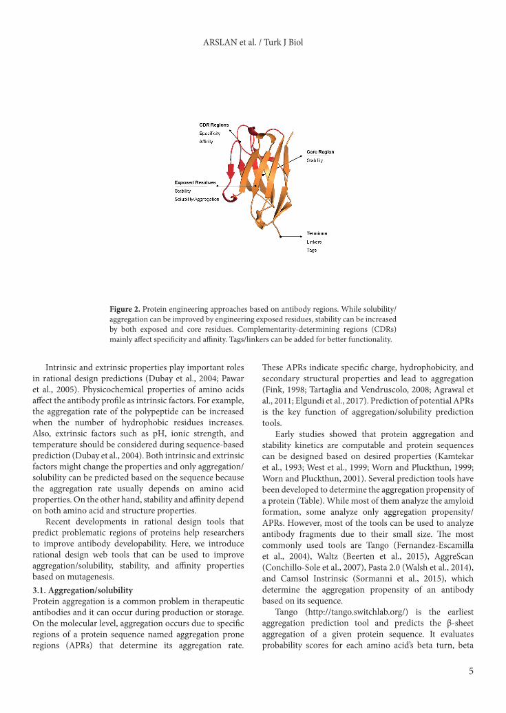
3. Rational design approachesAggregation, solubility, and stability are important factors that affect the developability of an antibody. These challenges can occur during the production process due to the protein’s large complex profile and can cause reduced antigen binding affinity, immunogenic responses, and waste of resources. Aggregation/solubility and stability properties of an antibody depend on both its sequence and structure (Figure 2). It is advantageous to control these properties with rational design before in vitro and in vivo studies. Rational design methods aim to demonstrate problematic regions of protein sequences or structures. Thus, combining rational design methods and in vitro/in vivo studies enhances the chance of antibodies with better solubility and stability in the early production phase.
ARSLAN et al. / Turk J Biol
5
Intrinsic and extrinsic properties play important roles in rational design predictions (Dubay et al., 2004; Pawar et al., 2005). Physicochemical properties of amino acids affect the antibody profile as intrinsic factors. For example, the aggregation rate of the polypeptide can be increased when the number of hydrophobic residues increases. Also, extrinsic factors such as pH, ionic strength, and temperature should be considered during sequence-based prediction (Dubay et al., 2004). Both intrinsic and extrinsic factors might change the properties and only aggregation/solubility can be predicted based on the sequence because the aggregation rate usually depends on amino acid properties. On the other hand, stability and affinity depend on both amino acid and structure properties.
Recent developments in rational design tools that predict problematic regions of proteins help researchers to improve antibody developability. Here, we introduce rational design web tools that can be used to improve aggregation/solubility, stability, and affinity properties based on mutagenesis.3.1. Aggregation/solubilityProtein aggregation is a common problem in therapeutic antibodies and it can occur during production or storage. On the molecular level, aggregation occurs due to specific regions of a protein sequence named aggregation prone regions (APRs) that determine its aggregation rate.
These APRs indicate specific charge, hydrophobicity, and secondary structural properties and lead to aggregation (Fink, 1998; Tartaglia and Vendruscolo, 2008; Agrawal et al., 2011; Elgundi et al., 2017). Prediction of potential APRs is the key function of aggregation/solubility prediction tools.
Early studies showed that protein aggregation and stability kinetics are computable and protein sequences can be designed based on desired properties (Kamtekar et al., 1993; West et al., 1999; Worn and Pluckthun, 1999; Worn and Pluckthun, 2001). Several prediction tools have been developed to determine the aggregation propensity of a protein (Table). While most of them analyze the amyloid formation, some analyze only aggregation propensity/APRs. However, most of the tools can be used to analyze antibody fragments due to their small size. The most commonly used tools are Tango (Fernandez-Escamilla et al., 2004), Waltz (Beerten et al., 2015), AggreScan (Conchillo-Sole et al., 2007), Pasta 2.0 (Walsh et al., 2014), and Camsol Instrinsic (Sormanni et al., 2015), which determine the aggregation propensity of an antibody based on its sequence.
Tango (http://tango.switchlab.org/) is the earliest aggregation prediction tool and predicts the β-sheet aggregation of a given protein sequence. It evaluates probability scores for each amino acid’s beta turn, beta
Figure 2. Protein engineering approaches based on antibody regions. While solubility/aggregation can be improved by engineering exposed residues, stability can be increased by both exposed and core residues. Complementarity-determining regions (CDRs) mainly affect specificity and affinity. Tags/linkers can be added for better functionality.

ARSLAN et al. / Turk J Biol
6
Tabl
e. P
rote
in se
quen
ce a
nd st
ruct
ure-
base
d w
eb to
ols f
or ra
tiona
l des
ign
appr
oach
es.
Sequ
ence
-bas
ed P
redi
ctio
n W
eb T
ools
Tool
nam
eD
efini
tion
Refe
renc
es
Agg
rega
tion/
Solu
bilit
y
Past
a 2.
0Pr
edic
ts a
ggre
gatio
n-pr
one,
diso
rder
ed re
gion
s(W
alsh
et a
l., 2
014)
Tang
oEv
alua
tes t
he a
ggre
gatio
n sc
ores
for e
ach
resid
ue b
ased
on
phys
icoc
hem
ical
prin
cipl
es(F
erna
ndez
-Esc
amill
a et
al.,
200
4)
Wal
tzC
ompu
tes t
he p
ositi
on-s
peci
fic sc
ores
to d
eter
min
e ag
greg
atio
n-pr
one
regi
ons
(Bee
rten
et a
l., 2
015)
Cam
sol I
ntrin
sicG
ives
out
put s
core
s for
eac
h re
sidue
bas
ed o
n so
lubi
lity
profi
le o
f seq
uenc
e(S
orm
anni
et a
l., 2
015)
Agg
reSc
anPr
edic
ts a
ggre
gatio
n-pr
one
regi
ons a
nd e
stim
ates
the
effec
t of m
utat
ion
on a
ggre
gatio
n pr
ofile
(Con
chill
o-So
le e
t al.,
200
7)
Fısh
Am
yloi
dId
entifi
es a
myl
oido
geni
c reg
ions
in p
rote
in se
quen
ces
(Gas
ior a
nd K
otul
ska,
201
4)
Soda
Focu
ses o
n eff
ect o
f the
mut
atio
ns o
n in
trin
sic so
lubi
lity
profi
le o
f pro
tein
sequ
ence
s(P
alad
in e
t al.,
201
7)
Pon-
Sol
Det
erm
ines
the
effec
t of a
min
o ac
id v
aria
tion
on so
lubi
lity
profi
le(Y
ang
et a
l., 2
016)
Prot
ein-
Sol
Giv
es g
raph
ical
out
puts
of h
ighl
ight
ed ly
sine
argi
nine
cont
ents
and
solu
bilit
y pr
ofile
(Heb
ditc
h et
al.,
201
7)
Stru
ctur
e Ba
sed
Pred
ictio
n W
eb-T
ools
Agg
rega
tion/
solu
bilit
y
Cam
sol S
truc
tura
lly
Cor
rect
edG
ives
stru
ctur
ally
corr
ecte
d so
lubi
lity
profi
le to
visu
aliz
e po
orly
solu
ble
regi
ons o
n th
e su
rfac
e, de
term
ines
the
prop
er re
sidue
s for
mut
atio
n(S
orm
anni
et a
l., 2
015)
Agg
reSc
an3D
Iden
tifies
the
poor
ly so
lubl
e re
sidue
s bas
ed o
n bo
th p
ositi
on o
f am
ino
acid
and
am
ino
acid
stru
ctur
e(Z
ambr
ano
et a
l., 2
015)
Stab
ility
ProM
aya
Cal
cula
tes p
rote
in st
abili
ty b
ased
on
diffe
renc
es b
etw
een
prot
ein’s
wild
type
and
m
utat
ed ty
pe fr
ee e
nerg
ies
(Wai
nreb
et a
l., 2
011)
SDM
Eval
uate
s the
stab
ility
diff
eren
ces b
etw
een
the
wild
type
and
mut
ated
type
pro
tein
st
ruct
ure
(Pan
dura
ngan
et a
l., 2
017)
I-M
utan
tD
eter
min
es th
e st
ruct
ure
and
sequ
ence
-bas
ed st
abili
ty ch
ange
s dep
endi
ng o
n sin
gle
poin
t mut
atio
n of
pro
tein
(Cap
riotti
et a
l., 2
005)
Cups
atU
ses a
min
o ac
id–a
tom
pot
entia
l and
tors
ion
angl
e di
strib
utio
n in
form
atio
n to
iden
tify
chan
ges i
n pr
otei
n st
abili
ty-b
ased
on
mut
atio
ns(P
arth
iban
et a
l., 2
006)
Affi
nity
mC
SM-A
BPr
edic
ts a
ntig
en–a
ntib
ody
affini
ty ch
ange
s upo
n m
utat
ions
(Pire
s and
Asc
her,
2016
)

ARSLAN et al. / Turk J Biol
7
sheet, alpha helix, and beta and alpha aggregation considering given extrinsic conditions (pH, temperature, ionic strength, concentration). The algorithm assumes that specific regions of protein have high aggregation propensity if they involve at least five consecutive residues with a probability to populate the β-aggregate state higher than 5% per residue. It was shown that Tango has a success rate of 87% , correctly predicting 155 out of 179 peptides, with 21 false positives and 3 false negatives (Fernandez-Escamilla et al., 2004).
Waltz (http://waltz.switchlab.org/) and Pasta 2.0 (http://protein.bio.unipd.it/pasta2/) give highly aggregation-prone/amyloid-forming regions as output. While Waltz uses a position-specific scoring matrix (PSSM) with physicochemical information to identify amyloid forming regions (Beerten et al., 2015), Pasta 2.0 identifies amyloid forming regions by calculating the pairing energies for each pair of residues facing one another on parallel or antiparallel neighboring strands within a β-sheet (Walsh et al., 2014).
AggreScan and Camsol are listed in two subsections of the Table because they can analyze protein aggregation propensity based on both sequence and structure information. Aggrescan (http://bioinf.uab.es/aggrescan/) calculates aggregation propensity scores for each residue in the sequence by averaging the aggregation propensity score per residue over a given length (Conchillo-Sole et al., 2007). Aggrescan3D (A3D) (http://biocomp.chem.uw.edu.pl/A3D/) is an improved version of Aggrescan that overcomes the limitations of sequence-based analyses. A3D identifies aggregation prone residues, which are related to folded states. Also, designed/desired mutation effects on aggregation propensity of any protein can be determined by using A3D (Zambrano et al., 2015).
The Camsol method can be used in two different modes, ‘Camsol Intrinsic’ and ‘Camsol Structurally Corrected’ (http://www-vendruscolo.ch.cam.ac.uk/camsolmethod.html), to evaluate aggregation scores of any protein. Camsol Intrinsic calculates the solubility profile scores per amino acid by using the given protein sequence and identifies the regions that are poorly soluble when the score is smaller than –1. It evaluates the aggregation propensity per residue using the sequence, charge, hydrophobicity, and secondary structure propensity as intrinsic factors. Camsol Structurally Corrected analyzes the protein structure like Camsol Intrinsic but it shows the poorly soluble regions on the surface that can be used to identify suitable mutations to increase the solubility of the protein. These poorly soluble regions can also be visualized by using output structure (Sormanni et al., 2015, 2017).
These methods can be used separately or combined to predict aggregation/solubility profiles and the combination of different methods can provide higher accuracy for
mutagenesis studies. Van Der Kant et al. used only Tango for prediction of APRs as a part of a study analyzing the relationship between intrinsic aggregation propensity and the local thermodynamic stability of over 2000 antibody structures from the abYsis database (Van Der Kant et al., 2017). Wang et al. combined Tango with structure-based methods to predict APRs in antibody sequences based on 29 published Fab-antigen complexes (Wang et al., 2010). They tested two different thresholds and they found that Tango was more than 92% correct in their experimental validation studies. In another study, estimations of Tango, Aggrescan, and Pasta 2.0 were used to identify APRs that were mostly confirmed by experimental results (Yageta et al., 2015).
Lately several sequence-based aggregation propensity prediction tools have also been developed. Gasior and Kotulska proposed a classification method called Fish Amyloid (http://comprec-lin.iiar.pwr.edu.pl/) that is able to recognize amyloidogenic fragments based on well-defined patterns of residue distribution and cooccurrence of position-specific amino acids in protein sequences (Gasior and Kotulska, 2014). Fish Amyloid was trained on different lengths of sequences and offered good potential for prediction. PonSol (http://structure.bmc.lu.se/PON-Sol) determines the effect of amino acid variations solubility profiles. The tool uses 443 amino acid substitutions from 71 proteins and these amino acid substitutions are classified as increasing, decreasing, and not affecting solubility (Yang et al., 2016). Protein-Sol (https://protein-sol.manchester.ac.uk/) is another recent sequence-based prediction tool that uses datasets of Escherichia coli protein solubility for comparison and calculates 35 sequence-based properties. The tool gives graphical output of predicted solubility, fold propensity, and net segment charge. Predicted solubility scales from 0 to 1 and more than 0.45 solubility scores are accepted as soluble. Also, lysine and arginine contents are highlighted for modifying protein solubility (Hebditch et al., 2017). Soda (http://protein.bio.unipd.it/soda/) predicts the protein solubility changes based on calculations of several physicochemical properties for given mutations. The method compares the mutant type and wild type profile properties and estimates the changes. Soda provides convenience for different types of variations such as point mutation, deletion, or insertion (Paladin et al., 2017).
As a case study, an scFv sequence used in our lab was analyzed with some of the sequence-based tools introduced above (Figure 3). The full scFv sequence was given as input. As output, every residue had an aggregation/solubility score based on the tool’s calculation and they were highlighted as aggregation-prone according to the tool’s corresponding thresholds. We determined multiple regions of the scFv as aggregation-prone (at least 6 of 8 tools gave predicted aggregation-prone residues).

ARSLAN et al. / Turk J Biol
8
One of those regions is shown as an example in Figure 3. Our future mutations will be focused on those regions to improve the biophysical characteristics of our protein. 3.2. StabilityProtein stability can be predicted by calculating the change in the Gibbs free energy due to substitution of an amino acid and more negative values of free energy present better stability (Thiltgen and Goldstein, 2012). Different approaches can be used for prediction of protein stability, such as physical, statistical, empirical, and/or machine learning methods. While the first three approaches are limited and are more time- and cost-intensive, machine learning methods can quickly perform predictions based on input mutation, protein sequence, and structural information at the same time (Capriotti et al., 2004; Cheng et al., 2006).
Several web-based tools were developed to predict protein stability. ProMaya (http://bental.tau.ac.il/ProMaya/) calculates the stability free energy change upon mutations by combining a collaborative filtering-based algorithm (CF) and random forest regression. The tool uses different available datasets of mutations in the same and different positions. ProMaya suggests that using known free energy values of mutations at a specific position corrects the prediction of free energy differences for other mutations (Wainreb et al., 2011).
SDM (http://marid.bioc.cam.ac.uk/sdm2) evaluates the stability change between the wild type and mutant protein by using a conformationally constrained environment-specific substitution table (ESST). The method analyzes the amino acid alteration with specific structural parameters based on residue packing density and the ESST. The webserver gives predicted stability
difference scores interpreted as reduced, induced, or unaffected stability (Pandurangan et al., 2017).
I-Mutant (http://gpcr2.biocomp.unibo.it/cgi/predictors/I-Mutant3.0/I-Mutant3.0.cgi) predicts protein stability changes based on a support vector machine and allows users to use protein structure or sequences for prediction. It was shown that I-Mutant has an accuracy of 77%–80% for the dataset derived from ProTherm (Bava et al., 2004; Capriotti et al., 2005).
Cupsat (http://cupsat.tu-bs.de/) uses atom potential and torsion angle distribution information of amino acids to identify protein stability free energy change upon mutations. The tool analyzes the protein structure and gives information about mutation site, solvent accessibility, and torsion angle and whether the mutated amino acid has suitable torsion angles or not. It was shown that Cupsat achieved 80% prediction success for both thermal and chemical stability (Parthiban et al., 2006).3.3. Affinity/specificityIf affinity improvement is desired, in vitro/vivo methods explained Section 2 of this review can be used. There are many available affinity maturation strategies based on directed evolution methods. Generally, mutations in complementarity-determining regions (CDRs) for improving antigen–antibody affinity cannot be predicted by using rational design approaches because it is hard to estimate the dynamic antigen–antibody complex structure. However, there is a newly developed tool called mCSM-AB (http://biosig.unimelb.edu.au/mcsm_ab/) that uses free energy change upon mutation and estimates the affinity change. In the tool, a negative sign means that the selected mutation reduces affinity and a positive sign means that the selected mutation increases affinity. It is important to
Figure 3. A case study for web tools. Several tools introduced in this review were used to determine aggregation-prone regions of an scFv sequence used in our lab. Blue highlighted regions are outputs of tools as aggregation prone regions. Each web tool has a different threshold, which was not shown in this figure. Mutation site is selected according to common predicted regions of different tools (at least 6 of 8 tools gave same residues as aggregation-prone).

ARSLAN et al. / Turk J Biol
9
know that this tool allows users to select more than one mutation (Pires and Ascher, 2016).
4. DiscussionThe main aims of protein engineering approaches are usually to improve affinity/specificity or to prevent aggregation and increase solubility and stability while not changing affinity/specificity. Although there are some trade-offs during these processes, there are many successful examples in the literature that improved the biophysical characteristics of antibodies.
Enever et al. used a new approach called phage display stress selection to screen for more stable human nanobodies (Enever et al., 2015). Their goals were to improve thermodynamic stability and to make nanobodies resistant to aggregation. They generated error-prone PCR phage libraries and subjected these libraries to various stress conditions. Stress conditions were related to temperature (incubation at 50–80 °C for various amounts of time), pH (incubation at pH 3.2 for various amounts of time), and protease (incubation with trypsin, elastase, leucozyme). Selection results revealed that beneficial mutations (both on CDRs and framework residues) were common to most of the stress conditions. This means that antibodies tend to mutate generic amino acids to improve their biophysical properties.
Dudgeon et al. introduced a general strategy to improve biophysical properties of antibody variable domains (Dudgeon et al., 2012). They identified specific positions in CDR regions (28, 30–33, 35 in VH and 24, 49–53, 56 in VL) and mutated those to aspartate or glutamate. This strategy led to increased aggregation resistance, which is advantageous for both diagnostic and therapeutic applications. Although most of those mutations were located in CDR regions, they showed that binding performances were not significantly affected for nearly half of the mutants.
Courtois et al. rationally designed a biobetter drug candidate by mutating or engineering aggregation-prone residues of a Fab fragment (Courtois et al., 2016). They removed aggregation-prone residues by single point mutations (hydrophobic residues to charged aspartate or lysine) and found that stability increased up to 4-fold. They also added a glycosylation site near aggregation-prone regions to increase solubility and up to 3-fold increases in stability were obtained. Most importantly,
these engineering approaches did not alter binding to the target.
Before designing mutations to decrease aggregation and/or increase stability of antibodies, three important points should be considered carefully: (i) CDR regions of the sequence should not be selected for mutation although they have high predicted scores because they are usually important for antigen binding and affinity/specificity might be impaired. (ii) Exposed hydrophobic amino acids are widely known to contribute to aggregation, and those residues should be considered first for mutation. They are preferentially mutated to hydrophilic, even charged amino acids such as aspartate, glutamate (Dudgeon et al., 2012), or lysine (Courtois et al., 2016) to circumvent aggregation problems. (iii) Designed mutations should also be compared with a natural repertoire because mutating a residue to its naturally conserved amino acid might improve its properties. The abYsis database is a web-based tool that integrates sequence data from the European Molecular Biology Laboratory European Nucleotide Archive (EMBL-ENA) and structure data from the Protein Data Bank (PDB). The abYsis database can be used to determine location-specific amino acid distribution of the natural repertoires of different organisms (Swindells et al., 2017).
It is important to note that there could be some trade-offs while improving the desired properties of an antibody (solubility, stability, affinity). Thus, the designed change should be considered for all properties. For example, while a mutation increases the solubility, it might also decrease stability at the same time. Affinity maturation can lead to a better binder but this higher affinity antibody might fail in the development phase due to its poor biophysical characteristics. It is important to keep in mind that trade-offs can occur while improving antibody fragments and one should design their computational/experimental setup accordingly.
AcknowledgmentsWe would like to thank the İzmir Biomedicine and Genome Center and YÖK (Council of Higher Education) 100/2000 fellowship program for funding our research group. We thank Hasan Buğra Çoban for his valuable input during writing process. We thank all of our research group members for carefully reviewing this article before submission.
References
Agrawal NJ, Kumar S, Wang XL, Helk, B, Singh SK, Trout BL (2011). Aggregation in protein-based biotherapeutics: computational studies and tools to identify aggregation-prone Regions. Journal of Pharmaceutical Sciences 100: 5081-5095.
Bain B, Brazil M (2003). Adalimumab. Nature Reviews Drug Discovery 2: 693-694.

ARSLAN et al. / Turk J Biol
10
Bava KA, Gromiha MM, Uedaira H, Kitajima K, Sarai A (2004). ProTherm, version 4.0: thermodynamic database for proteins and mutants. Nucleic Acids Research 32: D120-D121.
Beerten J, Van Durme J, Gallardo R, Capriotti E, Serpell L, Rousseau F, Schymkowitz J (2015). WALTZ-DB: a benchmark database of amyloidogenic hexapeptides. Bioinformatics 31: 1698-1700.
Bradbury A, Pluckthun A (2015). Standardize antibodies used in research. Nature 518: 27-29.
Brodel AK, Isalan M, Jaramillo A (2018). Engineering of biomolecules by bacteriophage directed evolution. Current Opinion in Biotechnology 51: 32-38.
Capriotti E, Fariselli P, Casadio R (2004). A neural-network-based method for predicting protein stability changes upon single point mutations. Bioinformatics 20: 63-68.
Capriotti E, Fariselli P, Casadio R (2005). I-Mutant2.0: predicting stability changes upon mutation from the protein sequence or structure. Nucleic Acids Research 33: W306-W310.
Chan CE, Lim AP, MacAry PA, Hanson BJ (2014). The role of phage display in therapeutic antibody discovery. International Immunology 26: 649-657.
Chao G, Lau W L, Hackel BJ, Sazinsky SL, Lippow SM, Wittrup KD (2006). Isolating and engineering human antibodies using yeast surface display. Nature Protocols 1: 755-768.
Cheng JL, Randall A, Baldi P (2006). Prediction of protein stability changes for single-site mutations using support vector machines. Proteins-Structure Function and Bioinformatics 62: 1125-1132.
Chiu ML, Gilliland GL (2016). Engineering antibody therapeutics. Current Opinion in Structural Biology 38: 163-173.
Conchillo-Sole O, de Groot NS, Aviles FX, Vendrell J, Daura X, Ventura S (2007). AGGRESCAN: a server for the prediction and evaluation of “hot spots” of aggregation in polypeptides. BMC Bioinformatics 8: 65.
Courtois F, Agrawal NJ, Lauer TM, Trout BL (2016). Rational design of therapeutic mAbs against aggregation through protein engineering and incorporation of glycosylation motifs applied to bevacizumab. MAbs 8: 99-112.
Dubay KF, Pawar AP, Chiti F, Zurdo J, Dobson CM, Vendruscolo M (2004). Prediction of the absolute aggregation rates of amyloidogenic polypeptide chains. Journal of Molecular Biology 341: 1317-1326.
Dudgeon K, Rouet R, Kokmeijer I, Schofield P, Stolp J, Langley D, Stock D, Christ D (2012). General strategy for the generation of human antibody variable domains with increased aggregation resistance. Proceedings of the National Academy of Sciences of the United States of America 109: 10879-10884.
Elgundi Z, Reslan M, Cruz E, Sifniotis V, Kayser V (2017). The state-of-play and future of antibody therapeutics. Advanced Drug Delivery Reviews 122: 2-19.
Enever C, Pupecka-Swider M, Sepp A (2015). Stress selections on domain antibodies: ‘What doesn’t kill you makes you stronger’. Protein Engineering Design & Selection 28: 59-66.
Feldhaus MJ, Siegel RW (2004). Yeast display of antibody fragments: a discovery and characterization platform. Journal of Immunological Methods 290: 69-80.
Fernandez-Escamilla AM, Rousseau F, Schymkowitz J, Serrano L (2004). Prediction of sequence-dependent and mutational effects on the aggregation of peptides and proteins. Nature Biotechnology 22: 1302-1306.
Fink AL (1998). Protein aggregation: folding aggregates, inclusion bodies and amyloid. Folding & Design 3: R9-R23.
Frenzel A, Kugler J, Helmsing S, Meier D, Schirrmann T, Hust M, Dubel S (2017). Designing human antibodies by phage display. Transfusion Medicine and Hemotherapy 44: 312-318.
García Merino A (2011). Monoclonal antibodies. Basic features. Neurología (English Edition) 26: 301-306.
Gasior P, Kotulska M (2014). FISH Amyloid - a new method for finding amyloidogenic segments in proteins based on site specific co-occurrence of aminoacids. BMC Bioinformatics 15: 54.
Georgiou G, Poetschke HL, Stathopoulos C, Francisco JA (1993). Practical applications of engineering gram-negative bacterial-cell surfaces. Trends in Biotechnology 11: 6-10.
Groves MAT, Amanuel L, Campbell JI, Rees DG, Sridharan S, Finch DK, Lowe DC, Vaughan TJ (2014). Antibody VH and VL recombination using phage and ribosome display technologies reveals distinct structural routes to affinity improvements with VH-VL interface residues providing important structural diversity. MAbs 6: 236-245.
Gustafson HH, Olshefsky A, Sylvestre M, Sellers DL, Pun SH (2018). Current state of in vivo panning technologies: Designing specificity and affinity into the future of drug targeting. Advanced Drug Delivery Reviews 130: 39-49.
Hairul Bahara NH, Tye GJ, Choong YS, Ong EB, Ismail A, Lim TS (2013). Phage display antibodies for diagnostic applications. Biologicals 41: 209-216.
Hanes J, Pluckthun A (1997). In vitro selection and evolution of functional proteins by using ribosome display. Proceedings of the National Academy of Sciences of the United States of America 94: 4937-4942.
Harel Inbar N, Benhar I (2012). Selection of antibodies from synthetic antibody libraries. Archives of Biochemistry and Biophysics 526: 87-98.
Hebditch M, Carballo-Amador MA, Charonis S, Curtis R, Warwicker J (2017). Protein-Sol: a web tool for predicting protein solubility from sequence. Bioinformatics 33: 3098-3100.
Ho M, Nagata S, Pastan I (2006). Isolation of anti-CD22 Fv with high affinity by Fv display on human cells. Proceedings of the National Academy of Sciences of the United States of America 103: 9637-9642.
Ho M, Pastan I (2009). Mammalian cell display for antibody engineering. Methods in Molecular Biology 525: 337-352.
Jeong KJ, Seo MJ, Iverson BL, Georgiou G (2007). APEx 2-hybrid, a quantitative protein-protein interaction assay for antibody discovery and engineering. Proceedings of the National Academy of Sciences of the United States of America 104: 8247-8252.
ARSLAN et al. / Turk J Biol
11
Jijakli K, Khraiwesh B, Fu W, Luo L, Alzahmi A, Koussa J, Chaiboonchoe A, Kirmizialtin S, Yen L, Salehi-Ashtiani K (2016). The in vitro selection world. Methods 106: 3-13.
Kamtekar S, Schiffer JM, Xiong HY, Babik JM, Hecht MH (1993). Protein design by binary patterning of polar and nonpolar amino-acids. Science 262: 1680-1685.
Kaplon H, Reichert JM (2018). Antibodies to watch in 2018. MAbs 10: 183-203.
Karimi M, Mirshekari H, Moosavi Basri SM, Bahrami S, Moghoofei M, Hamblin MR (2016). Bacteriophages and phage-inspired nanocarriers for targeted delivery of therapeutic cargos. Advanced Drug Delivery Reviews 106: 45-62.
Kiguchi Y, Oyama H, Morita I, Katayama E, Fujita M, Narasaki M, Yokoyama A, Kobayashi N (2018). Antibodies and engineered antibody fragments against M13 filamentous phage to facilitate phage-display-based molecular breeding. Biological & Pharmaceutical Bulletin 41: 1062-1070.
Ledsgaard L, Kilstrup M, Karatt-Vellatt A, McCafferty J, Laustsen AH (2018). Basics of antibody phage display technology. Toxins (Basel) 10: E236.
Li W, Caberoy NB (2010). New perspective for phage display as an efficient and versatile technology of functional proteomics. Applied Microbiology and Biotechnology 85: 909-919.
Liu R, Li X, Xiao W, Lam KS (2017). Tumor-targeting peptides from combinatorial libraries. Advanced Drug Delivery Reviews 110-111: 13-37.
Loset GA, Sandlie I (2012). Next generation phage display by use of pVII and pIX as display scaffolds. Methods 58: 40-46.
Mei M, Zhou Y, Peng W, Yu C, Ma L, Zhang G, Yi L (2017). Application of modified yeast surface display technologies for non-Antibody protein engineering. Microbiological Research 196: 118-128.
Nagumo Y, Fujiwara K, Horisawa K, Yanagawa H, Doi N (2016). PURE mRNA display for in vitro selection of single-chain antibodies. Journal of Biochemistry 159: 519-526.
Nelson AL (2010). Antibody fragments: hope and hype. MAbs 2: 77-83.
Nixon AE, Sexton DJ, Ladner RC (2014). Drugs derived from phage display: from candidate identification to clinical practice. MAbs 6: 73-85.
Paladin L, Piovesan D, Tosatto SCE (2017). SODA: prediction of protein solubility from disorder and aggregation propensity. Nucleic Acids Research 45: W236-W240.
Pandurangan AP, Ochoa-Montano B, Ascher DB, Blundell TL (2017). SDM: a server for predicting effects of mutations on protein stability. Nucleic Acids Research 45: W229-W235.
Parthiban V, Gromiha MM, Schomburg D (2006). CUPSAT: prediction of protein stability upon point mutations. Nucleic Acids Research 34: W239-W242.
Pawar AP, DuBay KF, Zurdo J, Chiti F, Vendruscolo M, Dobson CM (2005). Prediction of “aggregation-prone” and “aggregation-susceptible” regions in proteins associated with neurodegenerative diseases. Journal of Molecular Biology 350: 379-392.
Pires DEV, Ascher DB (2016). mCSM-AB: a web server for predicting antibody-antigen affinity changes upon mutation with graph-based signatures. Nucleic Acids Research 44: W469-W473.
Qin CF, Li GC (2014). Mammalian cell display technology coupling with AID induced SHM in vitro: an ideal approach to the production of therapeutic antibodies. International Immunopharmacology 23: 380-386.
Sergeeva A, Kolonin MG, Molldrem JJ, Pasqualini R, Arap W (2006). Display technologies: application for the discovery of drug and gene delivery agents. Advanced Drug Delivery Reviews 58: 1622-1654.
Smith GP (1985). Filamentous fusion phage - novel expression vectors that display cloned antigens on the virion surface. Science 228: 1315-1317.
Sormanni P, Amery L, Ekizoglou S, Vendruscolo M, Popovic B (2017). Rapid and accurate in silico solubility screening of a monoclonal antibody library. Scientific Reports 7: 8200.
Sormanni P, Aprile FA, Vendruscolo M (2015). The CamSol method of rational design of protein mutants with enhanced solubility. Journal of Molecular Biology 427: 478-490.
Stohl W, Hilbert DM (2012). The discovery and development of belimumab: the anti-BLyS-lupus connection. Nature Biotechnology 30: 69-77.
Swindells MB, Porter CT, Couch M, Hurst J, Abhinandan KR, Nielsen JH, Macindoe G, Hetherington J, Martin ACR (2017). abYsis: Integrated antibody sequence and structure-management, analysis, and prediction. Journal of Molecular Biology 429: 356-364.
Tartaglia GG, Vendruscolo M (2008). The Zyggregator method for predicting protein aggregation propensities. Chemical Society Reviews 37: 1395-1401.
Teixeira D, Gonzalez-Pajuelo, M (2018). Phage display technology for selection of antibody fragments. In: Sarmento B, Das Neves J (editors). Biomedical Applications of Functionalized Nanomaterials. Amsterdam, the Netherlands: Elsevier, pp. 67-88.
Thiltgen G, Goldstein RA (2012). Assessing predictors of changes in protein stability upon mutation using self-consistency. PLoS One 7: e46084.
van der Kant R, van der Kant R, Karow-Zwick AR, Durme JV, Blech M, Gallardo R, Seeliger D, Aßfalg K, Baatsen P, Compernolle G et al. (2017). Prediction and reduction of the aggregation of monoclonal antibodies. Journal of Molecular Biology 429: 1244-1261.
Wainreb G, Wolf L, Ashkenazy H, Dehouck Y, Ben-Tal N (2011). Protein stability: a single recorded mutation aids in predicting the effects of other mutations in the same amino acid site. Bioinformatics 27: 3286-3292.
Walsh I, Seno F, Tosatto SCE, Trovato A (2014). PASTA 2.0: an improved server for protein aggregation prediction. Nucleic Acids Research 42: W301-W307.
Wang XL, Singh SK, Kumar S (2010). Potential aggregation-prone regions in complementarity-determining regions of antibodies and their contribution towards antigen recognition: a computational analysis. Pharmaceutical Research 27: 1512-1529.

ARSLAN et al. / Turk J Biol
12
West MW, Wang WX, Patterson J, Mancias J D, Beasley JR, Hecht MH (1999). De novo amyloid proteins from designed combinatorial libraries. Proceedings of the National Academy of Sciences of the United States of America 96: 11211-11216.
Worn A, Pluckthun A (1999). Different equilibrium stability behavior of ScFv fragments: identification, classification, and improvement by protein engineering. Biochemistry 38: 8739-8750.
Worn A, Pluckthun A (2001). Stability engineering of antibody single-chain Fv fragments. Journal of Molecular Biology 305: 989-1010.
Xiao XD, Chen Y, Mugabe S, Gao C, Tkaczyk C, Mazor Y, Pavlik P, Wu H, Dall’Acqua W, Chowdhury PS (2017). A high-throughput platform for population reformatting and mammalian expression of phage display libraries to enable functional screening as full-length IgG. MAbs 9: 996-1006.
Yageta S, Lauer TM, Trout BL, Honda S (2015). Conformational and colloidal stabilities of isolated constant domains of human immunoglobulin G and their impact on antibody aggregation under acidic conditions. Molecular Pharmaceutics 12: 1443-1455.
Yang Y, Niroula A, Shen BR, Vihinen M (2016). PON-Sol: prediction of effects of amino acid substitutions on protein solubility. Bioinformatics 32: 2032-2034.
Zambrano R, Jamroz M, Szczasiuk A, Pujols J, Kmiecik S, Ventura S (2015). AGGRESCAN3D (A3D): server for prediction of aggregation properties of protein structures. Nucleic Acids Research 43: W306-W313.