protein biosynthesis in the testis. v. mechanism of stimulation by follicle-stimulating hormone
TRANSCRIPT
V O L . 8, N O . 11, N O V E M B E R 1 9 6 9
Protein Biosynthesis in the Testis. V. Concerning tkc Mechanism of Stimulation by Follicle-Stimulating Hormone*
Anthony R. Meansf and Peter F. Hall1
ABSTRACT: The mechanism by which follicle-stimulating hormone stimulates testicular protein biosynthesis was in- vestigated by studying the action of the hormone upon the be- havior of testicular polyribosomes. (1) It was found that within 1 hr of a single injection of follicle-stimulating hormone the proportion of testicular ribosomes appearing as polyribo- somes and the relative proportions of various polyribosomal species were not demonstrably altered. On the other hand, when testis was incubated with [ 14C]valine before polyribo- somes were prepared, the specific activity of protein associated with all polysomal fractions was higher in particles prepared from rats receiving follicle-stimulating hormone than from control animals. (2) Incorporation of [ 'Clvaline into protein by polyribosomes in citro was more rapid with particles from treated than from control animals whether the pH 5 enzyme fraction was prepared from testes of treated or control an- imals, or from liver. Polyuridylic acid increased protein syn- thesis in citro (incorporation of [ l4C]pheny1alanine) by poly- ribosomes from treated and from control animals but in the
I t has been demonstrated that a single injection of FSH ad- ministered either to the immature (20 days of age) or to mature hypophysectomized male rats stimulated testicular protein bio- synthesis in citro (Means and Hall, 1967, 1968). Moreover, it was suggested that this stimulatory action of FSH was not de- pendent upon increased transport of amino acids into testicular cells (Means and Hall, 1967). In order to elucidate the mech- anism by which FSH stimulates testicular protein biosyn- thesis it was decided to investigate the influence of this gona- dotrophin upon the activity of testicular polyribosomes. To this end we have established the optimal conditions for the in- corporation of amino acids into peptides by a cell-free system (Means et al., 1969). The experiments to be reported will show that a single injection of FSH administered to hypophysecto- mized male rats stimulates the incorporation of ['Clamino acid into peptide by testicular polyribosomes in cifro.
* From the Department of Molecular Biology, Southwest Founda- tion for Research and Education, San Antonio, Texas 78228, and the Department of Obstetrics and Gynecology, Vanderbilt University Medical School, Nashville, Tennessee 37203. Receiued April 30, 1969. This investigation was supported in part by Research Grant RA-5 from the Morrison Trust of San Antonio, Texas, and by U. S. Public Health Service Health Sciences Advancement Award 5-SO4-FR06067 to Vanderbilt University.
of the Vanderbilt address.
Melbourne, Victoria, Australia.
t Requests for reprints should be sent to Anthony R
Dr. Hall is Professor of Biochemistry at the
Means in care
University of
presence of excess polyuridylic acid incorporation was equal with polyribosomes from these two sources. (3) Injection of' actinomycin D 1 hr before injection of follicle-stimulating hormone prevented the stimulation of protein biosynthesis by testicular polyribosomes in vitro without decreasing the rate of synthesis by particles from control animals. The dose of ac- tinomycin D used inhibited the testicular synthesis of ribo- nucleic acid by 95%. (4) Sodium fluoride (10 mM) inhibited protein biosynthesis by polyribosomes in cirro with particles from rats receiving follicle-stimulating hormone and from controls.
In the presence of sodium fluoride net incorporation of ['Clvaline into protein was the same with particles from treated and control rats. These observations suggest that follicle-stimulating hormone stimulates testicular protein bio- synthesis by increasing the synthesis of ribonucleic acid (pos- sibly messenger ribonucleic acid) although additional effects upon translation of messenger ribonucleic acid cannot be ex- cluded.
Methods
Materials. Uniformly labeled [ 'Clvaline (209.5 mCi/ mmole; lot 391-01 3) and uniformly labeled [lClphenylalanine (409 mCi/mmole; lot 459-007) were purchased from New England Nuclear Corp. The purity of these amino acids was tested in two systems of chromatography as previously de- scribed (Means and Hall, 1967). [5-3H]Uridine (27.8 Ci/mmole; lot TRK 178) was obtained from Nuclear-Chicago.
Polyuridylic acid was obtained from Miles Laboratories. FSH (NIH-FSH-S6) was a gift of the Endocrine Study Section of the National Institutes of Health, and actinomycin D was generously supplied by Merck Sharp & Dohme. The sources of other chemicals used herein have been given in a previous communication (Means et al., 1969).
Animals. The animals used in these experiments were hy- pophysectomized male rats of the Sprague-Dawley strain and were purchased from Hormone Assay Co. Rats weighed 150- 160 g at the time of hypophysectomy and were used between 3 and 4 weeks postoperatively. Animals were maintained on Purina laboratory chow and were fasted for 15 hr prior to killing. Animals which did not show the expected decrease in body weight and testis weight following hypophysectomy were eliminated from the study. Moreover, the pituitary fossa was routinely examined at autopsy.
FSH (200 pg/lOO g of body weight) was dissolved in 0.9% saline (w/v) and administered as a single injection cia the fem- oral vein 1 hr before killing. Control animals received an in- jection of saline administered r;ia the same route. In most ex-
P R O T E I N B I O S Y N T H E S I S I N T H E T E S T I S 4293

E I O C FT E M I S 1' R Y
300 CONTROL
FSH
EFFLUENT
FIGURE 1 : Sucrose gradient profiles of testicular polyribosomes isolated from control (A), or FSH-treated (B), hypophysectomized rats. FSH (200 pg/100 g of body weight) was administered as a single intravenous injection 1 hr prior to killing. Polyribosomes were isolated from testis and analyzed on sucrose gradients as described in Methods. Equivalent amounts of polyribosomes from control or FSH-treated testis were applied to gradients in order to obtain the profiles shown in A and B (10 200 pg of protein); 76 S is the s ~ ~ , ~ value obtained from monosomes (Means ef al., 1969).
periments sufficient numbers of animals were used to provide between 10 and 15 g of testicular tissue per group; the weight of one testis in the hypophysectomized rats was approximately 500 mg.
Preparation of Polyribosomes. Rats were killed by cervical dislocation and the testes were rapidly excised and chilled to 0-4"; all subsequent procedures were performed a t this tem- perature. Following removal of the tunica, testes were ho- mogenized in 2.5 volumes of medium H (0.035 M Tris, 0.025 M KCl, 0.01 M MgClp, and 0.25 M sucrose at pH 7.6) per g of tissue by four up-and-down passes in a mechanically driven glass homogenizer fitted with a Teflon pestle. The clearance between vessel and pestle was 0.9 mm. Polyribosomes were then isolated from the homogenate as previously described (Means et a!., 1969).
Preparation of the p H 5 Enzyme Fraction. The p H 5 enzyme fraction was obtained by lowering the p H of a 269,OOOg super- natant from testis to 5.1 with 1 M acetic acid (Means et al., 1969).
Sucrose Gradient Centrijiugation. Sucrose gradients (0.3- 1.0 M) were prepared as previously described (Means et a[., 1969). Polyribosomes equivalent to 10.0 AZj4 (approximately 200 pg of protein) were applied to a 27-ml gradient using the Spinco band-forming caps for the SW-25 rotor. Gradients were centrifuged at 0" and 53,500g (&) for 2 hr in a Spinco Model L2-65 preparative ultracentrifuge. Following centrif- ugation the Apj4 was monitored continuously using an ISCO Model D density gradient fractionater (Instrumentation Specialties Co.) and 1.0-ml fractions were collected. In some experiments the protein in each fraction was precipitated with trichloroacetic acid after the addition to each tube of 100 pg of bovine serum albumin. Precipitates were then washed with acid, heated to 85" for 30 min, and collected on fiber glass filter disks, and the radioactivity was determined by liquid scintilla- tion spectrometry (Means e f al., 1969).
Protein-Synthesizing System. The complete system for mea- surement of protein synthesis by testicular polyribosomes has been previously described (Means et al., 1969) and contained the following components: Tris, 30 mM; MgC12, 6.4 m ~ ; thio- glycerol, 2 mM; ATP, 1.0 mM; GTP, 0.4 m ~ ; phosphoenol- pyruvic acid, 10 mM; pyruvic kinase, 20 EU; 19 amino acids (excluding the [14C]amino acid), 0.05 m~ each; [14C]amino acid, 1.0 pCi; sucrose, 0.24 M; a p H 5 enzyme fraction, 250 pg;
FIGURE 2: The effect of FSH administered in vico upon the radio- active labeling of testicular polyribosomes in vitro. FSH (200 Fg/lOO g of body weight) was administered as a single intravenous injec- tion 1 hr before killing. Testis tissue (2.5 g) from control and FSH- treated hypophysectomized rats was incubated in separate flasks in Krebs-Ringer bicarbonate buffer containing 10 pCi of [l4CC]- valine, Final volume was 9.0 ml. Incubation was performed at 37" for 30 min in a Dubnoff metabolic incubator set at 144 oscillations min and flasks were continuously gassed with 02-CO2 (95:5, v/v). Polyribosomes were then isolated from the tissue contained in each flask and analyzed on sucrose gradients by the procedures described in Methods. Equivalent amounts of polysomes were applied to each gradient (10 A254; 200 pg of protein). The A251 was monitored continuously and 1-ml fractions were collected. The AZjg profile for control polyribosomes is shown as the bottom line of the figure (scale, right ordinate). Bovine serum albumin (100 pg) was added to each fraction followed by trichloroacetic acid to a final concentration of 10%. Precipitates were collected by centrif- ugation, resuspended in 5 trichloroacetic acid, heated for 30 min at 8 5 " , and collected on fiber glass filter disks (Means et al., 1969). Filters were placed in scintillation viais and protein was dissolved by adding 1 ml of NCS solubilizer (Packard). Radio- activity was determined after addition of a toluene scintillation fluid (Means et a / . , 1969) and absolute values were determined by automatic external standardization. For disintegration per minute values: - - -, control; -, FSH (scale, left ordinate).
and polyribosomes, 400 pg, measured as protein or 400-440 pg measured as RNA. For each experiment the ratio of absor- bancy at 260 and 280 was the same in polyribosomes from rats receiving FSH and from controls, although it varied somewhat between experiments (Le., 1.5-1.6). The final volume was 1.0 ml.
Samples were incubated a t 37" in a Dubnoff metabolic in- cubator set a t 144 oscillations/min with air as the gas phase. Reactions were terminated by addition of 10 pmoles of [lZCc]- valine or [ 12C]phenylalanine, followed by trichloroacetic acid to a final concentration of 10 %. Precipitates were prepared for determination of radioactivity as previously described (Means etal., 1969).
Protein content of the polyribosomal preparations was determined by the method of Lowry et a[. (1951) and results are expressed as pmoles of [ '4CIvaline incorporated per mg of polyribosomal protein; 1 pmole of [ '4CIvaline corresponds to 420 dpm in the acid-precipitable material, whereas 1 pmole of [14C]phenylalanine corresponds to 818 dpm. Counting efficiency for [ 14C] varied from 81 to 84 %.
Results
Sucrose Gradient Analysis of Polyribosomes. Typical sucrose gradient from control (A) and FSH-treated (B) rats are shown in Figure 1. Direction of sedimentation is from left to right and the peak representing monosomes is identified by the S Z O . ~ of
4294 M E A N S A N D H A L L

V O L . 8, N O . 1 1 , N O V E M B E R 1 9 6 9
30r--- -1 TABLE I: Incorporation of [14C]Valine into Peptide by Tes- ticular Polysomes from Control and FSH-Treated Rats."
Source of Fraction pmoles of ['TI- Valine/mg of
Polysomes pH 5 Enzyme Polysomal Protein
Control Control 12.1 Control FSH 11.8 FS H Control 18.1 FSH FSH 17.9
FSH was injected intravenously 1 hr before killing (200 pg/lOO g of body weight). Polyribosomes and a pH 5 enzyme fraction were isolated from both control and FSH-treated tissue as previously described (Means et ai., 1969). Incubation was for 30 min a t 37". Each tube contained 1.0 pCi of ['C]- valine, 400 pg of polyribosomes, and 250 pg of pH 5 enzyme fraction, together with additions shown under Methods. Each value represents the means of triplicate determinations and are corrected for zero control (see legend to Figure 3).
this species (76 S). Beginning with the monosome peak, six peaks of absorbance at 254 mp can be seen in both 1A and 1B. These six peaks have been shown to represent monomers through hexamers, respectively, and the reproducibility of such profiles has been demonstrated previously (Means ei al., 1969). It can be seen that there are no detectable differences between the profiles shown in Figure lA,B. It should be noted that care was taken to load each gradient with exactly the same amount of material, i.e., equivalent to 10.0 Ayjd units.
Figure 2 shows the distribution of radioactivity associated with polyribosomes isolated from testes of control and FSH- treated rats following incubation of testicular tissue with [ 14C]- valine. Polyribosomes were prepared and centrifuged through sucrose gradients as described above, and the radioactivity of the protein from individual fractions (1 ml) was determined. The Ansa profile of polyribosomes isolated from the control tissue in this experiment is also shown in the figure as a marker. Again, however, no differences in A231 patterns were observed between polyribosomal profiles from control and hormone- treated animals. It can be seen that the radioactivity associated with polyribosomes from FSH-treated testis is greater throughout the gradient than that associated with control polyribosomes. I t should be pointed out that values of radio- activity shown in Figure 2 represent total counts per fraction. However, since FSH has not altered the (Figure 1) these values for total radioactivity are proportional to the specific activity in each fraction (disintegrations per minute per unit of absorbance at 254 mp).
Effect of FSH on Protein Synthesis by Testicular Polyribo- somes in Vitro. The effect of FSH on the incorporation of [ 14C]valine into peptide by testicular polyribosomes in oitro is shown in Figure 3 as a function of the duration of incubation. The values for both treated and control animals show that the rate of incorporation was linear for 30 min and thereafter de- clined. However, a t all time intervals tested incorporation of [lT]valine was greater in polyribosomes from the rats re- ceiving FSH. Previous studies have revealed that the condi-
~
I 5 30 45 GO lncubat#On Time (m n l
FIGURE 3: The effect of FSH upon incorporation of ['C]valine into peptide by testicular polyribosomes as a function of the dura- tion of incubation. FSH (200 pg/lOO g of body weight) was ad- ministered as a single intravenous injection 1 hr prior to killing. Testis tissue from 20 animals was pooled for each group, Poly- ribosomes were isolated and incubated in a cell-free system at 3 7 O for the times shown (see Methods and Means et nl., 1969, for de- tails). Zero controls were those tubes incubated without the addi- tion of polyribosomes. In no case did the radioactivity of such controls exceed 30 dpm, and each value plotted on the graph was corrected by this factor. Points represent f =k SE of triplicate de- terminations; (0-0) control, and (0-0) FSH.
tions used in these studies are optimal for testicular protein synthesis in ciiro (Means et a[., 1969).
Table I shows the results of an experiment in which poly- ribosomes and pH 5 enzyme fraction from both FSH-treated and control testis were incubated together in all possible com- binations. These data show that FSH stimulates protein syn- thesis by increasing the activity of polyribosomes without demonstrable effect on the activity of the p H 5 enzyme frac- tion (compare lines 3 and 4 with lines 1 and 2 in Table I). Moreover, pH 5 enzyme fraction from rat liver could be used without demonstrable effect upon polyribosomal activity or upon the stimulation by FSH. In all subsequent studies the pH 5 enzyme fraction used was isolated from control testis.
Finally the effect of FSH upon polyribosomal incorporation activity as a function of time following a single injection of the hormone is shown in Table 11. No effect of the hormone upon protein synthesis was demonstrable until 1 hr after ad- ministration. The response reached a maximum at 2 hr and then began to decrease until by 8 hr no detectable difference was observed between incorporation activity by polyribosomes from control or FSH-treated animals.
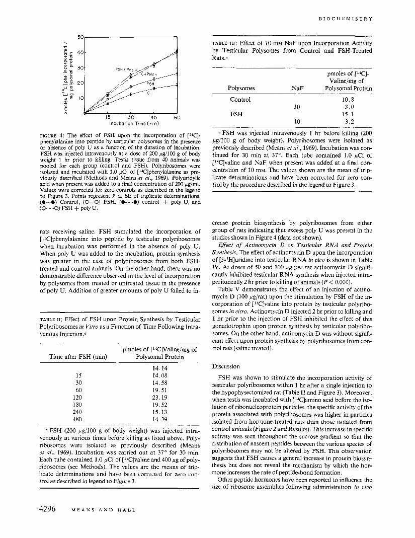
Influence oj NaF on Protein Synthesis by Testicular Polyribo- somes in Vitro. The influence of NaF (10 mM) upon protein biosynthesis by testicular polyribosomes from FSH-treated and control rats is shown in Table 111. Testicular polyribo- somes from rats receiving FSH incorporate more [ 14C]valine into peptides than do polyribosomes from rats receiving saline. On the other hand it can be seen that NaF inhibits protein bio- synthesis by testicular polyribosomes from both groups of rats. Moreover, net incorporation of [ l4C1valine (picomoles per milligram of polyribosomal protein) in the presence of NaF is approximately the same whether polyribosomes were derived from treated or untreated (control) rats.
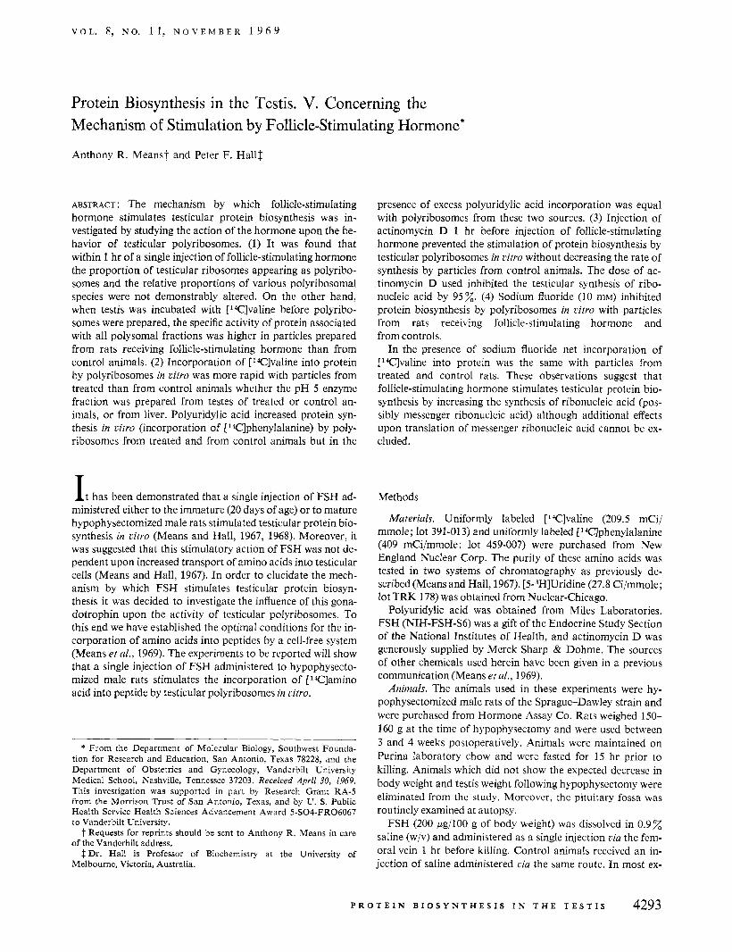
Effect ojPoly U on Polj~ribosoniril Protein Synthesis. Figure 4 shows the rate of incorporation of ['Elphenylalanine into protein in the presence and absence of polyuridylic acid using testicular polyribosomes from rats receiving FSH and from
P K O I ' E I N B I O S Y N T I - ~ ~ . S I S I N T H E T E S T I S 4295
B I O C H E M I S T R Y
W 0 - E a
15 30 45 60 Incubat ion Time (min)
FIGURE 4: The eflect of FSH upon the incorporation of [W]- phenylalanine into peptide by testicular polysomes in the presence or absence of poly U as a function of the duration of incubation. FSH was injected intravenously at a dose of 200 pg/100 g of body weight 1 hr prior to killing. Testis tissue from 40 animals was pooled for each group (control and FSH). Polyribosomes were isolated and incubated with 1.0 pCi of [14C]phenylalanine as pre- viously described (Methods and Means ef al., 1969). Polyuridylic acid when present was added to a final concentration of 200 pg/ml. Values were corrected for zero controls as described in the legend to Figure 3. Points represent R i SE of triplicate determinations. (0-0) Control, (0-0) FSH, (0- - -0) control + poly U, and (0- - -0) FSH + poly U.
rats receiving saline. FSH stimulated the incorporation of [ 14Clphenylalanine into peptide by testicular polyribosomes when incubation was performed in the absence of poly U. When poly U was added to the incubation, protein synthesis was greater in the case of polyribosomes from both FSH- treated and control animals. On the other hand, there was no demonstrable difference observed in the level of incorporation by polysomes from treated or untreated tissue in the presence of poly U. Addition of greater amounts of poly U failed to in-
TABLE 11: Effect of FSH upon Protein Synthesis by Testicular Polyribosomes in Vitro as a Function of Time Following Intra- venous 1njection.a
Time after FSH (min)
15 30 60
120 180 240 480
pmoles of I: 14C]Valine/mg of Polysomal Protein
14.14 14.08 14.58 19.51 23.19 19.52 15.13 14.39
FSH (200 pg/lOO g of body weight) was injected intra- venously at various times before killing as listed above. Poly- ribosomes were isolated as previously described (Means et al., 1969). Incubation was carried out at 37" for 30 min. Each tube contained 1 .O pCi of [ 14C]valine and 400 pg of poly- ribosomes (see Methods). The values are the means of trip- licate determinations and have been corrected for zero con- trol as described in legend to Figure 3.
TABLE III: Effect of 10 mM NaF upon Incorporation Activity by Testicular Polysomes from Control and FSH-Treated Rats.a
pmoles of [IC]- Valine/mg of
Polysomes NaF Polysomal Protein ~ ~ _ _
Control 10.8 10 3 . 0
FSH 15.1 10 3 . 2
Q FSH was injected intravenously 1 hr before killing (200 bg/lOO g of body weight). Polyribosomes were isolated as previously described (Means et a[., 1969). Incubation was con- tinued for 30 min at 37". Each tube contained 1.0 pCi of [14C]valine and NaF when present was added at a final con- centration of 10 mM. The values shown are the mean of trip- licate determinations and have been corrected for zero con- trol by the procedure described in the legend to Figure 3.
crease protein biosynthesis by polyribosomes from either group of rats indicating that excess poly U was present in the studies shown in Figure 4 (data not shown).
Effect of Actinomycin D on Testicular RNA and Protein Synthesis. The effect of actinomycin D upon the incorporation of [5-aH]uridine into testicular RNA in civo is shown in Table IV. Ai doses of 50 and 100 pg per rat actinomycin D signifi- cantly inhibited testicular RNA synthesis when injected intra- peritoneally 2 hr prior to killing of animals (P < 0.001).
Table V demonstrates the effect of an injection of actino- mycin D (100 pgirat) upon the stimulation by FSH of the in- corporation of [ 14C]valine into protein by testicular polyribo- somes in citro. Actinomycin D injected 2 hr prior to killing and 1 hr prior to the injection of FSH inhibited the effect of this gonadotrophin upon protein synthesis by testicular polyribo- somes. On the other hand, actinomycin D was without signifi- cant effect upon protein synthesis by polyribosomes from con- trol rats (saline treated).
Discussion
FSH was shown to stimulate the incorporation activity of testicular polyribosomes within 1 hr after a single injection to the hypophysectomized rat (Table I1 and Figure 3). Moreover, when testis was incubated with [14C]amino acid before the iso- lation of ribonucleoprotein particles, the specific activity of the protein associated with polyribosomes was higher in particles isolated from hormone-treated rats than those isolated from control animals (Figure 2 and Results). This increase in specific activity was seen throughout the sucrose gradient so that the distribution of nascent peptides between the various species of polyribosomes may not be altered by FSH. This observation suggests that FSH causes a general increase in protein biosyn- thesis but does not reveal the mechanism by which the hor- mone increases the rate of peptide-bond formation.
Other peptide hormones have been reported to influence the size of ribosome assemblies following administration in civo
4296 M E A N S A N D H A L L

V O L . 8, N O . 11 , N O V E M B E R 1 9 6 9
TABLE IV: Effect of Actinomycin D on Incorporation of [5-3H]Uridine into Testicular RNA in Viao.5
Actinomycin D Sp Act. of RNA (cldrat) (dpm/mg) Inhibn (%)
1234 50 209 85 100 64 95
0 Actinomycin D dissolved in propylene glycol was in- jected intraperitoneally 2 hr prior to killing the animals and 90 min before the intravenous injection of 10 pCi of [5-3H]- uridine; 30 min following the injection of isotope, rats were sacrificed by cervical dislocation, and testes were quickly excised and kept on ice. Following removal of the tunica, testis was homogenized in 2.5 volumes of ice water per g of tissue and HClOl was added to a final concentration of 0.4 M. Nucleic acids were then extracted from the precipitate by the method of Means and Hamilton (1966). Radioactivity was determined in the acid extract by liquid scintillation spec- trometry; 1 ml of the extract was placed in a scintillation vial and 15 ml of toluene scintillator containing 10% (v/v) collo- solve solubilizer (Beckman). Counting efficiency for 3H was 30-34x and absolute values were determined by auto- matic external standardization. RNA content was deter- mined by the method of Ceriotti (1955). Values represent the mean of duplicate determinations for each of six testes.
(Korner, 1964; Stirewalt ef al., 1967; Cohen and Stastny, 1968). Diabetes reduces the number of large polyribosomes that can be isolated from rat skeletal muscle. Insulin was dem- onstrated to cause a reassembly of ribosomes resulting in a complete restoration of polyribosomes compared with the non- diabetic animal (Stirewalt ef al., 1967). Similarly epidermal growth factor causes a conversion of preexisting ribosomal monomers into functional polysomal structures in the epi- dermis of the chick embryo (Cohen and Stastny, 1968). Clearly this is not the case with FSH acting upon the testis since no change is observed in the proportion of testicular ribosomes appearing as polyribosomes (as opposed to monosomes), nor is the distribution of ribosomes between the various species of polysomes demonstrably altered (Figure 1). Such a change in ribosomal distribution might be expected if FSH increased the synthesis of a single protein or group of proteins (Heywood et al., 1967; Heywood and Rich, 1968). It appears, therefore, that FSH does not stimulate testicular protein biosynthesis by recruiting monosomes to increase the number of polysomes with the existing levels of mRNA and also that this hormone probably produces a general increase in the synthesis of tes- ticular proteins.
The facts that polyribosomal preparation from rats re- ceiving FSH incorporate amino acids into proteins in citro more rapidly than preparations from saline-treated rats (Figure 3) and that this difference is independent of the source of the pH 5 enzyme fraction used (Table I) indicate that FSH does not accelerate testicular protein biosynthesis only by in- creasing amino acid transport or activation. It seems likely that FSH increases either the activity of ribosomes or the syn-
TABLE v: Effect of Actinomycin D upon the Stimulation by FSH of Amino Acid Incorporation by Testicular Polyribo- s0mes.a
~ ~~
pmoles of [14C]Valine/mg of Treatment Polysomal Protein
Control 14.86 + 1.62 14.74 i 2.01 Actinomycin D
FSH 22.31 =k 2.68 Actinomycin D + FSH 14.47 =t 1.88
5 Actinomycin D (100 pg/rat) was injected intraperitoneally 2 hr before killing and 1 hr before the intravenous injection of FSH (200 pg/lOO g of body weight). Testis tissue from eight animals was pooled for each treatment. Polyribosomes were isolated as previously described (Means et al., 1969). Incu- bation was for 30 min at 37" with ['Clvaline as the radio- active amino acid (Means et a/., 1969). Values represent R + SE of six determinations and were corrected for zero control (see legend to Figure 3).
thesis of mRNA. Since the same amount of polysomal prep- aration from treated and control animals (measured as RNA or as protein, see Results) was added to each flask in these ex- periments (Figure 3 and Table I) and since the distribution of ribosomes between the various species of polyribosomes was the same in both groups of rats (Figure I), the stimulation caused by FSH in the present system cannot be attributed to an increase in the number of ribosomes although our data do not exclude an additional effect of FSH upon the biosynthesis of ribosomes.
Addition of polyuridylic acid to ribonucleoprotein particles stimulates incorporation of phenylalanine into protein by particles from hormone-treated and from saline-treated rats (Figure 4) but in the presence of excess poly U incorporation was not significantly different with particles from either group of rats (Figure 4). It therefore appears that the capacity of polyribosomes to support peptide synthesis is the same whether these particles are isolated from treated or control animals. Similar results have been reported for the action of insulin upon protein synthesis by ribosomes from skeletal muscle of diabetic rats. Addition of poly U stimulated incor- poration by ribosomes from both normal and diabetic an- imals but restored to normal the efficiency of diabetic ribo- somes, thus negating the effect of insulin (Wool and Cavicchi, 1967). On the other hand, the differences between normal and diabetic ribosomes isolated from heart muscle are still ap- parent in the presence of excess poly U (Rampersad and Wool, 1965).
The data presented here concerning the abolition of the FSH effect upon polyribosomal incorporation activities by poly U suggest that this hormone may stimulate testicular protein synthesis by first increasing the synthesis of mRNA. However, in interpreting the results of experiments with poly U, it should be pointed out that we are by no means sure that this syn- thetic compound is a good model for natural mRNA (Oka- mot0 and Takanami, 1Y63). The observation that actinomycin D (in doses which inhibit the synthesis of testicular RNA by
P R O T E I N B I O S Y N T H E S I S I N T H E T E S T I S 4297

I3 I O C H E M I S 1 K Y
95 (Table IV)) prevents the increased protein biosynthesis produced by FSH without inhibiting testicular protein syn- thesis in saline-treated rats (Table V) supports the suggestion that this FSH effect is mediated by a prior enhancement of ksticular RNA synthesis.
Inhibition of the FSH-mediated increase in protein syn- thesis by actinomycin D illustrates yet another difference be- tween the action of this gonadotrophin compared with other peptide hormones. Wool and Cavicchi (1966) have presented evidence that a deficiency of template RNA may not be the basis for the primary defect in protein biosynthesis in diabetic animals. Insulin restores polysomal incorporation activity by muscle ribosomes to normal even in diabetic animals pre- treated with actinomycin D in doses sufficient to completely suppress RNA synthesis. Again although growth hormone stimulates the synthesis of RNA (Talwar et al., 1962), it has been argued that the primary anilbolic effect is not a stimula- tion of mRNA synthesis since effects of this hormone have been obtained when RNA synthesis was inhibited by actino- mycin D (Korner, 1964; Martin and Young, 1965).
Korner (1969) has suggested that the growth hormone effect on liver ribosomes is to stimulate the translation process once the ribosome is attached to the message, that is, upon com- pletion of previously initiated peptide chains. This conclusion was based on the fact that dextran sulfate, which has been re- ported to specifically block chain initiation (Munro, 1965), had no effect on the stimulation of protein synthesis by growth hormone (Korner, 1969). Sodium fluoride has also been dem- onstrated to specifically inhibit chain initiation when added to a ribosomal system from reticulocytes to a final concentration of 10 mM (Lin et al., 1966). The same concentration of NaF in the present studies caused considerable inhibition of incorpo- ration of [14C]valine in polyribosomal preparations from both hormone-treated and control animals. Moreover, this inhibi- tion reduced net incorporation of [ lC]valine (picomoles per milligram of ribosomal protein) to the same level by particles from FSH-treated and control rats (Table 111). If the findings of Lin el al. (1966) apply equally to the testis, it seems reasonable to suggest that FSH stimulates protein synthesis by primarily accelerating chain initiation, thus raising the possibility of another difference between the action of FSH and growth hor- mone. Clearly, however, other interpretations of these obser- vations is possible.
At present the nature of chain initiation in mammalian cells is not well understood, so that further speculation concerning the mechanism by which FSH influences this process in the
testis is not likely to prove helpful at this time. However, these studies suggest that the influence of FSH upon the synthesis of testicular RNA may throw further light upon the mechanism by which this gonadotrophin stimulates testicular protein bio- synthesis. To this end studies on effects of FSH on testicular RNA metabolism are at present being undertaken in this lab- oratory.
Acknowledgment
lent technical assistance. The authors are grateful to Mr. Charles R. Mena for excel-
References
Ceriotti, G. (1955),J. Biol. Chem. 214,59. Cohen, S., and Stastny, M. (1968), Biochim. Biophys. Acta
Heywood, S. M., Dawben, R. M., and Rich, A. (1967), Proc.
Heywood, S. M., and Rich, A. (1968), Proc. Natl. Acad. Sci.
Korner, A. (1964), Biochem. J . 92,449. Korner, A. (1969), Biochim. Biophys. Acta 174,351. Lin, S., Mosteller, R. D., and Hardesty, B. (1966), J . Mol.
Lowry, 0. H., Rosebrough, N. J., Farr, A. L., and Randall,
Martin, T. E., and Young, F. G. (1965), Nature 208,684. Means, A. R., and Hall, P. F. (1967), Endocrinology 81, 1151. Means, A. R., and Hall, P. F. (1968), Endocrinology 82,597. Means, A. R., Hall, P. F., Nicol, L. W., Sawyer W. H., and
Means, A. R., and Hamilton, T. H. (1966), Proc. Natl. Acad.
Munro, A. J. (1965), Absrr. 2nd Meeting Fed. European
Okamoto, T., and Takanami, M. (1963), Biochim Biophys.
Rampersad, 0. R., and Wool, I. G. (1965), Science 149, 1102. Stirewalt, W. S., Wool, I. G., and Cavicchi, P. (1967), Proc.
Talwar, G. P., Panda, N. A., Sarin, G. S., and Tolani, A. J.
Wool, I. G., and Cavicchi, P. (1967), Biochemistry 6, 1231.
166,427.
Natl. Acad. Sci. U. S . 57, 1002.
U. S. 59,590.
Biol. 21, 51.
R. J. (1951), J . Biol. Chem. 193,265.
Baker, C. A. (1969), Biochemistry 8, 1488.
Sci. U. S. 56, 686.
Biochem. SOC., Vienna, 2.
Acta 76,266.
Natl. Acad. Sci. U. S. 57, 1885.
(1962), Biochem. J. 82, 173.
4298 M L A N S A N D H A L L