protease inhibitors as models for the study of oxidative...
TRANSCRIPT
FORUM REVIEW ARTICLE
Protease Inhibitors as Models for the Studyof Oxidative Folding
Joan L. Arolas* and Salvador Ventura
Abstract
The correct balance between proteases and their natural protein inhibitors is of great importance in livingsystems. Protease inhibitors usually comprise small folds that are crosslinked by a high number of disulfidebonds, making them perfect models for the study of oxidative folding. To date, the oxidative folding of nu-merous protease inhibitors has been analyzed, revealing a great diversity of folding pathways that differ mainlyin the heterogeneity and native disulfide-bond content of their intermediates. The two extremes of this diversityare represented by bovine pancreatic trypsin inhibitor and hirudin, which fold, respectively, via few nativeintermediates and heterogeneous scrambled isomers. Other proteins, such as leech carboxypeptidase inhibitor,share characteristics of both models displaying mixed folding pathways. The study of the oxidative folding oftwo-domain inhibitors, such as secretory leukocyte protease inhibitor, tick carboxypeptidase inhibitor, andAscaris carboxypeptidase inhibitor, has provided some clues about how two-domain protease inhibitors mayfold, that is, either by folding each domain autonomously or with one domain assisting in the folding of theother. Finally, the recent determination of the structures of the major intermediates of protease inhibitors hasshed light on the molecular mechanisms guiding the oxidative folding of small disulfide-rich proteins. Antioxid.Redox Signal. 14, 97–112.
Introduction
Proteolytic enzymes, commonly referred to as proteases,are essential in nature for the survival of all living or-
ganisms. Despite their important physiological functions,enzymes that cleave other proteins are potentially verydamaging to their environment, so their activities must bekept strictly under control. Distinct mechanisms exist for theregulation of the catalytic activity of proteases, very importantamong them being the interactions of proteases with theirprotein inhibitors (17). Protein inhibitors of proteolytic en-zymes comprise the largest and structurally most diversegroup of naturally occurring enzyme inhibitors. A compre-hensive list of the different inhibitor families is availableon the web at http:==merops.sanger.ac.uk= (62). Protease in-hibitors are usually classified either by their mechanismsof action, which are surprisingly diverse, or by the type ofprotease they inhibit, that is, serine, cysteine, aspartic, ormetalloproteases—protein inhibitors of threonine and gluta-myl proteases are yet to be discovered [for review, see (58,63)]. Protease inhibitors are often small in size, with one ormultiple tandem domains, and are crosslinked by several
disulfide bonds. These characteristics provide protease in-hibitors with high stability and a long half-life and makethem very attractive models for the study of protein folding.Besides enzymes such as ribonuclease A (RNase A) andlysozyme, growth factors, and toxins [for review, see (2)],numerous serine and metalloprotease inhibitors have beeninvestigated using the technique of oxidative folding.
Oxidative folding is the in vitro process by which a fullyreduced and unfolded protein gains both its native disulfidebonds (disulfide-bond formation) and native structure (con-formational folding). The folding processes of many proteaseinhibitors can be efficiently tracked owing to the small anddisulfide-rich nature of their protein folds. Thus, their foldingintermediates are chemically trapped in a time-course man-ner, either irreversibly by alkylation or reversibly by acidifi-cation, and further analyzed=purified by reversed-phasechromatography [for review, see (55)]. The isolation of thesepartially folded species allows their characterization in termsof disulfide-bond connectivity (number and type—native ornonnative) and structure. The derived information will helpto define the studied protein’s oxidative folding pathwayon the basis of the following features: (i) the extent of the
Departament de Bioquımica i Biologia Molecular, Institut de Biotecnologia i Biomedicina, Universitat Autonoma de Barcelona, Bellaterra(Barcelona), Spain.
*Current affiliation: Proteolysis Lab, Department of Structural Biology, Molecular Biology Institute of Barcelona, CSIC, Barcelona, Spain.
ANTIOXIDANTS & REDOX SIGNALINGVolume 14, Number 1, 2011ª Mary Ann Liebert, Inc.DOI: 10.1089=ars.2010.3456
97
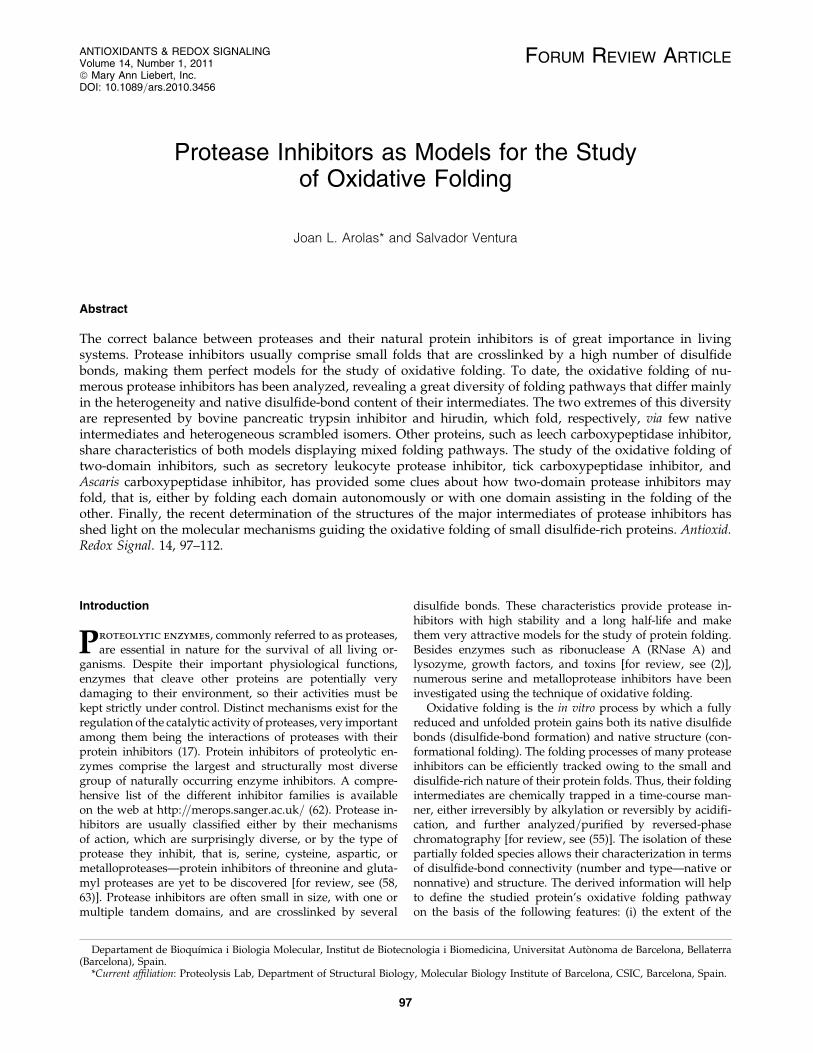
heterogeneity of folding intermediates, (ii) the predominanceof intermediates containing native disulfide bonds andnative-like structures, and (iii) the presence of scrambledisomers (fully oxidized species containing at least two non-native disulfides) as intermediates along the folding reaction(32, 33). The two opposite extremes of the oxidative foldingpathways are exemplified by proteins that fold through theformation of a few intermediates adopting native disulfidebonds, and proteins that fold via highly heterogeneous inter-mediates containing nonnative disulfides, that is, their foldingpathways are populated by scrambled isomers (Fig. 1). On theother hand, other proteins share similarity and dissimilaritywith both models, displaying mixed folding pathways.
The present work reviews the pathways of oxidative fold-ing reported thus far for single- and two-domain proteaseinhibitors. The determinants that account for the large di-versity of folding landscapes in these proteins are discussed,together with the recent structural determination of disulfideintermediates that provide fundamental insights into a betterunderstanding of oxidative folding.
Oxidative Folding via Few Native Intermediates
Bovine pancreatic trypsin inhibitor
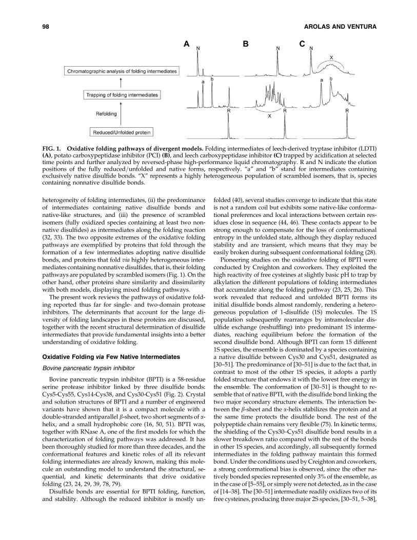
Bovine pancreatic trypsin inhibitor (BPTI) is a 58-residueserine protease inhibitor linked by three disulfide bonds:Cys5-Cys55, Cys14-Cys38, and Cys30-Cys51 (Fig. 2). Crystaland solution structures of BPTI and a number of engineeredvariants have shown that it is a compact molecule with adouble-stranded antiparallel b-sheet, two short segments of a-helix, and a small hydrophobic core (16, 50, 51). BPTI was,together with RNase A, one of the first models for which thecharacterization of folding pathways was addressed. It hasbeen thoroughly studied for more than three decades, and theconformational features and kinetic roles of all its relevantfolding intermediates are already known, making this mole-cule an outstanding model to understand the structural, se-quential, and kinetic determinants that drive oxidativefolding (23, 24, 29, 39, 78, 79).
Disulfide bonds are essential for BPTI folding, function,and stability. Although the reduced inhibitor is mostly un-
folded (40), several studies converge to indicate that this stateis not a random coil but exhibits some native-like conforma-tional preferences and local interactions between certain res-idues close in sequence (44, 46). These contacts appear to bestrong enough to compensate for the loss of conformationalentropy in the unfolded state, although they display reducedstability and are transient, which means that they may beeasily broken during subsequent conformational folding (28).
Pioneering studies on the oxidative folding of BPTI wereconducted by Creighton and coworkers. They exploited thehigh reactivity of free cysteines at slightly basic pH to trap byalkylation the different populations of folding intermediatesthat accumulate along the folding pathway (23, 25, 26). Thiswork revealed that reduced and unfolded BPTI forms itsinitial disulfide bonds almost randomly, rendering a hetero-geneous population of 1-disulfide (1S) molecules. The 1Spopulation subsequently rearranges by intramolecular dis-ulfide exchange (reshuffling) into predominant 1S interme-diates, reaching equilibrium before the formation of thesecond disulfide bond. Although BPTI can form 15 different1S species, the ensemble is dominated by a species containinga native disulfide between Cys30 and Cys51, designated as[30–51]. The predominance of [30–51] is due to the fact that, incontrast to most of the other 1S species, it adopts a partlyfolded structure that endows it with the lowest free energy inthe ensemble. The conformation of [30–51] is thought to re-semble that of native BPTI, with the disulfide bond linking thetwo major secondary structure elements. The interaction be-tween the b-sheet and the a-helix stabilizes the protein and atthe same time protects the disulfide bond. The rest of thepolypeptide chain remains very flexible (75). In kinetic terms,the shielding of the Cys30–Cys51 disulfide bond results in aslower breakdown ratio compared with the rest of the bondsin other 1S species, and accordingly, all subsequently formedintermediates in the folding pathway maintain this formedbond. Under the conditions used by Creighton and coworkers,a strong conformational bias is observed, since the other na-tively bonded species represented only 3% of the ensemble, asin the case of [5–55], or simply were not detected, as in the caseof [14–38]. The [30–51] intermediate readily oxidizes two of itsfree cysteines, producing three major 2S species, [30–51, 5–38],
FIG. 1. Oxidative folding pathways of divergent models. Folding intermediates of leech-derived tryptase inhibitor (LDTI)(A), potato carboxypeptidase inhibitor (PCI) (B), and leech carboxypeptidase inhibitor (C) trapped by acidification at selectedtime points and further analyzed by reversed-phase high-performance liquid chromatography. R and N indicate the elutionpositions of the fully reduced=unfolded and native forms, respectively. ‘‘a’’ and ‘‘b’’ stand for intermediates containingexclusively native disulfide bonds. ‘‘X’’ represents a highly heterogeneous population of scrambled isomers, that is, speciescontaining nonnative disulfide bonds.
98 AROLAS AND VENTURA
[30–51, 5–14], and [30–51, 14–38], among which only the lastone contains two native disulfide bonds; that is, it may betermed des[5–55]. Although the formation of the Cys5–Cys55disulfide bond in des[5–55] would lead to native BPTI, thisstep finds a high energy barrier for its completion due to thehighly folded structure of this intermediate. In contrast, thisintermediate can rearrange its second disulfide bond to formeither of the nonnative Cys5–Cys14 or Cys5–Cys38 bonds andtherefore is in rapid equilibrium with the other two 2S inter-mediates. Subsequently, in the slowest intramolecular reac-tion of the pathway, these two intermediates reshuffle theirnonnative disulfide bonds to render [30–51, 5–55], which
contains two of the three native BPTI disulfides (des[14–38]).In this intermediate the free thiols of Cys14 and Cys38 are onthe surface and spatially close, and may oxidize rapidly toform the native inhibitor.
Weissman and Kim investigated BPTI folding with adifferent experimental approach using acid-trapping andreversed-phase high-performance liquid chromatographyanalysis of the intermediates. They proposed a revised path-way for the oxidative folding of this inhibitor, which, incontrast to that proposed by Creighton and coworkers, ischaracterized by the absence of stable intermediates contain-ing nonnative disulfide bonds (Fig. 2, model a vs. b) (78, 79).
FIG. 2. Oxidative folding of bovine pancreatic trypsin inhibitor (BPTI)-like proteins. The amino acid sequence, sec-ondary structure elements, and disulfide pairings of BPTI (3 SS and 58 residues), LDTI (3 SS and 46 residues), and Momordicacochinchinensis trypsin inhibitor II (MCoTI-II; 3 SS and 34 residues) are schematically shown. The Protein Data Bank ID codesfor the structures of BPTI, LDTI, and MCoTI-II are, respectively, 1PIT, 1LDT, and 1IB9 (ribbon plots prepared with programPyMOL). The disulfide bonds are shown as black sticks. An overview of the oxidative folding pathway of BPTI [modelsproposed by Creighton and coworkers (a) and Weissman and Kim (b)], LDTI, and MCoTI-II is shown below each sequence. Rand N indicate the fully reduced=unfolded and native forms, respectively. The disulfide pairings of major folding inter-mediates are shown between parentheses, and the missing disulfides between parentheses with the prefix ‘‘des’’ (des species).1S and 2S are ensembles of molecules with the corresponding number of disulfide bonds.
OXIDATIVE FOLDING OF PROTEASE INHIBITORS 99

This results in a less heterogeneous pathway with only sixsignificantly populous intermediates. The 1S population ini-tially forms its disulfide bonds almost randomly, but thenreshuffles rapidly to render two natively bonded species: thealready-described [30–51] intermediate and a new interme-diate [5–55], which also likely possesses a folded conforma-tion. In both forms, the free Cys14 and Cys38 would belocated in flexible regions preferentially exposed to solventwhen compared with the accessibility of the other two freethiols. Accordingly, the formation of the Cys14–Cys38 dis-ulfide in this 1S species would render only two 2S interme-diates: [5–55, 14–38] (named des[30–51] or N*) and [30–51, 14–38] (named des[5–55] or N0). These two forms act as strongkinetic traps, freezing the folding reaction. Importantly, onlyhalf of the unfolded and reduced BPTI molecules are able tofold productively into the native form. The rest of the proteinremains kinetically trapped as N*. Different structural studieshave shown that N* has a native-like conformation in whichthe free thiols of Cys30 and Cys50 are buried and protectedfrom the solvent, which precludes their oxidation. In this way,N* is defined as an off-pathway intermediate that is stable forweeks in this conformation. Confirming this view, it has beenrecently reported that redox conditions that disfavor the ac-cumulation of N* tend to accelerate and increase the pro-ductivity of the BPTI folding reaction (52). As observed byCreighton and coworkers, N0 is a well-folded intermediate inwhich the missing disulfide bond between Cys5 and Cys55cannot be directly formed to render the native BPTI structure.According to Weissman and Kim, the burial of the reactivecysteine residues inside the N0 structure forces this interme-diate to unfold, to gain its native conformation. However,fluctuations inside the N0 structure promote a rearrangementreaction in which the free thiols attack the already formeddisulfides in the intermediate and promote the conversion ofN0 into N* and the [5–55, 30–51] intermediate (named des[14–38] or NSH-SH). The stability of the N0 structure makes un-folding reactions energetically expensive so that the rate ofthis rearrangement is slow. In addition, the formation of N*and NSH-SH from N0 is not a direct reaction but implies thecreation of a transient population of two intermediates con-taining nonnative disulfide bonds, [30–51, 5–14] and [30–51,5–38], which correspond to the productive nonnative inter-mediates in Creighton’s scheme. Once NSH-SH is formed, asdescribed above, the flexibility and accessibility of the freeCys14 and Cys34 thiols allow rapid oxidation to form thenative protein.
The disparity in the BPTI folding pathways proposed byboth laboratories conforms to the different solution conditionsin which the refolding reactions were performed and thedifferent strategies used to trap and analyze the intermedi-ates. With time, Weissman and Kim’s view has prevailed andBPTI is considered the archetype of a disulfide-rich protein inwhich the folding pathway proceeds rapidly through theformation of a limited number of stable, native-like interme-diates in the absence of scrambled isomers.
An important point raised by Creighton and coworkers isthat inside the cell BPTI is synthesized as a pre-pro-inhibitor,containing a 22-residue pre N-terminal extension that leads toits translocation into the endoplasmic reticulum and two pro-segments of 13 and 7 residues at the N- and C-terminal sidesof the mature sequence, respectively. It is not the mature formthat folds in the endoplasmic reticulum but the pro-inhibitor.
The pro-segment contains a cysteine residue that has beenshown to react with the mature BPTI cysteines (80). However,no significant differences were observed between the foldingrates of mature and pro-BPTI in microsomes (27) and thetruncation of the side chain of the pro-segment’s cysteine bymutation to serine did not affect the folding reaction either.Unfortunately, a detailed picture of the folding pathway ofthe physiologically relevant pro-BPTI form is still missing.
Leech-derived tryptase inhibitor
Human tryptase b is a trypsin-like serine protease involvedin the pathogenesis of disorders such as asthma and arthritis(71). Leech-derived tryptase inhibitor (LDTI) is a nonclassicalKazal-type inhibitor isolated from the medicinal leech Hirudomedicinalis (14, 72). It is one of two proteins shown to bindtightly to human tryptase b, making it a molecule of consid-erable biomedical interest. LDTI comprises 46 residues andthree disulfide bonds (Cys4-Cys29, Cys6-Cys25, and Cys14-Cys40) (Fig. 2). Its structure, solved both in solution by nu-clear magnetic resonance (NMR) and in a crystalline complexwith trypsin, shows a short central a-helix and a small triple-stranded antiparallel b-sheet (41, 56, 73). The first two dis-ulfides stabilize the a-helix, joining it to the N-terminus of theprotein in a cysteine-stabilized a-helical motif that is widelyfound in other bioactive peptides such as endothelins andtoxins from insects and snakes. In the third disulfide, Cys14 inthe b1-strand is linked to Cys40, restricting the mobility of theb3-strand, and thereby stabilizing the small b-sheet. Thisparticular topology and disulfide connectivity makes LDTI aninteresting model protein for oxidative folding studies.
The oxidative folding pathway of LDTI has been re-cently analyzed using acid-trapping, reversed-phase high-performance liquid chromatography and disulfide-pairinganalysis of its intermediates (3, 59). The folding of LDTI fromthe reduced and unfolded state proceeds through a sequentialoxidation of its cysteine residues toward the native confor-mation. As in the case of BPTI, although 75 different disulfide-bonded intermediates can be theoretically formed during therefolding reaction, only 5 of these species accumulate to asignificant extent. First, LDTI oxidizes one of its disulfides torender 1S intermediates that rapidly reach equilibrium. Thedistribution of disulfide bonds in this ensemble significantlydeviates from that expected from simple loop entropy contri-butions, with a preferential accumulation of the nativelybonded [14–40] species. NMR studies of [14–40] strongly sug-gest that the characteristic small triple-stranded antiparallelb-sheet of LDTI is already present in this intermediate. It islikely that secondary structure propensities promote thetransient formation of the b-sheet approaching Cys14 andCys40. The formation of the disulfide bond linking the twoexternal b-strands would further stabilize the b-sheet. Thestructural properties of [14–40] resemble those of [30–51] BPTIin the sense that the presence of a folded secondary structurepreferentially protects the disulfide bond from reduction andkinetically favors the prevalence of this intermediate in the 1Spopulation. Two of the three 2S folding intermediates, des[6–25] and des[4–29], are generated directly from the oxidation oftwo of its free cysteines. The rest of the 1S population re-arranges and oxidizes its disulfide and free cysteines, respec-tively, to form the third 2S intermediate: des[14–40]. Therefore,the three 2S intermediates contain exclusively native disulfide
100 AROLAS AND VENTURA
bonds. In principle, the oxidation of the two free cysteines inany of these species would result in the formation of a nativeand functional LDTI. However, when the intermediates weretrapped and isolated and their folding was re-initiated atneutral pH, it became clear that neither des[14–40] nor des[6–25] can form directly native LDTI but rather reshuffle theirdisulfides to form des[4–29], which turns out to be the onlyproductive intermediate in the folding pathway. The conver-gence of all 2S intermediates into des[4–29] is due to both therelative conformational properties of these species and thedifferent reactivity of their free cysteines. The recently deter-mined structures of these three 2S intermediates show thatthey are highly native (see structural discussion later) (59). Inthe des[4–29] structure, Cys29 at the C-terminus of the a-helixis fully exposed and can react with Cys4 at the highly flexibleN-terminus to produce the native inhibitor. The des[6–25] in-termediate has a very short half-life and rapidly reshuffles intodes[4–29]. Cys6 has a highly reactive thiolate group. However,in the structure of the intermediate it is far away from Cys25,which precludes the direct oxidation reaction. Instead, Cys6attacks the closer Cys4-Cys29 disulfide bond promoting a fastreshuffling to form des[4–29]. The oxidative folding of BPTIillustrated how the formation of a disulfide bond is kineticallyimpeded, when, once formed, becomes protected in a stableand structured conformation. This is likely the reason thedes[14–40] intermediate, similar to des[5–55] and des[30–51] inBPTI, cannot oxidize directly into the native form and insteadrearranges to form des[4–29]. In contrast to what is observedfor des[6–25], the rearrangement of des[14–40] into the pro-ductive intermediate is a slow reaction, which promotes theaccumulation of this intermediate during the folding reaction.Overall, the folding pathway of LDTI is dominated by thepresence of four natively bonded intermediates without sig-nificant accumulation of scrambled forms. This simplicity inthe conformational space that the protein has to sample toattain the native structure results in a fast and efficient foldingpathway.
Momordica cochinchinensis trypsin inhibitor II
Momordica cochinchinensis trypsin inhibitor II (MCoTI-II) isa serine protease inhibitor belonging to the cyclotide sub-family (22). It comprises 34 residues and three disulfidebonds, with a structure similar to that of squash protease in-hibitors, with their cysteine residues displaying a I–IV, II–V,and III–VI connectivity forming a classical cystine knot motif(Fig. 2) (42, 49). However, in the case of MCoTI-II the N- andC-terminus of the molecule are covalently linked resulting in acyclic molecule (20). The combination of these two structuralfeatures endows this inhibitor with an exceptionally highstability. The oxidative folding pathway of MCoTI-II andother cyclotides has been studied and reviewed by Craik andcoworkers (19, 20). The folding pathway of MCoTI-II is ex-tremely simple, with a predominant 2S intermediate (des[1–18]; see structural discussion later) arising from the oxidationof an initially heterogeneous 1S population. This intermediatehas the two native Cys8-Cys20 and Cys14-Cys26 disulfidebonds and lacks the Cys1-Cys18 disulfide that is formed bydirect oxidation leading to the native protein. The formationof the third disulfide bond is the rate-limiting step in the ox-idative folding reaction. Interestingly, apart from this mainroute there is a minor formation of other 2S intermediates that
can also attain the native structure. The energy barrier pre-cluding the rapid oxidation of the des[1–18] intermediate intonative MCoTI-II is thought to depend on a structural re-arrangement that would allow orienting the reactive thiols inthe free cysteines in proximity (21). The circular structure ofMCoTI-II strongly reduces the entropy associated with theunfolded and reduced state of the inhibitor relative to that ofother linear molecules and is likely responsible for the limitednumber of conformational states the protein has to samplebefore attaining the native structure. Again, as in the cases ofBPTI and LDTI, the presence of a reduced number of nativelybonded intermediates and the absence of scrambled isomersresults in a fast and efficient folding process.
Oxidative Folding via HeterogeneousScrambled Isomers
Hirudin
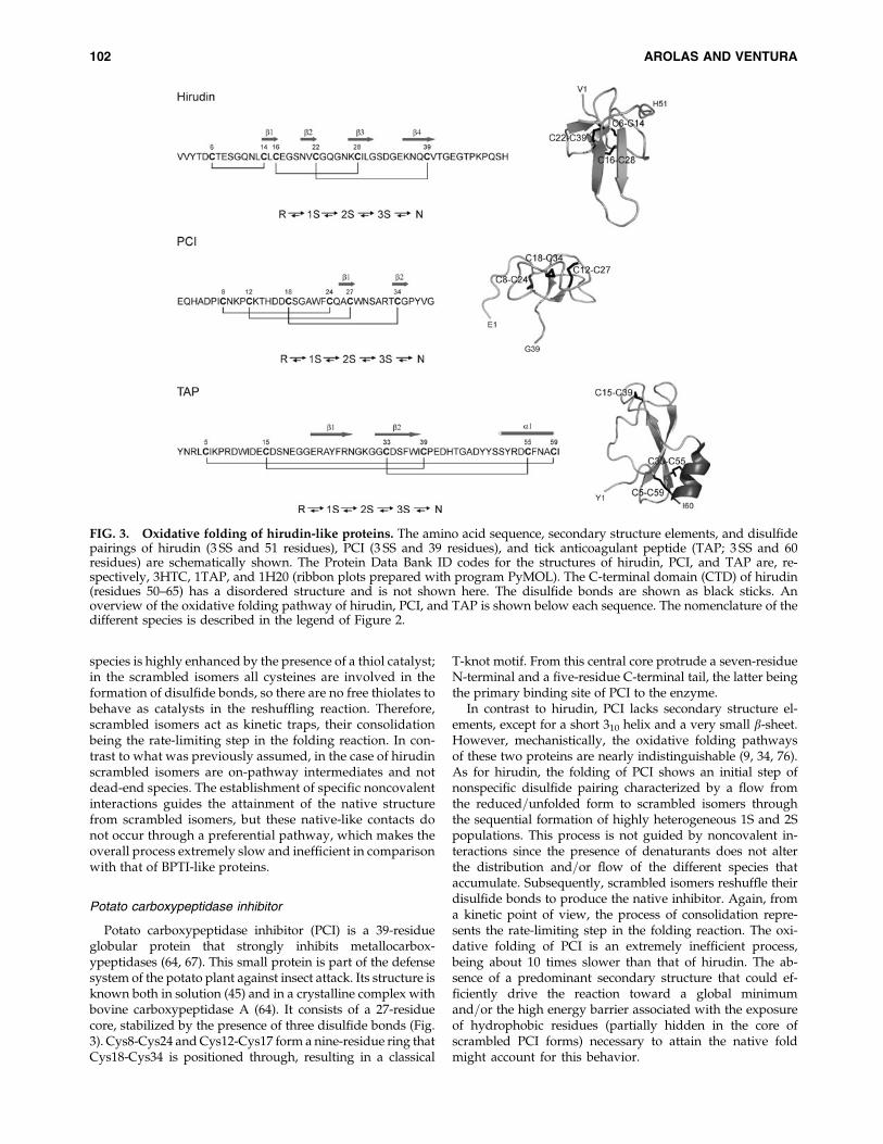
Hirudin is a thrombin-specific protease inhibitor iso-lated from the medicinal leech H. medicinalis (30). It is a65-residue protein consisting of two functional domains: aglobular N-terminal domain (NTD) formed by four b-sheetscrosslinked by three disulfide bonds (Cys6-Cys14, Cys16-Cys28, and Cys22-Cys39) and a disordered negativelycharged C-terminal extension (Fig. 3). The isolated functionaldomains possess anticoagulant activity and bind to differentregions of the enzyme: the globular domain docks into thecatalytic site of thrombin, whereas the C-terminal tail interactswith its fibrinogen binding site (43, 48, 68).
Chatrenet and Chang studied the oxidative folding ofhirudin in the absence and presence of a thiol agent (37). Incontrast to the proteins discussed in the previous section, nopredominant folding pathways or preferentially populatedintermediates were detected. Hirudin appears to fold fol-lowing a trial-and-error mechanism in which the six cyste-ines are equally involved in the disulfide reshufflingreactions that lead to the native state. The main intermedi-ates that populate the folding process of hirudin are non-natively bonded 3S species, that is, scrambled isomers. In afurther study the same authors investigated the folding re-action of the globular domain of hirudin (38). The folding ofthis protein can be dissected into two well-differentiatedstages: an initial stage of nonspecific packing and a final stepof consolidation that leads to the native structure. In the firststage, reduced and unfolded hirudin oxidizes its free cyste-ines to sequentially form equilibrated populations of 1S, 2S,and 3S (scrambled) intermediates. All these ensembles arehighly heterogeneous and there is no apparent contributionof noncovalent interactions to the intermediate distribution.Of a total of 60 potential 1S and 2S species, at least 30 ac-cumulate in the reaction, and 11 of the 14 theoretically pos-sible scrambled isomers could be detected (36). This findingsuggests that the driving force at this initial folding stage isan unspecific hydrophobic collapse to attain a compactconformation through a nearly random disulfide pairing.Scrambled hirudin forms are more compact than 1S or 2Sintermediates, but they do not possess a unique restrictedconformation and accordingly, in contrast to native hirudin,are highly sensitive to proteolytic attack. During the laststage of folding this heterogeneous population of scrambledintermediates reshuffles its disulfide bonds to attain na-tive disulfide connectivity. The consolidation of scrambled
OXIDATIVE FOLDING OF PROTEASE INHIBITORS 101
species is highly enhanced by the presence of a thiol catalyst;in the scrambled isomers all cysteines are involved in theformation of disulfide bonds, so there are no free thiolates tobehave as catalysts in the reshuffling reaction. Therefore,scrambled isomers act as kinetic traps, their consolidationbeing the rate-limiting step in the folding reaction. In con-trast to what was previously assumed, in the case of hirudinscrambled isomers are on-pathway intermediates and notdead-end species. The establishment of specific noncovalentinteractions guides the attainment of the native structurefrom scrambled isomers, but these native-like contacts donot occur through a preferential pathway, which makes theoverall process extremely slow and inefficient in comparisonwith that of BPTI-like proteins.
Potato carboxypeptidase inhibitor
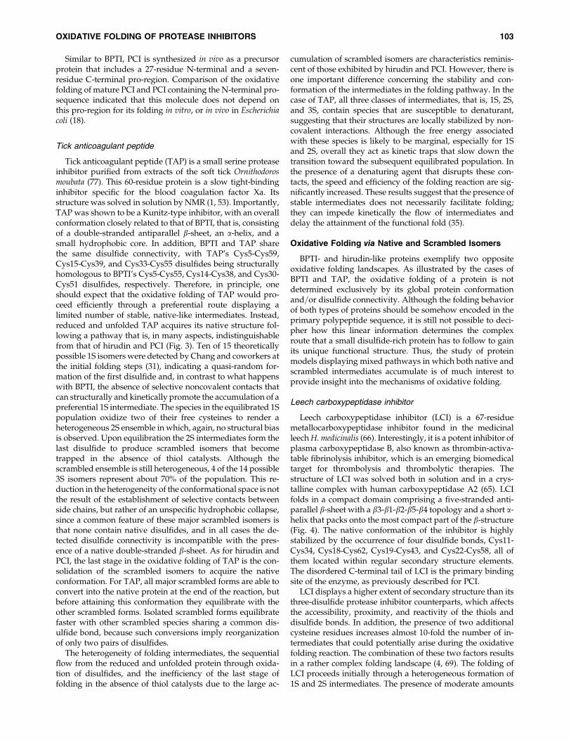
Potato carboxypeptidase inhibitor (PCI) is a 39-residueglobular protein that strongly inhibits metallocarbox-ypeptidases (64, 67). This small protein is part of the defensesystem of the potato plant against insect attack. Its structure isknown both in solution (45) and in a crystalline complex withbovine carboxypeptidase A (64). It consists of a 27-residuecore, stabilized by the presence of three disulfide bonds (Fig.3). Cys8-Cys24 and Cys12-Cys17 form a nine-residue ring thatCys18-Cys34 is positioned through, resulting in a classical
T-knot motif. From this central core protrude a seven-residueN-terminal and a five-residue C-terminal tail, the latter beingthe primary binding site of PCI to the enzyme.
In contrast to hirudin, PCI lacks secondary structure el-ements, except for a short 310 helix and a very small b-sheet.However, mechanistically, the oxidative folding pathwaysof these two proteins are nearly indistinguishable (9, 34, 76).As for hirudin, the folding of PCI shows an initial step ofnonspecific disulfide pairing characterized by a flow fromthe reduced=unfolded form to scrambled isomers throughthe sequential formation of highly heterogeneous 1S and 2Spopulations. This process is not guided by noncovalent in-teractions since the presence of denaturants does not alterthe distribution and=or flow of the different species thataccumulate. Subsequently, scrambled isomers reshuffle theirdisulfide bonds to produce the native inhibitor. Again, froma kinetic point of view, the process of consolidation repre-sents the rate-limiting step in the folding reaction. The oxi-dative folding of PCI is an extremely inefficient process,being about 10 times slower than that of hirudin. The ab-sence of a predominant secondary structure that could ef-ficiently drive the reaction toward a global minimumand=or the high energy barrier associated with the exposureof hydrophobic residues (partially hidden in the core ofscrambled PCI forms) necessary to attain the native foldmight account for this behavior.
FIG. 3. Oxidative folding of hirudin-like proteins. The amino acid sequence, secondary structure elements, and disulfidepairings of hirudin (3 SS and 51 residues), PCI (3 SS and 39 residues), and tick anticoagulant peptide (TAP; 3 SS and 60residues) are schematically shown. The Protein Data Bank ID codes for the structures of hirudin, PCI, and TAP are, re-spectively, 3HTC, 1TAP, and 1H20 (ribbon plots prepared with program PyMOL). The C-terminal domain (CTD) of hirudin(residues 50–65) has a disordered structure and is not shown here. The disulfide bonds are shown as black sticks. Anoverview of the oxidative folding pathway of hirudin, PCI, and TAP is shown below each sequence. The nomenclature of thedifferent species is described in the legend of Figure 2.
102 AROLAS AND VENTURA
Similar to BPTI, PCI is synthesized in vivo as a precursorprotein that includes a 27-residue N-terminal and a seven-residue C-terminal pro-region. Comparison of the oxidativefolding of mature PCI and PCI containing the N-terminal pro-sequence indicated that this molecule does not depend onthis pro-region for its folding in vitro, or in vivo in Escherichiacoli (18).
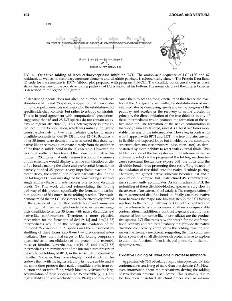
Tick anticoagulant peptide
Tick anticoagulant peptide (TAP) is a small serine proteaseinhibitor purified from extracts of the soft tick Ornithodorosmoubata (77). This 60-residue protein is a slow tight-bindinginhibitor specific for the blood coagulation factor Xa. Itsstructure was solved in solution by NMR (1, 53). Importantly,TAP was shown to be a Kunitz-type inhibitor, with an overallconformation closely related to that of BPTI, that is, consistingof a double-stranded antiparallel b-sheet, an a-helix, and asmall hydrophobic core. In addition, BPTI and TAP sharethe same disulfide connectivity, with TAP’s Cys5-Cys59,Cys15-Cys39, and Cys33-Cys55 disulfides being structurallyhomologous to BPTI’s Cys5-Cys55, Cys14-Cys38, and Cys30-Cys51 disulfides, respectively. Therefore, in principle, oneshould expect that the oxidative folding of TAP would pro-ceed efficiently through a preferential route displaying alimited number of stable, native-like intermediates. Instead,reduced and unfolded TAP acquires its native structure fol-lowing a pathway that is, in many aspects, indistinguishablefrom that of hirudin and PCI (Fig. 3). Ten of 15 theoreticallypossible 1S isomers were detected by Chang and coworkers atthe initial folding steps (31), indicating a quasi-random for-mation of the first disulfide and, in contrast to what happenswith BPTI, the absence of selective noncovalent contacts thatcan structurally and kinetically promote the accumulation of apreferential 1S intermediate. The species in the equilibrated 1Spopulation oxidize two of their free cysteines to render aheterogeneous 2S ensemble in which, again, no structural biasis observed. Upon equilibration the 2S intermediates form thelast disulfide to produce scrambled isomers that becometrapped in the absence of thiol catalysts. Although thescrambled ensemble is still heterogeneous, 4 of the 14 possible3S isomers represent about 70% of the population. This re-duction in the heterogeneity of the conformational space is notthe result of the establishment of selective contacts betweenside chains, but rather of an unspecific hydrophobic collapse,since a common feature of these major scrambled isomers isthat none contain native disulfides, and in all cases the de-tected disulfide connectivity is incompatible with the pres-ence of a native double-stranded b-sheet. As for hirudin andPCI, the last stage in the oxidative folding of TAP is the con-solidation of the scrambled isomers to acquire the nativeconformation. For TAP, all major scrambled forms are able toconvert into the native protein at the end of the reaction, butbefore attaining this conformation they equilibrate with theother scrambled forms. Isolated scrambled forms equilibratefaster with other scrambled species sharing a common dis-ulfide bond, because such conversions imply reorganizationof only two pairs of disulfides.
The heterogeneity of folding intermediates, the sequentialflow from the reduced and unfolded protein through oxida-tion of disulfides, and the inefficiency of the last stage offolding in the absence of thiol catalysts due to the large ac-
cumulation of scrambled isomers are characteristics reminis-cent of those exhibited by hirudin and PCI. However, there isone important difference concerning the stability and con-formation of the intermediates in the folding pathway. In thecase of TAP, all three classes of intermediates, that is, 1S, 2S,and 3S, contain species that are susceptible to denaturant,suggesting that their structures are locally stabilized by non-covalent interactions. Although the free energy associatedwith these species is likely to be marginal, especially for 1Sand 2S, overall they act as kinetic traps that slow down thetransition toward the subsequent equilibrated population. Inthe presence of a denaturing agent that disrupts these con-tacts, the speed and efficiency of the folding reaction are sig-nificantly increased. These results suggest that the presence ofstable intermediates does not necessarily facilitate folding;they can impede kinetically the flow of intermediates anddelay the attainment of the functional fold (35).
Oxidative Folding via Native and Scrambled Isomers
BPTI- and hirudin-like proteins exemplify two oppositeoxidative folding landscapes. As illustrated by the cases ofBPTI and TAP, the oxidative folding of a protein is notdetermined exclusively by its global protein conformationand=or disulfide connectivity. Although the folding behaviorof both types of proteins should be somehow encoded in theprimary polypeptide sequence, it is still not possible to deci-pher how this linear information determines the complexroute that a small disulfide-rich protein has to follow to gainits unique functional structure. Thus, the study of proteinmodels displaying mixed pathways in which both native andscrambled intermediates accumulate is of much interest toprovide insight into the mechanisms of oxidative folding.
Leech carboxypeptidase inhibitor
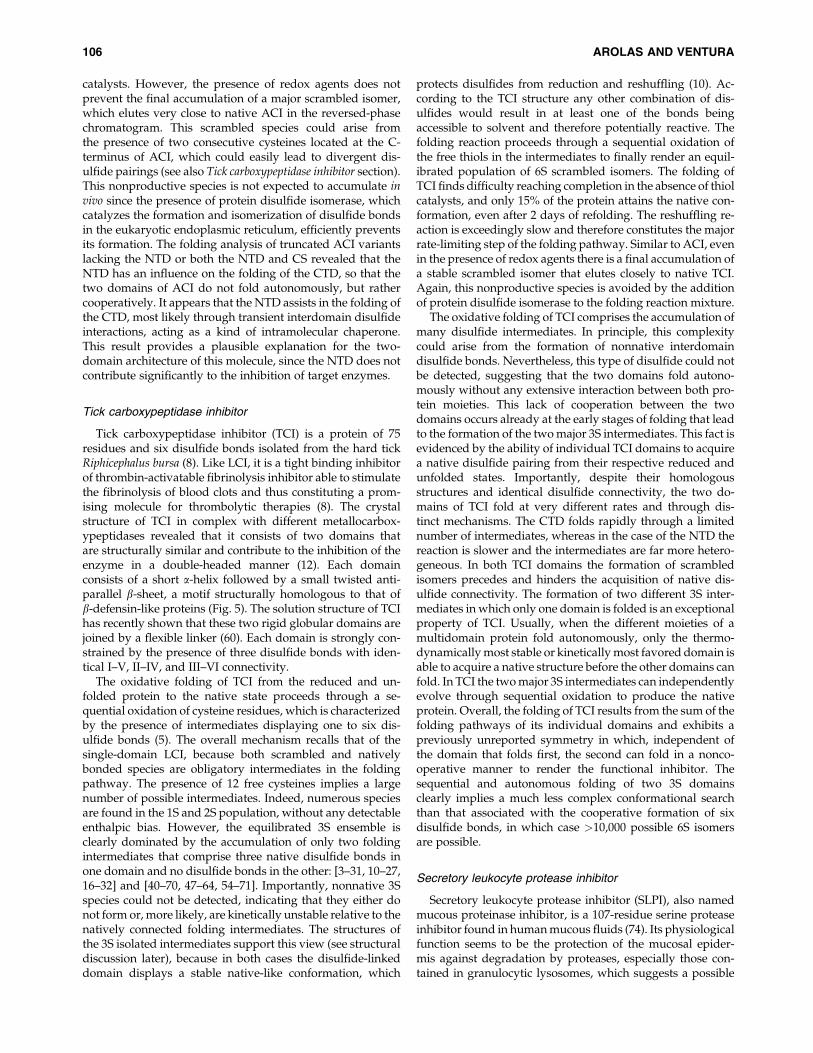
Leech carboxypeptidase inhibitor (LCI) is a 67-residuemetallocarboxypeptidase inhibitor found in the medicinalleech H. medicinalis (66). Interestingly, it is a potent inhibitor ofplasma carboxypeptidase B, also known as thrombin-activa-table fibrinolysis inhibitor, which is an emerging biomedicaltarget for thrombolysis and thrombolytic therapies. Thestructure of LCI was solved both in solution and in a crys-talline complex with human carboxypeptidase A2 (65). LCIfolds in a compact domain comprising a five-stranded anti-parallel b-sheet with a b3-b1-b2-b5-b4 topology and a short a-helix that packs onto the most compact part of the b-structure(Fig. 4). The native conformation of the inhibitor is highlystabilized by the occurrence of four disulfide bonds, Cys11-Cys34, Cys18-Cys62, Cys19-Cys43, and Cys22-Cys58, all ofthem located within regular secondary structure elements.The disordered C-terminal tail of LCI is the primary bindingsite of the enzyme, as previously described for PCI.
LCI displays a higher extent of secondary structure than itsthree-disulfide protease inhibitor counterparts, which affectsthe accessibility, proximity, and reactivity of the thiols anddisulfide bonds. In addition, the presence of two additionalcysteine residues increases almost 10-fold the number of in-termediates that could potentially arise during the oxidativefolding reaction. The combination of these two factors resultsin a rather complex folding landscape (4, 69). The folding ofLCI proceeds initially through a heterogeneous formation of1S and 2S intermediates. The presence of moderate amounts
OXIDATIVE FOLDING OF PROTEASE INHIBITORS 103
of denaturing agents does not alter the number or relativeabundance of 1S and 2S species, suggesting that their distri-bution at equilibrium does not respond to the establishment ofspecific side chain contacts, but rather to entropy constraints.This is in good agreement with computational predictions,suggesting that 1S and 2S LCI species do not contain an ex-tensive regular structure (6). This heterogeneity is stronglyreduced in the 3S population, which was initially thought toconsist exclusively of two intermediates displaying nativedisulfide connectivity: des[19–43] and des[22–58]. Because noother 3S forms were detected it was assumed that these twonative-like species could originate directly from the oxidationof the third disulfide bond in the 2S ensemble. However, thelack of an enthalpic bias toward the formation of native dis-ulfides in 2S implies that only a minor fraction of the isomersin this ensemble would display a native combination of dis-ulfide bonds, making the direct and preferential formation ofnatively bonded 3S species a very improbable reaction. In arecent study, the contribution of each particular disulfide tothe folding of LCI was investigated by constructing a series ofsingle and multiple mutants lacking one to four disulfidebonds (6). This work allowed reformulating the foldingpathway of this protein, specifically the formation, distribu-tion, and role of 3S isomers in the folding reaction. The resultsdemonstrated that in LCI 3S isomers can be effectively formedin the absence of the fourth disulfide bond and, more im-portantly, that these wrongly bonded species can rearrangetheir disulfides to render 3S forms with native disulfides andnative-like conformations. Therefore, a more plausiblemechanism for the formation of des[19–43] and des[22–58]intermediates would be the nonspecific oxidation of theunfolded 2S ensemble to 3S species and the subsequent re-shuffling of these forms into these two predominant inter-mediates. Thus, the initial stages of LCI folding comprise aquasi-stochastic consolidation of the protein, and resemblethose of hirudin. Nevertheless, des[19–43] and des[22–58]intermediates are reminiscent of the intermediates present inthe oxidative folding of BPTI, in the sense that in contrast tothe other 3S species, they have a highly folded structure. Thisendows them with the highest stability in the ensemble, and atthe same time protects their native disulfide bonds from re-duction and=or reshuffling, which kinetically favors the largeaccumulation of these species in the 3S ensemble (7, 11). Thehigh stability and low reactivity of des[19–43] and des[22–58]
cause them to act as strong kinetic traps that freeze the reac-tion at the 3S stage. Consequently, the destabilization of suchintermediates by denaturing agents allows the progress of thepathway and accelerates the recovery of native protein. Inprinciple, the direct oxidation of the free thiolates in any ofthese intermediates would promote the formation of the na-tive inhibitor. The formation of the native conformation isthermodynamically favored, since it is at least two times morestable than any of the intermediates. However, in contrast towhat happens with BPTI and LDTI, the free thiolates are notin flexible and exposed loops but shielded by the secondarystructure elements (see structural discussion later), as dem-onstrated by their inability to react with external thiols. Thishidden location of the free cysteines in the intermediates hasa dramatic effect on the progress of the folding reaction be-cause structural fluctuations expose both the thiols and thedisulfide bonds, thus promoting their reshuffling instead ofthe oxidation of free thiols into the native disulfide pairing.Therefore, the gained native structure becomes lost and apopulation of compact but unstructured 4S scrambled iso-mers subsequently accumulates. As for hirudin and PCI, thereshuffling of these disulfide-blocked species is very slow inthe absence of an external thiol catalyst. The reorganization ofthe misconnected disulfide bonds to finally attain the nativeform becomes the major rate-limiting step in the LCI foldingreaction. In the folding pathway of LCI both scrambled andnative intermediates are necessary to attain a unique stableconformation. In addition, in contrast to general assumptions,scrambled but not native-like intermediates are the produc-tive species. LCI illustrates how the search for the conforma-tional stability and reduced flexibility that provide the nativedisulfide connectivity complicates the folding reaction andmakes it extremely inefficient, suggesting that the conforma-tional space that small disulfide-rich proteins have to exploreto attain the functional form is shaped primarily in thermo-dynamic terms.
Oxidative Folding of Two-Domain Protease Inhibitors
Approximately 75% of eukaryotic protein sequences fold intoconformations consisting of more than one domain (15). How-ever, information about the mechanisms driving the foldingof two-domain proteins is still scarce. This is mainly due tothe limitation of indirect structural probes such as intrinsic
FIG. 4. Oxidative folding of leech carboxypeptidase inhibitor (LCI). The amino acid sequence of LCI (4 SS and 67residues), as well as its secondary structure elements and disulfide pairings, is schematically shown. The Protein Data BankID code for the structure is 1DTV (ribbon plot prepared with program PyMOL). The disulfide bonds are shown as blacksticks. An overview of the oxidative folding pathway of LCI is shown at the bottom. The nomenclature of the different speciesis described in the legend of Figure 2.
104 AROLAS AND VENTURA

fluorescence or ellipticity to monitor folding=unfolding transi-tions in two-domain proteins where the two protein moietiescan fold=unfold autonomously or in a cooperative way. Duringoxidative folding the formation of disulfide bonds acts as acovalent structural probe, providing unbiased informationabout the proximity of the different regions of the polypeptidechain. Therefore, studies of multidomain proteins in which thedifferent moieties are stabilized by disulfide bonds might allowcharacterization of the extent to which the folding reactions ofthis kind of polypeptide relies on the establishment of inter-domain interactions. Recent studies on the oxidative folding oftwo-domain protease inhibitors have provided valuable in-sights into the complicated folding landscape that multidomainproteins must face before attaining their native conformation.
Ascaris carboxypeptidase inhibitor
The metallocarboxypeptidase inhibitor Ascaris carboxy-peptidase inhibitor (ACI) isolated from Ascaris, the most
common human parasite of the gastrointestinal tract thatcauses the pandemic disease ascariasis, is 67 residues long andcontains five disulfide bonds (70). The crystal structure of ACIin complex with human carboxypeptidase A1 reveals twostructurally similar tandem modules (Fig. 5), an NTD and a C-terminal domain (CTD), which are linked by a connectingsegment (CS). Each domain comprises a small helix and a C-terminal b-sheet that are stabilized by two disulfide bonds(Cys6-Cys18, Cys12-Cys25; and Cys43-Cys58, Cys49-Cys63).The CS includes an a-helix, which is linked to the b-sheet ofthe CTD by the Cys34-Cys62 disulfide bond.
The oxidative folding of ACI has been recently examined,showing that this protein folds through the sequential for-mation of 1S, 2S, 3S, and 4S intermediates that subsequentlyevolve into a mixture of 5S (scrambled) isomers (13). Similar tothe cases of hirudin, PCI, TAP, and LCI, the reshuffling of thisheterogeneous population of scrambled isomers into the na-tive protein represents the rate-limiting step of the foldingreaction and is efficiently promoted by the presence of thiol
FIG. 5. Oxidative folding of two-domain protease inhibitors. The amino acid sequence, secondary structure elements, anddisulfide pairings of Ascaris carboxypeptidase inhibitor (ACI; 5 SS and 67 residues), tick carboxypeptidase inhibitor (TCI; 6 SSand 75 residues), and secretory leukocyte protease inhibitor (SLPI; 8 SS and 107 residues) are schematically shown. TheProtein Data Bank ID codes for the structures of ACI and TCI are, respectively, 3FJU and 2JTO (ribbon plots prepared withprogram PyMOL). The disulfide bonds are shown as black sticks. The N-terminal domain (NTD) and CTD and the con-necting segment (CS) are indicated. An overview of the oxidative folding pathway of ACI, TCI, and SLPI is shown beloweach sequence. The nomenclature of the different species is described in the legend of Figure 2.
OXIDATIVE FOLDING OF PROTEASE INHIBITORS 105
catalysts. However, the presence of redox agents does notprevent the final accumulation of a major scrambled isomer,which elutes very close to native ACI in the reversed-phasechromatogram. This scrambled species could arise fromthe presence of two consecutive cysteines located at the C-terminus of ACI, which could easily lead to divergent dis-ulfide pairings (see also Tick carboxypeptidase inhibitor section).This nonproductive species is not expected to accumulate invivo since the presence of protein disulfide isomerase, whichcatalyzes the formation and isomerization of disulfide bondsin the eukaryotic endoplasmic reticulum, efficiently preventsits formation. The folding analysis of truncated ACI variantslacking the NTD or both the NTD and CS revealed that theNTD has an influence on the folding of the CTD, so that thetwo domains of ACI do not fold autonomously, but rathercooperatively. It appears that the NTD assists in the folding ofthe CTD, most likely through transient interdomain disulfideinteractions, acting as a kind of intramolecular chaperone.This result provides a plausible explanation for the two-domain architecture of this molecule, since the NTD does notcontribute significantly to the inhibition of target enzymes.
Tick carboxypeptidase inhibitor
Tick carboxypeptidase inhibitor (TCI) is a protein of 75residues and six disulfide bonds isolated from the hard tickRiphicephalus bursa (8). Like LCI, it is a tight binding inhibitorof thrombin-activatable fibrinolysis inhibitor able to stimulatethe fibrinolysis of blood clots and thus constituting a prom-ising molecule for thrombolytic therapies (8). The crystalstructure of TCI in complex with different metallocarbox-ypeptidases revealed that it consists of two domains thatare structurally similar and contribute to the inhibition of theenzyme in a double-headed manner (12). Each domainconsists of a short a-helix followed by a small twisted anti-parallel b-sheet, a motif structurally homologous to that ofb-defensin-like proteins (Fig. 5). The solution structure of TCIhas recently shown that these two rigid globular domains arejoined by a flexible linker (60). Each domain is strongly con-strained by the presence of three disulfide bonds with iden-tical I–V, II–IV, and III–VI connectivity.
The oxidative folding of TCI from the reduced and un-folded protein to the native state proceeds through a se-quential oxidation of cysteine residues, which is characterizedby the presence of intermediates displaying one to six dis-ulfide bonds (5). The overall mechanism recalls that of thesingle-domain LCI, because both scrambled and nativelybonded species are obligatory intermediates in the foldingpathway. The presence of 12 free cysteines implies a largenumber of possible intermediates. Indeed, numerous speciesare found in the 1S and 2S population, without any detectableenthalpic bias. However, the equilibrated 3S ensemble isclearly dominated by the accumulation of only two foldingintermediates that comprise three native disulfide bonds inone domain and no disulfide bonds in the other: [3–31, 10–27,16–32] and [40–70, 47–64, 54–71]. Importantly, nonnative 3Sspecies could not be detected, indicating that they either donot form or, more likely, are kinetically unstable relative to thenatively connected folding intermediates. The structures ofthe 3S isolated intermediates support this view (see structuraldiscussion later), because in both cases the disulfide-linkeddomain displays a stable native-like conformation, which
protects disulfides from reduction and reshuffling (10). Ac-cording to the TCI structure any other combination of dis-ulfides would result in at least one of the bonds beingaccessible to solvent and therefore potentially reactive. Thefolding reaction proceeds through a sequential oxidation ofthe free thiols in the intermediates to finally render an equil-ibrated population of 6S scrambled isomers. The folding ofTCI finds difficulty reaching completion in the absence of thiolcatalysts, and only 15% of the protein attains the native con-formation, even after 2 days of refolding. The reshuffling re-action is exceedingly slow and therefore constitutes the majorrate-limiting step of the folding pathway. Similar to ACI, evenin the presence of redox agents there is a final accumulation ofa stable scrambled isomer that elutes closely to native TCI.Again, this nonproductive species is avoided by the additionof protein disulfide isomerase to the folding reaction mixture.
The oxidative folding of TCI comprises the accumulation ofmany disulfide intermediates. In principle, this complexitycould arise from the formation of nonnative interdomaindisulfide bonds. Nevertheless, this type of disulfide could notbe detected, suggesting that the two domains fold autono-mously without any extensive interaction between both pro-tein moieties. This lack of cooperation between the twodomains occurs already at the early stages of folding that leadto the formation of the two major 3S intermediates. This fact isevidenced by the ability of individual TCI domains to acquirea native disulfide pairing from their respective reduced andunfolded states. Importantly, despite their homologousstructures and identical disulfide connectivity, the two do-mains of TCI fold at very different rates and through dis-tinct mechanisms. The CTD folds rapidly through a limitednumber of intermediates, whereas in the case of the NTD thereaction is slower and the intermediates are far more hetero-geneous. In both TCI domains the formation of scrambledisomers precedes and hinders the acquisition of native dis-ulfide connectivity. The formation of two different 3S inter-mediates in which only one domain is folded is an exceptionalproperty of TCI. Usually, when the different moieties of amultidomain protein fold autonomously, only the thermo-dynamically most stable or kinetically most favored domain isable to acquire a native structure before the other domains canfold. In TCI the two major 3S intermediates can independentlyevolve through sequential oxidation to produce the nativeprotein. Overall, the folding of TCI results from the sum of thefolding pathways of its individual domains and exhibits apreviously unreported symmetry in which, independent ofthe domain that folds first, the second can fold in a nonco-operative manner to render the functional inhibitor. Thesequential and autonomous folding of two 3S domainsclearly implies a much less complex conformational searchthan that associated with the cooperative formation of sixdisulfide bonds, in which case >10,000 possible 6S isomersare possible.
Secretory leukocyte protease inhibitor
Secretory leukocyte protease inhibitor (SLPI), also namedmucous proteinase inhibitor, is a 107-residue serine proteaseinhibitor found in human mucous fluids (74). Its physiologicalfunction seems to be the protection of the mucosal epider-mis against degradation by proteases, especially those con-tained in granulocytic lysosomes, which suggests a possible
106 AROLAS AND VENTURA
therapeutic use against local inflammatory processes. Thecrystal structure of SLPI in complex with a-chymotrypsinshows that the inhibitor folds in two independent domains ina boomerang-like structure (Fig. 5) (47). The C-terminal se-quence of each domain organizes into two b-strands linked bya hairpin turn, whereas the N-terminal part consists of a loopcovalently linked to the b-sheet, which constitutes the primarybinding site of the protease. Each domain is stabilized by thepresence of four disulfide bonds with I–VI, II–VII, III–V, andIV–VIII connectivity.
The presence of 16 free cysteine residues in the reducedand unfolded state of SLPI could result in an extremelycomplex oxidative folding reaction for this inhibitor. Morethan 40 million different species could accumulate alongthe pathway, with 2 million corresponding to 8S scrambledisomers; that is, the folding reaction would be exceedinglyinefficient. Lin and Chang analyzed the oxidative folding ofSLPI, finding that, surprisingly, despite the complicatedtheoretical folding landscape of SLPI, this protein foldsvery efficiently into the two-domain native architecturewithout requiring a thiol catalyst (54). This property isunique and intriguing in light of the SLPI native structure,which lacks an extensive regular structure that could drivea coupled conformational and oxidative folding reaction.Even in the case that the protein adopts a folding pathwayin which only native disulfide bonds are formed, 254 dif-ferent species would populate the pathway. Moreover, thefolding pathway of SLPI comprises a highly heterogeneouspopulation of folding intermediates that includes scram-bled isomers, which discredits the idea that folding occursthrough a BPTI-like mechanism. In fact, the folding of SLPIfollows a two-stage mechanism that combines features ofboth BPTI- and hirudin-like proteins. During the first stageof folding, reduced and unfolded SLPI sequentially oxi-dizes its disulfide bonds to form a highly heterogeneousmixture of intermediates that finally converges in the for-mation of 6S and 7S isomers comprising nonnatively andnatively bonded intermediates. The main 6S intermediate isdes[18–43, 71–97], which contains the three cysteines lo-cated in the b-strands of each domain correctly bonded,forming I–VI, III–V, and IV–VIII disulfide bonds, and lacksthe II–VII bond joining the protease binding loops in bothdomains. The main 7S intermediate corresponds to des[18–43], which lacks the II–VII bond in the first domain. Pre-sumably both intermediates display a partially foldedconformation, which protects preferentially the linked dis-ulfides promoting its accumulation and its role as a kinetictrap in the flow of the reaction. In the second stage offolding, des[18–43, 71–97] converts into des[18–43] throughdirect oxidation of its Cys71 and Cys97 residues, and thenewly formed and previously accumulated des[18–43] ox-idizes into the native SLPI. Similar to BPTI, these reactionsare favored by the presence of free cysteines in flexible andexposed loops. This is likely the reason that they can act asthiol catalysts, facilitating the reshuffling of the remainingnonnatively bonded isomers toward native pairings thatfurther evolve into native intermediates and finally formthe functional inhibitor. It also explains why SLPI foldsefficiently in the absence of external redox agents. There-fore, in contrast to TCI and more similar to ACI, the foldingof SLPI comprises interdomain interactions and is a coop-erative reaction.
Structures of Protease Inhibitors’Folding Intermediates
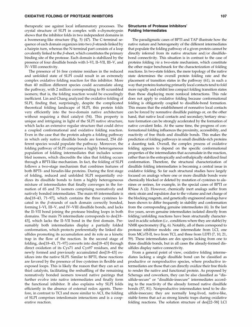
The paradigmatic cases of BPTI and TAP illustrate how thenative nature and heterogeneity of the different intermediatesthat populate the folding pathway of a given protein cannot bedirectly inferred from its native structure and=or disulfide-bond connectivity. This situation is in contrast to the case ofproteins folding via a two-state mechanism, which constitutethe other major benchmark for the characterization of foldingreactions. In two-state folders, the mere topology of the nativestate determines the overall protein folding rate and theplacement of transition states in the pathway (61), in such away that proteins featuring primarily local contacts tend to foldmore rapidly and exhibit less compact folding transition statesthan those displaying more nonlocal interactions. This ruledoes not apply to oxidative folding because conformationalfolding is obligatorily coupled to disulfide-bond formation.This means that the establishment of nonnative local contactscan be forced by nonnative disulfide pairings or, on the otherhand, that native local contacts and secondary=tertiary struc-ture formation can be strongly accelerated by the formation ofnative covalent links. At the same time, the progress of con-formational folding influences the proximity, accessibility, andreactivity of free thiols and disulfide bonds. This makes theprediction of folding pathways in small disulfide-rich proteinsa daunting task. Overall, the complex process of oxidativefolding appears to depend on the specific conformationalproperties of the intermediates that accumulate in the reaction,rather than in the entropically and enthalpically stabilized finalconformation. Therefore, the structural characterization ofdisulfide folding intermediates is becoming a central issue inoxidative folding. So far such structural studies have largelyfocused on analogs where one or more disulfide bonds werechemically blocked or deleted by mutation of cysteines to ala-nines or serines, for example, in the special cases of BPTI orRNase A (2). However, chemically inert analogs suffer fromsteric strain and repulsion due to the volume and net charge ofthe blocking reagents, and genetically engineered analogs havebeen shown to differ frequently in stability and conformationfrom the corresponding genuine intermediates (6). In the lastfive years, seven genuine intermediates isolated directly fromfolding=unfolding reactions have been structurally character-ized in acidic solution (i.e., conditions where they are stable) byNMR spectrometry (Fig. 6). Notably, all of them correspond toprotease inhibitor models: one intermediate from LCI, onefrom MCoTI-II, two from TCI, and three from LDTI (7, 10, 21,59). These intermediates are des species lacking from one tothree disulfide bonds, but in all cases the already-formed dis-ulfides display native connectivity.
From a general point of view, oxidative folding interme-diates lacking a single disulfide bond can be classified asproductive or nonproductive species, where productive in-termediates are those that can directly oxidize their free thiolsto render the native and functional protein. As proposed byScheraga and coworkers, they can be also classified as ‘‘dis-ulfide-secure’’ or ‘‘disulfide-insecure’’ intermediates accord-ing to the reactivity of the already formed native disulfidebonds (57, 81). Nonproductive intermediates tend to be dis-ulfide-insecure; they are normally long-lived, native-like,stable forms that act as strong kinetic traps during oxidativefolding reactions. The solution structure of des[22–58] LCI
OXIDATIVE FOLDING OF PROTEASE INHIBITORS 107
shows the molecular determinants responsible for such be-havior (7). This intermediate possesses a well-defined globu-lar conformation that closely resembles that of the nativeinhibitor, including a four-stranded antiparallel (b3-b1-b2-b5)b-sheet, although certain regions are more disordered than inthe native state and the a-helix is not properly formed. Despitethe increased flexibility and lower compactness of the inter-mediate, proton=deuterium exchange experiments demon-strated that both free cysteines (Cys22 and Cys58) and thethree native disulfide bonds are located in protected regionsand have limited accessibility. This restricted exposure tosolvent is also evident from the fact that both free thiols anddisulfide bonds are insensitive to the presence of external thiolagents. The burial of the free thiol groups inside a native-likestructure would hinder any further progress in the foldingreaction. However, the increased overall flexibility of thebackbone promotes structural fluctuations that allow the in-termediate to escape from this trap. Because of their similardegrees of protection, fluctuations that expose the free thiolsalso expose the disulfide bonds, resulting in disulfide re-shuffling into 4S scrambled forms instead of direct oxidationof the missing disulfide bond into the native structure. In
des[22–58] LCI reshuffling can compete effectively with oxi-dation due to the conformational similarity between a re-shuffling reaction and the second step of an oxidation reaction(81).
In contrast to des[22–58] LCI, the des[1–18] intermediate ofMCoTI-II is a productive species able to directly render thenative inhibitor by direct oxidation of its free thiols. The so-lution structure of this intermediate shows that it displaysnative secondary and tertiary structure, the only exceptionbeing a flexible loop, the movement of which in the nativestate is restricted by the Cys1–Cys18 disulfide bond, forcingits collapse against the protein core. The absence of the co-valent link in the intermediate results in a loop more looselyassociated with the rest of the protein (21). However, proton=deuterium exchange experiments demonstrated that thenumber of slowly exchanging amide protons in the interme-diate is comparable to that of the native protein, indicatingthat both forms display similar rigidity. The rigidity in theoverall scaffold of des[1–18] MCoTI-II turns it into a disulfide-secure intermediate in which structural fluctuations prefer-entially change the environment of their thiol groups whilekeeping their disulfide bonds in a protected native location.
FIG. 6. Solution structures of major folding intermediates. Ca-structure representation of the average structures of LCI,MCoTI-II, TCI, and LDTI (native forms), together with those of their major folding intermediates—missing the disulfidebonds shown between parentheses (figure prepared with program PyMOL). The Protein Data Bank ID codes are as follows:LCI (1ZFI), des[22–58] (1ZFL), MCoTI-II (1IB9), des[1–18] (2PO8), TCI (2K2X), des[CTD] (2K2Y), des[NTD] (2K2Z), LDTI(2KMO), des[4–29] (2KMP), des[6–25] (2KMQ), and des[14–40] (2KMR). For clarity, the molecules are in the same orientationas their ribbon plot structures in Figures 2, 4, and 5. Regions with increased flexibility are indicated by arrows. Dashed linesrepresent the unfolded and unpaired domains of TCI intermediates.
108 AROLAS AND VENTURA
This is reflected by the surface accessibilities of the two freecysteines in the intermediate, which are much larger thanthose in the native state, where they become part of the hy-drophobic core. Molecular dynamic simulations suggest thatin the absence of the disulfide bond the rigid native confor-mation is thermodynamically destabilized with respect to themore flexible intermediate structure. Therefore, although thefree cysteines are ready for oxidation and do not compete withthe disulfide bonds, the fact that the protein backbone mustadopt an energetically unfavorable conformation before bothcysteines can oxidize slows down significantly the productiveformation of the native form.
The two main 3S folding intermediates of the two-domaininhibitor TCI illustrate how the preferential burial of nativedisulfide bonds into a compact native structure protects themfrom reshuffling reactions that could be initiated by reactivefree cysteines. As discussed above, each intermediate com-prises three native disulfide bonds in one domain and nodisulfide bonds in the other. This means that each interme-diate has six free thiols that can potentially initiate a disulfideexchange reaction with the preformed disulfide bonds.Moreover, the solution structures of these two intermediatesindicate that these free cysteines are found in a disordered andhighly flexible context, which in principle would stronglyfavor the disulfide attack (10). However, this type of reactiondoes not occur and the already formed covalent bonds remainintact in both intermediates during the rest of the folding re-action. The reason for this protection is that the structures ofthe folded NTD and CTD in the intermediates are exactly thesame as the corresponding ones in the native inhibitor, withall the disulfide bonds highly buried and protected fromsolvent in a globular and highly compact conformation.Therefore, the folding pathway proceeds with the oxidation ofthe free cysteines in the unfolded domain.
LDTI is the first protein model for which all the genuinedes species preceding the formation of the native structurehave been structurally characterized (59). As discussedabove, they consist of three intermediates each containingtwo native disulfide bonds and a pair of free thiols: des[4–29], des[6–25], and des[14–40]. Among these species onlydes[4–29] is a productive intermediate that yields the nativeinhibitor by direct oxidation of its free thiols. The other twointermediates reorganize their disulfide bonds to convergein the productive intermediate. LDTI shows a reducedamount of secondary structure, that is, a short a-helix and asmall triple-stranded antiparallel b-sheet in a b2-b1-b3 to-pology. Despite the overall packing of LDTI, which wasthought to rely on the special Kazal-type connectivity of itsdisulfide bonds, the three des intermediates display essen-tially the same fold and secondary structure motifs as nativeLDTI, except des[14–40], which lacks the b3-strand due tothe missing disulfide. Importantly, the nonproductivedes[6–25] and the productive des[4–29] intermediates possesa well-packed and structurally identical core, with a rms-deviation of only 1.3A, the main difference between thembeing a more flexible N-terminus in des[4–29]. In contrast toLCI, in LDTI these productive and nonproductive interme-diates display almost identically accessible free thiols anddisulfide bonds. In both intermediates the Cys14–Cys40disulfide is buried in the native b-sheet, whereas the otherdisulfide connecting the a-helix to the N-terminal loop andthe free thiols are fully accessible. This indicates that the
productivity of a folding intermediate depends on moresubtle structural determinants than the mere exposure of itsfree or bonded cysteine residues. In fact, the balance be-tween reshuffling and oxidation depends on the accessibil-ity, but also on the proximity and reactivity, of the groupsinvolved. In des[6–25] the two latter factors shift the balancetoward reshuffling into des[4–29] rather than toward theformation of the native inhibitor via oxidation. Cys25 is lo-cated at the N-terminus of the a-helix where a partial pos-itive charge of the dipole stabilizes the thiolate, lowering theeffective pKa of this group and making it highly reactive.The flexibility of the backbone in the intermediate bringsthis reactive cysteine close to the accessible Cys4–Cys29disulfide bond instead of to the free Cys6, which results indisulfide exchange instead of oxidation. Together, thestructures of the LDTI folding intermediates suggest that theaccessibility of the free thiols ensures that an intermediate isa disulfide-secure, productive species only if all the nativedisulfide bonds are fully buried and cannot be attacked bythe free thiols.
Concluding Remarks
Protease inhibitors comprise an extremely large number ofmolecules with different sequential and structural character-istics. They are usually small proteins crosslinked by severaldisulfides, which have turned them into outstanding modelsfor the study of the complex process of oxidative folding.Although only two classes of protease inhibitors have beenstudied to date, namely, serine and metalloprotease inhibi-tors, a surprisingly high diversity of folding scenarios havebeen detected. Expanding folding studies toward the unex-plored cysteine and aspartic protease inhibitors would likelyuncover new folding processes. Unfortunately, it is still notpossible to forecast the folding landscapes that small dis-ulfide-rich proteins have to face by using only the confor-mation and covalent connectivity of their native structures.The aforementioned structures of genuine folding interme-diates are providing the first clues to the molecular determi-nants that guide the last stages of oxidative folding. However,structural information is still scarce regarding the initialfolding steps preceding the formation of stable intermediates.In the forthcoming years research should be focused onthese fast and intricate but crucial events. The use of well-established approaches for the study of fast-folding proteinswill be of much help. Understanding the general rules thatgovern oxidative folding at different stages would provide aunique opportunity to attract theoretical groups to this re-search area and guide the development of bioinformaticsapproaches to assist in the prediction and design of the fold-ing pathways of small disulfide-rich proteins. What becomesobvious is that protease inhibitors will, in the years to come,provide one of the main bedrocks from which to understandthe complexity of oxidative folding.
Acknowledgments
The authors are indebted to Prof. Francesc X. Aviles andDrs. Josep Vendrell and Sılvia Bronsoms for their continuoussupport and advice. The authors also thank Prof. WolframBode for kindly providing the pdb file of the SLPI structure.This work has been supported by the Spanish Ministry ofScience and Innovation, grant BIO2007-68046, and by the
OXIDATIVE FOLDING OF PROTEASE INHIBITORS 109
National Catalan Government, grants 2005-SGR01037 and2009-SGR760.
References
1. Antuch W, Guntert P, Billeter M, Hawthorne T, Grossen-bacher H, and Wuthrich K. NMR solution structure of therecombinant tick anticoagulant protein (rTAP), a factor Xainhibitor from the tick Ornithodoros moubata. FEBS Lett 352:251–257, 1994.
2. Arolas JL, Aviles FX, Chang JY, and Ventura S. Folding ofsmall disulfide-rich proteins: clarifying the puzzle. TrendsBiochem Sci 31: 292–301, 2006.
3. Arolas JL, Bronsoms S, Aviles FX, Ventura S, and Som-merhoff CP. Oxidative folding of leech-derived tryptase in-hibitor via native disulfide-bonded intermediates. AntioxidRedox Signal 10: 77–85, 2008.
4. Arolas JL, Bronsoms S, Lorenzo J, Aviles FX, Chang JY, andVentura S. Role of kinetic intermediates in the folding of leechcarboxypeptidase inhibitor. J Biol Chem 279: 37261–37270, 2004.
5. Arolas JL, Bronsoms S, Ventura S, Aviles FX, and Calvete JJ.Characterizing the tick carboxypeptidase inhibitor: molecu-lar basis for its two-domain nature. J Biol Chem 281: 22906–22916, 2006.
6. Arolas JL, Castillo V, Bronsoms S, Aviles FX, and Ventura S.Designing out disulfide bonds of leech carboxypeptidaseinhibitor: implications for its folding, stability and function. JMol Biol 392: 529–546, 2009.
7. Arolas JL, D’Silva L, Popowicz GM, Aviles FX, Holak TA,and Ventura S. NMR structural characterization and com-putational predictions of the major intermediate in oxidativefolding of leech carboxypeptidase inhibitor. Structure 13:1193–1202, 2005.
8. Arolas JL, Lorenzo J, Rovira A, Castella J, Aviles FX, andSommerhoff CP. A carboxypeptidase inhibitor from the tickRhipicephalus bursa: isolation, cDNA cloning, recombinant ex-pression, and characterization. J Biol Chem 280: 3441–3448, 2005.
9. Arolas JL, Lorenzo J, Rovira A, Vendrell J, Aviles FX, andVentura S. Secondary binding site of the potato carboxy-peptidase inhibitor. Contribution to its structure, folding,and biological properties. Biochemistry 43: 7973–7982, 2004.
10. Arolas JL, Pantoja-Uceda D, Ventura S, Blanco FJ, and AvilesFX. The NMR structures of the major intermediates of thetwo-domain tick carboxypeptidase inhibitor reveal symme-try in its folding and unfolding pathways. J Biol Chem 283:27110–27120, 2008.
11. Arolas JL, Popowicz GM, Bronsoms S, Aviles FX, Huber R,Holak TA, and Ventura S. Study of a major intermediate inthe oxidative folding of leech carboxypeptidase inhibitor:contribution of the fourth disulfide bond. J Mol Biol 352: 961–975, 2005.
12. Arolas JL, Popowicz GM, Lorenzo J, Sommerhoff CP, HuberR, Aviles FX, and Holak TA. The three-dimensional struc-tures of tick carboxypeptidase inhibitor in complex withA=B carboxypeptidases reveal a novel double-headedbinding mode. J Mol Biol 350: 489–498, 2005.
13. Arolas JL, Sanglas L, Lorenzo J, Bronsoms S, and Aviles FX.Insights into the two-domain architecture of the metallo-carboxypeptidase inhibitor from the Ascaris parasite inferredfrom the mechanism of its oxidative folding. Biochemistry 48:8225–8232, 2009.
14. Auerswald EA, Morenweiser R, Sommerhoff CP, PiechottkaGP, Eckerskorn C, Gurtler LG, and Fritz H. Recombinantleech-derived tryptase inhibitor: construction, production,
protein chemical characterization and inhibition of HIV-1replication. Biol Chem Hoppe Seyler 375: 695–703, 1994.
15. Batey S, Scott KA, and Clarke J. Complex folding kinetics ofa multidomain protein. Biophys J 90: 2120–2130, 2006.
16. Berndt KD, Guntert P, Orbons LP, and Wuthrich K. De-termination of a high-quality nuclear magnetic resonancesolution structure of the bovine pancreatic trypsin inhibitorand comparison with three crystal structures. J Mol Biol 227:757–775, 1992.
17. Bode W and Huber R. Structural basis of the endoproteinase-protein inhibitor interaction. Biochim Biophys Acta 1477: 241–252, 2000.
18. Bronsoms S, Villanueva J, Canals F, Querol E, and Aviles FX.Analysis of the effect of potato carboxypeptidase inhibitorpro-sequence on the folding of the mature protein. Eur JBiochem 270: 3641–3650, 2003.
19. Cemazar M, Daly NL, Haggblad S, Lo KP, Yulyaningsih E,and Craik DJ. Knots in rings. The circular knotted proteinMomordica cochinchinensis trypsin inhibitor-II folds via astable two-disulfide intermediate. J Biol Chem 281: 8224–8232, 2006.
20. Cemazar M, Gruber CW, and Craik DJ. Oxidative folding ofcyclic cystine knot proteins. Antioxid Redox Signal 10: 103–111, 2008.
21. Cemazar M, Joshi A, Daly NL, Mark AE, and Craik DJ. Thestructure of a two-disulfide intermediate assists in eluci-dating the oxidative folding pathway of a cyclic cystine knotprotein. Structure 16: 842–851, 2008.
22. Craik DJ, Cemazar M, Wang CK, and Daly NL. The cyclo-tide family of circular miniproteins: nature’s combinatorialpeptide template. Biopolymers 84: 250–266, 2006.
23. Creighton TE. Renaturation of the reduced bovine pancre-atic trypsin inhibitor. J Mol Biol 87: 563–577, 1974.
24. Creighton TE. Electrophoretic analysis of the unfolding ofproteins by urea. J Mol Biol 129: 235–264, 1979.
25. Creighton TE. Toward a better understanding of protein fold-ing pathways. Proc Natl Acad Sci U S A 85: 5082–5086, 1988.
26. Creighton TE. Protein folding coupled to disulphide bondformation. Biol Chem 378: 731–744, 1997.
27. Creighton TE, Bagley CJ, Cooper L, Darby NJ, Freedman RB,Kemmink J, and Sheikh A. On the biosynthesis of bovinepancreatic trypsin inhibitor (BPTI). Structure, processing,folding and disulphide bond formation of the precursorin vitro and in microsomes. J Mol Biol 232: 1176–1196, 1993.
28. Creighton TE, Darby NJ, and Kemmink J. The roles of partlyfolded intermediates in protein folding. FASEB J 10: 110–118,1996.
29. Creighton TE and Goldenberg DP. Kinetic role of a meta-stable native-like two-disulphide species in the foldingtransition of bovine pancreatic trypsin inhibitor. J Mol Biol179: 497–526, 1984.
30. Chang JY. The functional domain of hirudin, a thrombin-specific inhibitor. FEBS Lett 164: 307–313, 1983.
31. Chang JY. The disulfide folding pathway of tick anticoagu-lant peptide (TAP), a Kunitz-type inhibitor structurally ho-mologous to BPTI. Biochemistry 35: 11702–11709, 1996.
32. Chang JY. Evidence for the underlying cause of diversity of thedisulfide folding pathway. Biochemistry 43: 4522–4529, 2004.
33. Chang JY. Diversity of folding pathways and folding mod-els of disulfide proteins. Antioxid Redox Signal 10: 171–177,2008.
34. Chang JY, Canals F, Schindler P, Querol E, and Aviles FX.The disulfide folding pathway of potato carboxypeptidaseinhibitor. J Biol Chem 269: 22087–22094, 1994.
110 AROLAS AND VENTURA

35. Chang JY and Li L. Divergent folding pathways of two ho-mologous proteins, BPTI and tick anticoagulant peptide:compartmentalization of folding intermediates and iden-tification of kinetic traps. Arch Biochem Biophys 437: 85–95, 2005.
36. Chang JY, Schindler P, and Chatrenet B. The disulfide struc-tures of scrambled hirudins. J Biol Chem 270: 11992–11997, 1995.
37. Chatrenet B and Chang JY. The folding of hirudin adopts amechanism of trial and error. J Biol Chem 267: 3038–3043, 1992.
38. Chatrenet B and Chang JY. The disulfide folding pathway ofhirudin elucidated by stop=go folding experiments. J BiolChem 268: 20988–20996, 1993.
39. Dadlez M. Hydrophobic interactions accelerate early stagesof the folding of BPTI. Biochemistry 36: 2788–2797, 1997.
40. Darby NJ and Creighton TE. Dissecting the disulphide-coupled folding pathway of bovine pancreatic trypsin in-hibitor. Forming the first disulphide bonds in analogues ofthe reduced protein. J Mol Biol 232: 873–896, 1993.
41. Di Marco S and Priestle JP. Structure of the complex ofleech-derived tryptase inhibitor (LDTI) with trypsin andmodeling of the LDTI-tryptase system. Structure 5: 1465–1474, 1997.
42. Felizmenio-Quimio ME, Daly NL, and Craik DJ. Circularproteins in plants: solution structure of a novel macrocyclictrypsin inhibitor from Momordica cochinchinensis. J Biol Chem276: 22875–22882, 2001.
43. Folkers PJ, Clore GM, Driscoll PC, Dodt J, Kohler S, andGronenborn AM. Solution structure of recombinant hirudinand the Lys-47—-Glu mutant: a nuclear magnetic resonanceand hybrid distance geometry-dynamical simulated an-nealing study. Biochemistry 28: 2601–2617, 1989.
44. Goldenberg DP and Zhang JX. Small effects of amino acidreplacements on the reduced and unfolded state of pancre-atic trypsin inhibitor. Proteins 15: 322–329, 1993.
45. Gonzalez C, Neira JL, Ventura S, Bronsoms S, Rico M, andAviles FX. Structure and dynamics of the potato carboxy-peptidase inhibitor by 1H and 15N NMR. Proteins 50: 410–422, 2003.
46. Gottfried DS and Haas E. Nonlocal interactions stabilize com-pact folding intermediates in reduced unfolded bovine pan-creatic trypsin inhibitor. Biochemistry 31: 12353–12362, 1992.
47. Grutter MG, Fendrich G, Huber R, and Bode W. The 2.5 A X-ray crystal structure of the acid-stable proteinase inhibitorfrom human mucous secretions analysed in its complex withbovine alpha-chymotrypsin. EMBO J 7: 345–351, 1988.
48. Grutter MG, Priestle JP, Rahuel J, Grossenbacher H, Bode W,Hofsteenge J, and Stone SR. Crystal structure of the throm-bin-hirudin complex: a novel mode of serine protease inhi-bition. EMBO J 9: 2361–2365, 1990.
49. Heitz A, Hernandez JF, Gagnon J, Hong TT, Pham TT,Nguyen TM, Le-Nguyen D, and Chiche L. Solution structureof the squash trypsin inhibitor MCoTI-II. A new family forcyclic knottins. Biochemistry 40: 7973–7983, 2001.
50. Housset D, Kim KS, Fuchs J, Woodward C, and WlodawerA. Crystal structure of a Y35G mutant of bovine pancreatictrypsin inhibitor. J Mol Biol 220: 757–770, 1991.
51. Huber R, Kukla D, Bode W, Schwager P, Bartels K, Dei-senhofer J, and Steigemann W. Structure of the complexformed by bovine trypsin and bovine pancreatic trypsininhibitor. II. Crystallographic refinement at 1.9 A resolution.J Mol Biol 89: 73–101, 1974.
52. Kibria FM and Lees WJ. Balancing conformational and oxi-dative kinetic traps during the folding of bovine pancreatictrypsin inhibitor (BPTI) with glutathione and glutathionedisulfide. J Am Chem Soc 130: 796–797, 2008.
53. Lim-Wilby MS, Hallenga K, de Maeyer M, Lasters I, VlasukGP, and Brunck TK. NMR structure determination of tickanticoagulant peptide (TAP). Protein Sci 4: 178–186, 1995.
54. Lin CC and Chang JY. Pathway of oxidative folding of se-cretory leucocyte protease inhibitor: an 8-disulfide proteinexhibits a unique mechanism of folding. Biochemistry 45:6231–6240, 2006.
55. Mamathambika BS and Bardwell JC. Disulfide-linked pro-tein folding pathways. Annu Rev Cell Dev Biol 24: 211–235,2008.
56. Muhlhahn P, Czisch M, Morenweiser R, Habermann B,Engh RA, Sommerhoff CP, Auerswald EA, and Holak TA.Structure of leech derived tryptase inhibitor (LDTI-C) insolution. FEBS Lett 355: 290–296, 1994.
57. Narayan M, Welker E, Wedemeyer WJ, and Scheraga HA.Oxidative folding of proteins. Acc Chem Res 33: 805–812,2000.
58. Otlewski J, Jelen F, Zakrzewska M, and Oleksy A. The manyfaces of protease-protein inhibitor interaction. EMBO J 24:1303–1310, 2005.
59. Pantoja-Uceda D, Arolas JL, Aviles FX, Santoro J, Ventura S,and Sommerhoff CP. Deciphering the structural basis thatguides the oxidative folding of leech-derived tryptase in-hibitor. J Biol Chem 284: 35612–35620, 2009.
60. Pantoja-Uceda D, Arolas JL, Garcia P, Lopez-Hernandez E,Padro D, Aviles FX, and Blanco FJ. The NMR structure anddynamics of the two-domain tick carboxypeptidase inhibitorreveal flexibility in its free form and stiffness upon binding tohuman carboxypeptidase B. Biochemistry 47: 7066–7078, 2008.
61. Plaxco KW, Simons KT, and Baker D. Contact order, tran-sition state placement and the refolding rates of single do-main proteins. J Mol Biol 277: 985–994, 1998.
62. Rawlings ND, Morton FR, Kok CY, Kong J, and Barrett AJ.MEROPS: the peptidase database. Nucleic Acids Res 36: D320–D325, 2008.
63. Rawlings ND, Tolle DP, and Barrett AJ. Evolutionary fami-lies of peptidase inhibitors. Biochem J 378: 705–716, 2004.
64. Rees DC and Lipscomb WN. Refined crystal structure of thepotato inhibitor complex of carboxypeptidase A at 2.5 Aresolution. J Mol Biol 160: 475–498, 1982.
65. Reverter D, Fernandez-Catalan C, Baumgartner R, PfanderR, Huber R, Bode W, Vendrell J, Holak TA, and Aviles FX.Structure of a novel leech carboxypeptidase inhibitor deter-mined free in solution and in complex with human car-boxypeptidase A2. Nat Struct Biol 7: 322–328, 2000.
66. Reverter D, Vendrell J, Canals F, Horstmann J, Aviles FX,Fritz H, and Sommerhoff CP. A carboxypeptidase inhibitorfrom the medical leech Hirudo medicinalis. Isolation, se-quence analysis, cDNA cloning, recombinant expression,and characterization. J Biol Chem 273: 32927–32933, 1998.
67. Ryan CA, Hass GM, and Kuhn RW. Purification and prop-erties of a carboxypeptidase inhibitor from potatoes. J BiolChem 249: 5495–5499, 1974.
68. Rydel TJ, Ravichandran KG, Tulinsky A, Bode W, Huber R,Roitsch C, and Fenton JW, 2nd. The structure of a complexof recombinant hirudin and human alpha-thrombin. Science249: 277–280, 1990.
69. Salamanca S, Li L, Vendrell J, Aviles FX, and Chang JY.Major kinetic traps for the oxidative folding of leech car-boxypeptidase inhibitor. Biochemistry 42: 6754–6761, 2003.
70. Sanglas L, Aviles FX, Huber R, Gomis-Ruth FX, and ArolasJL. Mammalian metallopeptidase inhibition at the defensebarrier of Ascaris parasite. Proc Natl Acad Sci U S A 106:1743–1747, 2009.
OXIDATIVE FOLDING OF PROTEASE INHIBITORS 111
71. Sommerhoff CP and Schaschke N. Mast cell tryptase beta asa target in allergic inflammation: an evolving story. CurrPharm Des 13: 313–332, 2007.
72. Sommerhoff CP, Sollner C, Mentele R, Piechottka GP,Auerswald EA, and Fritz H. A Kazal-type inhibitor ofhuman mast cell tryptase: isolation from the medical leechHirudo medicinalis, characterization, and sequence analysis.Biol Chem Hoppe Seyler 375: 685–694, 1994.
73. Stubbs MT, Morenweiser R, Sturzebecher J, Bauer M, BodeW, Huber R, Piechottka GP, Matschiner G, Sommerhoff CP,Fritz H, and Auerswald EA. The three-dimensional structureof recombinant leech-derived tryptase inhibitor in complexwith trypsin. Implications for the structure of human mastcell tryptase and its inhibition. J Biol Chem 272: 19931–19937,1997.
74. Thompson RC and Ohlsson K. Isolation, properties, andcomplete amino acid sequence of human secretory leukocyteprotease inhibitor, a potent inhibitor of leukocyte elastase.Proc Natl Acad Sci U S A 83: 6692–6696, 1986.
75. van Mierlo CP, Darby NJ, Keeler J, Neuhaus D, andCreighton TE. Partially folded conformation of the (30–51)intermediate in the disulphide folding pathway of bovinepancreatic trypsin inhibitor. 1H and 15N resonance assign-ments and determination of backbone dynamics from 15Nrelaxation measurements. J Mol Biol 229: 1125–1146, 1993.
76. Venhudova G, Canals F, Querol E, and Aviles FX. Mutationsin the N- and C-terminal tails of potato carboxypeptidaseinhibitor influence its oxidative refolding process at the re-shuffling stage. J Biol Chem 276: 11683–11690, 2001.
77. Waxman L, Smith DE, Arcuri KE, and Vlasuk GP. Tick an-ticoagulant peptide (TAP) is a novel inhibitor of blood co-agulation factor Xa. Science 248: 593–596, 1990.
78. Weissman JS and Kim PS. Reexamination of the folding ofBPTI: predominance of native intermediates. Science 253:1386–1393, 1991.
79. Weissman JS and Kim PS. Kinetic role of nonnative speciesin the folding of bovine pancreatic trypsin inhibitor. ProcNatl Acad Sci U S A 89: 9900–9904, 1992.
80. Weissman JS and Kim PS. The pro region of BPTI facilitatesfolding. Cell 71: 841–851, 1992.
81. Welker E, Narayan M, Wedemeyer WJ, and Scheraga HA.Structural determinants of oxidative folding in proteins. ProcNatl Acad Sci U S A 98: 2312–2316, 2001.
Address correspondence to:Dr. Salvador Ventura
Departament de Bioquımica i Biologia MolecularInstitut de Biotecnologia i Biomedicina
Universitat Autonoma de BarcelonaE-08193 Bellaterra (Barcelona)
Spain
E-mail: [email protected]
Date of first submission to ARS Central, July 6, 2010; date ofacceptance, September 2, 2010.
Abbreviations Used
ACI¼Ascaris carboxypeptidase inhibitorBPTI¼ bovine pancreatic trypsin inhibitor
CS¼ connecting segmentCTD¼C-terminal domainLCI¼ leech carboxypeptidase inhibitor
LDTI¼ leech-derived tryptase inhibitorMCoTI-II¼Momordica cochinchinensis trypsin
inhibitor IIN¼native protein
NMR¼nuclear magnetic resonanceNTD¼N-terminal domain
PCI¼potato carboxypeptidase inhibitorR¼ fully reduced and unfolded protein
RNase A¼ ribonuclease ASLPI¼ secretory leukocyte protease inhibitorTAP¼ tick anticoagulant peptideTCI¼ tick carboxypeptidase inhibitorXS¼molecules with X number of disulfide
bonds
112 AROLAS AND VENTURA