prospective study of high dose rate brachytherapy in
TRANSCRIPT

PROSPECTIVE STUDY OF HIGH DOSE RATE
BRACHYTHERAPY IN CERVICAL CANCER TREATMENT
USING COBALT-60 RADIONUCLIDE SOURCE
INSTITUTION
DEPARTMENT OF RADIOTHERAPY
MADRAS MEDICAL COLLEGE
RAJIV GANDHI GOVERNMENT GENERAL HOSPITAL CHENNAI - 600003.
DISSERTATION SUBMITTED IN PARTIAL FULFILLMENT OF MD
BRANCH IX RADIOTHERAPY EXAMINATION MAY 2019
THE TAMIL NADU DR.M.G.R MEDICAL UNIVERSITY CHENNAI - 600032.

CERTIFICATE
This is to certify that the dissertation entitled
“PROSPECTIVE STUDY OF HIGH DOSE RATE
BRACHYTHERAPY IN CERVICAL CANCER TREATMENT
USING COBALT-60 RADIONUCLIDE SOURCE” submitted by
Dr.MEENAKSHI.S.B, in partial fulfillment for the award of the
degree of Doctor of Medicine in Radiotherapy by the Tamil Nadu
Dr.MG.R. Medical University, Chennai is a bonafide record of the
work done by her in the Department of Radiotherapy, Madras
Medical College during the academic year 2016-2019.
DEAN, Madras Medical College, Rajiv Gandhi Government General Hospital Chennai - 600 003.
PROFESSOR & HOD, Department of Radiotherapy, Madras Medical College, Rajiv Gandhi Government General Hospital Chennai - 600 003.
CERTIFICATE OF THE GUIDE
This is to certify that the dissertation entitled
“PROSPECTIVE STUDY OF HIGH DOSE RATE
BRACHYTHERAPY IN CERVICAL CANCER TREATMENT
USING COBALT-60 RADIONUCLIDE SOURCE” submitted by
Dr.MEENAKSHI.S.B, in partial fulfillment for the award of the
degree of Doctor of Medicine in Radiotherapy by the Tamil Nadu
Dr.MG.R. Medical University, Chennai is a bonafide record of
original work done by her under my guidance and supervision in
the Department of Radiotherapy, Madras Medical College during
the academic year 2016-2019.
Place : Date :

DECLARATION
I solemnly declare that the dissertation titled “ PROSPECTIVE STUDY OF
HIGH DOSE RATE BRACHYTHERAPY IN CERVICAL CANCER
TREATMENT USING COBALT-60 RADIONUCLIDE SOURCE.’’ was done in
department of radiotherapy, Madras Medical college and Rajiv Gandhi Government
General Hospital, Chennai during June 2017 to September 2018 under guidance and
supervision of Prof. Dr .N. V .KALAIYARASI.
The dissertation is submitted to The Tamil Nadu Dr. M.G.R Medical
UNIVERSITY towards the fulfilment for the award of M.D. Degree (Branch IX) in
Radiotherapy.
Dr.S.B.MEENAKSHI
MD Radiotherapy
Post Graduate,
Madras Medical College
Chennai
Date

ACKNOWLEDGEMENT
I thank THE LORD ALMIGHTY, for his eternal grace and guidance in
helping me finish this study.
I express my sincere gratitude to Prof.Dr.R.JAYANTHI, M.D.FRCP, Dean,
Madras Medical College, Chennai - 03, who has been a continuous source of
encouragement. I am grateful to her for permitting me to conduct this study.
I express my sincere gratitude to Prof. SUDHA SESHAIYAN M.D., Vice
Principal, Madras Medical College, Chennai – 03 for her kind words of encouragement.
I express my gratitude to Chairman Dr.C. RAJENDRAN M.D.and the members
of the Ethical Committee Council of Madras Medical College and Rajiv Gandhi Govt.
General Hospital, Chennai - 03, affiliated to The Tamil Nadu Dr. M. G. R Medical
University, Chennai - 32 for having approved my study and for his valuable suggestions.
I am extremely grateful to Prof. Dr.N.V.KALAIYARASI, MDRT, DCH,
Professor & Head, Department of Radiotherapy, Madras Medical College and Rajiv
Gandhi Government General Hospital, Chennai - 03 for bringing out an attitude of
questioning everything and not accepting anything at face value. For her probing
questions to get to the basics of all subjects and making us think. I have benefitted
immensely from her discussions.

I am gratified to Prof. Dr.GIRIDHARAN., M.D,DMRD, Additional
Professor , for his practical understanding of the subjects, the assignments he gave
which helped me to improve my knowledge about the subject and he plays a major
role in instituting the habit of daily reading. The feedback what he gives for the
assignment and project paved way to explore many areas of the subject.
I am grateful to Prof. MUTHUVEL., Professor & HOD, Department of
Radiological Physics, for the support, encouragement and motivation rendered
throughout the study period.
I wish to express my sincere gratitude to all the Assistant professors of our
department (past and present) for guiding me during my study period. They guided me
in acquiring the cases, planning the treatment, executing the chemotherapy and
radiotherapy, manage the side effects during the treatment and much more.
Dr. P. K. BASKAR, M.D.R.T
Dr. S. MADHUMATHI, M.D.R.T
Dr.SUNDARESAN, M.D.R.T
Dr.SANJAL KUMAR, M.D.R.T
Dr.VIJEY KARTHIK, M.D.R.T
Dr.POONKODI, M.D.R.T
Dr.JEYASHANKAR, M.D.R.T
Dr.SENTHIL KUMARAN, M.D.R.T
I am indebted to the Medical Physicists of The Department of Radiological
Physics who helped plan the radiotherapy and offered guidance during difficult
situations, Mrs. A. KOPPERUNDEVI, Mr. SURENDRAN.

My words of appreciation and gratitude also extends to our department
radiographers Mr. VELU, Mr. VIVEK, Mr. UDAY KUMAR and their team of
radiographers and students for their sincere execution of the treatment, kind support
and service rendered for the patients in this study.
My sincere gratitude goes out to my fellow post graduates and friends of our
department for the magnanimous assistance offered to me throughout the study period.
Finally, I wish to acknowledge my family and the co-operation of my patients
during the study period without whom this study would have been impossible.
.

CONTENTS
Sl. No. TITLE PAGE No
1. INTRODUCTION 1
2. LITERATURE REVIEW 3
3. AIM OF THE STUDY 43
4. MATERIALS AND METHODS 44
5. RESULTS AND ANALYSIS 54
6. DISCUSSION 64
7. CONCLUSION 75
8. BIBLIOGRAPHY

1
INTRODUCTION
Cancer cervix is the second commonest malignancy among women,
globally and accounts for nearly 500,000 cases and 250,000 deaths annually.
According to ICMR 2012 Chennai metropolitan ranks first compared to other
metropolitan with 236 per 100,000.Average annual number of cases have been
on a rise since 2012. Over 80% of patients present in locally advanced stage.
Around 80,000 deaths were reported due to cervical cancer in India.
Radiotherapy is an effective treatment modality for carcinoma of uterine cervix.
Radiotherapy in carcinoma cervix comprises usually of a combination of
external beam radiation and intracavitary brachytherapy. The curative potential
of radiotherapy is greatly enhanced by intracavitary brachytherapy. There are
three methods for intracavitary brachytherapy dose delivery system - Low dose
rate (LDR), medium dose rate and High dose rate (HDR) with pros and cons for
each system. The success of brachytherapy depends on the delivery of high
radiation dose to the uterine cervical tumor volume and considerable sparing of
surrounding normal structures. HDR brachytherapy was developed to overcome
the potential disadvantages of LDR brachytherapy radiation exposure to
medical personnel, prolonged treatment time, mandatory hospitalization. HDR
brachytherapy although used successfully for over 30 years, the primary
disadvantage of HDR brachytherapy is the potential late toxicity of large dose
per fraction. But still late tissue complications can be minimized more
effectively in HDR than in LDR brachytherapy because greater normal tissue

2
displacement (bladder -anteriorly and rectum-posteriorly) is possible because of
shorter treatment time and available retraction devices.
Several studies (including randomized and non-randomized prospective
clinical trials survey of published studies and meta-analysis) have compared
LDR-BT with HDR-BT in the management of cervical cancer2. In summary
these have demonstrable comparable local control, survival and morbidity.
RTOG, GOG have incorporated HDR as a component in the treatment of cancer
cervix. With 5 year survival rate after radiotherapy in the range of 30% to 50%
even for advanced cases of carcinoma cervix, brachytherapy with EBRT has
become the standard of care .For HDR various dose fraction schedules have
been used worldwide. Though iridium 192 has been widely used as a
radionuclide source, in our institute it was our first time using cobalt 60
radionuclide source for treatment. Though there has been an apprehension of
toxicity due to its higher energy [average-1.25MeV], studies have proved that
the two radionuclide sources have comparable physical and dosimetric
properties. Due to their similarity in properties the clinical outcomes on toxicity
are comparable with an additional advantage of less number of change of source
for cobalt 60.

3
LITERATURE REVIEW
EPIDEMIOLOGY
There is a wide geographical variation in incidence of cancer cervix. The
highest incidence rates are reported from Asia, South America and Africa. Most
of the women belong to lower socioeconomic stratum.
RISK FACTORS3
The predisposing factors include
• Early Sexual intercourse
• Multiple sexual partners
• HPV, HIV (Human immunodeficiency virus), HSV (Herpes simplex
virus but controversial).
• Smoking
Natural History:
Squamous cell carcinoma of the uterine cervix originates at the
squamocolumnar junction (transformation zone) of the endocervical canal. The
lesion is frequently associated with severe cervical dysplasia and carcinoma in
situ usually progressing over 10 to 20 years. The malignant process breaks
through the basement membrane of the epithelium and invades the cervical
stroma. The lesion may eventually manifest as superficial ulceration, exophytic
tumor in the ectocervix, or extensive infiltration of the endocervix. Later on the

4
tumor may spread to the vaginal fornices or to the paracervical and parametrial
tissues with eventual direct invasion of the bladder, rectum or both. Lymphatic
metastasis depends on the stage of the tumor. Distant metastasis commonly
involves lung, spine, and supraclavicular node.
Clinical Presentation
• CIS and early invasive carcinoma can be detected before it becomes
symptomatic by cytological smears.
• Frequent and first manifestation of cancer cervix is post coital bleeding
which may increase to metrorrhagia or menorrhagia.
• Sero sanguineous or yellowish foul-smelling discharge is also noted.
• Fatigue, weakness related to anemia if chronic bleeding occurs.
• Pain in pelvis or hypogastrium due to tumor necrosis or associated ,pelvic
inflammatory disease
• Lumbosacral pain due to para aortic node involvement
• Hematuria, rectal bleeding may appear due to bladder or rectal invasion
by the tumor.
PATHOLOGY
Over 90% of tumors are squamous cell carcinomas. There are 3 types:
• large cell keratinizing
• non-keratinizing

5
• Small cell type.
They are sub divided according to the degree of differentiation into well,
moderately or poorly differentiated. Verrucous carcinoma is a variant of a very
well differentiated SCC which has a tendency to occur locally but not to
metastasize.
Adenocarcinoma arises from the cylindrical mucosa of the endocervix or
the mucous secreting endocervical glands. Approximately they form 7-10% of
cervical tumors. Mucinous is the most common sub type. Other sub types are
clear cell adenocarcinoma, adenosquamous carcinoma, adenoid cystic
carcinoma, and adenoid basal cell carcinoma. Small cell carcinoma,
neuroendocrine tumors, undifferentiated carcinomas, lymphomas and sarcomas
are also reported rarely4.

6
STAGING [AJCC- 8 th edition]5
T category FIGO T criteria
Tx Primary tumor cannot be assessed
T0 No evidence of tumor
T1 I Cervical carcinoma confined to cervix
T1a I A Invasive carcinoma diagnosed only by microscopy. Stromal invasion with a maximum depth of 5 mm measured from the base of the epithelium and a horizontal spread of 7mm or less .vascular space invasion ,venous or lymphatic does not affect classification.
T1a1
IA1
Measured stromal invasion of 3mm or less in depth and 7mm or less in horizontal spread.
T1a2
IA2
Measured stromal invasion of more than 3mm and not more than 5mm with a horizontal spread of 7mm or less.
T1b IB Clinically visible lesion confined to the cervix or microscopic lesion greater than T1a/IA2.Includes all macroscopically visible lesions, even those with superficial invasion.
T1b1 IB1 Clinically visible lesion 4cm or less in greatest dimension.
T1b2
IB2
Clinically visible lesion more than 4cm in greatest dimension.
T2
II
Cervical carcinoma invading beyond the
uterus but not to the pelvic wall or to lower third of the vagina
T2a IIA Tumor without parametrial invasion
T2a1 IIA1 Clinically visible lesion 4.0 cm or less
in greatest dimension

7
T category FIGO T criteria
T2a2 IIA2 Clinically visible lesion more than
4.0 cm in greatest dimension
T2b IIB
Tumor with parametrial invasion
T3 III Tumor extending to the pelvic sidewall*
and/or involving the lower third of the
vagina and/or causing hydronephrosis or
nonfunctioning kidney
T3a IIIA Tumor involving the lower third of the vagina but not extending to the pelvic wall.
T3b IIIB Tumor extending to the pelvic wall and/or causing hydronephrosis or nonfunctioning kidney.
T4a IVA Tumor invading the mucosa of the bladder or rectum and/or extending beyond the true pelvis (bullous edema is not sufficient to classify a tumor as T4).
The pelvic sidewall is defined as the muscle, fascia, neurovascular
structures and skeletal portions of the bony pelvis. On rectal examination, there
is no cancer-free space between the tumor and pelvic sidewall

8
Definition of Regional Lymph Node (N)
N category FIGO N criteria
Nx Regional lymph nodes cannot be assessed
N0 No regional lymph node metastasis
N0(i+) Isolated tumor cells in regional lymph node(s) no greater than 0.2 mm
N1 Regional lymph node metastasis
Definition of Distant Metastasis (M)
M Category FIGO Stage M Criteria
M0 No distant metastasis
M 1 IVB Distant metastasis

9
STAGE GROUPING
WHEN T is AND N is AND M is Then STAGE GROUPING IS
T1 Any N M0 I
T1a Any N M0 IA
T1a1 Any N M0 IA1
T1a2 Any N M0 IA2
T1b Any N M0 IB
T1b1 Any N M0 IB1
TIb2 Any N M0 IB2
T2 Any N M0 II
T2a Any N M0 IIA
T2a1 Any N M0 IIA1
T2a2 Any N M0 IIA2
T2b Any N M0 IIB
T3 Any N M0 IIII
T3a Any N M0 IIIA
T3b Any N M0 IIIB
T4 Any N M0 IV A
Any T Any N M1 IVB

10
PROGNOSTIC FACTORS
PATIENT FACTORS
AGE
The study conducted by Delaloye et al6 says that, age is not a prognostic
factor in carcinoma of the cervix. But Dattoli et al7 reported decreased survival
in women younger than 35 or 40 years, who have a greater frequency of poorly
differentiated tumors.
TUMOUR VOLUME
Piver and Chung8 showed a greater incidence of lymphatic and distant
metastasis and lower survival rates in patients with bulky and barrel-shaped
stage IB and IIA tumors treated by radical hysterectomy. A higher incidence of
pelvic recurrences and distant metastases and a decreased survival rate were
reported by Fletcher9 , Eifel et al10. , and Perez et al11 in patients with larger
tumors treated with irradiation. In stages IB and IIA, higher radiation doses or
combination with an extrafascial hysterectomy improved local tumor
control12,13,14..
ANEMIA
The strongest evidence that anemia plays a causative role in pelvic
recurrence comes from a small randomized study conducted by Princess
Margaret Hospital15 .In all patients, the hemoglobin level was maintained at
least 10 gm%, but in patients in the treatment arm, the hemoglobin level was
maintained, through the use of transfusions, at least 12.5 gm%. The loco

11
regional recurrence rate was significantly higher for the 25 anemic patients in
the control arm than it was for the patients who received transfusions.
Treatment related factors
There is higher chance of recurrence if the duration of treatment is more than 7
Weeks16
OVERVIEW OF TREATMENT POLICY IN CANCER CERVIX
All the three standard modalities of oncology namely radiation, surgery
and chemotherapy have stamped their role in the treatment of different stages of
the disease.
ROLE OF SURGERY IN CANCER CERVIX THERAPY
The role of radical surgery is limited to pre-invasive and early stages of
invasive growth. In some selected cases it is combined with RT and used as a
salvage procedure to treat local failure after RT. Radical curative surgery can be
done up to FIGO stage IIA which has 5 year survival rates similar to radiation
alone. In early disease surgery is preferred over RT in:
• Patients with Carcinoma in situ, severe dysplasia
• Small volume disease (<4cm),
• Young patients for better preservation of sexual life.
• Endocervical cancers and adenocarcinomas,
• Patients with co-existing diseases like uterus, ovarian cysts and prolapse.

12
Types of surgeries in carcinoma cervix17:
Radical hysterectomy - Excision of the uterus en bloc with the parametrium
(i.e., round, broad, cardinal, and uterosacral ligaments) and the upper one-third
to one-half of the vagina. There are 5 types of radical hysterectomies
• Total (extrafascial) abdominal hysterectomy (class I) consists of removal
of the cervix and adjacent tissues as well as a small cuff of the upper
vagina in a plane outside the pubocervical fascia. There is minimal
disturbance of the ureters and the trigone of the bladder.
• In modified radical extended hysterectomy (class II), the cervix and upper
vagina are removed, including paracervical tissues, and the ureters are
dissected in the paracervical tunnel to their point of entry into the bladder.
This operation may be performed with or without lymphadenectomy.
• Radical abdominal hysterectomy (class III) with bilateral pelvic
lymphadenectomy consists of a wider resection of the parametrial tissues
to the pelvic wall, with dissection of the ureters and mobilization of the
bladder as well as the rectum to allow for more extensive removal of
tissues. Also, a vaginal cuff of at least 2 to 3 cm is always included in the
procedure. A bilateral pelvic lymphadenectomy is usually carried out.
This operation is often referred to as the Wertheim or Meigs procedure.

13
More extensive radical hysterectomies (class IV and V) have been
described, but they are rarely performed.
MICROINVASIVE CARCINOMA (STAGE IA)
The standard treatment for patients with stage IA1 disease is cervical
conization or total (type I) or vaginal hysterectomy. 18The risk of lymph node
metastasis in minimally invasive carcinoma cervix is less than 1%. Because of
this prophylactic lymphadenectomy is not recommended19,20. For patients whose
tumors invade 3 to 5 mm into the stroma (FIGO stage IA2), the risk of nodal
metastases is approximately 5%. Therefore, in such patients, a bilateral pelvic
lymphadenectomy should be performed in conjunction with a modified radical
(type II) hysterectomy. Modified radical hysterectomy is a less extensive
procedure than a classic radical (type III) hysterectomy.
Stage IB and IIA Disease
Early stage IB cervical carcinomas can be treated effectively with
combined external-beam irradiation and brachytherapy or with radical
hysterectomy and bilateral pelvic lymphadenectomy. The goal of both
treatments is to destroy malignant cells in the cervix, para cervical tissues, and
regional lymph nodes21.
In 1997, Landoni et al22 conducted a prospective trial comparing radical surgery
with radiotherapy alone. In their study, patients with stage IB or IIA disease

14
were randomly assigned to receive treatment with type III radical hysterectomy
or a combination of external-beam and Low-Dose Rate intracavitary
radiotherapy. In the surgical arm, findings of parametrial involvement, positive
margins, deep stromal invasion, or positive nodes led to the use of postoperative
pelvic irradiation in 62(54%) of 114 patients with tumors 4 cm or smaller in
diameter and in 46 (84%) of 55 patients with tumors measuring more than 4 cm.
Patients in the radiotherapy arm received a relatively low total dose of radiation
to the cervix, with a median dose to point A of 76 Gy. With a median follow-up
of 87 months, the 5-year actuarial disease-free survival rates for patients treated
in the surgery and radiotherapy groups were 80% and 82%, respectively, for
patients with tumors that were 4 cm or smaller and 63% and 57%, respectively,
for patients with larger tumors. The authors reported a significantly higher rate
of complications in the patients treated with initial surgery, and they attributed
this finding to the frequent use of combined-modality treatment in this group.

15
EXTERNAL BEAM RADIATION TREATMENT
Radiotherapy has been used successfully to treat cervical cancer for nearly a
century. Most of the patients present with locally advanced stage of the disease,
and nearly 70% belong to FIGO Stage IIB or III at presentation23.
The combination of external beam radiotherapy (EBRT) and intracavitary
brachytherapy (ICBT) has become a standard treatment modality for locally
advanced cervical cancer. As with radical surgery, the goal of radical
radiotherapy is to sterilize disease in the cervix, paracervical tissues, and
regional lymph nodes in the pelvis. Patients are usually treated with a
combination of external beam irradiation to the pelvis and brachytherapy.
Clinicians balance between external and intracavitary treatment in different
ways, weighting one or the other component more heavily. A homogenous
dose distribution over a large volume can be achieved only by teletherapy. The
external beam radiation is followed by ICA to achieve the highest local control
rate possible and this sequence is also ideal in that the tumor shrinkage caused
by the initial EBRT will bring the anatomy to near normal resulting in an
optimal and uniform dose distribution from a subsequent ICA. EBRT is
delivered before brachytherapy in patients with bulky primary tumors,
exophytic bleeding tumors, necrotic or infected tumors and tumors with
parametrial involvement. However brachytherapy is a critical element in the
curative radiation treatment of all carcinomas of the cervix. Even relatively

16
small tumors that involve multiple quadrants of the cervix are usually treated
with total doses of 80 to 85 Gy to point A. Although patients with small tumors
may be treated with somewhat smaller fields than patients with more advanced
loco regional disease, care must still be taken to adequately cover the obturator,
external iliac, low common iliac, and presacral nodes24 .
External-beam irradiation is used to deliver a homogeneous dose to the primary
cervical tumor and to potential sites of regional spread. An initial course of
external irradiation may also improve the efficacy of subsequent intracavitary
treatment by shrinking bulky tumor and bringing it within the range of the high-
dose portion of the brachytherapy dose distribution. For this reason, patients
with locally advanced disease usually begin with a course of external-beam
treatment. Subsequent brachytherapy exploits the inverse square law to deliver a
high dose to the cervix and parametrial tissues while minimizing the dose to
adjacent normal tissues25. Whitney et al26,27 in the Gynaecologic Oncology
Group (GOG 85) randomly assigned patients with stage IIB to IVA disease to
receive either hydroxyurea or cisplatin-containing chemotherapy during
external-beam irradiation. All three of the cisplatin-containing regimens in these
trials produced local control and survival rates superior to those for the control
arms (hydroxyurea and radiation). Keys et al28 conducted a study in patients
with stage IB tumors measuring at least 4 cm in diameter. They were randomly
assigned to receive radiation alone or radiation plus weekly cisplatin before
extrafascial hysterectomy. Patients who received cisplatin were more likely to

17
have a complete histologic response and were more likely to be disease-free at
the time of preliminary analysis.
In a study conducted by the Southwest Oncology Group and the
Gynecology and Oncology Group (GOG)29, included patients who were treated
with radical hysterectomy and were found to have pelvic lymph node
metastases. Positive margins, or parametrial involvement. Patients were
randomly assigned to receive postoperative pelvic radiation alone or combined
with cisplatin and 5-FU. In the preliminary analysis, patients who received
chemotherapy had a better disease-free survival rate.
The Radiation Therapy Oncology Group30 also conducted a trial in which
radiotherapy alone (including prophylactic para-aortic irradiation) was
compared with pelvic irradiation plus concurrent cisplatin and 5-FU [(8-year OS
(67 vs. 41%)]. This is the only study in which chemotherapy was administered
during both the brachytherapy and external-beam components of treatment. The
results of this trial showed significant difference in the rates of local control,
distant metastasis, overall survival, and disease-free survival favoring in the
treatment arm with chemotherapy.
Eifel et al (RTOG90-01)31,32 conducted a study showed that chemo
radiation followed by brachytherapy improved overall survival in locally
advanced carcinoma cervix compared to external beam radiation and
brachytherapy alone[8-year OS (67%vs. 41%)].

18
Concurrent chemo radiation is the treatment of choice for locally
advanced carcinoma cervix. EBRT with concurrent chemotherapy showed
consistent improvement in local disease control, distant metastasis and
survival33.
In Feb 1999 National Cancer Institute (NCI), in clinical announcement
advised that Cisplatin based chemotherapy administered concurrently exhibited
a marked superiority over standard radiotherapy alone regimens in locally
advanced cancer cervix and that furthermore Cisplatin based chemo radiation
has been the standard of treatment for this disease.
BRACHYTHERAPY
Brachytherapy is also known as internal radiotherapy, endocurietherapy
or sealed source radiotherapy. It is a form of radiotherapy where a radiation
source is placed inside or next to the area requiring treatment34. Brachytherapy
procedures were initially performed by inserting the radioactive material
directly into the tumor ("hot" loading), thereby exposing the physicians and
caregivers to high radiation doses. Manually after loaded techniques, whereby
hollow needles, catheters or applicators are first inserted into the tumor, then
loaded with radioactive materials, increased placement accuracy while reducing
the radiation hazards. The introduction of remote controlled insertion of sources
eliminated radiation exposure to visitors and medical personnel, since the

19
patient was housed in a shielded room while the caregivers were in an adjacent
room monitoring the patient treatment remotely35.
PRINCIPLE OF ICA
ICA delivers a very high dose to the central tumor volume including the
cervix and adjacent tissues with maximum tumor control without crossing the
tolerance of surrounding normal tissue. This is possible because the normal
uterus and vagina are relatively radio-resistant and tolerate relatively high doses
of radiation and there is a rapid fall of dose at a distance from the cervix
protecting the rectum, bladder and small bowel. Intracavitary brachytherapy can
be delivered by low dose rate (LDR), moderate dose rate (MDR) and high dose
rate (HDR) dose delivery systems. The dose rate of LDR is 0.4- 2Gy/ hr., MDR
is 2-12 Gy/hr and HDR is > 12 Gy/hr. As many studies have demonstrated
comparable local control, survival and morbidity HDR-ICA has been widely
incorporated as a component in the treatment of cancer cervix.
ADVANTAGES OF HDR VS. LDR IN CANCER OF CERVIX
• Eliminates radiation exposure hazard for caregivers, visitors.
• Allows shorter treatment times: Less patient discomfort, elimination of
prolonged bed rest reduces hospitalization (due to outpatient therapy),
possibly allows greater displacement of nearby normal tissues (by
packing or using rectal retractor) which could potentially

20
reduce rectal and bladder morbidity, possible to treat large number of
patients in institutions having high volume of cervical cancer patients.
• Allows use of smaller diameter sources than are used in LDR-reduces the
need for dilatation of the cervix and need for heavy sedation or general
anesthesia is reduced, physically easier to insert applicator into the
cervix.
• Makes treatment dose distribution (dwell time, dwell position)
optimization possible.
• Allows integration of EBRT and HDR which can lead to a shorter overall
treatment duration and potentially to better tumor control.
THE AMERICAN BRACHYTHERAPY SOCIETY DOSE
RECOMMENDATIONS 36-
1) Recommended Prescription [earlier]: Low-dose-rate prescription may be
in milligram-hours or in cGy to Point A or LDR primary treatment of 45-
50 Gy external-beam plus 40-60 cGy/hr to a cumulative dose of 40-45
Gy. Goal TD should be >85 Gy. High-dose-rate typically prescribed in
one of the following fractionation regimes: 5.5 Gy x 5, 6 Gy x 5, 7 Gy x 4
2) Timing- All treatment, including external-beam and brachytherapy, must
be completed within 56 days from initiation of treatment. High-dose-rate
brachytherapy commences after 39.6 Gy or 45Gy with up to 2 fractions

21
being given per week during the conclusion of external beam and during
the parametrial boost portion of treatment. Brachytherapy can be initiated
earlier, but not earlier than approximately 20 Gy, if the applicator placed
at this time point would provide adequate tumor coverage and sparing of
normal tissues. Alternatively, if 45 Gy is delivered to the whole pelvis
prior to brachytherapy, two brachytherapy insertions per week should be
given to avoid treatment prolongation of treatment.
3) The updated ABS 2011 Guideline recommends that 3D imaging with
ultrasound, CT or MRI be performed when feasible to estimate the
cervical tumor dimensions and ensure adequate coverage of the tumor.
Normal-tissue dosimetry using 3D parameters results in a more accurate
reflection of doses administered and may provide more reliable indicators
of the risk of toxicity. The dose to point A should be recorded, the goal
should be good coverage (i.e., a D90) of the involved region with EQD2
≥ 80 Gy for patients with either a complete response or a partial response
with residual disease less than 4 cm. For non-responders or those with
tumors larger than 4 cm at the time of brachytherapy, tumor dose
escalation to an EQD2 of 85–90 Gy is recommended for maximizing
local control. Other fractionation regimens with EQD2 in the range of
80–85 Gy are also acceptable, although the larger fraction size, the higher
the risk for normal-tissue toxicity. For the normal tissues, it is
recommended that for each fraction of brachytherapy, the DVH values
22
are calculated and the final dose to the bladder, rectum and sigmoid
calculated. Dose limits for the normal tissues are the EQD2 limit for the
rectum and sigmoid is 70–75 Gy and for the bladder is 90 Gy.
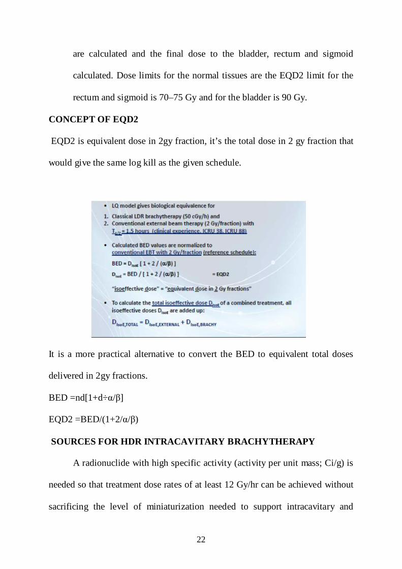
CONCEPT OF EQD2
EQD2 is equivalent dose in 2gy fraction, it’s the total dose in 2 gy fraction that
would give the same log kill as the given schedule.
It is a more practical alternative to convert the BED to equivalent total doses
delivered in 2gy fractions.
BED =nd[1+d÷α/β]
EQD2 =BED/(1+2/α/β)
SOURCES FOR HDR INTRACAVITARY BRACHYTHERAPY
A radionuclide with high specific activity (activity per unit mass; Ci/g) is
needed so that treatment dose rates of at least 12 Gy/hr can be achieved without
sacrificing the level of miniaturization needed to support intracavitary and

23
interstitial brachytherapy. A source no larger than 1 mm in diameter by 4mm
long with an exposure rate of at least IR/sec at l cm is required. The exposure
rate achieved by a small source depends on the chemical form (i.e. relative mass
of non-radioactive atoms) of the source, its density, exposure rate constant of
the radionuclide and photon self-absorption.
SOURCES
The need for high specific activity sources limits the number of
radioisotopes suitable for HDR remote after loaders. Most HDR units use
Iridium-192 (192Ir) or cobalt 60 (60Co). 192Ir offer smaller source sizes but
sources must be changed frequently. Most centers exchange their 192Ir sources
every 3 or 4 months (half life 73.8 days) whereas a similar decay fraction for
60Co takes 5-8years (half life 5.26 years). 60Co is used as an intracavitary HDR
source in the form of small spherical pellets and it emits two high energetic
gamma rays(1.17 &1.34 MeV). The smaller sources permit access to more body
sites via interstitial or intraluminal applications. Based solely on specific
activity considerations 192Ir has been the optimal choice for HDR brachytherapy
and is being widely used radionuclide for the application. With the introduction
of cobalt as a brachytherapy source paradigm has shifted to its use for cost
effectiveness in high burden setups.
COMPARISON BETWEEN COBALT AND IRIDIUM SOURCE
Source Specifications of cobalt and iridium source

24
Fig-2.1
Fig- 2.2
Beyond 200 Kev all isotopes show similar absorption in tissues

25
Fig- 2.3
Fig- 2.4
Anisotropy- No difference between the two radio isotopes except dip in the
direction of source axis.

26
Fig -2.5
Fig -2.6- Isodose distribution for 60Co and 192Ir

27
Fig -2.7 source change data for both radionuclides
DOSE SPECIFICATION
Since the first application of radium in the treatment of cancer of the
uterus in 1908, several techniques have evolved, most of which are
modifications of the Stockholm technique and the Paris technique. Stockholm37
based techniques was introduced by Forsell and Heyman' at the Radium
hemmet in Stockholm. It is a fractionated course of radiation delivered over a
period of one month. In Stockholm there are three insertions each of 22 hours
separated by 1 -3 weeks. Total prescribed dose - 6500-7100 mg Ra and 4500
mg Ra is contributed by the vaginal box and dose rate is 110 R/hr or 2500mg/hr

28
per fraction. Paris technique38 is a single application of radium for 120 hrs
(45R/hr). It delivers a dose of 7000- 8000mg-hrs of radium
The Manchester system, which evolved from the Paris technique, uses a
Rubber uterine tandem to hold one to three radium tubes and rubber ovoids,
separated by a rubber spacer, to each hold a radium tube. The radiation is
delivered in at least two applications. In the Fletcher-Suit applicator the tandem
and the ovoids (or the colpostats) are made of stainless steel and then secured to
hollow handles to permit after loading of the sources39.
Brachytherapy plays a very important role in obtaining high cure rates with
minimum complications. Ideal placement of the uterine tandem and vaginal
ovoids produces a pear-shaped distribution, delivering a high dose to the cervix
and paracervical tissues and a reduced dose to the rectum and bladder40. If the
intracavitary placement has been optimized, this can usually be accomplished
without exceeding a dose of 75 Gy to the bladder reference point or 70 Gy to
the rectal reference point, doses that are usually associated with an acceptably
low risk of major complications. The dose to the surface of the lateral wall of
the apical vagina should not usually exceed 120 to 140 Gy. Suboptimal
placements occasionally force compromises in the dose to tumor or normal
tissues. To choose a treatment that optimizes the therapeutic ratio in these
circumstances requires experience and a detailed understanding of factors that
influence tumor control and normal tissue complications41,42,43.

29
HIGH DOSE RATE APPLICATORS
Fletcher-Suit or Fletcher-Suit-DeIco's
These applicators are used for the treatment of gynecological malignancies of
the uterus, cervix and pelvic side walls. The applicator set typically consists of
three rigid intrauterine tandems, with curvature of 15-, 30-, and 45-degree
angles, and a pair of ovoids or colpostats. The tandem and the ovoids (or the
colpostats) are made of stainless steel and then secured to hollow handles to
permit after loading of the sources. CT and MRI compatible applicators are now
available.
The Manchester System
The Manchester system is one of the oldest and the most extensively used
systems in the world. It is characterized by doses to four points: point A, point
B, a bladder point, and a rectum point. The duration of the implant is based on
the dose rate calculated at point A, although the dose at the other points is taken
into consideration in evaluating a treatment plan. With the availability of the
treatment planning computers, most users of the Manchester system examine
the isodose distributions in the frontal and sagittal planes in addition to
obtaining dose at the four designated points. Point A still remains the point of
dose prescription. Point A was originally defined as 2 cm superior to the lateral
vaginal fornix and 2 cm lateral to the cervical canal. Later, it was redefined to
be 2 cm superior to the external cervical os (or cervical end of the tandem), and
2 cm lateral to the cervical canal. Point B is defined 3 cm lateral to point A.
30
Ideally, a point A represents the location where the uterine vessels cross the
ureter. It is believed that the tolerance of these structures is the main limiting
factor in the irradiation of the uterine cervix. Point B represents obturator node.
The International Commission on Radiation Units and Measurements
System [ICRU]
The ICRU has recommended a system of dose specification that relates
the dose distribution to the target volume, instead of the dose to a specific point.
The dose is prescribed as the value of an isodose surface that just surrounds the
target volume.
Data required for reporting intracavitary therapy [ICRU-38]41
• Description of the Technique: Minimum information should
include the applicator type, source type and loading and orthogonal
radiographs of the application.
• Total Reference Air Kerma: By this parameter, it is meant the total
air kerma strength of sources times the implant duration. This is
similar to the total milligram-hours of radium or total mg-Ra eq-h
except that the sources are calibrated in units of air kerma strength,
that is, ^Gy m2 h-1.
• Reference Volume. The reference volume is the volume of the
isodose surface that just surrounds the target volume. The
prescription isodose value of 60 Gy includes the dose contribution

31
from the external beam. The reference volume for the intracavitary
part of the treatment should be identified and its dimensions
recorded.
Absorbed Dose at Reference Points
Bladder Point: The bladder point is localized by using a Foley catheter,
with the balloon filled with 7 ml of a contrast material. On the frontal
radiograph, the bladder point is marked at the center of the balloon; on the
lateral radiograph, the bladder point is obtained on a line drawn antero
posteriorly through the center of the balloon, at the posterior surface.
Rectal Point: The rectal point is identified on the frontal radiograph at the
midpoint of the ovoid sources (or at the lower end of the intrauterine source).
On the lateral radiograph, the rectal point is located on a line drawn from the
middle of the ovoid sources, 5 mm behind the posterior vaginal wall. The
posterior vaginal wall may be visualized by using radiopaque gauze for the
vaginal packing.
Pelvic Wall Points: On the anteroposterior radiograph, the pelvic wall
points are located at the intersection of a horizontal tangent to superior aspect of
the acetabulum and a vertical line touching the medial aspect of the acetabulum.
On the lateral view, these points are marked as the highest mid distance points
of the right and left acetabulum.

32
CHARACTERISTICS OF AN IDEAL APPLICATION IN
BRACHYTHERAPY 42
• The ovoids should fill the vaginal fornices - largest ovoid size to be
used.
• The ovoids should be separated by 0.5 -1.0 cm, admitting the flange on
the tandem.
• The axis of the tandem should be central between the ovoids
• Tandem - 1/3 of the way between Sl -S2 and the symphysis pubis.
• The tandem - midway between the bladder and Sl -S2.
• Ovoids should be against the cervix (marker seeds).

33
• Tandem should bisect the ovoids. The bladder and rectum should be
packed away from the implant.
DOSIMETRIC CHARACTERISTICS OF BRACHYTHERAPY
SOURCES
In general, four factors influence the single-source dose distribution for
photon-emitting sources:
• distance (inverse-square law);
• absorption and scattering in the source core and encapsulation;
• photon attenuation and
• Scattering in the surrounding medium.
RADIOBIOLOGY OF BRACHYTHERAPY AND THE DOSE-RATE
EFFECT
The biological effects of radiotherapy depend on dose distribution,
treated volume, dose rate, fractionation and treatment duration. These factors,
have an important role in determining the outcome of external beam
radiotherapy or of brachytherapy.
The biological damage inflicted by irradiation of human cells with
ionizing radiation can be divided into three consecutive steps:
A very short initial physical phase (about 10-18 S), during which photons
interact with orbital electrons, raising them to higher energy levels inside the

34
atoms (excitation), or ejecting some of them from the atoms (ionization). This is
the energy deposition phase.
A chemical phase, again very short (about 10-3 s), during which ionized
and excited atoms interact, leading either directly or indirectly through the
formation of free radicals to the breakage of chemical bonds. Free radicals are
highly reactive and can induce chemical changes in biologically important
molecules like DNA. Single-strand or double-strand breaks in DNA appears to
be the basic damage leading to biological effects.
A biological phase, much longer (seconds to years), during which the
cells react to the inflicted chemical damage43.Specific repair enzymes can
successfully repair the vast majority of lesions in DNA. A few lesions however
may not be repaired, and may therefore lead to cell death. Cell death is not
immediate and usually occurs during the next cell division (apoptosis is a minor
process in most human cells). The early reactions are seen during the first days
or weeks after irradiation (for example diarrhea or acute mucositis). They are
temporary because the cell deficit is compensated by the repopulation of stem
cells, and subsequently of differentiated cells. Late reactions due to damage to
the late-reacting tissues, for instance blood vessel damage, fibrosis,
telangiectasia, etc., may be seen after months or years. Damage to these late
reacting normal tissues is poorly repaired and is responsible for most severe
complications of radiotherapy. Tolerance of these tissues is the limiting factor
for radiation therapy.

35
THE FIVE R'S OF RADIOBIOLOGY
A number of biological processes take place during irradiation and
modify the radiation response. These processes are often described as the 5 R’s
of radiobiology44
Each follows a specific time pattern:
• Repair of DNA damage. Both experimental and clinical studies have shown
that human tumors strongly differ in radio sensitivity and radio curability
thought to stem from differences in capacity for repair of sub lethal
damage45,46.
• Reassortment or redistribution in the cell cycle. The cell cycle is divided
into four consecutive stages: Gl, S, G2 and M. Gl is a gap of apparent
inactivity after a mitosis (M), before DNA synthesis (S-phase) resumes in
view of the following cell division. G2 is a second gap of apparent inactivity
between S phase and M.
• Radiosensitivity - varies along the cell cycle, S being the most resistant
phase, and G2 and M the most sensitive. Therefore, cells surviving an
exposure are preferentially in a stage of low sensitivity (Gl), i.e.
synchronized in a resistant cell cycle phase. They progress thereafter
together into S and then to the more sensitive G2 and M phases. A new
irradiation exposure at this time will have a larger biological effect (more
cell kill). However, while this synchronization effect has explained some

36
experimental results, redistribution has never been shown to play a
measurable role in the clinic of radiotherapy.
• Repopulation - Cells surviving an irradiation keep proliferating. This
increases the number of clonogenic cells, i.e. the number that must
eventually be sterilized to eradicate cancer. Repopulation therefore has a
detrimental effect as far as cancer control is concerned. Stem cells do also
proliferate in normal tissues, which has in this case a protective effect (it
helps the tissue to recover from radiation damage and it adds to DNA repair
in cells).
• Reoxygenation - Because of an inappropriate development of intra tumoral
vasculature, every tumor of clinically detectable size contains a large
proportion of poorly oxygenated cells. Also, the proportion of hypoxic cells
increases with the tumor size. Acutely hypoxic cells are far more radio
resistant than well oxygenated cells. This is expressed by the Oxygen
Enhancement Ratio (OER), i.e. the ratio between radiation doses required in
hypoxia and air to produce the same biological effect. Its value is 3, and it
varies very little with dose or with the biological system. Hypoxic cells
usually survive irradiation, but they progressively (re)oxygenate, due to the
better supply of oxygen available after well oxygenated cells have died .This
restores radio sensitivity in the tumor. Several mechanisms are involved, but
reoxygenation occurring at long intervals is probably due to tumor shrinkage
leading to a reduction of the intercapillary distance.

37
DOSE RATE EFFECTS IN BRACHYTHERAPY
Biological effects of radiation are strongly dependent upon the rate of
dose delivery. The radiobiological processes involved in high dose rate
brachytherapy are in all respects similar to those involved in fractionated
external beam radiation therapy, except for the volume effect, as mentioned
earlier. Repair, repopulation, and reoxygenation, are the main factors
determining outcome. They do not occur during the very short duration of
irradiation (up to 10-15 minutes), but take place between consecutive fractions,
provided the interval is adequate.
Repair. - For brief exposures, the survival fraction S of a cell population
decreases with increasing dose D. It has been mathematically modelled as the
sum of two type of lesions:
Lethal (non-repairable) lesions, with a survival fraction S = exp (-
αD), represented by the tangent to the survival curve at its origin.
Sublethal lesions, non-lethal and potentially repairable, but the
accumulation of which can cause cell death, with a survival fraction S = (-βD2)
The sum of these two components leads to the classical linear-quadratic
Equation proposed by Chadwick & Leenhouts and Kellerer & Rossi.
S=exp-(αD+(βd2).
The survival curve displayed on a semi-logarithmic graph exhibits an
initial shoulder. According to the model, it is proportional to DNA repair
capacity. Hence, a broad shoulder is associated with a large repair capacity. The
38
ratio α/β corresponds to the dose at which the contribution of the two factors to
the survival fraction is equal, αD = βD2, and D = α/β. A large α/β corresponds
to a small shoulder (small repair capacity) and a small α/β to a broad shoulder
(large repair capacity).
In summary, a low α/β is characteristic of late-responding normal tissues
and some tumors (0.5 to 6Gy, average 3 Gy), while a higher α/β ratio
characterizes the early-responding normal tissues and carcinomas. (7 to 20 Gy,
average 10 Gy
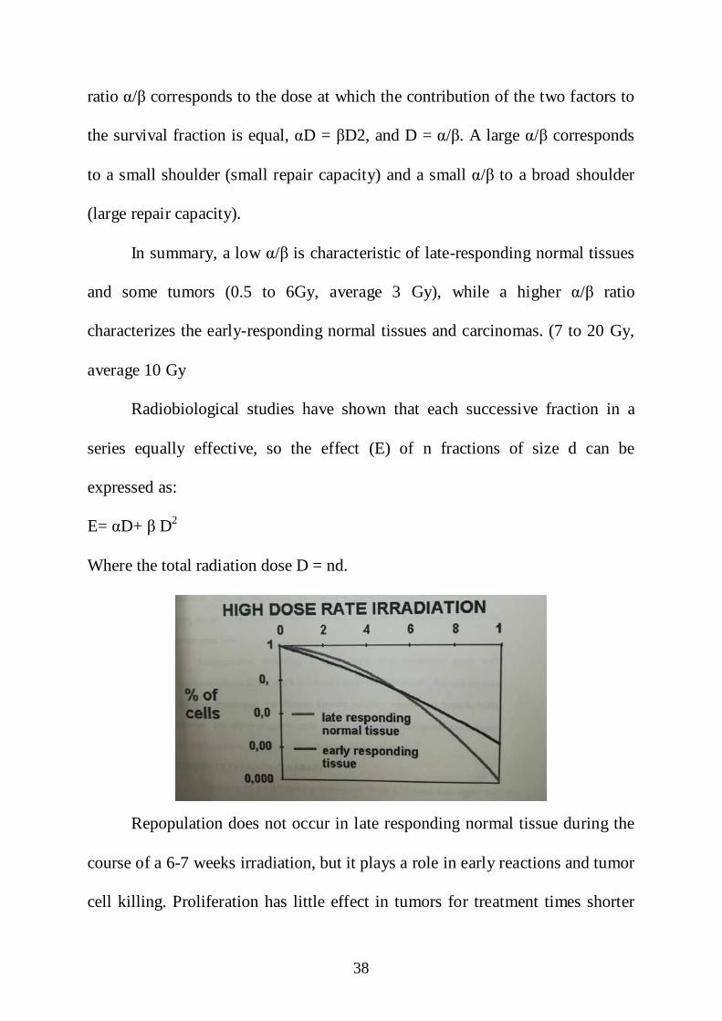
Radiobiological studies have shown that each successive fraction in a
series equally effective, so the effect (E) of n fractions of size d can be
expressed as:
E= αD+ β D2
Where the total radiation dose D = nd.
Repopulation does not occur in late responding normal tissue during the
course of a 6-7 weeks irradiation, but it plays a role in early reactions and tumor
cell killing. Proliferation has little effect in tumors for treatment times shorter

39
than 3-4 weeks47 . After this time, accelerated repopulation of fast-growing
tumors may be observed48. For early effects on skin and mucosa (desquamation
and mucositis), the spontaneous tissue kinetics are unchanged until about 10
days after the initiation of Irradiation, when the rate of cell replacement is
accelerated49. It remains very active during the two weeks following irradiation,
and then tends to drop quickly, back to its physiological level.
Reoxygenation. Following a large single dose irradiation, most well
oxygenated cells are killed, and hypoxic cells survive predominantly. Because
aerobic cells have disappeared, the distance between capillary vessels and
hypoxic cells decreases. This allows oxygen to reach hypoxic cell, which
reoxygenate and become more radiosensitive. The process takes between hours
and weeks50.

40
COMPLICATIONS
RTOG-ACUTE TOXICITY [Table-2.1]
Toxicity Grade 1
Grade 2
Grade 3
Grade 4
Grade 5
GENITO
URI
NARY
Frequency of
urination or
nocturia twice
pretreatment
habit/dysuria,
urgency not
requiring
medication.
Frequency of
urination or
nocturia that is
less frequent than
every hour.
dysuria urgency,
bladder spasm
requiring local
anaesthetic.
Frequency with
urgency and
nocturia hourly
or more
frequently/
dysuria, pelvis
pain or bladder
spasm
requiring
regular, frequent
narcotic/ gross
haematuria with
/ without clot
passage.
Hematuria
requiring
transfusion,
acute bladder
obstruction
not secondary
to clot
passage,
ulceration or
necrosis.
Death

41
Toxicity Grade 1
Grade 2
Grade 3
Grade 4
Grade 5
Small
bowel
toxicity
Anorexia with
≤5% weight loss
from pretreatment
baseline/nausea
not requiring
antiemetic’s/
abdominal
discomfort not
requiring
parasym
patholytic drugs
or analgesics.
Anorexia with
≤15% weight loss
from pretreatment
baseline/ nausea
and or vomiting
requiring
antiemetic’s/
abdominal pain
requiring
analgesics.
Anorexia
with≥15%weight
loss from
pretreatment
baseline or
requiring NG
tube or parentral
support. Nausea
and/or vomiting
requiring tube or
parentral
support/
abdominal pain
,severe despite
medication
/hematemesis or
melena/
abdominal
distention[ x ray
demonstrating
distended
bowel loops]
Ileus, subacute
or acute
obstruction,
perforation, GI
bleeding
requiring
transfusion/
abdominal
pain requiring
tube,
decompression
or bowel
diversion.
Death

42
Toxicity Grade 1
Grade 2
Grade 3
Grade 4
Grade 5
Rectal
toxicity
Increased
frequency or
change in quality
of bowel habits
not requiring
medication/Rectal
discomfort not
requiring
analgesics.
Diarrhea
requiring
parasympatholytic
drugs/Mucous
discharge not
necessitating
sanitary
pads/rectal or
abdominal pain
requiring
analgesics.
Diarrhea
requiring
parentral
support/ severe
mucous or
bloody discharge
necessitating
sanitary pads/
abdominal
distention
[X-ray
demonstrates
distended loops]
Acute or sub
acute
obstruction,
fistula or
perforation, GI
bleeding
requiring
transfusion,
abdominal
pain or
tenesmus
requiring tube
decom
pression or
bowel
diversion.
Death

43
AIM OF THE STUDY
To evaluate the acute gastrointestinal and genitourinary toxicity profile in
carcinoma cervix patients using cobalt 60 radionuclide source.
SECONDARY END POINTS
• Feasibility of twice weekly brachytherapy
• Response assessment after completion of treatment.
STUDY CENTRE-
• Dept. of Radiotherapy, Barnard Institute of Radiotherapy, Madras
Medical College, Chennai-3
• The study was reviewed and approved by the institutional ethical
committee.
STUDY PERIOD
From June 2017 to September 2018
STUDY DESIGN – Single arm prospective study.

44
MATERIALS AND METHODS
CASE SELECTION
Carcinoma cervix patients who had completed their EBRT and slated for
brachytherapy with minimal or no parametrial disease.
NO. OF PATIENTS - 38
INCLUSION CRITERIA
• Biopsy proven newly diagnosed carcinoma cervix
• Age - 30-65 years
• Stage – IB2 –IIIB
• Histology – Squamous cell carcinoma and its variants.
• ECOG 0-2
• Previously not exposed to any chemotherapy or radiotherapy.
• No major life threatening complications
• HIV negative
• Patient should be fit for anesthesia [GA,SA,IV sedation]
• Cystoscopy – for ruling out bladder invasion
• Urine routine, culture and sensitivity-to rule out other causes of cystitis
before brachytherapy.
• Written and informed consent.

45
EXCLUSION CRITERIA
• Age - < 30 and > 65 years.
• ECOG -3 or more
• Stage IVA –involvement of bladder and rectum.
• Inadequate hepatic and renal functions
• Patient not consenting to chemotherapy.
• Previously treated for any other malignancy.
• Metastatic or recurrent disease
• HIV positive patients
• Patient unfit for anesthesia.
INVESTIGATIONS
• Biopsy from the tumor
• Complete blood count, liver function test, renal function test, viral
markers.
• CT scan abdomen and pelvis or MRI- plain and contrast [pretreatment
and post treatment at 6-8 weeks]
• Chest X ray –PA view ,ECG, blood grouping
• Cardiology evaluation for fitness.
• Weekly CBC and RFT before each fraction of brachytherapy.

46
DATA COLLECTION AND METHODS
Eligible patients treated with radiotherapy in the form of external beam
radiotherapy to a total dose of 45-50Gy to the whole pelvis. [45-50Gy/180-200
cGy/#/25#/5 days a week [Monday to Friday] using cobalt teletherapy machine.
Followed by brachytherapy to a dose of 21Gy/7 Gy/#/3# with a minimum
gap of 72 hours between each fraction using cobalt 60 source with BEBIG
BRACHYTHERAPY MACHINE.
CONCURRENT CHEMOTHERAPY
Cisplatin 40mg/m2 weekly along with proper premedication.
Patients to be clinically assessed during treatment for toxicity of acute
gastrointestinal and genitourinary symptoms and response graded accordingly.
CT abdomen and pelvis or MRI to be taken 6-8 weeks post treatment for
response assessment. Toxicity to be graded according to RTOG acute toxicity
criteria. Patients were also assessed for their compliance during brachytherapy
sessions.
TREATMENT PLANNING
EBRT
The whole pelvis including cervix, vagina, and parametrium with the iliac
and pelvic lymph nodes treated.
47
RT PORTALS AND BORDERS
SUPERIOR - L4-L5 interspace [to include iliac and hypogastric nodes]
INFERIOR – If vagina uninvolved –lower border of obturator foramen.
If vagina involved- entire vagina upto introitus was included.
LATERAL- 2cm lateral to bony pelvis.
Treatment field was verified using X-ray simulation
PORTALS
AP and PA portals treated if field separation was less than 20cm
Field separation of more than 20cm were treated with 4 field box technique
where the anterior border was kept in front of pubic symphysis and posterior
border at S2-S3 junction.
CHEMOTHERAPY DRUG - cisplatin
DOSE - 40mg/m2 –weekly
SCHEDULE - Weekly once
PREMEDICATIONS – Inj.Dexamethasone
Inj.Ranitidine
Inj.Ondansetron
With adequate hydration before and after chemotherapy.

48
INTENT OF ADDING CHEMOTHERAPY - Radio sensitizer.
TOTAL NUMBER OF CYCLES - 4 -5 cycles
INTRACAVITARY PROTOCOL
After Teletherapy all patients were assessed for intracavitary application.
Those who were found fit for brachytherapy were subjected to the procedure.
HDR BRACHYTHERAPY
Technique - Remote after loading with cobalt 60 source.
Machine - BEBIG
Activity - 1.82 curie [67.484 GBq]
Intracavitary applicator – Modified fletcher suit with 15 and 20 degree
angulation.
No. of fractions -3 # with a minimum gap of 72 hrs between each fraction.
Dose prescribed to Point A - 7Gy.
PROCEDURE
Under anesthesia [ IV sedation/SA/GA] with patient in lithotomy position
the perineum and upper half of thighs were cleaned with beta dine and draped.
Per vaginal examination done .Urinary bladder was catheterized and 7ml of
diluted contrast(3ml of contrast +4 ml of distilled water) was injected into the
Foleys balloon. Uterine sound was introduced and uterine length measured. The
cervical stopper was adjusted according to the uterine length and fixed. Then
the two ovoids were introduced and positioned accordingly .Adequate vaginal

49
packing was done. Rectal tube inserted. CT simulation was done. The films
were used in our treatment planning system for applicator reconstruction,
defining Point A, Point B, dose prescription to point A and calculating bladder
and rectum points as per ICRU38 .Dose to bladder and rectum was kept less
than 80% of point A. Optimization done using change in dwell position and
time .After obtaining the desired prescription isodose, patient was connected to
the BEBIG machine with cobalt 60 source through catheters (transfer tubes) and
treated.
The Assessment of GI and GU toxicities was done as per schedule
First assessment -after the application of first HDR ICRT brachytherapy
Second assessment – at the time of last HDR ICRT application
Third assessment- one month later to last HDR ICRT application
Fourth assessment-two month later to last HDR ICRT application
Fifth assessment- three month later to last HDR ICRT application

50
ANATOMICAL TOPOGRAPHY AND BRACHYTHERAPY APPLICATOR
INSERTIONS. [Fig -4.1]

51
FLETCHER SUIT DELCLOS APPLICATOR [Fig - 4.3]
BEBIG HDR BRACHYTHERAPY MACHINE [Fig -4.4]
52
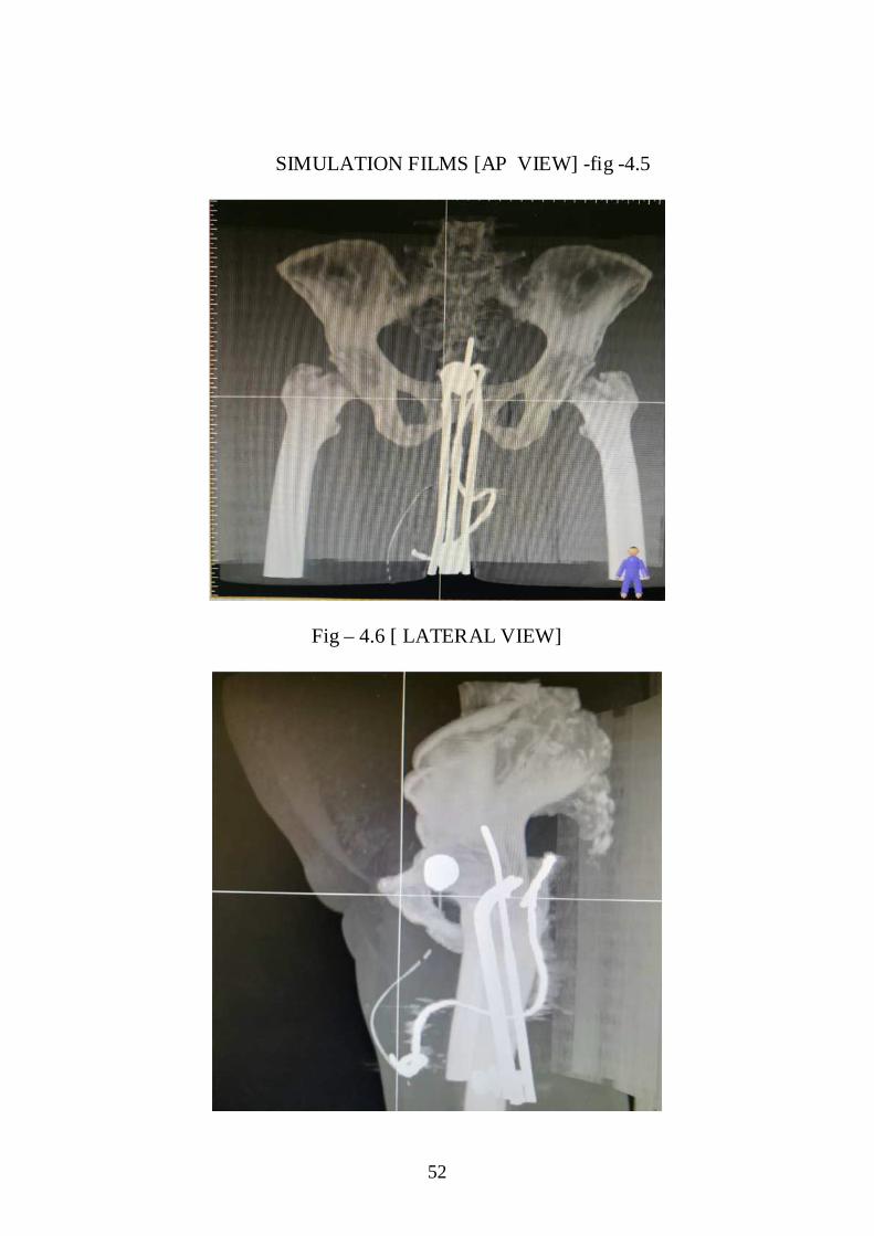
SIMULATION FILMS [AP VIEW] -fig -4.5
Fig – 4.6 [ LATERAL VIEW]

53
ISODOSE DISTRIBUTION IN AP AND LATERAL VIEW
Fig – 4.7
Fig-4.8

54
CASE ANALYSIS AND RESULTS
STUDY POPULATION AND COMPLIANCE TO TREATMENT
From June 2017-September 2018 a total of 38 patients of carcinoma
cervix were studied for acute toxicity analysis with brachytherapy using cobalt
60 source. All insertions were done when patient had completed EBRT and fit
for intracavitory insertions. All patients were available for final analysis
CHARACTERISTICS
AGE (TABLE-5.1)
AGE [Years] Number of patients [percentage]
30- 40 yrs 01 [3%]
41 -50 yrs 10 [27%]
51-60 yrs 18 [49%]
61 -65 yrs 08 [21%]
FIGURE [5.1]
30-40 yrs
3% 41-50 yrs
27%
51-60 yrs
[PERCENTAGE]
61-65 yrs
21%
No. of patients
30-40 yrs
41-50 yrs
51-60 yrs
61-65 yrs
55
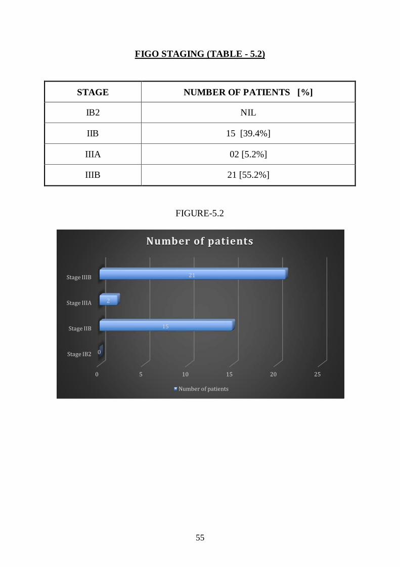
FIGO STAGING (TABLE - 5.2)
STAGE NUMBER OF PATIENTS [%]
IB2 NIL
IIB 15 [39.4%]
IIIA 02 [5.2%]
IIIB 21 [55.2%]
FIGURE-5.2
0 5 10 15 20 25
Stage IB2
Stage IIB
Stage IIIA
Stage IIIB
0
15
2
21
Number of patients
Number of patients

56
Histology (Table- 5.3)
FIGURE-5.3
1, 3%
18, 47%
11, 29%
8, 21%
SQUAMOUS CELL CRCINOMA
WELL DIFFERENTIATED MODERATELY DIFFERENTIATED
POORLY DIFFERENTIATED LARGE CELL
SQUAMOUS CELL CARCINOMA NO.OF PATIENTS
WELL DIFFERENTIATED 01 [2.6%]
MODERATELY DIFFERENTIATED 18 [47.3%]
POORLY DIFFERENTIATED 11 [29%]
LARGE CELL NON KERATINIZING 08 [21%]

57
EBRT+CHEMO (Table-5.4)
EBRT+CHEMO NUMBER OF PATIENTS
RT+CHEMO 38
RT ONLY NIL
TIME INTERVAL BETWEEN EBRT AND BRACHYTHERAPY
(Table - 5.5)
TIME INTERVAL NUMBER OF PATIENTS [%]
< 1 WEEK 23 [60.5%]
>1 WEEK 15 [39.4%]

58
CHEMOTHERAPY CYCLES (Table - 5.6)
CISPLATIN [CYCLE] NUMBER OF PATIENTS
PERCENTAGE
2 CYCLES 02 5.2%
3 CYCLES 06 15.7%
4 CYCLES 12 31.5%
5 CYCLES 18 47.36%
FIGURE-5.4
Majority of the patients in the study were able to tolerate chemotherapy
cycle and were able to complete all 5 weekly schedules.
2 cycles, 2
3 cycles, 6
4 cycles, 12
5 cycles, 18
Cisplatin
2 cycles 3 cycles 4 cycles 5 cycles

59
PERFORMANCE STATUS (Table-5.7)
ECOG NO.OF PATIENTS
0 03 [7.8%]
1 21 [55.2%]
2 14 [36.8%]
FIGURE-5.5
There was a delay of more than a week to 12 days for nearly half of the
patients due to grade 3 - 4 skin reactions due to EBRT and referral from other
centers but this was compensated with twice weekly brachytherapy with a
minimum gap of 72 hrs between each fractionation.
3
21
14
0 5 10 15 20 25
ECOG 0
ECOG 1
ECOG 2
PERFORMANCE STATUS
PERFORMANCE STATUS

60
SECONDARY ASSESMENT
There was no patient related factors for delay in between the fractions
except for 1 patient who had grade III GI toxicity and was managed with
symptomatic and supportive care.
TUMOUR ASSESMENT AFTER EBRT (Table- 5.8)
TUMOUR RESPONSE [CLINICAL] NUMBER OF PATIENTS
COMPLETE RESPONSE 27 [71%]
PARTIAL RESPONSE 11 [29%]
FIGURE-5.6
27, 71%
11, 29%
TUMOUR RESPONSE
COMPLETE RESPONSE
PARTIAL RESPONSE

61
BRACHY THERAPY DOSE CHARACTERISTICS . (Table-5.9)
CA CERVIX[1-III] MEDIAN RANGE
EBRT[WHOLE PELVIS] 50GY 45-50 GY
HDR 7/3# -
PT A 7gy 7-7.5GY
ICRU BLD PT 5.6GY 4.2-6GY
ICRU REC PT 5.4GY 3.2-6GY
BED RECT[EBRT+HDR] 124.4 120-133
EQD2 TUMOUR[EBRT+HDR] 83.6GY 78.8-86GY
REACTIONS ASSESED
• NAUSEA
• VOMITING
• DIARRHEA
• URINARY FREQUENCY
• URINARY URGENCY
62
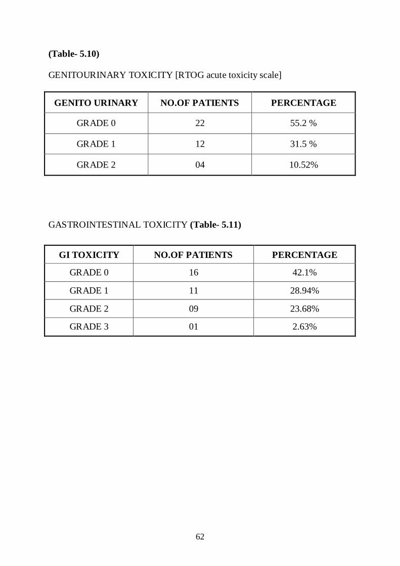
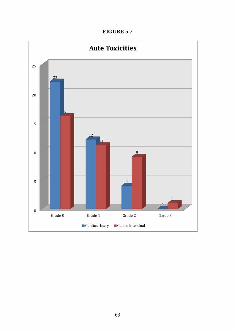
(Table- 5.10)
GENITOURINARY TOXICITY [RTOG acute toxicity scale]
GENITO URINARY NO.OF PATIENTS PERCENTAGE
GRADE 0 22 55.2 %
GRADE 1 12 31.5 %
GRADE 2 04 10.52%
GASTROINTESTINAL TOXICITY (Table- 5.11)
GI TOXICITY NO.OF PATIENTS PERCENTAGE
GRADE 0 16 42.1%
GRADE 1 11 28.94%
GRADE 2 09 23.68%
GRADE 3 01 2.63%
63
FIGURE 5.7
0
5
10
15
20
25
Grade 0 Grade 1 Grade 2 Garde 3
22
12
4
0
16
11
9
1
Aute Toxicities
Genitourinary Gastro intestinal
64
DISCUSSION
Atara ntekim51 et al compared the acute gastrointestinal and genitourinary
Toxicity associated with Co-60 source in the brachytherapy of cervical cancer.
His study assessed 70 patients with cervical cancer who received 45 Gy in 22
fractions of pelvic external beam radiotherapy and 19.5 Gy in 3 fractions of
HDR with Co-60 source using tandem and ring applicators with 6 courses of
cisplatin 50 mg/m2 and 5 fluorouracil 1000 mg/m2. (3%) had grade 3
gastrointestinal toxicity while all others had grade 2 toxicity.
Jain Abhay Kumar et al52 studied 65 patients of carcinoma cervix for acute
toxicity using cobalt brachytherapy .all patients completed EBRT 45-50GY/
180-200 CGY /#/25 #/ 5 days a week using telecobalt machine followed by
intracavitory application after 1 week. 7gy in 3 # were given and toxicity
assessed by CTCAE 4.03 version. Only 2 patients [3%] had acute diarrhea. This
was comparable to iridium source.
Montien Pesee et al53 did a retrospective study of uterine cervical cancer
patients, stages IB-IVB treated by radiotherapy alone. The patients received
teletherapy 50Gy / 25 fractions, five fractions per week to the whole pelvis
together with HDR Cobalt -60 after loading brachytherapy of 850 cGy/ fraction
weekly to point A for 2 fractions for 141 patients of cervical cancer. 96.5%
had complete response rates but morbidity rates of grade1 and grade 2 radiation

65
proctitis of 27.0%, and 10.6 % were reported .The grade 1 and grade 2 radiation
cystitis were reported as 1.4%, and 1.4 %. Grade 3 radiation complications
were 0.71% of radiation proctitis and 0.71% small bowel obstruction . The
mean onset time to develop radiation proctitis after treatment was 15 months
with a range of 6-61 months, for radiation cystitis it was 30 months (range 9 -
47 months) and for small bowel obstruction it was 53 months. So treatment with
HDR-60 brachytherapy less than 850 cGy per fractionation for decreasing the
grade 2 and grade 3 radiation morbidity was recommended in the study.
Om Prakash Gurjar et al54 reported the dosimetric parameters of Co-60 based
high dose rate (HDR) brachytherapy plans for patients of carcinoma uterine
cervix .study on ten patients with locally advanced carcinoma of the uterine
cervix . Computed tomography (CT) images were taken after three channel
applicator insertions. The planning for 7 Gray per fraction (7 Gy/#) was done
for Co-60 HDR brachytherapy following the American Brachytherapy Society
(ABS) guidelines. All the patients were treated with 3# with one week interval
between fractions The mean dose to high risk clinical target volumes (HRCTV)
for D90(dose to 90%volume) was found to be 102.05% (Standard Deviation
(SD): 3.07). the mean D2cc (dose to 2cubic centimeter volume) of the bladder,
rectum and sigmoid were found to be 15.9 Gy (SD: 0.58), 11.5 Gy (SD: 0.91)
and 4.1 Gy (SD: 1.52), respectively. The target coverage and doses to organs at
risk (OARs) were achieved as per the ABS guidelines. This study concluded

66
that the Co-60 HDR brachytherapy unit is a good choice especially for the
centers with a small number of brachytherapy procedures as no frequent source
replacement is required like in an Ir-192 HDR unit.
Upendra Nandwana et al55 examined the dosimetry of intracavitary radiotherapy
(ICRT) in carcinoma of the cervix using cobalt-60 (Co-60) as source of ICRT,
and as an alternative to iridium-192 (Ir-192) retrospectively. The study was
done on 80 ICRT patients. . The dose to point A, 60 Gy isodose reference
volume, and bladder and rectum maximum and mean doses were defined. It was
found that dosimetry of Co-60 as a brachytherapy source was consistent with
the International Commission on Radiation Units (ICRU 38) recommendations.
Co-60 is a logical alternative to Ir-192 in low socio-economic settings when
repeated changing of the source is not an option.
Thanatip Tantivatana et al56 did a retrospective cohort study of patients with
cervical cancer and treated with radiotherapy Ir-192 group and cobalt-60 group.
The 2- and 5-year disease-free survival rate in Ir-192 group were 80.4% and
73.1% and in Co-60 group were 82.5% and 74.7%, respectively (p=0.365).
Overall survival rates at 2 and 5 years were 89.4% and 77% of the Ir-192 group,
and 91.6% and 81.9% in the Co-60 group, respectively (p=0.238). The
complications were primarily grade 1 or 2. Grade 3 and 4 complications were
found in 13 of 274 and 7 of 206 in Ir-192 and Co-60 groups, respectively

67
(p=0.232). Grade and clinical stage of cancer significantly affected the survival
outcome. patients who were treated with HDR Co-60 brachytherapy were
comparable in survival and toxicity outcomes of those with HDR Ir-192
brachytherapy concluding Co-60 source has economic advantages over Ir-192
and hence suitable for low resource setting.
Stefan Strohmaier et al57 compared the isotopes 60Co and 192Ir as radiation
sources for high-dose-rate (HDR) afterloading brachytherapy in view of
availability with identical geometrical dimensions. The paper compared the
characteristics of both nuclides in different fields of brachytherapy .It
investigated the advantages or disadvantages of both radionuclides for HDR
brachytherapy due to their physical differences. Results of this work showed
that no advantages or disadvantages exist for 60Co sources compared to 192Ir
sources with regard to clinical aspects. Nevertheless, there are potential
logistical advantages of 60Co sources due to its longer half-life (5.3 years vs. 74
days), making it an interesting alternative especially in developing countries.
Y. Lee et al58 investigated that dose rate (HDR) brachytherapy contributed
about 7% of the total Equivalent Dose in 2-Gray Fractions (EQD2) of external
beam radiation therapy (EBRT) and HDR brachytherapy .Due to the location in
the pelvis and subsequent distance from the implant, LN groups receive
differing amounts of brachytherapy contribution. . Twenty-one patients with 45

68
positive pelvic LNs (9common iliac (CI), 15 external iliac (EI), 12 internal iliac
(II) and 9 obturator (Ob) LNs) treated in two institutions from Oct 2007 to Aug
2011 were included in this retrospective analysis. All patients received EBRT to
the pelvis with a supplemental boost to the involved pelvic node, plus HDR
brachytherapy. Pathologically involved LNs were contoured on the planning
EBRT image as well as the 4 to 5 brachytherapy planning images. The mean
received dose of each LN from the EBRT and brachytherapy plans was
recorded and EQD2 was calculated .brachytherapy EQD2 was significantly
different among 4 pelvic LN groups. The average prescribed doses from the
EBRT, including the initial pelvic fields and boost contribution to CI, EI, II and
Ob LNs, were 54.60Gy, 54.53Gy, 53.15Gy and 54.42Gy respectively. The
average prescribed HDR doses to International Commission on Radiation Units
and Measurements (ICRU) point A were 26.83Gy, 27.84Gy, 29.79Gy and
28.49Gy accordingly. The average dose delivered to CI, EI, II and Ob LNs were
53.19Gy, 55.14Gy, 53.26Gy and 55.10Gy (EBRT), and 2.65Gy, 4.31Gy,
5.46Gy and 5.77Gy (HDR) respectively, with the Corresponding EQD2 of
52.26Gy, 54.36Gy, 52.42Gy and 54.42Gy (EBRT), and 2.36Gy, 4.00Gy,
5.09Gy and 5.47Gy (HDR). The HDR contribution to CI, EI, II and Ob LNs
was 4.10%, 6.93%, 8.83% and 9.48% of the total EQD2 (EBRT+HDR,
57.69Gy) of all LN groups respectively. There was a statistically significant
difference in brachytherapy EQD2 among the 4 pelvic LN groups (p < 0.05),
with the Ob LN receiving the most dose. This study highlights that there is
69
4.1% to 9.5% variation in brachytherapy dose contribution of the total EQD2
among pelvic LN groups. This difference in HDR contribution needs to be
considered when prescribing EBRT boost dose to each pelvic LN group for the
optimal therapeutic total dose.
Amir Shahram et al59 in locally advanced stage did a cross sectional-analytic
study to report outcome 154 patients with carcinoma of cervix treated with
external beam radiation therapy (EBRT) and high dose rate (HDR)
brachytherapy with cobalt 60 (Co-6o) remote after loading system. They were
analyzed for three-year disease-free survival (DFS), three-year overall survival
(OS) incidence of acute and late complications for HDR brachytherapy.
Fourteen patients (9.1 %) were in Stage I (FIGO classification), 8 (5.2%) were
in Stage IIA, 26 (16.9%) were in Stage IIB, 100 (64.9%) were in Stage III, and
6 (3.9 %) were in Stage IVA. The follow up duration was between - 60 months
with a median of 38 months. Overall rectal and bladder treatment toxicity rates
were 33.7%. The three-year DFS Rate was 85.7%, 70.7 %, 41% and 16.6% for
Stages I, II, III, IVA.
DS Nikam,et al60 reported the feasibility and cost effectiveness of high dose rate
(HDR) cobalt60 (60Co) source versus Iridium-192 (192Ir) source brachytherapy
in government funded hospitals and treatment interruption gap because of
exchange of sources.it was a retrospective study of gynecological cancer
70
patients, The dates for 192Ir sources installation and the last date and first date of
brachytherapy procedure before and after source installation respectively were
also analyzed and calculated the gap in days for brachytherapy interruptions
where eight 192Ir sources were installed. The mean gap between treatment
interruptions was 123.12 days (range 1647 days). Around 52.25% of patients
who received EBRT at their institute were referred to outside hospital for
brachytherapy because of unavailability of Iridium source. The cost for 5 year
duration for single cobalt source is approximately 20-22 lakhs while for
Iridium sources is approximately 52-53 lakhs. This study concluded that the
treatment interruption because of source exchange is longer and can be
minimized by using cobalt source as it is cost effective and has 5 year working
life. Thus, Co60 source for brachytherapy is a feasible option for government
funded institutions.
Some of the studies that showed similar toxicity with iridium source were
71
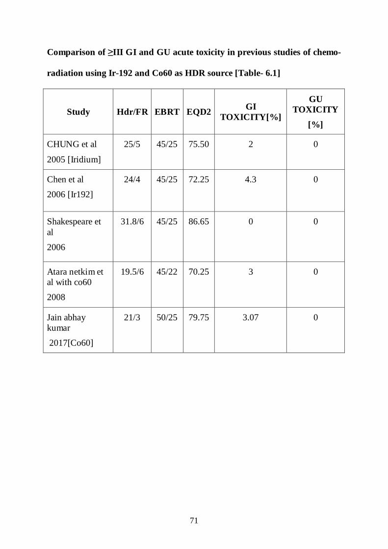
Comparison of ≥III GI and GU acute toxicity in previous studies of chemo-
radiation using Ir-192 and Co60 as HDR source [Table- 6.1]
Study Hdr/FR EBRT EQD2 GI TOXICITY[%]
GU TOXICITY
[%]
CHUNG et al
2005 [Iridium]
25/5 45/25 75.50 2
0
Chen et al
2006 [Ir192]
24/4 45/25 72.25 4.3 0
Shakespeare et al
2006
31.8/6 45/25
86.65 0 0
Atara netkim et al with co60
2008
19.5/6 45/22 70.25 3 0
Jain abhay kumar
2017[Co60]
21/3 50/25 79.75 3.07 0
72
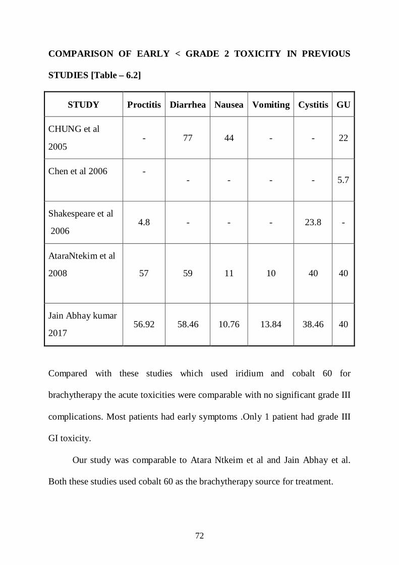
COMPARISON OF EARLY < GRADE 2 TOXICITY IN PREVIOUS
STUDIES [Table – 6.2]
STUDY Proctitis Diarrhea Nausea Vomiting Cystitis GU
CHUNG et al
2005 - 77 44 - - 22
Chen et al 2006 -
- - - - 5.7
Shakespeare et al
2006 4.8 - - - 23.8 -
AtaraNtekim et al
2008
57 59 11 10 40 40
Jain Abhay kumar
2017 56.92 58.46 10.76 13.84 38.46 40
Compared with these studies which used iridium and cobalt 60 for
brachytherapy the acute toxicities were comparable with no significant grade III
complications. Most patients had early symptoms .Only 1 patient had grade III
GI toxicity.
Our study was comparable to Atara Ntkeim et al and Jain Abhay et al.
Both these studies used cobalt 60 as the brachytherapy source for treatment.

73
In our study the average age group of presentation was 53 years where as it was
45 Years and 50 years in the above two studies. Majority of patients were stage
IIIB. Predominant HPE was moderately differentiated squamous cell carcinoma.
All patients had chemotherapy all though some of them could not complete all
the 5 cycles.3 fractions of 7 Gy with a gap of 72 hrs between each fraction was
a feasible option similar to Jain Abhay et al study and it compensated the time
delay between EBRT and brachytherapy . Dose per fraction in our study was
7Gy as per ABS guidelines .Secondary end points were response rates of
primary tumor .The acute toxicity profile was assessed using RTOG scale which
differed from the above two studies who had used common Terminology
Criteria for Adverse Events . All patients were able to complete the treatment
within the 7 weeks though 1 patient who had grade III toxicity during
brachytherapy could not. 35 [92.1%] patients had complete clinical and
radiological response at the end of 3 months.

74
PRE TREATMENT CT SCAN (FIGURE 6.1)
POST TREATMENT CT SCAN [EBRT +ICA] AT 8 WEEKS [fig -6.2]

75
CONCLUSION
Cervical cancer is the third most common malignancy in women
worldwide of which 85% are detected in developing countries. According to
GLOBOCON 2018 in INDIA, it is the second most common malignancy
among women. Carcinoma cervix turn over accounts for 10-15% in our hospital
with additional patients being referred for brachytherapy from most government
hospitals in Tamil Nadu. A shorter overall treatment time in treating this
malignancy would result in better patient compliance as long as there is no
increased toxicity or loco regional failure. This study highlighted the fact that
use of cobalt radionuclide brachytherapy has comparable toxicity profile as
Iridium source .This in turn would translate to treat large number of patients in
low resource settings with high patient load with no frequent change of source
as cobalt 60 has long half-life with similar dosimetric properties and acute
toxicity profile. The follow up period of this study was limited to 90 days post
treatment .Further follow up is needed to assess the late toxicity effects of cobalt
brachytherapy.

BIBLIOGRAPHY
1) Indian Council of Medical Research [ICMR] in 2012.
2) Int J Radiat Oncol Biol Phys.1994 jan 15,28
3) Perez CA: Principles and practice of Radiation Oncology
JB.Lippincott Company 6th
edition.Uterine Cervix.
4) Perez CA; Principles and practice of Radiation Oncology
JB.Lippincott Company 6th
edition.Uterine Cervix.
5) The International Federation of Gynaecology and Obstetrics [FIGO]
revised staging system in 2017.
6) Delaloye JF,Pampallona S,Coucke PA,et al.Younger age as a bad
prognostic factor in patients with carcinoma cervix. Eur J Obstet
Gynaecol Reprod Biol 1996,64;201-205.
7) Dattoli MJ,Gretz HF III,Beller U,et al.Analysis of multiple prognostic
factor in patients in stage IB cervical cancer,age as a major
determinant.Int J Radiat Oncol Biol Phys 1989;17;41-47.
8) Piver MS ,Chung WS. Prognostic significance of cervical lesion size
and pelvic node metastasis in cervical carcinoma.Obstet Gynaecol
1975;46;507-510.
9) Fletcher GH,Cancer of the uterine cervix:Janeway lecture AJR Am J
Roentgenol Radium Ther Nucl Med 1971;111:225-242.
10) Eifel.PJ,Morris M,Wharton JT,et al.The influence of tumor size and
morphology on outcome of patients with FIGO stage IB squamous

cell carcinoma of uterine cervix.Int J Radiat Oncol Biol Phys
1994;29:9-16.
11) Perez CA ,Grigsby PW,Nene SM,et al.Effect of tumor size on the
prognosis pf carcinoma of uterine cervix treated with irradiation
alone.Cancer 1992;69:2796-2806.
12) Eifel PJ, Thoms WW Jr,Smith TL,et al.The relationship between
brachytherapy dose and outcome in patients with bulky endocervical
tumours treated with radiation dose.Int J Radiat Oncol Biol Phys
1994;28;113-118.
13) Perez CA Grigsby PW,Chao KSC,et al.Tumour size,irradiation
dose,and long term outcome of carcinoma cervix.Int J Radiat Oncol
Biol Phys 1998;41:307-317.
14) Eifel PJ,Thomas WW Jr,Smith TL,et al.The relationship between
brachytherapy dose and outcome in patients with bulky endocervical
tumours treated with radiation dose.Int J Radiat Oncol Biol Phys
1994;28:113-118.
15) Douple EB .Platinum –radiation interactions.J Natl Cancer Inst
Monogr 1988;6:315-319.
16) Perez CA,Grigsby PW,Castro-Vita H,et al.Carcinoma of uterine
cervix;I.Impact of prolongation of treatment time and timing of
brachytherapy on outcome of radiation therapy.Int J Radiat Oncol Biol
Phys 1995;32:1275-1288.

17) Piver MS,Rutledge F,Smith JP.Five classes of extended hysterectomy
for women with cervical cancer.Obstet Gynaecol 1974;44:265-272.
18) Kolstad P.follow up study of stage IA1 and IA2 squamous cell
carcinoma cervix .Gynaecol Oncol 1989;33:265
19) Delgado G,Bundy BN,Fowler WC,et al.A prospective surgical
pathological study of stage I squamous carcinoma of the cervix:a
Gynaecologic Oncology Group study.
20) Gynaecol Oncol 1989;36:314 Maimam MA ,Frutcher RG ,DiMaio
TM,et al.Superficiallly invasive squamous cell carcinoma of the
cervix. Obstet Gynecol 1988;72:399.
21) Peters WA ,3rd
,Liu PY,Barrett RJ,2nd
,et al.Concurrent chemotherapy
and pelvic radiation therapy alone as adjuvant therapy after radical
surgery in high risk early stage cancer of the cervix.J Clin Oncol
2000;18:1606
22) Landoni F,Maneo A,Colombo A, et al.Randomised study of radical
surgery versus radiotherapy for stage IB,IIA cervical cancer.Lancet
1997;350:535
23) Patel PD,Sharma SC,Negi PS,et al.Low dose rate vs high dose rate
brachytherapy in th e treatment of carcinoma of uterine cervix;a
clinical trial.Int J Radiat Oncol Biol Phys 1994;28:335-341.
24) Principles and practice of Oncology Vincent T Devita 8th edition .
25) Principles and practice of Oncology Vincent T Devita 8th edition

26) Whitney CW ,Sause W, Bundy BN, et al. A randomized comparison
of fluorouracil plus cisplatin versus hydroxyurea as an adjunct to
radiation therapy in stages IIB-IVA carcinoma of cervix with negative
para aortic lymph nodes ;A Gynaecologic Oncology Group and
Southwest Oncology Group study .J Clin Oncol 1999;17:1339..
27) Rose PG, Bundy BN, Watkins J. et al. Concurrent cisplatin based
chemotherapy and radiotherapy for locally advanced cervical cancer.
N Engl J Med 1999; 340;1144.
28) Keys HM, Bundy BN, Stehman FB, et al. Cisplatin, radiation, and
adjuvant hysterectomy for bulky stage IB cervical carcinoma. N Engl
J Med 1999; 340: 1154.
29) Peters WA ,3rd
, Liu PY, Barrett RJ, 2nd
, et al. Concurrent
chemotherapy and pelvic radiation therapy compared with pelvic
radiation therapy alone as adjuvant therapy after radical surgery in
high risk early stage cancer of the cervix.J Clin Oncol 2000;18:1606
30) Fagundes H, Perez CA, Grigsby PW, et al. Distant metastases after
irradiation alone in carcinoma of the uterine cervix. Int J Radiat
Oncol Biol Phys 1992; 24:197-204.
31) Eifel PJ, Winter K, Morris M, et al. Pelvic radiation with concurrent
chemotherapy versus pelvic and para-aortic radiation for high-risk
cervical cancer: an update of RTOG 90-01.Int J Radiat Oncol Biol
Phys 2002;54:1.

32) Eifel PJ, Winter K, Morris M, et al. Pelvic radiation with concurrent
chemotherapy versus pelvic and para-aortic radiation for high-risk
cervical cancer: an update of RTOG 90-01.Int J Radiat Oncol Biol
Phys 2004;22:872
33) Chemoradiotherapy for Cervical Cancer Meta-analaysis
Collaboration(2008) reducing uncertainities about the effects of
chemoradiotherapy for cervical cancer; a systematic review and
metaanalysis of individual patient data from 18 randomized trials. J
Clin Oncol 26; 5802-581;
34) Gerbaulet Alain; Potter, Richard; Mazeron,Jean-Jacques et al.,
eds.(2002).The GEC ESTRO handbook of brachytherapy. Leuven,
Belgium; European Society for Therapeutic Radiology and Oncology.
ISBN 90-804532-6
35) Nag S. (2004). “High dose rate brachytherapy; its clinical applications
and treatment guidelines”. Technology in Cancer Research and
Treatment 3(3):269-87, PMID 15161320
36) Heyman J. The technique in the treatment of cancer uteri at radium-
hemmet. Acta Radiol 1929; 10:49.
37) Regaud C.Radium therapy of cancer at the radium institute of Paris.
AJR 1929; 1: 1
38) Tod MC, Meredith WJ. A dosage system for use in the treatment of
cancer of uterine cervix.Br J Radiol 1938;11: 809

39) Potish RA, Gerbi BJ Engeler GP.Dose prescription, dose
specification and applicator geometry in intracavitary therapy. In:
Williamson JF, Thomadsen BR, Nath R, eds. Brachytherapy physics.
Madison, WI: Medical Physics Publishing, ru1995
40) International Commission on Radiation Units and Measurements
[ICRU]:Dose and volume specifications for reporting intracavitory
therapy in gynaecology.Bethesda, MD: ICRU ;1985
41) Book of Radiation Oncology-Leibel Philip P-1086.
42) Lea DE. A theory of the action of radiation on biological materials
capable of recovery.Br J Radiol 1938;11:554-66.
43) Withers HR, The four R”s of Radiobiology.Adv Radiol Biol
1975:5:241-7.
44) Steel GG. Basic clinical radiobiology for radiation oncologists.
Arnold, London,1993.
45) Steel GG, Deacon JM, Duchesne GM ,et al. The dose rate effect in
human tumor cells.
46) Dische S, Saunders MI. Continuous hyperfractionated ,accelerated
radiotherapy[CHART]:An interim report upon late morbidity .
Radiother Oncol 1989;16:65-72
47) Bentzen SM, Thames HD ,Clinical evidence for tumor clonogen
regeneration :intepretations of the data. Radiother Oncol
1991;22:161-6.
48) Dutreix J, Wambersie A, Bounhik C. Cellular recovery in human
skin reactions;application to dose –fraction number- overall time
relationship in radiotherapy. European J Cancer 1973;9:156-67.
49) Radiobiology of Brachytherapy and the dose rate effect JJ Mazeron, P
Scalliet, E Van Limbergen.
50) Atara Ntkeim ,Adeniyi Adaenipekum et al; High dose rate
brachytherapy in treatment of cervical cancer;Preliminary experience
with cobalt 60 radionuclide source-A prospective study;Clinical
Medicine Insights: Oncology 2010:4 89-94.
51) Jain Abhay Kumar et al ,Assesment of acute gastrointestinal genito
urinary toxicities in the cervical cancer patients using cobalt-60 based
based high dose rate intracavitary brachytherapy; Int J of Current
Research,9(06), 53142-53145.
52) Monteen Pesee et al-High dose rate Co60 after loading intracavitary
therapy of uterine cervical carcinomas in Sringarind Hospital-Analysis
of complications. Asian Pacific J Cancer Prev,11,491-494.
53) Om Prakash Gurjar et al- Dosimetric analysis of Co60 source based
high dose rate brachytherapy;A case series of ten patients with
carcinoma of uterine cervix.Reports of Practical Oncology and
Radiotherapy 2 [2016]201-206.

54) Upendra Nandwana et al-Cobalt 60 is a logical,economical and
comparable alternative to Ir192;analysis and institutional experience
from western India, South afr J Gynaecol Oncol 2015;7(2):60-63.
55) Thanatip Tantivatana et al-Treatment outcomes of high dose rate
intracavitary brachytherapy for cervical cancer;Comparison of Ir 192
versus Co60 sources: J Gynacol Oncol.2018 sep:29(5);e86.
56) Stefan Strohmaier et al-Comparison of Co60 and Ir192 sources in
HDR brachytherapy. J of Contmporary Brachytherapy [2011/VOL
3/number 4].
57) Y.Lee et al-Variation in HDR brachytherapy dose contribution among
pelvic LN groups in locally advanced cervical cancer. Int J of Radiat
Oncol.[2674]
58) Amir Shahram ,Afshin Raksha et al-Evaluation of Survival and
treatment toxicity with HDR brachytherapy with CO60 in ca
cervix.Iran J Cancer Preven.2015 August; 8(4);e3573.

Annexure 1:
INFORMATION TO PARTICIPANTS
Title: PROSPECTIVE STUDY OF HIGH DOSE RATE
BRACHYTHERAPY IN CERVICAL CANCER TREATMENT
USING COBALT 60 RADIONUCLIDE SOURCE.
Name of Participant:
Name of the Principal(co–investigator): DR.S.B.MEENAKSHI.
Name of the institution: Department of radiotherapy, RGGGH,
MMC. You are invited to take part in this research/ study/procedures/tests. The
information in this document is meant to help you decide whether or not
to take part. Please feel free to ask if you have any queries or concerns.
What is the purpose of research?
Cervical cancer is one of the leading cancers in women.Concurrent
chemoradiation is a treatment modality with established benefits in both
organ preservation and survival. Brachytherapy increases the tumor dose
while decreasing the dose to organ at risk [bladder,rectum].
60CO radionuclide source has similar dosimetric properties with
comparable levels of gastrointestinal and genitourinary toxicity as that of 192
Ir source.
More over source replacement of 60
Co takes 5-8 years once which is
beneficial in low resource and high patient load settings.
Chemotherapy with weekly cisplatin is a more acceptable regimen than
three weekly cisplatin with 5flourouracil.Hence cisplatin 40mg/m2
was
used in this study.
Radiotherapy will be delivered by anteroposterior field or four field box
technique with a telecobalt machine in the form of
External beam radiotherapy 45-50gy/25-28#/180-200cgy/#/5 days a week
to the whole pelvis.
Followed by intracavitary brachytherapy using 60
Co radionuclide source
7gy/#/3# with a minimal gap of 72 hrs between each fraction.
Remaining dose will be given as parametrial boost using external beam
radiotherapy if required.
We want to test the efficacy and acceptable levels of toxicity of “ Using
High dose rate brachytherapy in cervical cancer treatment using cobalt 60
radionuclide source“.
We have obtained permission from the Institutional Ethics Committee.
The study design Single arm prospective study

Study Procedures
The study involves evaluation of outcomes with High dose rate
brachytherapy in cervical cancer using cobalt 60 radionuclide source.
chemotherapy in the form of injection weekly will be given once .blood
test, urine examination ,clinical examination and other necessary tests
will be carried out. These tests are essential to monitor your condition and
to asses the safety and efficacy of the treatment given to you.
In addition, if you notice any physical or mental change(s), you must
contact the persons listed at the end of the document.
You may have to come to the hospital (study site) for examination and
investigations apart from your scheduled visits, if required.
Possible benefits to other people
The results of the research may provide benefits to the society in terms of
advancement of medical knowledge and/or therapeutic benefit to future
patients.
Confidentiality of the information obtained from you You have the right to confidentiality regarding the privacy of your
medical information (personal details, results of physical examinations,
investigations, and your medical history). By signing this document, you
will be allowing the research team investigators, other study personnel,
sponsors, Institutional Ethics Committee and any person or agency
required by law like the Drug Controller General of India to view your
data, if required.
The information from this study, if published in scientific journals or
presented at scientific meetings, will not reveal your identity.
How will your decision to not participate in the study affect you? Your decision not to participate in this research study will not affect your
medical care or your relationship with the investigator or the institution.
You will be taken care of and you will not lose any benefits to which you
are entitled.
Can you decide to stop participating in the study once you start? The participation in this research is purely voluntary and you have the
right to withdraw from this study at any time during the course of the
study without giving any reasons. However, it is advisable that you talk
to the research team prior to stopping the treatment/discontinuing of
procedures etc.
Signature of Investigator Signature of
Participant Date Date

Annexure 2:
INFORMED CONSENT FORM
TITLE OF THE STUDY: PROSPECTIVE STUDY OF HIGH DOSE
RATE BRACHYTHERAPY IN CERVICAL CANCER TREATMENT USING COBALT60 RADIONUCLIDE SOURCE .
NAME OF THE PARTICIPANT:
NAMEOF THE PRINCIPAL (Co–Investigator): DR.S.B.MEENAKSHI.
NAME OF THE INSTITUTION: MADRAS MEDICAL COLLEGE
_____________________________ have read the information in this form (or it
has been read to me). I was free to ask any questions and they have been
answered. I am over 18 years of age and, exercising my free power of choice,
hereby give my consent to be included as a participant in”: Prospective study
of High dose rate Brachytherapy in Cervical cancer treatment using
Cobalt 60 radionuclide source” 1. I have read and understood this consent form and the information
provided to me.
2. I have had the consent document explained to me.
3. I have been explained about the nature of the study.
4. I have been explained about my rights and responsibilities by the
investigator.
5. I have been informed the investigator of all the treatments I am taking
or have taken in the past 12 months including any native (alternative)
treatment.
6. I have been advised about the risks associated with my participation in
this study*
7. I agree to cooperate with the investigator and I will inform him/her
immediately if I suffer unusual symptoms.
8. I have not participated in any research study within the past
12month(s). *
9. I agree to undergo complete blood count, renal and liver function test,
chest x ray, CT scan of the head and neck
10. I am aware of the fact that I can opt out of the study at any time
without having to give any reason and this will not affect my future
treatment in this hospital. *
11. I am also aware that the investigator may terminate my participation
in the study at any time, for any reason, without my consent. *
12. I hereby give permission to the investigators to release the
information obtained from me as result of participation in this study to
the sponsors, regulatory authorities, Govt. agencies, and IEC. I
understand that they are publicly presented.
13. I have understood that my identity will be kept confidential if my data
are publicly presented
14. I have had my questions answered to my satisfaction.
15. I have decided to be in the research study.
I am aware that if I have any question during this study, I should contact
the investigator. By signing this consent form I attest that the information
given in this document has been clearly explained to me and understood
by me, I will be given a copy of this consent document
Name and signature / thumb impression of the participant (or legal
representative if participant incompetent)
Name ________________ Signature_________________
Date________________
Name and Signature of impartial witness (required for illiterate patients):
Name ________________ Signature_________________
Date________________
Address and contact number of the impartial witness:
Name and Signature of the investigator or his representative obtaining
consent
Name ________________ Signature_________________
Date________________


CERTIFICATE
This is to certify that the dissertation entitled
“PROSPECTIVE STUDY OF HIGH DOSE RATE
BRACHYTHERAPY IN CERVICAL CANCER TREATMENT
USING COBALT-60 RADIONUCLIDE SOURCE” of the
candidate Dr.MEENAKSHI.S.B, with Registration Number
201619005 for the award of M.D. Degree in the Branch of
Radiotherapy. I personally verified the urkund.com website for the
purpose of plagiarism check. I found that the uploaded thesis file
contains from Introduction to Conclusion pages and result shows
___ Percentage of Plagiarism in the Dissertation.
Guide & Supervisor Sign with Seal