propositions - wur e-depot home
TRANSCRIPT
Propositions
1. The presence of proteolytic microorganisms in dairy concentrate production lines contributes to
the diversity and spore load of specific thermophiles in end products.
(this thesis)
2. The pattern of gene expression relating to the biofilm development is dependent on the
circumstance where biofilms are formed but is also largely dissimilar for different biofilm-
forming organisms.
(this thesis)
3. On the one hand, ‘omics’ data yield novel insights into the cellular inner workings of organisms;
on the other hand, the abundance of data also presents many hurdles.
4. The difficulty of translating biological insights to practical use is often underestimated by
scientists as well as the general public.
5. There is a need to educate the general public in food safety and nutrition related science.
6. Ideally a PhD study is a personal development trajectory, a process for generating scientific
understanding, and a process that delivers practical insights.
7. Success is an accumulation of small steps taken and it is crucial to go in the right direction and
not to give up.
8. Acceptance for the ownership of the problem is the first step towards solving a problem.
Propositions belonging to the thesis, entitled
Thermophilic sporeformers from dairy processing environments
Yu Zhao
Wageningen, 1 July 2020

Thermophilic sporeformers from dairy processing environments
Yu Zhao

Thesis committee
Promotor
Prof. Dr M.H. Zwietering
Professor of Food Microbiology
Wageningen University & Research
Other members
Prof. Dr M.A.J.S. van Boekel, Wageningen University & Research
Prof. Dr S. Brul, University of Amsterdam
Dr M.H.J. Wells-Bennik, NIZO food research, Ede
Dr M. van der Voort, Wageningen University & Research
This research was conducted under the auspices of the Graduate School VLAG
(Advanced studies in Food Technology, Agrobiotechnology, Nutrition and Health
Sciences)

Thermophilic sporeformers from dairy processing environments
Yu Zhao
Thesis
submitted in fulfilment of the requirements for the degree of doctor
at Wageningen University
by the authority of the Rector Magnificus,
Prof. Dr A. P. J. Mol,
in the presence of the
Thesis Committee appointed by the Academic Board
to be defended in public
on Wednesday 1 July 2020
at 4 p.m. in the Aula.

Yu Zhao
Thermophilic sporeformers from dairy processing environments, 160 pages.
PhD thesis, Wageningen University, Wageningen, the Netherlands (2020)
With references, with summary in English
ISBN 978-94-6395-345-0
DOI https://doi.org/10.18174/517519

Table of contents
Chapter 1 General introduction 7
Chapter 2 Abiotic and microbiotic factors controlling biofilm
formation of thermophilic spore formers
21
Chapter 3 Growth of dairy isolates of Geobacillus
thermoglucosidans in skim milk depends on lactose
degradation products supplied by Anoxybacillus
flavithermus as secondary species
53
Chapter 4 Biofilm dynamics of Geobacillus thermoglucosidans, a
dairy processing isolate
69
Chapter 5 Genomic comparison of dairy and non-dairy associated
thermophilic sporeformers
95
Chapter 6 General discussion 137
Summary 151
Acknowledgements 153
List of publications 155
Curriculum vitae 157
VLAG graduate school activities 159


Chapter 1 General introduction

General introduction
8
1.1 Dairy powder production
Dairy products are an important part of human diet. They contain many nutrients,
including high-quality protein, and essential nutrients like minerals (e.g. calcium) and
vitamins (e.g. vitamin D), which are considered necessary for homeostasis and
therefore good health. Storage of raw milk is challenging since it is prone to
microbial spoilage because of its neutral pH, high water content, and high nutrient
content. In order to extend its shelf-life, different preservation techniques are applied
(Law and Mabbitt, 1983). Powderization, for example, is a widely used method for the
preservation of various dairy products, such as whole milk, non-fat milk, and dry
buttermilk. Compared to butter, cheese or fluid cow milk products, milk powder
consumption is, however, smaller. It is an important diet option for many people,
especially for people in the countries where cooling facilities are not widely available.
For example, China was the largest whole dry milk consumption area in the world in
2018, and the consumption of whole milk powder takes up 14% of total cow milk
consumption (USDA/FAS, 2018).
In a nutshell, powderization is about transforming liquid milk into dry powder, which
requires removal of (almost) all the water. Two main water removal processes used in
milk powder industry are vacuum evaporation and spray drying, which can be further
supplemented by other fluid removing technologies such as membrane processes or
fluid bed drying (Pisecky, 2012). As an example of a dry powder process, the dairy
powder production process in a New Zealand whole milk powder factory, as Scott et
al. (2007) described, started with the raw milk’s separation, pasteurization, and
standardization. There the raw milk treatment runs were 6-8 hours in length. Raw
milk was first preheated using a plate heat exchanger (PHE), and then separated into
skim milk and cream. The skim milk and cream were then pasteurized separately. The
skim milk and cream were then mixed to achieve a specified composition, in a
process known as standardization. The standardized milk, which was stored at 4°C,
was then directed to different dairy powder production processes to make products
such as dry whole milk, non-fat dry milk, and dry dairy blends.

General introduction
9
Figure 1.1 Schematic diagram of the evaporation and drying process (Scott et al., 2007).
In the case of dry whole milk at this New Zealand whole powder factory described in
Scott et al. (2007), the milk powder manufacturing runs were approximately 18 hours
in length. To begin with, the temperature of the milk is increased by pushing it
through a plate heat exchanger (PHE) and a direct steam injection (DSI). Following
the heating treatment by PHE and DSI, the milk goes into evaporators. Following
evaporation, the concentrated milk is sent to a scraped surface pre-heater and then
undergoes homogenization before being dried and packed (Figure 1.1) (Scott et al.,
2007).
1.2 Spoilage organisms associated with dairy
powder products
Dairy powder products (dairy-concentrate end products) are generally considered to
be microbiologically stable, because of their low water content which prevents
microbial cells from growing. However, bacterial spores may be present in the
product. The ecology of spore formation is further discussed later in this chapter.
After the powder is reconstituted with other products that have high water content,
pre-existing spores can germinate and grow. This may result in enzyme and acid
production, with the consequential development of an off-flavour, loss of structure,
or coagulation in the end products (Chopra and Mathur, 1984; Chen et al., 2004).
Thermophilic bacilli are associated with contamination in dairy-concentrate
processing environments (Burgess et al., 2010), and they can out-compete other
bacteria in dairy-concentrate processing lines where high temperatures are applied,

General introduction
10
and are a primary concern in such facilities (Watterson et al., 2014). In the dairy
industry, thermophilic sporeformers are usually enumerated at 55 °C on aerobic plate
count agar. Those that have been isolated from dairy products at this temperature
can be divided into two groups: obligatory thermophiles and facultative
thermophiles (also known as thermo-tolerant microorganisms). According to
literature the obligate thermophiles grow at temperatures in the range of 30°C to
72°C; typical examples include Anoxybacillus spp. and Geobacillus spp. (Flint et al.,
2001; Ronimus et al., 2003; Scott et al., 2007). Facultative thermophiles can grow at
both mesophilic and thermophilic temperatures (approximately 15°C - 65°C).
Examples of facultative thermophiles include Bacillus licheniformis, Bacillus
coagulans, Bacillus sporothermodurans, and Bacillus subtilis (Crielly et al., 1994; Flint
et al., 2001; Ronimus et al., 2003, Scheldeman et al., 2005). Obligatory thermophilic
bacilli are less of a concern since they generally do not grow at temperatures below
37°C, while dairy-based concentrates are usually stored at temperatures below 37°C.
However, exceptions have been reported, for example, obligatory thermophilic bacilli
Geobacillus stearothermophilus are considered to be responsible for the flat sour
spoilage of evaporated milk, which is milk with lowered water content and has not
been subject to the final drying process which transforms it into milk powder
(Kalogridou-Vassiliadou, 1992; Olson and Sorrells, 1992).
Facultative thermophilic bacilli are reported to be more involved in incidents of
spoilage: strains of B. licheniformis are capable of producing a slimy extra-cellular
substance that can affect the quality of pasteurized milk and cream; B. subtilis has
been associated with ropiness in raw and pasteurized milk as well as the spoilage of
UHT and canned milk products; B. coagulans have been connected to the spoilage of
UHT and canned milk products due to their production of lactic acid (Burgess et al.,
2010). Under favourable environmental conditions, evaporated milk may undergo flat
sour spoilage when it contains viable spores capable of germinating and growing at
both mesophilic and thermophilic temperatures, depending on the strain (Gordon et
al., 1989). Table 1.1 presents the growth characteristics of several thermophilic bacilli.
In this thesis, obligatory thermophilic sporeformers are studied, which are here
defined to have a growth range from 30 to 75 °C. Mesophilic sporeformers are
defined to grow from 5 to 35 °C.

General introduction
11
Table 1.1 Growth characteristics of several thermophilic bacilli (Burgess et al., 2010).
Anoxybacillus
flavithermis
Geobacillus
stearothermo-
philus
Geobacillus
thermo-
leovorans
Bacillus
licheniformis
Bacillus
subtilis
Bacillus
coagulans
Bacillus
pumilus
Bacillus
sporo-
thermodurans
Maximum growth
temperature (C)
65-72 65-68 70 50-55 45-55 57-61 50-55 45-55
Minimum growth
temperature (C)
30-38 37 35-47 15 5.0-20.0 15-25 5.0-15.0 20
Anaerobic growth Yes No No Yes No Yes No No
pH range 6.0-9.0 6.0-8.0 5.2-8.0 5.5-8.5 5.5-8.5 4.0-10.5 5.5-8.5 unknown
Sporangium
swollen
Yes Yes Yes No No Variable No No
Spore position Terminal Terminal Terminal Central Central Sub-
terminal
Central Terminal
Voges-Proskauer Positive Negative Negative Positive Positive Variable Positive Negative
Growth in 7%
NaCl
No No No Yes Yes No Yes No
Nitrate reduced to
nitrite
Yes Variable Yes Yes Yes Variable No No
Casein hydrolysis Yes Variable Variable Yes Yes No Yes Weak
Gelatin hydrolysis No Yes Variable Yes Yes Variable Yes No
1.3 The origin of the contamination of dairy-
concentrate processing environments by
thermophilic bacilli
Thermophilic bacilli can be found in low numbers (< 1 log CFU/mL) in raw milk and
may originate from the feed and milking equipment, where high numbers of heat
resistant spores have been detected (Te Giffel et al., 2002). When animals consume
feed contaminated by spore-forming bacteria, large quantities of spores can end up
in their feces and in turn contaminate their udders and teats (Te Giffel et al., 2002).
These low numbers of thermophilic bacilli in raw milk serve as the initial inoculum
which could lead to the contamination by thermophilic bacilli in dairy-concentrate
end products; however, studies have shown that the degree of contamination of
thermophilic bacilli in dairy-concentrate end products is not related to the quality of
raw milk (Scott et al., 2007). When the low number (<1 log CFU/ml) of thermophilic
spores present in raw milk is introduced into the dairy-concentrate processing line,
they cannot lead to contamination with cell counts larger than 106 CFU/g (the cell
number which can lead to noticeable spoilage), provided that the cells are not given
the chance to reside and proliferate during the processing. It has been observed that

General introduction
12
a large increase of thermophilic bacilli spore counts occurs after the milk undergoes
the PHE and evaporation processing steps (see Figure 1.1) (Scott et al., 2007).
Moreover, the bacteria present in the fouling that remains in the DSI units and
evaporators after cleaning in place (CIP) were predominantly in their spore form. This
suggests that fouling can be a possible source of spores, which ultimately
contaminate dairy-concentrate end products (Scott et al., 2007).
1.4 Factors contributing to the contamination of
dairy powder products by thermophilic bacilli
There are several possible contributing factors to the capability of thermophilic bacilli
to be a relevant contaminant in dairy powder products (dairy-concentrate end
products). Although the initial levels of thermophilic sporeformers from the dairy
farm can contribute to the contamination, the contributing factors relevant to the
dairy-concentrates processing environment are discussed here.
The first factor is the ability of thermophilic bacilli to grow readily in dairy
environments if temperature allows. Although milk is a nutrient-rich food, not all
bacteria can grow in it, because milk contains several antimicrobial components,
including lactoferrin, lactoperoxidase, lysozyme, and possibly N-acetyl-ß-D-
glucosaminidase (Losnedahl et al., 1998), which are capable of reducing the growth
of bacteria. However, thermophilic bacilli can grow readily in dairy environments if
temperature allows, contributing to the growth or accumulation during dairy-
concentrate processes and the contamination of the final power products. After
rehydration, those sporeformers being present in the products can rapidly grow in
the products under favourable conditions. During the growth, those thermophilic
bacilli can produce acid, and degrade protein, this in turn will also lead to
unfavourable product characteristics.
The second factor is the ability of thermophilic bacilli to form heat-resistant spores –
i.e., bacteria in a dormant state – which contributes to the long survival in the dairy
powder products and survival of heat treatments. Bacteria form spores mainly when
they undergo environmental stresses (e.g. population density, lack of nutrients) (Tan
and Ramamurthi, 2014). Since the spores of thermophilic bacilli are resistant to heat
and chemicals (Setlow, 2006), when they are formed in a plant they are difficult to

General introduction
13
eradicate, even with extreme heat processes, and can therefore end up in the dairy
end products. The heat resistance of thermophilic bacilli spores varies widely,
because it can be influenced by temperature, pH, and medium composition during
the sporulation process (Burgess et al., 2010; Watterson et al., 2014). When the
spores end up in the dairy powder products, they will usually remain dormant since
well-kept dairy powder products will remain dry within the “best-before date” if
stored as recommended. However, if the contaminated products are exposed to
favourable conditions (so after mixing with other ingredients having more water), the
thermophilic bacilli can be activated. For example, thermophilic sporeformers can be
activated by heat, chemicals, or a decrease of pH (Kim and Foegeding, 1990; Rajan et
al., 2006; de Vries, 2006; Ghosh et al., 2009), or activated by nutrients (e.g. L-alanine)
or by high pressure, salts, or lysozyme (Setlow, 2003) and start germinating. This
germination can then lead to spoilage of the products if the condition is favourable
for the thermophilic sporeformers to grow. Thermophilic sporeformers are also
notorious in this since they have a very short doubling time (at high temperatures),
and they can spoil the product rapidly.
The third factor is the ability of thermophilic bacilli to form biofilms, which is also
considered a very important factor contributing to their contamination of dairy
powder products (Scott et al., 2007). It is well accepted that the biofilm life style is a
feature common to most microorganisms in natural, medical, and engineered
systems (Hobley et al., 2015). Biofilms consist of cells that are encompassed by
complex biopolymer layers known as the extracellular matrix. These biopolymers can
be proteins, DNA, and/or polysaccharides. There can exist single-species biofilms and
mixed-species biofilms. In single-species biofilms, cells are able to differentiate into
different variants, for example, more resistant variants can develop or cells can
transform into spores, in order to promote the survival of the species under harsh
conditions (Evans, 2015; Verplaetse et al., 2015). In mixed-species biofilms different
species can reside in the biofilm, cooperating to support each other (Periasamy and
Kolenbrander, 2009; Elias and Banin, 2012), or competing with each other for growth
in the biofilm structure (Rendueles and Ghigo, 2012). Biofilm development consists of
four stages: attachment, development, maturation, and dispersal. It has been
reported that the attachment stage can be initiated both actively, through signalling
molecules such as quorum sensing molecules (Davies et al., 1998; He et al., 2015),
and passively, as a consequence of surface attraction between cell surface and
substratum (Van Houdt and Michiels, 2005). After attaching to a substratum, while

General introduction
14
maintaining their surface-attaching lifestyle, cells start to proliferate and differentiate.
During the biofilm development, pathways important for regulation of biofilm
formation have often mapped to global regulatory systems that mediate broad
changes in cell physiology as a mean to adapt to specific environments (Monds et al.,
2009). When cells reach the late growth phase, they will usually also disperse from
the biofilm and return to planktonic environments, and the dispersed cells may then
colonize other surfaces. The trigger for dispersal from the biofilm can also be either
active or passive. Passive dispersal is caused by the sloughing of cells and erosion
from the biofilm; active dispersal, on the other hand, is a highly regulated process.
There are a range of environmental cues that trigger active dispersal from biofilm,
including alterations in the availability of nutrients, such as carbon sources, oxygen
depletion, low levels of nitric oxide, changes in temperature, and high or low levels of
iron. In addition, there are several bacterially derived signals that can induce
dispersal, including acyl-homoserine lactones, autoinducing peptides1, diffusible fatty
acids, and D-amino acids (McDougald et al., 2012). With regard to dairy powder
production, biofilm formation during the process can contribute to the persistence of
thermophilic bacilli in the processing line, because it increases the bacteria’s ability to
resist harsh conditions such as CIP cleaning.
1.5 Functional genomics as a tool to identify
potential spoilers of dairy powder products
Currently, our knowledge about the mechanisms connected to the growth in certain
niche environments and the biofilm formation of thermophilic bacilli, which can
contaminate and spoil dairy powder products, is limited. Our knowledge can be built
up through the application of functional genomics tools including genomics,
transcriptomics, proteomics, and metabolomics; these are the new generation high-
throughput tools which can be used to investigate the behaviour of bacteria at the
DNA, RNA, protein, and metabolite levels, respectively. Moreover, these functional
1 Extracellular peptides, ranging from 5 to 34 amino acids in length, that are generated by cleavage from precursor peptides and
then further post-transcriptionally modified. These peptides are used by Gram-positive bacteria as cell communication signals.
For example, the S. aureus quorum-sensing system is encoded by the accessory gene regulator (agr) locus and the communication
molecule that it produces and senses is called an autoinducing peptide (Boles et al., 2008).

General introduction
15
genomics techniques can generate high data volumes in the field in a short period.
This contrasts with classical methods, in which hypotheses are made based on the
prior knowledge and then limited numbers of selected aspects are assessed in
experimental settings. Functional genomics tools thus allow us to investigate with a
holistic, unbiased approach, in which a massive number of cellular molecules are
studied in chosen experimental settings and time frames (Brul et al., 2006). In
addition, biostatistics and the comparative analysis of the collected data, can result in
new biological insights. In our research, besides using transcriptomic analysis to
investigate biofilm-forming mechanisms (Chapter 4 of this thesis), we used a
comparative genomic approach, that is, we connected comparative genomics
analyses with phenotypic experiments, to explore genomic characteristics linked to
phenotypic indicators of milk spoilage at high temperature (Chapter 5 of the thesis).
Processes that are specifically active in biofilm-phase cells were found, and potential
biomarkers that could predict the contamination potential of a thermophilic bacillus
in dairy powder products were identified. Apart from creating some useful biological
insights and providing support for further knowledge-based hypotheses, functional
genomic analysis also generates a great deal of information which requires careful
interpretation and validation. This aspect of functional genomics approach is further
discussed in Chapter 6 of this thesis.
1.6 Thesis outline
Thermophilic sporeformers, mainly thermophilic bacilli, are a primary concern for
plants producing dairy-based concentrates. Over the last few years, some knowledge
has been gained regarding the prevalence of these bacteria, and several techniques
have been developed for cleaning contaminated environments. Nevertheless, the
prevalence and growth characteristics of thermophilic sporeformers in the dairy-
concentrate producing plants are not known. This sets major limits to the
development of an efficient method to control the contamination of thermophilic
bacilli in the end products.
This study was aimed to study the genomic, physiological and environmental aspects
contributing to the contamination of dairy powder products by thermophilic bacilli in
a dairy-concentrate processing plant, with a strong focus on the surface-attached
bacterial community. Another objective of the study was to investigate factors

General introduction
16
contributing to the outgrowth of the persistent and common thermophilic spoilers.
In addition, the genomic characteristics as indicators for the potential of thermophilic
bacilli to contaminate dairy powder products were explored.
Chapter 2 describes thermophilic sporeformers present at different locations in a
dairy-concentrate processing environment. This chapter also emphasizes the
importance of abiotic and microbiotic factors for niche colonization in dairy plants,
where the presence of thermophilic bacilli can affect the quality of end products.
Moreover, in Chapter 2, a growth dependence of one major thermophilic
contaminant, G. thermoglucosidans, on other dairy isolates was found.
Chapter 3 further communicates the study on the mechanism behind the previously
described growth dependence of G. thermoglucosidans in skim milk. Different
possibilities were investigated using both comparative genomic methods and
phenotypic assays. In the end, the release of glucose and galactose by the other
dairy isolate was found to be the element which G. thermoglucosidans was
dependent on for rapid growth in skim milk.
Chapter 4 documents the differences of gene expression profiles of G.
thermoglucosidans between biofilm and planktonic phases during biofilm development
from a transcriptomic study. The main objective of this study was to find out biofilm
formation specific gene expressions. The results indicate that there are discernible
differences of expression profiles between biofilm-phase cells and planktonic-phase
cells of G. thermoglucosidans. Categories of genes significantly up- or down-
regulated in biofilm development processes were further studied and also described
in this chapter.
Chapter 5 focuses on the study of spoilage capabilities of 22 thermophilic
sporeformers in dairy-concentrate processing environments. Their genomes and their
abilities in the spoilage related activities were evaluated. Together with the additional
experimental evidences and genomic analysis of selected hypothetical biomarkers,
the data described in this chapter can facilitate the identification of targets for the
detection and control of contamination of thermophilic sporeformers in dairy-
concentrate processing environments.
Finally, Chapter 6 integrates the topics discussed in this thesis, setting them in
perspective.

General introduction
17
1.7 References
Boles, B. R., and A. R. Horswill. 2008. Agr-mediated dispersal of Staphylococcus aureus biofilms. PLoS
Pathogens. 4: e1000052.
Brul, S., F. Schuren, R. Montijn, B. J. Keijser, H. Van Der Spek and S. J. Oomes. 2006. The impact of
functional genomics on microbiological food quality and safety. International Journal of Food
Microbiology. 112:195-199.
Burgess, S. A., D. Lindsay and S. H. Flint. 2010. Thermophilic bacilli and their importance in dairy
processing. International Journal of Food Microbiology. 144:215-225.
Bylund G., B. Malmgren, A. Holanowski, M. Hellman, G. Mattsson and B. Svensson. 2015. Dairy
processing handbook. Tetra pak processing system AB. Sweden.
Chen, L., T. Coolbear and R. M. Daniel. 2004. Characteristics of proteinases and lipases produced by
seven Bacillus sp. isolated from milk powder production lines. International Dairy Journal. 14:495-
504.
Chopra, A. K. and D. K. Mathur. 1984. Isolation, screening and characterization of thermophilic Bacillus
species isolated from dairy products. Journal of Applied Bacteriology. 57:263-271.
Crielly, E. M., N. A. Logan and A. Anderton. 1994. Studies on the Bacillus flora of milk and milk
products. Journal of Applied Microbiology. 77:256-263.
Davies, D. G., M. R. Parsek, J. P. Pearson, B. H. Iglewski, J. W. Costerton and E. P. Greenberg. 1998. The
involvement of cell-to-cell signals in the development of a bacterial biofilm. Science. 280:295-
298.
De Vries, Y. P. 2006. Bacillus cereus spore formation, structure, and germination. PhD Thesis,
Wageningen University.
Elias, S. and E. Banin. 2012. Multi-species biofilms: Living with friendly neighbours. FEMS Microbiology
Review. 36:990-1004.
Evans, T. J. 2015. Small colony variants of Pseudomonas aeruginosa in chronic bacterial infection of
the lung in cystic fibrosis. Future Microbiology. 10:231-239.
Flint, S. H., L. J. Ward, and K. M. Walker. 2001. Functional grouping of thermophilic Bacillus strains
using amplification profiles of the 16S–23S internal spacer region. Systematic and Applied
Microbiology. 244:539-548.

General introduction
18
Ghosh, S., P. Zhang, Y. Q. Li and P. Setlow. 2009. Super-dormant spores of Bacillus species have
elevated wet-heat resistance and temperature requirements for heat activation. Journal of
Bacteriology. 191:5584-5591.
Gordon, R. E., W. C. Haynes and C. H-N. Pang. 1973. The genus Bacillus. US Department of Agriculture
Handbook. Washington. D.C.
He, Z., J. Liang, Z. Tang, R. Ma, H. Peng and Z. Huang. 2015. Role of the luxS gene in initial biofilm
formation by Streptococcus mutans. Journal of Molecular Microbiology and Biotechnology.
25:60-68.
Hobley, L., C. Harkins, C. E. MacPhee and N. R. Stanley-Wall. 2015. Giving structure to the biofilm
matrix: An overview of individual strategies and emerging common themes. FEMS Microbiology
Review. 39:649-669.
Kalogridou-Vassiliadou, D. 1992. Biochemical activities of Bacillus species isolated from flat sour
evaporated milk. Journal of Dairy Science. 75:2681-2686.
Kim, J. and P. M. Foegeding. 1990. Effect of heat treatment, CaCl2 treatment and ethanol treatment on
activation of Bacillus spores. Journal of Applied Bacteriology. 69:414-420.
Law, B. A. and L. A. Mabbitt. 1983. New methods for controlling the spoilage of milk and milk
products. The Society for Applied Bacteriology Symposium Ser. 11:131-150.
Losnedahl, K. J., H. Wang, M. Aslam, S. Zou and W. L. Hurley. 1998. Antimicrobial factors in milk. Illinois
DairyNet Papers, University of Illinois.
McDougald, D., S. A. Rice, N. Barraud, P. D. Steinberg and S. Kjelleberg. 2012. Should we stay, or
should we go: Mechanisms and ecological consequences for biofilm dispersal. Nature Reviews
Microbiology. 10:39-50.
Monds, R. D., G. A. O'Toole. 2009. The developmental model of microbial biofilms: Ten years of a
paradigm up for review. Trends Microbiology. 17:73-87.
Olson, K. and K. Sorrells. 1992. Thermophilic flat sour sporeformers. Compendium of methods for the
microbiological examination of foods, 3rd Edition. American Public Health Association,
Washington, DC.
Periasamy, S. and P. E. Kolenbrander. 2009. Aggregatibacter actinomycetemcomitans builds
mutualistic biofilm communities with Fusobacterium nucleatum and Veillonella species in saliva.
Infection and Immunity. 77:3542-3551.
Písecký, J. 2012. Handbook of milk powder manufacture. GEA Process Engineering A/S. Copenhagen,
Denmark.

General introduction
19
Rajan, S., S. Pandrangi, V. M. Balasubramaniam and A. E. Yousef. 2006. Inactivation of Bacillus
stearothermophilus spores in egg patties by pressure-assisted thermal processing. LWT-Food
Science and Technology. 39:844-851.
Rendueles, O. and J. M. Ghigo. 2012. Multi-species biofilms: How to avoid unfriendly neighbours.
FEMS Microbiology Review. 36:972-989.
Ronimus, R. S., L. E. Parker and H. W. Morgan. 1997. The utilization of RAPD-PCR for identifying
thermophilic and mesophilic Bacillus species. FEMS Microbiology Letters. 147:75-79.
Scheldeman, P., A. Pil, L. Herman, P. De Vos and M. Heyndrickx. 2005. Incidence and diversity of
potentially highly heat-resistant spores isolated at dairy farms. Applied and Environmental
Microbiology. 71:1480-1494.
Scott, S. A., J. D. Brooks, J. Rakonjac, K. M. Walker and S. H. Flint. 2007. The formation of thermophilic
spores during the manufacture of whole milk powder. International Journal of Dairy Technology.
60:109-117.
Setlow, P. 2003. Spore germination. Current Opinion in Microbiology. 6:550-556.
Setlow, P. 2006. Spores of Bacillus subtilis: Their resistance to and killing by radiation, heat and
chemicals. Journal of Applied Microbiology. 101:514-525.
Tan, I. S. and K. S. Ramamurthi. 2014. Spore formation in Bacillus subtilis. Environmental Microbiology
Reports. 6:212-225.
Te Giffel, M. C., A. Wagendorp, A. Herrewegh and F. Driehuis. 2002. Bacterial spores in silage and raw
milk. Antonie Van Leeuwenhoek. 81:625-630.
USDA/FAS. 2018. Dairy: world markets and trade. <http://www.fas.usda.gov/
psdonline/circulars/dairy.pdf>.
Van Houdt, R. and C. W. Michiels. 2005. Role of bacterial cell surface structures in Escherichia coli
biofilm formation. Research in Microbiology. 156:626-633.
Verplaetse, E., L. Slamti, M. Gohar and D. Lereclus. 2015. Cell differentiation in a Bacillus thuringiensis
population during planktonic growth, biofilm formation, and host infection. MBio. 6:e00138-15.
Watterson, M. J., D. J. Kent, K. J. Boor, M. Wiedmann and N. H. Martin. 2014. Evaluation of dairy
powder products implicates thermophilic sporeformers as the primary organisms of interest.
Journal of Dairy Science. 97:2487-2497.


Chapter 2 Abiotic and microbiotic factors
controlling biofilm formation of thermophilic
spore formers
Yu Zhao,
Martien P. M. Caspers,
Karin I. Metselaar,
Paulo de Boer,
Guus Roeselers,
Roy Moezelaar,
Masja Nierop Groot,
Roy C. Montijn,
Tjakko Abee,
Remco Kort
This chapter was published in Applied and Environmental Microbiology (2013) 79
(18), 5652–5660

Abiotic and microbiotic factors controlling biofilm formation of thermophilic spore formers
22
2.1 Summary
One of the major concerns in the production of dairy concentrates is the risk of
contamination by heat-resistant spores from thermophilic bacteria. In order to
acquire more insight in the composition of microbial communities occurring in the
dairy concentrate industry, a bar-coded 16S-amplicon sequencing analysis was
carried out on milk, final products and fouling samples taken from dairy concentrate
production lines. The analysis of these samples revealed the presence of DNA from a
broad range of bacterial taxa, including a majority of mesophiles and a minority of
(thermophilic) spore forming bacteria. Enrichments of fouling samples at 55°C
showed the accumulation of predominantly Brevibacillus and Bacillus, whereas
enrichments at 65°C led to the accumulation of Anoxybacillus and Geobacillus
species. Bacterial population analysis of biofilms grown using fouling samples as an
inoculum indicated that both Anoxybacillus and Geobacillus preferentially form
biofilms on surfaces at air-liquid interfaces rather than on submerged surfaces. Three
of the most potent biofilm forming strains isolated from the dairy factory industrial
samples, including Geobacillus thermoglucosidans, Geobacillus stearothermophilus,
and Anoxybacillus flavithermus have been characterized in detail with respect to their
growth conditions and spore resistance. Strikingly, Geobacillus thermoglucosidans,
which forms the most thermostable spores of these three species, is not able to grow
in dairy intermediates as a pure culture but appears dependent for growth on other
spoilage organisms present, probably as a result of their proteolytic activity. These
results underscore the importance of abiotic and microbiotic factors on niche-
colonization in dairy factories, where the presence of thermophilic spore formers can
affect the quality of end products.
Abiotic and microbiotic factors controlling biofilm formation of thermophilic spore formers
23
2.2 Introduction
Contamination by spore-forming bacteria is an important concern in the production
of dairy concentrates. Besides mesophilic bacteria, thermophiles are problematic in
food-producing industrial facilities operating from 40°C to 65°C, as these
temperatures support growth and biofilm formation of thermophilic spore formers
(Burgess et al., 2010b). The growth of these thermophiles in biofilms in factories can
result in numbers of up to 106 CFU/g of bacteria and spores released in the final
products, including whey and milk concentrates (Scott et al. 2007). These spores
could germinate when the conditions are favourable, finally resulting in high
numbers of bacteria and off-flavour in end products (Scheldeman et al., 2005, Scott
et al., 2007). In order to prevent the presence and outgrowth of the accumulated
spores, costly precautions such as frequent cleaning, short production runs and
intensive microbial product control are required.
Most thermophilic spore formers, which have been identified so far in dairy
processing lines and products, belong to the genera of Bacillus, Geobacillus and
Anoxybacillus (Flint et al.,1997b, Scott et al., 2007, Yuan et al., 2012). Geobacillus spp.
and A. flavithermus are the most frequently reported species in thermophilic dairy
biofilms (Burgess et al., 2010b). The presence of spores from these thermophilic
bacilli in the final products most likely results from the detachment of spores from
biofilms on stainless steel surfaces found within a milk powder plant (Scott et al.,
2007). However, it is not evident that these organisms are the only organisms
important for biofilm formation in dairy processing environments. Insight in the
species diversity and the contribution of both thermophilic and mesophilic species in
microbial populations at the different sites in dairy concentrate production lines is
currently lacking. In this study we applied a bar-coded 16S-amplicon sequencing
approach (Nocker et al., 2010) to get insight in the microbial composition of fouling
samples in dairy concentrate processing plants and evaluated the effect of
enrichments at high temperatures, at air-liquid interface or on different surfaces. We
isolated three thermophilic species on the basis of their ability to grow at high
temperatures and efficiency to form biofilms under lab conditions. We provide
evidence that suggests that growth in milk-based media of G. thermoglucosidans is
dependent on proteolytic activity of other species present in dairy concentrate
processing environments.

Abiotic and microbiotic factors controlling biofilm formation of thermophilic spore formers
24
2.3 Results
2.3.1 Enrichment of Geobacillus and Anoxybacillus at high
temperatures.
The contribution of thermophilic spore formers to the contamination of the dairy
processing lines and end products was evaluated by an analysis of the microbial
composition of dairy fouling samples by bar-coded 16S-rRNA amplicon sequencing
up to the genus level. At the phylum level, the dairy fouling samples were dominated
by Firmicutes and Proteobacteria (55% and 42% respectively). The majority of the 16S
rRNA sequences in each sample represented a wide variety of mesophilic genera
(Figure 2.1), covering many genera of the classical milk microbial flora (Delbes et al.,
2007, Ercolini et al., 2009, Lafarge et al., 2004, Scheldeman et al., 2005, De Jonghe et
al., 2008); only a minor fraction of 16S-rRNA sequences were associated to the
thermophilic genera Anoxybacillus and Geobacillus. It should be noted that the
standard enumeration method for thermophilic species at 55°C also provides
conditions for some mesophilic species to grow. Therefore, the composition analysis
on the 14 dairy samples was also carried out after enrichment at 65°C (Figure 2.1).
Overnight (O/N) incubation of the 14 dairy samples at 55°C resulted in the
enrichment of spore forming genera Bacillus (four samples) or Brevibacillus (six
samples) and in some cases in the enrichment of thermophilic spore forming genera
Geobacillus (two samples) and Anoxybacillus (one sample). An increase of the
enrichment temperature to 65°C resulted in a higher predominance of thermophilic
genera, including Geobacillus (seven samples) and Anoxybacillus (three samples). In
eight samples little or no growth occurred (- or -/+), showing a similar composition
to that present in the samples prior to enrichment at 65°C.

Abiotic and microbiotic factors controlling biofilm formation of thermophilic spore formers
25
Fig
ure
2.1
Mic
rob
ial
inven
tory
of
raw
an
d e
nri
ch
ed
dair
y s
am
ple
s. G
en
om
ic D
NA
iso
late
d f
rom
in
du
stri
al sa
mp
les
(raw
, en
rich
ed
overn
igh
t at
55°C
or
65°C
in
TSB
med
ium
) w
as
an
aly
sed
by m
ass
-seq
uen
cin
g 1
6S-g
en
oty
pin
g (
500-2
000 s
eq
uen
ces
per
sam
ple
). G
rey levels
of
cells
rep
rese
nt
rela
tive a
bu
nd
an
ce o
f m
icro
bia
l
com
po
siti
on
at
the g
en
us
level o
r sp
eci
es
level;
fro
m b
lack
to
wh
ite in
dic
ate
s h
igh
an
d lo
w a
bu
nd
an
ce levels
. N
um
ber
rep
rese
nts
2lo
g (
rela
tive a
bu
nd
an
ce). F
rom
to
p
to t
he b
ott
om
of
the f
igu
re, each
ro
w d
esc
rib
es
the a
bu
nd
an
ce o
f o
ne g
en
us
or
speci
es
in e
ach
of
14 lo
cati
on
s se
lect
ed
fro
m a
dair
y p
roce
ssin
g p
lan
t. A
bb
revia
tio
ns:
A. fl
a: A
no
xyb
aci
llu
s fl
avit
herm
us;
G. st
e: G
eo
baci
llus
stearo
therm
op
hilu
s; G
. th
e.:
Geo
baci
llus
therm
og
luco
sid
an
s

Abiotic and microbiotic factors controlling biofilm formation of thermophilic spore formers
26
2.3.2 Preference of thermophiles for air-liquid-interface or
submerged biofilms.
The next experiment was aimed at the identification of thermophilic genera in
different types of biofilms formed at high temperatures. Static biofilm systems were
inoculated with three of our previously isolated dairy samples (two standard milk
samples, and one whey evaporator sample). The incubations were carried out at 55°C
and 65°C in the submerged steel biofilm system and the standing steel biofilm model
system, which includes an air-liquid interface, as described in the materials and
methods and displayed in Figure S2.1. The total viable counts of the different
fractions in the standing steel biofilm model (medium, standing steel coupon and
plastic well) were determined (Table S2.2). The counts in the planktonic fraction at
55°C and 65°C were approximately 1000-fold higher than the initial counts of the
dairy samples at 55°C, indicating that enrichment of thermophiles occurred at 55°C
and 65°C in milk.
Subsequently, the different fractions in the biofilm model system were analysed for
their microbiological composition by bar-coded 16S-amplicon sequencing. The
thermophilic genera Anoxybacillus and Geobacillus dominated in most of the
samples (Figure 2.2). Relatively high numbers of Anoxybacillus were found after
enrichment at both 55°C and 65°C, whereas 16S rRNA gene sequences affiliated to
Geobacillus dominated the population when samples were incubated at 65°C.
Besides, the mesophilic spore forming genus Anaerinibacillus was enriched at 55°C.
We observed that the contribution of the thermophilic genera Geobacillus and
Anoxybacillus in biofilms was higher in the air-liquid interface biofilms (standing
steel) compared to the submerged biofilms, where the genus Pseudomonas
dominated at 55°C and 65°C. Although the latter genus is not a known thermophilic
biofilm former, it should be noted that a thermophilic Pseudomonas species has
been described growing at 55°C (Manaia and Moore, 2002). Thermophilic
populations which adhere to steel and plastic surfaces were found to be nearly
identical in our model system (Figure 2.2). The presence of the species A.
flavithermus, G. stearothermophilus, and G. thermoglucosidans is shown in the three
bottom rows of Figure 2.2. While A. flavithermus, G. stearothermophilus were
frequently enriched in the standing steel biofilm system, G. thermoglucosidans was
not found in any of the samples enriched in milk medium. Apparently, this species
does not readily accumulate in milk medium, possibly resulting from a growth-
dependence, as described below.

Abiotic and microbiotic factors controlling biofilm formation of thermophilic spore formers
27
Fig
ure
2.2
Mic
rob
ial
inven
tory
of
therm
op
hil
ic b
acte
ria i
n a
sta
tic b
iofi
lm m
od
el. S
tan
dard
milk (
M1 a
nd
M2)
or
steri
le m
ilk in
ocu
late
d w
ith
in
du
stri
al fo
ulin
g f
rom
a w
hey e
vap
ora
tor
(i7)
was
cult
ure
d O
/N a
t 55 o
r 65°C
in
pla
stic
cu
ltu
re w
ells
con
tain
ing
ste
el co
up
on
s (s
tati
c b
iofi
lm m
od
el) in
ord
er
to s
tud
y t
herm
op
hile e
nri
chm
en
t
in t
he m
ed
ium
(p
lan
kto
nic
) an
d o
n s
teel an
d p
last
ic s
urf
ace
s (b
iofi
lm). G
en
om
ic D
NA
iso
late
d f
rom
raw
milk a
nd
fo
ulin
g (
raw
), m
ilk, st
eel o
r su
bm
erg
ed
ste
el an
d p
last
ic
well-w
all f
ract
ion
s w
ere
an
aly
sed
by m
ass
-seq
uen
cin
g 1
6S-g
en
oty
pin
g (
500-2
000 s
eq
uen
ces
per
sam
ple
). G
rey v
alu
es,
nu
mb
ers
an
d a
bb
revia
tio
ns
are
th
e s
am
e a
s in
Fig
1. M
: m
ed
ia; su
bm
.s: su
bm
erg
ed
-sta
inle
ss-s
teel-
surf
ace
att
ach
ed
bio
film
; St.
s: st
an
din
g-s
tain
less
-ste
el-
surf
ace
att
ach
ed
bio
film
; P
: p
last
ic-s
urf
ace
att
ach
ed
bio
film

Abiotic and microbiotic factors controlling biofilm formation of thermophilic spore formers
28
2.3.3 Isolation and characterization of novel thermophilic biofilm
and spore formers.
In this study, approximately 200 strains were isolated by selection of colonies from
TSA plates incubated at 55°C after inoculation with fouling samples from the dairy
concentrate production line. Twenty strains with morphologically different colonies
were characterized with respect to their 16S-rRNA genotype, growth rates and
biofilm-forming performance (data not shown). All culturable isolates from the
standard milk (M1 and M2) were typed as A. flavithermus, except for two isolates of
Bacillus licheniformis; the isolate from the dairy concentrate end product was typed
as G. stearothermophilus, and those from the fouling samples show a higher variety,
including A. flavithermus, G. stearothermophilus, and G. thermoglucosidans. The
occurrence of the thermophilic isolates A. flavithermus, G. stearothermophilus, and G.
thermoglucosidans in raw and enriched samples was confirmed by an exact match to
the 16S-rRNA sequences of these species (see the three bottom rows in Figure 2.1).
Most isolates showed significant biofilm formation at 60°C and 70°C, as derived from
the OD-values from crystal violet-staining of surface-attached biomass after growth.
On the basis of their ability to efficiently form biofilms in a laboratory model system,
the isolates A. flavithermus TNO-09.006, G. stearothermophilus TNO-09.008 and G.
thermoglucosidans TNO-09.020 were selected and their species identity was
confirmed by DNA-DNA hybridizations with genomic DNA isolated from the three
corresponding type strains. The percentage of relatedness to the type strain matched
the > 70% criterion for the assignment of all three bacterial species (Table 2.1). The
full genomes sequences of the three strains were determined (Zhao et al., 2012,
Caspers et al., 2013) and the strains were characterized regarding their temperature
growth range and optimum, sporulation efficiency, and spore heat resistance (Tables
2.1 and 2.2). In addition, their ability to sporulate was confirmed by microscopic
examination showing the formation of phase bright endospores at the poles (Figure
S2.3). Heat-resistant spores were enumerated in culture-medium and stainless-steel
biofilm fractions during a cultivation experiment of 30 hours, indicating an increase in
the number of spores over time in both fractions up to 105 CFU’s per ml (Figure S2.4).
Interestingly, the growth at high temperatures was observed over a temperature
window of 19°C for all three thermophilic species, including 43-62°C, 48-67°C, and
50-69°C for A. flavithermus, G. stearothermophilus and G. thermoglucosidans,
respectively. The preference for Geobacillus to grow at relatively high temperatures is
reflected in the enrichment experiments, showing accumulation at 65°C rather than

Abiotic and microbiotic factors controlling biofilm formation of thermophilic spore formers
29
55°C degrees (Figures 2.1, 2.2). The Geobacillus produce heat-resistant spores, with
decimal reduction values ranging from 18-20 min at 110°C, whereas the D-value of
Anoxybacillus is only 2 min. at this temperature (Table 2.2). The ability to efficiently
form biofilms and generate highly heat resistant spores with high efficiency under lab
conditions renders G. thermoglucosidans an interesting model organism. Biofilm
forming behaviour of the TNO-09.020 isolate on a stainless-steel coupon in the static
biofilm model system with Tryptone-based medium was analysed microscopically.
Examination of biofilms stained with Auramine indicated the presence of the
multicellular structures that predominantly formed at the air-liquid interphase (Figure
2.3A). The bacterial spores formed within these biofilms appeared more or less
randomly distributed (Figure 2.3 B, C).
Table 2.1 Typing and growth characteristics of selected model strains. Species assignment of model strains
was confirmed by DNA-DNA hybridizations with reference strains from the LMG culture collection. The Tmin (°C)
and Tmax (°C) are defined as the maximum and minimum temperatures at which still growth could be detected
under the conditions used (see Materials and Methods). The Topt (°C) is the temperature at the highest growth
rate, which is expressed in the doubling time tD (min).
Strain ID DNA-DNA hybridization
(% homology)
Growth temperature range tD (min)
Tmin
(°C)
Tmax (°C) Topt (°C)
TNO-09.006 Anoxybacillus flavithermus
LMG 18397T (75 ± 8 %)
43 62 57 52
TNO-09.008 Geobacillus stearothermophilus
LMG 6939T (86 ± 9 %)
48 67 61 35
TNO-09.020 Geobacillus thermoglucosidans
LMG 7137T (88 ± 13 %)
50 69 60 32

Abiotic and microbiotic factors controlling biofilm formation of thermophilic spore formers
30
Table 2.2 Sporulation efficiency of thermophilic spore formers and heat resistance of their spores. The
sporulation efficiency was expressed as the number spores (CFU after heat inactivation) divided by the total
number of bacterial cells and spores (CFU before heat inactivation). The D-values are expressed in minutes of
treatment at indicated temperature for a 10-fold CFU reduction; the z-values are expressed in °C temperature
increase required for a 10-fold reduction of the D-value; the calculations are described in detail in Material and
Methods and Figure S2.2.
Strain ID Sporulation (on NA++ agar plates) Heat resistance of spores
Average sporulation efficiency (%) D110 (min) z-value
(°C)
Anoxybacillus flavithermus
TNO-09.006
77 ± 40 % 2 13
Geobacillus
stearothermophilus
TNO-09.008
38 ± 31 % 18 11
Geobacillus
thermoglucosidans
TNO-09.020
91 ± 3% 20 8
Figure 2.3 Geobacillus thermoglucosidans TNO-09.020 biofilms at the air-liquid interface. A) Fluorescence
microscopy image of Auramine-stained biofilm on a standing stainless-steel coupon after 10 hours of batch
cultivation at 65°C. B) Bright field and C) fluorescence microscopy image of Auramine and Safranine-stained
biofilm on a standing glass coupon after 16 hours of batch cultivation at 65°C.

Abiotic and microbiotic factors controlling biofilm formation of thermophilic spore formers
31
2.3.4 Growth-dependence of Geobacillus thermoglucosidans.
The selected species A. flavithermus, G. steathermophilus, and G. thermoglucosidans
were further characterized for their ability to grow on different nutrient plates.
Interestingly, the G. thermoglucosidans strains TNO-09.020 and TNO-09.023 were
not capable of growing on milk-plates. However, they were capable of growing on
plates containing casein, the major protein component of milk, if the casein was
proteolytically digested (data not shown). Therefore, we hypothesized that G.
thermoglucosidans is dependent on the proteolytic activity of other bacteria for
growth in milk. To test this, we analysed growth of G. thermoglucosidans TNO-
09.020 and A. flavithermus TNO-09.006 in a cell culture insert setup that enables
cultivation of the two strains separated by a permeable membrane. This membrane
allows the diffusion of enzymes and small organic molecules between the two
compartments. A. flavithermus TNO-09.006 readily started growth after 3 hours and
continued growing until approximately 12 hours in the presence and absence of
TNO-09.020, after which the CFU number started to decrease (Figure 2.4). As
expected, G. thermoglucosidans TNO-09.020 inoculated in milk did not show any
growth, with CFUs remaining below 3 log units per ml. However, when G.
thermoglucosidans TNO-09.020 was inoculated in the presence of A. flavithermus
TNO-09.006, growth started after a long lag time of ≥ 12 hours, reaching a CFU value
of approx. 5 log units after 24 hours (Figure 2.4). Clearly, G. thermoglucosidans TNO-
09.020 is dependent on A. flavithermus TNO-09.006 for growth in the milk medium.
The second G. thermoglucosidans strain isolated in this study, TNO-09.023 was also
tested in this cell culture insert setup and showed similar behaviour (data not shown).

Abiotic and microbiotic factors controlling biofilm formation of thermophilic spore formers
32
Figure 2.4 Compartmentalized growth of Geobacillus thermoglucosidans TNO-09.020 and Anoxybacillus
flavithermus TNO-09.006. Graphical representation of bacterial counts from two strains in a compartmentalized
growth experiment in UHT skim milk with the BD FalconTM Cell Culture insert system, allowing growth of strains in
two compartments separated by a permeable membrane that permits diffusion of media components (pore size
0.4 m). (◼) Cell counts of TNO-09.020 (with TNO-09.006 in the other compartment); (⚫) Cell counts of TNO-09.006
(with TNO-09.020 in the other compartment); () Cell counts of TNO-09.020 in the absence of TNO-09.006; ()
Cell counts of TNO-09.006 in the absence of TNO-09.20. The bacterial cultures were enumerated at 6 different time
points, each point represents the mean and standard deviation, of triplicate measurements. The * indicates a
significant difference for growth (log CFU) of G. thermoglucosidans TNO-09.020 in the presence or absence of A.
flavithermus TNO-09.006 in the other compartment (T-test, P < 0.02).
Next, a co-culture experiment in milk was conducted with G. thermoglucosidans
TNO-09.020 and A. flavithermus TNO-09.006, and biofilm development was
monitored by determining the total number of viable cells in the biofilm attached to
stainless steel coupons, and the number of colony forming units of G.
thermoglucosidans TNO-09.020 was selectively determined as they appear as white
colonies on TSA-X-Gal plates at 55°C, in contrast to colonies of the A. flavithermus
TNO-09.006 strain that appear blue on TSA X-gal plates, as a result of its
galactosidase activity (Figure 2.5). In agreement with the results of the
Abiotic and microbiotic factors controlling biofilm formation of thermophilic spore formers
33
compartmentalized growth experiment, the strain TNO-09.020 is only able to form
biofilms when TNO-09.006 is present, and the number of colony forming units of
TNO-09.020 in the biofilm reached a level of approximately 105 CFU/biofilm after 24
hours, and approximately 107 CFU/biofilm fraction after 48 hours (Figure 2.5). The
TNO-09.006 strain grows well in milk in the absence of TNO-09.020, reaching
approximately 107 CFU/ml in the milk medium and 105 CFU/biofilm after 8 hours.
However, no CFU’s of this strain could be detected after 48 hours in either biofilm or
milk medium when TNO-09.020 was present (Figure 2.5).

Abiotic and microbiotic factors controlling biofilm formation of thermophilic spore formers
34
`
Figure 2.5 Colony forming units of planktonic cells and biofilms in co-culture of thermophiles. Figures A and
B show the results of a total of three batch cultivation experiments in milk of the standing steel biofilm model
system, including one co-culture and two monocultures. A) Bacterial cell counts in the 3-ml milk medium fraction
of a co-culture of planktonic cells of Geobacillus thermoglucosidans TNO-09.020 (◼), and Anoxybacillus
flavithermus TNO-09.006 (). For reference the results of monocultures of TNO-09.020 (⚫), and TNO-09.006 () in
milk were plotted. B) Bacterial cell counts of the biofilm attached to stainless steel in a co-culture of TNO-09.020
(◼), and TNO-09.006 cells (). For reference the results were plotted of biofilms obtained by monocultures of TNO-
09.020 cells (⚫), and TNO-09.006 cells (). The bacteria were enumerated at 4 different time points, each bar
represents the mean and the error bar the standard deviation, from two experiments of triplicate measurements.
The * indicates a significant difference for growth (log CFU) of G. thermoglucosidans TNO-09.020 in the presence
or absence of A. flavithermus TNO-09.006 during co-culture (T-test, P < 0.01).

Abiotic and microbiotic factors controlling biofilm formation of thermophilic spore formers
35
2.4 Discussion
In this study we developed and applied a number of novel approaches to study
growth and biofilm forming capacity of spore formers associated with the dairy
industry. This work included a cultivation-independent approach to study
contaminants in milk, factory fouling samples, end products and enrichments thereof.
We have grown biofilms with milk samples as an inoculum and screened factory
isolates for their ability to form biofilms under laboratory conditions in multi-well
plates. We have characterized three of these thermophilic biofilm forming isolates
and their spores in detail. Three major findings resulted from this work: (i) dairy
processing environments harbour species-rich microbial communities, (ii) the
thermophilic spore formers studied, preferentially form biofilms at air-liquid
interfaces, and (iii) the thermophilic spore former Geobacillus thermoglucosidans
depends on other thermophilic species present for growth and biofilm formation in
milk-based media.
The results revealed a wide diversity of genera in the processing lines. Fouling
samples taken from the processing line where high temperatures were applied, were
not dominated by thermophilic spore formers, but significant numbers mesophilic
bacteria were identified. The mass sequencing applied here detects DNA molecules,
encoding 16S-rRNA molecules, thus not necessarily viable bacteria, so this may lead
to overestimation of the viable microbiota present (see also reference (Ronaghi
2001)). Dairy-associated microbiota shows remarkable diversity, as previously
reported and reflected in the assignment of dairy farm isolates to seven spore-
forming genera, i.e., Aneurinibacillus, Bacillus, Brevibacillus, Geobacillus,
Paenibacillus, Ureibacillus, and Virgibacillus (Scheldeman et al., 2005). In the current
study, we confirmed the presence of the spore forming genera Aneurinibacillus,
Bacillus, Brevibacillus, Geobacillus and Anoxybacillus.
Enrichment at 55°C of the fouling materials resulted in selection of thermophilic
genera and spore formers. After enrichment at 65°C, the thermophilic genera (spore
formers or non-spore formers) are dominant in most samples. The fact that these
thermophiles are not always detected by this method in the fouling samples, is
because their numbers were below the detection limit of the method used. The
enrichment of fouling samples in biofilm model systems shows that the predominant
spoilage genera associated with biofilm formation are Geobacillus and Anoxybacillus,
of which species have been isolated from milk powders and dairy concentrate

Abiotic and microbiotic factors controlling biofilm formation of thermophilic spore formers
36
processing factories (Flint et al., 1997b). According to the bar-coded 16S-amplicon
sequencing data from this study, a number of other thermophilic genera are present
in the fouling samples, even after enrichments, including Thermus, Brevibacillus and
Aneuribacillus, but which do not appear among the cultured isolates; possibly these
species are easily outcompeted by Anoxybacillus and Geobacillus on TSB plates at
55°C.
Concerning the abiotic conditions which control biofilm formation of the
thermophiles studied here, we identified in this study no evident correlation between
composition of surface-attached microbiota and the nature of the surface, including
steel and plastic. However, a clear difference in preferred environment for biofilm
formation of microbial genera was identified, as Anoxybacillus and Geobacillus
preferentially reside at air-liquid interface, whereas Pseudomonas accumulated at the
surface of submerged steel. We hypothesize that the oxygen concentration may play
a crucial role in selective accumulation of bacteria and spores on the stainless-steel
surface at the air-liquid interface. This suggests that biofilms of thermophilic spore
formers and associated spores may particularly develop at elevated temperature and
in industrial piping systems that are only partly filled and as a result are exposed to
oxygen during operation.
Finally, we present data in this study suggesting that the G. thermoglucosidans strain,
which produces the most thermostable spores, is dependent on proteolytic strains
for outgrowth in the dairy environment. Several observations support this: the G.
thermoglucosidans was not enriched from the industrial milk or fouling samples in
milk medium, probably due to its long lag phase before outgrowth (bottom row,
Figure 2.2). In addition, the G. thermoglucosidans strain TNO-09.020 and the strain
TNO-09.023 cannot readily grow or form biofilms in undigested casein or milk
medium. However, they grow well and form biofilms in pre-digested casein or milk
medium or alternatively, when the proteolytic strain A. flavithermus TNO-09.006 was
also present in undigested casein or milk medium. Although there is no evidence for
a mutual relationship between TNO-09.020 and TNO-09.006, our observation may
bear some resemblance to that of the yogurt consortium, where the proteolytic
activity of L. bulgaricus results in the supply of amino acids for S. thermophiles
(Sieuwerts et al., 2008a). The ecology and interrelationship between the selected
isolates will be elucidated using gene-trait matching approaches based on whole
genome sequence information (Zhao et al., 2012, Caspers et al., 2013). Such
information would be relevant because our results suggest that the presence of

Abiotic and microbiotic factors controlling biofilm formation of thermophilic spore formers
37
proteolytic microorganisms in the dairy concentrate production line may contribute
to the diversity and spore load of specific thermophiles in end products.
2.5 Experimental procedures
Sampling, culturing and enrichment.
A number of fouling sites were selected along dairy concentrate production lines for
the bar-coded 16S-amplicon sequencing analysis of the microbial flora (Table S2.1).
Samples of standard milk, fouling material isolated from the processing line and final
products were collected. Standard milk was flash frozen by dripping in liquid
nitrogen. The frozen milk pellets were stored at -80°C. Fouling samples were scraped
from pipelines, dispersed 1:1 (w/v) in sterile antifreeze Microbank medium (Pro-Lab
Diagnostics, Canada) and stored at -80°C. Final products were dissolved in sterile
water (2 -10% w/v) and stored at -80°C.
Viable counts were carried out for all samples analysed with bar-coded 16S-amplicon
sequencing. Growth analysis of strains and colony forming unit (CFU) determination
were performed on Tryptone Soy Broth (TSB) or Tryptone Soy Agar (TSA) (Tritium
Microbiologie, The Netherlands). All CFU determinations in this study were
performed by plating 80 µl on TSA plates followed by overnight (O/N) incubation at
30°C (for non-thermophilic CFU determination) or at 55°C (for thermophilic CFU
determination). Dilution series were made in PPS (0.1% peptone, 0.9% NaCl).
Thermophilic aerobic spore counts at 55°C were similarly determined after pre-
treatment of the samples at 100°C for 30 minutes to eliminate vegetative cells and to
activate thermo-resistant spores (Scott et al., 2007). The CFU determinations of
samples after enrichment at 55 or 65°C were obtained by plating on TSA and
incubating at the respective enrichment temperatures.
Thermophilic enrichment was carried out by O/N culturing of 50 µl of a sample in 2
ml TSB at 55 or 65°C followed by inspection for growth by increase of optical density
(no growth (-), little growth (+/-) or outgrowth (+)). Initially, enrichment was
performed at 55°C in TSB, since it is a classical method to determine dairy
thermophilic bacterial loads (Scott et al., 2007). However, to prevent extensive
overgrowth of mesophilic species at 55°C, enrichment at 65°C was included as well,
in order to facilitate selection of the thermophilic species.

Abiotic and microbiotic factors controlling biofilm formation of thermophilic spore formers
38
Biofilm model systems.
In order to study biofilm formation by thermophilic spore forming dairy isolates on a
laboratory scale, a standing steel biofilm model system was developed. This biofilm
system included a sterile, vertically standing, 14 x 14 mm stainless steel coupon (P.
316 grade) in a well of a sterile 24-well-plate (Corning, The Netherlands). The plate
was incubated in a tight plastic bag containing a wetted paper towel to limit
evaporation of the culture media (Figure S2.1). In addition, a submerged steel biofilm
system was developed, consisting of a steel coupon lying horizontally on the well
bottom of a 24-well-plate.
For enrichment in the static biofilm models, 2 ml industrial milk samples
(standardized milk with a standardized composition), and 50 µl of a fouling sample in
2-ml heat sterilized milk (120°C, 20min), were cultured O/N at 55 or 65°C (non-
shaken) in the separately wells. After O/N incubation the various fractions (including
culture medium, polystyrene well wall and coupon surfaces) were harvested and
directly subjected to CFU determinations or stored at -80°C until DNA isolation (see
below). The medium fractions were directly harvested from the culture wells. The
metal coupons and empty wells were gently rinsed with sterile PPS (3 x 3 ml) and
separately swabbed (coupons were first transferred to clean sterile wells) with sterile
cotton swabs, each in 2 x 150 μl sterile PPS.
Air-liquid interface biofilms of industrial isolates were studied as well by the use of
vertical, sterile 15 x 15 mm glass coupons (cut from standard microscopy object
glasses) in 12-well-plates. After O/N cultivation, the glass coupons were gently
washed with demineralized water and fixed by drying for 10 min at 60°C. Culture
wells were washed with sterile water (3x 3ml/well) and fixed by incubation for 10
minutes at 60°C. Water washed and air-dried coupons or culture wells were used for
Crystal Violet (CV) staining (5 min 1% w/v CV, 3 x water washing). CV-stained
coupons were analysed by light microscopy. CV-stained culture wells were distained
for 5 minutes at room temperature with 33% acetic acid (1.1 x volume originally
cultured in well) and the OD between 580 – 600 nm was measured with a plate well-
reader (TECAN, Switzerland) to determine the amount of CV-stainable biofilm.

Abiotic and microbiotic factors controlling biofilm formation of thermophilic spore formers
39
Fluorescence microscopy
Coupons were incubated for 2 min with 0.1 % Auramine (Merck, The Netherlands) for
visualizing the attached cells (Bartholomew et al., 1965). Spores were stained in the
water-washed and air-dried coupons by the Auramine-Safranine method
(Bartholomew et al., 1965). Briefly, stainless steel or glass coupons were incubated for
2 min with 0.1 % Auramine (Merck, The Netherlands), water-washed, incubated for 1
minute with 0.25 % Safranin (BD Biosciences), water-washed, and air-dried for 10 min
at 60°C; Bright field (glass coupons) and fluorescence microscopy (Zeiss, Axio
Observer Z1, filter set “Endow GFP” Ex BP 470/40, BS FT 495, EM BP 525/55 ) was
performed directly on the stained, dried and covered-glass coupons.
DNA isolation
Genomic DNA (gDNA) was isolated from the (enriched) fouling samples and fractions
from the static biofilm model. The bacterial samples (50 – 200 µl) were added to a 1.5
ml screw-cap Eppendorf tube with 0.3 g zirconium-silica beads (0.1 mm bead size),
800 µl phenol (pH 8.0) and 400 µl Agowa buffer without detergent. Next, the samples
were homogenized with a BeadBeater Bio101 (Biospec Products, USA) for 2x 45
seconds with a 30 seconds interval of cooling on ice and spinned down for 10
minutes at 10,000 g. The upper, aqueous phase was taken and extracted with the
AGOWA mag Mini DNA Isolation Kit (AGOWA, Germany), eluted in 45 µl AGOWA BL-
buffer. Quality and quantity of gDNA was determined on agarose gel and by
Nanodrop ND-1000 (NanoDrop Technologies, USA).
Bar-coded 16S-amplicon sequencing.
Mass sequencing was performed as described earlier (Nocker et al., 2010). Briefly,
barcoded 16S rRNA fragments were amplified with forward 785F (5’-
gcctccctcgcgccatcagggattagatacccbrgtagtc-3’) and reverse primer 1175R (5’-
gccttgccagcccgctcagnnnn-acgtcrtccccdccttcctc-3’). Pyrosequencing of equimolar
mixes of 24 amplicon pools was performed by Keygene N.V. (The Netherlands) using
the Roche Genome Sequencer-20 (GS-20) and FLX 454 pyrosequencing technology
yielding on average 1145 reads per amplicon pool (standard deviation 456; minimum
277; maximum 2583). The FASTA format sequences and corresponding quality scores
were extracted from the .sff data files generated by the GS-FLX system using the GS
Amplicon software package (Roche, Branford, CT). Sequence data was processed

Abiotic and microbiotic factors controlling biofilm formation of thermophilic spore formers
40
using modules implemented in the Mothur v. 1.25.0 software platform (Schloss,
Westcott et al., 2009). Sequences were binned by sample of origin by the unique
barcodes sequences in each amplicon pool. For further downstream analyses,
barcodes and primer sequences were trimmed and low-quality reads were excluded
from the analyses. The data set was simplified by using the "unique.seqs" command
to generate a non-redundant (unique) set of sequences. Unique sequences were
aligned using the "align.seqs" command and an adaptation of the Bacterial SILVA
SEED database as a template (available at:
http://www.mothur.org/wiki/Alignment_database). In order to ensure that we were
analysing comparable regions of the 16S rRNA gene across all reads, sequences that
started before the 2.5-percentile or ended after the 97.5-percentile in the alignment
were filtered. Sequences were denoised using the "pre.cluster" command. This
command applies a pseudo-single linkage algorithm with the aim of removing
sequences that are likely due to pyrosequencing errors (Huse et al., 2010). Potentially
chimeric sequences were detected and removed using the "chimera.slayer"
command (Haas et al., 2011). High quality aligned sequences were classified using
the RDP-II naïve Bayesian Classifier implemented into the Mothur platform. Aligned
sequences were clustered into OTUs (defined by 97% similarity) using the average
linkage clustering method. Typing to the level of Anoxybacillus and Geobacillus
species was performed using the most abundant unique sequence of these OTUs in
the Seqmatch tool of RDP. Relative abundance of genera and species were calculated
as fractions of the total reads per sample.
Typing of industrial isolates.
A set of around 100 bacterial isolates (single colonies) were obtained from raw and
enriched samples. These isolates were cultured to determine growth and biofilm
formation at temperatures of 30, 60, 65 and 70 °C in TSB medium. Of all 100 isolates
tested, 20 isolates were able to grow (OD>0.08) and form biofilms (OD>0.11) at 60°C
and 70°C. DNA of 20 industrial isolates was isolated as described above. For typing of
industrial isolates, the 16S rRNA gene region 8-1408 was PCR-amplified from gDNA
using forward (F) and reverse (R) primers 8F (5’-agagtttgatchtggytcag-3’) and 1408R
(5’-tgacgggcggtgtgtacaa-3’). PCR amplicons were purified and bidirectionally
sequenced by GATC-biotech AG, Germany, using primers 8F, 27F (5’-
agagtttgatcmtggctcag-3’), 1408R, and 1392R (5’-acgggcggtgtgtgtrc-3’). The
sequences were typed at the species level with the RDP SeqMatch tool
(http://rdp.cme.msu.edu/) (Cole et al., 2009), and by selection of the best hit reported

Abiotic and microbiotic factors controlling biofilm formation of thermophilic spore formers
41
from the RDP database (type strains, non-type strains, unculturable strains and
isolates with a size of > 1200 bp and of good quality). Growth curves of selected
model strains from thermophilic spore forming species A. flavithermus TNO.09-006,
G. stearothermophilus TNO.09-008, G. thermoglucosidans TNO.09-020 were
determined in TSB medium at various temperatures by the use of a temperature
gradient in a PCR machine (100 µl of culture per well; range: 38 - 74°C; model DNA
Engine Tetrad, PTC-225), and multiple OD measurements during cultivation (each 20
min; 50 µl/well of 384-well plate, Tecan F500 plate reader at 600 nm). Exponential
growth rates µ and doubling time tD were calculated with the equations µ =
ln[d(OD)]/dt and tD = ln2/µ, respectively. The Tmin and Tmax are defined as the
minimum and maximum temperature at which growth could be detected under the
conditions used. The Topt is defined as the temperature with the highest growth rate,
as expressed in doubling time (tD).
DNA-DNA hybridizations.
Genomic DNA was extracted from pure cultures according to a modification of the
procedure by Gevers et al. (2001). Hybridizations were performed in the presence of
50% formamide at 39°C according to a method adapted from Ezaki et al. (1990). The
DNA-DNA hybridization percentages reported are the means of at least 6
hybridisations.
Assessment of casein degrading activity.
The selected model strains from thermophilic spore forming species A. flavithermus
TNO.09-006, G. stearothermophilus TNO.09-008, G. thermoglucosidans TNO.09-020
were examined for their capability to utilize milk protein. Media included casitone
plates (25g/L casitone, 5 g/L NaCl, 2.5g/L K2HPO4, 1.5% (w/v) agar), Tryptone plates
(10 g/L Tryptone, 5 g/L NaCl, 2.5 g/L K2HPO4, 1.5% (w/v) agar), Ca-caseinate plates
(1.25% (w/v) containing Ca-caseinate (Friesland-Campina, NL) and 0.8% agarose, and
pancreatin-digested Ca-caseinate plates. The latter plates were prepared by
digestion of Ca-caseinate (1.25% (w/v)) with 10 mg/ml pancreatin (Sigma P3292) for
3 hours at 37 C, followed by heat inactivation at 100 C for 10 min. Plates were
inoculated by transfer of bacterial cells taken from a TSA plate.

Abiotic and microbiotic factors controlling biofilm formation of thermophilic spore formers
42
Heat resistance of spores.
A suspension prepared from an overnight grown plate culture was spread on NA++
plates (Nutrient agar with supplementation of analytical grade 1.13 mM CaCl2 and
0.99 mM MnSO4) and incubated for 2 days at 55°C. Bacterial lawns containing spores
were harvested, and washed with sterile demi-water as described (Kort et al., 2005).
This water washing procedure was repeated three times in order to obtain pure
spore suspensions. The spore suspensions were stored at -20°C. The heat inactivation
kinetics of the spores were determined as follows: Micropipettes of 100 µl were filled
with spore suspensions and both ends of the micropipettes were sealed by heating.
Micropipettes were incubated within a time window at serial temperatures above
100°C in an oil bath filled with glycerol. The spore suspension was diluted 100 times
in PPS, series of dilutions were made, and plated on to TSA plates. The D-values of
the spore batches were derived from plots with log CFU versus incubation time by
fitting a log-linear model with tail to the data and the z-values were calculated by
plotting the logD value against the temperature and performing a linear regression
(Figure S2.2).
Compartmentalized growth experiments.
The determination of growth dependencies in ultra-heat treated (UHT) skim milk was
performed using the BD FalconTM Cell Culture insert system. This system allows
growth of strains in two compartments separated by a permeable membrane that
permits diffusion of media components (pore size 0.4 m). Both the well and the cell
culture insert were filled with 3 ml UHT skim milk and inoculated with approximately
4 x 10³ CFU (A. flavithermus TNO-09.006) and 3 x 10³ CFU (G. thermoglucosidans
TNO-09.020), respectively. As controls, wells were filled with 3 ml UHT skim milk and
inoculated with either A. flavithermus TNO-09.006 or G. thermoglucosidans TNO-
09.020 with the same amounts of CFUs. All measurements were performed in
triplicate. Following inoculation, the 6-well plates were wrapped in a plastic bag and
sealed in order to prevent evaporation and incubated at 65C at 50 RPM. Sampling
was performed at 3, 6, 9, 12, and 24 hours. CFU counts of each fraction were
determined by serial dilutions poured in TSA.

Abiotic and microbiotic factors controlling biofilm formation of thermophilic spore formers
43
Formation of biofilms on stainless steel coupons in co-cultures.
Stainless steel coupons were placed in the wells of a polystyrene 12-well plate
(Falcon, Becton Dickinson, France). The wells were half filled with 3ml of UHT skim
milk, which was inoculated with 1% (v/v) overnight culture of either a mixture or
single strain of A. flavithermus strain TNO-09.006, and G. thermoglucosidans TNO-
09.020. The plates were wrapped with plastic bags and wet tissues and incubated for
12h, 24h and 48 h at 65°C. The total number of bacterial cells present in the milk or
attached to the surface of the stainless-steel coupon, was determined by CFU
counting. The coupons were washed in sterile UHT skim milk 3 times. Then they were
placed in 50-ml tubes filled with 3 ml UHT skim milk and 0.5 g of glass beads (100
µm diameter). Tubes were mixed by vortex for 1 min to detach the cells from the
stainless-steel coupon. Serial dilutions were made and plated on TSA-X-Gal plates for
counting after 24h of incubation at 55°C. Biofilm formation was assessed in triplicate
in two independent experiments.
2.6 Acknowledgments
We thank Carlo Brouwer, RuiRui Liu and Rob Leer for expert assistance. This work was
supported by the Top Institute Food and Nutrition (TIFN).
2.7 References
Bartholomew J. W., M. D. Lechtman and H. Finkelstein. 1965. Differential spore and lipid staining at
room temperature by use of fluorescent dye. Journal of Bacteriology. 90:1146 –1147.
Burgess S. A, D. Lindsay and S. H. Flint. 2010. Thermophilic bacilli and their importance in dairy
processing. International Journal of Food Microbiology. 144:215–225.
Burgess S. A., J. D. Brooks, J. Rakonjac and K. M. Walker and S. H. Flint. 2009. The formation of spores
in biofilms of Anoxybacillus flavithermus. Journal of Applied Microbiology. 107:1012–1018.
Caspers M. P. M., J. Boekhorst, T. Abee, R. Siezen and R. Kort. 2013. Complete genome sequence of
Anoxybacillus flavithermus TNO-09.006, a thermophilic sporeformer associated with a dairy-
processing environment. Genome Announcement. 1:e00010-13. doi:10.1128/genomeA.00010-13.

Abiotic and microbiotic factors controlling biofilm formation of thermophilic spore formers
44
Cole J. R., Q. Wang, E. Cardenas, J. Fish, B. Chai, R. J. Farris, A. S. Kulam-Syed-Mohideen, D. M.
McGarrell, T. Marsh, G. M. Garrity and J. M. Tiedje. 2009. The Ribosomal Database Project:
improved alignments and new tools for rRNA analysis. Nucleic Acids Research. 37:D41–D145.
De Jonghe V., A. Coorevits, J. Vandroemme, J. Heyrman, L. Herman, P. De Vos and M. Heyndrickx.
2008. Intraspecific genotypic diversity of Bacillus species from raw milk. International Journal of
Dairy Technology. 18:496 –505.
Delbes C, L. Ali-Mandjee and M. C. Montel. 2007. Monitoring bacterial communities in raw milk and
cheese by culture-dependent and -independent 16S rRNA gene-based analyses. Journal of
Applied & Environmental Microbiology. 73:1882–1891.
Ercolini D, F. Russo, I. Ferrocino and F. Villani. 2009. Molecular identification of mesophilic and
psychrotrophic bacteria from raw cow’s milk. Food Microbiology. 26:228–231.
Ezaki T., S. Adnan and M. Miyake. 1990. Quantitative microdilution plate hybridization to determine
genetic relatedness among bacterial strains. Nihon Saikingaku Zasshi 45:851–857. (In Japanese.)
Flint S. H., P. J. Bremer and J. D. Brooks. 1997. Biofilms in dairy manufacturing plant— description,
current concerns and methods of control. Biofouling. 11:81–97.
Gevers D., G. Huys and J. Swings. 2001. Applicability of rep-PCR fingerprinting for identification of
Lactobacillus species. FEMS Microbiology Letters. 205:31–36.
Haas B. J., D. Gevers, A. M. Earl, M. Feldgarden, D. V. Ward, G. Giannoukos, D. Ciulla, D. Tabbaa, S. K.
Highlander, E. Sodergren, B. Methe, T. Z. DeSantis, The Human Microbiome Consortium, J. F.
Petrosino, R. Knight and B. W, Birren. 2011. Chimeric 16S rRNA sequence formation and detection
in Sanger and 454-pyrosequenced PCR amplicons. Genome Research. 21:494 – 504.
Huse S. M., D. M. Welch, H. G. Morrison HG and M. L. Sogin. 2010. Ironing out the wrinkles in the rare
biosphere through improved OTU clustering. Environmental Microbiology. 12:1889 –1898.
Kort R., A. C. O’Brien, I. H. M. Van Stokkum, S. J. C. M. Oomes, W. Crielaard, K. J. Hellingwerf and S. Brul.
2005. Assessment of heat resistance of bacterial spores from food product isolates by
fluorescence monitoring of dipicolinic acid release. Applied and Environmental Microbiology.
71:3556 –3564.
Lafarge V., J. C. Ogier, V. Girard, V. Maladen, J. Y. Leveau, A. Gruss, A. Delacroix-Buchet. 2004. Raw cow
milk bacterial population shifts attributable to refrigeration. Applied and Environmental
Microbiology. 70:5644 –5650.
Manaia C.M. and E. R. Moore. 2002. Pseudomonas thermotolerans sp. nov., a thermotolerant species
of the genus Pseudomonas sensu stricto. International Journal of Systematic and Evolutionary
Microbiology. 52:2203–2209.

Abiotic and microbiotic factors controlling biofilm formation of thermophilic spore formers
45
Nocker A., T. Richter-Heitmann, R. Montijn, F. Schuren and R. Kort. 2010. Discrimination between live
and dead cells in bacterial communities from environmental water samples analysed by 454
pyrosequencing. International Microbiology. 13:59–65.
Ronaghi M. 2001. Pyrosequencing sheds light on DNA sequencing. Genome Research. 11:3–11.
Scheldeman P., A. Pil, L. Herman, P. De Vos and M. Heyndrickx. 2005. Incidence and diversity of
potentially highly heat-resistant spores isolated at dairy farms. Applied and Environmental
Microbiology. 71:1480 –1494.
Schloss P. D., S. L. Westcott, T. Ryabin, J. R. Hall, M. Hartmann, F. B. Hollister, R. A. Lesniewski, B. B.
Oakley, D. H. Parks, C. J. Robinson, J. W. Sahl, B. Stres, G. G. Thallinger, D. J. Van Horn, C. F. Weber.
2009. Introducing mothur: open-source, platform-independent, community-supported software
for describing and comparing microbial communities. Applied and Environmental Microbiology.
75:7537–7541.
Scott S. A., J. D. Brooks, J. Rakonjac, K. M. R. Walker and S. H. Flint. 2007. The formation of thermophilic
spores during the manufacture of whole milk powder. International Journal of Dairy Technology.
60:109 –117.
Sieuwerts S., F. A. de Bok, J. Hugenholtz, J. E. van Hylckama Vlieg. 2008. Unraveling microbial
interactions in food fermentations: from classical to genomics approaches. Applied and
Environmental Microbiology. 74:4997– 5007.
Yuan D., G. Liu, D. Ren, L. Zhao, C. Kan, Y. Yang, W. Ma, Y. Li and L. Zhang. 2012. A survey on
occurrence of thermophilic bacilli in commercial milk powders in China. Food Control. 25:752–
757.
Zhao Y., M. P. M. Caspers, T. Abee, R. J. Siezen and R. Kort. 2012. Complete genome sequence of
Geobacillus thermoglucosidans TNO-09.020, a thermophilic sporeformer associated with a dairy-
processing environment. Journal of Bacteriology. 194: 4118-4118.

Abiotic and microbiotic factors controlling biofilm formation of thermophilic spore formers
46
2.8 Supplemental figures and tables
2.8.1 Supplemental figures
Figure S2.1 Standing steel biofilm model system. Geobacillus thermoglucosidans TNO-09.020 was cultured 0-
24h at 65°C. (A) Culture well with tryptone medium and stainless steel (SS) coupon. (B) Same well after 24h
culturing: horizontal biofilm visible on washed SS coupon. (C) Auramine-stained coupons harvested, washed and
stained after 8 or 24 hours of culturing. The submerged and air-exposed parts of the coupons have been
indicated (liquid air)
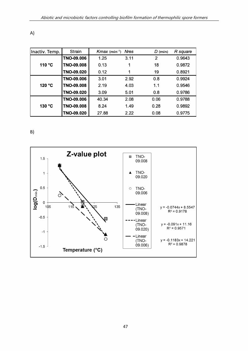
Abiotic and microbiotic factors controlling biofilm formation of thermophilic spore formers
47
A)
B)
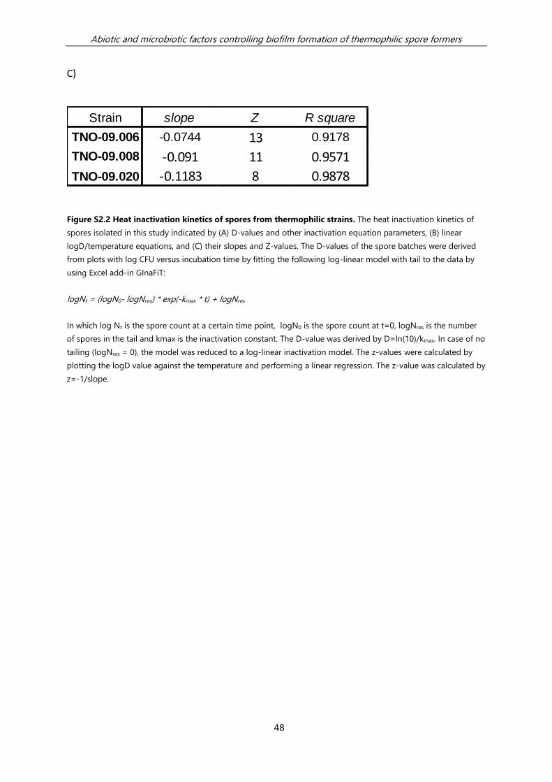
Abiotic and microbiotic factors controlling biofilm formation of thermophilic spore formers
48
C)
Figure S2.2 Heat inactivation kinetics of spores from thermophilic strains. The heat inactivation kinetics of
spores isolated in this study indicated by (A) D-values and other inactivation equation parameters, (B) linear
logD/temperature equations, and (C) their slopes and Z-values. The D-values of the spore batches were derived
from plots with log CFU versus incubation time by fitting the following log-linear model with tail to the data by
using Excel add-in GInaFiT:
logNt = (logN0- logNres) * exp(-kmax * t) + logNres
In which log Nt is the spore count at a certain time point, logN0 is the spore count at t=0, logNres is the number
of spores in the tail and kmax is the inactivation constant. The D-value was derived by D=ln(10)/kmax. In case of no
tailing (logNres = 0), the model was reduced to a log-linear inactivation model. The z-values were calculated by
plotting the logD value against the temperature and performing a linear regression. The z-value was calculated by
z=-1/slope.
Strain slope Z R square
TNO-09.006 -0.0744 13 0.9178
TNO-09.008 -0.091 11 0.9571
TNO-09.020 -0.1183 8 0.9878
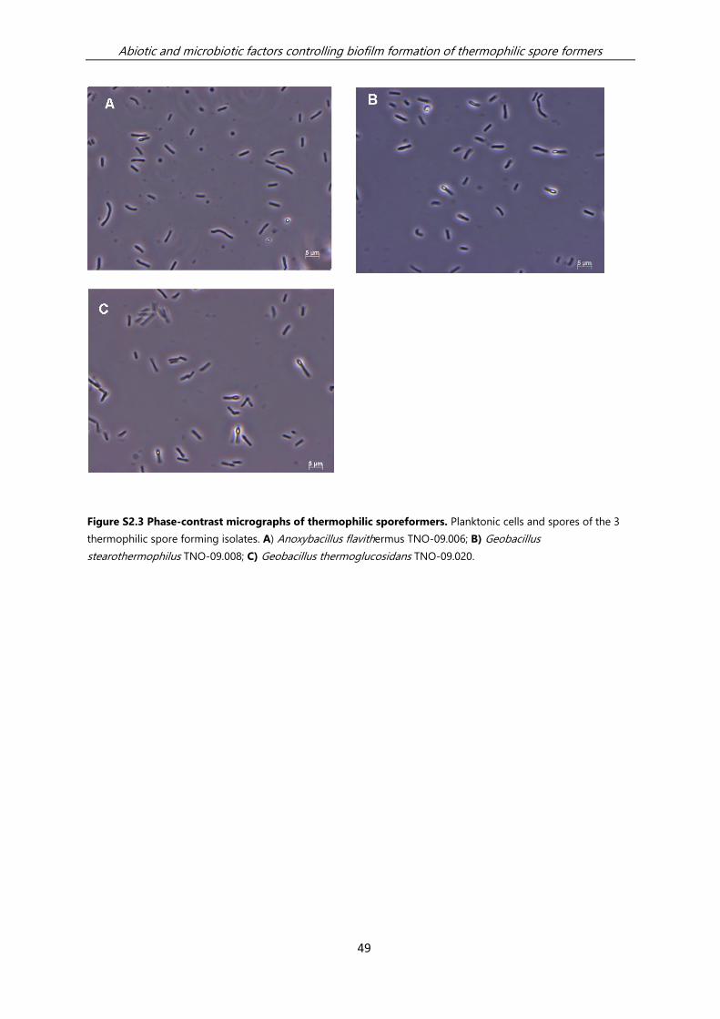
Abiotic and microbiotic factors controlling biofilm formation of thermophilic spore formers
49
Figure S2.3 Phase-contrast micrographs of thermophilic sporeformers. Planktonic cells and spores of the 3
thermophilic spore forming isolates. A) Anoxybacillus flavithermus TNO-09.006; B) Geobacillus
stearothermophilus TNO-09.008; C) Geobacillus thermoglucosidans TNO-09.020.

Abiotic and microbiotic factors controlling biofilm formation of thermophilic spore formers
50
A)
B)
Figure S2.4 Colony forming units of Geobacillus thermoglucosidans TNO-09.020 in the standing steal
biofilm system. Graphical representation of bacterial counts development (CFU/ml) in the static standing steal
biofilm system with 1% Tryptone at 60 °C. The 2 panels represent (A) the planktonic cells from the medium and
(B) biofilm cells from the steel coupon. The total CFUs were determined, vegetative cells and spores (◼), and for
the same samples treated for 30 min at 100 °C, allowing spore counts (). Means and standard deviations from 3
replicate static culture systems are shown.
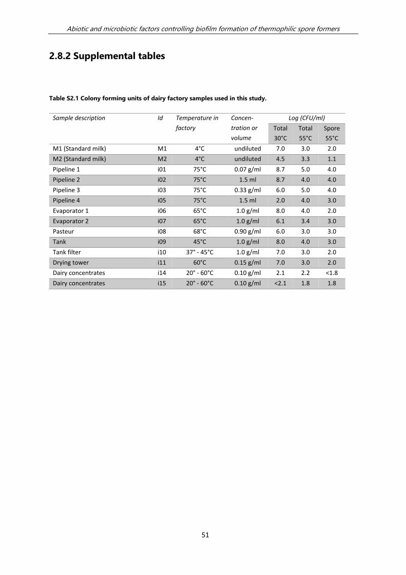
Abiotic and microbiotic factors controlling biofilm formation of thermophilic spore formers
51
2.8.2 Supplemental tables
Table S2.1 Colony forming units of dairy factory samples used in this study.
Sample description Id Temperature in
factory Concen-
tration or
volume
Log (CFU/ml)
Total
30°C Total
55°C Spore
55°C
M1 (Standard milk) M1 4°C undiluted 7.0 3.0 2.0
M2 (Standard milk) M2 4°C undiluted 4.5 3.3 1.1
Pipeline 1 i01 75°C 0.07 g/ml 8.7 5.0 4.0
Pipeline 2 i02 75°C 1.5 ml 8.7 4.0 4.0
Pipeline 3 i03 75°C 0.33 g/ml 6.0 5.0 4.0
Pipeline 4 i05 75°C 1.5 ml 2.0 4.0 3.0
Evaporator 1 i06 65°C 1.0 g/ml 8.0 4.0 2.0
Evaporator 2 i07 65°C 1.0 g/ml 6.1 3.4 3.0
Pasteur i08 68°C 0.90 g/ml 6.0 3.0 3.0
Tank i09 45°C 1.0 g/ml 8.0 4.0 3.0
Tank filter i10 37° - 45°C 1.0 g/ml 7.0 3.0 2.0
Drying tower i11 60°C 0.15 g/ml 7.0 3.0 2.0
Dairy concentrates i14 20° - 60°C 0.10 g/ml 2.1 2.2 <1.8
Dairy concentrates i15 20° - 60°C 0.10 g/ml <2.1 1.8 1.8

Abiotic and microbiotic factors controlling biofilm formation of thermophilic spore formers
52
Table S2.2 Colony forming units of enrichments at 55ºC and 65ºC. Standard milk (M1 and M2) or sterile milk
inoculated with industrial fouling from a whey evaporator (i07) was cultured O/N at 55 or 65°C in plastic culture
wells containing standing steel coupons (static biofilm model). The table shows the CFUs in the different fractions
in the biofilm system: fraction in the medium (2 ml), on the stainless-steel surface, and attached on the plastic
well.
Medium Temperature Fraction Log (CFU) /
Fraction
M2 (standard
milk)
55°C
medium 5.7
steel 5.4
well 5.5
65°C
medium 3.3
steel 1.6
well 2.1
M3 (Standard
milk)
55°C
medium 5.4
steel 5.5
well 5.6
65°C
medium 6.3
steel 4.8
well 5.8
Evaporator (i7)
55°C
medium 7.0
steel 5.5
well 5.4
65°C
medium 5.7
steel 4.2
well 4.7

Chapter 3 Growth of dairy isolates of
Geobacillus thermoglucosidans in skim milk
depends on lactose degradation products
supplied by Anoxybacillus flavithermus as
secondary species
Y. Zhao,
M. Kumar,
M. P. M. Caspers,
M. N. Nierop Groot,
J.M.B.M van der Vossen,
T. Abee
This chapter was published in the Journal of Dairy Science (2018) 101:1013-1019
Growth dependence of dairy isolates in skim milk
54
3.1 Summary
Thermophilic bacilli such as Anoxybacillus and Geobacillus are important
contaminants in dairy powder products. Remarkably, one of the common
contaminants, Geobacillus thermoglucosidans, displayed poor growth in skim milk,
whereas significant growth was observed in the presence of an Anoxybacillus
flavithermus dairy isolate. In the present study, the underlying reason for this growth
dependence of G. thermoglucosidans is investigated. Whole genome sequences of 4
A. flavithermus strains and 4 G. thermoglucosidans strains were acquired with special
attention given to carbohydrate utilization clusters and proteolytic enzymes.
Zooming in on traits relevant for dairy environments, comparative genomic analysis
revealed that all G. thermoglucosidans strains (i) lack genes necessary for lactose
transport and metabolism, (ii) show poor growth in skim milk and (iii) produce white
colonies on X-gal plates indicating the lack of β-galactosidase activity. Tested A.
flavithermus isolates scored positive in these tests, in line with the presence of a
putative lactose utilization gene cluster. All tested isolates from both species showed
proteolytic activity on MPCA plates. Subsequent experiments in liquid skim milk with
added glucose or galactose supported growth of G. thermoglucosidans isolates, in
line with the presence of respective monosaccharide utilization gene clusters in the
genomes. HPLC analysis of A. flavithermus TNO-09.006 culture filtrate indicated that
the previously described growth dependence of G. thermoglucosidans in skim milk, is
based on the supply of glucose and galactose by A. flavithermus TNO-09.006.
Growth dependence of dairy isolates in skim milk
55
3.2 Introduction
Heat resistant spores from thermophilic bacilli form a major concern in dairy powder
processing facilities posing a risk for product contamination. This group of bacteria is
able to grow in sections of skim milk manufacturing plants, such as heat exchangers
and evaporation sections where elevated temperatures, typically between 40 °C to
65 °C, are applied (Seale et al., 2015). Although these bacilli are not pathogenic, their
presence as spores in end products may lead to quality issues of respective
reconstituted products when conditions are favourable for germination and
outgrowth (Setlow and Johnson, 2013; Watterson et al., 2014; Wells-Bennik et al.,
2016). Obviously, such quality issues could also lead to severe economic losses.
Spore-forming bacteria survive through their innate ability to resist adverse
conditions in dairy manufacturing processes including heat, mechanical disruption,
and a wide variety of chemicals (Burgess et al., 2010). Moreover, both spores and
vegetative cells can attach to stainless steel and fouled surfaces in dairy processing
lines. Once attached to the surface, the spores may germinate, grow out, and form
biofilms. Spores from thermophilic bacilli detected in the end products conceivably
originate from biofilms formed in dairy processing lines (Seale et al., 2015).
Thermophilic bacteria generally have a high growth rate with generation times
typically in the range of 15-20 minutes under optimal conditions. Thus, high cell
counts can be reached in a short period of time. Additionally, the resistance of the
endospores to heat and chemicals makes it difficult to fully eliminate the
thermophilic bacteria from dairy processing environments (Burgess et al., 2010;
Wells-Bennik et al., 2016). Understanding of the proliferation and survival of spore
forming bacteria within the dairy processing environments and dairy products is
therefore a prerequisite to develop more effective methods to control and reduce
contamination.
Thermophilic bacteria predominantly isolated from the dairy processing industry are
of the species Anoxybacillus flavithermus and the genus Geobacillus spp., formerly
classified as Bacillus (Burgess et al., 2010; Zhao et al., 2013; Sadiq et al., 2016). A.
flavithermus typically dominates the preheating section of the skim milk powder
process, whereas a mix of A. flavithermus and Geobacillus spp. was found in the
evaporation and drying stages of the process. Geobacillus spp. was predominantly
isolated from fouling sites (Zhao et al., 2013).
Growth dependence of dairy isolates in skim milk
56
To proliferate in skim milk, bacteria must utilize the carbon and nitrogen sources in
skim milk. Raw milk is a nutrient-rich source supporting bacterial growth. It contains
approximately 3.4% protein, 3.7% fat, 4.6% lactose, and 0.7% ash by weight (Jensen,
1995). However, the G. thermoglucosidans strains TNO-09.020 and TNO-09.023,
previously isolated from a fouling site in the dairy-processing pipeline, showed poor
growth on skim milk plates and in liquid skim milk. This growth deficiency, and its
biofilm forming capacity in skim milk, was restored when A. flavithermus, a
proteolytic thermophilic sporefomer from the microbiota of the same dairy-
concentrate processing plant, was added as secondary species (Zhao et al., 2013). To
understand the mechanism of growth dependence of G. thermoglucosidans on the
presence of A. flavithermus as secondary species in skim milk, we performed a
comparative genomic analysis on the isolates of both species. Next to the 5 dairy
isolates (Anoxybacillus flavithermus TNO-09.006, TNO-09.014, TNO-09.016;
Geobacillus thermoglucosidans TNO-09.020, TNO-09.023), 1 A. flavithermus and 2 G.
thermoglucosidans hot spring isolates (Anoxybacillus flavithermus WK1, Geobacillus
thermoglucosidans C56-YS93, Geobacillus thermoglucosidans Y4-1MC1) available in
public genome databases were included in the study (Table 3.2). Particularly, the
presence and absence of genes involved in metabolic pathways including nitrogen
(proteolytic system) and carbohydrate metabolism, required for growth in skim milk,
were taken into consideration. Genes only present in 1 of the 2 species were
enumerated and their annotated function was inspected for possible roles in growth
in skim milk. Moreover, growth experiments in skim milk without and with added
supplements that conceivably could support the outgrowth of G. thermoglucosidans
were conducted. Finally, the cell free culture-filtrate of A. flavithermus was analyzed
to identify compounds produced by this organism, which could support the growth
of G. thermoglucosidans in skim milk.
3.3 Results and discussion
Comparative genomics revealed that genomes of A. flavithermus strains (except for
A. flavithermus WK1 isolated from a hot spring) encompass genes encoding a lactose
ABC transporter cassette and β-galactosidase activity that are involved in the uptake
and the utilization of lactose (De Vos and Vaughan, 1994), whereas these genes are
lacking in the G. thermoglucosidans genomes. Because G. thermoglucosidans lacks
these genes it is conceivably unable to utilize lactose present in skim milk for its

Growth dependence of dairy isolates in skim milk
57
activity and growth. Notably, genome analysis revealed all strains to contain an
extensive repertoire of proteolytic enzymes including genes encoding putative
proteases and peptidases (Y. Zhao, unpublished data). This would suggest that
acquisition of nitrogen sources by G. thermoglucosidans is not a limiting factor for
growth in skim milk, as previously suggested (Zhao et al., 2013). Based on this
genome analysis it was hypothesized that G. thermoglucosidans strains cannot utilize
lactose present in skim milk, and cannot grow readily in skim milk where lactose is
the sole energy source for the growth initiation; while A. flavithermus strains, apart
from the hot spring strain WK1, are able to do so because they are equipped with the
genes involved in the uptake and the utilization of lactose.
We subsequently validated this hypothesis by combining comparative genome
analysis with results obtained from growth experiments on X-gal plates, on skim milk
plates, in liquid skim milk, and on 1% MPCA plates, respectively. All colonies of G.
thermoglucosidans strains on X-gal plates appeared white indicating the lack of β-
galactosidase activity, while the A. flavithermus strains, except WK1, produced blue
colonies indicating hydrolysis of X-gal resulting from β-galactosidase activity (Table
3.1). This phenotypic result matches with the prediction from the comparative
genome analysis. Additionally, we determined the growth capacity of the G.
thermoglucosidans and A. flavithermus strains on skim milk plates and in liquid skim
milk. Results showed that all A. flavithermus strains, except A. flavithermus WK1,
could form visible colonies on skim milk plates, whereas tested G. thermoglucosidans
strains (like A. flavithermus WK1) did not show visible growth on skim milk plates.
Growth assays in liquid milk with the A. flavithermus and G. thermoglucosidans
strains showed the same trend compared to growth on skim milk plates (Table 3.2).
All A. flavithermus strains, except A. flavithermus WK1, were able to grow and
reached cell densities of 7.5 – 7.7 log CFU/mL within 6 hours in skim milk, whereas
the G. thermoglucosidans strains and A. flavithermus WK1 remained around the
inoculation cell density in 6 hours. In conclusion, the presence of genes encoding β-
galactosidase activity and a lactose ABC transporter cassette in these thermophilic
bacilli correlates with their capacity to grow rapidly in skim milk, which points to an
essential and differentiating role of the identified putative lactose utilization cluster
in these strains. Notably, all tested isolates from both species showed proteolytic
activity on MPCA plates (Table 3.1). This indicates that the inability of G.
thermoglucosidans to grow in skim milk is not due to the lack of proteolytic
digestion of milk proteins. Thus, we hypothesize that the inability to digest lactose

Growth dependence of dairy isolates in skim milk
58
and subsequent access to the monosaccharides (glucose and galactose) as the
lactose hydrolysing products, is a limiting factor for rapid growth of G.
thermoglucosidans strains in milk.
Table 3.1 Growth of Anoxybacillus flavithermus and Geobacillus thermoglucosidans strains on skim milk
plates, in liquid skim milk, on X-gal plates and 1% MPCA plates
Strains Growth on skim
milk agar plate1
Initial cell
counts
(log
CFU/mL)
Cell
counts at
6h (log
CFU/mL)
On MPCA
(halo)2
On X-gal
plate3
Anoxybacillus flavithermus
TNO-09.006
+ 3.6 (±0.1) 7.5(±0.4) + Blue
Anoxybacillus flavithermus
TNO-09.014
+ 4.1(±0.4) 7.7(±0.3) + Blue
Anoxybacillus flavithermus
TNO-09.016
+ 3.7(±0.5) 7.7(±0.2) + Blue
Anoxybacillus flavithermus
WK1
- 3.7(±0.2) 4.0(±1.0) + White
Geobacillus thermoglucosidans
TNO-09.020
- 3.8(±0.2) 4.4(±0.5) ++ White
Geobacillus thermoglucosidans
TNO-09.023
- 4.8(±0.5) 5.3(±0.8) ++ White
Geobacillus thermoglucosidans
C56_YS93
- 4.1(±0.2) 2.8(±1.0) + White
Geobacillus thermoglucosidans
Y4_1MC1
- 4.4(±0.5) 5.8(±1.6) ++ White
1 – means no visible colony; + means visible colonies detected on skim milk agar plates;
2 – means no halo; + means weak halo; ++ means large halo around the colony on MPCA plates indicative for
proteolytic activity;
3 blue colony on X-gal plates indicates positive for β-galactosidase activity, white colony indicates negative for β-
galactosidase activity. X-gal stands for 5-bromo-4-chloro-3-indolyl-β-D-galactopyranoside.
To test the hypothesis that indeed only the lactose uptake and hydrolysing capacity
were hampered, we conducted growth assays with G. thermoglucosidans in skim milk
without and with supplementation of glucose or galactose, as well as in an assay with
lactose-free milk, i.e., milk in which the lactose was hydrolysed by lactase resulting in
the availability of glucose and galactose (Figure 3.1). G. thermoglucosidans exhibited
limited growth in skim milk, having approximately 0.5 and 0.8 log unit increase in

Growth dependence of dairy isolates in skim milk
59
viable counts in six and 24 hours, respectively (Figure 3.1A). The presence of glucose,
galactose and hydrolysed lactose (glucose and galactose) enhanced growth of G.
thermoglucosidans reaching approximately 2 log units increase within 6 hours
(Figure 3.1B-D). The fact that glucose and galactose supported growth in skim milk of
G. thermoglucosidans suggests that in a co-culture, hydrolysis of lactose by A.
flavithermus may supply glucose, galactose or both to G. thermoglucosidans,
supporting its growth in skim milk. We therefore analysed the sugars and organic
acids present in cell free culture-filtrates of A. flavithermus TNO-09.006 and G.
thermoglucosidans TNO-09.020 grown in skim milk by using HPLC, and this
confirmed that lactose is degraded by A. flavithermus and that residual levels of
glucose and galactose were present. Additionally, β-galactosidase activity was
detected in the cell free culture-filtrate harvested after 24h growth of A. flavithermus
in skim milk at 55° C (Y. Zhao, unpublished data). Subsequent studies in skim milk
with added A. flavithermus TNO-09.006 culture filtrates, indeed showed stimulation
of growth of G. thermoglucosidans TNO-09.020 (Figure 3.2). Above mentioned
experiments support that the observed growth stimulation of G. thermoglucosidans
can be attributed to supply of lactose degradation products via β-galactosidase
activity of A. flavithermus TNO-09.006.

Growth dependence of dairy isolates in skim milk
60
5
5.5
6
6.5
7
0
2
4
6
8
10
0 6 14 24
pH
log
cfu
/ml
Time (h)
A
5
5.5
6
6.5
7
0
2
4
6
8
10
0 6 14 24
pH
log
cfu
/ml
Time (h)
B
5
5.5
6
6.5
7
0
2
4
6
8
10
0 6 14 24
pH
log
cfu
/ml
Time (h)
C
5
5.5
6
6.5
7
0
2
4
6
8
10
0 6 14 24
pH
log
cfu
/ml
Time (h)
D

Growth dependence of dairy isolates in skim milk
61
Figure 3.1 Growth of Geobacillus thermoglucosidans TNO-09.020 (A-D)
and Anoxybacillus flavithermus TNO-09.006 (E-H) in skim milk without and with supplements, and in
lactose-free milk. Growth in skim milk (A, E), skim milk + 0.25% glucose (B, F), skim milk + 0.25% galactose (C,
G), and lactose-free (lactase treated) 2.5% glucose and 2.5% galactose containing milk (D, H). Total counts (log
CFU/mL) are shown in bar graphs and pH measurements in filled squares. Error bars represent standard
deviations of biological duplicates.
5
5.5
6
6.5
7
0
2
4
6
8
10
0 6 14 24
pH
log
cfu
/ml
Time (h)
E
5
5.5
6
6.5
7
0
2
4
6
8
10
0 6 14 24
pH
log
cfu
/ml
Time (h)
F
5
5.5
6
6.5
7
0
2
4
6
8
10
0 6 14 24
pH
log
cfu
/ml
Time (h)
G
5
5.5
6
6.5
7
0
2
4
6
8
10
0 6 14 24
pH
log
cfu
/ml
Time (h)
H

Growth dependence of dairy isolates in skim milk
62
Figure 3.2 Effect of Anoxybacillus flavithermus TNO-09.006 cell free culture-filtrate (filtrate) on
the growth of Geobacillus thermoglucosidans TNO-09.020 in milk. A, skim milk; B, skim milk with added
filtrate; and C, filtrate only. Graph represents values of CFU measurements at the indicated sample times. Error
bars represent standard deviations of biological duplicates.
Notably, when G. thermoglucosidans was grown in skim milk with addition of
glucose, the viable counts after first growing, subsequently declined by almost 3 log
units after 24h (Figure 3.1B). A significant drop of the pH value to pH 5.4 in this G.
thermoglucosidans culture was also observed (Figure 3.1B). This acidification would
explain the decrease in viability of the G. thermoglucosidans cells, and previous
studies have reported the limited capacity of thermophilic bacilli to cope with low pH
conditions where relatively high minimum pH values (around pH 6) for growth were
documented (Tola and Ramaswamy, 2014). Notably, G. thermoglucosidans also
showed efficient growth in the lactose-free (glucose and galactose containing) milk,
likewise followed by a decline in cell numbers in 24h correlating with a significant
decline in pH (Figure 3.1D). On the contrary, in skim milk with supplementation of
galactose, the growth of G. thermoglucosidans was comparable to glucose added
and lactose-free milk but there is no significant drop of cell density observed at 24h.
In line with that, the drop in pH was also less significant (Figure 3.1C). Except for
growth in skim milk (Figure 3.1E), similar growth and acidification behaviour was
observed for A. flavithermus (Figure 3.1F-H).
Besides monitoring colony-forming unit (CFU) and pH change during growth, spore
counts at different time points were also monitored. Notably, we could not visually
0
2
4
6
8
10
0 24 48
log
cfu
/ml
Time (h)
A
0
2
4
6
8
10
0 24 48lo
g cf
u/m
l
Time (h)
B
0
2
4
6
8
10
0 24 48
log
cfu
/ml
Time (h)
C

Growth dependence of dairy isolates in skim milk
63
detect spores using qualitative microscopy in samples from the G.
thermoglucosidans and A. flavithermus cultures in skim milk, and in skim milk
supplemented with glucose and lactose-free milk (hydrolysed lactose). Nevertheless,
we could observe spores of G. thermoglucosidans and A. flavithermus after the
growth in the galactose-supplemented skim milk (final pH 6). The above observation
combined with the extensive drop in pH of the growth medium observed only in
skim milk supplemented with glucose or in glucose containing lactose-free skim milk,
suggests that rapid acidification of the growth medium provides inhibitory
conditions for sporulation of G. thermoglucosidans and A. flavithermus. The negative
influence of low pH conditions on sporulation has also been described by Yazdany
and Lashkari (1975). In their study, G. stearothermophilus ATCC 7953 and G.
stearothermophilus NCIB 8919 produced few spores when cultured in a medium with
final pH around 5.5, whereas when grown in a medium adjusted to pH 7.7 to 8.7,
sporulation of the G. stearothermophilus occurred (Yazdany and Lashkari, 1975). It is
conceivable that glucose metabolism-induced acidification leads to a reduction in
the total CFU counts, hence preventing the formation of spores. Factors that
contribute to the relatively high minimum pH for growth of a range of thermophilic
spore formers, and loss of sporulation capacity at weak acidic conditions, remain to
be elucidated.
In conclusion, this study indicated that the previously described growth stimulation
of G. thermoglucosidans during co-culture in skim milk with A. flavithermus TNO-
09.006 (Zhao et al., 2013) is not based on proteolytic activity of the latter, but rather
is due to the supply of glucose and galactose following lactose degradation by β-
galactosidase of A. flavithermus TNO-09.006. In addition, it is observed that glucose
metabolism–induced acidification in skim milk led to a reduction in total count in the
late growth stage of A. flavithermus and G. thermoglucosidans and, in turn,
hampered sporulation. These observations shed new insights into the interre-
lationship within microbiological communities of dairy sporeformers and could have
implications for industrial hygiene operations. Future studies will be directed at
studying whether these observations in A. flavithermus and G. thermoglucosidans
hold true for other known dairy or nondairy thermophilic spore-forming species such
as G. stearothermophilus. The latter suggestion is based on a recent paper by
Burgess et al. (2017) that provided novel insights in diversity of G.
stearothermophilus strains and presented genotypic and phenotypic evidences for
lactose utilization capacity in dairy strains of this species. Greater understanding of

Growth dependence of dairy isolates in skim milk
64
those thermophilic sporeformers is needed to develop better control measures in
milk processing environments.
3.4 Experimental procedures
The genomes of the 8 thermophilic bacilli of the species G. thermoglucosidans and A.
flavithermus (Table 3.2) have been compared to each other and to the reference non-
thermophilic strain Bacillus subtilis 168. For the newly sequenced genomes from this
study (Zhao et al., 2012; Caspers et al., 2013; Caspers et al., 2016), after assembly of
sequence reads into contigs, RAST (Aziz et al., 2008) was used to predict ORFs and to
do automatic annotation. Selected genes and protein sequences were manually
curated using BLASTP and InterPro. Orthologous groups (OGs; i.e. gene families) in
the 8 genomes were determined using OrthoMCL (Enright et al., 2002). This program
uses all-against-all protein BLAST where it groups proteins with more homology
within the species than homology with proteins outside the species. In this way,
orthologs (genes in different species that evolved from a common ancestral gene by
speciation) are separated from paralogs (genes related by duplication within a
genome). When an OG contained more than 1 gene per strain, i.e. highly similar
genes, this OG was manually split into separate OGs containing only 1 gene per
strain. An exception was made for transposons and other mobile elements.
Table 3.2 Presence/absence of genes for lactose metabolism in selected Anoxybacillus flavithermus and
Geobacillus thermoglucosidans strains
Strains NCBI Accession
number and
Reference
Origin
lactose ABC
transporter,
permease
protein
Genes1
lactose ABC
transporter,
lactose-binding
protein
Beta-
galactosi
dase (EC
3.2.1.23)
Anoxybacillus
flavithermus
TNO-09.006
AMCM00000000
Standard milk used for
dairy processing plant,
Netherlands
1 1 1
Anoxybacillus
flavithermus
TNO-09.014
LUFB00000000
Standard milk used for
dairy processing plant,
Netherlands
1 1 1
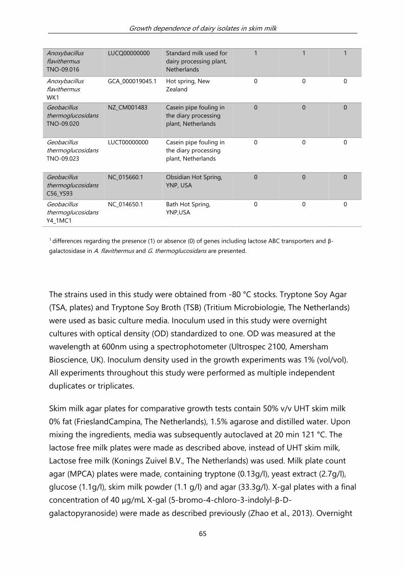
Growth dependence of dairy isolates in skim milk
65
Anoxybacillus
flavithermus
TNO-09.016
LUCQ00000000
Standard milk used for
dairy processing plant,
Netherlands
1 1 1
Anoxybacillus
flavithermus
WK1
GCA_000019045.1
Hot spring, New
Zealand
0 0 0
Geobacillus
thermoglucosidans
TNO-09.020
NZ_CM001483
Casein pipe fouling in
the diary processing
plant, Netherlands
0 0 0
Geobacillus
thermoglucosidans
TNO-09.023
LUCT00000000
Casein pipe fouling in
the diary processing
plant, Netherlands
0 0 0
Geobacillus
thermoglucosidans
C56_YS93
NC_015660.1
Obsidian Hot Spring,
YNP, USA
0 0 0
Geobacillus
thermoglucosidans
Y4_1MC1
NC_014650.1
Bath Hot Spring,
YNP,USA
0 0 0
1 differences regarding the presence (1) or absence (0) of genes including lactose ABC transporters and β-
galactosidase in A. flavithermus and G. thermoglucosidans are presented.
The strains used in this study were obtained from -80 °C stocks. Tryptone Soy Agar
(TSA, plates) and Tryptone Soy Broth (TSB) (Tritium Microbiologie, The Netherlands)
were used as basic culture media. Inoculum used in this study were overnight
cultures with optical density (OD) standardized to one. OD was measured at the
wavelength at 600nm using a spectrophotometer (Ultrospec 2100, Amersham
Bioscience, UK). Inoculum density used in the growth experiments was 1% (vol/vol).
All experiments throughout this study were performed as multiple independent
duplicates or triplicates.
Skim milk agar plates for comparative growth tests contain 50% v/v UHT skim milk
0% fat (FrieslandCampina, The Netherlands), 1.5% agarose and distilled water. Upon
mixing the ingredients, media was subsequently autoclaved at 20 min 121 °C. The
lactose free milk plates were made as described above, instead of UHT skim milk,
Lactose free milk (Konings Zuivel B.V., The Netherlands) was used. Milk plate count
agar (MPCA) plates were made, containing tryptone (0.13g/l), yeast extract (2.7g/l),
glucose (1.1g/l), skim milk powder (1.1 g/l) and agar (33.3g/l). X-gal plates with a final
concentration of 40 μg/mL X-gal (5-bromo-4-chloro-3-indolyl-β-D-
galactopyranoside) were made as described previously (Zhao et al., 2013). Overnight

Growth dependence of dairy isolates in skim milk
66
cultures (OD = 1) of the 8 selected strains were streaked separately to acquire single
colonies on the prepared plates; subsequently, the plates were incubated overnight
at 55°C for further observation. Growth experiments in liquid skim milk without and
with added supplements were performed using the following procedures. D-glucose
(Tritium Microbiologie, The Netherlands) or D-galactose (Tritium Microbiologie, The
Netherlands) were added to freshly obtained UHT skim milk in concentration of
0.25g/L for the growth assays; and Lactose free milk with concentrations of 2.5g/L
glucose and galactose was directly used for this experiment. These milk media were
subsequently inoculated with A. flavithermus or G. thermoglucosidans and
incubated for maximal 24h at 65°C, 100 rpm and selected time samples were
subsequently diluted and spread plated on TSA plates for enumeration. The pH of
the cultures was also measured using a pH meter (Mettler Toledo, Switzerland).
Finally, to study the effect of cell free culture supernatants of A. flavithermus on
growth of G. thermoglucosidans in skim milk, cell free culture supernatants of 24h
grown cultures of A. flavithermus in skim milk medium were prepared. The pH of the
cultures (pH 5.9) was set to pH 4.7 using 4N HCl to precipitate milk caseins and
subsequently filtered using a 0.45µm filter (Millipore, Merck, USA). The filtrate was
centrifuged (Allegra bench top centrifuge, Beckman Counter, USA) at 4000 g for 10
min. The clear supernatant was collected, and the pH of the supernatant was
readjusted to the initial pH 5.9 using 4N NaOH and stored at -20 °C for later use. The
filtrates of the 24h A. flavithermus cultures in skim milk were added to the skim milk
with the ratio 2:1 to assess the effect on growth of G. thermoglucosidans. These
filtrates were also subjected to high-performance liquid chromatography (HPLC)
assays in order to quantify sugars and organic acids present in cell free culture-
filtrates of A. flavithermus TNO-09.006. HPLC was performed using an Aminex HPX-
87H column (Biorad, USA) mounted in a Waters Alliance e2695 HPLC-apparatus. The
column was eluted with 0.6 mL/min 5 mM H2SO4 at 60°C. Peaks were detected by a
refractive-index detector and a dual-wavelength absorbance detector at 210nm and
270nm. Raw data were processed into Peak Areas and concentrations were
calculated using Waters Empower™ 2 software (Waters®, USA).

Growth dependence of dairy isolates in skim milk
67
3.5 Acknowledgments
We wish to thank Lars Wilms for assisting in conducting filtrate experiments, Karin
Overkamp for assisting HPLC experiments, and Roland Siezen for bio-IT support. The
project is funded by TI Food and Nutrition, a public-private partnership on
precompetitive research in food and nutrition. The public partners are responsible for
the study design, data collection and analysis, decision to publish, and preparation of
the manuscript. The private partners have contributed to the project through regular
discussion.
3.6 References
Aziz, R. K., D. Bartels, A. A. Best, M. DeJongh, T. Disz, R. A. Edwards, K. Formsma, S. Gerdes, E. M. Glass,
M. Kubal, F. Meyer, G. J. Olsen, R. Olson, A. L. Osterman, R. A. Overbeek, L. K. McNeil, D.
Paarmann, T. Paczian, B. Parrello, G. D. Pusch, C. Reich, R. Stevens, O. Vassieva, V. Vonstein, A.
Wilke and O. Zagnitko. 2008. The RAST server: Rapid annotations using subsystems technology.
BMC Genomics. 9:75.
Burgess, S. A., D. Lindsweay and S. H. Flint. 2010. Thermophilic bacilli and their importance in dairy
processing. International Journal of Food Microbiology. 144:215-225.
Burgess, S. A., S. H. Flint, D. Lindsay, M. P. Cox and P. J. Biggs. 2017. Insights into the Geobacillus
stearothermophilus species based on phylogenomic principles. BMC Microbiology. 17:140
Caspers, M. P. M., J. Boekhorst, T. Abee, R. J. Siezen and R. Kort. 2013. Complete genome sequence of
Anoxybacillus flavithermus TNO-09.006, a thermophilic sporeformer associated with a dairy-
processing environment. Genome Announcements. 1:e0010-13.
Caspers, M. P. M., J. Boekhorst, A. de Jong, R. Kort, M. N. Nierop Groot and T. Abee. 2016. Draft
genome sequences of four thermophilic spore formers isolated from a dairy-processing
environment. Genome Announcements. 4:e00757-16.
De Vos, W. M. and E. E. Vaughan. 1994. Genetics of lactose utilization in lactic acid bacteria. FEMS
Microbiology Reviews. 15:217-237.
Enright, A. J., S. Van Dongen and C. A. Ouzounis. 2002. An efficient algorithm for large-scale detection
of protein families. Nucleic Acids Research. 30:1575-1584.
Jensen, R. G. 1995. Handbook of Milk Composition. Page 3 in Introduction. Academic Press, London,
UK.

Growth dependence of dairy isolates in skim milk
68
Sadiq, F. A., Y. Li, T. Liu, S. Flint, G. Zhang, L. Yuan, Z. Pei and G. He. 2016. The heat resistance and
spoilage potential of aerobic mesophilic and thermophilic spore forming bacteria isolated from
Chinese milk powders. International Journal of Food Microbiology. 238:193-201.
Seale, B., S. Burgess, S. Flint, J. Brooks, P. Bremer and S. Parkar. 2015. Thermophilic spore-forming
bacilli in the dairy industry. Pages 112-137 in Biofilms in the Dairy Industry. John Wiley & Sons,
Ltd, Chichester, UK.
Setlow, P. and E. A. Johnson. 2013. Spores and their significance. Pages 45-79 in Food Microbiology.
Spores and their significance. ASM Press, Washington, DC.
Tola, Y. B. and H. S. Ramaswamy. 2014. Combined effects of high pressure, moderate heat and pH on
the inactivation kinetics of Bacillus licheniformis spores in carrot juice. Food Research
International. 62:50-58.
Watterson, M. J., D. J. Kent, K. J. Boor, M. Wiedmann and N. H. Martin. 2014. Evaluation of dairy
powder products implicates thermophilic sporeformers as the primary organisms of interest.
Journal of Dairy Science. 97:2487-2497.
Wells-Bennik, M. H. J., R. T. Eijlander, H. M. W. den Besten, E. M. Berendsen, A. K. Warda, A. O.
Krawczyk, M. N. Nierop Groot, Y. Xiao, M. H. Zwietering, O. P. Kuipers and T. Abee. 2016.
Bacterial spores in food: Survival, emergence, and outgrowth. Annual Review of Food Science and
Technology. 7:457-482.
Yazdany, S. and K. B. Lashkari. 1975. Effect of pH on sporulation of Bacillus stearothermophilus.
Applied Microbiology. 30:1-3.
Zhao, Y., M. P. M. Caspers, T. Abee, R. J. Siezen and R. Kort. 2012. Complete genome sequence of
Geobacillus thermoglucosidans TNO-09.020, a thermophilic sporeformer associated with a dairy-
processing environment. Journal of Bacteriology. 194:4118.
Zhao, Y., M. P. M. Caspers, K. I. Metselaar, P. de Boer, G. Roeselers, R. Moezelaar, M. N. Nierop Groot,
R. C. Montijn, T. Abee and R. Kort. 2013. Abiotic and microbiotic factors controlling biofilm
formation by thermophilic sporeformers. Applied and Environmental Microbiology. 79:5652-
5660.

Chapter 4 Biofilm dynamics of Geobacillus
thermoglucosidans, a dairy processing isolate
Y. Zhao

Biofilm dynamics of Geobacillus thermoglucosidans, a dairy processing isolate
70
4.1 Summary
Contamination by thermophilic sporeformers in end-products is a primary concern
for dairy concentrate-processing plants. Biofilms are known to be one of the major
sources of persistent contaminations in these plants, but little is known about the
biofilm-forming mechanism of thermophilic sporeformers in such environments. The
contaminant Geobacillus thermoglucosidans has been isolated at different locations
inside these plants. Compared to other common thermophilic contaminants – such
as Geobacillus stearothermophilus and Anoxybacillus flavithermus, G.
thermoglucosidans shows the best biofilm-forming capacity with high biofilm mass
produced in a reproducible manner under lab conditions. In this study, G.
thermoglucosidans was selected as a model organism to investigate biofilm-
formation mechanisms.
In the present study, a comparison was made of the gene expression profiles of G.
thermoglucosidans TNO-09.020 in the planktonic phase and the biofilm phase
during biofilm development. It was found that cells in these two phases showed
discernible behaviour in their decline stage and differences in expression profiles
over the course of biofilm development. The gene categories, with functions such as
methionine biosynthesis and phenylalanine degradation, were significantly
upregulated in biofilm-phase cells compared to planktonic-phase cells. The potential
roles of these newly identified pathways in biofilm formation were also investigated
and provided insight in the biofilm development of this organism.
This study described the expression profile of G. thermoglucosidans TNO-09.020
during biofilm formation and discovered some novel factors which might influence
biofilm development of this strain.

Biofilm dynamics of Geobacillus thermoglucosidans, a dairy processing isolate
71
4.2 Introduction
Besides mesophilic spore-formers, thermophilic sporeformers are especially
problematic in food-producing industrial facilities operating processes with
temperatures in the range of 40 to 65 °C, since these temperatures support the
growth and biofilm formation of these organisms (Burgess et al., 2010). The growth
of these thermophiles in biofilms can result in bacteria and spores being released
into the end-products, including whey and milk concentrates in numbers of up to 106
CFU/g (Scott et al., 2007; Watterson et al., 2014). These spores could germinate under
favourable conditions, ultimately resulting in high numbers of bacteria and off-
flavour in end-products (Scheldeman et al., 2005; Scott et al., 2007). In order to
prevent the presence and outgrowth of the accumulated spores, costly precautionary
measures are taken in dairy concentrate-processing environments, such as frequent
cleaning, short production runs, and intensive microbial product testing.
Most of the thermophilic sporeformers that have been identified to-date in dairy
concentrate-processing lines and products belong to the genera Bacillus,
Geobacillus, and Anoxybacillus (Flint et al., 1997; Scott et al., 2007; Yuan et al., 2012).
Geobacillus spp. and A. flavithermus are the most frequently reported species in
thermophilic dairy biofilms (Burgess et al., 2010). The presence of spores of these
thermophilic bacilli in the end-products most likely results from the detachment of
spores from biofilms from stainless steel surfaces in dairy concentrate-processing
plants (Scott et al., 2007). Moreover, it is also reported that biofilms render bacteria
tolerance to antimicrobial factors, making those cells residing in biofilm even more
difficult to be eradicated (Sadiq et al., 2017). This is why special interest is focused on
understanding the development of biofilm by thermophilic bacilli.
To begin with, three isolates from a dairy concentrate-processing plant were selected
according to their ability to grow at high temperatures and their biofilm-forming
capacity, namely: Anoxybacillus flavithermus TNO-09.006, Geobacillus
stearothermophilus TNO-09.008, and Geobacillus thermoglucosidans TNO-09.020. Of
these three model strains, G. thermoglucosidans TNO-09.020 (or GT20), which was
isolated in a bio-fouling sample from a dairy caseinate concentrate-processing plant,
showed the strongest and most robust biofilm-forming capacity at high
temperatures (60°C-70°C) in TSB (tryptone soy broth) under lab conditions (Zhao et
Biofilm dynamics of Geobacillus thermoglucosidans, a dairy processing isolate
72
al., 2013). GT20 was therefore chosen in the current study aiming to understand its
biofilm-forming mechanism. To this end, DNA microarray technology was applied to
identify global gene expression profiles in biofilm-phase cells during their biofilm
development and compare them to planktonic-phase cells in a static biofilm system.
Our objective was to use the gene expression profiles to discover unique patterns of
gene expression associated with biofilm development for thermophilic sporeformers;
these organisms are rarely studied but are highly relevant to plants with high-
temperature operating processes.
4.3 Results
4.3.1 Growth of G. thermoglucosidans TNO-09.020 in a static biofilm
system
The biofilm development cycle of GT20 was monitored by counting both biofilm-
phase and planktonic-phase cells by Colony Forming Units (CFU) (Figure 4.1) in a
static biofilm system over a time span of 30h. The total viable cell counts reached 7.2
log CFU/cm2 in the biofilm phase and 7.0 log CFU/ml in the planktonic phase both in
about 10h. Subsequently, a stationary phase, with a constant total viable cell count,
was observed for the planktonic-phase cells, while a significant drop in the total
viable cell count was observed for the biofilm-phase cells. At 8h and 10h, spore
counts remained similar to 4h, and from 16h onward the number reached a final
count around 5 log CFU/ml, both in the biofilm and the planktonic phase. At time
points 24h and 30h, total cell counts equalled spore counts in the biofilm phase. Two
spore-selection treatments were used in this study (80°C 10 min for all spores, 100°C
30 min for heat-resistant spores). At 30h, the results of these two treatments did
match, and before 30h, not all spores were very heat resistant, especially in the
planktonic phase. Under the microscope, a monolayer of biofilm cells was observed
from 8h onwards, and spores were observed in both the biofilm and the planktonic
phase from 16h onwards (data not shown). Taken together, based on the information
described above from CFU monitoring as well as microscopic observation, cells from

Biofilm dynamics of Geobacillus thermoglucosidans, a dairy processing isolate
73
the biofilm and the planktonic phase have discernible development characteristics,
especially in the late development phase.
Figure 4.1 Cell counts of G. thermoglucosidans TNO-09.020 in 1.05% tryptone medium at 65 °C, in both
the planktonic phase (A) and the biofilm phase (B). Each point with error bar represents the mean and
standard deviation from three independent trials. Triangles = total cells, Squares = spores, Circles = heat-resistant
spores. Biofilm phase cells (surface-attached cells) from each sample were dissolved in 6ml tryptone medium,
0
1
2
3
4
5
6
7
8
9
0 5 10 15 20 25 30 35
log
CFU
/ml
Time (hours)
0
1
2
3
4
5
6
7
8
9
0 5 10 15 20 25 30 35
log
CFU
/ml
Time (hours)
A
B

Biofilm dynamics of Geobacillus thermoglucosidans, a dairy processing isolate
74
later the cell number per ml (CFU/ml) was enumerated
2. Any point below the detection limit (dotted line)
indicates that no viable colonies were detected on TSA plate.
4.3.2 Evaluation of early stage differential expression in G.
thermoglucosidans TNO-09.020 (GT20)
In order to examine the mechanisms associated with biofilm formation during overall
biofilm development, a whole genome transcriptomic study was conducted. First of
all, to study the genes responsible for biofilm formation, without having to deal with
noise from genes involved in sporulation, the differentially expressed genes in the
early biofilm-development phase, namely at the 8h time point, were investigated. A
list of gene clusters was selected in which the majority of gene candidates were over-
expressed in the biofilm phase compared to planktonic phase (Table 4.1). The
comparison of biofilm-phase and planktonic-phase cells showed that upregulation in
biofilm-phase cells was mainly observed in categories such as methionine
biosynthesis, fatty acid utilization, and peptide utilization. Downregulation in biofilm-
phase cells was scattered in some functional groups in which at most five identified
gene members in GT20 were present (data not shown). The highest downregulation
percentage is in the category biosynthesis of glycogen, in which 4 out of 5 members
of the functional group were more than two times downregulated. Some of these
changes were initiated early and maintained throughout the process; others were
restricted to the earliest stages of the biofilm formation.
2 Biofilm of 3.92 cm 2 (1.4 * 1.4 cm2 *2 sides) was dissolved in 6 ml medium and then CFU/ml was determined. The conversion
factor between CFU/ml and CFU/cm2 is 1.5.

Biofilm dynamics of Geobacillus thermoglucosidans, a dairy processing isolate
75
Table 4.1 Categories (Subtiwiki gene function categories and regulon categories which contain more than
5 genes) of genes in which more than 50% of the genes are uniquely upregulated at 8h in biofilm phase.
The first column (genes/category) shows the total numbers in the specific category in the genome; the second
column (upregulated genes) shows the number of genes that are more than doubly upregulated in the biofilm
phase in comparison to the planktonic phase in the specific category; the third column (upregulated genes%)
presents the percentages of upregulated genes in the gene category. No downregulated gene is presented in the
table because all downregulated gene categories have no more than 5 genes.
Subtiwiki Categories Number of
Genes
Number of
Upregulated Genes
Upregulated Genes (%)
Biosynthesis/ acquisition of
methionine/ S-
adenosylmethionine
19 14 74
Utilization of glutamine/
glutamate
9 6 67
Utilization of peptides 16 9 56
Utilization of fatty acids 16 9 56
FadR regulon 14 9 64
SdpR regulon 17 12 71
TnrA regulon 19 12 63
In the functional category biosynthesis/acquisition of methionine/S-
adenosylmethionine (http://subtiwiki.uni-goettingen.de/wiki/), 74% of the genes are
more than doubly upregulated in biofilm-phase cells compared to planktonic-phase
cells at 8h (Table 4.1). Therefore, it can be hypothesized that the amino acids
produced by the methionine synthesis pathway – namely, methionine, aspartate, and
homocysteine – are needed for biofilm mass formation. If the hypothesis is true, then
increased availability of, for example, methionine inside the cells should lead to more
biofilm formation. This was tested by addition of the amino acids to the medium.
Effect of methionine is presented in Figure 4.2 (results for aspartate and
homocysteine not shown). In these tests, the addition of these amino acids in
tryptone medium, showed a significant influence (P<0.05) on biofilm formation at 48
hour when compared to tryptone medium without the addition: total cell count 48h
(P=0.03) (Figure 4.2C); spore count 48h (P=0.005)(Figure 4.2D); this was monitored by
the standard plate count method. This observation suggests that the increased
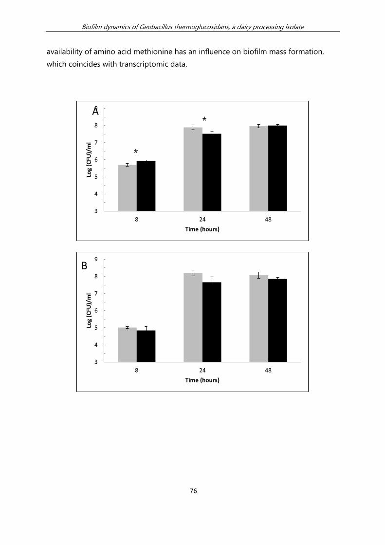
Biofilm dynamics of Geobacillus thermoglucosidans, a dairy processing isolate
76
availability of amino acid methionine has an influence on biofilm mass formation,
which coincides with transcriptomic data.
3
4
5
6
7
8
9
8 24 48
Log
(CFU
)/m
l
Time (hours)
3
4
5
6
7
8
9
8 24 48
Log
(CFU
)/m
l
Time (hours)
B
A
*
*

Biofilm dynamics of Geobacillus thermoglucosidans, a dairy processing isolate
77
Figure 4.2 Effect of methionine on the growth of G. thermoglucosidans TNO-09.020 in the planktonic
phase and the biofilm phase at 65°C. Data points represent CFU/ml in tryptone medium (grey) or in tryptone
medium with addition of 200µM methionine (black). Each bar with error bar represents the mean and standard
deviation, from three independent trials. An asterisk (*) indicates that there is a statistical difference between the
two samples verified by t-test (P<0.05).
A. Total cell counts in planktonic phase.
B. Spore counts in planktonic phase
C. Total cell counts in biofilm phase.
D. Spore counts in biofilm phase.
3
4
5
6
7
8
9
8 24 48
Log
(CFU
)/m
l
Time (hours)
C
3
4
5
6
7
8
9
8 24 48
Log
(CFU
)/m
l
Time (hours)
D
*
*

Biofilm dynamics of Geobacillus thermoglucosidans, a dairy processing isolate
78
Further, it is hypothesed that methionine biosynthesis accelerated the activities of the
LuxS catalysed side pathway, which plays a role in signalling biofilm cell attachment
by GT20 via AI-2 (AI-2 is a quorum sensing molecule). To test this hypothesis, the
presence of AI-2 in G. thermoglucosidans culture was first checked through an AI-2
assay. If AI-2 is present in the culture medium, the V. harveyi reporter strain BB170
will generate luminescence which can be captured and measured by a
spectrophotometer. This study observed that the luminescent signals generated from
the AI-2 reporter strain in cell-free extracts from GT20 were in proximity range of the
negative control, and even lower (Figure 4.3). Those signals indicated no (or low)
secretion of AI-2 by GT20; therefore, it is unlikely that methionine biosynthesis
influenced the biofilm formation via AI-2 generated from the LuxS catalysed side
pathway. The reasons behind over-expression of methionine biosynthesis-related
genes in biofilm-phase cells at 8h is not linked to AI-2 production, but it is linked to
biofilm mass formation.
Figure 4.3 AI-2 assay response of G. thermoglucosidans TNO-09.020 cell-free culture fluids. The responses
of V. harveyi reporter strains BB170 (sensor 12, sensor 21) to signalling substances present in cell-free culture
fluids of G. thermoglucosidans at 8h, 10h, and 16h culture in tryptone medium and sterile tryptone medium
(negative control), V. harveyi BB120 (positive control) are shown. An asterisk (*) indicates that there is a statistical
difference between the two samples verified by t-test (P<0.05).
0
5000
10000
15000
20000
25000
30000
35000
8h 10h 16h negativecontrol
positivecontrol
bio
lum
inac
en
ce
*

Biofilm dynamics of Geobacillus thermoglucosidans, a dairy processing isolate
79
4.3.3 Clustering of expression profiles of biofilm-phase and
planktonic-phase cells
Using the Neighbourhood Co-Regularized Spectral Clustering Algorithm, the
expression profiles of GT20 cells of all samples taken from both the planktonic and
the biofilm phase at time points 4h, 8h, 10h, 16h, 24h, and 30h were found to be
clustered into four distinguishable groups (Figure 4.4). These groups were: group 1
(8h and 10h samples), group 2 (16h planktonic-phase samples), group 3 (4h, 24h, 30h
planktonic-phase samples), and group 4 (16h, 24h, 30h biofilm-phase samples).
Biofilm-phase samples at 4h were not processed in the transcriptomic study due to
insufficient cell numbers. The clustering result of the samples shows early (8h and
10h) and late (16h, 24h, and 30h) biofilm development have discernible expression
profiles. Moreover, the separation of samples in group 3 and group 4 suggested that
there were important genes or gene clusters that differentiate planktonic cells and
biofilm cells of GT20.

Biofilm dynamics of Geobacillus thermoglucosidans, a dairy processing isolate
80
Figure 4.4 Consensus spectral clustering of expression profiles of biofilm- and planktonic-phase cells.
Samples are symmetrically ordered along both the x- and the y-axis, according to the co-occurrence matrix (i.e.,
the more similar the gene expression profiles of any two samples, the higher their tendency to cluster together).
Co-occurrence values range from 0.0 illustrated with colour blue (for samples that never cluster together) to 1.0
illustrated with red (for samples that always cluster together).
To investigate which dominant genes contributed to this clustering, Multi-View
Unsupervised Feature Selection (MVUFS) was used. Among the 30 selected genes
which contributed to the clustering, 8 genes with phenylalanine degradation function
stood out. More than double upregulation of these 8 genes in biofilm-phase cells in
comparison with planktonic-phase cells was observed (Table 4.2). It is hypothesized
that the late phase of biofilm development of GT20 is influenced by the availability of
phenylalanine.

Biofilm dynamics of Geobacillus thermoglucosidans, a dairy processing isolate
81
Table 4.2 Relative expression of 8 of the 30 most important genes contributing to clusters, in both
planktonic and biofilm phase at different time points, based on Consensus Spectral Clustering. This is the
output from the unsupervised feature selection method Multi-View Unsupervised Feature Selection (MVUFS).
Maxexpr = maximum expression. The ratio numbers in the table, ascribed to each time point and each gene, are
the actual expression divided by maximum expression (Maxexpr).
Planktonic phase Biofilm phase Annotation Gene MaxExpr 8h 10h 16h 24h 30h 8h 10h 16h 24h 30h
GT20_3943 phenylacetic acid
degradation protein paaI paaL 741 0.4 0.4 0.4 0.6 0.4 0.6 0.4 0.8 1.0 0.7 GT20_2466 phenylacetate-CoA ligase paaF 590 0.3 0.2 0.2 0.2 0.2 0.2 0.2 1.0 1.0 0.7
GT20_3362 phenylacetic acid
degradation protein paaA paaA 2011 0.1 0.1 0.1 0.2 0.2 0.1 0.1 0.9 1.0 0.8
GT20_1998
phenylacetic acid
degradation B family
protein paaB 2364 0.0 0.0 0.1 0.3 0.3 0.1 0.1 1.0 1.0 0.8
GT20_2592 phenylacetic acid
catabolic family protein paaC 2139 0.1 0.1 0.2 0.4 0.4 0.1 0.1 1.0 1.0 0.8
GT20_3950 phenylacetate-CoA
oxygenase, PaaJ subunit paaJ 1854 0.1 0.1 0.2 0.4 0.5 0.1 0.1 1.0 1.0 0.8
GT20_1100 phenylacetic acid
degradation protein PaaX paaX 12202 0.3 0.5 0.5 0.4 0.4 0.5 0.8 1.0 0.8 0.8
GT20_3612 phenylalanine
dehydrogenase pah 427 0.2 0.2 0.2 0.4 0.5 0.2 0.2 0.7 0.9 1.0
To test this hypothesis, the growth of G. thermoglucosidans in tryptone medium was
monitored, with and without the addition of phenylalanine. The results showed no
detectable significant difference in growth, when measured in a crystal violet assay
(data not shown). In addition, the influence of the added phenylalanine on the
increase of vegetative or spore cell counts in the static biofilm system was monitored
(Figure 4.5). No significant influence of phenylalanine on total cell numbers was
observed. However, both in the planktonic and the biofilm phase, at 16h specifically,
an increased number of spore counts in planktonic phase (P = 0.36) and spore
counts in biofilm phase (P = 0.31) were observed with the addition of 0.8%
phenylalanine (these differences were not significant ). This coincides with the time
point at which the differences in expression of the genes between planktonic and the
biofilm phase were first observed (Table 4.2). There was an increase of around on
average 1 to 1.5 log unit of spores in CFU/ml in tryptone medium with the addition
of 0.8% of phenylalanine compared to the medium without this addition. However,
the increase is not significant statistically (P>0.05), therefore, there is an observed

Biofilm dynamics of Geobacillus thermoglucosidans, a dairy processing isolate
82
difference, but the relation between significantly higher expression of phenylalanine
degradation-related genes in late developmental biofilm-phase cells and spore
formation is not concluded.
2
3
4
5
6
7
8
0 0.05% 0.8%
log
CFU
/ml
Phenylalanine percentage
A
2
3
4
5
6
7
8
0 0.05% 0.8%
log
CFU
/ml
Phenylalanine percentage
B

Biofilm dynamics of Geobacillus thermoglucosidans, a dairy processing isolate
83
Figure 4.5. Influence of phenylalanine on growth of G. thermoglucosidans TNO-09.020 in both planktonic
and biofilm phase. Data points represent CFU/ml in tryptone medium with no phenylalanine addition, 0.05%
phenylalanine addition and 0.80% phenylalanine addition. Each bar with error bar represents the mean and
standard deviation from two independent trials. Grey represents the result at 16h. Black represents the result at
24h. Biofilm phase cells (surface-attached cells) from each sample were dissolved in 6ml tryptone medium, later
the cell number per ml (CFU/ml) was enumerated3. Any point below the detection limit (dotted line) indicates that
3 Biofilm of 3.92 cm 2 (1.4 * 1.4 cm2 *2 sides) was dissolved in 6 ml medium and then CFU/ml was determined. The conversion
factor between CFU/ml and CFU/cm2 is 1.5.
2
3
4
5
6
7
8
0 0.05% 0.8%
log
CFU
/ml
Phenylalanine percentage
C
2
3
4
5
6
7
8
0 0.05% 0.8%
logC
FU/m
l
Phenylalanine percentage
D

Biofilm dynamics of Geobacillus thermoglucosidans, a dairy processing isolate
84
no viable colonies were detected on TSA plate. A) Total cell counts in planktonic phase. B) Spore counts in
planktonic phase. C) Total cell counts in biofilm phase. D) Spore counts in biofilm phase.
Taken together, this study’s results showed a discernible difference between biofilm-
phase cells and planktonic-phase cells at different development phases of GT20,
both in terms of growth patterns and expression profiles. Moreover, genes relating to
methionine biosynthesis were upregulated in early biofilm development. This is
associated with biofilm mass formation, but exact biological processes are not fully
elucidated on the basis of this study. Lastly, phenylalanine degradation-related genes
were significantly upregulated in the late biofilm development-phase cells. However,
the link between phenylalanine availability and biofilm development is not
concluded.
4.4 Discussion
The present study aimed at investigating the biofilm development mechanism of
thermophilic sporeformers from dairy concentrate-processing environments. For this
purpose, a whole genome transcriptomic study during biofilm development of a
dairy concentrate-processing plant isolate was conducted.
The observation that total viable counts, in the late phase of biofilm development,
dropped dramatically, and even reached the same level as the spore counts in the
biofilm phase, can be explained in two ways. Firstly, it could be caused by the
dispersal of the cells from the biofilm phase into the medium in the late biofilm-
development phase. The same fluctuation of cell counts, with the resulting dispersal
in the biofilm phase, has also been observed previously by Parkar et al. (2003).
Secondly, the viable cells may not have been able to resist the environmental stress
created by the over-crowding in the static environments in the biofilm where cells
grew and therefore died in the late phase of biofilm development. Following cell
death, the lysis of dead cells then could occur. It has been proven that cell lysis plays
an essential role in intercellular adhesion and biofilm stability (Bayles, 2007),
therefore, cell lysis is beneficial for the biofilm development. If these explanations
hold true, it is essential for dairy concentrate-processing plants to set up timely
cleaning practices to prevent the biofilm development proceeding into the late

Biofilm dynamics of Geobacillus thermoglucosidans, a dairy processing isolate
85
development phase, since this is when both cell dispersal and cell lysis occur; the two
phenomena could lead to either the recontamination of the product or to the
persistence of the biofilm in the processing environment.
Two different spore measurement techniques were used to allow for a differentiation
of the heat stability of the spores. The comparison of the outcomes of these two
measurements indicated that the spores from the early growth phase (particularly in
the planktonic phase) are less resistant to heat compared to the spores from the late
phase. It would be worthwhile to further investigate the spore maturation process of
thermophilic sporeformers. It is known that the matured spores are more resistant to
heat or other environmental stresses compared to the spores in the early
development stages (Sanchez-Salas et al., 2011). This suggests as a third reason that
if the food processing industries intend to eradicate the spores, they should take
hygienic measures to eliminate the possibility of the spores reaching a maturation
stage during food processing.
The genome-wide transcriptome analysis points to the molecular processes involved
in the biofilm development of the thermophilic sporeformers. The genes involved in
methionine biosynthesis are upregulated in biofilm-phase cells at the early stage of
biofilm formation. After observing an effect of the supplementation to the growth
medium of amino acids from the methionine biosynthesis pathway on biofilm mass
formation at late biofilm development phase, experiments were further done to
examine whether methionine is linked to the secretion of AI-2, a quorum-sensing
molecule side-product from this pathway and which has been reported to be related
to biofilm attachment (Lebeer et al., 2007). However, according to the AI-2 assay, the
presence of AI-2 in GT20 culture could not be detected. To avoid false negative
judgment of the AI-2 assay, the possible constraints of the assay were considered.
Other research indicates that low pH, low cell density, and the presence of glucose
might influence the results (DeKeersmaecker and Vanderleyden, 2003). However,
these factors were not relevant in the experiments conducted in this research. The
samples taken had a pH value of 7, came from full-grown cultures with sufficient
cells, and showed no measurable presence of glucose (data not shown).
Nevertheless, it cannot yet be excluded that GT20 produced very low amounts of AI-
2 which were below the detection limit of the current method. The upregulation of
methionine biosynthesis at early stage is now only proven in this study to be linked

Biofilm dynamics of Geobacillus thermoglucosidans, a dairy processing isolate
86
to the increased biofilm mass formation measured at late stage of biofilm
development phase. Future experiments could be directed at exploring the
mechanism behind this link.
A comparison of the results of this transcriptomic study with those of similar studies
on different thermophilic bacilli revealed that the gene categories described as being
associated with biofilm formation in other studies overlap very little with those found
in this study. Despite the difficulty in comparing different transcriptomic studies on
biofilm development, which make use of different methods and model organisms,
the discrepancy of the results of varies studies does suggest that there is no one
specific single pattern of gene expression relating to biofilm development that is
independent of the biofilm-forming organisms and circumstances where biofilms are
formed. This opinion has also been previously expressed in several papers (Lazazzera,
2005; Beloin and Ghigo, 2005; Kjelleberg and Givskov, 2007).
Nevertheless, the upregulation of some of the genes or gene clusters found in this
study are supported by existing knowledge. For example, upregulation of genes
related to fatty acid synthesis indicates that cells in biofilm are undergoing stressful
situations and need to maintain the membrane fluidity by enhancing fatty acid
synthesis. The importance of lipid metabolism in matrix production has also been
found by Sadiq (2017) for B. licheniformis, a facultative thermophilic sporeformer.
SdpR regulon encodes an auto-repressor which is associated with the regulation of
protection against SdpC (excretes extracellular toxic peptide). In the biofilm, it can be
extrapolated that sdpR is upregulated to protect neighbouring cells against SdpC
toxin. Moreover, sdpC is shut off once the global regulator Spo0A, the master
regulator for entry into sporulation in Bacillus subtilis, reaches a critical concentration
(Stragier, 2006). This agrees with the observation that only the upregulation of sdpR,
and not of sdpC was observed in the expression profile of biofilm-phase cells.
In conclusion, this global gene expression study provides insights into the
characteristics of growth and gene expression of the dairy processing isolate GT20
during biofilm development. Some of these insights highlight the importance of
earlier cleaning for the practical prevention of biofilms or spores in dairy
concentrate-processing environments. Moreover, other observations arising from
this study, in particular the upregulation of genes involved in methionine acquisition
in the early biofilm formation phase, and the upregulation of phenylalanine

Biofilm dynamics of Geobacillus thermoglucosidans, a dairy processing isolate
87
degradation-related genes in the late biofilm formation phase, provide interesting
leads for further investigation.
4.5 Experimental procedures
Bacterial strains, media, and growth conditions
The strains were obtained from the -80 °C stock. Tryptone Soy Broth (TSB), Tryptone
Soy Agar (TSA, plates) (Tritium Microbiologie, the Netherlands), and Tryptone
(Tritium Microbiologie, the Netherlands) were used as basic growth medium. Demi
water (Tritium Microbiologie, the Netherlands) was used for dilution of media in
growth assays.
RNA isolation
Biofilm samples were collected at 5 time points in triplicate and immediately frozen
in nitrogen. Samples were stored at −80°C until they were further processed as
described previously (Zoetendal et al., 2006).
Design of the Geobacillus thermoglucosidans TNO-09.020 (GT20) microarray
The probe sets covering the bacterial genome were designed by Roche NimbleGen
(US) according to the company's protocols and algorithms, using the custom array
design (Prok Expr 12x135K Custom Arr Del = 12 arrays x 135,000 probes per slide).
Max 10 probes per transcript were designed and were randomly spread over the
entire transcript length, with 3 copies per probe at randomized positions on the
arrays. All probes were manufactured directly onto the slides via photolithographic
synthesis of 60-mer oligonucleotides. Probe sets are based on the following whole
genome sequences deposited at NCBI Genbank (accession: NZ_CM001483
NZ_AJJN01000000). The probes had an average GC content of 47.1 ± 8.4%.
Labelling of cDNA, hybridization, and scanning
cDNA was synthesized and labelled as described by Ávila-Pérez et al. ( 2010). Probes
were hybridized to the oligos on the array using the NimbleGen hybridization system

Biofilm dynamics of Geobacillus thermoglucosidans, a dairy processing isolate
88
(Roche NimbleGen, US). Image analysis and data processing was performed as
described previously (Yzerman et al., 2010).
Labelling of cDNA
Fluorescently labelled cDNA was prepared from 12.5 µg of total RNA by random
hexamer pd(N)6 primer (Roche, Mannheim, Germany) polymerization using
Superscript II reverse transcriptase (Thermo Fisher Scientific, US). Concentrations of
nucleotides in labelling reaction mixture were 0.4 mM dATP, dGTP, dCTP and 0.2 mM
dTTP. The final concentration of Cy3-dUTP or Cy5-dUTP (GE Healthcare, US) was 0.1
mM. Unincorporated Cy-dye-labeled dUTP, dNTPs primers and salts were removed
by purification with AutoSeq G50 columns (GE Healthcare, US).
Hybridization and image analysis
Microarray slides were incubated for 45 min at 42°C with pre-hybridization solution
(1% bovine serum albumin, 5× SCC and 0.1% SDS, filtered), washed three times with
milliQ water, and dried using a nitrogen flow. The Cy3-labelled cDNA from untreated
cells sampled at 30°C, was mixed with Cy5-labelled cDNA from heat-treated cells for
all hybridizations. After 2 min of denaturation at 95°C, hybridizations of microarray
slides were performed overnight at 42°C in 40 µl of EasyHyb buffer (BioCat,
Heidelberg, Germany) with both labelled cDNAs and 2.5 mg/ml yeast tRNA (final
concentration). Microarray slides were washed at room temperature for 10 s in 1×
SSC/0.2% SDS at 37°C, 10 s in 0.5× SSC at 37°C, and twice for 10 min in 0.2× SSC at
room temperature. Slides were dried using a nitrogen flow and scanned with a
ScanArray 5000 laser scanner (Perkin Elmer Life Sciences, US). The TIFF images were
quantified with the software package ImaGene version 5.6.1 (BioDiscovery, El
Segundo, US).
Data processing and normalization
The customized oligo-nucleotide microarrays (Roche NimbleGen, US) contain135,000
features (10 oligo’s per gene, each gene in triplicate) covering 4334 annotated genes
of G. thermoglucosidans TNO.09-020 (Zhao et al., 2012). The spot-intensity data of
the set of hybridized and scanned arrays was normalized by log scale robust multi-
array analysis (RMA) (Irizarry et al., 2003) resulting in data matrix with normalized

Biofilm dynamics of Geobacillus thermoglucosidans, a dairy processing isolate
89
expression values. Average of the triplicate values for each gene was used for further
analysis.
Statistical analysis
The data analysis was performed in two sequential steps. For the first step, the
neighbourhood co-regularized spectral clustering algorithm developed by
Tsivtsivadze et al. ( 2013) was used. Spectral clustering was applied on the data
multiple times with varying parameters and results were aggregated in co-
occurrence matrix visualized as a heat map (Figure 4.4). Clustering labels were
retrieved based on the co-occurrence matrix using a probabilistic decomposition
algorithm developed by Ter Braak et al. ( 2009).
The first step revealed the clustering structure of the data. The most important
clustering features were discovered using Joint Embedding Learning and Sparse
Regression algorithm developed by Hou et al. (2011). This method uses regularized
spectral regression to calculate the importance of the weighting coefficients and
ranks them based on their importance with respect to clustering labels. The method
is unsupervised because, unlike traditional supervised methods, it does not need
labels. It uses information retrieved from the first step as an indication of number of
clusters, instead of using labels in a supervised way.
Growth assay in static biofilm system
To study thermophilic dairy biofilms on a laboratory scale, a standing steel biofilm
model system was developed. This biofilm system included a sterile, vertically
standing, 14 x 14 mm stainless steel coupon (Grade 316) in a well of a sterile 12-well
plate (Falcon, Becton Dickinson, France). The plate was incubated in a tight plastic
bag containing a wet paper towel to limit evaporation of the culture media. The wells
were half filled with 3ml of 1.05% tryptone medium, which was inoculated with 1%
(v/v) overnight culture of GT20. The plates were wrapped in a plastic bag and
incubated for 4h, 8h, 10h, 16h, 24h, and 30h at 65°C. The total number of bacterial
cells present in the tryptone medium or attached to the surface of the stainless-steel
coupon, was determined by CFU counts. The coupons were washed in sterile 1.05%
tryptone 3 times, the fluid was discarded. Then they were placed in 50 ml tubes filled
with 6 ml 1.05% tryptone and 0.5 g of glass beads (100 µm diameter). Tubes were

Biofilm dynamics of Geobacillus thermoglucosidans, a dairy processing isolate
90
vortexed for 1 min to detach the cells from the stainless-steel coupon to collect the
biofilm phase cells. Serial dilutions were made and plated on TSA plates for counting
after 24h of incubation at 55°C. Biofilm formation was assessed in biological
duplicates and technical triplicates. For enumeration, 10 µl culture was taken from
either 3ml medium or the 6ml medium in which the biofilm was dissolved of the 3.92
cm2 surface area. Serial dilutions were made and 10 µl was plated. Detection limit is
2.0 log CFU/ml (100 cells per ml) in the 3 ml medium samples. For the dissolved
biofilm this detection limit was also 2.0 log CFU/ml, which equals 2.18 log CFU/cm2
(600 cells in 6 ml fluid coming from 3.92 cm2 surface which equates to 152 cells per
cm2). In the experiments reported in figure 4.1, due to the low cell counts, 100 µl
culture was taken instead of 10 µl, there the detection limit was 1 log CFU/ml in
planktonic phase, 1.17 log CFU/ cm2 in biolfilm phase.
Preparation of cell-free culture fluid
GT20 was grown at 55°C in tryptone broth 1.05%. Cell-free culture fluids were
prepared by removing the cells from the growth medium by centrifugation at 15,000
rpm for 5 min in a micro-centrifuge. The cleared culture fluids were passed through
0.2 mm Millipore (Merck, US) and stored at -20°C. Cell-free culture fluids containing
V. harveyi autoinducer-2 were prepared from V. harveyi strain BB152 (autoinducer 12,
autoinducer 21). The V. harveyi strains were grown overnight at 30°C with aeration in
AB (autoinducer bioassay) medium (Greenberg et al., 1979). Cell-free culture fluids
from V. harveyi were prepared from the overnight culture exactly as described above
for GT20.
AI-2 assay
The method is adopted from the AI-2 assay described by Surette and Bassler (1998).
Cell-free culture fluids from GT20 were tested for the presence of signalling
substances that could induce luminescence in the V. harveyi reporter strain BB170 or
BB886. In the assays, 10 µl of cell-free culture fluids from GT20 harvested as
described above were added to 96-well microtiter dishes. The V. harveyi reporter
strain BB170 was grown for 16h at 30°C with aeration in AB medium and diluted
1:5,000 into fresh AB medium, and 90 µl of the diluted cells was added to the wells
containing the GT20 cell-free culture fluids. Positive control wells contained 10 µl of
cell-free culture fluid from strain V. harveyi BB152 (autoinducer-12, autoinducer-21).

Biofilm dynamics of Geobacillus thermoglucosidans, a dairy processing isolate
91
Negative control wells contained 10 µl of sterile growth medium. The microtiter
dishes were shaken in a rotary shaker at 175 rpm at 30°C. Every hour, luminescence
light production was measured by using a Tecan infinite 520 (Thermo Fisher
Scientific, US). Cell density of V. harveyi was measured at OD600. The measurements
of luminescent light production at 6h time point was taken for further analysis.
4.6 Acknowledgments
The project was funded by TI Food and Nutrition, a public-private partnership on
precompetitive research in food and nutrition. The founders had no role in study
design, data collection and analysis, or preparation of the manuscript. Microarray
slides were kindly provided by Nimblegen (Roche NimbleGen, US). We would like to
extend our special thanks to Sultan Imangaliyev for his contribution to the microarray
data analysis. We would also like to thank Jos Boekhorst for his contribution to
discussions about microarray data analysis.
4.7 References
Abee, T., Á. T. Kovács, O. P. Kuipers and S. van der Veen. 2011. Biofilm formation and dispersal in
Gram-positive bacteria. Current Opinion in Biotechnology. 22:172-179.
Ávila-Pérez, M., J. B. van der Steen, R. Kort and K. J. Hellingwerf. 2010. Red light activates the sigmaB-
mediated general stress response of Bacillus subtilis via the energy branch of the upstream
signaling cascade. Journal of Bacteriology. 192:755-762.
Bayles, K. W. 2007. The biological role of death and lysis in biofilm development. Nature Reviews
Microbiology. 5:721-726.
Beloin, C. and J. Ghigo. 2005. Finding gene-expression patterns in bacterial biofilms. Trends in
Microbiology. 13:16-19.
Burgess, S. A., D. Lindsay and S. H. Flint. 2010. Thermophilic bacilli and their importance in dairy
processing. International Journal of Food Microbiology. 144:215-225.

Biofilm dynamics of Geobacillus thermoglucosidans, a dairy processing isolate
92
DeKeersmaecker, S. C. J. and J. Vanderleyden. 2003. Constraints on detection of autoinducer-2 (AI-2)
signaling molecules using Vibrio harveyi as a reporter. Microbiology. 149:1953-1956.
Flint, S. H., P. J. Bremer and J. D. Brooks. 1997. Biofilms in dairy manufacturing plant - description,
current concerns and methods of control. Biofouling. 11:81-97.
Greenberg, E. P., J. W. Hastings and S. Ulitzur. 1979. Induction of luciferase synthesis in Beneckea
harveyi by other marine bacteria. Archives of Microbiology. 120:87-91.
Hou, C., F. Nie, D. Yi and Y. Wu. 2011. Feature selection via joint embedding learning and sparse
regression. Page 1324 in IJCAI proceedings-international joint conference on artificial
intelligence.
Irizarry, R. A., B. M. Bolstad, F. Collin, L. M. Cope, B. Hobbs and T. P. Speed. 2003. Summaries of
affymetrix GeneChip probe level data. Nucleic Acids Research. 31:e15.
Kjelleberg, S. and M. Givskov. 2007. The Biofilm Mode of Life: Mechanisms and Adaptations. Horizon
Bioscience, UK.
Lazazzera, B. A. 2005. Lessons from DNA microarray analysis: The gene expression profile of biofilms.
Current Opinion in Microbiology. 8:222-227.
Lebeer, S., S. C. J. De Keersmaecker, T. L. A. Verhoeven, A. A. Fadda, K. Marchal and J. Vanderleyden.
2007. Functional analysis of luxS in the probiotic strain Lactobacillus rhamnosus GG reveals a
central metabolic role important for growth and biofilm formation. Journal of Bacteriology.
189:860-871.
Parkar, S. G., S. H. Flint and J. D. Brooks. 2003. Physiology of biofilms of thermophilic bacilli potential
consequences for cleaning. Journal of Industrial Microbiology & Biotechnology. 30:553-560.
Sadiq, F. A., S. Flint, Y. Li, K. Ou, L. Yuan and G. Q. He. 2017. Phenotypic and genetic heterogeneity
within biofilms with particular emphasis on persistence and antimicrobial tolerance. Future
Microbiology. 12:1087-1107.
Sanchez-Salas, J. L., B. Setlow, P. Zhang, Y. Q. Li and P. Setlow. 2011. Maturation of released spores is
necessary for acquisition of full spore heat resistance during Bacillus subtilis sporulation. Applied
and Environmental Microbiology. 77:6746-6754.
Scheldeman, P., A. Pil, L. Herman, P. De Vos and M. Heyndrickx. 2005. Incidence and diversity of
potentially highly heat-resistant spores isolated at dairy farms. Applied and Environmental
Microbiology. 71:1480-1494.

Biofilm dynamics of Geobacillus thermoglucosidans, a dairy processing isolate
93
Scott, S. A., J. D. Brooks, J. Rakonjac, K. M. R. Walker and S. H. Flint. 2007. The formation of
thermophilic spores during the manufacture of whole milk powder. International Journal of Dairy
Technology. 60:109-117.
Stragier, P. 2006. To kill but not be killed: A delicate balance. Cell. 124:461-463.
Surette, M. G. and B. L. Bassler. 1998. Quorum sensing in Escherichia coli and Salmonella typhimurium.
Proceedings of the National Academy of Sciences. 95:7046-7050.
Ter Braak, C. J. F., Y. Kourmpetis, H. A. L. Kiers and M. C. A. M. Bink. 2009. Approximating a similarity
matrix by a latent class model: A reappraisal of additive fuzzy clustering. Computational Statistics
& Data Analysis. 53:3183-3193.
Tsivtsivadze, E., H. Borgdorff, J. van de Wijgert, F. Schuren, R. Verhelst and T. Heskes. 2013.
Neighborhood co-regularized multi-view spectral clustering of microbiome data. Page 80-90 in:
Zhou, Z.H. and F. Schwenker. (eds) Partially Supervised Learning. PSL 2013. Lecture Notes in
Computer Science, vol 8183. Springer, Berlin, Heidelberg.
Waite, R. D., A. Papakonstantinopoulou, E. Littler and M. A. Curtis. 2005. Transcriptome analysis of
Pseudomonas aeruginosa growth: Comparison of gene expression in planktonic cultures and
developing and mature biofilms. Journal of Bacteriology. 187:571-6576.
Wang, L., R. Grau, M. Perego and J. A. Hoch. 1997. A novel histidine kinase inhibitor regulating
development in Bacillus subtilis. Genes & Development. 11:2569-2579.
Watterson, M. J., D. J. Kent, K. J. Boor, M. Wiedmann and N. H. Martin. 2014. Evaluation of dairy
powder products implicates thermophilic sporeformers as the primary organisms of interest.
Journal of Dairy Science. 97:2487-2497.
Wray, L. V. Jr, A. E. Ferson, K. Rohrer and S. H. Fisher. 1996. TnrA, a transcription factor required for
global nitrogen regulation in Bacillus subtilis. Proceedings of the National Academy of Sciences.
93:8841-8845.
Yuan, D., G. Liu, D. Ren, D. Zhang, L. Zhao, C. Kan, Y. Yang, W. Ma, Y. Li and L. Zhang. 2012. A survey on
occurrence of thermophilic bacilli in commercial milk powders in China. Food Control. 25:752-
757.
Yzerman, E., J. W. den Boer, M. P. M. Caspers, A. Almal, B. Worzel, W. van der Meer, R. Montijn and F.
Schuren. 2010. Comparative genome analysis of a large dutch Legionella pneumophila strain
collection identifies five markers highly correlated with clinical strains. BMC Genomics. 11:433.

Biofilm dynamics of Geobacillus thermoglucosidans, a dairy processing isolate
94
Zhang, W., D. E. Culley, L. Nie and J. C. M. Scholten. 2007. Comparative transcriptome analysis of
Desulfovibrio vulgaris grown in planktonic culture and mature biofilm on a steel surface. Applied
Microbiology and Biotechnology. 76:447-457.
Zhao, Y., M. P. M. Caspers, T. Abee, R. J. Siezen and R. Kort. 2012. Complete genome sequence of
Geobacillus thermoglucosidans TNO-09.020, a thermophilic sporeformer associated with a dairy-
processing environment. Journal of Bacteriology. 194:4118-12.
Zhao, Y., M. P. M. Caspers, K. I. Metselaar, P. de Boer, G. Roeselers, R. Moezelaar, M. N. Nierop Groot,
R. C. Montijn, T. Abee and R. Kort. 2013. Abiotic and microbiotic factors controlling biofilm
formation by thermophilic sporeformers. Applied and Environmental Microbiology. 79:5652-
5660.
Zoetendal, E. G., C. C. G. M. Booijink, E. S. Klaassens, G. H. J. Heilig, M. Kleerebezem, H. Smidt and W.
M. de Vos. 2006. Isolation of RNA from bacterial samples of the human gastrointestinal tract.
Nature Protocols. 1:954-959.

Chapter 5 Genomic comparison of dairy and
non-dairy associated thermophilic
sporeformers
Yu Zhao

Genomic comparison of dairy and non-dairy associated thermophilic sporeformers
96
5.1 Summary
To gain a better understanding of genomic features of thermophilic sporeformers
that may facilitate spoilage in dairy processing environments, a comparative genomic
analysis was performed on lactose utilization, proteolysis, and biofilm formation. The
genomes of the 22 thermophilic bacilli (16 Geobacillus spp., 4 Anoxybacillus spp. and
2 Caldibacillus spp.) isolated from both dairy associated and non-dairy associated
environments were analysed and compared. Whole genome comparison distributed
the 22 genomes in five clades, including two clearly distinctive and well supported
clades representing Geobacillus spp. and Anoxybacillus spp. Most of the
thermophilic, sporeforming isolates (except Caldibacillus debilis DSM16016) that
were able to utilize lactose could also grow on skim milk plates. The presence of
genes involved in lactose-utilization correlated well with growth on lactose media for
Anoxybacillus flavithermus and Geobacillus thermoglucosidans strains, but this
correlation was not observed for all thermophilic sporeformer species. For example, 3
out of 5 Geobacillus stearothermophilus had the ability to utilize lactose, but in their
genomes, genes encoding the lactose hydrolysing β-galactosidase appeared absent.
This suggests that lactose utilization by some Geobacillus stearothermophilus strains
may involve a pathway that includes a different β-galactosidase. Although all
thermophilic sporeformers analysed appeared to encode a complete pathway for
proteolysis, not all strains could grow on skim milk plates. On the other hand, biofilm
formation is predominantly determined by the environmental conditions, rather than
strain specific genetics. Random forest-based gene-trait matching using the
differential growth on skim milk plates, led to the identification of 100 candidate
genes of which presence and absence is associated with this phenotype. The gene
with the strongest phenotype association encodes a niacin transporter, which was
shown to be relevant for growth in the dairy environment. These results revealed
several links between genomic signatures of thermophilic bacilli and their spoilage
potential in dairy industry. Moreover, they exemplify the genomic and phenotypic
diversity of this group of bacteria that plays an important role in the dairy industry.

Genomic comparison of dairy and non-dairy associated thermophilic sporeformers
97
5.2 Introduction
Thermophilic sporeformers are not pathogenic but can proliferate in dairy processing
environments with high temperatures and have the potential to spoil dairy products.
Therefore, strict specifications for thermophilic sporeformers in dairy powder
products are applied. For example, sporeformer specifications of international
customers for dairy powders have been reported as follows: aerobic mesophilic and
thermophilic spore counts < 500 to < 1,000 CFU/g for skim milk powder, non-fat dry
milk, and whole milk powder destined for infant formula; and < 500 to < 2,000
CFU/g for aerobic thermophilic spores in skim milk powder and whole milk powder
destined for recombined products or UHT products (Watterson et al., 2014). These
strict sporeformer specifications are very difficult to achieve, presenting an important
challenge to the dairy industry worldwide. Among the sporeformers able to survive in
dairy-powder processing environments, thermophilic sporeformers that are able to
grow at high temperatures are a particular concern with regard to spoilage of dairy
powder products processed at high temperatures (Watterson et al., 2014).
Thermophilic sporeformers have a number of properties that contribute to spoilage
in dairy processing environments, four of the main ones are the following. First, there
are multiple sources of contamination of thermophilic sporeformers in dairy-powder
processing environments such as soil, animal feed, and other ingredients (Carlin,
2011). Second, thermophilic sporeformers are fast growers (doubling time ranging
from 15 to 20 minutes under optimal growth conditions), indicating that upon
initiation of their proliferation in dairy-processing environment they can rapidly
surpass the contamination specifications for dairy powders. Third, thermophilic
sporeformers can persist in the dairy-powder processing environments in biofilms
(Burgess et al., 2011). Lastly, spores of thermophilic bacteria can survive the harsh
conditions encountered in dairy manufacturing processes, including heat and
chemical exposure, and mechanical disruption, and are thereby hard to remove and
can end-up in dairy products, like milk powder. Since numerous factors support
contamination by thermophilic sporeformers, to meet strict customer specifications
the reduction of their counts in finished dairy powder products requires a systematic
approach, which tackles adaptation, survival and accumulation of sporeforming
organisms, at the farm, during processing and during distribution.

Genomic comparison of dairy and non-dairy associated thermophilic sporeformers
98
The species Anoxybacillus spp. and Geobacillus spp. are the predominant aerobic
thermophilic sporeformers isolated from dairy powder products (Flint et al., 1997;
Zhao et al., 2013). Anoxybacillus spp. is mainly isolated - in the early processing line
like at heat exchangers, while Geobacillus spp. predominates in the late processing
line like at evaporators and in end-products (Zhao et al., 2013). This suggests that
Geobacillus spp. may, on a population basis, have a better ability to survive in dairy-
powder processing environments. Such differences in adaptation, survival and
growth abilities are highly relevant for measures to address the spoilage caused by
thermophilic sporeformers. Our knowledge of these differences is however relatively
limited, since those thermophilic sporeformers are rarely studied, especially when
compared to mesophilic sporeformers (Switt et al., 2014). Comparative genomic
analysis is an effective method to study the genomic features of thermophilic
sporeformers in terms of adaptation, survival- and growth abilities in dairy-
processing environments (Goh et al., 2014). Recently, a number of Anoxybacillus spp.
and Geobacillus spp. genomes, representing strains isolated from dairy and non-
dairy associated environments, were sequenced and their genomes were published
(Berendsen et al., 2016; Caspers et al., 2016).
Here we applied comparative genomics approaches to study the genomic features of
thermophilic sporeformers related to their spoilage capability in dairy-powder
processing environments. We analysed the relation between lactose utilization and
proteolytic activity with the ability to grow on skim milk plates. Moreover, we discuss
the predictive value of the presence of known lactose-utilization and proteolysis
associated genes for the ability of thermophilic sporeformers to grow on skim milk
plates. In addition, biofilm forming capability of the selected thermophilic
sporeformers was also assessed using comparative genomic approaches and biofilm
assay. Finally, gene trait matching identified candidate genomic features of
thermophilic sporeformers that imply potential relevance for spoilage in skim milk,
but further isogenic mutant controlled experiments are needed to further assess the
importance of those candidate genomic features.

Genomic comparison of dairy and non-dairy associated thermophilic sporeformers
99
5.3 Results and discussion
5.3.1 Phylogenetic analyses divide strains into 5 major clades
A maximum likelihood phylogeny tree was created based on multiple sequence
alignments of the core genes (as determined using OrthoMCL) of the 22 strains
(Figure 5.1). The 22 thermophilic strains analysed here can be clustered into five
clades, representing the species Caldibacillus debilis, Geobacillus vulcani,
Anoxybacillus flavithermus, Geobacillus thermoglucosidans and Geobacillus
stearothermophilus, respectively. The genome of G. vulcani clustered with the
genome of the reference strain Bacillus subtilis 168. Geobacillus caldoxulosilyticus
and Geobacillus toebii are clustered together with G. thermoglucosidans strains. One
notable observation is that G. stearothermophilus 10 did not cluster with other G.
stearothermophilus strains, but clustered with Geobacillus thermoleovorans. This
observation is in apparent agreement that in API tests, G. stearothermophilus 10
displays a distinct sugar utilization pattern relative to other G. stearothermophilus
strains (Figure 5.2). It is suspected that G. stearothermophilus 10 does not belong to
the G. stearothermophilus species and its taxonomic classification should be
readdressed. Notably, the difficulty in identifying G. stearothermophilus has been
reported before and we would recommend employing the high-resolution core
genome approach to refine the classification of this species (Burgess et al., 2017).

Genomic comparison of dairy and non-dairy associated thermophilic sporeformers
100
Figure 5.1 Maximum likelihood phylogeny from presence or absence of orthologous genes from the 22
thermophilic strains. The clade containing Bacillus subtilis 168 was selected to root the tree.

Genomic comparison of dairy and non-dairy associated thermophilic sporeformers
101
Figure 5.2 hierarchical clustering of the strains and their carbohydrate utilization. The tree with the highest
log likelihood is shown in the figure. High to low correlation of the strain and sugar is illustrated in the
dendrograms as value from 1 to 0.
It is notable that the strains belonging to the same species and with dairy origins are
clustered in one clade (Figure 5.1). For example, the three A. flavithermus and two G.
thermoglucosidans strains, originating from the same dairy environment, are
clustered together, suggesting that the specific genomic characteristics in these
strains would reflect their adaptation to the dairy environment (Rohmer et al., 2011;
Ceapa et al., 2015). To confirm this hypothesis, future research could expand the
number of total number of isolates as well as the number of dairy-processing
locations from which these isolates are collected.

Genomic comparison of dairy and non-dairy associated thermophilic sporeformers
102
5.3.2 Lactose utilization capability is an effective indication of the
ability for growth of thermophilic sporeformers on skim milk plates
A growth assay on skim milk plates demonstrated that of the 22 strains, 9 showed
growth and 13 did not (Table 5.1). Lactose is the major carbohydrate in milk. Zhao et
al. (2017) describes how the G. thermoglucosidans strain depends on lactose
utilization capability of A. flavithermus for rapid growth in skim milk. In order to
study the relation between lactose utilization capability and growth in skim milk for
the selected 22 thermophilic sporeformers, their lactose utilization capacity was
examined using an API test. Moreover, the production of active β-galactosidase, an
important enzyme which cleaves the glycosidic bond in D-lactose present in milk, can
hydrolyse X-gal (an analogue of lactose). Except for Geobacillus vacani B4164, the
results of the API test and X-gal assay (Table 5.1) showed a good match. Lactose can
be imported not only by a permease but also by PTS import system (Solopova et al.,
2012). In this study, after searching in all the genomes, no PTS cassettes related to
lactose importation were found (Data not shown). β-galactosidase is the primary
enzyme which is used to indicate lactose utilization for the selected strains.
Table 5.1 Phenotypic characterization of the strains used in this study.
Strain Growth on
skim milk
agar plate
(growth
+/no
growth -)
Growth on
MPCA
(growth
+/no
growth -)
On MPCA
(halo +/ no
halo -)
On X-gal
plate
(blue
+/white -)
on TSA 1
Lactose
(API
test)
Lactose
utilization and
transportation
related genes2
Geobacillus thermoglucosidans
C56YS93
- + + - - -
Anoxybacillus flavithermus
WK1
- + + - - -
Geobacillus thermoglucosidans
TNO-09.020
- + ++ - - -
Geobacillus thermoglucosidans
TNO-09.023
- + ++ - - -
Geobacillus thermoglucosidans
Y4.1MC1
- + ++ - - -
Anoxybacillus flavithermus
TNO-09.006
+ + + ++ + +
Anoxybacillus flavithermus
TNO-09.014
+ + + ++ + +
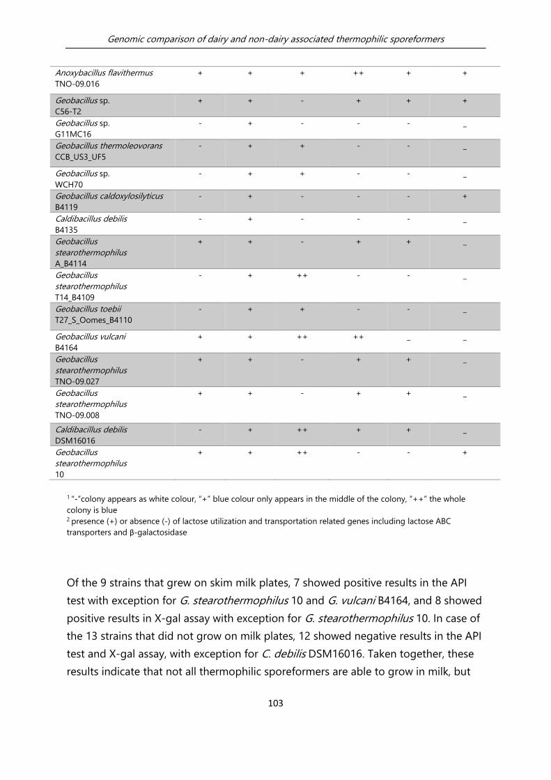
Genomic comparison of dairy and non-dairy associated thermophilic sporeformers
103
Anoxybacillus flavithermus
TNO-09.016
+ + + ++ + +
Geobacillus sp.
C56-T2
+ + - + + +
Geobacillus sp.
G11MC16
- + - - - _
Geobacillus thermoleovorans
CCB_US3_UF5
- + + - - _
Geobacillus sp.
WCH70
- + + - - _
Geobacillus caldoxylosilyticus
B4119
- + - - - +
Caldibacillus debilis
B4135
- + - - - _
Geobacillus
stearothermophilus
A_B4114
+ + - + + _
Geobacillus
stearothermophilus
T14_B4109
- + ++ - - _
Geobacillus toebii
T27_S_Oomes_B4110
- + + - - _
Geobacillus vulcani
B4164
+ + ++ ++ _ _
Geobacillus
stearothermophilus
TNO-09.027
+ + - + + _
Geobacillus
stearothermophilus
TNO-09.008
+ + - + + _
Caldibacillus debilis
DSM16016
- + ++ + + _
Geobacillus
stearothermophilus
10
+ + ++ - - +
1 “-“colony appears as white colour, “+” blue colour only appears in the middle of the colony, “++” the whole
colony is blue 2 presence (+) or absence (-) of lactose utilization and transportation related genes including lactose ABC
transporters and β-galactosidase
Of the 9 strains that grew on skim milk plates, 7 showed positive results in the API
test with exception for G. stearothermophilus 10 and G. vulcani B4164, and 8 showed
positive results in X-gal assay with exception for G. stearothermophilus 10. In case of
the 13 strains that did not grow on milk plates, 12 showed negative results in the API
test and X-gal assay, with exception for C. debilis DSM16016. Taken together, these
results indicate that not all thermophilic sporeformers are able to grow in milk, but

Genomic comparison of dairy and non-dairy associated thermophilic sporeformers
104
the strains that grow on milk quite consistently have the ability to utilize lactose and
produce active β-galactosidase. Thereby, the characterization of lactose utilization
and the production of active β-galactosidase can be employed as an indicator for the
ability of thermophilic sporeformers to grow on (skim) milk (plates).
5.3.3 The discrepancy between the presence of lactose-utilization
related genes and lactose utilization capability
Lactose fermentation contains three stages, namely: vectoral translocation and
phosphorylation of the disaccharide by the multi-component, lactose-specific PTS;
catabolism of lactose-6’P by energy-yielding pathways, and efflux of lactic acid from
the cell (Thompson et al., 1987). All 22 genomes analysed in this study were searched
for genes encoding these enzymes which can exert those functions, establishing
strain specific presence or absence of putative lactose utilization associated genes
(Table 5.1). There were three genes included in the analysis, namely, β-galactosidase
(EC 3.2.1.23), a permease protein (lactose ABC transporter) and a lactose-binding
protein (lactose ABC transporter). No PTS cassettes related to lactose importation
were found, and tagatose pathway known in lactic acid bacteria was not included in
this analysis. The three genes were always either all present, or all absent. API test
results (Table 5.1) appeared to show some discrepancies with the results of the
presence or absence analysis for lactose utilization genes. The hypothesis of those
discrepancies is discussed below: First, comparative genome analysis on the 22
strains revealed that G. stearothermophilus 10 and G. caldoxylosilyticus B4119
contain the set of lactose-metabolism related genes but did not demonstrate X-gal
nor lactose utilization activity. The comparative genome analysis was done with
algorithm OrthoMCL, a sequence comparison tool that performs bi-directional best
hit analyses to identify orthologous genes. In this analysis, identification of present
but inactive pseudogenes might occur due to the fact that in itself the OrthoMCL
tool does not consider domain presence, absence or intactness, and may tolerate
small sequence variations (e.g. truncations or mutations). Therefore, the occurrence
of errors should be checked for these 2 genomes like: (i) abnormal gene organization
(e.g. genomic reorganizations or loss of promoter- or cistronic- context), (ii)
dysfunctional promotors (e.g. missing essential promoter motives like TATA-boxes),
(iii) lacking or mutated protein domains like signal peptides, binding- and active

Genomic comparison of dairy and non-dairy associated thermophilic sporeformers
105
domains, (iv) N-, C- terminal truncations, (v) codon mutations leading to inactivity or
misfolding. To perform this analysis in full depth for all the reported lactose-
metabolism related orthologous genes is a massive task. Therefore, for this paper,
only the OrthoMCL generated orthologue alignments were examined. From this
examination, it is observed that G. caldoxylosilyticus B4119, one of two strains with
inactive lactose-metabolism genes, has only percentage identity ranging from 6% to
13% in the amino acid sequences of the putative lactose-metabolism related genes
((β-galactosidase (EC 3.2.1.23/OG_3249), a permease protein (lactose ABC
transporter/ OG_2883) and a lactose-binding protein (lactose ABC
transporter/OG_3549)) compared to the strains which had the consistence between
the presence of the genes and lactose utilization activity (e.g. A. flavithermus TNO-
09.006 and A. flavithermus TNO-09.016) (Table S5.1). This low percentage identity
explains the absence of lactose utilization ability. On the other hand, although the
sequences of G. stearothermophilus 10 showed percentage identity around 90%
compared with the sequences of consistent strains (e.g. A. flavithermus TNO-09.006
and A. flavithermus TNO-09.016) (Table S5.1), it is shown in the phenotypic
experiments that G. stearothermophilus 10 showed distinct sugar metabolism
patterns compared to other strains (Figure 5.2). It is suspected that the inactivity of
lactose utilization related genes of G. stearothermophilus 10 is related to
environmental reasons: for example, the effect of environmental conditions such as
oxygen availability and temperature, or carbon catabolite repression (CCR) on the
expression and regulation of sugar utilization systems (Mekalanos, 1992; Wassarman,
2002; Rohmer et al., 2011). Second, 3 G. stearothermophilus strains (G.
stearothermophilus TNO-09.027, G. stearothermophilus TNO-09.008, G.
stearothermophilus A_B4114), and C. debilis DSM16016 showed β-galactosidase
activity and lactose utilization capability. However, in the genome of those strains,
neither the lactose transporting system (including an ABC transporter or lactose
permease) and β-galactosidase gene, nor the functional domains of these genes
could be found. This observation suggests that the three G. stearothermophilus
strains and C. debilis DSM16016 may use different but equivalent cassettes for the
utilization of lactose.
The phenomenon that thermophilic bacteria have more variable lactose degradation
architectures and strategies is also suggested in the paper by Brumm et al. (2015).
Also, within the same species, there are different genes that are responsible for

Genomic comparison of dairy and non-dairy associated thermophilic sporeformers
106
lactose metabolism for lactic acid bacteria (Thompson et al., 1987). Those alternative
pathways can be used as a clue to search for alternatives in those above mentioned
thermophilic sporeformers.
Contrary, among mesophilic bacteria, the carbohydrate utilization genes were found
to be more conserved, particularly in case of lactose utilization (Warda et al., 2016).
As described above, the thermophilic bacteria have more variable carbohydrate
degradation architectures and strategies when compared to mesophilic bacteria.
Therefore, when using presence of lactose-utilization related genes as a predictive
factor for growth of thermophilic sporeformers in skim milk, more detailed
knowledge of these alternative pathways should be acquired to accurately detect
lactose-utilization genes in these species.
5.3.4 All thermophilic sporeformers sequenced encode proteolytic
systems with a putative role in casein breakdown
Casein, which makes up 80% of the proteins in cow's milk, is the main source of
nitrogen of microorganisms in milk (Mills and Thomas, 1981; Exterkate and de Veer,
1987). It is reported that Bifidobacterium’s ability to break down casein is linked to its
rapid growth in milk; conversely, the lack of ability to break down casein will retard
its growth (Klaver et al., 1993). To study this relationship for thermophilic
sporeformers, proteolytic activity of 22 thermophilic sporeformers was analysed
using MPCA (milk plate counting agar plates). The initial consideration in choosing
MPCA with the addition of yeast extract for testing proteolytic activity is due to the
fact that some thermophilic sporeformers cannot grow on complex milk protein.
Using only the milk plates, many strains would not grow due to the inability to use
lactose. Nevertheless, there is the presence of free peptide and amino acid in the
MPCA which can potentially inhibit proteolytic activity since proteins are broken
down in response to carbon, nitrogen or sulfur limitation (Sims, 2006; Sims and
Wander, 2002). When the environment is rich in small peptides and free amino acid,
the gene regulation of the overall proteolytic pathways will suppress protease and
peptidase expression levels in bacteria. In Bacillus subtilus, for example, CodY can
suppress exoprotease when cells are growing exponentially in a medium containing
abundant quantities of proteins or their degradation products (Barbieri et al., 2016).

Genomic comparison of dairy and non-dairy associated thermophilic sporeformers
107
In this study, 15 out of 22 collected thermophilic sporeformers showed halo
formation on MPCA plates, which indicates proteolytic ability is not suppressed. On
the contrary, the strains which do not appear a halo can also not be excluded for
having the potential for proteolytic activity, but the ability is likely to be limited due
to the ample presence of nitrogen source in the environment. With the knowledge of
this study, an improved version of MPCA plate to test proteolytic activity would be
without yeast extract but with the addition of monosaccharide (e.g. glucose or
galactose) to support initial growth but with the limitation of the nitrogen source.
Therefore, as for the link between growth on milk plates and the positive results on
MPCA plates, analysis of the proteolytic activity did show no clear correlation. This
indicates that the ability to grow on skim milk plates seems to have no clear
correlation with the ability of strains in casein breakdown as measured by MPCA
plates. This conflicts with the findings for dairy Bifidobacterium (Klaver et al., 1993).
However, it is observed that G. thermoglucosidans strains which presented biggest
halos on MPCA plates (data not shown) indicating a strong ability of casein
breakdown, could outcompete other species in growth in skim milk, as long as the
growth was initiated (Zhao et al., 2013). Since also a time-dependent phenomenon
has been mentioned for dairy lactic acid bacteria (Donkor et al., 2007), it can be
hypothesized that the extent of proteolysis varies among strains and appears to be
stage-dependent for thermophilic sporeformers, meaning lactose utilization is
essential for the initiation of growth in milk, and proteolytic activity is essential for
the continued growth of bacteria in milk (Bachmann et al., 2010).
In order to see the link between the proteolytic activity and the genomic features in
relation to proteolysis in these thermophilic sporeformers, searches for orthologous
genes in all 22 genomes sequenced were performed. The objective was to screen for
the presence of genes encoding proteolytic systems and proteins previously
reported as facilitating casein breakdown in Bacillus subtilis (Michna et al., 2014).
Peptidase encoding genes were identified in all 22 genomes, albeit it at highly
different levels of redundancy, ranging from 38 peptidase genes in A. flavithermus
TNO-09.006 to 67 peptidase genes in Geobacillus vulcani B4164. Genes unique to
each species were also identified (Table S5.2). For the species G. thermoglucosidans,
in which all strains showed the most apparent clearing zones; the species-specific
genes encoding proteases encoded subtilisin-like serine protease and hydrogenase
Genomic comparison of dairy and non-dairy associated thermophilic sporeformers
108
maturation protease. Overall, even though some of the 22 strains characterized here
were negative for proteolysis on MPCA agar, all strains appear to encode proteins for
all major steps required for casein breakdown (Table S5.2) (i.e., cell-wall proteinase
activity, peptide transport, and intracellular peptidases).
5.3.5 Biofilm forming capacity of thermophilic sporeformers
The biofilm forming capacity of strains in dairy-powder processing environments was
also assessed, since biofilm formation strongly contributes to the persistence of
certain spoilage organisms residing in the dairy processing line (Scott et al., 2007);
therefore being also of concern in respect to contamination of dairy powder
products. Among the 22 collected thermophilic sporeformers, only G. vulcani B4164
formed pellicles. The majority of the strains could form biofilm under at least one of
the conditions tested (Table 5.2). In fact, only A. flavithermus TNO-09.014 did not
form a biofilm under any condition tested. Although A. flavithermus TNO-09.014
showed turbidity in the medium, which means growth of the strain after overnight
culture, under test conditions (Table 5.2), biofilm formation could not be detected in
the crystal violet assay. In contrast, for G. stearothermophilus TNO-09.008 biofilm
was detected, but the turbidity in the medium was not very apparent. This could be
due to the fact that the bacteria were aggregated to the surface to form a biofilm,
which resulted in a clearance of the medium. This phenomenon, whereby cells
aggregate at the surface for biofilm formation, has also been described by Branda et
al. (2001). It is considered that biofilm is the surface associated structure, and crystal
violet assay is a good approach to evaluate this. However, the crystal violet assay
cannot directly indicate the amount of bacteria population aggregating to the
surface (Ramirez et al., 2015). Therefore, one should also be careful with the
interpretation of the results from the crystal violet assay.
According to the genome comparison analysis, every strain has a certain number of
genes involved in biofilm formation (Bacillus subtilis was used as a reference for
selecting genes involved in biofilm formation) (Figure S5.1). The presence of the
genes is an indication of the strains’ potential to form biofilm. Nevertheless, biofilm
formation can be influenced by different factors (Sadiq et al., 2017), such as surface
for attachment (Hinton et al., 2002), time of exposure to a surface (Jimenez-Flores,

Genomic comparison of dairy and non-dairy associated thermophilic sporeformers
109
2014), and accessible nutrients (Zhao et al., 2013). This study underpins that biofilm
formation is condition dependent, and the different strains did not form biofilms
under all conditions tested.
Table 5.2 A. Overview of biofilm-forming capacities of all 22 thermophilic sporeformers using microtiter
plate assay (“0” biofilm formation was not detectable, “1” biofilm formation was detected). B. Overview of
growth in the medium of all 22 strains using microtiter plate assay (“0” growth was not observed, “1” growth
was observed).
A. Biofilm formation capacity
of strains under the following
conditions:
B. Growth of strains in static
biofilm system under the
following conditions:
Thermophilic sporeformers 55°C 65°C 55°C 65°C
16h 25h 16h 25h 16h 25h 16h 25h
Geobacillus sp. C56-T2 1 0 1 1 1 1 1 1
Geobacillus sp. G11MC16 1 0 0 0 1 0 1 1
Geobacillus thermoglucosidans Y4-1MC1 1 1 1 1 1 1 1 1
Geobacillus thermoglucosidans C56-YS93 1 1 1 1 1 0 1 1
Geobacillus thermoleovorans CCB_US3_UF5 0 1 0 1 1 1 1 1
Geobacillus sp. WCH70 1 1 0 0 1 0 1 1
Geobacillus caldoxylosilyticus B4119 1 1 0 1 1 0 1 0
Caldibacillus debilis B4135 1 0 0 0 1 1 1 1
Geobacillus stearothermophilus A_B4114 1 1 1 1 1 0 1 0
Geobacillus stearothermophilus T14_B4109 0 1 1 1 0 0 1 0
Geobacillus toebii T27_S_Oomes_B4110 0 0 1 1 1 1 1 1
Geobacillus vulcani B4164 1 1 0 0 1 1 0 0
Anoxybacillus flavithermus TNO-09.014 0 0 0 0 1 1 1 1
Anoxybacillus flavithermus TNO-09.016 1 1 1 1 1 0 1 0
Geobacillus thermoglucosidans TNO-09.023 1 1 1 1 1 1 1 1
Geobacillus stearothermophilus TNO-09.027 0 0 1 1 1 0 1 0
Anoxybacillus flavithermus TNO-09.006 1 1 1 1 1 1 1 0
Geobacillus stearothermophilus TNO-09.008 1 1 1 1 0 0 0 0
Geobacillus thermoglucosidans TNO-09.020 0 1 1 1 1 1 1 1
Caldibacillus debilis DSM16016 0 1 0 1 1 1 1 1
Anoxybacillus flavithermus WK1 1 1 0 0 1 1 1 1
Geobacillus stearothermophilus 10 1 0 1 0 1 1 1 1

Genomic comparison of dairy and non-dairy associated thermophilic sporeformers
110
5.3.6 Specific genomic content relating to growth in milk plates in
thermophilic sporeformers
In order to find other thermophile specific genomic features relating to milk spoilage,
a gene-trait matching analysis (GTM) was conducted for the relevant phenotypes,
including ability to use different sugars, production of active β-galactosidase, and
ability to grow on skim milk plates etc. GTM identified 100 genes that specifically
help to differentiate strains by the ability to grow on skim milk plates (Table S5.3).
The gene annotated as “transporter, major facilitator superfamily (MFS)”, predicted to
be involved in the transport of niacin (niacin, known as nicotinic acid; together with
nicotinamide it composes vitamin B3 complex) weighted the heaviest in contributing
to the differentiation of growth from non-growth group indicated by the MDA (mean
decrease in accuracy) score. The Random Forest algorithm uses supervised
classification for discriminant selection, and “importance” is an arbitrary indicator for
the contribution of the discriminant to the classification. This gene appeared present
in the strains G. stearothermophilus TNO-09.027, TNO-09.008, A-B4114, and A.
flavithermus TNO-09.006, TNO-09.014, TNO-09.016 that could grow on skim milk
plates. However, there is not a complete association of this gene with this phenotype,
since several other skim-milk positive strains lacked this gene, i.e., Geobacillus sp.
C56-T2, Geobacillus stearothermophilus 10, and Geobacillus vulcani B4164.
Interestingly, niacin has been reported as an essential growth factor for lactic and
propionic acid bacteria (Sneli et al., 1939). Moreover, strains included in this study in
which the niacin transporter was identified were all isolated from diary-processing
environments and demonstrated rapid growth on skim milk plates. This observation
supports that the niacin transporter might be relevant for the growth of thermophilic
sporeformers in milk and may reflect an adaptation to the dairy environment. Further
investigation is required to understand this relation.
5.3.7 Concluding remarks
In conclusion, this study demonstrated that all 22 thermophilic sporeformers contain
casein breakdown related genes and biofilm formation related genes. A correlation
between lactose utilization and growth in skim milk was found. This suggests that the
ability to utilize lactose in skim milk is an essential factor for growth in milk. On one

Genomic comparison of dairy and non-dairy associated thermophilic sporeformers
111
hand, dairy powder-processing facilities can bear in mind this possibility of enhanced
growth capability for thermophilic sporeformers, when there is a presence of free
monomeric sugars in the ingredients. If stable product specifications are desired,
preventive measures to limit the presence of monomeric sugars can be taken into
account during the product formulation, raw material selection, or processing. For
example, the factory can prevent the addition of monomeric sugars in the product
formulation, or the factory could use a mild processing method to prevent sugar
break down in the products. On the other hand, if specific lactose-utilization related
genes for thermophilic sporeformers can be identified, together with the known
lactose-utilization related genes, the information can be used as a biomarker for the
prediction of growth potential of thermophilic sporeformers in skim milk. This
message might not be directly linked to spoilage control of the thermophilic
sporeformer in dairy processing environments, however, the knowledge which is
acquired in this study has shown the basis over what it is to be found through the
comparative genomic approach for the thermophilic sporeformers.
5.4 Experimental procedures
Whole genome comparison
Previously sequenced genomes representing Anoxybacillus spp. (n = 4), Geobacillus
spp. (n = 16) and Caldibacillus spp. (n = 2) were retrieved from NCBI (Table 5.3) and
used for comparative analyses. All genomes were (re)annotated using RAST (Aziz et
al., 2008) to allow a better comparison of all annotations. Orthologous groups (OGs;)
in the genomes were determined using OrthoMCL analysis, the settings used in the
analysis are default settings described in the paper from Li et al. (2003) (e-value
threshold used in the protein blast is 0.01). When OG (orthologous groups)
contained more than one gene per strain (i.e. highly similar genes), an effort was
made to manually split OG into separate OGs containing 1 gene per strain (except
for transposases or mobile elements). The analysis resulted in 16.455 OGs over all the
genomes. When an OG contained more than one gene per strain (i.e. highly similar
genes), an effort was made to manually split this OG into separate OGs containing
only one gene per strain (except for transposases/mobile elements). Phylogenetic
trees, created by MEGA5 (http://www.megasoftware.net/), were used to help indicate

Genomic comparison of dairy and non-dairy associated thermophilic sporeformers
112
how the OGs with questionable alignments should be split into subgroups.
Moreover, for every OG, a multiple sequence alignment was made on the amino acid
level using MUSCLE (Edgar, 2004) to facilitate identification of pseudogenes
(encoding incomplete proteins).
The protease, peptidase, and carbohydrate utilization systems of B. subtilis 168 listed
in the Subtiwiki database (Florez et al., 2009) were initially used to search for
orthologous systems in the selected thermophilic sporeformers OG table. Additional
genes and systems were found with keyword searches. Besides Subtiwiki database
(Florez et al., 2009), biofilm formation related genes are selected according to
Vlamakis et al. (2013). LocateP (http://www.cmbi.ru.nl/locatep-db/cgi-
bin/locatepdb.py/) was used to determine the subcellular locations of B. subtilis
protease and carbohydrate utilization systems.
Subsequently, selected genes and proteins were manually curated by comparison
with the reference Geobacillus genomes using databases (e.g. NCBI-BLASTP
http://blast.ncbi.nlm.nih.gov/), family and domain databases (e.g. Interpro (Mitchell
et al., 2015)), enzyme databases (e.g. Brenda (Schomburg et al., 2004) and pathways
databases (e.g. KEGG (Kanehisa and Goto, 2000)). Details of the protease system
cassette, biofilm formation related genes in the OG database can be found in Table
S5.1, Table S5.2.
Table 5.3 Name and genome description of strains used in this study.
Strain Species1 Environment/
isolation source
Sequence
statues
NCBI Acc # Replicons
/ contigs
Size
(Mb)
G+C
%2
C56-T2 Geobacillus sp. Double Hot
Springs, Nevada
complete +
plasmids
SAMN0017395 3 3.55 52.39
G11MC16 Geobacillus sp. No record scaffolds ABVH00000000 31 3.55 48.8
Y4.1MC1 Geobacillus
thermo-
glucosidans
Bath Hot spring,
YNP, USA
complete +
plasmids
NC_014650.1 2 3.84 44.02

Genomic comparison of dairy and non-dairy associated thermophilic sporeformers
113
C56YS93 Geobacillus
thermo-
glucosidans
Obsidian Hot
Spring, YNP, USA
complete +
plasmids
NC_015660.1 3 4 43.93
CCB_US3_UF5 Geobacillus
thermo-
leovorans
Ulu Slim hot
spring, Malaysia.
complete NC_016593.1 1 3.6 52.28
WCH70 Geobacillus sp. Middleton, USA complete NC_012793.1 3 3.51 42.8
B4119 Geobacillus
caldo-
xylosilyticus
Dairy scaffolds LQYS00000000.1 122 3.93 44
B4135 Caldibacillus
debilis
Dairy scaffolds LQYT00000000.1 129 3.22 50.9
A_B4114 Geobacillus
stearothermo-
philus
Buttermilk
powder,
Netherlands
scaffolds LQYY00000000.1 134 2.74 52.9
T14_B4109 Geobacillus
stearothermo-
philus
Pea soup,
Netherlands
scaffolds LQYV00000000.1 146 2.76 52.5
T27_S_Oomes
_B4110
Geobacillus
toebii
Pea soup,
Netherlands
scaffolds LQYW00000000.1 170 3.5 42.1
B4164 Geobacillus
vulcani
No record scaffolds unpublished data 31 4.09 46.2
TNO-09.014 Anoxybacillus
flavithermus
Standard milk
used for dairy
processing plant,
Netherlands
scaffolds LUFB00000000 180 2.56 41.8
TNO-09.016 Anoxybacillus
flavithermus
Evaporator,
Netherlands
scaffolds LUCQ00000000 187 2.65 41.1
TNO-09.023 Geobacillus
thermo-
glucosidans
Casein pipe
fouling in a diary
processing plant,
Netherlands
scaffolds LUCT00000000 52 3.7 43.8
TNO-09.027 Geobacillus
stearothermo-
philus
Casein pipe
fouling in a diary
processing plant,
Netherlands
scaffolds LUCR00000000 226 2.7 52.5
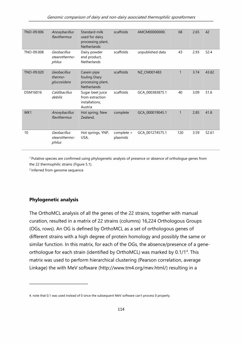
Genomic comparison of dairy and non-dairy associated thermophilic sporeformers
114
TNO-09.006 Anoxybacillus
flavithermus
Standard milk
used for dairy
processing plant,
Netherlands
scaffolds AMCM00000000. 68 2.65 42
TNO-09.008 Geobacillus
stearothermo-
philus
Dairy powder
end product,
Netherlands
scaffolds unpublished data 43 2.93 52.4
TNO-09.020 Geobacillus
thermo-
glucosidans
Casein pipe
fouling Diary
processing plant,
Netherlands
scaffolds NZ_CM001483 1 3.74 43.82
DSM16016 Caldibacillus
debilis
Sugar beet juice
from extraction
installations,
Austria
scaffolds GCA_000383875.1 40 3.09 51.6
WK1 Anoxybacillus
flavithermus
Hot spring, New
Zealand,
complete GCA_000019045.1 1 2.85 41.8
10 Geobacillus
stearothermo-
philus
Hot springs, YNP,
USA,
complete +
plasmids
GCA_001274575.1 120 3.59 52.61
1 Putative species are confirmed using phylogenetic analysis of presence or absence of orthologue genes from
the 22 thermophilic strains (Figure 5.1).
2 Inferred from genome sequence
Phylogenetic analysis
The OrthoMCL analysis of all the genes of the 22 strains, together with manual
curation, resulted in a matrix of 22 strains (columns) 16,224 Orthologous Groups
(OGs, rows). An OG is defined by OrthoMCL as a set of orthologous genes of
different strains with a high degree of protein homology and possibly the same or
similar function. In this matrix, for each of the OGs, the absence/presence of a gene-
orthologue for each strain (identified by OrthoMCL) was marked by 0.1/14. This
matrix was used to perform hierarchical clustering (Pearson correlation, average
Linkage) the with MeV software (http://www.tm4.org/mev.html/) resulting in a
4. note that 0.1 was used instead of 0 since the subsequent MeV software can’t process 0 properly.

Genomic comparison of dairy and non-dairy associated thermophilic sporeformers
115
heatplot with dendrograms for strains and/or OGs based on Pearson correlations
among either strains or OGs.
Bacterial strains and growth media
The strains (Table 5.3) were obtained from the -80 °C stock. Tryptone Soy Broth
(TSB), Tryptone Soy Agar (TSA plates, Tritium Microbiologie, the Netherlands) and
Tryptone (Tritium Microbiologie, the Netherlands) were used as basic growth
medium. UHT milk Friesche Vlag® Langlekker 0% fat was used for the growth
experiments in milk. Demi water (Tritium Microbiologie) was used for dilution of
media in growth assays.
Milk plate counting agar (MPCA) plates for testing protease activity
2.5g Tryptone, 1.25g yeast extract, 0.5g glucose, and 15g agar were dissolved in 450
ml demi water and autoclaved for 20 minutes at 120°C. Separately, 0.5g skim milk
powder was dissolved in 50 ml demi water and was autoclaved for 5 minutes at
120°C. The two above-prepared contents were poured together in a 47°C water bath.
MPCA Plates were made and stored in the cold room (-4°C). Strains were inoculated
on the MPCA plates and incubated for 24h at 55°C. Grown strains were observed for
halo formation on MPCA plates.
Plate Assay for testing β-galactosidase activity
200 mg X-gal was dissolved in 10 ml DMSO. TSA was melted and cooled down to lower
than 65°C. 500 μl X-gal stock solution was added into the TSA (final concentration 40
μg/ml). Plates were poured, dried, and stored in the cold room (-4°C) and in the dark
(covered with aluminium foil) for following inoculation. Overnight grown strains were
streaked on the previous made plates and incubated for 24h at 55°C. The strains which
showed blue colonies are strains which produced active β-galactosidase.
API growth test
API50CH (BioMerieux, France) test was used in combination with API 50CHB/E
medium (BioMerieux, France) according to the manufacturer’s instruction. Single
colonies were used to inoculate API 50CHB/E medium to a turbidity equivalent of
2McFarland. Test strips were filled with inoculated medium, incubated at 55°C, and

Genomic comparison of dairy and non-dairy associated thermophilic sporeformers
116
checked for media colour change after 24 and 48h. For each strain, two independent
repetitions were performed.
Gene trait matching (GTM)
GTM was performed using Phenolink, a web tool that associates bacterial
phenotypes with genomic data (Bayjanov et al., 2012), in order to correlate observed
phenotypes with the presence or absence of particular genes. For example, in
relation to spoilage in the dairy processing line, phenotypic data were divided into
strains that could grow on skim milk plates versus strains that could not grow on
skim milk plates. Growth (Yes) or non-growth (No) of 22 thermophilic sporeformers
strains on skim milk plates was measured. A gene that is found to be important to
distinguish strains of different phenotype is assumed as important. Selected genes
are ranked based on the mean decrease in accuracy (MDA). The higher the number
is, the more relevant statistically a gene is in differentiating the grower or non-
grower on skim milk plates.
In vitro biofilm assays for screening biofilm formation ability
Biofilm formation on polystyrene was measured as described previously (Wijman et
al., 2007), with some modifications. In short, polystyrene microtiter plates (Greiner
Bio-one, Germany) were filled with 200 µl TSB, and for each strain, six wells were
inoculated with 1.5% (vol/vol) suspended overnight cultures, grown on TSA plates,
respectively. The microtiter plates were incubated at 55 and 65°C. After incubation,
for either 16 or 25 h, the wells were gently washed three times with 220 µl of sterile
distilled water, and subsequently biofilm cells were stained with 220 µl of 0.1%
(wt/vol) crystal violet for 30 min. After this 30 min, the wells were washed twice with
220 µl sterile deionized water to remove unbound crystal violet. The remaining
crystal violet was dissolved in 220 µl 96% ethanol, and the absorbance was measured
at 595 nm (Quant, Bio-Tek Instruments). Strains with optical density at 595 nm
(OD595) values above two times the background signal, or higher were considered
positive for biofilm formation. The average biofilm formation and standard deviation
for six wells were calculated for each strain in the conditions tested. The turbidity of
medium measured by spectrophotometer at OD600 is used as an indication of
growth.

Genomic comparison of dairy and non-dairy associated thermophilic sporeformers
117
5.5 Acknowledgments
The project is funded by TI Food and Nutrition, a public-private partnership on pre-
competitive research in food and nutrition. We wish to thank Lars Wims for technical
assistance, Alicja Warda from the food microbiology group at Wageningen University
and other members of TNO microbiology and system biology group for stimulating
discussions on this topic.
5.6 References
Aziz, R. K., D. Bartels, A. A. Best, M. DeJongh, T. Disz, R. A. Edwards, K. Formsma, S. Gerdes, E.
M. Glass, M. Kubal, F. Meyer, G. J. Olsen, R. Olson, A. L. Osterman, R. A. Overbeek, L. K. McNeil, D.
Paarmann, T. Paczian, B. Parrello, G. D. Pusch, C. Reich, R. Stevens, O. Vassieva, V. Vontein, A.
Wilke and O. Zagnitko. 2008. The RAST server: Rapid annotations using subsystems technology.
BMC Genomics. 9:75.
Bachmann, H., D. Molenaar, M. Kleerebezem and J. E. van Hylckama Vlieg. 2011. High local substrate
availability stabilizes a cooperative trait. The ISME journal. 5:929.
Barbieri, G., A. M. Albertini, E. Ferrari, A. L. Sonenshein, B. R. Belitsky. 2016. Interplay of CodY and ScoC
in the regulation of major extracellular protease genes of Bacillus subtilis. Journal of
Bacteriology. 1986:907-920.
Bayjanov, J. R., D. Molenaar, V. Tzeneva, R. J. Siezen and S. A. van Hijum. 2012. PhenoLink a web-tool
for linking phenotype to ~omics data for bacteria: Application to gene-trait matching for
Lactobacillus plantarum strains. BMC Genomics. 13:170.
Berendsen, E. M., M. H. Wells-Bennik, A. O. Krawczyk, A. de Jong, A. van Heel, S. Holsappel, R. T.
Eijlander and O. P. Kuipers. 2016. Draft genome sequences of seven thermophilic spore-forming
bacteria isolated from foods that produce highly heat-resistant spores, comprising Geobacillus
spp., Caldibacillus debilis, and Anoxybacillus flavithermus. Genome Announcement. 4:e00105-16.
Branda, S. S., J. E. Gonzalez-Pastor, S. Ben-Yehuda, R. Losick and R. Kolter.
2001. Fruiting body formation by Bacillus subtilis. Proceedings of the National Academy of
Sciences. 98:11621-11626.
Brumm, P. J., P. De Maayer, D. A. Mead and D. A. Cowan. 2015. Genomic analysis
of six new Geobacillus strains reveals highly conserved carbohydrate degradation architectures a
nd strategies. Frontiers in Microbiology. 6:430.
Genomic comparison of dairy and non-dairy associated thermophilic sporeformers
118
Burgess, S. A., S. H. Flint, D. Lindsay, M. P. Cox and P. J. Biggs. 2017. Insights into the Geobacillus
stearothermophilus species based on phylogenomic principles. BMC Microbiology. 17:140.
Carlin, F. 2011. Origin of bacterial spores contaminating foods. Food Microbiology. 28:177-182.
Caspers, M. P. M., J. Boekhorst, A. de Jong, R. Kort, M. N. Nierop Groot and T. Abee. 2016. Draft
genome sequences of four thermophilic spore formers isolated from a dairy-processing
environment. Genome Announcement. 4: e00757-16.
Ceapa, C., J. Lambert, K. van Limpt, M. Wels, T. Smokvina, J. Knol and M. Kleerebezem. 2015.
Correlation of Lactobacillus rhamnosus genotypes and carbohydrate utilization signatures
determined by phenotype profiling. Applied and Environmental Microbiology. 81:5458-5470.
Donkor, O. N., A. Henriksson, T. Vasiljevic and N. P. Shah.
2007. Proteolytic activity of dairy lactic acid bacteria and probiotics as determinant
of growth and in vitro angiotensin converting enzyme inhibitory activity in fermented milk.
Le Lait, INRA Editions. 87:21-38.
Edgar, R. C. 2004. MUSCLE: A multiple sequence alignment method with reduced time and space
complexity. BMC Bioinformatics. 5:113.
Exterkate, F. A. and G. J. C. M. de Veer. 1987. Optimal growth of Streptococcus cremoris HP in milk is
related to β-and Χ-casein degradation. Applied Microbiology and Biotechnology. 25:471-475.
Flint, S. H., P. J. Bremer and J. D. Brooks. 1997. Biofilms in dairy manufacturing plant
- description, current concerns and methods of control. Biofouling. 11:81-97.
Flint, S., J. Palmer, K. Bloemen, J. Brooks and R. Crawford. 2001. The growth of Bacillus
stearothermophilus on stainless steel. Journal of Applied Microbiology. 90:151-157.
Florez, L. A., S. F. Roppel, A. G. Schmeisky, C. R. Lammers and J. Stulke. 2009. A community-
curated consensual annotation that is continuously updated: The
Bacillus subtilis centered wiki SubtiWiki. Database (Oxford). 2009:bap012.
Goh, K. M., H. M. Gan, K. Chan, G. F. Chan, S. Shahar, C. S. Chong, U. M. Kahar and K. P. Chai. 2014.
Analysis of Anoxybacillus genomes from the aspects of lifestyle adaptations, prophage diversity,
and carbohydrate metabolism. PLoS One. 9: e90549.
Griffiths, A., W. Gelbart and J. Miller. 1999. Regulation of the lactose system. Modern Genetic Analysis.
W. H. Freeman, US, New York.

Genomic comparison of dairy and non-dairy associated thermophilic sporeformers
119
Hinton, A. R., K. T. Trinh, J. D. Brooks and G. J. Manderson. 2002. Thermophile survival in milk fouling
and on stainless steel during cleaning. Food and Bio-products Processing Publication of the
Institution of Chemical Engineers Part C. 80:299-304.
Kanehisa, M. and S. Goto. 2000. KEGG: Kyoto encyclopedia of genes and genomes. Nucleic Acids
Research. 28:27-30.
Klaver, F. A., F. Kingma and A. H. Weerkamp. 1993. Growth and survival of Bifidobacteria in milk.
Nederlands Melk en Zuiveltijdschrift. 47:151-164.
Li, L., C. J. Stoeckert and D. S. Roos. 2003. OrthoMCL: Identification of ortholog groups
for eukaryotic genomes. Genome Research. 13:2178-2189.
Mekalanos, J. J. 1992. Environmental signals controlling expression of virulence
determinants in bacteria. Journal of Bacteriology. 174:1-7.
Michna, R. H., F. M. Commichau, D. Todter, C. P. Zschiedrich and J. Stulke. 2014. SubtiWiki-a database
for the model organism Bacillus subtilis that links pathway,
interaction and expression information. Nucleic Acids Resources. 42:D692-698.
Mills, O. E., and T. D. Thomas. 1981. Nitrogen sources for growth of lactic streptococci in milk. New
Zealand Journal of Dairy Science and Technology. 16:43-45.
Mitchell, A., H. Y. Chang, L. Daugherty, M. Fraser, S. Hunter, R. Lopez, C. McAnulla, C. McMenamin, G.
Nuka, S. Pesseat, A. Sangrador-Vegas, M. Scheremetjew, C. Rato, S. Y. Yong, A. Bateman,
M. Punta, T. K. Attwood, C. J. Sigrist, N. Redaschi, C. Rivoire, I. Xenarios, D. Kahn, D. Guyot, P. Bork,
I. Letunic, J. Gough, M. Oates, D. Haft, H. Huang, D. A. Natale, C. H. Wu, C. Orengo, I. Sillitoe, H.
Mi, P. D. Thomas and R. D. Finn. 2015. The InterPro protein families database:
The classification resource after 15 years. Nucleic Acids Resources. 43:D213-221.
Ramírez, M. D. F., E. J. Smid, T. Abee and M. N. Nierop Groot. 2015. Characterisation of biofilms formed
by Lactobacillus plantarum WCFS1 and food spoilage isolates. International Journal of Food
Microbiology. 207:23-29.
Rohmer, L., D. Hocquet and S. Miller. 2011. Are pathogenic bacteria just looking for food?
Metabolism and microbial pathogenesis. Trends in Microbiology. 19:341-348.
Sadiq, F. A., S. Flint, L. Yuan, Y. Li, T. Liu and G. He. 2017. Propensity for biofilm formation by aerobic
mesophilic and thermophilic spore forming bacteria isolated from Chinese milk powders.
International Journal of Food Microbiology. 262:89-98.

Genomic comparison of dairy and non-dairy associated thermophilic sporeformers
120
Schomburg, I., A. Chang, C. Ebeling, M. Gremse, C. Heldt, G. Huhn and D. Schomburg. 2004.
BRENDA, the enzyme database: Updates and major new developments. Nucleic Acids Resources.
32:D431- 433.
Scott, S. A., J. D. Brooks, J. Rakonjac, K. M. Walker and S. H. Flint. 2007. The formation of
thermophilic spores during the manufacture of whole milk powder. International Journal
of Dairy Technology. 60:109-117.
Siezen, R. J. and J. E. van Hylckama Vlieg. 2011. Genomic diversity and versatility of Lactobacillus
plantarum, a natural metabolic engineer. In Microbial cell factories (Vol.10, No. 1, p. S3). BioMed
Central.
Sims, G. K. 2006. "Nitrogen starvation promotes biodegradation of N-Heterocyclic compounds in
soil". Soil Biology & Biochemistry. 38:2478–2480.
Sims, G. K and M. M. Wander. 2002. "Proteolytic activity under nitrogen or sulfur limitation". Applied
Soil Ecology. 568:1–5.
Solopova, A., H. Bachmann, B. Teusink, J. Kok, A. R. Neves, O. P. Kuipers. 2012. A specific mutation in
the promoter region of the silent cel cluster accounts for the appearance of lactose-utilizing
Lactococcus lactis MG1363. Applied and Environmental Microbiology. 78:5612-5621.
Switt, A. I. M., A. D. Andrus, M. L. Ranieri, R. H. Orsi, R. Ivy, H. C. den Bakker, N. H. Martin, M. Wiedmann
and K. J. Boor. 2014. Genomic comparison of sporeforming bacilli isolated from milk. BMC
Genomics. 15:26.
Thompson, J. 1987. Regulation of sugar transport and metabolism in lactic acid bacteria. FEMS
Microbiology Reviews. 3:221-231.
Vlamakis, H., Y. Chai, P. Beauregard, R. Losick, and R. Kolter. 2013. Sticking together: building a biofilm
the Bacillus subtilis way. Nature Reviews Microbiology. 11:157-168.
Warda, A. K., R. J. Siezen, J. Boekhorst, M. H. J. Wells-Bennik, A. de Jong, O. P. Kuipers, M. N. Nierop
Groot and T. Abee. 2006. Linking Bacillus cereus genotypes and carbohydrate utilization capacity.
PloS One. 11:e156796.
Wassarman, K. M. 2002. Small RNAs in bacteria: Diverse regulators of gene expression in
response to environmental changes. Cell. 109:141-144.
Watterson, M. J., D. J. Kent, K. J. Boor, M. Wiedmann and N. H. Martin. 2014. Evaluation of dairy
powder products implicates thermophilic sporeformers as the primary organisms of interest.
Journal of Dairy Science. 97:2487-2497.

Genomic comparison of dairy and non-dairy associated thermophilic sporeformers
121
Wijman, J. G. E., P. P. L. A. de Leeuw, R. Moezelaar, M. H. Zwietering and T. Abee. 2007. Air-liquid
interface biofilms of Bacillus cereus: Formation, sporulation, and dispersion. Applied and
Environmental Microbiology. 73:1481-1488.
Zhao, Y., M. P. M. Caspers, K. I. Metselaar, P. de Boer, G. Roeselers, R. Moezelaar, M. N. Nierop
Groot, R. C. Montijn, T. Abee and R. Kort. 2013. Abiotic and microbiotic factors controlling
biofilm formation by thermophilic sporeformers. Applied and Environmental Microbiology.
79:5652-5660
Zhao, Y., M. Kumar, M. P. M. Caspers, M. N. Nierop Groot, J. M. B. M. van der Vossen, and T. Abee.
2017. Short communication: growth of dairy isolates of Geobacillus thermoglucosidans in skim
milk depends on lactose degradation products supplied by Anoxybacillus flavithermus as
secondary species. Journal of Dairy Science. 17:31071 – 31078

Genomic comparison of dairy and non-dairy associated thermophilic sporeformers
122
5.7 Supplemental figures and tables

Genomic comparison of dairy and non-dairy associated thermophilic sporeformers
123
5.7.1 Supplemental figures
Figure S5.1 Presence and absence of genes involved in biofilm formation (Bacillus subtilis was used as a
reference for selecting genes involved in biofilm formation). The first line from left to right are the name of the
Row
Gene/Protein
Genes
Bac
illu
s su
btili
s 1
68
Ano
xyba
cillu
s fl
avit
her
mu
s T
NO
.09
-00
6
Ano
xyba
cillu
s fl
avit
her
mu
s T
NO
.09
-01
4
Ano
xyba
cillu
s fl
avit
her
mu
s T
NO
.09
-01
6
Ano
xyba
cillu
s fl
avit
her
mu
s W
K1
Ge
oba
cillu
s th
erm
og
luco
sid
an
s TN
O.0
9-
020
Ge
oba
cillu
s th
erm
og
luco
sid
an
s TN
O.0
9-
023
Ge
oba
cillu
s th
erm
og
luco
sid
asi
us
C56
_Y
S93
Ge
oba
cillu
s st
ea
roth
erm
oph
ilus
TN
O.0
9-
008
Ge
oba
cillu
s st
ea
roth
erm
oph
ilus
TN
O.0
9-
027
Ge
oba
cillu
s st
ea
roth
erm
oph
ilus
10
Ge
oba
cillu
s st
ea
roth
erm
oph
ilus
A_B
411
4
Ge
oba
cillu
s_st
ea
roth
erm
op
hilu
s_T
14
_B4
109
Ge
oba
cillu
s ca
ldox
ylo
sily
ticu
s B
411
9
Ge
oba
cillu
s d
eb
ilis
B4
135
Ge
oba
cillu
s d
eb
ilis
DSM
_1
601
6
Ge
oba
cillu
s th
erm
ole
ovo
rans
CCB
_US3
_U
F5
Ge
oba
cillu
s to
ebi
i T
27
_S_
Oo
me
s_B
411
0
Ge
oba
cillu
s sp
. G
11M
C1
6
Ge
oba
cillu
s sp
C5
6-T
2
Ge
oba
cillu
s sp
. W
CH
70
Ge
oba
cillu
s sp
. Y
4_1
MC
1
Ge
oba
cillu
s vu
lcan
i B
41
64
1 YwcC BSU38220 1 0 0 0 0 0 0 0 0 0 0 0 0 0 0 0 0 0 0 0 0 0 0
2 SlrA BSU38229 1 0 0 0 0 0 0 0 0 0 0 0 0 0 0 0 0 0 0 0 0 0 1
3 sigM BSU09520 1 1 1 1 0 1 1 1 0 0 0 0 0 2 0 0 0 0 0 0 0 1 1
4 sigW BSU01730 1 1 1 1 1 1 1 1 1 1 1 1 1 1 1 1 1 1 1 1 1 1 1
5 sigX BSU23100 1 0 0 0 0 1 1 1 1 1 1 1 1 1 1 1 1 1 1 1 1 1 1
6 Abh BSU14480 1 0 0 0 0 0 0 0 0 0 0 0 0 0 0 0 0 0 0 0 0 0 1
7 SlrR BSU34380 1 0 0 0 0 0 0 0 0 0 0 0 0 0 0 0 0 0 0 0 0 0 1
8 SinR BSU24610 1 0 0 0 0 0 0 0 0 0 0 0 0 0 0 0 0 0 0 0 0 0 1
9 SinI BSU24600 1 0 0 0 0 0 0 0 0 0 0 0 0 0 0 0 0 0 0 0 0 0 1
10 Spo0A BSU24220 1 1 1 1 1 1 1 1 1 1 1 1 1 1 1 1 1 1 1 1 1 1 1
11 AbrB BSU00370 1 1 1 1 1 1 1 1 0 1 1 1 1 1 1 1 1 1 1 1 1 1 1
12 KinA BSU13990 1 0 0 0 0 0 0 0 0 0 0 0 0 0 0 0 0 0 0 0 0 0 1
13 KinB BSU31450 1 0 0 0 1 0 0 0 0 0 0 0 0 0 0 0 0 0 0 0 0 0 0
14 KinC BSU14490 1 0 0 0 0 0 0 0 0 0 0 0 0 0 0 0 0 1 0 0 1 0 1
15 KinD BSU13660 1 1 1 1 1 0 0 0 0 0 0 0 0 0 0 0 0 0 0 0 0 0 1
16 DegS BSU35500 1 1 1 1 1 1 1 1 1 1 1 1 1 1 1 1 1 1 1 1 1 1 1
17 DegU BSU35490 1 1 1 1 1 1 1 1 1 1 1 1 1 1 1 1 1 1 1 1 1 1 1
18 epsA BSU34370 2 1 1 1 0 0 0 0 1 1 0 1 1 1 1 1 1 1 1 0 1 0 2
19 epsB BSU34360 2 1 1 1 0 0 0 0 1 1 0 1 1 1 1 1 1 1 1 0 1 0 1
20 epsC BSU34350 1 1 1 0 0 1 2 0 0 0 1 0 0 0 1 1 1 0 0 1 0 1 2
21 epsD BSU34340 1 1 0 1 0 1 1 0 1 0 0 0 0 0 0 0 2 0 0 0 1 0 1
22 epsE BSU34330 1 1 0 0 0 1 1 0 0 0 0 0 0 0 0 0 0 0 1 1 0 0 1
23 epsF BSU34320 1 0 0 0 0 1 1 0 0 0 0 0 0 0 1 0 1 0 0 0 1 0 1
24 epsG BSU34310 1 0 0 0 0 0 0 0 0 0 0 0 0 0 0 0 0 0 0 0 0 0 1
25 epsH BSU34300 1 0 0 0 0 1 1 0 0 0 0 0 0 0 0 0 1 0 1 0 0 0 1
26 epsI BSU34290 1 0 0 0 0 0 0 0 0 0 0 0 0 0 0 0 0 0 0 0 0 0 1
27 epsJ BSU34280 1 0 0 0 0 0 0 0 0 0 0 0 0 0 1 0 0 0 0 0 0 0 1
28 epsK BSU34265 1 0 0 0 0 0 0 0 0 0 0 0 0 0 0 0 0 0 0 0 0 0 1
29 epsL BSU34250 1 2 1 1 0 1 1 1 1 2 0 1 1 1 1 1 1 1 0 1 1 0 2
30 epsM BSU34240 1 1 0 0 0 0 0 1 0 0 0 0 0 0 0 0 1 0 0 0 0 0 1
31 epsN BSU34230 1 2 0 1 0 0 1 1 1 0 0 0 0 1 0 0 1 0 0 0 1 0 1
32 epsO BSU34220 1 0 0 0 0 0 0 0 0 0 0 0 0 0 0 0 0 0 0 0 0 0 1
33 tapA BSU24640 1 0 0 0 0 0 0 0 0 0 0 0 0 0 0 0 0 0 0 0 0 0 1
34 sipW BSU24630 1 0 0 0 0 0 0 0 0 0 0 0 0 0 0 0 0 0 0 0 0 0 1
35 tasA BSU24620 1 0 0 0 0 0 0 0 0 0 0 0 0 0 0 0 0 0 0 0 0 0 1
36 bslA BSU31080 1 0 0 0 0 0 0 0 0 0 0 0 0 0 0 0 0 0 0 0 0 0 1
37 capB BSU35900 1 0 0 0 0 0 0 0 0 0 0 0 0 0 0 0 0 0 0 0 0 0 1
38 capC BSU35890 1 0 0 0 0 0 0 0 0 0 0 0 0 0 0 0 0 0 0 0 0 0 1
39 pgsA BSU16920 1 1 1 1 1 1 1 1 1 1 1 1 1 1 1 1 1 1 1 1 1 1 1
40 capE BSU35870 1 0 0 0 0 0 0 0 0 0 0 0 0 0 0 0 0 0 0 0 0 0 1
41 remB BSU00050 1 0 0 0 0 0 0 0 0 0 0 0 0 0 0 0 0 0 0 0 0 0 1
42 yaaO BSU00270 1 1 1 1 1 1 1 1 1 1 1 1 1 1 0 0 1 1 1 1 1 1 1
43 veg BSU00440 1 1 1 1 1 1 1 1 1 1 1 1 1 1 1 1 1 1 1 1 1 1 1
44 ftsH BSU00690 1 1 1 1 1 1 1 1 1 1 1 1 1 1 1 1 1 1 1 1 1 1 1
47 gabT BSU03900 1 0 0 0 1 1 1 1 0 0 0 0 0 1 0 0 0 0 0 0 0 1 1
48 yisP BSU10810 1 0 0 0 1 0 0 0 0 0 0 0 0 0 0 0 0 0 0 0 0 0 0
49 ampS BSU14450 1 1 1 1 1 1 1 1 1 1 1 1 1 1 1 1 1 1 1 1 1 1 1
50 speA BSU14630 1 1 1 1 1 1 1 1 1 1 1 1 1 1 1 1 1 1 1 1 1 1 1
51 ylbF BSU14990 1 1 1 1 1 1 1 1 1 1 1 1 1 1 1 1 1 1 1 1 1 1 1
52 ylmE BSU15380 1 1 1 1 1 1 1 1 1 1 1 1 1 1 1 1 1 1 1 1 1 1 1
53 remA BSU15670 1 0 0 0 0 1 1 1 1 1 1 1 1 1 0 0 1 1 1 1 1 1 1
54 rny BSU16960 1 1 1 1 1 1 1 1 1 1 1 1 1 1 1 1 1 1 1 1 1 1 1
55 ymdB BSU16970 1 1 1 1 1 1 1 1 1 1 1 1 1 1 1 1 1 1 1 1 1 1 1
56 ymcA BSU17020 1 1 1 1 1 1 1 1 1 1 1 1 1 1 1 1 1 1 1 1 1 1 1
57 luxS BSU30670 1 1 1 1 1 1 1 1 1 1 1 1 0 1 1 1 1 1 1 1 1 1 1
58 floT BSU31010 1 0 0 0 0 1 1 1 1 1 1 1 1 1 0 0 1 1 1 1 1 1 0
59 degQ BSU31720 1 0 0 0 0 0 0 0 0 0 0 0 0 0 0 0 0 0 0 0 0 0 1
60 racX BSU34430 1 0 0 0 0 0 0 0 0 0 0 0 0 0 0 0 0 0 0 0 0 0 0
61 yvcA BSU34850 1 0 0 0 0 1 1 1 0 0 0 0 0 1 0 0 0 0 1 0 0 1 0
64 ptpZ BSU36240 1 1 1 1 0 1 1 1 1 1 1 1 1 1 1 1 1 1 1 0 0 1 1
65 ptkA BSU36250 2 1 1 1 0 0 0 0 1 1 0 1 1 1 1 1 1 1 1 0 1 0 1
66 tkmA BSU36260 2 1 1 1 0 0 0 0 1 1 0 1 1 1 1 1 1 1 1 0 1 0 2
67 galE BSU38860 1 1 1 0 0 1 1 0 0 0 0 0 0 0 1 0 1 0 1 0 0 0 0
68 yxaB BSU40030 1 0 0 0 0 0 0 0 0 0 0 0 0 0 0 0 0 0 0 0 0 0 0
45 sfp/1 BSU03569
46 sfp/2 BSU03570
62 swrAA/1 BSU35230
63 swrAA/2 BSU35239

Genomic comparison of dairy and non-dairy associated thermophilic sporeformers
124
Bacillus subtilis 164 and the 22 collected thermophilic sporeformers. To the left of the black square listed the
selected genes involved in biofilm formation. Within the black square, “red square” indicates presence, and
“white” indicates absence.
5.7.2 Supplemental tables
Table S5.1 Percentage identity of the lactose utilization related genes from each strain in comparison to
the orthologue genes identified through OrthoMCL.
Percentage identity A permease protein/ lactose ABC transporter
β-galactosidase (EC 3.2.1.23)
a lactose binding protein/ lactose ABC transporter
Anoxybacillus flavithermus TNO-09.006
100% 100% 100%
Anoxybacillus flavithermus TNO-09.016
100% 99% 100%
Geobacillus caldoxylosilyticus B4119 13% 6% 6%
Geobacillus stearothermophilus 10 97% 99% 90%
Table S5.2 Genomic features in relation to proteolysis in the 22 selected thermophilic sporeformer.
(Bacillus subtilis was used as a reference for selecting genomic features involved in proteolytic activity). The first
line from left to right are the name of 22 collected thermophilic sporeformers and Bacillus subtilis 168. “1”
indicates presence, “0” indicates absence, “P” possible presence.
OG
An
oxy
baci
llus
flavi
therm
us
TN
O-0
9.0
06
An
oxy
baci
llus
flavi
therm
us
TN
O-0
9.0
14
An
oxy
baci
llus
flavi
therm
us
TN
O-0
9.0
16
An
oxy
baci
llus
flavi
therm
us
WK
1
Geo
baci
llus
cald
oxy
losi
lyti
cus
B4
119
Geo
baci
llus
deb
ilis
B41
35
Geo
baci
llus
deb
ilis
DSM
16
01
6
Geo
baci
llus
sp C
56-T
2
Geo
baci
llus
sp G
11M
C16
Geo
baci
llus
stearo
therm
op
hilu
s 10
Geo
baci
llus
stearo
therm
op
hilu
s A
_B41
14
Geo
baci
llus
stearo
therm
op
hilu
s T1
4_B
4109
Geo
baci
llus
stearo
therm
op
hilu
s TN
O-0
9.0
08
Geo
baci
llus
stearo
therm
op
hilu
s TN
O-0
9.0
27
Geo
baci
llus
therm
og
luco
sid
an
s TN
O-0
9.0
20
Geo
baci
llus
therm
og
luco
sid
an
s TN
O-0
9.0
23
Geo
baci
llus
therm
og
luco
sid
asi
us
C56Y
S93
Geo
baci
llus
therm
og
luco
sid
an
s Y
4.1
MC
1
Geo
baci
llus
therm
ole
ovo
ran
s C
CB
_US
3_U
F5
Geo
baci
llus
toeb
ii T27_S
_Oo
mes_
B4
11
0
Geo
baci
llus
vulc
an
i B
416
4
Geo
baci
llus
sp. W
CH
70
Baci
llus
sub
tilis
16
8
improved annotation
OG_199 1 1 1 1 1 1 1 1 1 1 1 1 1 1 1 1 1 1 1 1 1 1 BSU00430
Sporulation-specific protease
YabG
OG_4365a 0 0 0 0 0 0 0 0 0 0 0 0 0 0 0 0 0 0 0 0 1 0 BSU02240
extracellular glutamyl
endopeptidase
OG_311 1 1 1 1 1 1 1 1 1 1 1 1 1 1 1 1 1 1 1 1 1 1 BSU06010
CAAX amino terminal protease,
self-immunity protein
OG_3271 1 1 1 1 1 0 0 0 0 0 0 0 0 0 0 0 0 0 0 1 1 p BSU10290
membrane-bound peptidase
HtpX
OG_2682b 0 0 0 0 1 0 0 0 0 0 0 0 0 0 0 0 0 0 0 0 0 1 BSU10300
serine alkaline protease
(subtilisin E)
OG_6727 0 0 0 0 0 0 0 0 0 0 0 0 0 0 0 0 0 0 0 0 1 0 BSU10770
cell wall-associated subtilisin-
like serine protease
OG_2833 0 0 0 0 1 0 0 1 0 1 p p p p 0 0 0 0 1 0 0 0 BSU11100 extracellular neutral protease B
OG_1652a 0 0 0 0 1 1 1 1 1 1 1 1 1 1 1 1 1 1 1 1 1 0 BSU12900
Serine protease, DegP/HtrA,
do-like

Genomic comparison of dairy and non-dairy associated thermophilic sporeformers
125
OG_2939a 1 1 1 0 1 0 0 0 1 0 0 0 0 0 1 1 1 1 0 0 1 0 BSU13190
Major intracellular serine
protease precursor
OG_9928 0 0 0 0 0 0 0 0 0 0 0 0 0 0 0 0 0 0 0 0 0 0 BSU14700
extracellular neutral
metalloprotease
OG_457 1 1 1 1 1 1 1 1 1 1 1 1 1 1 1 1 1 1 1 1 1 1 BSU15310
Sporulation sigma-E factor
processing peptidase (SpoIIGA)
OG_511 1 1 1 1 1 1 1 1 1 1 1 1 1 1 1 1 1 1 1 1 1 1 BSU16150 ATP-dependent protease HslV
OG_548 1 1 1 1 1 1 1 1 1 1 1 1 1 1 1 1 1 1 1 1 1 1 BSU16560
membrane-associated zinc
metalloprotease, peptidase
OG_561 1 1 1 1 1 1 1 1 1 1 1 1 1 1 1 1 1 1 1 1 1 1 BSU16710 Zn-dependent peptidase
OG_570 1 1 1 1 1 1 1 1 1 1 1 1 1 1 1 1 1 1 1 1 1 1 BSU16845 Zn-dependent peptidase
OG_571 1 1 1 1 1 1 1 1 1 1 1 1 p 1 1 1 1 1 1 1 1 1 BSU16860 Zn-dependent peptidase
OG_2456 0 0 0 0 1 0 0 1 1 1 0 0 0 0 1 1 1 1 1 0 1 0 BSU17260 subtilisin-like serine protease
OG_5457 0 0 0 0 0 0 1 0 0 0 0 0 0 0 0 0 0 0 0 0 1 0 BSU19590
carboxy-terminal processing
protease, peptidase S41 family
OG_676 1 1 1 1 1 1 1 1 1 1 1 1 1 1 1 1 1 1 1 1 1 1 BSU22940
protease required for RsiW
anti-sigma (W) degradation
OG_679 1 1 1 1 1 1 1 1 1 1 1 1 1 1 1 1 1 1 1 1 1 1 BSU23010
CAAX amino terminal protease,
self-immunity protein
OG_717 1 1 1 1 1 1 1 1 1 1 1 1 1 1 1 1 1 1 1 1 1 1 BSU24230 Stage IV sporulation protein B
OG_798 1 1 1 1 1 1 1 1 1 1 1 1 1 1 1 1 1 1 1 1 1 1 BSU25540
Endopeptidase spore protease
Gpr
OG_854 1 1 1 1 1 1 1 1 1 1 1 1 1 1 1 1 1 1 1 1 1 1 BSU27970
Stage IV sporulation pro-sigma-
K processing enzyme (SpoIVFB)
OG_1247 1 1 1 1 1 1 1 1 1 1 1 0 1 1 1 1 1 1 1 1 1 1 BSU28070
Prepilin peptidase, membrane-
bound
OG_873 1 1 1 1 1 1 1 1 1 1 1 1 p 1 1 1 1 1 1 1 1 1 BSU28210
ATP-dependent protease La,
LonB Type I
OG_53a 1 1 1 1 1 1 1 1 1 1 1 1 1 1 1 1 1 1 1 1 1 1 BSU31310
membrane neutral zinc
metallopeptidase
OG_1652b 0 0 0 0 0 0 0 0 0 0 0 0 0 0 0 0 0 0 0 0 1 0 BSU33000 HtrA-like serine protease
OG_1012 1 1 1 1 1 1 1 1 1 1 1 1 1 1 1 1 1 1 1 1 1 1 BSU34540
ATP-dependent Clp protease
proteolytic subunit
OG_1027 1 1 1 1 1 1 1 1 1 1 1 1 1 1 1 1 1 1 1 1 1 1 BSU35240
ATP-dependent Clp protease
proteolytic subunit
OG_4929a 0 0 0 0 0 0 0 0 0 0 0 0 0 0 0 0 0 0 0 0 1 0 BSU38090 Minor extracellular protease vpr
OG_153c 0 0 0 0 0 0 0 0 0 0 0 0 0 0 0 0 0 0 0 0 1 0 BSU38400 minor extracellular protease epr
OG_3174 0 0 0 0 0 0 0 0 1 1 1 1 0 1 0 0 0 0 1 0 0 0 BSU38780
membrane-bound peptidase
HtpX
OG_9741 0 0 0 0 0 0 0 0 0 0 0 0 0 0 0 0 0 0 0 0 0 0 BSU40160 membrane metalloprotease
OG_1112 1 1 1 1 1 1 1 1 1 1 1 1 1 1 1 1 1 1 1 1 1 1 BSU40360
Serine protease, DegP/HtrA,
do-like
OG_183 1 1 1 1 1 1 1 1 1 1 1 1 1 1 1 1 1 1 1 1 1 1 BSU00100
D-alanyl-D-alanine
carboxypeptidase
OG_221 1 1 1 1 1 1 1 1 1 1 1 1 1 1 1 1 1 1 1 1 1 1 BSU00690
ATP-dependent
metalloprotease FtsH
OG_9723 0 0 0 0 0 0 0 0 0 0 0 0 0 0 0 0 0 0 0 0 0 0 BSU01140 proline iminopeptidase
OG_1298 1 1 1 1 1 0 0 1 1 1 1 1 1 1 1 1 1 1 1 1 1 1 BSU01380 Methionine aminopeptidase
OG_2171b 0 0 0 0 0 0 0 1 0 1 0 1 1 1 0 0 0 0 1 0 1 0 BSU02650
Pyrrolidone-carboxylate
peptidase
OG_3028 0 0 0 1 1 0 0 1 0 0 0 0 1 0 0 0 1 1 0 1 0 1 BSU02810
D-alanyl-D-alanine
carboxypeptidase VanY
OG_9612 0 0 0 0 0 0 0 0 0 0 0 0 0 0 0 0 0 0 0 0 0 0 BSU04010 type I signal peptidase

Genomic comparison of dairy and non-dairy associated thermophilic sporeformers
126
OG_304 1 1 1 1 1 1 1 1 1 1 1 1 1 1 1 1 1 1 1 1 1 1 BSU04790
Zn-dependent metalloprotease,
SprT family
OG_1792 0 0 0 0 1 1 1 1 p 1 1 1 1 1 1 1 1 1 1 1 1 1 BSU06350
cysteine proteinase,
membrane-bound
OG_6479 0 0 0 0 0 0 0 0 0 0 0 0 0 0 0 0 0 0 0 0 1 0 BSU07690 methionine aminopeptidase
OG_4857 0 0 0 0 0 1 1 0 0 0 0 0 0 0 0 0 0 0 0 0 1 0 BSU07850
intracellular cysteine peptidase
(Pfp1 endopeptidase)
OG_4173 0 0 0 0 1 0 1 0 0 0 0 0 0 0 0 0 0 0 0 p 0 p BSU08780 Peptidase E
OG_43a 0 0 0 0 1 1 1 1 1 1 1 1 1 1 0 0 0 0 1 0 1 0 BSU09200 sortase A
OG_10017 0 0 0 0 0 0 0 0 0 0 0 0 0 0 0 0 0 0 0 0 0 0 BSU09370
gamma-D-glutamate-meso-
diaminopimelate
muropeptidase
OG_4866 0 0 0 0 0 1 1 0 0 0 0 0 0 0 0 0 0 0 0 0 1 0 BSU10200 glutamyl aminopeptidase
OG_1969 0 0 0 0 1 0 0 1 1 1 1 1 1 1 1 1 1 1 1 p 1 1 BSU10490 Signal peptidase I
OG_1187 1 1 1 1 1 1 1 1 1 1 1 1 1 1 1 1 1 1 1 1 1 1 BSU11350
Zn-dependent
protease,DUF2268 family
OG_380 1 1 1 1 1 1 1 1 1 1 1 1 1 1 1 1 1 1 1 1 1 1 BSU11540 Oligoendopeptidase F
OG_3114 0 0 0 0 1 0 0 0 0 0 0 0 0 0 1 1 1 1 0 0 1 0 BSU12920
zinc-dependent, D-specific
aminopeptidase DppA
OG_6598 0 0 0 0 0 0 0 0 0 0 0 0 0 0 0 0 0 0 0 0 1 0 BSU12970
LD-carboxypeptidase;
Muramoyl-tetrapeptide
carboxypeptidase
OG_3292 0 0 0 0 1 0 0 0 1 0 0 0 0 0 1 1 0 1 0 0 p 1 BSU12990
cell wall endopeptidase,
NLP/P60 family
OG_5448a 0 0 0 0 0 0 0 0 0 0 0 0 0 0 0 0 0 0 0 0 1 0 BSU13490
membrane-bound peptidase
HtpX
OG_1632 0 0 0 1 1 1 1 1 1 1 1 1 1 1 1 1 1 1 1 1 1 1 BSU13860 Proline dipeptidase
OG_3213a 0 0 0 0 0 1 1 0 0 0 0 0 0 0 1 1 1 1 0 0 1 0 BSU14410 Signal peptidase I
OG_628 1 1 1 1 1 1 1 1 1 1 1 1 1 1 1 1 1 1 1 1 1 1 BSU14450
Aminopeptidase S (Leu, Val,
Phe, Tyr preference)
OG_437 1 1 1 1 1 1 1 1 1 1 1 1 1 1 1 1 1 1 1 1 1 1 BSU15050
Lon-like protease with PDZ
domain
OG_4188a 0 0 0 0 0 1 1 0 0 0 0 0 0 0 0 0 0 0 0 0 1 0 BSU15300 bacillopeptidase F
OG_466 1 1 1 1 1 1 1 1 1 1 1 1 1 1 1 1 1 1 1 1 1 1 BSU15450 Lipoprotein signal peptidase
OG_567 1 1 1 1 1 1 1 1 1 1 1 1 1 1 1 1 1 1 1 1 1 1 BSU16790
ATP-dependent Clp protease,
protease subunit
OG_6390 0 0 0 0 0 0 0 0 0 0 0 0 0 0 0 0 0 0 0 0 1 0 BSU16950
penicillin-binding
endopeptidase X
OG_590 1 1 1 1 1 1 1 1 1 1 1 1 1 1 1 1 1 1 1 1 1 1 BSU17850
SOS-response repressor and
protease LexA
OG_6662 0 0 0 0 0 0 0 0 0 0 0 0 0 0 0 0 0 0 0 0 1 0 BSU18350
D-alanyl-D-alanine
carboxypeptidase
OG_6585 0 0 0 0 0 0 0 0 0 0 0 0 0 0 0 0 0 0 0 0 1 0 BSU18410
membrane bound gamma-
glutamyltranspeptidase
OG_2689a 0 0 0 0 0 p p 0 0 0 0 0 0 0 1 1 0 1 0 1 0 1 BSU19170
LD-
carboxypeptidase ;Muramoyl-
tetrapeptide carboxypeptidase
OG_6695 0 0 0 0 0 0 0 0 0 0 0 0 0 0 0 0 0 0 0 0 1 0 BSU19410
cell wall endopeptidase,
NLP/P60 family, with LysM
domains
OG_613 1 1 1 1 1 1 1 1 1 1 1 1 1 1 1 1 1 1 1 1 1 1 BSU19620
D-alanyl-D-alanine
carboxypeptidase
OG_608 1 1 1 1 1 1 1 1 1 1 1 1 1 1 1 1 1 1 1 1 1 1 BSU22080
Thermostable carboxypeptidase
1

Genomic comparison of dairy and non-dairy associated thermophilic sporeformers
127
OG_688 1 1 1 1 1 1 1 1 1 1 1 1 1 1 1 1 1 1 1 1 1 1 BSU23190
D-alanyl-D-alanine
carboxypeptidase
OG_1196 1 1 1 1 1 1 1 1 1 1 1 1 1 1 1 1 1 1 1 1 0 1 BSU23310 Signal peptidase I
OG_699 1 1 1 1 1 1 1 1 1 1 1 1 1 1 1 1 1 1 1 1 1 1 BSU23480
D-alanyl-D-alanine
carboxypeptidase
OG_710 1 1 1 1 1 1 1 1 1 1 1 1 1 1 1 1 1 1 1 1 1 1 BSU23910 Peptidase T
OG_737 1 1 1 1 1 1 1 1 1 1 1 1 1 1 1 1 1 1 1 1 1 1 BSU24460
Xaa-Pro aminopeptidase,
peptidase M24 family
OG_6752 0 0 0 0 0 0 0 0 0 0 0 0 0 0 0 0 0 0 0 0 1 0 BSU24630 type I signal peptidase
OG_755 1 1 1 1 1 1 1 1 1 1 1 1 1 1 1 1 1 1 1 1 1 1 BSU24830
Gamma-D-glutamyl-L-diamino
acid endopeptidase I
OG_757 1 1 1 1 1 1 1 1 1 1 1 1 1 1 1 1 1 1 1 1 1 1 BSU24870
membrane associated protease,
rhomboid family
OG_3406b 0 0 0 0 0 1 1 0 0 0 0 0 0 0 0 0 0 0 0 0 1 0 BSU25390
Membrane-bound serine
protease (ClpP class)
OG_1211a 0 1 1 0 1 1 1 1 1 1 1 1 1 1 1 1 1 1 1 1 1 1 BSU27020 intracellular cysteine peptidase
OG_1242 1 1 1 1 1 1 1 1 0 1 1 1 1 1 1 1 1 1 1 1 1 1 BSU27340
peptidase, U32 family large
subunit [C1]
OG_1243 1 1 1 1 1 1 1 1 0 1 1 1 1 1 1 1 1 1 1 1 1 1 BSU27350
peptidase, U32 family small
subunit [C1]
OG_855 1 1 1 1 1 1 1 1 1 1 1 1 1 1 1 1 1 1 1 1 1 1 BSU27980 endopeptidase
OG_872 1 1 1 1 1 1 1 1 1 1 1 1 1 1 1 1 1 1 1 1 1 1 BSU28200
ATP-dependent protease La,
Type I
OG_1252 1 1 1 1 1 1 1 1 0 1 1 1 1 1 1 1 1 1 1 1 1 1 BSU28820 glutamyl aminopeptidase PepA
OG_1865 0 0 0 0 1 1 1 1 1 1 1 1 1 1 1 1 1 1 1 1 1 1 BSU29530
Signal peptide peptidase A
(SppA)
OG_946 1 1 1 1 1 1 1 1 1 1 1 1 1 1 1 p 1 1 1 1 1 1 BSU29860 Glutamyl aminopeptidase
OG_1521 1 1 1 1 1 0 0 1 1 1 1 1 1 1 1 1 1 1 1 1 1 1 BSU29980 Xaa-His dipeptidase (PepV)
OG_2022 0 0 0 0 1 0 0 1 1 1 1 1 1 1 1 1 1 1 1 1 1 1 BSU30580 Dipeptidyl aminopeptidase
OG_2099 0 0 0 0 1 0 0 1 1 1 1 1 1 1 1 1 1 1 p 1 1 1 BSU32050 aminopeptidase PepA
OG_1670a 0 0 0 0 1 1 1 1 1 1 p 1 1 p 1 1 1 1 p 0 1 1 BSU32230 Dipeptidyl aminopeptidase
OG_989 1 1 1 1 1 1 1 1 1 1 1 1 1 1 1 1 1 1 1 1 1 1 BSU32340
sporulation-specific L-Ala-D-
Glu endopeptidase
OG_6405 0 0 0 0 0 0 0 0 0 0 0 0 0 0 0 0 0 0 0 0 1 0 BSU34800
cell wall DL-endopeptidase,
NPLC/P60 family
OG_1878 0 0 0 0 1 1 1 1 1 1 1 p 1 p 1 1 1 1 1 1 1 1 BSU36100
Gamma-
glutamyltranspeptidase
OG_1061 1 1 1 1 1 1 1 1 1 1 1 1 1 1 1 1 1 1 1 1 1 1 BSU36550 endopeptidase
OG_9559 0 0 0 0 0 0 0 0 0 0 0 0 0 0 0 0 0 0 0 0 0 0 BSU37420 subtilosin production peptidase
OG_1099 1 1 1 1 1 1 1 1 1 1 1 1 1 1 1 1 1 1 1 1 1 1 BSU37530
membrane-associated zinc
metalloprotease
OG_6952 0 0 0 0 0 0 0 0 0 0 0 0 0 0 0 0 0 0 0 0 1 0 BSU38470 metallopeptidase
OG_1828 0 0 0 0 1 1 1 1 1 1 1 1 1 1 1 1 1 1 1 1 1 1 BSU38920
Tripeptide aminopeptidase
PepT
OG_1125 1 1 1 1 1 1 1 1 1 1 1 1 1 1 1 1 1 1 1 1 1 1 BSU40950
sporulation protein YyaC (spore
protease Gpr family)
OG_4188b 0 0 0 0 0 0 0 0 0 0 0 0 0 0 0 0 0 0 0 0 1 0 0 bacillopeptidase F
OG_153a 0 0 0 0 1 0 0 1 1 1 1 1 1 1 0 0 0 0 1 1 0 1 0
extracellular subtilisin-like
serine protease
OG_153b 1 1 1 1 1 0 0 0 0 0 0 0 0 0 1 1 1 1 0 1 0 1 0
extracellular subtilisin-like
serine protease

Genomic comparison of dairy and non-dairy associated thermophilic sporeformers
128
OG_4929b 0 0 0 0 0 1 1 0 0 0 0 0 0 0 0 0 0 0 0 0 0 0 0
extracellular subtilisin-like
serine protease, peptidoglycan
anchored
OG_53b 0 0 0 0 0 0 0 0 0 0 0 0 0 0 1 1 1 0 0 1 0 1 0
membrane neutral zinc
metallopeptidase
OG_4365b 0 0 0 0 1 0 0 0 1 0 0 0 0 0 0 0 0 0 0 0 0 0 0
extracellular glutamyl
endopeptidase
OG_2171a 0 0 0 0 1 1 1 0 0 0 0 0 0 0 1 1 1 1 0 0 0 0 0
Pyrrolidone-carboxylate
peptidase
OG_2689b 0 0 0 0 0 0 0 0 0 p p p p p 0 0 0 0 p 0 0 0 0
LD-
carboxypeptidase ;Muramoyl-
tetrapeptide carboxypeptidase
OG_3213b 0 0 0 0 0 0 0 0 0 0 0 0 0 0 0 0 0 0 0 0 1 0 0 Signal peptidase I
OG_2978 0 0 0 0 0 0 0 1 0 1 0 p 1 1 0 0 0 0 p 0 0 1 0 L/D-Ala aminopeptidase DmpA
OG_2682a 0 1 p 1 0 0 0 0 0 0 0 0 0 0 1 1 1 1 0 1 0 0 0
alkaline serine protease, with S-
layer homology domains
OG_583a 1 0 0 0 1 0 0 1 0 1 p p 1 1 1 1 1 1 1 1 0 1 0
cell wall-associated hydrolase
(endopeptidase),NLPC/P60
family, with N-terminal LysM
domains
OG_3870 0 0 0 0 0 0 0 0 0 0 0 0 0 0 1 1 1 1 0 1 0 p 0
membrane-associated zinc
metalloprotease
OG_1646a 0 1 p 1 1 0 0 1 1 1 1 1 1 1 1 1 1 1 1 1 0 1 0
cell wall-associated hydrolase
(endopeptidase),NLPC/P60
family
OG_1977 0 0 0 0 1 1 1 1 p 1 1 1 1 1 1 1 1 1 1 1 0 1 0 Oligopeptidase F (PepF)
OG_1209 1 1 1 1 1 1 1 1 1 1 1 1 1 1 1 1 1 1 1 1 1 1 0 Membrane dipeptidase
OG_2006 0 0 0 0 1 1 1 1 1 1 1 1 1 1 1 1 1 1 1 1 0 1 0 Methionine aminopeptidase
OG_2645 0 0 0 0 1 0 0 1 1 1 1 p 1 1 0 0 0 0 1 0 0 p 0 metalloendopeptidase InhA
OG_2173 0 0 0 0 1 0 0 1 1 1 1 1 1 1 1 1 1 1 1 1 0 1 0 Oligopeptidase F (PepF)
OG_2172 0 0 0 0 1 0 0 1 1 1 1 1 1 1 1 1 1 1 1 1 0 1 0 metallopeptidase
OG_2934 0 1 0 1 p 1 1 0 0 0 0 0 0 0 1 1 1 1 0 1 0 1 0 tripeptidase
OG_1271 1 1 1 1 1 1 1 1 1 1 1 1 1 1 1 1 1 1 1 1 1 1 0 endopeptidase
OG_4196a 0 0 0 0 0 0 p 0 0 0 0 0 0 0 0 0 0 0 0 0 0 1 0 metallopeptidase
OG_4196b 0 0 0 0 0 0 1 0 0 0 0 0 0 0 0 0 0 0 0 0 0 0 0 metallopeptidase
OG_4196c 0 0 0 0 0 0 p 0 0 0 0 0 0 0 0 0 0 0 0 0 0 0 0 metallopeptidase
OG_4000 0 0 0 1 0 0 0 0 0 0 0 0 0 0 1 1 1 0 0 1 0 0 0
peptidase family S1C (protease
Do subfamily)
OG_3993a 0 1 0 0 0 0 0 0 0 0 0 1 0 0 0 0 1 0 0 0 0 0 0
SOS-response repressor and
protease LexA
OG_3993b 0 0 0 1 0 0 0 0 0 0 0 1 0 0 0 0 0 0 0 0 0 0 0
SOS-response repressor and
protease LexA
OG_3250a 0 0 1 0 1 0 0 0 0 0 0 1 0 1 0 0 1 0 0 0 1 0 0
Clp protease (caseinolytic
protease; ClpP)
OG_3250b 0 0 0 0 1 0 0 0 0 0 0 0 0 0 0 0 0 0 0 0 0 0 0
Clp protease (caseinolytic
protease; ClpP)
OG_4448 0 0 0 0 0 0 0 0 0 0 0 0 0 0 1 1 1 1 0 0 0 0 0 subtilisin-like serine protease
OG_4411 0 0 0 0 0 0 0 0 0 0 0 0 0 0 1 1 1 1 0 0 0 0 0
Hydrogenase maturation
protease
OG_4522 0 0 0 0 0 0 0 0 0 0 0 0 0 0 1 1 1 1 0 0 0 0 0
Hydrogenase maturation
protease
OG_4523 0 0 0 0 0 0 0 0 0 0 0 0 0 0 1 1 1 1 0 0 0 0 0
Hydrogenase maturation
protease

Genomic comparison of dairy and non-dairy associated thermophilic sporeformers
129
OG_3662 1 0 p 0 0 0 0 0 0 0 0 0 0 0 1 1 1 1 0 0 0 0 0
Hydrogenase maturation
protease
OG_3865 0 0 0 0 0 p 1 0 0 0 0 0 0 0 1 1 1 1 0 0 0 0 0
membrane-associated zinc
metalloprotease
OG_10880 0 0 0 0 0 0 0 0 0 0 0 0 0 0 0 0 p 0 0 0 0 0 0 peptidase
OG_10922 0 0 0 0 0 0 0 0 0 0 0 0 0 0 0 0 1 0 0 0 0 0 0 Xaa-Pro aminopeptidase
OG_5533 0 0 0 0 0 0 0 0 0 0 0 0 0 0 1 1 0 0 0 0 0 0 0 peptidase
OG_5625 0 0 0 0 0 0 0 0 0 0 0 0 0 0 1 1 0 0 0 0 0 0 0
CAAX amino terminal protease,
self-immunity protein
OG_10447 0 0 0 0 0 0 0 0 p 0 0 0 0 0 0 0 0 0 0 0 0 0 0 Oligopeptidase F (PepF)
OG_4696a 0 0 0 0 1 0 0 0 1 0 0 0 0 0 0 0 0 0 0 0 0 0 0 D-alanyl-D-alanine dipeptidase
OG_4696b 0 0 0 0 1 0 0 0 0 0 0 0 0 0 0 0 0 0 0 0 0 0 0 D-alanyl-D-alanine dipeptidase
OG_5862 0 0 0 0 1 0 0 0 1 0 0 0 0 0 0 0 0 0 0 0 0 0 0 Xaa-Pro aminopeptidase PepP
OG_7818 0 0 0 0 0 0 0 0 1 0 0 0 0 0 0 0 0 0 0 0 0 0 0 Xaa-Pro aminopeptidase PepP
OG_10384 0 0 0 0 0 0 0 0 1 0 0 0 0 0 0 0 0 0 0 0 0 0 0
serine protease (ClpP class)-like
protein
OG_3921a 0 0 0 0 1 0 0 0 1 0 0 0 0 0 0 0 0 1 1 0 0 0 0 signal peptidase I
OG_3921b 0 0 0 0 0 0 0 0 0 0 0 0 0 0 0 0 0 0 1 0 0 0 0 signal peptidase I
OG_7869 0 0 0 0 0 0 0 0 1 0 0 0 0 0 0 0 0 0 0 0 0 0 0 serine protease, trypsin-like
OG_10565 0 0 0 0 0 0 0 0 p 0 0 0 0 0 0 0 0 0 0 0 0 0 0 glutamyl aminopeptidase
OG_10445 0 0 0 0 0 0 0 0 p 0 0 0 0 0 0 0 0 0 0 0 0 0 0 peptidase
OG_5193 0 0 0 0 1 0 0 1 1 0 0 0 0 0 0 0 0 0 0 0 0 0 0 endopeptidase
OG_3801 0 0 0 1 0 0 0 p 1 0 0 0 0 0 0 0 0 0 0 1 1 0 0
bacteriocin-processing
endopeptidase /ABC
transporter
OG_4301 1 1 1 1 0 0 0 0 0 0 0 0 0 0 0 0 0 0 0 0 0 0 0 aminopeptidase
OG_5365 0 0 0 1 0 0 0 0 0 0 0 0 0 0 0 0 0 1 0 0 0 0 0
phage prohead protease, HK97
family
OG_5398 0 0 0 1 0 0 0 0 0 0 0 0 0 0 0 0 0 0 0 0 1 0 0
CAAX amino terminal protease,
self-immunity protein
OG_4325 1 1 1 1 0 0 0 0 0 0 0 0 0 0 0 0 0 0 0 0 0 0 0 peptidase
OG_14144 0 0 0 1 0 0 0 0 0 0 0 0 0 0 0 0 0 0 0 0 0 0 0 metallopeptidase
OG_14348 0 0 0 0 0 0 0 0 0 0 0 0 0 0 0 0 0 0 0 0 1 0 0
Lantibiotic specific maturation
protease, serine peptidase
OG_9124 0 0 0 0 0 0 0 1 0 0 0 0 0 0 0 0 0 0 0 0 0 0 0 peptidase
OG_8211 0 0 0 0 0 1 1 0 0 0 0 0 0 0 0 0 0 0 0 0 0 0 0
Gamma-D-glutamyl-meso-
diaminopimelate peptidase I
OG_6019 1 1 1 0 0 0 0 0 0 0 0 0 0 0 0 0 0 0 0 0 0 0 0 peptidase
OG_11196 0 0 0 0 1 0 0 0 0 0 0 0 0 0 0 0 0 0 0 0 0 0 0 subtilisin-like serine protease
OG_3150 0 0 0 0 0 0 0 0 0 1 1 1 1 1 0 0 0 0 1 0 0 0 0
C-terminal processing
peptidase
OG_16044 0 0 0 0 0 0 0 0 0 0 0 0 0 0 0 0 0 0 p 0 0 0 0 prolyl oligopeptidase
OG_8837 0 0 1 0 0 0 0 0 0 0 0 0 0 0 0 0 0 0 0 0 0 0 0
Clp protease subunit
2(caseinolytic protease; ClpP)
OG_7630 0 0 0 0 0 0 0 0 0 p 0 0 0 0 0 0 0 0 0 0 0 0 0 endopeptidase
OG_8128 0 0 0 0 0 1 1 0 0 0 0 0 0 0 0 0 0 0 0 0 0 0 0 Membrane dipeptidase
OG_8377 0 0 0 0 0 1 1 0 0 0 0 0 0 0 0 0 0 0 0 0 0 0 0
neutral zinc metallopeptidase,
membrane bound
OG_11681 0 0 0 0 0 1 0 0 0 0 0 0 0 0 0 0 0 0 0 0 0 0 0 zinc metallopeptidase

Genomic comparison of dairy and non-dairy associated thermophilic sporeformers
130
OG_14554 0 0 0 0 0 0 0 0 0 0 0 0 0 0 0 0 0 0 0 0 1 0 0
Clp protease subunit
2(caseinolytic protease; ClpP)
OG_5438 0 0 0 0 0 1 1 0 0 0 0 0 0 0 0 0 0 0 0 0 1 0 0 Dipeptidyl aminopeptidase
OG_5475 0 0 0 0 0 p 1 0 0 0 0 0 0 0 0 0 0 0 0 0 1 0 0
D-alanyl-D-alanine
carboxypeptidase
OG_5476 0 0 0 0 0 1 1 0 0 0 0 0 0 0 0 0 0 0 0 0 1 0 0 Oligopeptidase F (PepF)
OG_4692a 0 0 0 0 0 0 0 0 0 0 0 0 0 0 0 0 0 0 0 0 0 0 0
Gamma-D-glutamyl-meso-
diaminopimelate peptidase I
OG_9112 0 0 0 0 0 0 0 1 0 0 0 0 0 0 0 0 0 0 0 0 0 0 0
extracellular serine protease,
with S-layer homology domains
OG_6147a 0 0 0 0 0 0 0 0 0 0 0 0 0 0 0 0 0 0 0 0 0 0 0
membrane-associated zinc
metalloprotease
OG_6147b 0 0 0 0 0 0 0 0 0 0 0 0 0 0 0 0 0 0 0 0 0 0 0
membrane-associated zinc
metalloprotease
OG_7648 0 0 0 0 0 1 0 0 0 0 0 0 0 0 0 0 0 0 0 1 0 0 0 membrane serine protease
OG_16017 0 0 0 0 0 0 0 0 0 0 0 0 0 0 0 0 0 0 p 0 0 0 0 prolyl oligopeptidase
OG_15459 0 0 0 0 0 0 0 0 0 0 0 0 0 0 0 0 0 0 0 p 0 0 0
Serine protease, DegP/HtrA,
do-like
OG_9255 0 0 0 0 0 0 0 1 0 0 0 0 0 0 0 0 0 0 0 0 0 0 0 Zn-Peptidase
OG_15045 0 0 0 0 0 0 0 0 0 0 0 0 0 0 0 0 0 0 0 0 1 0 0
Cell wall endopeptidase, with
LysM domains
OG_5697 0 0 0 0 0 0 0 1 0 0 0 0 1 1 0 0 0 0 0 0 0 0 0
subtilisin-like serine protease,
with FlgD Ig-like domains
OG_8173 0 0 0 0 0 1 1 0 0 0 0 0 0 0 0 0 0 0 0 0 0 0 0 carboxypeptidase
OG_12053 0 0 0 0 0 1 0 0 0 0 0 0 0 0 0 0 0 0 0 0 0 0 0
CAAX amino terminal protease,
self-immunity protein
OG_5169 1 1 1 0 0 0 0 0 0 0 0 0 0 0 0 0 0 0 0 1 0 0 0 Zn-dependent peptidase
OG_8379 0 0 0 0 0 1 1 0 0 0 0 0 0 0 0 0 0 0 0 0 0 0 0
murein hydrolase activator
NlpD
OG_5409 0 0 0 0 0 1 1 0 0 0 0 0 0 0 0 0 0 0 0 0 1 0 0 Oligopeptidase F (PepF)
OG_13708 0 0 0 0 0 0 0 0 0 1 0 0 0 0 0 0 0 0 0 0 0 0 0 Ubiquitin specific protease
OG_8448 1 0 1 0 0 0 0 0 0 0 0 0 0 0 0 0 0 0 0 0 0 0 0
sortase C, membrane-bound
cysteine transpeptidase
OG_8395 0 0 0 0 0 1 1 0 0 0 0 0 0 0 0 0 0 0 0 0 0 0 0 Xaa-Pro dipeptidase, Prolidase
OG_7981a 0 0 0 0 0 0 0 0 0 0 0 1 0 0 0 0 0 0 0 0 0 0 0 subtilisin-like serine protease
OG_7981b 0 0 0 0 0 0 0 0 0 0 0 p 0 0 0 0 0 0 0 0 0 0 0 subtilisin-like serine protease
OG_8197 0 0 0 0 0 1 1 0 0 0 0 0 0 0 0 0 0 0 0 0 0 0 0
CAAX amino terminal protease,
self-immunity protein
OG_5448b 0 0 0 0 0 0 0 0 0 0 0 0 0 0 0 0 0 0 0 0 0 1 0 peptidase
OG_43b 1 1 1 1 1 0 0 0 0 0 p 0 0 0 1 1 1 1 1 1 1 1 0 sortase family protein
Sum 6 3 6 5 6 4 6 8 9 2 8 2 8 4 8 2 7 9 7 9 7 2 7 5 7 5 7 6 9 1 9 0 9 1 9 0 7 8 8 1 102 8 0 0

Genomic comparison of dairy and non-dairy associated thermophilic sporeformers
131
Table S5.3 Presence and absence of GTM identified 100 genes that specifically help to differentiate strains
by the ability to grow on skim milk plates in the selected thermophilic sporeformers. The number indicates
the number of copies found in the genome.
Gen
e/O
G
imp
ort
an
ce (
MD
A)
An
no
tati
on
An
oxy
baci
llus
flavi
therm
us
WK
1
Geo
baci
llus
cald
oxy
losi
lyti
cus
B4
119
Geo
baci
llus
deb
ilis
B41
35
Geo
baci
llus
deb
ilis
DSM
_16
01
6
Geo
baci
llus
sp G
11M
C16
Geo
baci
llus
stearo
therm
op
hilu
s T1
4_B
4109
Geo
baci
llus
therm
og
luco
sid
an
s TN
O-0
9.0
20
Geo
baci
llus
therm
og
luco
sid
an
s TN
O-0
9.0
23
Geo
baci
llus
therm
og
luco
sid
asi
us
C56_Y
S93
Geo
baci
llus
therm
ole
ovo
ran
s C
CB
_US
3_U
F5
Geo
baci
llus
toeb
ii T27_S
_Oo
mes_
B4
11
0
Geo
baci
llus
sp. W
CH
70
Geo
baci
llus
therm
og
luco
sid
an
s Y
4.1
MC
1
An
oxy
baci
llus
flavi
therm
us
TN
O-0
9.0
06
An
oxy
baci
llus
flavi
therm
us
TN
O-0
9.0
14
An
oxy
baci
llus
flavi
therm
us
TN
O-0
9.0
16
Geo
baci
llus
sp. C
56
-T2
Geo
baci
llus
stearo
therm
op
hilu
s 10
Geo
baci
llus
stearo
therm
op
hilu
s A
_B41
14
Geo
baci
llus
stearo
therm
op
hilu
s TN
O-0
9.0
08
Geo
baci
llus
stearo
therm
op
hilu
s TN
O-0
9.0
27
Geo
baci
llus
vulc
an
i B
416
4
no(#13) yes (#9)
OG_2621 0.009985 Outer surface
protein of unknown
function, cellobiose
operon
0 1 1 1 1 0 1 1 1 1 1 1 1 0 0 0 0 0 0 0 0 0
OG_3740 0.008929 transporter, major
facilitator
superfamily
0 0 0 0 0 0 0 0 0 0 0 0 0 1 1 1 0 0 1 1 1 0
OG_2238 0.005417 Xylose ABC
transporter,
substrate-binding
component
1 1 1 1 1 0 1 1 1 1 1 1 1 0 0 0 1 1 0 0 0 0
OG_2234 0.005417 Butyryl-CoA
dehydrogenase (EC
1.3.99.2)
1 1 1 1 1 0 1 1 1 1 1 1 1 0 0 0 1 1 0 0 0 0
OG_2216 0.005128 Methylcrotonyl-
CoA carboxylase
carboxyl transferase
subunit (EC 6.4.1.4)
1 1 1 1 1 0 1 1 1 1 1 1 1 0 0 0 1 0 0 0 0 1
OG_2215 0.005128 Biotin carboxyl
carrier protein of
methylcrotonyl-
CoA carboxylase
1 1 1 1 1 0 1 1 1 1 1 1 1 0 0 0 1 0 0 0 0 1
OG_2214 0.005128 Biotin carboxylase
of methylcrotonyl-
CoA carboxylase
(EC 6.3.4.14)
1 1 1 1 1 0 1 1 1 1 1 1 1 0 0 0 1 0 0 0 0 1
OG_2213 0.005128 Isovaleryl-CoA
dehydrogenase (EC
1.3.99.10)
1 1 1 1 1 0 1 1 1 1 1 1 1 0 0 0 1 0 0 0 0 1
OG_2691 0.004706 FIG00672976:
hypothetical
protein
0 1 1 1 1 0 1 1 1 1 0 1 1 0 0 0 0 0 0 0 0 0
OG_3219 0.004357 toxin-antitoxin
system, antitoxin
0 0 0 0 0 0 0 0 0 1 0 0 0 1 1 1 0 1 1 1 1 0

Genomic comparison of dairy and non-dairy associated thermophilic sporeformers
132
component,
Xrefamily
OG_3218 0.004357 toxin-antitoxin
system, toxin
component,
RelEfamily
0 0 0 0 0 0 0 0 0 1 0 0 0 1 1 1 0 1 1 1 1 0
OG_2446 0.004064 Glycerol-3-
phosphate ABC
transporter,
periplasmic
glycerol-3-
phosphate-binding
protein (TC
3.A.1.1.3)
1 1 1 1 1 0 1 1 0 1 1 1 1 0 0 0 1 0 0 0 0 0
OG_3461 0.003926 urease accessory
protein UreH
0 1 0 0 0 0 0 0 0 0 0 0 0 1 1 1 1 1 1 0 0 0
OG_2212 0.00366 transcriptional
regulator, MerR
family protein
0 1 1 1 1 1 1 1 1 1 1 1 1 0 0 0 1 0 0 0 0 1
OG_2567 0.003394 SpoIID/LytB family
protein
1 0 0 0 0 1 0 0 0 1 0 0 0 1 1 1 1 1 1 1 1 1
OG_2665 0.003267 ABC transporter
permease protein
0 1 1 1 1 0 1 1 1 1 1 0 1 0 0 0 0 0 0 0 0 0
OG_4533 0.002992 PAS domain S-
box/diguanylate
cyclase (GGDEF)
domain
0 0 0 0 0 0 0 0 0 0 0 0 0 0 0 0 1 0 1 1 1 0
OG_4532 0.002992 hypothetical
protein
0 0 0 0 0 0 0 0 0 0 0 0 0 0 0 0 1 0 1 1 1 0
OG_1949 0.002819 Ribokinase (EC
2.7.1.15)
1 1 1 1 1 1 1 1 1 1 1 1 1 0 0 0 1 1 0 0 1 1
OG_4686 0.002793 hypothetical
protein
0 0 0 0 0 0 0 0 0 0 0 0 0 0 1 1 1 1 0 0 0 0
OG_4096 0.002783 CRISPR-associated
protein Cas7
0 0 0 0 0 0 0 0 0 0 0 0 0 1 0 1 1 0 0 1 1 0
OG_2525 0.002655 Transcriptional
regulator, TetR
family
1 1 0 0 1 0 1 1 1 1 1 1 1 0 0 0 1 0 0 0 0 0
OG_3570 0.00259 hypothetical
protein
0 0 0 0 0 0 0 0 0 1 0 0 0 0 0 0 1 1 1 1 0 1
OG_3735 0.002576 hypothetical
protein
0 0 0 0 0 1 0 0 0 0 0 0 0 0 0 0 1 1 1 1 1 0
OG_3731 0.002576 hypothetical
protein
0 0 0 0 0 1 0 0 0 0 0 0 0 0 0 0 1 1 1 1 1 0
OG_3078 0.002343 sugar ABC
transporter
periplasmic protein
1 1 1 1 1 1 0 0 0 1 0 0 0 0 0 0 0 1 0 0 0 0
OG_1983 0.002317 hypothetical
protein
2 0 1 0 1 2 1 1 3 1 0 1 1 0 1 1 0 0 0 0 0 0
OG_2322 0.00225 DNA-binding
transcriptional
regulator
0 1 1 1 1 0 1 1 1 2 1 1 1 0 0 0 0 1 0 0 0 0
OG_4599 0.002208 FMN-dependent
NADH-
azoreductase
0 0 0 0 0 0 0 0 0 0 0 0 0 0 0 0 0 1 1 1 1 0
OG_2526 0.00211 Transcriptional
regulator, PadR
family
1 0 1 1 1 0 1 1 1 0 1 1 1 0 0 0 0 0 0 0 0 1

Genomic comparison of dairy and non-dairy associated thermophilic sporeformers
133
OG_2541 0.002068 3-hydroxybutyryl-
CoA
dehydrogenase (EC
1.1.1.157); 3-
hydroxyacyl-CoA
dehydrogenase (EC
1.1.1.35)
1 1 0 0 1 0 1 1 1 1 1 1 1 0 0 0 0 1 0 0 0 0
OG_1839 0.002037 B12 binding
domain / kinase
domain /
Methylmalonyl-CoA
mutase (EC
5.4.99.2)
1 1 1 1 1 1 1 1 1 1 1 1 1 0 0 0 1 1 1 1 1 0
OG_2394 0.001936 Dihydrodipicolinate
synthase (EC
4.2.1.52)
0 1 1 1 1 1 1 1 1 1 1 0 1 0 1 0 0 1 0 0 0 0
OG_2369 0.001895 Sialic acid
utilization
regulator, RpiR
family
0 1 1 1 1 0 1 1 1 1 1 1 1 0 0 0 0 0 1 0 0 1
OG_2829 0.001883 hypothetical
protein
0 1 1 1 0 0 1 1 1 0 1 1 1 0 0 0 0 0 0 0 0 0
OG_2827 0.001883 hypothetical
protein
0 1 1 1 0 0 1 1 1 0 1 1 1 0 0 0 0 0 0 0 0 0
OG_2814 0.001883 Beta-mannosidase
(EC 3.2.1.25)
0 1 1 1 0 0 1 1 1 0 1 1 1 0 0 0 0 0 0 0 0 0
OG_4174 0.001833 hypothetical
protein
0 0 0 0 0 0 0 0 0 0 0 0 0 1 2 1 0 1 0 0 0 0
OG_4564 0.001769 hypothetical
protein
0 0 0 0 0 0 0 0 0 0 0 0 0 0 0 0 1 1 0 1 1 0
OG_4787 0.001717 DNA translocase
FtsK
1 1 0 0 0 1 0 0 0 0 0 0 0 0 0 0 0 0 0 0 0 0
OG_1915 0.001702 Stage V sporulation
protein AF
(SpoVAF)
1 1 0 0 1 0 1 1 1 1 0 0 1 1 1 1 1 1 1 1 1 1
OG_2521 0.001675 FIG00675231:
hypothetical
protein
0 1 1 1 0 0 1 1 1 0 2 2 1 0 0 0 0 0 0 0 0 0
OG_3795 0.001661 hypothetical
protein
1 0 0 0 0 1 0 0 0 1 0 0 1 0 0 1 0 0 0 0 0 0
OG_4528 0.001533 CRISPR-associated
helicase Cas3
0 0 0 0 0 0 0 0 0 0 0 0 0 1 0 0 1 0 0 1 1 0
OG_4527 0.001533 CRISPR-associated
protein Cas5
0 0 0 0 0 0 0 0 0 0 0 0 0 1 0 0 1 0 0 1 1 0
OG_4526 0.001533 CRISPR-associated
protein Cas8
0 0 0 0 0 0 0 0 0 0 0 0 0 1 0 0 1 0 0 1 1 0
OG_1477 0.001479 CDS_ID OB1060 2 1 1 1 1 0 1 1 2 1 1 2 2 1 1 0 1 1 0 0 0 0
OG_4377 0.00146 Uncharacterized
protein conserved
in bacteria
0 0 0 0 0 1 0 0 0 0 0 0 0 0 0 0 1 1 0 0 0 1
OG_718 0.001357 3-ketoacyl-CoA
thiolase (EC
2.3.1.16) @ Acetyl-
CoA
acetyltransferase
(EC 2.3.1.9)
1 2 1 1 1 1 1 1 1 2 1 1 1 0 0 0 2 2 1 1 1 0
OG_4246 0.001292 FIG00673613:
hypothetical
protein
1 0 0 0 0 1 0 0 1 0 0 0 0 0 0 1 0 0 0 0 0 0

Genomic comparison of dairy and non-dairy associated thermophilic sporeformers
134
OG_4245 0.001292 hypothetical
protein
1 0 0 0 0 1 0 0 1 0 0 0 0 0 0 1 0 0 0 0 0 0
OG_4244 0.001292 Phage major tail
protein
1 0 0 0 0 1 0 0 1 0 0 0 0 0 0 1 0 0 0 0 0 0
OG_4243 0.001292 hypothetical
protein
1 0 0 0 0 1 0 0 1 0 0 0 0 0 0 1 0 0 0 0 0 0
OG_4242 0.001292 hypothetical
protein
1 0 0 0 0 1 0 0 1 0 0 0 0 0 0 1 0 0 0 0 0 0
OG_4389 0.00125 transcriptional
regulator, Xre
family
0 0 0 0 0 0 0 0 0 0 0 0 0 1 1 1 0 0 0 0 0 1
OG_4362 0.00125 potassium/proton
antiporter
0 0 0 0 0 0 0 0 0 0 0 0 0 1 1 1 0 0 0 0 0 1
OG_4332 0.00125 RNA polymerase
sigma-70 factor
0 0 0 0 0 0 0 0 0 0 0 0 0 1 1 1 0 0 0 0 0 1
OG_4331 0.00125 hypothetical
protein
0 0 0 0 0 0 0 0 0 0 0 0 0 1 1 1 0 0 0 0 0 1
OG_2574 0.001202 spore
peptidoglycan
hydrolase (N-
acetylglucosaminid
ase) (EC 3.2.1.-)
0 1 1 1 1 0 1 1 1 1 1 0 1 0 0 0 0 1 0 0 0 0
OG_97 0.001176 Sulfur carrier
protein
adenylyltransferase
ThiF
1 1 0 0 1 1 1 1 1 1 1 1 1 2 2 2 1 1 1 1 1 2
OG_1808 0.001176 SPFH domain/band
7 family protein
1 1 1 1 1 1 1 1 1 1 1 1 1 0 1 1 1 1 1 0 0 0
OG_2474 0.001175 N-acetylmuramic
acid 6-phosphate
etherase (EC 4.2.-.-)
0 1 1 1 1 0 1 1 1 1 1 1 1 0 0 0 0 0 0 0 0 1
OG_2471 0.001175 PTS system, N-
acetylmuramic
acid-specific IIB
component (EC
2.7.1.69) / PTS
system, N-
acetylmuramic
acid-specific IIC
component (EC
2.7.1.69)
0 1 1 1 1 0 1 1 1 1 1 1 1 0 0 0 0 0 0 0 0 1
OG_2096 0.00116 Hypothetical
protein, ydbS
homolog
1 1 1 1 1 0 1 1 1 1 1 1 1 0 0 0 1 1 0 0 0 1
OG_2088 0.00116 DNA-binding
response regulator,
AraC family
1 1 1 1 1 0 1 1 1 1 1 1 1 0 0 0 1 1 0 0 0 1
OG_1753 0.001158 HNH endonuclease 0 0 0 0 0 0 0 0 0 1 0 0 0 12 2 1 0 0 1 1 1 0
OG_2642 0.001114 nitric oxide
synthase,
oxygenase domain
1 0 0 0 0 1 0 0 0 1 0 0 0 1 1 1 0 1 1 1 1 1
OG_2491 0.001113 Putative regulatory
protein
0 1 1 0 1 0 1 1 1 1 1 1 1 0 0 0 0 1 1 0 0 0
OG_3032 0.0011 hypothetical
protein
1 2 0 0 0 2 0 0 1 1 0 0 0 0 1 0 0 0 0 0 0 0
OG_4648 0.00106 hypothetical
protein
0 0 0 0 0 0 0 0 0 0 0 0 0 1 1 1 0 0 0 0 1 0

Genomic comparison of dairy and non-dairy associated thermophilic sporeformers
135
OG_2761 0.001035 sugar ABC
transporter
permease
1 1 1 1 1 1 0 0 0 1 0 0 1 0 0 0 0 1 0 0 0 1
OG_3216 0.001033 CRISPR-associated
RAMP protein,
Cmr6 family
0 0 0 0 0 0 0 0 0 0 1 1 0 1 0 1 0 1 1 1 1 0
OG_3215 0.001033 CRISPR-associated
RAMP protein,
Cmr4 family
0 0 0 0 0 0 0 0 0 0 1 1 0 1 0 1 0 1 1 1 1 0
OG_2318 0.000993 Succinate-
semialdehyde
dehydrogenase
[NADP+] (EC
1.2.1.16)
1 3 1 1 0 0 1 1 1 0 1 1 1 0 0 0 0 0 0 0 0 1
OG_2880 0.000976 TOMM biosynthesis
dehydrogenase
(protein B)
1 1 0 0 0 0 1 1 1 0 1 1 1 0 0 0 0 0 0 0 0 0
OG_2649 0.000958 phosphate
transport system
regulatory
proteinPhoU
1 0 0 0 1 1 0 0 0 0 0 0 0 1 1 1 1 1 1 1 1 0
OG_2648 0.000958 phosphate ABC
transporter,
substrate-
bindingprotein PstS
1 0 0 0 1 1 0 0 0 0 0 0 0 1 1 1 1 1 1 1 1 0
OG_1816 0.00095 Transcriptional
regulator, TetR
family
1 1 1 1 1 1 1 1 1 1 1 1 1 0 1 0 0 1 1 1 1 0
OG_2449 0.000942 small, acid-soluble
spore protein H
1 0 0 0 1 1 0 0 0 1 0 0 0 1 1 1 1 1 1 1 1 1
OG_2448 0.000942 sulfite reductase
[NADPH]
flavoprotein, alpha-
component
1 0 0 0 1 1 0 0 0 1 0 0 0 1 1 1 1 1 1 1 1 1
OG_2674 0.000917 Nitrite reductase
[NAD(P)H] small
subunit (EC1.7.1.4)
0 1 0 0 0 1 0 0 0 1 0 0 0 0 1 1 1 1 1 1 1 1
OG_2999 0.000902 Cytochrome c
biogenesis protein
0 1 1 0 0 1 1 1 0 1 0 2 0 0 0 0 0 0 0 0 0 0
OG_3418 0.00075 putative type II
restriction enzyme
methylase subunit
0 0 1 0 1 1 1 1 0 0 0 1 0 0 0 0 0 0 0 0 0 0
OG_2637 0.0007 transposase,
IS200/IS605 family
0 1 0 0 0 0 0 0 0 0 1 2 0 1 1 1 0 0 1 2 1 0
OG_3214 0.000693 CRISPR-associated
protein, Csx1 family
0 0 0 0 0 0 0 0 1 0 0 1 0 1 0 1 0 1 1 1 1 0
OG_3213 0.000693 CRISPR-associated
protein, Csm6
family
0 0 0 0 0 0 0 0 1 0 0 1 0 1 0 1 0 1 1 1 1 0
OG_3138 0.000664 Spore coat protein 0 0 0 0 0 1 0 0 0 1 0 0 0 0 0 0 1 1 1 1 1 1
OG_3085 0.000664 cell envelope-
related function
transcriptional
attenuator
common domain
0 0 0 0 0 1 0 0 0 1 0 0 0 0 0 0 1 1 1 1 1 1
OG_2063 0.000617 2-dehydropantoate
2-reductase (EC
1.1.1.169)
0 1 1 1 1 1 1 1 1 1 1 1 1 0 1 0 0 1 1 0 1 0

Genomic comparison of dairy and non-dairy associated thermophilic sporeformers
136
OG_4 0.00061 Mobile element
protein
1 1 1 1 8 1 2 2 8 7 2 14 10 1 1 1 0 0 1 2 1 0
OG_2418 0.00061 Methylglutaconyl-
CoA hydratase (EC
4.2.1.18)
1 1 0 0 1 0 1 1 1 1 1 1 1 0 0 0 1 0 0 0 0 1
OG_3709 0.000583 tryptophan
synthase subunit
beta
0 1 0 0 0 1 0 0 0 1 1 1 0 0 0 0 0 0 0 1 0 0
OG_2967 0.000583 ABC transporter
substrate-binding
protein
0 1 0 0 1 0 1 1 1 1 1 0 1 0 0 0 0 0 0 0 0 0
OG_2934 0.000581 FMN reductase (EC
1.5.1.29)
0 1 0 0 1 0 1 1 1 1 0 1 1 0 0 0 0 0 0 0 0 0
OG_2398 0.000576 Predicted beta-
glucoside-
regulated ABC
transport system,
permease
component 1,
COG1175
0 1 1 1 1 0 1 1 1 1 1 1 1 0 0 0 1 1 0 0 0 0
OG_2517 0.00057 Late competence
protein ComEC,
DNA transport
0 1 1 1 1 1 1 1 0 0 1 1 0 0 0 0 2 0 0 0 0 0
OG_2434 0.000531 amino acid
permease family
protein
0 1 0 1 0 0 1 2 2 0 1 1 2 0 0 0 0 0 0 0 0 0
OG_2534 0.000526 FIG00674644:
hypothetical
protein
1 1 1 1 0 0 1 1 1 0 1 1 1 0 1 0 0 0 0 0 0 0
OG_5237 0.00045 FIG00675263:
hypothetical
protein
0 0 0 0 0 0 0 0 0 0 0 0 0 0 0 0 0 1 0 1 1 0
OG_5204 0.00045 hypothetical
protein
0 0 0 0 0 0 0 0 0 0 0 0 0 0 0 0 0 1 0 1 1 0

Chapter 6 General discussion

General discussion
138
6.1 Introduction
Thermophilic sporeformers are primary contaminants in the dairy concentrate-
processing industry. The contamination of thermophilic sporeformers is not reported
to cause human disease, but it can cause undesirable quality of the dairy products
when temperature and humidity is favorable for them to germinate and grow.
Therefore, this group of bacteria has been used as the hygienic indicator for the dairy
concentrate-processing industry to ensure a desirable quality for these products. It is
hypothesized that this group of bacteria is mostly originating from the dairy farm
environment (e.g. silage, feed and soil) (Scheldeman et al., 2005; Pereira and
Sant’Ana, 2018). During the processing of dairy-concentrates, while most bacteria
cannot withstand harsh environmental conditions as well as treatment (e.g., heating,
drying and the use of cleaning in place (CIP) etc.) commonly applied in dairy
concentrate-processing facilities, those thermophilic sporeformers survive those
conditions due to their capacity in spore formation. Moreover, biofilm formation and
rapid growth at high temperature can help them to ultimately be present in the dairy
concentrate-processing facilities. Due to the resistant properties of thermophilic
sporeformers, more effective hygienic treatments are required to control their
contamination of dairy powder products. Traditional methods aimed at controlling
thermophilic sporeformers include shorter production runs, temperature alteration,
reduction of surface areas at the optimal temperature zone, and the use of dual
equipment (Burgess et al., 2010). However, because of the insufficient understanding
of thermophilic sporeformers in dairy concentrate-processing environments, the
development of more cost-effective and efficient hygienic treatments against these
microorganisms is challenging, particularly considering the increased complexity of
production plant design and product composition. With the emergence of high-
throughput techniques, this exploration has allowed the gaining of insights at the
molecular level. This study focused on the behavior of thermophilic sporeformers,
including their origin, prevalence, and lifecycle, in dairy concentrate-processing
environments with the help of the high-throughput molecular techniques.
Taken from the entire study, this chapter (General discussion) summarizes the biotic
and abiotic factors influencing the growth and accumulation of thermophilic
sporeformers; elucidates the characteristics of biofilm development of thermophilic
sporeformers; addresses factors potentially involved in spore formation and heat
General discussion
139
resistance of thermophilic sporeformers; lastly, elaborates current hurdles in using
genomic approaches for predicting of microbial behavior.
6.2 Factors influencing the growth and
accumulation of thermophilic sporeformers
Free monosaccharides
A carbon source is a critical requirement for bacterial growth, as this element is a
principal component of biomolecules, which ultimately contribute to the bacterial
structure and metabolism. In the context of milk, the most relevant carbon source is
lactose (C12H22O11), a disaccharide originated from the condensation of galactose
and glucose. The metabolism of lactose requires two enzymes: a permease or an ABC
transporter to transport lactose into the cell, and β-galactosidase to cleave the
lactose molecule to yield glucose and galactose (Griffiths et al., 1999). On the basis of
comparative genome analysis, this thesis showed that G. thermoglucosidans strains
lack lactose-utilization related genes; in contrast, most A. flavithermus strains
collected in this study are equipped with the genes involved in lactose uptake and
utilization. Growth experiments showed that all A. flavithermus strains, except A.
flavithermus WK1, could form visible colonies on skim milk plates, whereas the G.
thermoglucosidans strains tested showed no visible growth on skim milk plates (like
A. flavithermus WK1). Growth assays in liquid milk with the A. flavithermus and G.
thermoglucosidans strains showed the same trend compared to growth on skim milk
plates; nevertheless, with the addition of glucose and galactose to the skim milk, the
growth of G. thermoglucosidans in skim milk is restored within a short time (Chapter
3). This suggests that free monosaccharide can promote growth of G.
thermoglucosidans in skim milk when lactose is the sole energy source. Chapter 5
showed that other thermophilic sporeformers that do not have the lactose utilization
capability were likely also unable to readily grow in milk. It is assumed that the
supplementation of monosaccharides can restore their growth in skim milk. Further
study is needed to confirm this assumption. From the recent literature, this variation
of the ability of different thermophilic sporeformers to grow in skim milk was not

General discussion
140
reported. This is the first study which discussed the relation between the capability of
lactose utilization and the spoilage of thermophilic sporeformers in skim milk. This
relation also suggests an enhanced growth capability for thermophilic sporeformers
when there is a presence of free monomeric sugars in the ingredients. Therefore,
limiting the presence of monomeric sugars during the product formulation, raw
material selection, or processing in the dairy powder-processing facilities can be
considered as a countermeasure for preventing growth of thermophilic sporeformers
that cannot utilize lactose in milk ; along with this, other parameters including
physicochemical and microbial properties of milk must be evaluated to ensure the
product quality.
Other members in the dairy-associated microbiota
Dairy-associated microbiota show remarkable diversity, as previously reported and
reflected in the study of dairy farm isolates on seven sporeforming genera, i.e.,
Aneurinibacillus, Bacillus, Brevibacillus, Geobacillus, Paenibacillus, Ureibacillus, and
Virgibacillus (Scheldeman et al., 2005). The present study confirmed the presence of
the sporeforming genera Aneurinibacillus, Bacillus, Brevibacillus, Geobacillus, and
Anoxybacillus in the dairy concentrate-processing plant studied(Chapter 2).
Moreover, in various samples taken from the dairy concentrate-processing plant, the
co-existence of a diverse group of microorganisms was observed. This implies a
potential dependence among these microorganisms for survival and growth. One
such dependence which was discovered and studied in this thesis is that of G.
thermoglucosidans on A. flavithermus.
This ecology and the interrelationship between G. thermoglucosidans and A.
flavithermus were elucidated in Chapter 3. It is confirmed in this study that, in a co-
culture, the hydrolysis of lactose via β-galactosidase activity by A. flavithermus
supplies glucose, galactose, or both, to G. thermoglucosidans, thus supporting its
growth in skim milk (Chapter 3). The most frequently described example of different
species which have influence on each other in dairy environments relates to the
yogurt consortium, where the proteolytic activity of Lactobacillus delbrueckii
subsp. bulgaricus results in the supply of amino acids for Streptococcus thermophiles
growth (Sieuwerts et al., 2008). Such information is relevant because it suggests that
the presence of proteolytic microorganisms in the dairy concentrate production line
may contribute to the diversity and spore load of specific thermophiles in end-

General discussion
141
products. On the basis of this finding, it is suspected that by targeting at one type of
less heat-resistance sporeformers, the growth of the more heat-resistance
sporeformers can be prevented. However, how realistic this approach would be
requiring further understanding of this dependence and further studying the
characteristics of other thermophilic sporeformers.
6.3 Characteristics of biofilm development of
thermophilic sporeformers in a static biofilm system
Biofilm formation at the air-liquid interface
In this study, a clear difference in the preferred environment for biofilm formation of
microbial genera was identified: Anoxybacillus and Geobacillus preferentially reside
at the air-liquid interface, whereas Pseudomonas accumulated at the surface of
submerged steel (Chapter 2). It is hypothesized that the ability to grow in relation to
the concentration of oxygen may play a crucial role in selective accumulation of
bacteria and spores on the stainless-steel surface at the air-liquid interface. This
suggests that biofilms of thermophilic sporeformers and associated spores may
particularly develop at elevated temperatures and in industrial piping systems that
are only partly filled and, as a result, are exposed to oxygen during operation causing
aerotaxis (migration towards oxygen) of thermophilic sporeformers towards oxygen
(Laszlo et al., 1984). This selective attachment of biofilm of different bacteria species
have also been reported for mesophilic Bacillus spp. (Morikawa et al., 2006; Wijman
et al., 2007). The observations from this study about the preferred formation of
thermophilic sporeformers at air-liquid interface underlines the importance of the
good closure of piping systems in preventing biofilm formation by thermophilic
sporeformers in dairy-processing facilities. Moreover, when studying diversity in
biofilm formation by thermophilic sporeformers, it is important to bear in mind that
environmental conditions have a large impact on the results.

General discussion
142
Biofilm developmental stages
The present study investigated the biofilm development mechanism of thermophilic
sporeformers in dairy concentrate-processing environments. For this purpose, a
whole genome transcriptomic study during biofilm development of one model strain,
G. thermoglucosidans TNO-09.020, isolated from a dairy concentrate-processing
plant, was conducted (Chapter 4). Throughout the study, based on the growth
patterns, three distinct stages of biofilm development for G. thermoglucosidans in
the static biofilm system were observed: the initiation, development, and maturation
stages. In the initiation stage, the cells attach to abiotic surfaces. In the development
stage the cells proliferate and aggregate themselves to the surface. In the maturation
stage the cells disperse into the medium (Parkar et al., 2003), or lyse after their death
caused by environmental stress, and lysis increases over time in the static biofilm
system. In this context, it is important to remember that cell lysis has been shown to
play an essential role in intercellular adhesion and biofilm stability (Bayles, 2007).
Referring to Chapter 4, the initiation and development stages constitute the early
biofilm phase; the maturation stage including cell dispersal and cell lysis is the late
biofilm phase. In this chapter, a significant difference in the expression profiles of the
cells was observed between cells in early biofilm phase and cells in late biofilm
phase. Regarding the role of cell dispersal and cell lysis in biofilm resistance and
stability, to prevent the biofilm development reaching the maturation stage is
needed, for example by using a more frequent cleaning scheme to prevent the
progression of biofilm development into the maturation stage, since the two
phenomena (both cell dispersal and cell lysis) could contribute to the
recontamination of the product and/or to the persistence of the biofilm in the
processing environment.

General discussion
143
6.4 Factors potentially involved in spore formation
and heat resistance of thermophilic sporeformers
pH
Concerning the sporulation of thermophilic sporeformers, several studies have dealt
with the locations where the spores were frequently detected in the manufacturing
plants (e.g. direct steam injection [DSI] and evaporator) (Scott et al., 2007; Zhao et al.,
2013). Only a few investigations have focused on the study on factors affecting the
production of spores by thermophilic sporeformers. Spore formation by
thermophilic sporeformers has been reported to be influenced by aeration (Long et
al., 1959), manganese (Thompson et al., 1967), and pH (Yazdany and Lashkari., 1975).
In this study, the inhibitory effect of rapid acidification of the growth medium on the
sporulation of thermophilic sporeformers was also observed. In skim milk
supplemented with glucose and in lactose-free milk (hydrolyzed lactose), no visible
spores were detected using qualitative microscopy in samples from the G.
thermoglucosidans and A. flavithermus cultures; and significant drop in the pH of the
growth medium was observed (final pH < 5.5). Nevertheless, spores of G.
thermoglucosidans and A. flavithermus were observed after the growth in the
galactose-supplemented skim milk (final pH of 6). Similarly, in the study of Yazdany
and Lashkari (1975), G. stearothermophilus ATCC 7953 and G. stearothermophilus
NCIB 8919 produced few spores when cultured in a medium with final pH around
5.5, whereas when grown in a medium adjusted to pH 7.7 to 8.7, sporulation of the
G. stearothermophilus occurred. It is conceivable that glucose metabolism-induced
acidification leads to a reduction in the total CFU counts, hence preventing the
formation of spores.
Development stages
In the present study, two different spore measurement techniques were used to
allow for a differentiation of the heat stability of the spores (80C, 10min and 100C,
30min). Only at the late growth phase did the results of these two treatments match.
At the early growth phase, not all spores were very heat resistant, especially in the
planktonic phase. The comparison of the outcomes of these two measurements

General discussion
144
indicated that the spores from the early growth phase are less resistant to heat
compared to the spores from the late phase. It would be worth further investigating
the spore maturation process of thermophilic sporeformers. It is known that the
matured spores are more resistant to heat or other environmental stresses,
compared to the spores in the early development stages (Sanchez-Salas et al., 2011).
This suggests that if the food processing industries intend to eradicate the spores,
they could take hygienic measures to eliminate the possibility of the spores reaching
a maturation stage during food processing.
6.5 Using genomic analysis to study the behavior of
thermophilic sporeformers in dairy-powder
processing environments
Thanks to the emergence of various “omics” technologies such as metagenomics,
proteomics, transcriptomics, metabolomics etc., researchers were able to produce
genome-scale, or “omics” data sets5 which reveal not only the static sequence of the
genes and proteins, but also the biological function of the gene product in a high-
throughput manner. For this study, using pyrosequencing, the location of the
thermophilic sporeformers in a dairy concentrate-processing plant was determined.
Transcriptomic analysis permitted the identification of genes that are correlated to
the biofilm-development stages. Comparative genomics was used to explore the
genomic features in those thermophilic sporeformers, related to contaminations in
dairy concentrate-processing environments
This ‘omics’ data yields novel insights on the cellular inner workings of those not
well-known thermophilic sporeformers. However, this abundance of information also
presents many hurdles, the main one being the extraction of discernible biological
5 Omics data set: A generic term that describes the genome-scale data sets that are emerging from high-throughput
technologies. Examples include whole genome sequencing data (genomics) and microarray-based genome-wide expression
profiles (transcriptomics).

General discussion
145
meaning from multiple omics data sets. Some hurdles encountered in this study are
discussed below.
In order to explore biofilm-formation related genes, expression profiles of
planktonic-phase cells were compared with those of biofilm-phase cells. Moreover,
to investigate the dairy-contamination related genes, gene-trait matching analysis
(GTM) was applied. The genome-wide transcriptome analysis points to the molecular
processes involved in the biofilm development of the thermophilic sporeformers. In
the late phase of biofilm development of G. thermoglucosidans, since there is a
significant difference between the expression profiles of the planktonic cells and
biofilm cells, we investigated the genes that contributed to this differentiation by
using un-supervised mathematical algorithms and selected phenylalanine-
degradation related genes which rank highest in determining the difference between
biofilm phase cells and planktonic phase cells in the late phase. In one experiment it
was observed that phenylalanine promoted spore formation for G.
thermoglucosidans. In the literature, one reference to the effect of phenylalanine on
biofilm formation in a crystal violet assay was found (Bernier et al., 2011), but no
significant effect of phenylalanine on biofilm was observed in our study. In other
words, it is clear that the mechanism behind the promotion of spore formation of G.
thermoglucosidans by phenylalanine remains unknown and needs to be further
studied. Meanwhile, in the early stage of biofilm formation, it was found that genes
related to methionine biosynthesis are upregulated. After observing no effect of
amino acids from the methionine biosynthesis pathway on biofilm mass formation,
experiments were done to examine whether the effect of methionine on biofilm is
linked to the secretion of AI-2, a quorum-sensing molecule which has been reported
to be related to biofilm attachment (Lebeer et al., 2007; Kaur et al., 2018). However, in
the AI-2 assay, the presence of AI-2 in G. thermoglucosidans culture could not be
detected. Nevertheless, it is possible that the upregulation of methionine
biosynthesis is related to early cell attachment. The above-mentioned example shows
the relevance of the genomic information revealed by an unsupervised method,
given our current knowledge, sometimes cannot be easily determined.
Moreover, when one refers to the already existing knowledge to interpret the data
collected from high throughput “omics” analyses, the specific conditions and
possible deviations need to be taken into careful consideration. For example,
General discussion
146
following a transcriptomic study on biofilm development (Chapter 4), the results
were compared with those of similar studies. This revealed that the gene categories
described as being associated with biofilm formation in other studies coincide very
little with those found in this one because different researchers made use of different
methods, conditions and model organisms. It is proven that biofilm development is
dependent on the biofilm-forming organisms and the circumstances under which
biofilms form (Lazazzera, 2005; Beloin and Ghigo, 2005; Kjelleberg and Givskov,
2007). It indicates that either the thermophilic sporeformer or the conditions applied
in the research required the cells to follow different strategies in developing biofilm,
thus it is important to note that the conclusion made from a transcriptomic study is
only a reflection of gene expression under the particular experiment conditions,
unless proven otherwise.
All in all, ‘omics’ has triggered a paradigm shift in experimental study design,
expanding beyond hypothesis-driven approaches to research that is basically
explorative (Mayer et al., 2011). With the increasing amount of data and knowledge
in the relevant fields, we should be able to make more accurate interpretations and
deduce the data’s underlying implications. This abundance of information also
presents many hurdles, so as the extraction of discernable biological meaning from
multiple omics data sets. However, investigators are making progress in identifying,
extracting and interpreting biological insights from omics data sets. One successful
approach to do so requires the integration of omics data (Joyce et al., 2006), which
means the use of multiple sources of omics data to provide a better understanding
of a process or a system. In short, this approach uses a range of experimental and
statistical methods to quantitate and integrate intermediate phenotypes, such as
transcript, protein and metabolite levels. For example, a process which is not well
captured in transcriptomics in one study might be supplemented by proteomics data
from another study to facilitate understanding. This research area attracts the
attention of many researchers who indicate various advantages of such an integrated
approach and, at the same time, the need to develop novel data integration
methodologies (Gomez-Gabrero et al., 2014).

General discussion
147
6.6 Final remarks
This study’s observations of the dependence between A. flavithermus and G.
thermoglucosidans shed new light on the inter-relationship within microbiological
communities of dairy sporeformers. The new insights could have relevant
implications for industrial hygiene operations. The comparisons of the physiology of
cells in the early growth and late growth phases under various circumstances also
produced some insights. These underline the importance of earlier cleaning for the
practical prevention of biofilms or spores in dairy concentrate-processing
environments. Moreover, the growth-promotion effect of free monomeric sugars was
also emphasized in this study. This implies, on one hand, that dairy powder-
processing facilities can consider the possibility of enhanced growth capability for
thermophilic sporeformers when free monomeric sugars are present in the
ingredients. Therefore, limiting the presence of monomeric sugars can be considered
as a preventive measure against contamination by G. thermoglucosidans during the
product formulation, raw material selection, or processing. On the other hand, if
specific lactose-utilization related genes for thermophilic sporeformers can be
identified, together with the known lactose-utilization related genes, this information
can be used as a biomarker for the prediction of growth potential of thermophilic
sporeformers in skim milk. In practice, this biomarker can be identified at either DNA
or RNA level. With regard to the transcriptomic studies, the upregulation of genes
involved in methionine acquisition in the early biofilm formation phase, and the
upregulation of phenylalanine-degradation related genes in the late biofilm
formation phase, provide interesting leads for further investigation. Lastly, the results
of “omics” analyses can provide important clues about the key characteristics of
thermophilic sporeformers with regard to the contamination of dairy concentrate-
processing environments. A better understanding of these thermophilic
sporeformers can lead to the development of more effective control measures to
prevent contaminations from thermophilic sporeformers in dairy concentrate-
processing environments.

General discussion
148
6.7 References
Bayles, K. W. 2007. The biological role of death and lysis in biofilm development. Nature Reviews
Microbiology. 5:721-726.
Beloin, C. and J. M. Ghigo. 2005. Finding gene-expression patterns in bacterial biofilms. Trends in
Microbiology. 13:16-19.
Bernier, S. P., D. G. Ha, W. Khan, J. H. Merritt and G. A. O’Toole. 2011. Modulation of Pseudomonas
aeruginosa surface-associated group behaviors by individual amino acids through c-di-GMP
signaling. Research in Microbiology. 162:680-688.
Burgess, S. A., D. Lindsay and S. H. Flint. 2010. Thermophilic bacilli and their importance in dairy
processing. International Journal of Food Microbiology. 144:215-225.
Gomez-Cabrero, D., I. Abugessaisa, D. Maier, A. Teschendorff, M. Merkenschlager, A. Gisel, E. Ballestar,
E. Bongcam-Rudloff, A. Conesa, J. Tegnér. 2014. Data integration in the era of omics: current and
future challenges. BMC Systems Biology. 8 (suppl2):I1
Griffiths, A. J. F., W. M. Gelbart and J. H. Miller. 1999. Regulation of the lactose system-Modern genetic
analysis. W. H. Freeman, New York, US.
Joyce, A. R., B. Ø. Palsson. 2006. The model organism as a system: integrating 'omics' data sets. Nature
Reviews Molecular Cell Biology. 7:198.
Kaur, A., N. Capalash and P. Sharma. 2018. Quorum sensing in thermophiles: prevalence of
autoinducer-2 system. BMC Microbiology. 18:62.
Kjelleberg, S. and M. Givskov. 2007. The Biofilm Mode of Life: Mechanisms and Adaptations. Horizon
Bioscience, Lyngby, Denmark.
Laszlo, D. J., Niwano, M., W. W. Goral and B. L. Taylor. 1984. Bacillus cereus electron transport and
proton motive force during aerotaxis. Journal of Bacteriology.159:820-824.
Lazazzera, B. A. 2005. Lessons from DNA microarray analysis: The gene expression profile of biofilms.
Current Opinion in Microbiology. 8:222-227.
Lebeer, S., S. C. J. De Keersmaecker, T. L. A. Verhoeven, A. A. Fadda, K. Marchal and J. Vanderleyden.
2007. Functional analysis of luxS in the probiotic strain Lactobacillus rhamnosus GG reveals a
central metabolic role important for growth and biofilm formation. Journal of Bacteriology.
189:860-871.

General discussion
149
Long, S. K. and O. B. Williams. 1960. Factors affecting growth and spore formation of Bacillus
stearothermophilus. Journal of Bacteriology. 79:625.
Mayer, B. 2011. Bioinformatics for omics data: methods and protocols. Humana Press, Vienna, Austria.
Morikawa, M. 2006. Beneficial biofilm formation by industrial bacteria Bacillus subtilis and related
species. Journal of Bioscience and Bioengineering. 101:1-8.
Parkar, S. G., S. H. Flint and J. D. Brooks. 2003. Physiology of biofilms of thermophilic bacilli - potential
consequences for cleaning. Journal of Industrial Microbiology and Biotechnology. 30:553-560.
Pereira, A. P. M. and A. S. Sant’Ana. 2018. Diversity and fate of spore forming bacteria in cocoa
powder, milk powder, starch and sugar during processing: A review. Trends in Food Science &
Technology. 76:101-118.
Sanchez-Salas, J. L., B. Setlow, P. Zhang, Y. Q. Li and P. Setlow. 2011. Maturation of released spores is
necessary for acquisition of full spore heat resistance during Bacillus subtilis sporulation. Applied
and Environmental Microbiology. 77:6746-6754.
Scheldeman, P., A. Pil, L. Herman, P. De Vos and M. Heyndrickx. 2005. Incidence and diversity of
potentially highly heat-resistant spores isolated at dairy farms. Applied and Environmental
Microbiology. 71:1480-1494.
Scott, S. A., J. D. Brooks, J. Rakonjac, K. M. Walker and S. H. Flint. 2007. The formation of thermophilic
spores during the manufacture of whole milk powder. International Journal of Dairy Technology.
60:109-117.
Sieuwerts, S., F. A. M. de Bok, J. Hugenholtz and J. E. T. van Hylckama Vlieg. 2008. Unraveling microbial
interactions in food fermentations: From classical to genomics approaches. Applied and
Environmental Microbiology. 74:4997-5007.
Tan, S. Y. E., S. C. Chew, S. Y. Y. Tan, M. Givskov and L. Yang. 2014. Emerging frontiers in detection and
control of bacterial biofilms. Current Opinion in Biotechnology. 26:1-6.
Thompson, P. J. and O. A. Thames. 1967. Sporulation of Bacillus stearothermophilus. Applied
Microbiology. 15:975-979.
Wijman, J. G., P. P. de Leeuw, R. Moezelaar, M. H. Zwietering and T. Abee. (2007). Air-liquid interface
biofilms of Bacillus cereus: formation, sporulation, and dispersion. Applied and Environmental
Microbiology. 73:1481-1488.
Yazdany, S. and K. B. Lashkari. 1975. Effect of pH on sporulation of Bacillus stearothermophilus.
Applied Microbiology. 30:1-3.

General discussion
150
Zhao, Y., M. P. M. Caspers, K. I. Metselaar, P. de Boer, G. Roeselers, R. Moezelaar, M. N. Nierop Groot,
R. C. Montijn, T. Abee, R. Kort. 2013. Abiotic and microbiotic factors controlling biofilm formation
by thermophilic sporeformers. Applied and Environmental Microbiology. 79:5652-5660.
Summary
151
Summary
Thermophilic sporeformers are a group of bacteria that proliferate at high
temperatures (mostly above 50°C as optimum growth temperatures) and can form
spores as a mean to survive in unfavourable environmental conditions. They are of
primary concern for food processing plants that apply high temperatures. Even
though they have been reported as not pathogenic, they could pose a threat to the
quality of food products when they grow because their growth produces off flavour
and changes the texture of the products. For this reason, they are sometimes used as
a hygiene indicator in food processing environments. Customers of intermediate
products also often set strict specifications for thermophilic sporeformers. Those
specifications are difficult to achieve, thus present an important challenge to the
dairy industry worldwide. To meet the strict specifications, a systematic approach to
understanding the source of the contamination and the transmission routes of
thermophilic bacilli in dairy-concentrate processing environments is needed.
In this thesis, the composition of the natural flora of thermophilic sporeformers
present along an entire dairy-concentrate processing line is studied (Chapter 2), and
the environmental and physiological factors contributing to their rapid growth in
dairy-concentrate processing environments is explored (Chapter 2, Chapter 3).
Furthermore, it has been reported that biofilms contribute to the persistence of
thermophilic sporeformers in dairy-concentrate processing environments. For this
reason, specific genes that are involved in biofilm formation and development in
thermophilic sporeformers, were also subjects of study in this thesis (Chapter 4). This
has produced a better understanding of the mechanism of biofilm formation in
thermophilic sporeformers. Lastly, we applied comparative analysis to explore the
genomic and physiological characteristics of a group of dairy and non-dairy
associated thermophilic bacilli in relation to the contamination in the dairy-
concentrate processing environments (Chapter 5). Altogether, the research described
here has produced new insights into the life cycle of thermophilic sporeformers in
dairy-concentrate processing environments. First of all, the presence of thermophilic
sporeformers was observed in dairy-concentrate processing lines, particularly at
locations where high temperatures were applied. Secondly, for the sporeformers to
readily grow in dairy environments, the ability to utilize lactose seems essential. If this
ability is absent, monomeric sugar such as glucose and galactose is required for

Summary
152
growth in such environments. Thirdly, genes involved in certain processes, such as
methionine biosynthesis and phenylalanine utilization, seem to be relevant in the
formation of thermophilic bacilli biofilms. Lastly, a niacin transporter, which is only
present in thermophilic bacilli that can readily grow in skim milk, appears to be a
relevant genetic marker for ability to grow in dairy-concentrate processing
environments.

Acknowledgements
153
Acknowledgements
For the final finishing of this PhD work, I would like to thank all the people who have
provided help and support. Without you, this PhD thesis would not be successful.
First, I would like to thank Marcel and Michel. Even though it was difficult, and you
were also very busy, you did not give up on me and provided me the last chance to
finish up this piece of work on biofilm thermophilic sporeformers. I also like to thank
Remco, Jos, Tjakko and Masja for your supervision. I have learned a lot about
conducting research from you.
Second, I would like to thank TIFN and TNO who provided me the financial and
infrastructural support for my PhD. Without those support, it would not be possible
for the curious minds to look into the behaviour of those thermophiles. Of course,
the opportunities provided by TIFN to periodically talk about our progress with other
scientists were also very meaningful. I often could talk about my hurdles during my
research with other scientists and experts. The exchange of ideas enriched my
research and also myself as a young scientist.
Further, I like to thank the technical support, with special thanks to Martien. Martien
was always there when I needed some form of support, technical, bioinformatic and
even sometimes a bit of coaching when the PhD project got difficult. Besides
Martien, Marian, Trifa, Lars, and Jos were really supportive. Jos was really smart with
data analysis and really a professional support, although sometimes, I needed extra
explanation from Martien for me to understand all the ins and outs of the analysis.
Lars, on the other hand, was very creative in designing and operating experiments. In
the last phase of the PhD work, without his creativity, the high-throughput
phenotypic assay would not have succeeded.
Hereby, I also would like to thank all the colleagues at both Food microbiology
group and also Microbiology and system biology group. Those coffee break
conversations and also the afterwork parties have definitely made the stressful and
sometimes lonely PhD period social and also entertaining.
And lastly, I like to thank my family and friends for your support and trust in me. I am
extremely thankful that because of my ambition to finish my PhD, my family in China

Acknowledgements
154
did not force me to follow the “normal” life journey of a Chinese woman. On the
contrary, in the midst of difficulties, my grandfather, who passed away a couple of
years ago, always encouraged me. I am also very thankful towards my dear friend
Hanne and my Dutch mother Jul. During the most difficult period, you were there to
support me and hold me. Those loving messages and words sometimes really could
lift me from a moment of frustration. Last couple of months was a stressful period. I
also like to thank Jan for your love, patient listening and support which gave me so
much confidence for bring my PhD work towards its finish line.
List of publications
155
List of publications
Zhao, Y., M. P. M. Caspers, T. Abee, R. J. Siezen and R. Kort. (2012). Complete
genome sequence of Geobacillus thermoglucosidans TNO-09.020, a thermophilic
sporeformer associated with a dairy-processing environment. Journal of Bacteriology.
194(15), 4118-4118.
Zhao, Y., M. P. M. Caspers, K. I. Metselaar, P. de Boer, G. Roeselers, R. Moezelaar
and R. Kort. (2013). Abiotic and microbiotic factors controlling biofilm formation by
thermophilic sporeformers. Applied and Environmental Microbiology. 79(18), 5652-
5660.
Zhao, Y., M. Kumar, M. P. M. Caspers, M. N. Nierop Groot, J. M. B. M. van der
Vossen and T. Abee. (2018). Growth of dairy isolates of Geobacillus
thermoglucosidans in skim milk depends on lactose degradation products supplied
by Anoxybacillus flavithermus as secondary species. Journal of Dairy Science. 101(2),
1013-1019.


Curriculum vitae
157
Curriculum vitae
Yu Zhao was born on May 5th,1986 in Changsha, Hunan, China. In 2004, she
graduated from Yali secondary school. She continued her bachelor study in bio-
safety at Hunan Agricultural University. After her bachelor, she came to the
Netherlands for her master study in Food safety at Wageningen University. She
conducted her master thesis at NIZO studying the stress responses of probiotics. She
did her second internship at DSM with a focus on high-throughput test methods for
yogurt culture collection. In the end of the master study, she received the offer to
continue a study in the area of food microbiology towards a PhD degree. Her PhD
work is titled “Thermophilic sporeformers from dairy processing environments”. The
results of this work are described in this thesis. Currently, Yu is working as an
integrated science teacher at International school Laren.


Overview of training activities
159
VLAG graduate school – Overview of completed training activities
Discipline- specific activities
• Systems biology course " statistics of ~omics data analysis" VLAG
• Conference: Biofilm 5 INRA
• Course "BioIT for biologist" TIFN
• Conference: China International food safety & quality conference China FDA
• Course "Genetics and physiology of food-associated micro-organisms" VLAG
• Lab Training in microphages handling food microbiology group (WUR)
• TIFN Experts meetings TIFN
• Symposium: TIFN we day TIFN
• Course "Food fermentation" VLAG
General courses
• VLAG PhD week VLAG
• Expatriate Training TNO
• Project and time management WGS
• Career perspectives WGS
• Teaching supervising MSc thesis students WU
• IP workshop TIFN
• English writing course for publication Babel
• Effective behavior in your professional surroundings WGS
• PhD peer consultation WGS
Optional courses
• Preparation of research proposal TIFN
• Weekly group meetings Microbiology and system biology (TNO)
• PhD study tour. Food microbiology
• PhD study tour. Food microbiology

160
The studies presented in this thesis were performed within the framework of TI food
and Nutrition.
Cover & Layout design: Jan Zoutendijk || zoutendijk.co
Cover photo: Mae Mu || unsplash.com/@picoftasty
Printing: ProefschriftMaken || proefschrifmaken.nl