properties of reca441 protein-catalyzed dna strand...
TRANSCRIPT

THE JOURNAL OF BIOLOGICAL CHEMWTRY 0 1990 by The American Society for Biochemistry and Molecular Biology, Inc.
Vol. 265, No. 7, Issue of March 5. pp. 4004-4010, 1990 Printed in U. S. A.
Properties of recA441 Protein-catalyzed DNA Strand Exchange Can Be Attributed to an Enhanced Ability to Compete with SSB Protein*
(Received for publication, August 30, 1989)
Polly E. Lavery and Stephen C. Kowalczykowski From the Department of Molecular Biology, Northwestern University Medical School, Chicago, Illinois 60611
We have investigated the recombinase activity of recA441 protein by comparing its in vitro DNA strand exchange activity to that of wild-type recA protein. Consistent with its proficiency in recombination in vivo, recA441 protein is able to catalyze the in vitro exchange of a circular single-stranded DNA molecule for a homologous strand in a linear double-stranded DNA molecule. Under conditions optimal for wild-type recA protein, the rates of joint molecule formation are the same for the two recA proteins, but the wild-type protein converts these intermediate species to gapped circular heteroduplex DNA product molecules more rapidly than recA441 protein. In the recA441 protein reaction, joint molecules are instead converted to ex- tensive homology-dependent DNA networks via pre- sumed reinitiation reactions. Under some conditions, the DNA strand exchange activity of recA441 protein is enhanced relative to the wild-type. These conditions include when single-stranded DNA*SSB protein (where SSB is Escherichia coli single-stranded DNA- binding protein) complexes are formed prior to the addition of recA protein, at low magnesium ion concen- tration in the presence of spermidine, and at low ATP concentrations. Under the conditions examined, recA441 protein competes more effectively with SSB protein for DNA-binding sites; thus, the differences between the strand exchange activities of the wild- type and recA441 proteins can be attributed to this enhanced ability in SSB protein competition.
The recA protein of Escherichia coli has been shown to be necessary for both genetic recombination (for reviews see Radding, 1988; Cox and Lehman, 1987; Kowalczykowski, 1987) and induction of the genes that comprise the SOS system (for reviews see Little and Mount, 1982; Walker, 1984). Induction of the SOS system occurs when recA protein stim- ulates the cleavage of the LexA repressor protein in response to treatments that cause DNA damage or inhibit DNA repli- cation (Witkin, 1976). The lytic repressors of lambdoid phage are also targets of this protease activity of recA protein, accounting for the recA-dependent induction of prophage (Roberts et al., 1978). In uitro, recA protein becomes active as a protease when it binds ssDNA’ and ATP or dATP, suggest- ing that activation in vivo results from the binding of recA
* This work was supported by National Institutes of Health Grant AI-18987 (to S. C. K.) and bv National Institutes of Health Predoc- toral Training Grant bM08061 (to P. E. L.). The costs of publication of this article were defrayed in part by the payment of page charges. This article must therefore be hereby marked “aduertisement” in accordance with 18 U.S.C. Section 1734 solely to indicate this fact.
’ The abbreviations used are: ssDNA, single-stranded DNA, dsDNA, double-stranded DNA, recAwt protein, wild-type recA pro- tein; SSB protein, E. coli single-st,randed DNA-binding protein.
protein to single-stranded regions or gaps in damaged DNA (Craig and Roberts, 1980; Phizicky and Roberts, 1981).
There are mutations in the recA gene that affect the regu- lation of the SOS response (Tessman and Peterson, 1985). Some mutant recA proteins are constitutively activated for repressor cleavage, i.e. they do not require DNA damaging treatment for SOS or prophage induction. The recA441 pro- tein exhibits constitutive protease activity under some con- ditions, most notably at elevated temperature (Goldthwait and Jacob, 1964; Kirby et al., 1967). There are two mutations in recA441, one that changes amino acid 38 from glutamic acid to lysine and another that changes amino acid 298 from isoleucine to valine (Knight et aZ., 1984). In a previous report (Lavery and Kowalczykowski, 1988), we compared biochemi- cal properties of recA441 protein to those of wild-type recA protein in an effort to account for the constitutive protease activity observed in recA441 strains. We found that, compared with recAwt protein, recA441 protein does not appear to have a greater equilibrium binding affinity for ssDNA, nor a slower rate of dissociation from ssDNA. However, recA441 protein is more capable of using ssDNA that has been complexed with SSB protein as a cofactor for ATPase and protease activities, implying that the recA441 protein is more proficient at dis- placing SSB protein from ssDNA. We hypothesized that the enhanced ability of recA441 protein in the competition with SSB protein for ssDNA sites must be related to an increased rate of association with ssDNA. Additionally, we concluded that recA441 protein can be activated to cleave repressors in the absence of DNA damage by displacing SSB protein from the limited ssDNA that occurs naturally in E. cob.
In uitro, recA protein catalyzes the unique reaction of DNA strand exchange (Cox and Lehman, 1981). In this reaction, one strand of a linear duplex DNA molecule is exchanged for a homologous circular ssDNA molecule, ultimately producing a gapped circular heteroduplex DNA product molecule and a displaced linear ssDNA molecule. This reaction is generally thought to be a good model for in uiuo recombination. Con- sistent with this, examination of mutant recA proteins has shown that proteins that are incapable of recombination in uiuo are unable to catalyze the DNA strand exchange reaction in uitro (see Kowalczykowski and Roman, 1990) e.g. recA1 protein (Rusche et al., 1985), recA56 protein,* and recA142 protein (Kowalczykowski et al., 1989). Additionally, mutant recA proteins that are capable of at least some level of recombination in uiuo are able to catalyze the DNA strand exchange reaction in uitro, e.g. recA430 protein (Menetski and Kowalczykowski, 199Ob) and recA803 protein.3
The effect of the recA441 mutation on in viva recombination has been examined, and varying conclusions have been
*S. D. Lauder and S. C. Kowalczykowski, unpublished observations.
3 M. V. V. S. Madiraju, P. E. Lavery, S. C. Kowalczykowski, and A. J. Clark, unpublished observations.
4004
recA441 Protein-catalyzed DNA Strand Exchange 4005
reached. From an examination of both conjugal recombination and recombination between X phage deficient in their auton- omous recombination systems, Castellazzi et al. (1972) sug- gested that the recA441 mutation does not affect the recom- binase activity of the recA protein either at 30 “C or after a temperature pulse at 42 “C for 40 min. Lloyd (1978) studied conjugal recombination in strains where synthesis of recA protein is increased, e.g. due to the recA441 mutation at 37 “C or due to the lexA51 mutation, which yields a defective LexA repressor protein. He demonstrated that, while recombination proficiency is only slightly elevated by an increase in the level of wild-type recA protein or by the recA441 mutation, the frequency of genetic exchanges/unit length of chromosome (as inferred from the reduced linkage of multiple donor alleles) is increased by each. Additionally, the linkage of donor alleles was reduced more dramatically when the recipient strain carried the recA441 mutation in addition to the lexA51 mu- tation as compared with the lexA.51 mutation alone. Thus, Lloyd (1978) concluded that the recA441 mutation may indeed have some stimulatory effect on the recombinase activity of the recA protein.
In this study we address the recombinase activity of the recA441 protein by comparing its in vitro DNA strand ex- change activity to that of the wild-type recA protein under a variety of experimental conditions, particularly those where we have previously seen enhanced ssDNA-dependent ATPase and LexA-protease activity by recA441 protein (Lavery and Kowalczykowski, 1988). We find that, under optimal condi- tions, recA441 protein appears to be no more proficient at joint molecule formation than wild-type recA protein, and in fact, produces gapped circular heteroduplex DNA product molecules more slowly than wild-type recA protein (for rea- sons discussed below). However, under suboptimal conditions, recA441 protein does show enhanced DNA strand exchange activity relative to wild-type recA protein. This enhanced activity correlates with the enhanced ability of recA441 pro- tein in the displacement of SSB protein from ssDNA.
MATERIALS AND METHODS
Chemicals and Buffers-All chemicals were reagent grade and solutions were made using glass-distilled water. ATP was purchased from Boehringer Mannheim and was dissolved as a concentrated stock at pH 7.5. Reactions were performed in TD buffer (25 mM Tris- acetate, pH 7.5, and 1 mM dithiothreitol) and contained magnesium acetate as indicated in the figure legends.
Proteins-Wild-type recA protein was purified from strain JC12772 (Uhlin and Clark, 1981) using a preparative procedure4 based on spermidine precipitation (Griffith and Shores, 1985). RecA441 protein was purified from strain BEU397 (kindly provided bv A. John Clark of the-university of California, Berkeley), using the procedure described bv Cox et al. (1981). Strain BEU397 is strain JC10289 containing pBEU54, a derivative of pBEU28 (Uhlin et al., 1983), into which the BamHI fragment with the recA441 gene has been inserted. SSB protein was purified from strain RLM727 using a preparative protocol provided by Roger McMacken of The Johns Hopkins Uni- versity. Protein concentrations were determined using molar extinc- tion coefficients of 2.7 X lo4 M-’ cm-’ for recA protein and 3 X lo4 M-’ cm-’ for SSB protein, both at 280 nm (Ruyechan and Wetmur, 1976).
Pyruvate kinase was purchased from Sigma as an ammonium sulfate suspension. A working solution of this enzyme was made by centrifuging a homogenous sample of the suspension and dissolving the protein pellet in reaction buffer.
DNA-Single and double-stranded DNA were prepared from bac- teriophage M13mp7 using the procedures described by Messing (1983). The duplex DNA was linearized by digestion with EcoRI restriction endonuclease. pBEU41 was prepared from strain BEU293 (kindly provided by A. John Clark of the University of California, Berkeley); pBEU41 is a derivative of pBEU2 (Uhlin et al., 1979)
4 S. C. Kowalczykowski, manuscript in preparation.
containing the cloned recA56 gene. X DNA was purchased from U. S. Biochemical Corporation. Molar nucleotide concentrations were de- termined using extinction coefficients of 6500 Me1 cm-’ for duplex DNA and 8780 M-’ cm-’ for single-stranded DNA, at 260 nm.
DNA Strand Exchange Assay-The agarose gel assay for DNA strand exchange was conducted as described previously (Cox and Lehman, 1981; Roman and Kowalczykowski, 1986). However, in this study, reaction time points were subjected to electrophoresis in 0.8% agarose gels in the absence of ethidium bromide. The gels were then stained with 2 fig/ml ethidium bromide for 1 h to visualize the DNA bands. This procedure allows for the separation of the gapped circular heteroduplex DNA product molecules from intermediate species (Menetski and Kowalczykowski, 1989). Intermediates in this reaction are homologously paired plectonemic joint molecule structures con- taining both strands of the linear duplex DNA molecule and the invading ssDNA molecule (Cox and Lehman, 1981).5
The stained gels were photographed, and the negatives were scanned using a Zeineh soft laser scanning densitometer. The amount of intermediate species and product molecules present at each time point was computed using a Hewlett-Packard 3390A integrator. The percentage of product and joint molecule intermediates formed were determined as the amount of each species present divided by the total amount of dsDNA present at each time point. The rates of joint molecule and product formation reported represent the most rapid rate of appearance observed for a particular species.
Unless otherwise indicated, DNA strand exchange reactions were performed at 37 “C in TD buffer containing 8 mM magnesium acetate, 1 mM ATP, 7.5 mM phosphoenolpyruvate, 10 units of pyruvate kinase/ml, 6 @M recA protein, 0.9 pM SSB protein, 9.9 PM ssM13 DNA, and 16.8 FM dsM13 DNA. RecA protein was incubated for 1 min in reaction buffer containing ssDNA prior to the addition of SSB protein. The reaction was then started by the addition of dsDNA.
Coaggregation Assay-Coaggregation, as defined by Tsang et al. (1985), of ssM13 DNA and pBEU41 dsDNA or X dsDNA was meas- ured using an agarose gel assay (Kowalczykowski et al., 1989). Reac- tions were performed by preincubating (5 min at 37 “C) recA protein with ssDNA in TD buffer, in the presence of magnesium acetate, spermidine, ATP, and SSB protein, as indicated. The dsDNA was added, the reaction mixture was incubated at 37 “C for 10 more min and then subjected to centrifugation in an Eppendorf centrifuge for 10 min. The amount of DNA present in both the supernatant and the pellet (resuspended in 1% SDS) was assayed by agarose gel electrophoresis.
RESULTS
RecA441 Protein Can Promote DNA Strand Exchange- The recA441 protein is able to catalyze the exchange of a circular ssM13 DNA molecule for the homologous strand in a linear dsM13 DNA molecule (Fig. 1). Since our agarose gel assay for DNA strand exchange permits separation of heter- oduplex DNA product molecules from plectonemic joint mol- ecule intermediate species, both can be quantitated in a single experiment. Fig. 2 compares the time course for the appear- ance of joint molecules and final product molecules in the wild-type protein and recA441 protein reactions, as derived from densitometric scanning of data like that in Fig. 1. In this reaction, the rates of joint molecule appearance are essentially the same for the two recA proteins, suggesting that the recA441 protein-ssDNA complex and the recAwt protein- ssDNA complex interact comparably with dsDNA.
While the initial rates of joint molecule formation are the same for both proteins, the wild-type protein converts these intermediate species to product molecules about six times more rapidly than recA441 protein (Fig. 2). However, in the recA441 protein reaction, DNA species that do not migrate into the gel are formed instead (Fig. 1). The time course for the appearance of these species is also shown in Fig. 2. Their appearance coincides with the disappearance of joint molecule intermediate species, suggesting that they are formed from the joint molecules. The DNA molecules that fail to enter the
’ J. P. Menetski, D. G. Bear, and S. C. Kowalczykowski, manuscript in preparation.

4006 recA441 Protein-catalyzed DNA Strand Exchange
12 3 4 5 12 3 4 5 FIG. 1. RecAwt protein and recA441 protein-catalyzed
DNA strand exchange. Reactions were performed as described under “Materials and Methods.” The bands are A, linear dsMl3 DNA substrate molecules; R, gapped circular heteroduplex DNA product molecules: C, joint molecule intermediate DNA species; D, homolof+y- dependent DNA networks. Lanes 1-J are 5, 10, 20, 30, and 60 min, respectively. I’uncl I is wild-type recA protein and Panel II is recA441 protein.
9 .‘.
\ : . . \
: .
I
: * , , ‘.
li _- -- L . .I” I-
.
.:
FIG. 2. Time course for recAwt protein and recA441 pro- tein-catalyzed DNA strand exchange reactions. Reactions were performed as described under “Materials and Methods.” For recAwt protein, open cir&s indicate joint molecules and filled circles indicate product molecules. For recA441 protein, open triangles indicate joint molecules, filled triangles indicate product molecules, and squares indicate DNA networks.
gel probably represent extensive networks of single-stranded and double-stranded DNA molecules formed via reinitiation of pairing by the ssDNA that is partially displaced from the initial joint molecules (Chow et al., 1988). These reactions were performed at recA protein concentrations 2-fold in ex- cess of available ssDNA-binding sites. When recA protein concentration was decreased to 3 or 1.5 pM, homology-de- pendent DNA networks still appeared in the recA441 protein reaction; however, at 1 pM recA protein and below, no net- works were formed (data not shown).
The Order of SSB Protein Addition Affects DNA Strand Exchange-DNA strand exchange is affected by the order in which recA protein and SSB protein are added to the reaction mixture, i.e. the reaction occurs much less efficiently when recA protein is added to ssDNA after SSB protein (Cox and Lehman, 1982; Menetski and Kowalczykowski, 1989). Com- pared with recAwt protein, recA441 protein is more capable of using ssDNA that has been complexed with SSB protein as a cofactor for ATP hydrolysis and repressor cleavage, implying that it can displace SSB protein from ssDNA more quickly and completely than recAwt protein (Lavery and Kowalczykowski, 1988). To determine whether this enhanced ability is significant to the DNA strand exchange reaction, this activity was examined using ssDNA complexed with SSB protein. The results in Fig. 3 indicate that the rates of ap- pearance of both joint molecules and final product molecules are much greater in the recA441 protein-catalyzed reaction.
.
.
: :
I . . - *
FIG. 3. Time course for DNA strand exchange reactions using ssDNA that has been complexed with SSB protein, (MB-first). Reactions were performed as described under “Mate- rials and Methods,” except that SSB protein was incuhat.ed for 1 min in reaction buffer containing ssDNA prior to the addition of recA protein. The reaction was then started by the addition of dsDNA. For recAwt protein, open circles indicate joint molecules, and filled circles indicate product molecules. For recA441 protein, open triangles indicate joint molecules, and filled triangles indicate product mole- cules.
TABLE I The effect of temperature on DNA strand exchange
All reactions were performed in TD buffer and contained 8 mM magnesium acetate, 1 mM ATP, 10 units of pyruvate kinase/ml, 7.5 mM phosphoenolpyruvate, 6 pM recA protein, 0.9 ,uM SSB protein, 9.9 pM ssMl3 DNA, and 16.8 pM linear dsM13 DNA.
Rate of species appearance (%/minY
‘r Protein SSH secondh SSB first’
Intermediate Product Networks Intermediate Product “C 30 recAwt 2.4 2.6 0 <o. 1 0 30 recA441 2.5 0.3 2.8 0.6 0 37 recAwt 8.6 5.5 0 0.6 0.3 37 recA441 8.8 0.9 4.7 2.9 1.2 42 recAwt 15.6 7.0 0 1.1 6.0 42 recA441 14.2 1.7 8.8 6.0 5.2
” The rat.es of species appearance reported represent the maximum rate of appearance of each species, as deduced from a reaction time course (like that shown in Fig. 2).
’ SSB second designates reactions in which recA protein was added to ssDNA in reaction buffer. After 1 min SSB protein was added, and then dsDNA was added to start the strand exchange reaction.
’ SSB first designates reactions in which SSB protein was added to ssDNA in reaction buffer. After 1 min recA protein was added, and then dsDNA was added to start the strand exchange reaction.
Nearly all of the input linear dsDNA substrate is taken up into stable heteroduplex DNA structures (i.e. either joint molecule intermediate species or product molecules) within 80 min in the recA441 protein reaction. In contrast, less than 30% of the linear dsDNA is found in stable heteroduplex DNA structures after 80 min in the recAwt protein-catalyzed reaction.
A comparison of Figs. 2 and 3 shows that the rate of joint molecule formation by recA441 protein in the SSB-first re- action is only about one-third of that observed when SSB protein is added after recA protein (see Table I). Additionally, in the SSB-first reaction, homology-dependent DNA net- works are not formed by recA441 protein. The slower rate of joint molecule formation presumably restricts their accumu- lation and subsequent involvement in the putative reinitiation reactions that lead to extensive DNA network formation.
The Effect of Temperature on the DNA Strand Exchange Reaction-The enhanced activity of recA441 protein in ATP hydrolysis and repressor cleavage is strongly dependent on temperature (Lavery and Kowalczykowski, 1988). To deter-

recA441 Protein-catalyzed DNA Strand Exchange 4007
mine if differences in the strand exchange activities of the recA441 and wild-type recA proteins were also temperature- dependent, we examined their activities at various tempera- tures (Table I). For reactions using the SSB-second protocol, the trends observed are similar at all temperatures (Table I). The rates of joint molecule formation are essentially the same for the two recA proteins. In all cases, the wild-type protein converts these joint molecules to final product molecules at a significantly greater rate than recA441 protein. However, instead of producing gapped circular product molecules, recA441 protein produces homology-dependent DNA net- works at a rate comparable to the rate at which recAwt protein produces product molecules.
When the SSB-first protocol is followed, neither recA441 protein nor recAwt protein is able to promote significant DNA strand exchange at 30 “C (Table I). At 42 “C, like 37 “C, joint molecules appear more rapidly in the recA441 protein reaction than in the recAwt protein reaction (Table I). A comparison of recAwt protein activity in the SSB-first and SSB-second reactions at 42 “C shows that product molecules begin to appear about 15 min later in the reaction time course of the SSB-first reaction (data not shown), presumably due to the slower rate of joint molecule formation under these conditions. However, the maximum rates at which joint mol- ecules are converted to gapped circular product molecules are essentially the same in both of these reactions (Table I). Thus, it appears that once joint molecules are formed, branch migration is unaffected by the order in which recA protein and SSB protein were added to the reaction.
In the SSB-first reaction at 42 “C, homology-dependent DNA networks are not formed by recA441 protein, and the rate at which product molecules appear approaches that ob- served for recAwt protein. This rate is significantly greater than that observed in the recA441 protein SSB-second reac- tion, where homology-dependent DNA networks are formed. Thus, the formation of these DNA networks from joint mol- ecules appears to be inhibitory to the completion of DNA strand exchange.
The Effect of Varying Magnesium Ion Concentration on DNA Strand Exchange-Because ATP hydrolysis and repres- sor cleavage activities of recA441 protein are enhanced con- siderably at low (1 mM) magnesium ion concentration (Lavery and Kowalczykowski, 1988), we compared the DNA strand exchange activities of the wild-type and recA441 proteins at various magnesium acetate concentrations. At 1 mM magne- sium acetate, neither wild-type recA protein nor recA441 protein is able to catalyze DNA strand exchange (Fig. 4). Since the ATPase activity of recAwt protein is inhibited by SSB protein at this magnesium ion concentration, it has been proposed that this contributes to the deficiency in DNA strand exchange (Roman and Kowalczykowski, 1986). How- ever, the ATPase activity of recA441 protein is not inhibited by SSB protein at 1 mM magnesium ion and still strand exchange does not occur (Lavery and Kowalczykowski, 1988). This suggests that elevated magnesium ion concentration is required for more than just allowing recA protein to compete with SSB protein, i.e. magnesium ion fulfills some other requirement in DNA strand exchange.
Above 1 mM magnesium ion, both the wild-type and rec- A441 proteins are able to carry out DNA strand exchange (Fig. 4). However, at 2 mM magnesium acetate, neither protein catalyzes DNA strand exchange very effectively. As magne- sium ion concentration increases, the rates at which joint molecule intermediate species appear increase more or less equivalently for both recA proteins. The rate of product molecule appearance also increases with increasing magne-
FIG. 4. Magnesium acetate concentration dependence of DNA strand exchange. Reactions were performed as described under “Materials and Methods,” except that magnesium acetate concentration was varied as indicated. The marimum rates of species appearance are represented as follows: for recAwt protein, open circles indicate joint molecules and filled circles indicate product molecules; for recA441 protein open triangles indicate joint molecules, filled triangles indicate product molecules, and squares indicate homology- dependent DNA networks.
sium acetate concentration for recAwt protein. However, for recA441 protein, the rate of product molecule appearance only increases up to 4 mM magnesium ion. Beyond this concentra- tion, a significant amount of homology-dependent DNA net- works are produced, and subsequently the rate of product molecule formation remains low (Fig. 4). The combined rates of network formation plus product molecule formation by recA441 protein parallel product molecule formation by recAwt protein, i.e. networks seem to be formed in lieu of product molecules in the recA441 protein reaction.
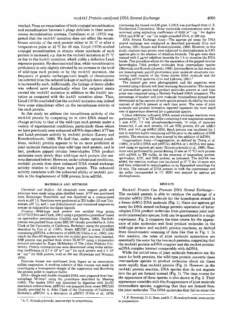
Spermidine Reduces the Magnesium Requirement for DNA Strand Exchange-Buffer containing 1 mM magnesium ion does not support DNA strand exchange by either recAwt protein or recA441 protein. However, if spermidine is added to this buffer, both recA proteins are able to catalyze DNA strand exchange (Fig. 5). At least 0.5 mM spermidine is required in order for any joint molecules to be formed in the recAwt protein-catalyzed reaction. As spermidine concentra- tion increases to 0.75 mM, the rate of joint molecule formation saturates at a value that is less than 20% of that observed under optimal DNA strand exchange conditions (i.e. at 8 mM magnesium acetate). Additionally, the rate at which product molecules appear is only about 30% of that observed at 8 mM magnesium acetate, suggesting that joint molecule formation limits product molecule formation by recAwt protein.
In contrast, some joint molecule formation occurs in the recA441 protein reaction when as little as 0.05 mM spermidine is present (Fig. 5). Above 0.05 mM spermidine, the rate of joint molecule formation increases, saturating at a rate com- parable to that observed at 8 mM magnesium acetate. Addi- tionally, at saturating spermidine concentration (1 mM), the profile for the conversion of these intermediate species to product molecules is similar to that seen at 8 mM magnesium acetate; homology-dependent DNA networks are produced, and product molecules appear at approximately equivalent rates. Thus, at 1 mM magnesium acetate, the presence of 1 mM spermidine can completely supplant the requirement for elevated magnesium ion concentration in the recA441 protein- catalyzed DNA strand exchange reaction, but not in the recAwt protein-catalyzed reaction.
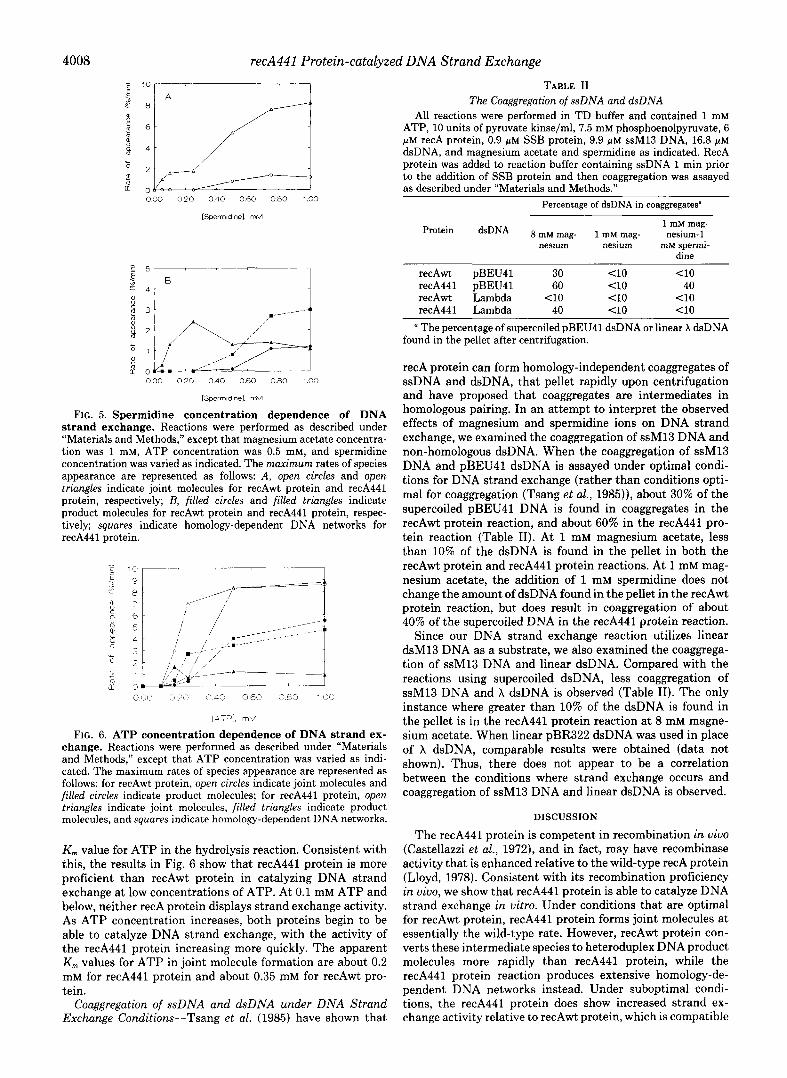
RecA441 Protein Can Catalyze DNA Strand Exchange at Low ATP Concentrations-DNA strand exchange normally requires the presence of ATP or dATP (Cox and Lehman, 1981; Menetski and Kowalczykowski, 1989). Phizicky and Roberts (1981) have shown that, compared with the wild-type recA protein, recA441 protein has a slightly lower apparent
4008 recA441 Protein-catalyzed DNA Strand Exchange
000 0.20 0.40 0.60 0.80 1 cl0
[Sperm,dl”el. rnM
FIG. 5. Spermidine concentration dependence of DNA strand exchange. Reactions were performed as described under “Materials and Methods,” except that magnesium acetate concentra- tion was 1 mM, ATP concentration was 0.5 mM, and spermidine concentration was varied as indicated. The maximum rates of species appearance are represented as follows: A, open circles and open triangfes indicate joint molecules for recAwt protein and recA441 protein, respectively; B, filled circles and filled triangles indicate product molecules for recAwt protein and recA441 protein, respec- tively; squares indicate homology-dependent DNA networks for recA441 protein.
FIG. 6. ATP concentration dependence of DNA strand ex- change. Reactions were performed as described under “Materials and Methods,” except that ATP concentration was varied as indi- cated. The maximum rates of species appearance are represented as follows: for recAwt protein, open circles indicate joint molecules and filled circles indicate product molecules; for recA441 protein, open triangles indicate joint molecules, filled triangles indicate product molecules, and squares indicate homology-dependent DNA networks.
Km value for ATP in the hydrolysis reaction. Consistent with this, the results in Fig. 6 show that recA441 protein is more proficient than recAwt protein in catalyzing DNA strand exchange at low concentrations of ATP. At 0.1 mM ATP and below, neither recA protein displays strand exchange activity. As ATP concentration increases, both proteins begin to be able to catalyze DNA strand exchange, with the activity of the recA441 protein increasing more quickly. The apparent K,,, values for ATP in joint molecule formation are about 0.2 mM for recA441 protein and about 0.35 mM for recAwt pro- tein.
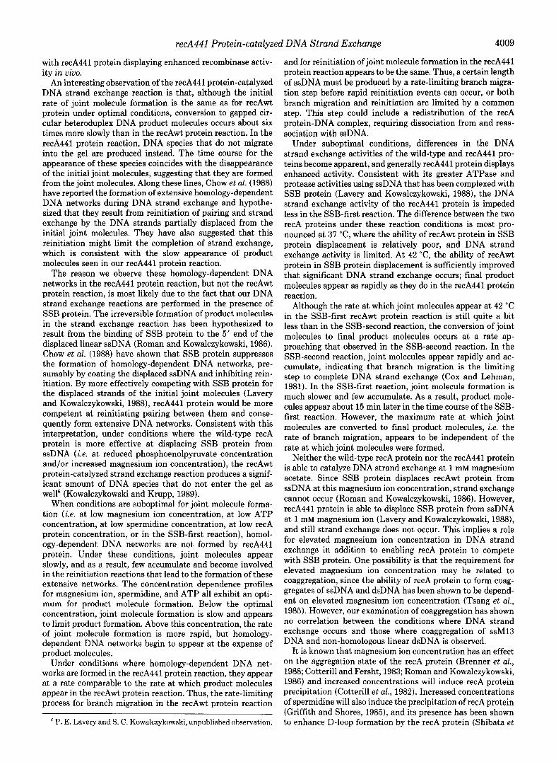
Couggregution of SSDNA and dsDNA under DNA Strand Exchange Conditions-Tsang et al. (1985) have shown that
TABLE II The Coaggregation of s.sDNA and dsDNA
All reactions were performed in TD buffer and contained 1 mM ATP, 10 units of pyruvate kinse/ml, 7.5 mM phosphoenolpyruvate, 6 pM recA protein, 0.9 pM SSB protein, 9.9 pM ssM13 DNA, 16.8 pM dsDNA, and magnesium acetate and spermidine as indicated. RecA protein was added to reaction buffer containing ssDNA 1 min prior to the addition of SSB protein and then coaggregation was assayed as described under “Materials and Methods.”
Percentage of dsDNA in coaggregates”
Protein dsDNA 1 InM mag- 8 mM mag- 1 mM mag- nesium-1
nesium nesium mM spermi- dine
recAwt pBEU41 30 Cl0 40 recA441 pBEU41 60 40 40 recAwt Lambda <lO <lO <lO recA441 Lambda 40 <lO 40
a The percentage of supercoiled pBEU41 dsDNA or linear X dsDNA found in the pellet after centrifugation.
recA protein can form homology-independent coaggregates of ssDNA and dsDNA, that pellet rapidly upon centrifugation and have proposed that coaggregates are intermediates in homologous pairing. In an attempt to interpret the observed effects of magnesium and spermidine ions on DNA strand exchange, we examined the coaggregation of ssM13 DNA and non-homologous dsDNA. When the coaggregation of ssMl3 DNA and pBEU41 dsDNA is assayed under optimal condi- tions for DNA strand exchange (rather than conditions opti- mal for coaggregation (Tsang et al., 1985)), about 30% of the supercoiled pBEU41 DNA is found in coaggregates in the recAwt protein reaction, and about 60% in the recA441 pro- tein reaction (Table II). At 1 mM magnesium acetate, less than 10% of the dsDNA is found in the pellet in both the recAwt protein and recA441 protein reactions. At 1 mM mag- nesium acetate, the addition of 1 mM spermidine does not change the amount of dsDNA found in the pellet in the recAwt protein reaction, but does result in coaggregation of about 40% of the supercoiled DNA in the recA441 protein reaction.
Since our DNA strand exchange reaction utilizes linear dsMl3 DNA as a substrate, we also examined the coaggrega- tion of ssMl3 DNA and linear dsDNA. Compared with the reactions using supercoiled dsDNA, less coaggregation of ssMl3 DNA and X dsDNA is observed (Table II). The only instance where greater than 10% of the dsDNA is found in the pellet is in the recA441 protein reaction at 8 mM magne- sium acetate. When linear pBR322 dsDNA was used in place of X dsDNA, comparable results were obtained (data not shown). Thus, there does not appear to be a correlation between the conditions where strand exchange occurs and coaggregation of ssM13 DNA and linear dsDNA is observed.
DISCUSSION
The recA441 protein is competent in recombination in viva (Castellazzi et al., 1972), and in fact, may have recombinase activity that is enhanced relative to the wild-type recA protein (Lloyd, 1978). Consistent with its recombination proficiency in Go, we show that recA441 protein is able to catalyze DNA strand exchange in vitro. Under conditions that are optimal for recAwt protein, recA441 protein forms joint molecules at essentially the wild-type rate. However, recAwt protein con- verts these intermediate species to heteroduplex DNA product molecules more rapidly than recA441 protein, while the recA441 protein reaction produces extensive homology-de- pendent DNA networks instead. Under suboptimal condi- tions, the recA441 protein does show increased strand ex- change activity relative to recAwt protein, which is compatible
recA441 Protein-catalyzed DNA Strand Exchange 4009
with recA441 protein displaying enhanced recombinase activ- ity in vivo.
An interesting observation of the recA441 protein-catalyzed DNA strand exchange reaction is that, although the initial rate of joint molecule formation is the same as for recAwt protein under optimal conditions, conversion to gapped cir- cular heteroduplex DNA product molecules occurs about six times more slowly than in the recAwt protein reaction. In the recA441 protein reaction, DNA species that do not migrate into the gel are produced instead. The time course for the appearance of these species coincides with the disappearance of the initial joint molecules, suggesting that they are formed from the joint molecules. Along these lines, Chow et al. (1988) have reported the formation of extensive homology-dependent DNA networks during DNA strand exchange and hypothe- sized that they result from reinitiation of pairing and strand exchange by the DNA strands partially displaced from the initial joint molecules. They have also suggested that this reinitiation might limit the completion of strand exchange, which is consistent with the slow appearance of product molecules seen in our recA441 protein reaction.
The reason we observe these homology-dependent DNA networks in the recA441 protein reaction, but not the recAwt protein reaction, is most likely due to the fact that our DNA strand exchange reactions are performed in the presence of SSB protein. The irreversible formation of product molecules in the strand exchange reaction has been hypothesized to result from the binding of SSB protein to the 5’ end of the displaced linear ssDNA (Roman and Kowalczykowski, 1986). Chow et al. (1988) have shown that SSB protein suppresses the formation of homology-dependent DNA networks, pre- sumably by coating the displaced ssDNA and inhibiting rein- itiation. By more effectively competing with SSB protein for the displaced strands of the initial joint molecules (Lavery and Kowalczykowski, 1988), recA441 protein would be more competent at reinitiating pairing between them and conse- quently form extensive DNA networks. Consistent with this interpretation, under conditions where the wild-type recA protein is more effective at displacing SSB protein from ssDNA (i.e. at reduced phosphoenolpyruvate concentration and/or increased magnesium ion concentration), the recAwt protein-catalyzed strand exchange reaction produces a signif- icant amount of DNA species that do not enter the gel as well6 (Kowalczykowski and Krupp, 1989).
When conditions are suboptimal for joint molecule forma- tion (i.e. at low magnesium ion concentration, at low ATP concentration, at low spermidine concentration, at low recA protein concentration, or in the SSB-first reaction), homol- ogy-dependent DNA networks are not formed by recA441 protein. Under these conditions, joint molecules appear slowly, and as a result, few accumulate and become involved in the reinitiation reactions that lead to the formation of these extensive networks. The concentration dependence profiles for magnesium ion, spermidine, and ATP all exhibit an opti- mum for product molecule formation. Below the optimal concentration, joint molecule formation is slow and appears to limit product formation. Above this concentration, the rate of joint molecule formation is more rapid, but homology- dependent DNA networks begin to appear at the expense of product molecules.
Under conditions where homology-dependent DNA net- works are formed in the recA441 protein reaction, they appear at a rate comparable to the rate at which product molecules appear in the recAwt protein reaction. Thus, the rate-limiting process for branch migration in the recAwt protein reaction
6 P. E. Lavery and S. C. Kowalczykowski, unpublished observation.
and for reinitiation of joint molecule formation in the recA441 protein reaction appears to be the same. Thus, a certain length of ssDNA must be produced by a rate-limiting branch migra- tion step before rapid reinitiation events can occur, or both branch migration and reinitiation are limited by a common step. This step could include a redistribution of the recA protein-DNA complex, requiring dissociation from and reas- sociation with ssDNA.
Under suboptimal conditions, differences in the DNA strand exchange activities of the wild-type and recA441 pro- teins become apparent, and generally recA441 protein displays enhanced activity. Consistent with its greater ATPase and protease activities using ssDNA that has been complexed with SSB protein (Lavery and Kowalczykowski, 1988), the DNA strand exchange activity of the recA441 protein is impeded less in the SSB-first reaction. The difference between the two recA proteins under these reaction conditions is most pro- nounced at 37 “C, where the ability of recAwt protein in SSB protein displacement is relatively poor, and DNA strand exchange activity is limited. At 42 “C, the ability of recAwt protein in SSB protein displacement is sufficiently improved that significant DNA strand exchange occurs; final product molecules appear as rapidly as they do in the recA441 protein reaction.
Although the rate at which joint molecules appear at 42 “C in the SSB-first recAwt protein reaction is still quite a bit less than in the SSB-second reaction, the conversion of joint molecules to final product molecules occurs at a rate ap- proaching that observed in the SSB-second reaction. In the SSB-second reaction, joint molecules appear rapidly and ac- cumulate, indicating that branch migration is the limiting step to complete DNA strand exchange (Cox and Lehman, 1981). In the SSB-first reaction, joint molecule formation is much slower and few accumulate. As a result, product mole- cules appear about 15 min later in the time course of the SSB- first reaction. However, the maximum rate at which joint molecules are converted to final product molecules, i.e. the rate of branch migration, appears to be independent of the rate at which joint molecules were formed.
Neither the wild-type recA protein nor the recA441 protein is able to catalyze DNA strand exchange at 1 mM magnesium acetate. Since SSB protein displaces recAwt protein from ssDNA at this magnesium ion concentration, strand exchange cannot occur (Roman and Kowalczykowski, 1986). However, recA441 protein is able to displace SSB protein from ssDNA at 1 mM magnesium ion (Lavery and Kowalczykowski, 1988), and still strand exchange does not occur. This implies a role for elevated magnesium ion concentration in DNA strand exchange in addition to enabling recA protein to compete with SSB protein. One possibility is that the requirement for elevated magnesium ion concentration may be related to coaggregation, since the ability of recA protein to form coag- gregates of ssDNA and dsDNA has been shown to be depend- ent on elevated magnesium ion concentration (Tsang et al., 1985). However, our examination of coaggregation has shown no correlation between the conditions where DNA strand exchange occurs and those where coaggregation of ssM13 DNA and non-homologous linear dsDNA is observed.
It is known that magnesium ion concentration has an effect on the aggregation state of the recA protein (Brenner et al., 1988; Cotterill and Fersht, 1983; Roman and Kowalczykowski, 1986) and increased concentrations will induce recA protein precipitation (Cotterill et al., 1982). Increased concentrations of spermidine will also induce the precipitation of recA protein (Griffith and Shores, 1985), and its presence has been shown to enhance D-loop formation by the recA protein (Shibata et

recA441 Protein-catalyzed DNA Strand Exchange
al., 1980). These observations, together with our finding that the presence of spermidine reduces the magnesium ion con- centration required for DNA strand exchange, suggest that the requirement for elevated magnesium ion concentration in strand exchange could be related to a requirement for a certain degree of recA protein aggregation.
Lloyd (1978) observed slightly heightened recombinase ac- tivity for recA441 protein in uiuo. However, under optimal conditions for DNA strand exchange, we do not see increased activity by recA441 protein. Thus, these conditions may not be stringent enough to allow for the detection of slight differ- ences between the activities of the two recA proteins. How- ever, the increased activity that we observe under suboptimal conditions (i.e. at low ATP concentration, at low magnesium ion concentration in the presence of spermidine, or in the SSB first reaction), is consistent with the idea that these conditions are necessary to provide a window for observing an enhancement in recA441 protein strand exchange activity. This enhancement would be manifest in heightened recom- binase activity in the more demanding in uiuo situation.
AcknoLuledgments-We thank Dr. A. John Clark of the University of California, Berkeley for kindly providing strains BEU397 and BEU293, and Dan A. Dixon, Angela K. Eggelston, Scott D. Lauder, William M. Rehrauer, and Linda J. Roman for critical reading of the manuscript.
REFERENCES
Brenner, S. K., Zlotnick, A. & Griffith, J. D. (1988) J. Mol. Biol. 204, 959-972
Castellazzi, M., George, J. & Buttin, G. (1972) Mol. Gen. Genet. 119, 139-152
Chow, S. A., Rao, B. J. & Radding, C. M. (1988) J. Biol. Chem. 263, 200-209
Cotterill, S. M. & Fersht, A. R. (1983) Biochemistry 22, 35253531 Cotterill, S. M., Satterthwait, A. C. & Fersht, A. R. (1982) Biochem-
istry 21,4332-4337 Cox, M. M. & Lehman, I. R. (1981) Proc. N&l. Acud. Sci. U. S. A.
78,3433-3437 Cox, M. M. & Lehman, I. R. (1982) J. Biol. Chem. 257,8523-8532 Cox. M. M. & Lehman, I. R. (1987) Annu. Reu. Biochem. 56, 229-
262 Cox, M. M., McEntee, K. & Lehman, I. R. (1981) J. Biol. Chem. 256,
4676-4678 Craig, N. L. & Roberts, J. W. (1980) Nature 283, 26-29
Goldthwait, D. A. &Jacob, F. (1964) Compt. Rend. 258,661 Griffith, J. & Shores, C. G. (1985) Biochemistry 24, 158-162 Kirby, E. P. & Goldthwait, D. A. (1967) Proc. Natl. Acad. Sci. U. S. A.
58,1903-1910 Knight, K. L., Aoki, K. H., Ujita, E. L. & McEntee, K. (1984) J. Biol.
Chem. 259,11279-11283 Kowalczykowski, S. C. (1987) Trends Biochem. Sci. 12, 141-145 Kowalczykowski, S. C. & Krupp, R. A. (1987) J. Mol. Biol. 193,97-
113 Kowalczykowski, S. C. & Krupp, R. A. (1989) J. Mol. Biol. 207,735-
747 Kowalczykowski, S. C. & Roman, L. J. (1990) in Molecular Mecha-
nisms of Replication and Recombination: UCLA Symposia on Mo- lecular and Cellular Biology (Richardson, C. & Lehman, I., eds) Alan R. Liss, Inc., New York, in press
Kowalczykowski, S. C., Burk, D. L. & Krupp, R. A. (1989) J. Mol. Biol. 207, 719-733
Lavery, P. E. & Kowalczykowski, S. C. (1988) J. Mol. Biol. 203,861- 874
Little, J. W. & Mount, D. W. (1982) Cell 29, 11-22 Lloyd, R. G. (1978) J. Bacterial. 134, 929-935 Menetski, J. P. & Kowalczykowski, S. C. (1989) Biochemistry 28,
5871-5881 Menetski, J. P. & Kowalczykowski, S. C. (1990) J. Mol. Biol., in press Messing, j. (1983) Methods Enzymol. 101, 20-78 Phizickv. E. M. & Roberts. J. W. (1981) Cell 25.259-267 Radding,‘C. M. (1988) in Genetic Recombination.(Kucherlapati, R. &
Smith, G. R., eds) pp. 193-229, American Society for Microbiology, Washington, D. C.
Roberts, J. W., Roberts, C. W. & Craig, N. L. (1978) Proc. Natl. Acad. Sci. U. S. A. 75,4714-4718
Roman, L. J. & Kowalczykowski, S. C. (1986) Biochemistry 25,7375- 7385
Rusche, J. R., Konigsberg, W. & Howard-Flanders, P. (1985) J. Biol. Chem.260,949-955
Ruyechan, W. T. & Wetmur, J. G. (1976) Biochemistry 15, 5057- 5064
Shibata, T., DasGupta, C., Cunningham, R. P. & Radding, C. M. (1980) Proc. Natl. Acad. Sci. U. S. A. 77. 2606-2610
Tessman, E. S. & Peterson, P. K. (1985) JI Bacterial. 163, 677-687 Tsang, S. S., Chow, S. A. & Radding, C. M. (1985) Biochemistry 24,
3226-3232 Uhlin, B. E. & Clark, A. J. (1981) J. Bacterial. 148, 386-390 Uhlin, B. E., Molin, S., Gustafsson, P. & Nordstrom, K. (1979) Gene
(Amst.) 6,91-106 Uhlin, B. E., Schweikart, V. & Clark, A. J. (1983) Gene (Amst.) 22,
255-265 Walker, G. C. (1984) Microbiological Reuiews 48,60-93 Witkin, E. (1976) Bacterial. Reu. 40, 869-907